bd biocoat cellware
TRANSCRIPT

BD BiosciencesClontechDiscovery LabwareImmunocytometry SystemsPharmingen
BD BioCoat™ Cellware

Table of Contents
About the cover:Computer colorization of, from top to bottom:
Rat cerebellar granule (RCG) cells cultured on BD BioCoat™ PDL (page 6)
Human umbilical vein endothelial cells (HUVECs)grown for seven days on BD BioCoat™ Gelatin 6-well Multiwell Plates (page 5)
BHK-21 fibroblasts cultured on BD BioCoat™
Fibronectin CultureSlides (page 9)
Neurotransmitter induced calcium waves in astrocytes plated on BD BioCoat™
Laminin/Fibronectin (page 13)
Introduction 1
BD BioCoat™ Cellware Vessel Options 2
BD BioCoat™ Manufacturing Facilities 3
BD BioCoat™ Collagen I Cellware 4
BD BioCoat™ Gelatin Cellware 5
BD BioCoat™ Poly-Lysine Cellware 6
BD BioCoat™ Collagen IV Cellware 7
BD BioCoat™ EHS Natrix Cellware 8
BD BioCoat™ Fibronectin Cellware 9
BD BioCoat™ Laminin Cellware 10
BD BioCoat™ Matrigel™ Matrix Cellware 11
BD BioCoat™ Osteologic™ Bone Cell Culture System 12
BD BioCoat™ Poly-D-Lysine/Laminin, Poly-L-Ornithine/Laminin and Laminin/Fibronectin Cellware 13
BD BioCoat™ T-Cell Activation Plates 14
BD BioCoat™ Variety Pack Cellware Products,Vented Caps for BD BioCoat™ Flasks 15
BD BioCoat™ Collagen and PDL 96- and 384-well Plates for High Throughput Screening 16
BD BioCoat™ Services 18
Ordering Information 19

Introduction
The development and normal functioning of cells depends on interactions with molecules in theirmicro-environment. The major classesof molecules that regulate cellulardevelopment and function includegrowth and differentiation factors, cell adhesion molecules and the components of the extracellularmatrix (ECM). The ECM is composed of a number of differentmacromolecules whose structuralintegrity and functional compositionare important in maintaining normaltissue architecture, in developmentand in tissue-specific function. TheECM exerts influences on behavior(adherence, spreading, differentiationand migration) and the pattern ofgene expression of the cells in contactwith it. The ECM, however, is notstatic but changes during both normaldevelopment and in tissue repair and regeneration and is intimatelyinvolved in both normal biologicalfunction and response to injury.1
To create physiologically relevant in vitro models that support normalcell culture and function, the compo-nents of the in vivo environment mustbe incorporated. The use of an ECM as a coating for tissue culture surfacespermits the development of model systems which closely mimic in vivoconditions. The choice of ECM is animportant component to considerwhen optimizing in vitro culture systems.
BD BioCoat™
CellwareBD BioCoat™ Cellware is a unique line of tissueculture vessels with variousECM components appliedto vessel surfaces by a proprietary manufacturingprocess. The result is a uniform, optically clearmatrix substrate. This technology, together with our exacting qualitycontrol, guarantees the
performance of each lot, as well asconsistency from lot-to-lot.
BD BioCoat Cellware promotes cellattachment, spreading, growth anddifferentiation of a variety of primarycells and cell lines in serum-free orserum-containing cultures.
Applications include:Cell adhesion assays
Receptor-ligand-binding assays
Routine drug screening assays
Studies of tissue morphogenesis
Studies of cell-matrix interactions
Regulation of signal transductionand gene expression
Get the BD BioCoat™ Advantages
Ready to Use ConvenienceSpend more time performing yourexperiments rather than preparingfor them. Precoated BD BioCoatCellware saves time and labor costswhile increasing productivity.
Quality Assurance TestingEach lot of BD BioCoat Cellware is thoroughly tested for bioactivityand guaranteed to perform asclaimed so you can use with complete confidence.
Reliable PerformanceBD BioCoat Cellware improves cell attachment and increases proliferation rates for a variety of normal and transformed cells.
Lot-to-Lot ConsistencyBD Biosciences prides itself on maintaining highly controlled ISO 9001 production environmentsand validated manufacturing procedures that result in uniformityand consistent performance.
Wide SelectionAvailable with a wide range of ECM proteins and attachment factors, BD BioCoat helps optimizeconditions for attachment, growth or differentiation for your cell type.
Readily AvailableBD BioCoat Cellware is availablefrom stock for immediate shipments.Our service options include standingorder management and lot numberreservations.
1. Alberts, B., et al., Mol Bio of the Cell (Third Edition). Garland Publishing, NY (1994).
www.bdbiosciences.com/discovery_labware1

The first company to produce sterile,disposable labware, BD Biosciences isa world leader in providing researcherswith top-quality cell culture products.BD Falcon™ Cultureware is treatedusing a unique vacuum gas plasmaprocess resulting in a pure and consistent tissue culture surface. BD BioCoat™ Cellware is manufacturedusing BD Falcon products to ensureconsistent, reliable results. Trust BD Biosciences, the first name in cell culture.
BD BioCoat™……Flasks are available in various sizes
and designsto meet allof your cellcultureneeds instandardplug-sealand ventedcapoptions.
…Multiwell Plates are manufacturedusing acrystal-grade poly-styrene andfeature apatentedlabyrinthlid, conden-sation ringsand deep
well design to control contaminationwhile reducing evaporation and minimizing edge effects.
…Dishes are exceptionally flat for distortion-free opticsand featurestackingrings forbetterstacking.Easy Gripdesignensures
save handling.
…CultureSlides have an innovativesealingdesign thatminimizesleakageand a plasticchamberaffixed to a speciallycleaned
glass slide that can be removed withan easy-to-use disposable safetyremoval tool.
…Coverslips are Number 1 Germanglass andprovide anopticallyclear surfacewhich isnon-neuro-toxic andexhibitslow back-
ground fluorescence. The convenientpackage also acts as a storage container and allows for easy coverslip manipulation.
…Coverslip-Bottom Dishes are 35 mmstyle disheswith acoverslipbottomthat is easyto use andfacilitatespreparationof cells
for microscopic analysis. The cover-slip floor is Number 1 German glass.This format is ideal for use in highresolution and inverted microscopy,fluorescence imaging in live cells, con-focal microscopy, phase contrastmicroscopy and micro-manipulations.
BD BioCoat™ Cellware Vessel Options
For BD BioCoatTM Cell Culture Inserts and Assay Systems, please see our general BD FalconTM/BD BioCoatTM catalog.
www.bdbiosciences.com/discovery_labware2
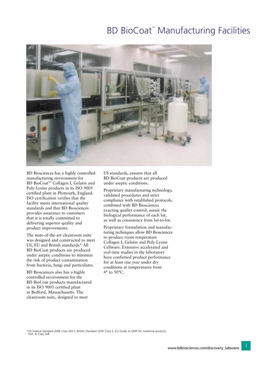
BD BioCoat™ Manufacturing Facilities
BD Biosciences has a highly controlledmanufacturing environment for BD BioCoat™ Collagen I, Gelatin and Poly-Lysine products in its ISO 9001certified plant in Plymouth, England.ISO certification verifies that the facility meets international qualitystandards and that BD Biosciencesprovides assurance to customers that it is totally committed to delivering superior quality and product improvements.
The state-of-the-art cleanroom suitewas designed and constructed to meetUS, EU and British standards.* All BD BioCoat products are producedunder aseptic conditions to minimizethe risk of product contaminationfrom bacteria, fungi and particulates.
BD Biosciences also has a highly controlled environment for the BD BioCoat products manufacturedin its ISO 9001 certified plant in Bedford, Massachusetts. The cleanroom suite, designed to meet
US standards, ensures that all BD BioCoat products are produced under aseptic conditions.
Proprietary manufacturing technology,validated procedures and strict compliance with established protocols,combined with BD Biosciences exacting quality control, assure thebiological performance of each lot, as well as consistency from lot-to-lot.
Proprietary formulation and manufac-turing techniques allow BD Biosciencesto produce room temperatureCollagen I, Gelatin and Poly-LysineCellware. Extensive accelerated andreal-time studies in the laboratoryhave confirmed product performancefor at least one year under dry conditions at temperatures from 4° to 50°C.
*US Federal Standard 209E Class M3.5, British Standard 5295 Class E, EU Guide to GMP for medicinal products (Vol. 4) Class A/B
www.bdbiosciences.com/discovery_labware3

BD BioCoat™ Collagen I Cellware
Collagen I, found in most tissues andorgans, is most plentiful in dermis,tendon and bone. It is an integral part of the framework that holds cells and tissues together and has been recognized as a useful matrix for improving cell culture. In vitrouse of collagen can exert effects onthe adherence, morphology, growth,migration and differentiation of avariety of cell types.1
BD BioCoat™ Collagen ICellware applications include:
Promotion of cell attachment and spreading
Rapid expansion of cell populations
Serum-free or reduced serum culture
Cell adhesion assays
Improving survival of primary cells in culture
BD BioCoat™ Collagen I has been used to culture:
Primary murine cardiac myocytes2
Human vascular SMC3
PC12 cells and SH-SY5Y cells4
Mouse primary keratinocytes5
SK-MEL-28-N1 cells6
Murine myoblast C2C12 cells7
HUVEC8
HEK-293 cells9
Rat Kupffer cells10
MDA-231 breast cancer cells11
Source:Rat tail tendon
Quality Control:Tested for ability to promoteattachment and spreading of HT-1080 human fibrosarcoma cells
Tested and found negative for bacteria and fungi
Collagen I purity >90% by SDS-PAGE
Storage and Stability:Cellware stable for at least six monthsfrom date of shipment when stored at 4° to 30°C under dry conditions.Coverslips and CultureSlides stablefor at least three months from date ofshipment when stored at 2° to 8°C.
Effects of BD BioCoat™ Collagen I Cellware on Fetal Bovine Heart Endothelial (FBHE) Cells
FBHE cells grown for five days in basalmedium containing 10% FBS on tissue-culture plastic show sparsegrowth.
FBHE cells grown for five days usingthe BD BioCoat Endothelial CellGrowth Environment (Collagen ICellware) form a confluent monolayerand show numerous mitotic cells.
References1. Kleinman, H.K., et al., Analytical Biochemistry 166:1 (1987).2. Bjorkegren, J., et al., J. Biol. Chem. (Accepted for pub July 31, 2001).3. Flaherty, P., et al., BD Tech Bulletin (1996).4. Ivankovic-Dikic, I., et al., Nat. Cell. Biol. 2:574-581 (2000).5. Maatta, A., et al., J. Biol. Chem. 275(26):19857 (2000).6. Nakano, J., et al., J. Invest. Dermatology Symposium Proc. 4(2):173 (1999).7. Ogilvie, M., et al., J. Biol. Chem. 275(50):39754 (2000).8. Rajagopalan, L.E., et al., J. Neurochemistry 74(1):52 (2000).9. Smith, J.S., et al., J. Neurosci. 21(4):1096 (2001).10. Takeyama, O., et al., Transplantation 69(7):1283 (2000).11. Yoneda, T., et al., J. Clin. Invest. 99(10):2509 (1997).
www.bdbiosciences.com/discovery_labware4

BD BioCoat™ Gelatin Cellware
BD BioCoat™ Gelatin Cellware provides an attachment and growthpromoting substrate for the culture of a variety of cell types. Gelatin isused commonly in the culture of vascular endothelial cells, muscle,embryonic stem (ES) cells and F9 teratocarcinoma cells. It is also suitable for promoting adhesion of transfected cell types. Gelatin is a heterogeneous mixture of water-soluble proteins derivedthrough the hydrolysis of Collagen.
BD BioCoat™ GelatinCellware applicationsinclude:
Promotion of cell attachment andspreading of:
• Vascular endothelial cells, i.e. BME1, BAEC2
• Embryonic stem (ES) cells3
• C2C12 myoblasts4 and MM14myoblasts5
Culture of normal and transfected F9 teratocarcinomacells for gene expression studies6
Culture of HUVEC for E-Selectin7 expression and VEGF induction8
Source:Gelatin, porcine
Quality Control: Tested for ability to promote proliferation of HUVECs
Tested and found negative forbacteria and fungi
Storage and Stability:Stable for at least six months fromdate of shipment when stored at 4° to 30°C under dry conditions.
References1. Zimrin, A.B., et al., J. Biol. Chem. 271(51):32499 (1996).2. Gou, D., et al., J. Biol. Chem. 270(12):6729 (1995).3. Ernst, M., et al., J. Biol. Chem. 271(47):30136 (1996).4. Stuart, C.E., et al., J. Biol. Chem. 271(19):11330 (1996).5. Patrie, K.M., et al., J. Biol. Chem. 270(48):29018 (1995).6. Laurance, M.E., et al., J. Biol. Chem. 272(5):2646 (1997).7. Read, et al., J. Biol. Chem. 272(5):2753 (1997).8. Gitay-Goren, et al., J. Biol. Chem. 271(10):5519 (1996).
Effect of BD BioCoat Gelatin Cellware of HUVEC
Human umbilical vein endothelial cells (HUVECs) grown for seven days on BD BioCoat Gelatin 6-well Multiwell Plates seeded at a density of 2x10⁴ in the presence of E-STIM™ Endothelial Cell Culture Medium (440x).
www.bdbiosciences.com/discovery_labware5

BD BioCoat™ Poly-Lysine Cellware
Poly-D-Lysine (PDL) and Poly-L-Lysine(PLL) are synthetic compounds thatenhance cell adhesion and proteinabsorption by altering surface chargeson the culture substrate. In additionto promoting cell adhesion, poly-lysinesurface treatments support neuriteoutgrowth and improve the survivalof many central nervous system(CNS) primary cells in culture. AsPDL and PLL are synthetic molecules,they do not stimulate biological activity in the cells cultured on them,and they do not introduce impuritiescarried by natural polymers.
BD BioCoat™ Poly-LysineCellware applicationsinclude:
Attachment and spreading of a variety of cell types
Cell differentiation and neurite outgrowth
Attachment of fastidious transfected cell lines
Support survival of primary neurons in culture
Serum-free or reduced serum culture
BD BioCoat™ Poly-Lysinehas been used to culture:
Primary mouse brain capillaries1
HEK-293 cells2-5
MDA-231 breast cancer cells6
Mouse cerebellar granuleneurons7
Transfected rat 1 cells8
Rat anterior pituitary cells9
Transfected COS-7 cells10
Transiently transfected primaryrat astrocytes11
Rat primary cerebellar granuleneurons12-13
Murine microglia MG-7 cells14
Source:PDL, synthetic (MW 75-150 kD)
PLL, synthetic (MW 30-70 kD)
Quality Control:Tested for ability to promote firmattachment of RCG cells
Tested and found negative forbacteria and fungi
Storage and Stability:Cellware stable for at least six monthsfrom date of shipment when stored at 4° to 30°C under dry conditions.Coverslips and CultureSlides stablefor at least three months from date of shipment when stored at 2° to 8°C.
References1. Santambrogio, L., et al., PNAS 98(11):6295 (2001).2. Sugawara, T., et al., PNAS 98(11):6384 (2001).3. Bdeir, K., et al., J. Biol. Chem. 275:28532 (2000).4. Fitzgerald, L.W., et al., J. Neurochemistry 72(5):2127 (1991).5. Hu, L.A., et al., J. Biol. Chem. 275:38659 (2000).6. Yoneda, T., et al., J. Clin. Invest. 99(10):2509 (1997).7. Armstrong, R.C., et al., J. Neuroscience 17(2):553 (1997).8. Bertin, J., et al., J. Biol. Chem. 276(15):11877 (2001).9. Hinuma, S., et al., Nature 393(6682):272 (1998).10. Kirsch. K.H., et al., PNAS 96(11):6211 (1999).11. Little, E.B., et al., PNAS 98(5):2238 (2001).12. Segal, J.A., et al., J. Neurochemistry 74(1):60 (2000).13. Wood, M.W., et al., J. Neurochemistry 74(5):2033 (2000).14. Szczepanik, A.M., et al., J. Neurochemistry 77(1):304 (2001).
Effect of BD BioCoat PDL on Cortical Neurons
Mixed culture of cortical neurons andastrocytes cultured on BD BioCoat PDLCellware. Neurons are highly branchedwith very long processes. Astrocytesshow similar process elongation.
Effect of BD BioCoat PDL on RCG Cells
Rat cerebellar granule (RCG) cellscultured on BD BioCoat PDL showfirm attachment (similar resultsobtained on PLL).
www.bdbiosciences.com/discovery_labware6

BD BioCoat™ Collagen IV Cellware
Type IV Collagen is a ubiquitouscomponent in basement membranesand provides the major structural support for this matrix. When theCollagen IV meshwork is assembled,it provides a scaffold for the assemblyof other basement membrane components through interactions with laminin, entactin/nidogen andheparan sulfate proteoglycan.Collagen IV is useful as a substratefor growth of epithelial, endothelial,muscle and nerve cells. Collagen playsa role in the regulation of cell growth,differentiation and adhesion, as wellas tissue formation.
BD BioCoat™ Collagen IVCellware applicationsinclude:
Promotion of cell attachment and spreading
Cell differentiation and neurite outgrowth
Increased proliferation of PC12 cells
Studies of effects of Collagen IVon cell behavior
Cell Adhesion Assays
BD BioCoat™ Collagen IVCellware has been used to culture:
PC12 cells1-3
SH-SY5Y cells4
Human melanoma cells lines SK-MEL-28-N1 and SK-MEL-285
Primary murine hepatocytes6
Source:Engelbreth-Holm-Swarm (EHS) lathrytic mouse tumor
Quality Control:Tested for ability to initiate differentiation (neurite outgrowth)of NG-108 rat glioma/mouse neuroblastoma cells
Tested and found negative for bacteria and fungi
Collagen IV purity > 90% by SDS-PAGE
Storage and Stability:Stable for at least three months from date of shipment when stored at 2° to 8°C.
Effects of BD BioCoat Collagen IV Cellware on PC12 Rat Pheochromocytoma Cells
PC12 cells cultured on tissue-cultureplastic do not attach well and tend tofloat in clumps in the culture medium.
PC12 cells cultured on BD BioCoatCollagen IV Cellware show 90%attachment and rapid proliferation.
References1. Ivankovic-Dikic, I., et al., Nat. Cell. Biol. 2:574 (2000).2. Marchetti, D., et al., Int. J. Cancer 55:692 (1993).3. Muda, M., et al., J. Biol. Chem. 271:4319 (1996).4. Ivankovic-Dikic, I., et al., Nat. Cell. Biol. 2:574 (2000).5. Nakano, J., et al., J. Invest. Derm. Symp. Proc. 4(2):173 (1999).6. Swift, L.L., et al., J. Biol. Chem. 276(25):22965 (2001).
www.bdbiosciences.com/discovery_labware7

BD BioCoat™ EHS Natrix Cellware
EHS Natrix is a natural ECM that is synthesized, secreted and formed in vitro by a continuous cell lineestablished from primary tissue of the EHS tumor. The ECM secreted by these cells is similar to that whichsurrounds the in vivo EHS tumor.This in vitro ECM is composed of laminin, collagen IV and othercomponents which have not as yetbeen fully characterized.1
BD BioCoat™ EHS NatrixCellware has been used to culture:
Neural cell attachment and differentiation
Transduction of neuroblastomas2
Culture of fish hepatocytes
Amniocyte attachment and culture
Primary tumor-cell culture
Dunning R-3327 rat prostaticadenocarcinoma cells3
Source:Monolayers of a cell line establishedfrom the EHS mouse tumor
Quality Control:Tested for ability to promote differentiation (neurite outgrowth)of NG-108 rat glioma/mouse neuroblastoma cells
Tested and found negative forbacteria and fungi
Storage and Stability:Stable for at least three months at 2° to 8°C. Do not freeze.
Effects of BD BioCoat EHS Natrix Cellware on NG-108Rat Glioma/Mouse Neuroblastoma Cells
NG-108 rat glioma/mouse neuroblastoma cells cultured on BD BioCoat EHS NatrixCellware.
References1. Early, E.M., et al., Third Intl. Conf. on the Mol. Bio.
and Path. of Matrix. Philadelphia, PA (1990).2. Bowman, et al., Blood. 92(6):1941(1998).3. Donald, C.D., et al. Invasion Metastasis 18(4):165 (1998-99).
www.bdbiosciences.com/discovery_labware8

BD BioCoat™ Fibronectin Cellware
Human Fibronectin (HFN) is a widelydistributed glycoprotein that is usedas a substrate to promote attachmentof cells through its central-bindingdomain RGD sequence. HFN is aproduct of most mesenchymal andepithelial cells and is present in boththe ECM and plasma. The principalfunction of HFN appears to be in cellular migration during wound healing and development, regulationof cell growth and differentiation and haemostasis/thrombosis.
BD BioCoat™ FibronectinCellware applicationsinclude:
Promotion of cell attachment and spreading
Rapid expansion of cell populations
Serum-free or reduced serumculture
Cell adhesion assays
Studies of effects of HFN on cell behavior
Improving survival of primarycells in culture
BD BioCoat™ FibronectinCellware has been used to culture:
3T3 Preadipocytes1
Transfected 293T and transfectedH1299 cells2
MCF-10A cells3
Primary cord blood mononuclear cells4
SK-MEL-28 (human melanoma cells)5
NIH3T3 cells6
MDA-231 human breast cancer cells7
Source:Human plasma NOTE: Source material tested for hepatitis B antigenand HIV-1 antibody
Quality Control:Tested for ability to promoteattachment and spreading ofBHK-21 hamster kidney cells
Tested and found negative for bacteria and fungi
Fibronectin purity > 90% by SDS-PAGE
Storage and Stability:Stable for at least three months fromdate of shipment when stored at 2° to 8°C. Do not freeze.
Effects of BD BioCoat Fibronectin Cellware on BHK-21 Cells
BHK-21 fibroblasts cultured on glassCultureSlides do not spread.
BHK-21 fibroblasts cultured on BD BioCoat Fibronectin CultureSlidesattach and spread within one hour.
References1. Guller, S., et al., Endocrinology 130:2609 (1992).2. Lavoie, J.N., et al., J. Cell Biol. 150:1037 (2000).3. Miller, K.A., et al., J. Biol. Chem. 275:8176 (2000).4. Murohara, T., et al., J. Clin. Invest. 105:1527 (2000).5. Nakano, J., et al., J. Invest. Derm. Symp. Proc. 4:173 (1999).6. Shaw, R.J., J. Biol. Chem. 273:7757 (1998).7. Yoneda, T., et al., J. Clin. Invest. 99:2509 (1997).
www.bdbiosciences.com/discovery_labware9

BD BioCoat™ Laminin Cellware
Laminin (LM), a major component of basement membranes, is a multifunctional glycoprotein that is used as a substrate to culture and maintain differentiated functionof a wide variety of cells. Laminin has been shown in culture to stimulate neurite outgrowth, promotecell attachment, chemotaxis, cell differentiation and neuronal survival.
BD BioCoat™ LamininCellware applicationsinclude:
Promotion of cell attachment and spreading
Induction of cell differentiationand neurite outgrowth
Increases proliferation ofmyoblasts1
Studies of effects of laminin on cell behavior
Cell adhesion assays
BD BioCoat™ LamininCellware has been used to culture:
SH-SY5Y (human neuroblastoma),Neuro-2A (mouse neuroblastoma),N1-E115 (rat neuroblastoma)2
MCF-10A cells3,4
SK-MEL-28 cells5
HVSMC6
MDA-231 breast cancer cell line7
Source:Engelbreth-Holm-Swarm (EHS)mouse tumor
Quality Control:Tested for ability to initiate differentiation (neurite outgrowth)of NG-108 rat glioma/mouse neuroblastoma cells
Tested and found negative for bacteria and fungi
Laminin purity > 90% by SDS-PAGE (contains entactin)
Storage and Stability:Stable for at least three months fromdate of shipment when stored at 2° to 8°C. Do not freeze.
References1. Ocalan, M., et al., Dev. Biol. 125:158 (1988).2. Leventhal, P.S. and Feldman, E.L., J. Biol. Chem. 271:5957 (1996).3. Miller, K.A., et al., J. Biol. Chem. 275:8176 (2000).4. Salas, P.J., et al., J. Cell Biol. 137:359 (1997).5. Nakano, J., et al., J. Investig. Derm. Symp. Proc. 4:173 (1999).6. Tyagi, S.C., Am. J. Physiol. 274:C396 (1998).7. Yoneda, T., et al., J. Clin. Invest. 99:2509 (1997).
Effects of BD BioCoat Laminin Cellware on NG-108 RatGlioma/Mouse Neuroblastoma Cells
NG-108 rat glioma/mouse neuroblastomacells cultured on tissue-culture plastic areloosely adhered and remain rounded.
NG-108 rat glioma/mouse neuroblastomacells cultured on BD BioCoat LamininCellware exhibit a spindle-shaped morphology and dendritic processes.
www.bdbiosciences.com/discovery_labware10

BD BioCoat™ Matrigel™ Matrix Cellware
BD Matrigel™ Basement MembraneMatrix is a solubilized basementmembrane preparation extracted from the Engelbreth-Holm-Swarm(EHS) mouse sarcoma, a tumor rich in ECM proteins. Its major component is laminin, followed by collagen IV, heparan sulfate proteoglycans, entactin and nidogen.BD Matrigel Matrix is effective forthe attachment and differentiation of both normal and transformedanchorage-dependent epithelial andother cell types including neurons and oligodendrocytes.
BD BioCoat™ Matrigel™Matrix Cellware applicationsinclude:
Elicitation of tissue-specific cellular morphology and proteinproduction in epithelial cells
Differentiation of endothelial,muscle and neuronal cells
Development of three-dimensionalmatrix model systems
BD BioCoat™ Matrigel™Matrix has been used to culture:
Rat hepatocytes1
Primary human hepatocytes2
Mouse pituitary gland tissue3
Rabbit colonocytes4
Human urothelial cells5
Osteopontin (OPN) deficient ratvascular smooth muscle cells6
Source:EHS mouse tumor
Formulation:Dulbecco’s Modified Eagles’ Medium with 50 µg/ml gentamycin.BD Matrigel Matrix is compatiblewith all culture media.
Quality Control:Tested for ability to promote neurite outgrowth from chickdorsal root ganglia in the absence of NGF
Tested and found negative for bacteria and fungi
Storage and Stability:Cellware stable for at least three months at -20°C. Keep frozen until use.
Thin layer cellware stable for at least three months from date of shipment when stored at 2° to 8°C.
For more information on BD Matrigel Matrix, please request literature on our ExtracellularMatrices (“Driving CellularCommunications”).
Rat Hepatocytes Cultured in the BD BioCoat HepatocyteDifferentiation Environment
Intracellular structures activenucleus (5000x)Numerous mitochondria with calciumdeposits; rough ER stacks near cell surfaces and surrounding mitochondria;glycogen stores; golgi, ribosomalrosette, lipid droplets.
Intercellular structures (8600x)Frequent interdigitation of apposingcells; gap and tight junctions; intercellular lumens with microvilli,characteristic of bile canaliculi.
Transmission Electron Micrograph of thin sections shows similar intracellularand intercellular structures, indicative of healthy differentiated hepatocytes, in four-week-old cultures.
References1. Fabrega, A.J., et al., Transplantation 62(12):1866 (1996).2. Krams, S.M., et al., Transplantation 65(5):713 (1998).3. Lee, E.J., et al., Neurosurgery 46(6):1461 (2000).4. Reddy, P.M., et al., Pediatric Research 39(2):287 (1996).5. Solomon, L.Z., et al., J. Lab. & Clin. Medicine 132(4):279 (1998).6. Weintraub, A.S., et al., Lab. Invest. 80(11):1603 (2000).
www.bdbiosciences.com/discovery_labware11

BD BioCoat™ Osteologic™ Bone Cell Culture System
Historically, biologically derived hardtissues such as de-vitalized bone, dentine or ivory slices are used as culture substrates for in vitro assays.However, preparation and handling ofthese matrices is often expensive andtime consuming. Ceramic biomaterialsubstrates are now being consideredas an alternative to biologicallyderived substrates. BD BioCoat™
Osteologic™ Bone Cell Culture Systemconsists of sub-micron synthetic calcium phosphate thin films coatedonto various culture vessels. This system has been used as an alternativemethod for compound screening fordirect assessment of osteoclast1-4 andosteoblast5 activity in vitro. The thinfilm design permits easy and reliablequantification of results.
BD BioCoat™ Osteologic™
MultiTest SlidesThis unique multi-well test format is ideal for screening applications.Also designed for customers requiringmultiple assays on a common substrate, and for parallel tests ofresorption and bone growth in vitro.
Proprietary bone biomaterial
High test density — 16 discretewells per quartz slide
Allows parallel resorption andbone growth studies — minimizesvariables
Machine readable results usingMicrost™ Image Analyzer*
Cost effective
BD BioCoat™ Osteologic™
DiscsThis unique system incorporates a resorbable artificial bone analog in the form of sub-micron calciumphosphate films on transparent quartz substrates.
Proprietary bone biomaterial
Direct assessment of osteoclastand osteoblast activity in vitro
Machine readable results usingMicrost™ Image Analyzer*
Coverslip configurations also available for confocalmicroscopy, immunofluorescenceand electrophysiology studies
* Please contact your local BD Biosciences representative for additional information.
BD BioCoat™ Osteologic™
Disposables TechnicalSpecificationsFilm Composition: Proprietary Calcium
Phosphates
Film Thickness: Approximately 0.6 µm
Dimensions: Discs — 12.7 mm diam. x 1.0 mm thick
Slides —76.0 mm x 25.0 x 1.0 mm thick
Coverslips — 12.7 mm diam. x 0.16 mm thick
Quality Control:Tested for resorptive activity by rat primary osteoclasts
Sterilized by ETO gas
Osteoclasts - Resorption
Assessment of new bone tissue on BDBioCoat Osteologic Substrate usingfreeze-fractured cross-section SEM.Note the formation of the cement line.
Osteoclasts -Bone Growth
Osteoclast resorption event in a chamber from a 16-well BD BioCoat™
Osteologic™ slide. This complex resorption pattern closely resemblesthat seen in normal bone tissue.
References:1. Gu, W., et al., Fifth World Biomaterials Congress (1996). 2. Kurihara, N., et al., Experimental Hematology 26:1080 (1998). 3. Loomer, P.M., et al., ASBMR 18th Annual Meeting Abstract (1996). 4. Schneider, M.A., et al., ASBMR 18th Annual Meeting Abstract (1996).5. Sindrey, D., et al., ASBMR 21st Annual Meeting Abstract (1999).
QuartzSubstrate
CollagenousMatrix
Interfacial Layerwith IntimateCement Line
www.bdbiosciences.com/discovery_labware12

BD BioCoat™ Poly-D-Lysine/Laminin, Poly-L-Ornithine/Laminin and Laminin/Fibronectin Cellware
For some applications, the use of a combination of ECM proteins, such as Laminin (LM) andFibronectin (HFN) or LM and attachment factors such as Poly-D-Lysine (PDL) or Poly-L-Ornithine(PLO) has been shown superior to the use of either alone.
BD BioCoat™ PDL/LM and PLO/LMCellware is suitable for culturingmany different types of PeripheralNervous System (PNS) and CentralNervous System (CNS) networks and is useful for promoting neural cell attachment and differentiation.BD BioCoat LM/HFN Cellware provides an in vitro environment that promotes cell attachment andextensive process formation.
BD BioCoat™ PDL/LM,PLO/LM and LM/HFNCellware applicationsinclude:
Enhancement of neuronal cellattachment to plastic and glass
Promotion of neurite outgrowth
Culture of glial cells as a feederlayer for neurons
Construction of neural cell model systems to study CNSfunction, development and diseases
BD BioCoat™ PDL/LM hasbeen used to culture:
SH-SY5Y (human neuroblastoma),Neuro-2A (mouse neuroblastoma),N1-E115 (rat neuroblastoma)1
Primary rat hippocampus2
Murine T11-L3 DRGNs3,4
Transfected PC12 cells4
MCF-10A cells5
Rat primary dorsal root ganglion neurons6
Source:PDL, synthetic (MW 75-150 kD)
PLO, synthetic (MW 30-70 kD)
Laminin, EHS mouse tumor
Fibronectin, human plasmaNOTE: Source material tested for hepatitis B antigen and HIV-1 antibody
Quality Control:PDL/LM and PLO/LM tested forability to initiate differentiation(neurite outgrowth) of NG-108rat glioma/mouse neuroblastomacells
Tested and found negative for bacteria and fungi
Storage and Stability:Stable for at least three months at 2° to 8°C. Do not freeze.
Effects of BD BioCoat LM/HFN on Primary Brain Cells
Neurotransmitter induced calcium waves in CNS cells plated on BD BioCoat Laminin/Fibronectin.
References1. Leventhal, P.S. and Feldman, E.L., J. Biol. Chem. 271:5957 (1996).2. Maiese, K., et al., J. Neursci. 13:3034 (1993).3. Nakashima, K., et al., J. Neurosci. 19:5429 (1999).4. Riederer, B.M., et al., PNAS USA, 94:741 (1997).5. Salas, P.J., et al., J. Cell Biol. 137:359 (1997).6. Tanner, S.L., et al., J. Neurochem. 75:553 (2000).
www.bdbiosciences.com/discovery_labware13

BD BioCoat™ T-Cell Activation Plates
Plate-bound antibodies against the T-cell receptor complex have beenused to induce activation of T-cellsfrom a variety of species without thehelp of accessory cells. BD BioCoat™
T-Cell Activation Plates are precoatedwith high-quality BD PharmingenCD3 antibodies. Available for usewith mouse or human T-cells, BD BioCoat™ T-Cell Activation Plates offer lot-to-lot consistency and come individually packaged with lids for ease of use.
BD BioCoat™ T-CellActivation Plate applicationsinclude:
T-Cell activation
Cytokine production
Cytokine mRNA quantitation
Co-stimulation
Studies of drug effects on T-cell function
Quality Control:Tested for ability to proliferatemouse splenocytes or humanPBMCs
Tested and found negative forpresence of bacteria and fungi
Storage and Stability:Stable for at least three months at 2° to 8°C. Do not freeze.
00.5 1.0
0.5
1.0
1.5
2.0
2.5
1.5 2.0 2.5 3.0
Uncoated
Anti-mouse CD3
OD
490
-700
nm
Seeding Cell Density (cells/well x 105)
Anti-Mouse CD3 Plates
48-hour incubation with mouse splenocytes on mouse anti-CD3 plates followed by afour hour MTS assay.
02.0 1.0
0.5
1.0
1.5
2.0
2.5
0.5 0
Uncoated
Anti-human CD3
OD
370
-492
nm
Seeding Cell Density (cells/well x 105)
Anti-Human CD3 Plates
48-hour incubation with human PBMCs followed by cell proliferation assay usingBrdU (six hour labeling).
www.bdbiosciences.com/discovery_labware14

BD BioCoat™ Variety Pack Cellware Products/Vented Caps for BD BioCoat™ Flasks
BD BioCoat™ Variety Packs each contain 6-well Multiwell Plates or CultureSlides with a selection of different extracellular matrix proteins and attachment factors.
BD BioCoat™ Variety PackCellware applicationsinclude:
Determination of optimal substrate for growth or differentiation of particular cell types
Studies of effects of various ECM components on cell behavior
Cell adhesion assays
Quality Control:Tested for ability to promote cell growth or differentiation (cell type used is indicated foreach individual BD BioCoatCellware product)
Tested and found negative for bacteria and fungi
Storage and Stability:Stable for at least three months at 2° to 8°C. Do not freeze.
Vented Caps for BD BioCoat™ FlasksVented caps are available for use with BD BioCoat™ Flasks. The ventedcaps are made from polyethylene and contain a 0.2 µm membrane vent that allows consistent gasexchange but prevents passage of bacteria and fungi. The special designreduces the risk of contaminationassociated with standard cell cultureopen incubation.
Storage & Stability:Store at ambient temperature.
0
6.6
15 30 45 60
6.8
7.0
7.2
7.4
7.6
7.8
8.0
90 120 150 180
Loose Plug-Seal Cap
Closed Vented Cap
pH
Time (minutes)pH equilibration using vented caps after flasks are placed in anincubator (175 cm2 Flasks, 5% CO2 incubator).
www.bdbiosciences.com/discovery_labware15

BD BioCoat™ Collagen and PDL 96- and 384-well Plates for High Throughput Screening
Application Focus: Analysis of Transfected Cell LinesBD BioCoat™ products coated with extracellular matrix proteins(ECM) and cell attachment factors are widely used to promote cellattachment, proliferation and differentiation. For high throughputscreening during the drug discoveryprocess, cell-based assays are used toidentify drug candidates that exhibit a desired effect upon target function.
To provide an appropriate biologicalbackground, functional assays can be performed using transfected celllines that express a wild-type or
mutated gene of interest. Many transfected cell lines are susceptible to reduced adherence when 96- or384-well assay plates are subjected to standard wash protocols duringhigh throughput sample processing.Although vigorous washing is essential for reducing backgroundnoise, this treatment can result insample loss due to disruption of the cell monolayer. Therefore, theacquisition of reliable data can be dramatically compromised whentransfected cells exhibit weak attachment to the culture substrate. In contrast, transfected cell linesadhere strongly to assay plates thatare coated with a cell attachment substrate.
A number of cell types are used forhigh throughput transfection analyses.The human embryonic kidney cell lineHEK-293 is a common choice for stable transfections. Although thesecells are useful for expressing a widevariety of transfected genes, HEK-293cells are especially susceptible toreduced adherence on standard assay plates during high throughputsample processing. However, strongadherence has been observed whentransfected HEK-293 cells are cultured on BD BioCoat PDL [Figure on next page], PLL orCollagen I Cellware. BD BioCoatCellware is also effective for highthroughput applications that utilizeneuronal cell lines. For example,transfected human astrocytoma cells(1321N1) and PC12 cells have beencultured on BD BioCoat PDL andCollagen I, respectively. Also, a varietyof BD BioCoat products are availablethat support neurite outgrowth andneurotransmitter receptor function.
High throughput studies of a variety of cell types (transfected and untransfected) have also been carried out using BD BioCoat HFN,Collagen IV and PDL/LM. Overall,BD BioCoat Cellware has been found to dramatically improve celladherence during high throughputsample processing. In this regard, the appropriate culture substrate willcontribute to the reliability of highthroughput transfection analyses by providing optimal conditions for cell attachment and growth.
Applications:Reporter Gene AssaysIon Channel ActivityReceptor BindingCytotoxicity AssaysApoptosis AssaysCell Adhesion KineticsCell Proliferation AssaysCalcium Flux Assays
BD BioCoat 96- and 384-well Microplates
BD Biosciences Discovery Labware offers a wide selection of microplates for cell-based fluorescence, luminescence, colorimetric and radiometric assays.
For a complete listing of High Troughput Screening products please visit our web site or see our Drug Discovery Catalog.
www.bdbiosciences.com/discovery_labware16

Culture Substrates for Transfected Cells
Cell Attachment Substrate Cell Type
BD BioCoat™ Poly-D-Lysine HEK-293293 EBNACardiomyocyteHuman Astrocytoma (1321N1)Mouse Pituitary (AtT-20)Pancreatic Islet (RIN-m)COS-7
BD BioCoat Poly-L-Lysine HEK-293PC12
BD BioCoat Collagen I HEK-293PC12
BD BioCoat Fibronectin Pancreatic Tumor (AR42J)COS-7
BD Cell-Tak™ Cell and Tissue Adhesive HEK-293L9 Mouse Fibroblasts
Specialized experimental conditions or the unique properties of a transfectedcell line may result in poor adhesion to poly-D-lysine or other cell attachmentsubstrates. In these cases, the BD BioCoat Custom Coating Service is dedicatedto meeting your needs by developing specialized formulations of ECM proteinsand/or cell attachment molecules.
Improved HEK-293 Cell Adhesion Post-Transfection withBD BioCoat™ Assay Plates
Transfected HEK-293 cells exhibit poor adhesion to tissue culture-treated96-well assay plates following multiple washes (top row). In contrast, thesecells exhibit strong attachment to BD BioCoat PDL 96-well assay platesfollowing vigorous washing (bottom row).
0 Washes 2 Washes 4 WashesTC Plastic
Poly-D-Lysine
www.bdbiosciences.com/discovery_labware17

BD BioCoat™ Services
BD BioCoat™ CustomCoating ServiceCustom Coating, the way YOU want it. BD BioCoat Custom CoatingService offers an extensive selection of cell-based coatings on a wide variety of BD Falcon™ vessels — from roller bottles to flasks to 384-well plates. We can even developspecial coatings for you. Simply tell us the type of vessel and coating youwould like applied and we’ll make a trial sample for you to evaluate.
BD BioCoat™
Consulting ServiceBD Biosciences is committed to helping you optimize your cell cultureconditions. The process of selectingthe most appropriate substratum
is oftenempiricaland timeconsuming.Our consultingservice canfacilitatethis processfor you.
Our highly trained technical servicestaff will assist you in determiningwhich BD BioCoat product is best for your cell type and application. LetBD Biosciences put its comprehensivetechnical database and expertise incell culture to work for you.
BD BioCoat™
Bar Coding ServiceGet on the road to increased productivity with BD BioCoat. The BD Biosciences Discovery
LabwareBar CodingServiceprovideshigh-quality bar codelabelsaffixed to any
side of a microplate. Bar codes havebeen quality tested for optimal readability, chemical resistance and temperature durability.
www.bdbiosciences.com/discovery_labware18

Ordering Information
BD BioCoat™ Collagen I CellwareMultiwell and Assay Plates354400 6-well 5356400 6-well 50354500 12-well 5356500 12-well 50354408 24-well 5356408 24-well 50354505 48-well 5356505 48-well 50354407 96-well clear 5356407 96-well clear 50354649 96-well black/clear 5356649 96-well black/clear 50354650 96-well white/clear 5356650 96-well white/clear 50354519 96-well white 5356519 96-well white 50354666 384-well clear 5356666 384-well clear 50354667 384-well black/clear 5356667 384-well black/clear 50354664 384-well white/clear 5356664 384-well white/clear 50354665 384-well white 5356665 384-well white 50
Culture Dishes354456 35 mm 20356456 35 mm 100354401 60 mm 20356401 60 mm 100354450 100 mm 10356450 100 mm 40354551 150 mm 5
Flasks354531 25 cm2, plug-seal cap† 10356531 25 cm2, plug-seal cap† 50354484 25 cm2, vented-cap cap† 10356484 25 cm2, vented-cap cap† 50354462 75 cm2, plug-seal cap† 5356462 75 cm2, plug-seal cap† 50354485 75 cm2, vented-cap cap† 5356485 75 cm2, vented-cap cap† 50354645 150 cm2, plug-seal cap 5356645 150 cm2, plug-seal cap 40354486 150 cm2, vented-cap cap 5356486 150 cm2, vented-cap cap 40354478 175 cm2, plug-seal cap 5356478 175 cm2, plug-seal cap 40354487 175 cm2, vented-cap cap 5356487 175 cm2, vented-cap cap 40
Coverslips354089 22 mm round No.1 60
German glass
CultureSlides354556 1-well 12354627 2-well 12354557 4-well 12354630 8-well 12
BD BioCoat™ Poly-D-Lysine CellwareMultiwell and Assay Plates354413 6-well 5356413 6-well 50354470 12-well 5356470 12-well 50354414 24-well 5356414 24-well 50354509 48-well 5356509 48-well 50354461 96-well clear 5356461 96-well clear 50354640 96-well black/clear 5356640 96-well black/clear 50354651 96-well white/clear 5356651 96-well white/clear 50354620 96-well white 5356620 96-well white 50354662 384-well clear 5356662 384-well clear 50354663 384-well black/clear 5356663 384-well black/clear 50354660 384-well white/clear 5356660 384-well white/clear 50354661 384-well white 5356661 384-well white 50
Culture Dishes354467 35 mm 20356467 35 mm 100354468 60 mm 20356468 60 mm 100354469 100 mm 10356469 100 mm 40354550 150 mm 5
Flasks354479 25 cm2, plug-seal cap† 10356479 25 cm2, plug-seal cap† 50354536 25 cm2, vented-cap† 10356536 25 cm2, vented-cap† 50354524 75 cm2, plug-seal cap† 5356524 75 cm2, plug-seal cap† 50354537 75 cm2, vented-cap† 5356537 75 cm2, vented-cap† 50354495 150 cm2, plug-seal cap 5356495 150 cm2, plug-seal cap 40354538 150 cm2, vented-cap 5356538 150 cm2, vented-cap 40354529 175 cm2, plug-seal cap 5356529 175 cm2, plug-seal cap 40354539 175 cm2, vented-cap 5356539 175 cm2, vented-cap 40
Coverslips354086 12 mm round No.1 80
German glass 354077 35 mm Coverslip-bottom Dishes 20
CultureSlides354566 1-well 12354629 2-well 12354577 4-well 12354632 8-well 12
BD BioCoat™ Poly-L-Lysine CellwareMultiwell and Assay Plates354515 6-well 5356515 6-well 50354516 96-well clear 5356516 96-well clear 50
Culture Dishes354518 35 mm 20356518 35 mm 100354517 60 mm 20356517 60 mm 100
Coverslips354085 12 mm round No.1 60
German glass
BD BioCoat™ Gelatin CellwareMultiwell and Assay Plates354652 6-well 5356652 6-well 50354689 96-well 5356689 96-well 50
Culture Dishes354653 100 mm 10356653 100 mm 40
Flasks354654 75 cm2, plug-seal cap† 5356654 75 cm2, plug-seal cap† 50354488 75 cm2, vented-cap cap† 5356488 75 cm2, vented-cap cap† 50
BD BioCoat™ Collagen IV Cellware Multiwell and Assay Plates354428 6-well 5354430 24-well 5354429 96-well 5
Culture Dishes354459 35 mm 20354416 60 mm 20354453 100 mm 10354554 150 mm 5
Flasks354534 25 cm2, plug-seal cap† 10354523 75 cm2, plug-seal cap† 10354528 175 cm2, plug-seal cap 5
BD BioCoat™ EHS Natrix Cellware Multiwell Plates354418 6-well 5354419 24-well 5
Cat. No. Description Qty/Pack Cat. No. Description Qty/Pack
† BD BioCoat 25 cm2 Flasks are 70 ml canted neck; BD BioCoat 75 cm2 Flasks are 250 ml canted neck.
Cat. No. Description Qty/Pack
www.bdbiosciences.com/discovery_labware19

BD BioCoat™ Laminin Cellware Multiwell and Assay Plates354404 6-well 5354502 12-well 5354412 24-well 5354507 48-well 5354410 96-well 5
Culture Dishes354458 35 mm 20354405 60 mm 20354452 100 mm 10354553 150 mm 5
Flasks354533 25 cm2, plug-seal cap† 10354522 75 cm2, plug-seal cap† 10
BD BioCoat™ Fibronectin Cellware Multiwell and Assay Plates354402 6-well plates 5354501 12-well plates 5354411 24-well plates 5354506 48-well plates 5354409 96-well plates 5
Culture Dishes354457 35 mm 20354403 60 mm 20354451 100 mm 10354552 150 mm 5
Flasks354532 25 cm2, plug-seal cap† 10354521 75 cm2, plug-seal cap† 10354646 150 cm2, plug-seal cap 5354526 175 cm2, plug-seal cap 5
Coverslips354088 22 mm round No.1 60
German glass
CultureSlides354558 1-well 12354628 2-well 12354559 4-well 12354631 8-well 12
BD BioCoat™ Laminin/Fibronectin CellwareAssay Plates354670 96-well 5
BD BioCoat™ Matrigel™ Matrix CellwareMultiwell Plates354432 6-well 2354503 12-well 2354433 24-well 2354508 48-well 2
Culture Dishes354460 35 mm 8
BD BioCoat™ Matrigel™ Matrix Cellware — Thin Layer Multiwell and Assay Plates354603 6-well 5 354605 24-well 5 354607 96-well 5
Culture Dishes354602 35 mm 20 354601 60 mm 20 354600 100 mm 10
BD BioCoat™ Matrigel™ Matrix Cellware for HepatocytesMultiwell Plates 354510 6-well 5
Culture Dishes354634 100 mm 5
BD BioCoat™ GFR Matrigel™ Matrix Cellware for Smooth Muscle CellsMultiwell Plates354635 24-well 5
BD BioCoat™ Osteologic™
Bone Cell Culture System Multitest Slides354608 16-well 2354609 16-well 8
Discs354610 12.7 mm discs in a 24-well plate 24
Coverslips354611 12 mm round 5
BD BioCoat™ Poly-D-Lysine/Laminin CellwareMultiwell and Assay Plates354595 6-well 5354619 24-well 5354596 96-well 5
Culture Dishes354455 100 mm 10
Coverslips354087 12 mm round No.1
German glass 80
CultureSlides354687 2-well 12354688 8-well 12
BD BioCoat™ Poly-L Ornithine/Laminin Cellware Multiwell and Assay Plates354658 6-well 5354659 24-well 5354657 96-well 5
BD BioCoat™ T-Cell Activation Assay Plates354720 Mouse Anti-CD3 96-well clear 5354725 Human Anti-CD3 96-well clear 5354730 Uncoated Control 5
Vented Caps for BD BioCoat™ Flasks354637 25 cm2, vented cap 100354638 75 cm2, vented cap 100354639 175 cm2, vented cap 50
BD BioCoat™ Variety Pack CellwareMultiwell Plates354417 6-well 5
includes: Collagen I, Fibronectin, Laminin, Poly-D-Lysine and BD Falcon™ Plates
354431 6-well 5includes: Collagen I, Collagen IV, Fibronectin, Laminin and Poly-D-Lysine Plates
CultureSlides354655 2-well 12
includes: Collagen I, Fibronectin, Poly-D-Lysine and BD Falcon™
CultureSlides354656 8-well 12
includes: Collagen I, Fibronectin, Poly-D-Lysine and BD Falcon™
CultureSlides
Cat. No. Description Qty/Pack Cat. No. Description Qty/Pack Cat. No. Description Qty/Pack
For BD BioCoatTM Cell Culture Inserts and Assay Systems, please see our general BD FalconTM/BD BioCoatTM catalog.
www.bdbiosciences.com/discovery_labware20

BD, BD logo, BioCoat, Matrigel, Cell-Tak, E-STIM and Falcon trademarks are the property of Becton, Dickinson and Company. ©2002 BDOsteologic and Microst are registered trademarks of Millenium Biologix, Kingston, Ontario, Canada.
Local Offices
BD Biosciences Discovery LabwareWorldwide
Two Oak ParkBedford, MA 01730USATel.: (978) 901.7300Fax: (978) 901.7493
BD BiosciencesEurope
Tullastrasse 8-1269126 HeidelbergGermany
AUSTRIAAm Concorde Park E1/72320 SchwechatScientific SupportTel.: (43) 1 706.36.60.44Fax: (43) 1 [email protected]
Customer ServiceTel.: (43) 1 706.36.60.20Fax: (43) 1 706.36.60.11
BELGIUMPOB 13, Denderstraat 24 B-9320 ErembodegemTel.: (32) 53 720.600Fax: (32) 53 [email protected]
Customer ServiceTel.: (32) 53 720.550Fax: (32) 53 [email protected]
DENMARKPark Allé 2902605 BrøndbyTel.: (45) 43 43.45.66Fax: (45) 43 [email protected]
EAST AFRICASurgipharm LtdBD East Africa Operations2nd Floor, Comcraft House,Haile Selassie AvenueP.O. Box 46043, NairobiTel.: (254) 2 34.11.57Fax: (254) 2 34.11.61bdafricaonline.co.ke
EASTERN EUROPETullastrasse 8-1269126 HeidelbergGermanyTel.: (49) 6221.305.161Fax: (49) 6221 [email protected]
EGYPT203 Airport RoadHeliopolis-Cairo 11361Tel.: (202) 268 0181Fax: (202) 266 7562
FINLANDPL 197 (Melkonkatu 28 E 20)00211 HelsinkiTel.: (358) 9 88.70.7832Fax: (358) 9 [email protected]
FRANCE11, Rue Aristide Bergès38800 Le Pont de ClaixCustomer ServiceTel.: (33) 4 76.68.36.40Fax: (33) 4 76.68.35.06Scientific SupportTel.: (33) 4 76.68.37.11Fax: (33) 4 [email protected]
GERMANYTullastrasse 8 - 1269126 HeidelbergScientific SupportTel.: (49) 6221.305.525Fax: (49) [email protected]
Customer ServiceTel.: (49) 6221.305.551Fax: (49) [email protected]
GREECE5 Amfitheas Avenue17122 Nea SmyrniAthens GreeceTel.: (30) 10 940.77.41Fax: (30) 10 940.77.40
HUNGARYSzerena u. 60/C1025 BudapestTel.: (36) 1 345 7090Fax: (36) 1 345 7093
ITALYVia Caldera 2120153 MilanoTel.: (39) 02.48.24.01Fax: (39) 02 48.20.33.36
MIDDLE EASTP.O.Box 52279DubaiU.A.E.Tel.: (971) 4 337.95.25Fax: (971) 4 337.95.51
NORWAYE. Pedersen & Sønn A/STel.: (47) 22 95.59.59Fax: (47) 22 95.59.40
POLANDOkrezna 902-916 WarszawaPolandTel.: (48) 22 651.79.22Fax: (48) 22 651.79.24
PORTUGALEnzifarmaDiagnóstica e Farmacêutica, LdaTagusparkParque de Ciência e TecnologiaNúcleo Central 1842780 - 920 OeirasTel.: (351) 21 422.01.00Fax: (351) 21 422.01.10
SAUDI ARABIAEl Seif BuildingExit 9, GhornataPO Box 222Riyadh 11352Tel.: (966) 1 26.00.805/806Fax: (966) 1 26.00.804
SOUTH AFRICANorbuy Park372 Rivonia Boulevard2128 RivoniaSouth AfricaTel.: (27) 11 807.15.31Fax: (27) 11 807.19.53
SPAINCamino de Valdeoliva, s/nSan Agustín de Guadalix28750 MadridScientific SupportPhone No.: (34) 91 848.81.77Fax No.: (34) 91 848.81.05Customer ServiceTel.: (34) 90 227.17.27Fax: (34) 91 848.81.04
SWEDENMain office Nordic + BaltecInstrumentvägen 19Box 32054126 11 StockholmTel.: (46) 8 775.51.10Fax: (46) 8 [email protected]
SWITZERLANDPostfach4002 BaselScientific SupportTel.: (41) 61 485.22.91Fax: (41) 61 [email protected]
Customer ServiceTel.: (41) 61 485.22.22Fax: (41) 61 [email protected]
THE NETHERLANDSPostbus 7572400 AT Alphen aan den RijnTel.: (31) 20 582.94.25Fax: (31) 20 [email protected]
Customer ServiceTel.: (31) 20 582.94.20Fax: (31) 20 [email protected]
TURKEYFamas Is MerkeziNo: 45 B Blok Kat: 1Daire 3-4Sisli, 80270 IstanbulTel.: (90) 212 222.87.77Fax: (90) 212 222.87.76
UK21 Between Towns RoadCowleyOxford OX4 3LYTel.: (44) 1865 748 844Fax: (44) 1865 781 627Customer ServiceTel.: (44) 1865 781 [email protected]
WEST AFRICASOBIDISImmeuble Les AcaciasBoulevard Clozel1er étage, Appt.102Abidjan - Plateau09 BP 4670 Abidjan 09Ivory CoastTel.: (225) 20 33.40.32Fax: (225) 20 33.40.28
XEUR 4055-00
BD BiosciencesClontechDiscovery LabwareImmunocytometry SystemsPharmingen
www.bdbiosciences.com/discovery_labware