bd facservice technotes
TRANSCRIPT

BD FACService™ TECHNOTESCustomer Focused SolutionsVol. 9 No. 4 October, 2004
BD Bioscienceswww.bdbiosciences.com
United States Canada Europe Japan Asia Pacific Latin America/Caribbean877.232.8995 888.259.0187 32.53.720.211 0.120.8555.90 65.6861.0633 55.11.5185.9995For country-specific contact information, visit www.bdbiosciences.com/how_to_order/BD, BD Logo and all other trademarks are the property of Becton, Dickinson and Company. ©2004 BD340051 9/04
Tips on Cell Preparation forFlow Cytometric Analysisand Sorting
BD FACSAria Sorting
Upcoming Workshops 2004
4
12
1Special Sorting Issue
Increasingly, investigators need to isolate unique cell populationsbased on multiple phenotypic andfunctional characteristics that can be measured by flow cytometry. This special issue of BD FACServiceTECHNOTES addresses manyimportant technical requirements for successful cell sorting, usinginstruments with either analog ordigital electronic systems.* This issueassumes the reader is familiar withthe principles of cell sorting by flowcytometry. The first article identifiesfactors related to cell preparationthat affect flow cytometric analysisand sorting. This article includes a discussion of factors that affectresolution and maintenance of cellviability, and offers suggestions onsample preparation. The second article describes how digital sorting with the BD FACSAria can be optimized with appropriate sort gates, taking into accountfluorescence resolution and cell aggregates.
* More fundamental information can be obtained from the BD FACSAria™ or BD FACSVantage™ SE user’s guides or operator training courses, andfrom textbooks on this topic.1,2
Tips on Cell Preparation for Flow Cytometric Analysis andSorting
Introduction
Multiple factors need to be considered when analyzing flow cytometry data, especially when sorting cells. Many problems come up not related toinstrumentation. Based on customer feedback, the following tips have beencompiled to help flow cytometrists resolve common problems with proteinconcentration, buffering, cell conditions, viability, and autofluorescence.
Contributors:
Burt HoutzJoe TrotterDennis Sasaki

2 www.bdbiosciences.com
Tips on Cell Preparation forFlow Cytometric Analysisand Sorting
Sample Identification and Resolution
Protein Concentration and Refractive Index
Gustav Mie described light scattering of particles in 1908.His theory describes how light scatter is related to themagnitude of the refractive mismatch of the particle to thesurrounding medium coupled with the surface size of therefractive mismatch. Any refractive mismatches betweensample buffer and surrounding sheath will play a role inscatter measurements as well.
The maintenance of healthy cell populations in culturerequires the presence of protein such as bovine serumalbumin (BSA) or fetal calf serum (FCS). Cells differing intheir protein requirements and labile cell populations mayrequire higher concentrations of protein to survive.Conversely, any sample buffer hydrodynamically focusedinto an optical pathway consisting of sheath fluid maycreate optical interference. A particle surrounded by thesample core stream requires minimal optical interferencefor maximal resolution. Sufficient protein concentrations ina sample buffer can create a Schlieren Effect and act as adynamic fluid lens at the sheath/sample boundary.3 Proteinin saline creates a refractive bias with surrounding protein-free sheath buffer because of the increased optical densityand refractive index of the buffer containing protein. Aslight passes through media possessing different indices ofrefraction, the light is bent, ultimately resulting in dynamicdistortions in the light-scatter signals.
If you need to sort cells in sample buffer containing proteinand you require the maximum resolution possible, it maybe necessary to use a different buffer. Use the least amountof protein necessary to keep the cells viable, to minimizeFSC and SSC signal distortion. In other words, if aprotocol calls for 5% FCS in the sample buffer, yet the cellswill survive and perform well using anywhere from 0.5%to 2.0% BSA instead, the data quality will often besignificantly better using a lower concentration of BSA.
Autofluorescence and High Background
Fluorescence sensitivity is an important consideration whenevaluating flow cytometric data. In addition toinstrumentation factors, cell sample autofluorescence canreduce fluorescence sensitivity by increasing relativebackground “noise.” NADH and flavins are intracellularco-enzymes that are also known to increase cellularautofluorescence. The presence of unbound antibody andphenol red, a cell culture media pH indicator, will increasebackground fluorescence. Although the system baselinerestore algorithm will correct the signal levels in thepresence of a high background such as fluorescent buffer,
the standard deviation contributions will remain from themeasurement and ultimately decrease sensitivity. The 488nm (blue) laser line will excite each of these sources andthe fluorescence emission is generally green (530 nm). Thesignal from autofluorescence can be reduced with theaddition of Trypan Blue.4 Since Trypan Blue will fluorescered, it is necessary to avoid using red detectors when usingthis dye as a counterstain. Wash cells and avoid phenol redto minimize background fluorescence. Use red-excitablefluorochromes, such as APC or APC-Cy7, whenautofluorescence is an issue, since cells exhibit littleautofluorescence when excited by a red laser and theemission is measured above 650 nm.
Sheath Fluids and Cell Buffers
For applications that require extremely good resolution,investigators have found that using the same buffer for thesample and sheath fluid improves resolution. This is thecase for chromosome analysis and sorting wherebyminimizing fluorescence signal CVs is a critical feature.Large-scale chromosome sorting has been successfullyperformed by using a chromosome-stabilizing polyaminebuffer as the sheath fluid.5 If you use a protein-containingsheath fluid, avoid high concentrations of protein tominimize foaming.
Maintenance of Cell Viability andFunctionality
Buffering Capacity and Sorting
Common phosphate and carbonate buffers are frequentlyused in culture media and saline solutions to maintain a pHoptimal for cells in solution. As these cells are exposed toincreased pressure prior to and during a sort (10 to 100psi), the partial pressure of CO2 also increases and willreduce the pH of the solution unless it is adequatelybuffered.
According to Henry’s Law, the concentration of a solutegas in a solution is directly proportional to the partialpressure of that gas above the solution. Additionally, thepresence of dissolved CO2 readily equilibrates with waterto form H2CO3, carbonic acid. Quantitatively, bufferingcapacity is defined as the number of moles of a strong acidor strong base required to change the pH of 1 liter of thesolution by 1 pH unit.6,7 Therefore, increasing the partialpressure of CO2 will require a buffer with a strong pH-buffering capacity to minimize the effects of acid insolution.6,8 What buffers are best at physiological pH? Wehave found that HEPES buffer has an optimal bufferingcapacity with a useful pH range from 6.8 – 8.2, and a pKaof 7.5. A final concentration of 25 mM of HEPES in an

Tips on Cell Preparation forFlow Cytometric Analysis
and Sorting
www.bdbiosciences.com 3
appropriate sample buffer (PBS, HBSS, or phenol-freeculture media) with a neutral pH has been found tominimize these effects.
UV Irradiation of Cell Lines and Cell Viability
For many flow cytometric applications it is necessary to excite cells with UV light. This includes monitoringexpression of blue fluorescent protein, identifying “sidepopulation” stem cells, or measuring DNA content ofviable cells using Hoescht 33342. Some investigators havealso reported reduced viability of sorted cells after thesecells were excited with UV light.1 In these cases, it isnecessary to determine the cause of the low cell viability,which may not be due to UV excitation. Although somecells may be more sensitive to the effects of high-speedsorting and high-energy wavelengths of light, these factors,independently, will not necessarily be the cause of cell loss.Investigators testing the effect of UV radiation on bothhuman skin fibroblasts and CHO cells have cultured cellsafter exposure to laser powers ranging from 25 mW to 500mW, with no resulting loss of survival.9
Sheath Pressure and Sheath Formulation
Sasaki has also reported using sheath pressures as high as60 psi while sorting peripheral blood mononuclear cellswith no significant loss in cell viability. Other possiblesources of accelerated cell death include high sheathpressures used on fragile or aged samples, and collectiontube conditions, such as media and temperature.10 It is alsoimportant to ensure that cells are sorted to areas on tubesor slides that have liquid protein–containing media. It isnot uncommon for the cell yield to be reduced because thetrajectory of the sort streams caused the cells to land ondry plastic or glass.
Use care when selecting commercial sheath fluids, as somecontain preservatives such as ethanolamine, and may affectresults from some applications. Sterile 25 mM HEPES inthe sample buffer will help keep cells viable both duringsorting and afterwards (when mixed and diluted withsheath). Once the cells are sorted and are no longer underhigh pressure, the extra buffering capacity of the HEPES isno longer as critical.
Sample Preparation
Mechanical and enzymatic procedures have been used to disperse cells from tissues, a necessary requirement forsorting cells derived from solid tissue. It is important toprocess cells as quickly as possible after dissection tominimize cell death. The following steps help maximize cell recovery and minimize cell loss prior to sorting.
Tissue Disaggregation
Organs such as spleen can be minced, teased, and pushedthrough stainless steel mesh filters in the presence ofculture media. Rubbing tissue between frosted glass slidescan similarly release cells into media. This process can belaborious and lacks standardization. Alternatively, theMedimachine™ sample preparation system is forautomated, mechanical disaggregation of solid tissues forflow cytometric analysis and sorting. This system has threecomponents: the Medimachine, Medicons™ (tissue holders),and Filcons™ (filters). Various pore sizes of disposable,sterile, and non-sterile Medicons and Filcons allow anoperator to optimize experimental conditions to suit thetissue under study,* ensuring clean, viable preparations ofsingle cells, cell clusters, or cell nuclei.
Cell Dissociation
Cell clumping can result in poor sort purity when sortedtarget cells are attached to nontarget cells, and poorrecovery when coincident aborts exclude all clumped cells.DNA from lysed cells in the medium can cause cells toclump. DNAse I (Sigma Cat. No. D-4513) in the presenceof magnesium chloride (Sigma Cat. No. M-2670) will helpreduce cellular aggregation. Treat cells for 15 to 30 minutesin a solution of 100 µg/mL DNAse and 5 mM MgCl2 inHBSS at room temperature. Wash the cells once in thepresence of 5 mM MgCl2 in HBSS. Gently suspend the cellsin BD Pharmingen™ Stain Buffer (BSA) containing MgCl2and 25-50 µg/mL DNAse (as a maintenance dose) prior toand during the sort.† DNAse I requires a concentration ofat least 1 mM magnesium to work effectively, although 5mM is optimal. Note: It is important to minimize thepresence of dead cells during this procedure, since actinreleased from dead cells irreversibly inhibits DNAse I.11
* Medimachine Filcon:• 50 µm Sterile, Syringe-Type, Pkg of 100, Cat. No. 340601 • 100 µm Sterile, Syringe-Type, Pkg of 100 Cat. No. 340609
† BD Pharmingen Stain Buffer (BSA) Cat. No. 554657

4 www.bdbiosciences.com
BD FACSAria Sorting
Cell Filtration
Cell clumping can also be reduced by filtering samples justprior to analysis or cell sorting. In addition to theMedimachine Filcon, the 12 x 75-mm tube with cellstrainer cap offers a convenient way to filter laboratorysamples.* A 35-µm nylon mesh is incorporated into thetube cap, which can be used to collect the filtered samplefor sorting.† As the 35-µm nylon mesh size is suitable forleucocytes, it is important to choose an appropriate meshsize for larger cell types.
Summary
Conditions that can adversely impact your cell sample priorto sorting include cell media, sheath fluid, bufferingcapacity, and protein concentration. Low cellular viability,autofluorescence, and cell aggregates will result in poorsorts. Identification of these factors, and taking theappropriate action to remedy the problem, will improvesort purity, yield, and cell viability.
BD FACSAria Sorting
Introduction
The expanding applications used in drug discovery andproteomics, and improvements in flow cytometrytechnology, have made cell sorting a more demanding task.Increasing use of transfected cell lines, dendritic cells, andother cell types require more sophisticated gating strategiesfor sorting. Appropriate expectations, an optimal gatingstrategy, and the proper sort “tools” are essential forsuccessful sorting results. Beam geometry, noise, and properdata interpretation can have a profound effect on gatingand resulting sort gate purity. The following factors, casestudy, and recommendations are presented to assist users ofthe BD FACSAria on these issues.
Factors Influencing Sort Gates
Fluidics and Particle Orientation
The BD FACSAria is designed to be a sorter that combinesthe benefits of optical interrogation within a quartz cuvettewith jet-in-air sorting performance. To achieve thenecessary stability for proper timing throughout the cuvetteinto the emerging jet, the system design makes use of along fluidic path within the cuvette and fully developedlaminar flow. As a result, nonspherical particles experiencetorque and may slowly rotate from their orientation fromwhen they enter the cuvette channel to when they passthrough the laser beams and emerge within the jet.
This means nonspherical objects in the FACSAria may bein any rotation when analyzed, as opposed to passingthrough the beams primarily on-axis as in other systems.Previously developed BD cytometers have shorter fluidicpaths and the particles are predominately oriented on-axis.Since the BD FACSAria has a longer fluidic path toaccommodate the required sort timing precision, otherdoublet figures are present.
Beam Geometry
The BD FACSAria has highly elliptical beam geometry (9:1)that produces a core interrogation focus spot that isapproximately 9 x 81 microns. This optical featureconcentrates light energy in the vertical plane anddistributes it over the sample stream to a greater degree inthe horizontal plane, providing uniform illumination andbetter resolution of most cell types. The smaller opticalinterrogation zone of the sample in the vertical planeenables the sort electronics to make more accurate sortdecisions while cells are analyzed at higher speeds. Incontrast, many previous BD instruments use a more ovalbeam geometry (3:1) with a spot size that is approximately25 x 75 microns.‡ As a result, the optical interrogation andsubsequent analysis, while still accurate, can besignificantly different from other cytometers. See Figure 1aand 1b.
To use an analogy for analysis, if a BD FACSCalibur is aknife, then the BD FACSAria is a scalpel. The dissection ofthe data is performed differently. The larger spot size in aBD FACSVantage SE or a BD FACSCalibur allows forpassive integration of most particles as they pass throughthe beam because most commonly sorted particles are less
Figure 1a. Single 6.0-micron particles are shown in the BD FACSAria onthe left and the BD FACSCalibur (or BD FACSDiva) on the right. Theresulting pulses and data will be similar.
Figure 1b. With a 12-micron particle, however, the BD FACSAria actsmore as a slit-scanning cytometer because the cell is larger than the beamheight.
* BD Falcon™ Round Bottom Test Tubes with Cell Strainer Cap, BD Falcon Cat. No. 352235† http://www.bdbiosciences.com/discovery_labware/Products/tubes/round_bottom/‡ The BD LSR II has a beam spot height that is normally intermediate between that of a BD FACSCalibur and
BD FACSAria.

BD FACSAria Sorting
www.bdbiosciences.com 5
than 25 microns in diameter. In contrast, any particlesignificantly larger than 9 microns will be essentially slitscanned by the BD FACSAria, making use of the pulsearea, essential for proper measurement.12,*
Because of this optical difference, detecting doublet figuresand coincident events are important considerations.Coincident events occur when two (or more) unattachedparticles are in the beam at the same time. Doublets andaggregates occur when two (or more) particles are stucktogether, sometimes weakly, and are seen by the system as asingle, larger particle. With a larger beam spot, as in theBD FACSVantage or BD FACSCalibur, cell doublets areoften seen as two cells and are relatively easy to distinguishfrom single cells. In contrast, the cell doublet angle ofrotation in the BD FACSAria beam will determine if it isdifficult to correctly classify. See Figure 2.
Cell doublet profiles appear differently in a graphicaldisplay according to their orientation and the parametersused (height, area, width). Particle rotation occurs onmultiple axes, data is represented in all rotations. On aproperly setup BD FACSAria,† sorts that do not meetpurity expectations are typically caused by the inclusion of
doublets that appear as a single particle and cannot becorrectly identified without the appropriate sort gates.
Beam geometry and particle size are key to understandingwhere doublets are likely to occur. This is a key conceptbecause it is necessary to use one or more gates based onbeam geometry to discriminate doublets. This is the casewith analog and digital sorters when a “single cell” FSC vsSSC gate region (R3) fails to isolate single cells. See Figures2 and 3.
Traditionally, pulse width was required to identify singlecells. This is shown in the example using Enhanced GreenFluorescent Protein (EGFP)+ CHO cells on the BDFACSVantage SE. Note in Figure 4 that a FSC-W vs EGFP(FITC-H) gate follows a FSC-H vs SSC-H gate. Incombination, these gates serve to exclude doublets by usingFSC-H, FSC-W, and FL1-H.‡ By using signal width andheight we are taking advantage of beam geometry.Similarly, height, area, and width parameters are importanttools to discriminate single cells on the BD FACSAria. Themain differences between the BD FACSAria and othersorters in terms of doublet discrimination, are in beamgeometry and identification of cell aggregates.§
* Slit-scanning cytometry makes high-resolution measurements, whereby fluorescence and light scatter measurements can be collected from a narrow strip of a cell in a given instant in time.
† Where particle size does not exceed approximately 1/6 the nozzle diameter and fluidics are stable.‡ Where two cells are stuck together and behave as a single particle as opposed to two single cells that are
coincidently in the laser beam at the same time.§ The BD FACSAria uses fully developed laminar flow, and particles can experience torque, which will cause
them to rotate within the sample core stream.
Figure 2. Cell doublets in rotation as diagramed on the laser beam axis and the corresponding voltage pulse.

6 www.bdbiosciences.com
BD FACSAria Sorting
Noise
Multiple factors, including background light, poor opticalefficiency, Raman light scatter, dark current, and “photoncounting statistics” will impact resolution (the ability toresolve dim cell populations). Collectively, these factors arecommonly referred to as noise. The primary sources oferror in measuring dim fluorescence signals are 1) highbackground noise due to autofluorescence, unboundantibody, and/or spectral overlap, and 2) the total numberof photoelectron signals measured and the optical efficiencyof the system. Error associated with photon-countingstatistics refers to the inherent inaccuracies in measuringthe relative brightness of a dim population. Statistically, asmall sampling of photons will produce greater variation innumber than a larger sampling. All these factors willimpact sort purity when sorting dim populations frombackground.
Fluorescence resolution can be measured on a relative basisusing “hard-dyed” beads. Cell or bead population CV
broadening at the lower end of the fluorescence scale is dueprimarily to variance in the number of photons striking thephotocathode of the PMT. See Figure 4. We can use this topredict how well some sorts will successfully isolate thecells of interest and attempt to optimize the sort gatelocation.
Figure 3. CHO cells transfected with BD Clontech EGFP. Pulse width as well as FSC and SSC are used to discriminatecell aggregates from singlets. Note that cells gated only on R3 do not eliminate cell multiplets, and will result in a
larger FSC-W signal.
Figure 4. SPHERO™ Rainbow Calibration Particles. Note broadening ofthe bead peaks at the lower end of the fluorescence range.

BD FACSAria Sorting
www.bdbiosciences.com 7
Dendritic Cell Case Study—Optimizing Your Sort Gates
Background
Dendritic cells (DCs) are highly efficient antigen-presentingcells that have an essential function in the development ofvarious kinds of immune responses. These cells interact invivo with T cells in a very dynamic way, physicallyinteracting with hundreds of T cells per hour, and with upto 10 or more simultaneously.13 These cell “clusters” areoften difficult to break apart, and even the bestdisaggregation procedures may still result in a high numberof cluster figures. Additionally, unless you are workingwith purified preparations, DCs are rare cell populations,which can adversely impact sort yield. DCs are alsosticky—they tend to clump to each other and stick toplastic. It is also not uncommon for investigators to useproteolytic enzymes such as collagenase to attempt toincrease dendritic cell yield by digesting the clusters for aperiod of time to help break cells apart. Poor enzymepurity and other factors can easily cause the preparation tobecome stickier, and result in a larger fraction ofdendritic–T cell clusters. See Figure 5.
Problem
Incompletely disaggregated cell clusters will often slipthrough simple sort gates and decrease sort purity, or resultin statistical errors in population estimates. A customerwanted to sort DCs from mouse spleen, had fluidic stabilityproblems, and had a sticky cell preparation with lots ofclusters. This is the worst-case scenario for “passenger cell”contamination. Additionally, a region for one of thepopulation boundaries did not provide sufficient separationbetween negative and dim populations. This increased theprobability that a sorted cell did not have the desiredcharacteristics due to photon counting error. As mentionedpreviously, background light and photon statistics cancause impure sorts and will result in suboptimal isolationof the population of interest. If the sort gate is too close tothe negative population, reanalysis of the sorted cells mayresult in the presence of “negative” and “dim” cells withinthe positive fraction.
Figure 5. Dendritic–T cell interactions resultin cell aggregates, which make sorting difficult.
Figure 6. Using a DNA-binding dye such asDAPI can help identify doublets and verify thathierarchical scatter gates eliminate doubletcontaminants.

* Note the region on the lower left dot plot in Figure 6. Its lower boundary is dangerously close to B220 single positives.
8 www.bdbiosciences.com
BD FACSAria Sorting
Solution
To check for cell clusters, we counterstained DNA withDAPI to allow “G2/M” level (doublet) events to beidentified. By using a series of hierarchical scatter gates toisolate single particles, it was possible to increase theCD11c+ B220+ purity to over 90%. By tightening the gateregions further, it was possible to increase purity to over95%. Additionally, it was clear the majority ofcontaminants were passenger cells were doublets thatexpressed B220, and were included in the original sortgates. Some of these contaminants were most likely a resultof measurement error.* This sort became quite pure byusing a scatter gate strategy as shown in Figure 6, and byensuring region boundaries were separated from ambiguousparts of the distribution.
Recommendations
As suggested previously, height, area, and width parametersfor FSC and SSC will help discriminate single cells on theBD FACSAria. Specifically, these are particularly usefulsince the particles tend to rotate, and these measurementsare impacted by beam geometry, particle size, and shape.
Take the following steps to improve your sort results:
1. Adjust the FSC-H and SSC-H detector gains so thesignals are on scale (as much as possible).
2. Check the Area Scaling Factor (ASF), a secondary gain
adjustment, to ensure the Area and Height intensitiesmatch reasonably for the particles to be sorted, using thecurrent instrument setup (nozzle, pressure/velocity, detectorgain, etc.). Since large cells require more time to passthrough the laser, it is important to get an accurate Areameasurement. Refer to the BD FACSAria User’s Guide ortraining manual for more information on theseadjustments.
3. Monitor the system’s ability to discriminate doublets oraggregates with 6-micron beads. Large cells such as Raji orDaudi cell lines can also be used as positive controls. Thesecells are particularly useful for monitoring the ASF basedon their size. Look for the “arc” in FSC-A vs FSC-H, andSSC-A vs SSC-H.
4. Note the impact of ASF on SSC-H vs SSC-W. This is nota “set and forget” instrument setting. This setting will needto be checked and optimized for particles if their size issignificantly different than the standard setup particles,since the relationship between area and height will differwith different cells. As mentioned previously, cells withgreater area require more time to pass through the laser,and detector gain adjustments alone may not give optimalresults.
Figure 7. The Area Scaling Factor (ASF) can be adjusted to ensure the signal area and height intensities areboth on scale and of equal intensity.

Conclusion
Design improvements found in the BD FACSAria haveresulted in changes that can impact sort results. The mostsignificant change is to the laser beam geometry at thelaser–cell interface. Successful cell sorting requires effectivediscrimination of singlets from aggregates. Effective use ofFSC and SSC height, area, and width parameters enableidentification of aggregates to eliminate them from a sort.Noise from a variety of sources (electronic noise, photoncounting statistics, population variances), as well asaggregates, need to be considered when drawing sortregions for effective singlet isolation.
References1. Shapiro H. Practical Flow Cytometry. Fourth Edition. New York, NY. John
Wiley and Sons. 2003
2. Melamed MR, Lindmo T, Mendelsohn ML. Flow Cytometry and Sorting.Second Edition. New York, NY. Wiley-Liss, Inc. 1990
3. Settles GS, Hackett EB, Miller JD, Weinstein LM. Full-Scale Schlieren FlowVisualization. In: Flow Visualization VII. New York, NY: Begell House, Inc;1995:2-13.
4. Mosiman VL, Patterson BK, Canterero L, Goolsby CL. Reducing cellularautofluorescence in flow cytometry: An in situ method. Cytometry.(Communications in Clinical Cytometry). 1997;30:151-156
5. Darzynkiewics Z, Robinson JP, Crissman H. Flow Cytometry, 2nd Ed. Part B.San Diego, CA. Academic Press, Inc. 1994
6. Segel IH. Biochemical Calculations, 2nd Ed. New York, NY John Wiley &Sons. 1976.
7. pH and Buffering Capacity. http://www.kyantec.com/Tips/phbuffering.htm.June 2, 2004.
8. Henry's Law and the Solubility of Gases.http://dwb.unl.edu/Teacher/NSF/C09/C09Links/www.chem.ualberta.ca/courses/plambeck/p101/p01182.htm. June 2, 2004.
9. Crissman HA, Hofland MH, Stevenson AP, Wilder ME, Tobey RA. Use ofDiO-C5-3 to improve Hoechst 33342 uptake, resolution of DNA content,and survival of CHO cells. Exp Cell Res. 1988;174:388-96.
10. Sasaki DT, et al. Development of a clinically applicable high-speedcytometer for the isolation of transplantable human hematopoieticstem cells. J of Hematotherapy. 1995;4:503-514
11. Crissman HA, Mullaney PF, Steinkamp JA. Methods and applications offlow systems for analysis and sorting of mammalian cells. Methods CellBiol. 1975;9(0):179-246.
12. Wheeless LL. Slit-Scanning. Flow Cytometry and Sorting. New York, NY:Wiley-Liss, Inc; 1990:109-125.
13. Bousso B. & Robey E. Dynamics of CD8+ T cell priming by dendritic cellsin intact lymph nodes. Nat. Immunol. 4, 579-585 (2003).
BD FACSAria Sorting
www.bdbiosciences.com 9
Figure 8. FSC-H vs FSC-W, SSC-H vs SSC-W, and FSC-A vs FSC-H dotplots are used to more effectively eliminate aggregates. Note thatreduction of background light with a neutral density (ND) filter in frontof the FSC detector improved FSC resolution.*
* FSC ND filter is now standard on all BD FACSAria systems.

BD FACSArray™ Bioanalyzer
Unlimited Possibilities
The BD FACSArray™ bioanalyzer is anew flow cytometry platform for fast andsensitive high-content analysis of cells andproteins in cell biology, immunology, andproteomics applications.
Capable of performing a wide range ofapplications, from cell based assays tomultiplexed bead assays, theBD FACSArray bioanalyzer is a uniquelyflexible platform that is easy to use,yet provides high performance.
This instrument, with our key applicationsand over 1500 reagents, is one powerfulsolution to accelerate your discovery.
BD FACSArray™ Bioanalyzer System:• Fast microtiter plate sampler• Two laser system• Six-parameter detection (two scatter
and four fluorescence) • Intuitive software • Digital signal processing with up
to 15,000 events per second • Compact benchtop unit • BD Biosciences reagents and
applications
www.bdbiosciences.com/bdfacsarray
04-8100040-10
BD Bioscienceswww.bdbiosciences.com
United States Canada Europe Japan Asia Pacific Latin America/Caribbean877.232.8995 888.259.0187 32.53.720.211 0120.8555.90 65.6861.0633 55.11.5185.9995For country-specific contact information, visit www.bdbiosciences.com/how_to_order/
For Research Use Only. Not for use in diagnostic or therapeutic procedures. Not for resale. All applications are either tested in-house or reported in the literature. See Technical Data Sheets for details. BD flow cytometers are class I (1) laser products. BD, BD Logo and all other trademarks are the property of Becton, Dickinson and Company. ©2004 BD

BD Biosciences Offers
A multidisciplinary approach to analytical cytology requires multiple technologies. BD Biosciences has provided technology and tools to members of ISAC since its inceptionby creating innovative instrumentation, reagents, and software to meet the challengingdemands of the cytometry community.
Solutions include the followingrevolutionary products:• BD FACSCantoTM* flow cytometer, the
newest digital benchtop analyzer.†
• BD FACSArrayTM bioanalyzer, a plate-based system.†
• BD FACSAriaTM cell sorter, a revolution in high-speed cell sorting.†
• BDTM LSR II flow cytometer, the mostflexible tool for multicolor analysis.†
• BD FACSTM Sample Prep Assistant II* andthe BD FACSTM Lyse/Wash Assistant* forsample preparation automation.
• BD Biosciences multicolor flow cytometry reagents.
• BDTM Cytometric Bead Array multiplex immunoassay system.
• BDTM PhosFlow reagents.
BD Bioscienceswww.bdbiosciences.com
United States Canada Europe Japan Asia Pacific Latin America/Caribbean877.232.8995 888.259.0187 32.53.720.211 0120.8555.90 65.6861.0633 55.11.5185.9995For country-specific contact information, visit www.bdbiosciences.com/how_to_order/
* For In Vitro Diagnostic Use† Class I (1) Laser ProductFor Research Use Only. Not for use in diagnostic or therapeutic procedures. Not for resale.BD, BD Logo and all other trademarks are the property of Becton, Dickinson and Company. ©2004 BD23-7210-00
CompleteCytometry Solutions
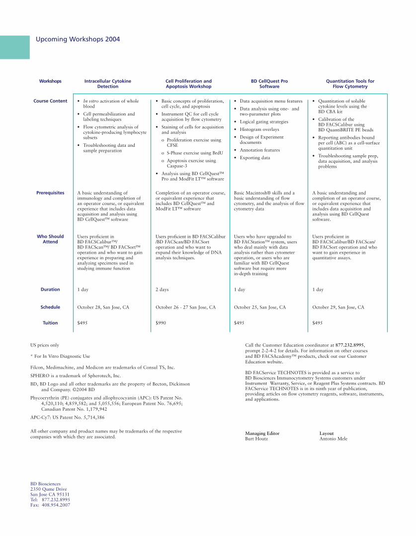
Upcoming Workshops 2004
Call the Customer Education coordinator at 877.232.8995, prompt 2-2-4-2 for details. For information on other courses and BD FACSAcademy™ products, check out our Customer Education website.
BD FACService TECHNOTES is provided as a service to BD Biosciences Immunocytometry Systems customers underInstrument Warranty, Service, or Reagent Plus Systems contracts. BDFACService TECHNOTES is in its ninth year of publication,providing articles on flow cytometry reagents, software, instruments,and applications.
BD Biosciences 2350 Qume DriveSan Jose CA 95131Tel: 877.232.8995 Fax: 408.954.2007
Intracellular Cytokine Detection
Cell Proliferation and Apoptosis Workshop
BD CellQuest ProSoftware
Quantitation Tools for Flow Cytometry
Workshops
• In vitro activation of wholeblood
• Cell permeabilization andlabeling techniques
• Flow cytometric analysis ofcytokine-producing lymphocytesubsets
• Troubleshooting data andsample preparation
• Basic concepts of proliferation,cell cycle, and apoptosis
• Instrument QC for cell cycleacquisition by flow cytometry
• Staining of cells for acquisitionand analysis
o Proliferation exercise usingCFSE
o S-Phase exercise using BrdU
o Apoptosis exercise usingCaspase-3
• Analysis using BD CellQuest™Pro and ModFit LT™ software
• Data acquisition menu features
• Data analysis using one- andtwo-parameter plots
• Logical gating strategies
• Histogram overlays
• Design of Experimentdocuments
• Annotation features
• Exporting data
• Quantitation of soluble cytokine levels using theBD CBA kit
• Calibration of theBD FACSCalibur usingBD QuantiBRITE PE beads
• Reporting antibodies boundper cell (ABC) as a cell-surface quantitation unit
• Troubleshooting sample prep, data acquisition, and analysis problems
Course Content
A basic understanding ofimmunology and completion ofan operator course, or equivalentexperience that includes dataacquisition and analysis using BD CellQuest™ software
Completion of an operator course,or equivalent experience thatincludes BD CellQuest™ andModFit LT™ software
Basic Macintosh® skills and abasic understanding of flowcytometry, and the analysis of flowcytometry data
A basic understanding andcompletion of an operator course,or equivalent experience thatincludes data acquisition andanalysis using BD CellQuestsoftware.
Prerequisites
Users proficient inBD FACSCalibur™/ BD FACScan™/ BD FACSort™operation and who want to gainexperience in preparing andanalyzing specimens used instudying immune function
Users proficient in BD FACSCalibur/BD FACScan/BD FACSortoperation and who want toexpand their knowledge of DNAanalysis techniques.
Users who have upgraded toBD FACStation™ system, userswho deal mainly with dataanalysis rather than cytometeroperation, or users who arefamiliar with BD CellQuestsoftware but require morein-depth training
Users proficient inBD FACSCalibur/BD FACScan/BD FACSort operation and whowant to gain experience inquantitative assays.
Who ShouldAttend
1 day 2 days 1 day 1 dayDuration
October 28, San Jose, CA October 26 - 27 San Jose, CA October 25, San Jose, CA October 29, San Jose, CASchedule
$495 $990 $495 $495Tuition
US prices only
* For In Vitro Diagnostic Use
Filcon, Medimachine, and Medicon are trademarks of Consul TS, Inc.
SPHERO is a trademark of Spherotech, Inc.
BD, BD Logo and all other trademarks are the property of Becton, Dickinsonand Company. ©2004 BD
Phycoerythrin (PE) conjugates and allophycocyanin (APC): US Patent No.4,520,110; 4,859,582; and 5,055,556; European Patent No. 76,695;Canadian Patent No. 1,179,942
APC-Cy7: US Patent No. 5,714,386
All other company and product names may be trademarks of the respectivecompanies with which they are associated.
Managing EditorBurt Houtz
LayoutAntonio Mele