beng 260- supplementary neurophysiology slides
TRANSCRIPT

BENG 260 Supplementary neurophysiology slides
Fall 2013
Slides are taken from Vander’s Human Physiology, 11th edition, McGraw Hill (ISBN 0077216091)"
These slides cover:"Chapter 6, Neuronal Signaling and the Structure of the Nervous System"Chapter 8, Consciousness, Brain, and Behavior"Chapter 10, Control of Body Movement"

A typical neuron has a dendritic region and an axonal region. The dendritic region is specialized to receive information whereas the axonal region is specialized to deliver information.
Communication by neurons is based on changes in the membrane’s permeability to ions. Two types of membrane potentials are of major functional significance: graded potentials and action potentials.
Chapter 6 Neuronal Signaling and the Structure
of the Nervous System

The two major divisions in the nervous system are the central nervous system (CNS) and the peripheral nervous system (PNS).
Within the PNS, major divisions are the somatic nervous system and the autonomic nervous system, which has two branches: the parasympathetic and the sympathetic branches.
Chapter 6 Neuronal Signaling and the Structure
of the Nervous System (cont.)

Dendrites: receive information, typically neurotransmitters, then undergo graded potentials.
Figure 6-1
Axons: undergo action potentials to deliver information, typically neurotransmitters, from the axon terminals.

Among all types of neurons, myelinated neurons conduct action potentials most rapidly.
Figure 6-2
Oligodendrocytes form myelin on central neuronal axons.
Schwann cells form myelin on peripheral neuronal axons.

PNS = afferent neurons (their activity “affects” what will happen
next) into the CNS +
efferent neurons (“effecting” change: movement, secretion, etc.) projecting out of the CNS.
CNS PNS
CNS = brain +
spinal cord; all parts of
interneurons are in the CNS.
Figure 6-4

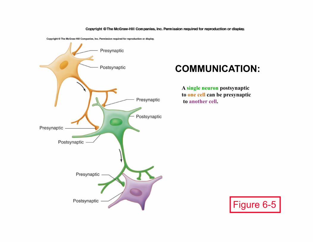
COMMUNICATION:
Figure 6-5
A single neuron postsynaptic to one cell can be presynaptic to another cell.

Opposite charges attract each other and will move toward each other if not separated by some barrier.
Figure 6-7

Figure 6-8

Only a very thin shell of charge difference is needed to establish a membrane potential.
Figure 6-9
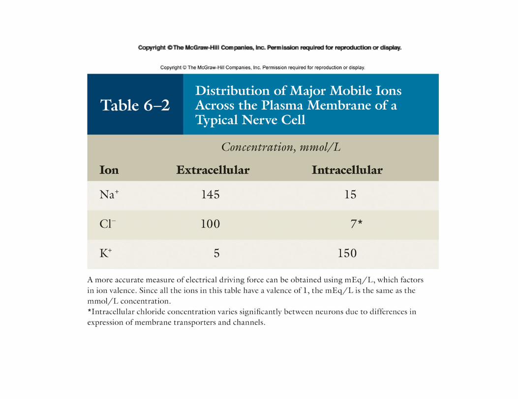

Figure 6-10
Begin: K+ in Compartment 2, Na+ in Compartment 1; BUT only K+ can move.
Ion movement: K+ crosses into Compartment 1; Na+ stays in Compartment 1.
buildup of positive charge in Compartment 1 produces an electrical potential that exactly offsets the K+ chemical concentration gradient.
At the potassium equilibrium potential:

Begin: K+ in Compartment 2, Na+ in Compartment 1; BUT only Na+ can move.
Ion movement: Na+ crosses into Compartment 2; but K+ stays in Compartment 2.
buildup of positive charge in Compartment 2 produces an electrical potential that exactly offsets the Na+ chemical concentration gradient.
At the sodium equilibrium potential:

Establishment of resting membrane potential: Na+/K+ pump establishes concentration gradient generating a small negative potential; pump uses up to 40% of the ATP produced by that cell!
Figure 6-13

Depolarization occurs when ion movement reduces the charge imbalance.
A cell is “polarized” because its interior is more negative than its exterior.
Overshoot refers to the development of a charge reversal.
Repolarization is movement back toward the resting potential.
Hyperpolarization is the development of even more negative charge inside the cell.
Figure 6-14


The size of a graded potential (here, graded depolarizations) is proportionate to the intensity of the stimulus.
Figure 6-15
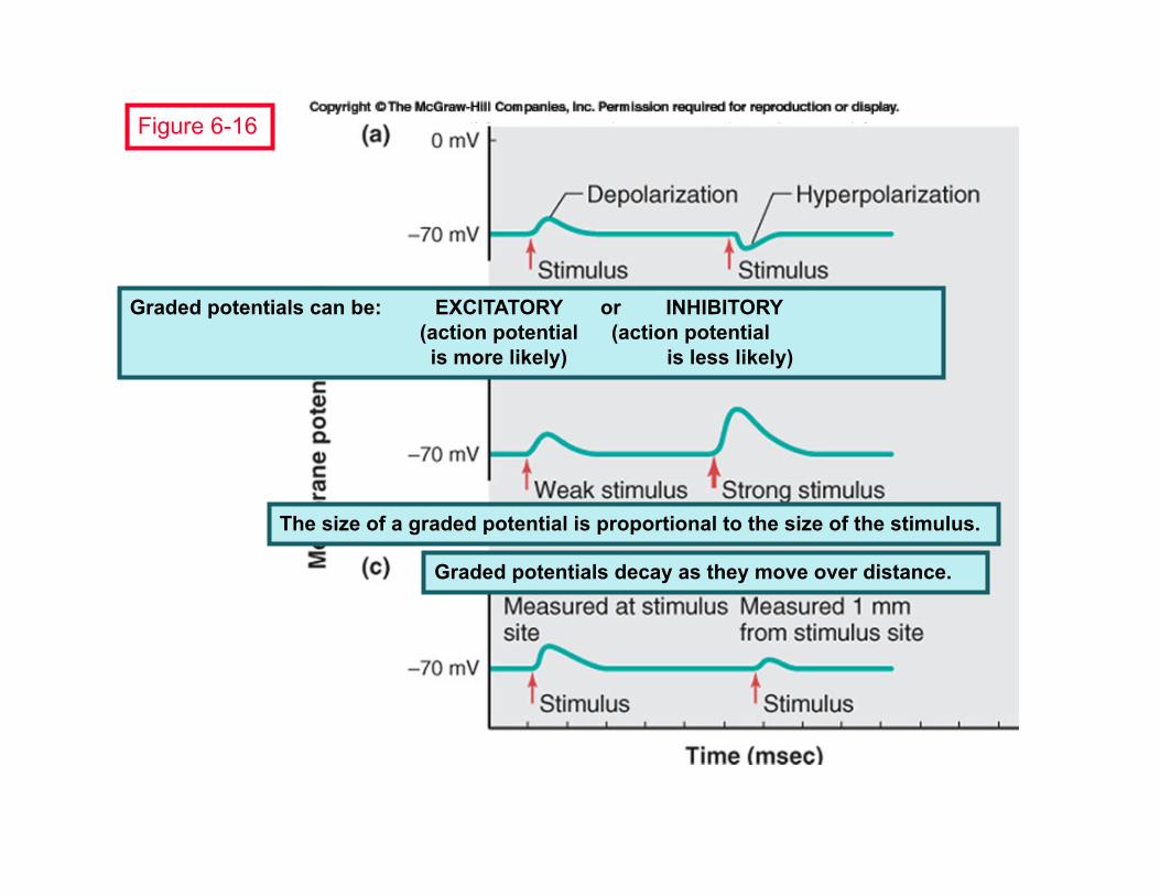
Graded potentials can be: EXCITATORY or INHIBITORY (action potential (action potential is more likely) is less likely)
The size of a graded potential is proportional to the size of the stimulus.
Graded potentials decay as they move over distance.
Figure 6-16

Graded potentials decay as they move over distance.
Figure 6-17
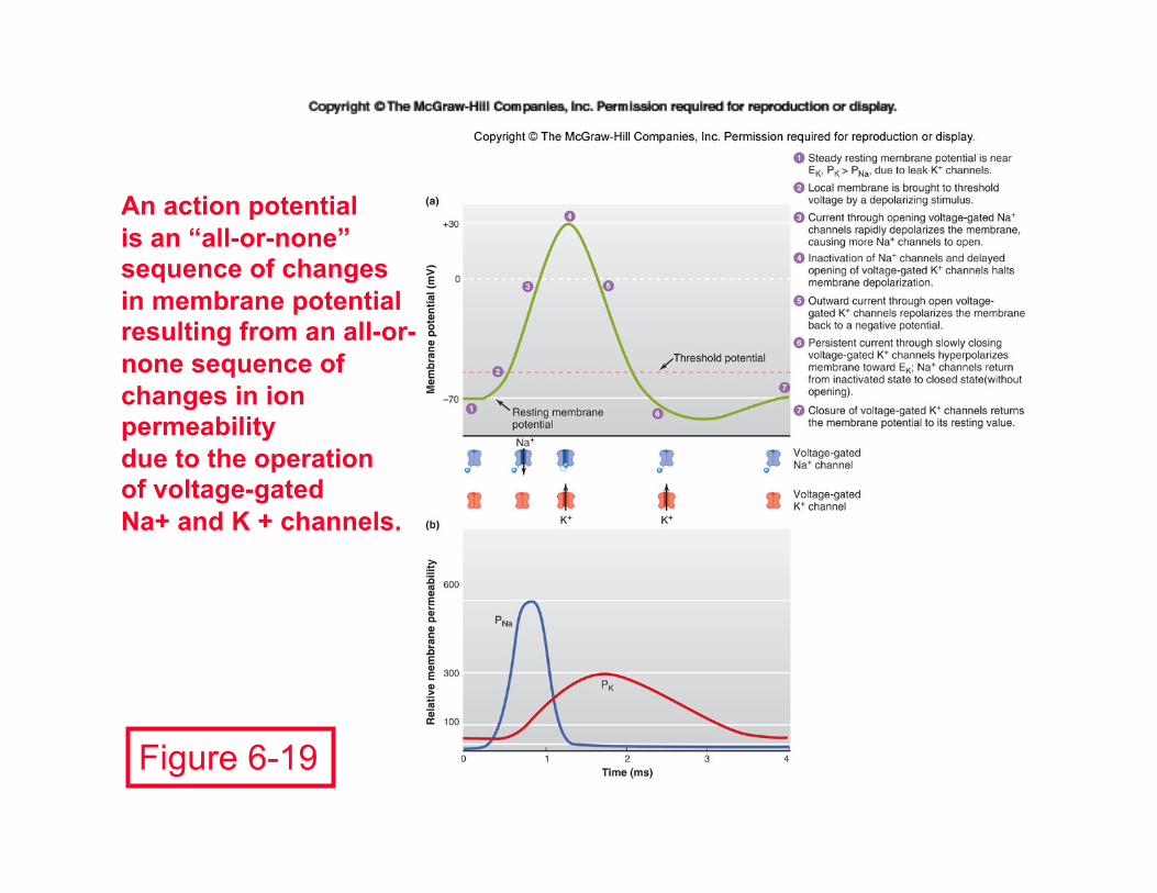
Figure 6-19
An action potential is an “all-or-none” sequence of changes in membrane potential resulting from an all-or-none sequence of changes in ion permeability due to the operation of voltage-gated Na+ and K + channels.
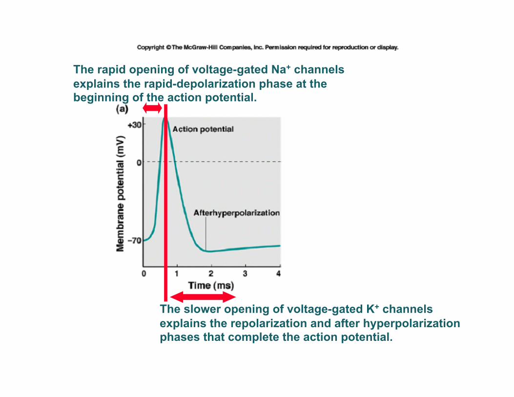
The rapid opening of voltage-gated Na+ channels explains the rapid-depolarization phase at the beginning of the action potential.
The slower opening of voltage-gated K+ channels explains the repolarization and after hyperpolarization phases that complete the action potential.

Figure 6-21
Four action potentials, each the result of a stimulus strong enough to cause deloplarization,are shown in the right half of the figure.

The propagation of the action potential from the dendritic to the axon-terminal end is typically one-way because the absolute refractory period follows along in the “wake” of the moving action potential.
Figure 6-22

Figure 6-23
Saltatorial Conduction: Action potentials jump from one node to the next as they propagate along a myelinated axon.


Figure 6-24
Four primary neurons communicate to one secondary neuron.
One primary neuron communicates to four secondary neurons.

Figure 6-25
The synapse is the point of communication between two neurons that operate sequentially.

Figure 6-26
Diversity in synaptic form allows the nervous system to achieve diversity and flexibility.
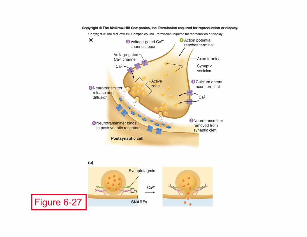
Figure 6-27

Figure 6-28
An excitatory postsynaptic potential (EPSP) is a graded depolarization that moves the membrane potential closer to the threshold for firing an action potential (excitement).

Figure 6-29
An inhibitory postsynaptic potential (IPSP) is a graded hyperpolarization that moves the membrane potential further from the threshold for firing an action potential (inhibition).

Figure 6-30
The membrane potential of a real neuron typically undergoes many EPSPs (A) and IPSPs (B), since it constantly receives excitatory and inhibitory input from the axons terminals that reach it.

Figure 6-31
Panel 1: Two distinct, non-overlapping, graded depolarizations. Panel 2: Two overlapping graded depolarizations demonstrate temporal
summation. Panel 3: Distinct actions of stimulating neurons A and B demonstrate
spatial summation. Panel 4: A and B are stimulated enough to cause a suprathreshold graded
depolarization, so an action potential results. Panel 5: Neuron C causes a graded hyperpolarization; A and C effects
add, cancel each other out.

Real neurons receive as many as 200,000 terminals.

Figure 6-32

Axo-axonal communication (here, between A & B) can modify classical synaptic communication (here, between B & C); this can result in presynaptic inhibition or presynaptic facilitation.
Figure 6-33 Note: the Terminal B must have receptors for the signal released from A.

Figure 6-34
Possible drug effects on synaptic effectiveness: A. release and degradation of the
neurotransmitter inside the axon terminal.
B. increased neurotransmitter release into the synapse.
C. prevention of neurotransmitter release into the synapse.
D. inhibition of synthesis of the neurotransmitter.
E. reduced reuptake of the neurotransmitter from the synapse.
F. reduced degradation of the neurotransmitter in the synapse. G. agonists (evoke same response as neurotransmitter) or
antagonists (block response to neurotransmitter) can occupy the receptors.
H. reduced biochemical response inside the dendrite.
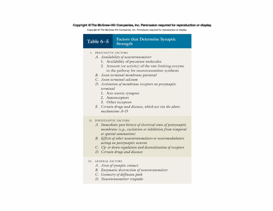


Figure 6-35
The catecholamines are formed from the amino acid tyrosine and share the same two initial steps in their biosynthetic pathway.

Figure 6-38 Major landmarks of the Central Nervous System
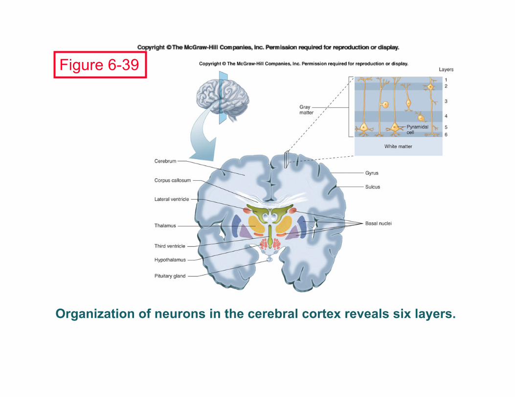
Organization of neurons in the cerebral cortex reveals six layers.
Figure 6-39

Figure 6-40
Functions of the limbic system: • learning • emotion • appetite (visceral function) • sex • endocrine integration

Figure 6-41 Anterior view of one vertebra and the nearby section of the spinal cord.


Figure 6-43
M o t o r n e u r o n
Preganglionic neuron Postganglionic neuron

Figure 6-44
Parasympathetic: “rest and digest”
Sympathetic: “emergency responses”

Figure 6-45
The sympathetic trunks are chains of sympathetic ganglia that are parallel to either side of the spinal cord; the trunk interacts closely with the associated spinal nerves.

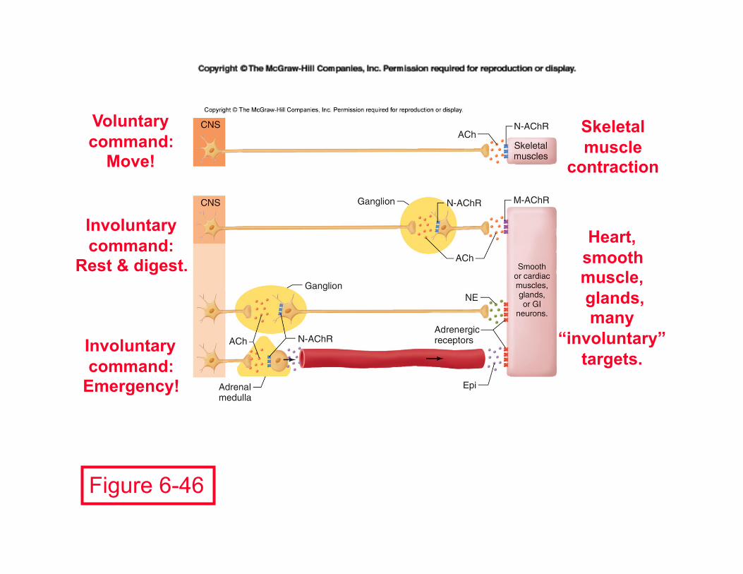
Figure 6-46
Voluntary command:
Move!
Skeletal muscle
contraction
Involuntary command:
Rest & digest. Heart,
smooth muscle, glands, many
“involuntary” targets.
Involuntary command:
Emergency!
M o t o r n e u r o n


Chapter 8 Consciousness, Brain, and Behavior
Electroencephalography: a window on the brain
• States of wakefulness and sleep
• Limbic system: motivation and reward
• Neurochemistry of drug abuse
• Learning and memory

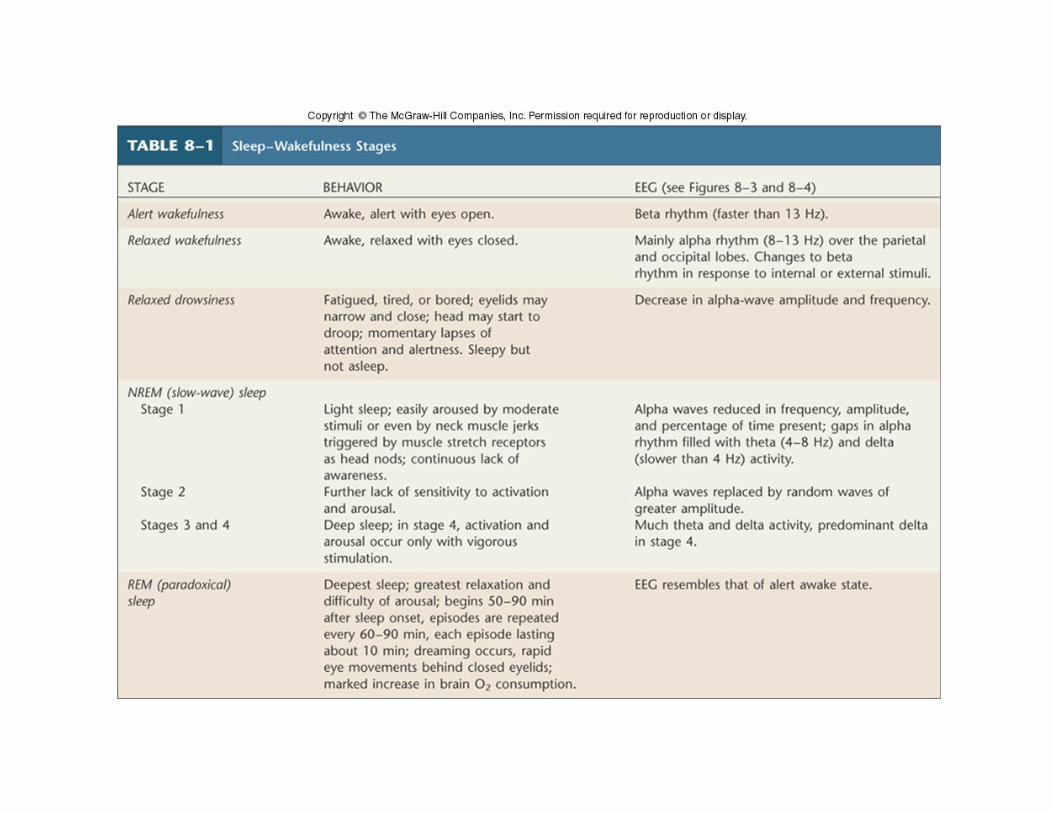
The electroencephalograph (EEG) is the printout of an electronic device that uses scalp electrodes to monitor the internal neural activity in the brain; this is a record from the parietal or occipital lobes of an awake person.
VO
LTA
GE
(typ
ical
ly 2
0-10
0 m
icro
volts
)
Figure 8-1

VO
LTA
GE
(20
to 1
00 m
icro
volts
)
EEGs provide diagnostic information about the location of abnormal activity in the brain, such as shown in this record typical of a patient undergoing an epileptic seizure.
Figure 8-2

Figure 8-3
VO
LTA
GE
(20
to 1
00 m
icro
volts
) V
OLT
AG
E (2
0 to
100
mic
rovo
lts)
EEGs reflect mental state: contrasted here are mental relaxation (a) versus concentration (b).
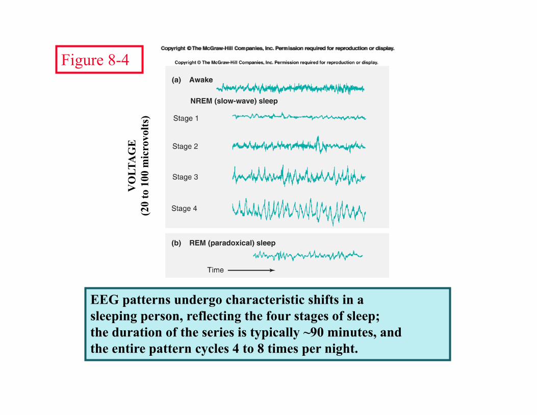
EEG patterns undergo characteristic shifts in a sleeping person, reflecting the four stages of sleep; the duration of the series is typically ~90 minutes, and the entire pattern cycles 4 to 8 times per night.
VO
LTA
GE
(20
to 1
00 m
icro
volts
)
Figure 8-4

The EEG pattern was analyzed to produce this graph of a full night’s sequence of sleep stages; also shown are cyclic patterns in the periphery.
Figure 8-5

A model of some of the neurochemical changes across the sleep-wake continuum; cause-and-effect relationships are under study.
Figure 8-6

Neuronal changes in these CNS structures appear to be essential participants in sleep-wake transitions and in biological rhythms.
Figure 8-7

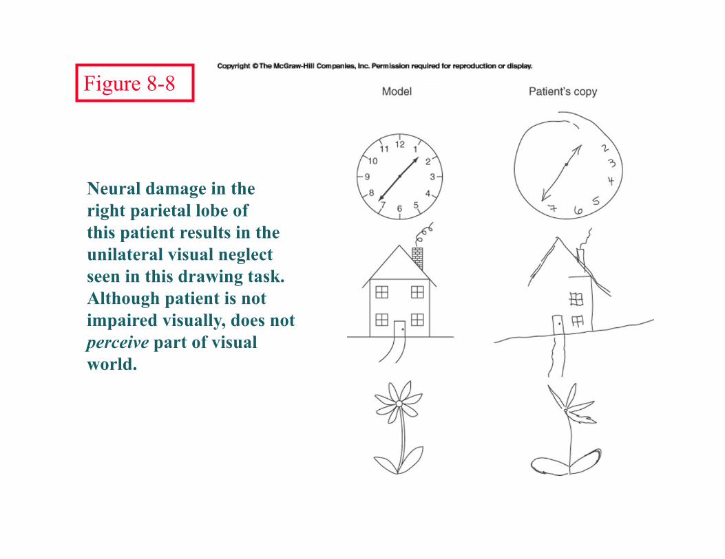
Neural damage in the right parietal lobe of this patient results in the unilateral visual neglect seen in this drawing task. Although patient is not impaired visually, does not perceive part of visual world.
Figure 8-8

Alterations in the mesolimbic dopamine pathway (shown here) appear to be a primary mechanism by which psychoactive drugs change behavior.
Figure 8-9
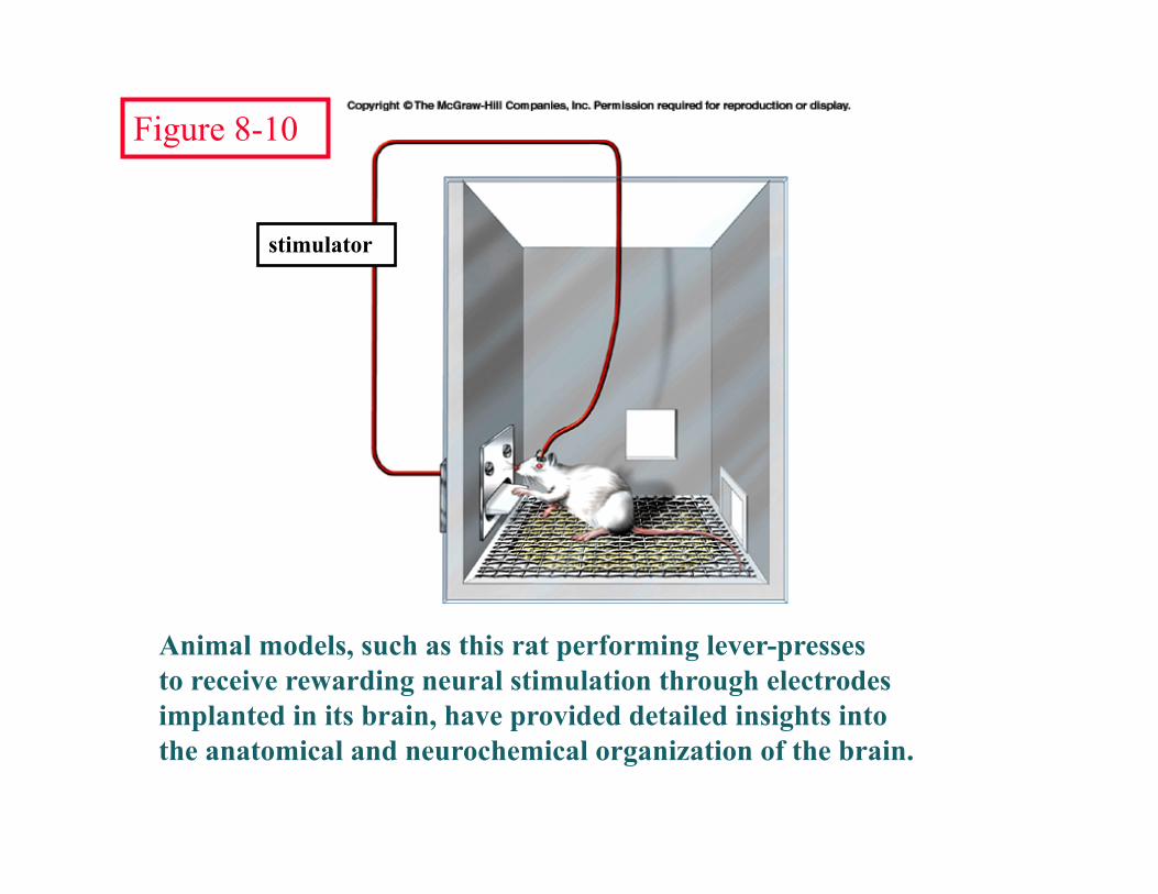
Animal models, such as this rat performing lever-presses to receive rewarding neural stimulation through electrodes implanted in its brain, have provided detailed insights into the anatomical and neurochemical organization of the brain.
stimulator
Figure 8-10

Changes in activity of the limbic system underlie some of the primary needs of the organism, including learning, motivation, appetite, and emotional response; its malfunction is associated with affective disorders.
Figure 8-11

Psychoactive drugs that affect serotonin- receptors share structural similarities with serotonin.
Psychoactive drugs that affect dopamine- receptors share structural similarities with dopamine.
Figure 8-13


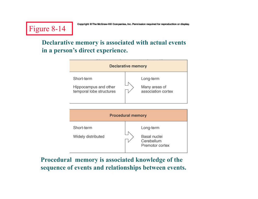
Declarative memory is associated with actual events in a person’s direct experience.
Procedural memory is associated knowledge of the sequence of events and relationships between events.
Figure 8-14


The primary loci underlying the comprehension of speech are in Wernicke’s area, whereas the primary loci for the production of speech are located in Broca’s area.
Figure 8-17
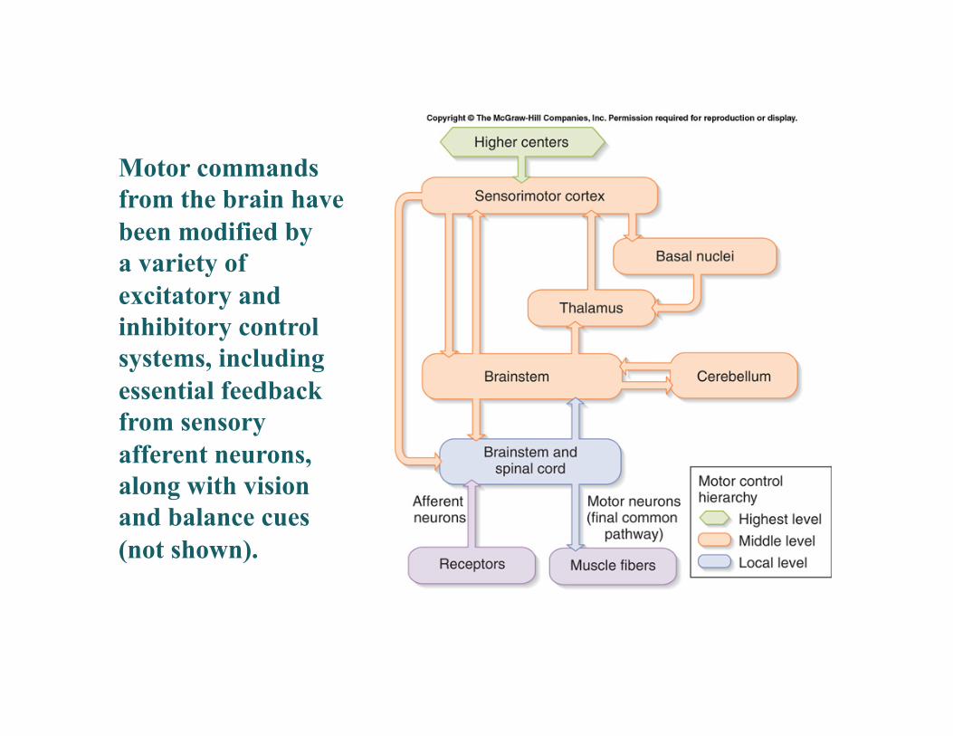
Motor commands from the brain have been modified by a variety of excitatory and inhibitory control systems, including essential feedback from sensory afferent neurons, along with vision and balance cues (not shown).
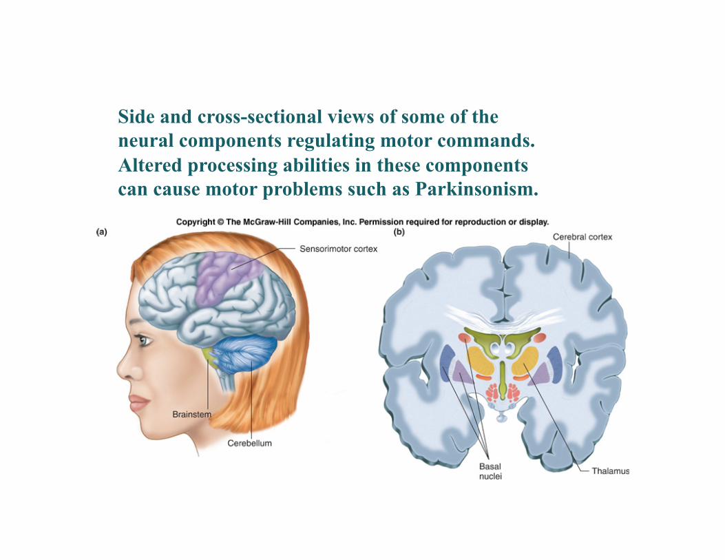
Side and cross-sectional views of some of the neural components regulating motor commands. Altered processing abilities in these components can cause motor problems such as Parkinsonism.


Examples of the categories of information and their underlying neuronal substrates modifying the production of motor commands from the brain.

Acting on local reflex circuits and by relaying impulses to the brain, muscle spindles and Golgi tendon organs provide information about muscle position and stretch in order to finely regulate the speed and intensity of muscle contraction.

Regardless of the reason for a change in length, the stretched spindle in scenario (a) generates a burst of action potentials as the muscle is lengthened; in scenario (b), the shortened spindle produces fewer action potentials from the spindle.

Tapping the patellar tendon lengthens the stretch receptor in the associated extensor muscle in the thigh; responses include:
compensatory contraction in that muscle (A and C), relaxation in the opposing flexor (B), and sensory afferent delivery to the brain. Note: NMJ = neuromuscular junction

Activation of Golgi tendon organs. Compared to when a muscle is contracting, passive stretch of the relaxed muscle produces less stretch of the tendon and fewer action potentials from the Golgi tendon organ.
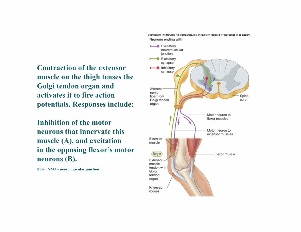
Contraction of the extensor muscle on the thigh tenses the Golgi tendon organ and activates it to fire action potentials. Responses include:
Inhibition of the motor neurons that innervate this muscle (A), and excitation in the opposing flexor’s motor neurons (B). Note: NMJ = neuromuscular junction

Figure 10-9
2
1
3
The neural components of the pain-withdrawal reflex in this example proceed as follows: Pain sensory afferents detect pain in foot and send action potentials via dorsal horn of spinal cord.
Interneurons in the cord activate extensor muscles on the “pained” side of the body and flexor muscles on the opposite side of the body.
Muscles move body away from painful stimulus.
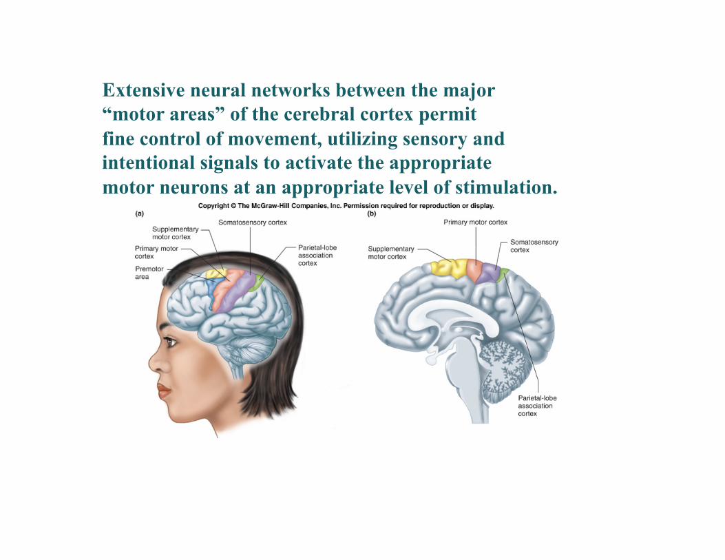
Extensive neural networks between the major “motor areas” of the cerebral cortex permit fine control of movement, utilizing sensory and intentional signals to activate the appropriate motor neurons at an appropriate level of stimulation.
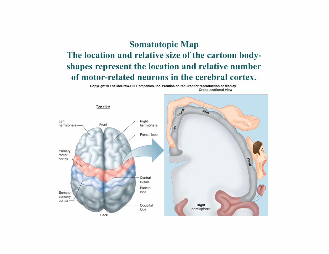
Somatotopic Map The location and relative size of the cartoon body- shapes represent the location and relative number of motor-related neurons in the cerebral cortex.

Efferent motor commands from the cerebral cortex are contralateral or “crossed,” meaning that the left cortex controls the muscles on the right side of the body (and vice versa), whereas the brainstem influences ipsilateral (same side) motor activity.

Motor activity must be informed about the body’s center of gravity in order to make adjustments in the level of stimulation to muscles whose contraction prevents unstable conditions (falling).