berberine protects homocysteic acid-induced ht-22 cell...
TRANSCRIPT
RESEARCH ARTICLE
Berberine protects homocysteic acid-induced HT-22 cell death:involvement of Akt pathway
Meihui Chen & Min Tan & Minghua Jing & Anmin Liu & Qinyu Liu & Shijun Wen &
Ziwei Chen & Xiaojuan Chao & Xixin He & Charles Ramassamy & Youheng Gao &
Rongbiao Pi
Received: 18 April 2014 /Accepted: 23 June 2014 /Published online: 23 July 2014# Springer Science+Business Media New York 2014
Abstract Berberine (BBR), one of the major constituents ofChinese herb Rhizoma coptidis, has been reported to exertbeneficial effects to various diseases, including Alzheimer’sdisease (AD). In the present work, we aimed to investigate theeffects of BBR on neuronal cell death induced by homocysteicacid (HCA), which was considered as a risk of AD. BBRsignificantly reduced HCA-induced reactive oxygen species(ROS) generation, lactate dehydrogenase release and subse-quent cell death. LY294002, the PI3K inhibitor, blocked theprotection as well as the up-regulation of Akt phosphorylationof BBR. Taken together, our results indicate that BBR protectsHCA-induced HT-22 cell death partly via modulating Akt
pathway, suggesting BBR may be a promising therapeuticagent for the treatment of HCA-related diseases, includingAD.
Keywords Berberine . Alzheimer’s disease . Homocysteicacid . Cytotoxicity . Akt
Introduction
Homocysteic acid (HCA) is an oxidized metabolite of Homo-cysteine (Obeid and Herrmann 2006) and an excitatory aminoacid analogue of glutamate (Glu) (Ikonomidou et al. 1996).Previous studies have suggested that elevated blood HCAmay be one of the pathological biomarkers in the brain withAD and could induce the accumulation of intraneuronal betaamyloid peptides (Hasegawa et al. 2010). In addition, HCAresults in accumulation of reactive oxygen species (ROS) athigh concentrations and inhibits the mitochondrial respirationand disordering of cell signaling mechanisms, which wereinvolved in the pathogenesis of AD (Folbergrova et al. 2007).
The phosphoinositide 3-kinase (PI3K)/Akt pathway iswidely expressed during central nervous system development(Ryder et al. 2004). Akt, which is activated by growth factorsto function in a pathway involving PI3K, could deactivate pro-apoptotic mediators and activate anti-apoptotic proteins (Shuet al. 2013). Previous studies suggested that PI3K/Akt signal-ing is impaired in AD brain and that treatments activating thispathway should enhance neuronal survival and block ADpathology (Shu et al. 2013).
Berberine (BBR, Fig. 1a), a natural isoquinoline alkaloidhas been used as a Chinese medicine for bacterial diarrhea andintestinal parasite infections for hundreds of years (Hsu et al.2012). Increasing evidence suggested that BBR might be apromising drug for the treatment of AD (Kulkarni and Dhir2010) and that BBR conferred protection against 6-
Meihui Chen and Min Tan contributed equally to this work
M. Chen :M. Tan : S. Wen : Z. Chen :X. Chao :R. Pi (*)Department of Pharmacology & Toxicology, School ofPharmaceutical Sciences, Sun Yat-Sen University,Guangzhou 510006, Chinae-mail: [email protected]
M. Tan :X. He :Y. Gao (*)Department of Traditional Chinese Medicine Chemistry, College ofChinese Materia Madica, Guangzhou University of ChineseMedicine, Guangzhou 510006, Chinae-mail: [email protected]
M. JingDepartment of Traditional Chinese Medicine, The First AffiliatedHospital, Sun Yat-Sen University, Guangzhou 510006, China
A. Liu :Q. LiuSun Yat-Sen Memorial Hospital, Sun Yat-Sen University,Guangzhou, China
S. WenState Key Laboratory of Oncology in South China, Sun Yat-senUniversity Cancer Center, Guangzhou 510060, China
C. RamassamyINRS-Institut Armand Frappier, Laval, QC, Canada
Metab Brain Dis (2015) 30:137–142DOI 10.1007/s11011-014-9580-x
hydroxydopamine (6-OHDA)-induced neuron injury (Baeet al. 2013) and H2O2-induced cytotoxicity in motor neuron-like cells (Hsu et al. 2012) via regulation of PI3K/Akt path-way. However, the mechanism underlying its potentialprotective effects against HCA-induced toxicity on neuro-nal cells has not yet been fully studied. We here demon-strated that BBR induced activation of PI3K/Akt, whichmight result in its protection against HCA-induced dam-age in HT-22 cells.
Experimental procedures
Materials
HT-22 cells were obtained from the Second Affiliated Hospi-tal, Sun Yat-Sen University (Guangzhou, China). BBR wasextracted by our laboratory and the purity is>99.0 %. HCAwas purchased from Tokyo Chemical Industry (Tokyo, Ja-pan). Lactate dehydrogenase (LDH), Annexin V-FITC/PIand glutathione (GSH) assay kits were purchased fromJiancheng Biochemical (Nanjing, China). Dulbecco’s modi-fied Eagle’s medium (DMEM) and fetal bovine serum (FBS)were obtained from Gibco-BRL (NY, USA). Others unlessstated elsewhere were purchased from Sigma-Aldrich (MO,USA).
Cell culture and treatment
HT-22 murine hippocampal neuronal cells were maintained inDMEM supplemented with 10 % (v/v) FBS and incubated at37 °C under 5 % CO2. To study the protective effect of BBRon HCA-induced neuronal death, cells were seeded in 96-wellplates (1×10⁴ cells/well). For the experiments, cells werepreincubated with BBR at indicated concentrations for30 min, and followed by treatment with HCA for differenthours.
Assessment of cell viability by MTT assay
Cells were grown on 96-well plates at a density of 1×10⁴ for24 h. After treatment with BBR (1–30 μM) for the indicatedtime, and MTT (5 mg/mL) 10 μL was then added to each welland the mixture was incubated for 2 h at 37 °C. MTT reagentwas then replaced with DMSO (100 μL per well) to dissolveformazan crystals. Absorbance was determined at 570 nmusing a microplate reader. Results were expressed as thepercentage of MTT reduction and the absorbance of controlcells was set as 100 %.
LDH release assay
For LDH assay, the medium was collected and assayed forLDH activity using a cytotoxicity detection kit. Briefly, the
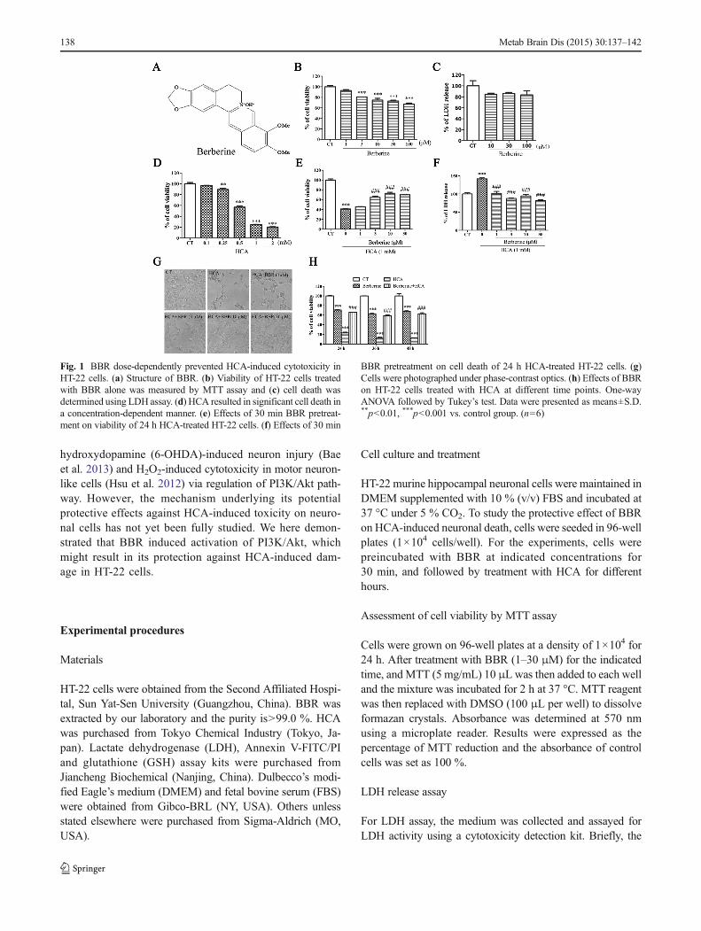
Fig. 1 BBR dose-dependently prevented HCA-induced cytotoxicity inHT-22 cells. (a) Structure of BBR. (b) Viability of HT-22 cells treatedwith BBR alone was measured by MTT assay and (c) cell death wasdetermined using LDH assay. (d) HCA resulted in significant cell death ina concentration-dependent manner. (e) Effects of 30 min BBR pretreat-ment on viability of 24 h HCA-treated HT-22 cells. (f) Effects of 30 min
BBR pretreatment on cell death of 24 h HCA-treated HT-22 cells. (g)Cells were photographed under phase-contrast optics. (h) Effects of BBRon HT-22 cells treated with HCA at different time points. One-wayANOVA followed by Tukey’s test. Data were presented as means±S.D.**p<0.01, ***p<0.001 vs. control group. (n=6)
138 Metab Brain Dis (2015) 30:137–142

release of LDH ismeasuredwith a coupled enzymatic reactionthat results in the conversion of a tetrazolium saltinto red-colored formazan. The formazan product was measured with amicroplate reader at 450 nm. Results were expressed as thepercentage of LDH release and the absorbance of control cellswas set as 100 %.
Hochest 33258 and PI staining
HT-22 cells were treated with BBR with or without HCA for12 h, and then stained with Hochest 33258 (1 μg/mL) or PI(10 μM) for 20 min in the dark. Results were determined byvisual observation of nuclear morphology through fluores-cence microscopy (Olympus, Japan) equipped with a UVfilter.
Apoptosis analysis by flow cytometry
The apoptotic ratios of cells were determined with theAnnexin V-FITC apoptosis detection kit (20 T, NanjingJiancheng, China). HT-22 cells were collected and washedtwice with cold PBS buffer, resuspended in 200 μL of bindingbuffer, incubated with 5 μL of Annexin V-FITC conjugated toFITC for 10 min, and then added with 10 μL PI for 15 min atroom temperature, and analyzed by flow cytometry. Cellstreated with DMSO were used as the negative control.
Measurement of ROS
Intracellular ROS formation was measured by fluorescenceusing H2DCF-DA. Briefly, after treatment, cells were washedand then stained with 10 μM H2DCF-DA in serum-free me-dium for 30 min at 37 °C in the dark. DCF fluorescence wasanalyzed by visual observation of cell morphology throughfluorescence microscopy equipped with a UV filter.
Estimation of intracellular GSH
A GSH assay kit (48 T, Nanjing Jiancheng, China) was usedto measure intracellular GSH concentration. In brief, aftertreatment, whole-cell lysate was prepared according to manu-facturer’s instructions. The basal contents of GSH in untreatedcontrol cells were taken as 100 %.
Determination of mitochondrial membrane potential
Rhodamine 123 (Rh123) was used to determine the Mito-chondrial Membrane Potential (MMP). For fluorescence pho-tomicrographs, cells were incubated with Rh123 (5 μg/mL) inPBS for 15 min at 37 °C in the dark. After incubation, cellswere washed with PBS. For flow cytometric analysis, cellswere then trypsinized, washed with PBS, resuspended in500 μL PBS containing 5 μg/mL mitochondrial dye Rh123.
After incubated for 15 min at 37 °C in the dark, cells werewashed once with PBS. Relative fluorescence intensities weremonitored using the flow cytometry (Beckman, USA), andmean fluorescence intensity of 1×10⁴ cells per sample waspresented.
Western blot analysis
Proteins were separated by SDS–PAGE, and transferred elec-trophoretically onto the PVDF membranes (Millipore, Biller-ica, MA). After blocking the membrane with 5 % skim milk,immune complexes were formed by incubation of primaryantibodies (table 1) for overnight at 4 °C. The dilutions andsources were as follows: anti-phospho-Akt (1:1000) anti-β-actin (1:10000), all obtained fromCell Signalling Technology,Hitchin, UK. PVDF membranes were washed three times inTBS-T, and incubated with horseradish peroxidase-linkedanti-mouse or anti-rabbit IgG (1:1000), depending on primaryantibodies used, in TBS-T for 1 h, followed by washing inTBS-T. Immunoreactive followed by sequential incubationwith horseradish peroxidase-conjugated secondary antibodiesfor 1 h at 37 °C. Enhanced chemiluminescence was used fordetection. All blots were compared with anti-β-actin antibodyto ascertain equal loading of proteins. The intensities of bandswere performed using Quantity One Software (Bio-Rad,Hercules, CA).
Statistical analysis
The data were presented as the means±standard error. Statis-tical analyses between two groups were performed by un-paired Student’s t-test. Differences among groups were testedby one-way analysis of variance (ANOVA). FollowingANOVA analyses, the Tukey’s test was used and p<0.05was accepted to be statistically significant.
Results
Effects of BBR on HT-22 cells with or without HCA
To investigate the effects of drugs on the cell viability, HT-22cells were treated with various concentrations of drugs for24 h and then cell viability and cytotoxicity were monitoredby the MTT test and LDH assay respectively (Fig. 1b or c).The results showed that BBR inhibited the cell proliferationbut did not cause cell death, whereas HCA caused cell death ina concentration-dependent manner (Fig. 1d). Moreover, BBR(10 μM) significantly increased cell survival following HCA(1 mM) treatment (Fig. 1e or f) and persistently block thetoxicity of HCA at different durations indicated (Fig. 1h).
Metab Brain Dis (2015) 30:137–142 139

BBR reduced HCA-induced apoptosis-like and necrosis-likedeath in HT-22 cells
To observe whether HCA induced apoptotic death, AnnexinV/Propidium Iodide (PI) staining assay and Hoechst 33258nuclear condensation staining assay were used. Necrosis wasidentified by PI, for their membrane impermeable and gener-ally excluded from viable cells (Jung et al. 2010). Our resultsshowed that HCA induced nuclear condensation (Fig. 2a),necrotic death (Fig. 2b) and apoptotic death (Fig. 2c). All ofabove changes induced byHCAwere significantly blocked byBBR (Fig. 2a or b).
BBR decreased HCA-induced ROS accumulation in HT-22cells
Previous study in our laboratory showed that HCA causedoxidative stress in HT-22 cells (Tan et al. 2013). Therefore, weinvestigated the effects of BBR on ROS production andendogenous antioxidant, glutathione (GSH) using fluorescentprobe H2DCF-DA and a GSH assay kit. Results showed thatHCA increased the intracellular ROS production, and BBRprofoundly reversed the process at the tested concentrations(Fig. 3a). To further investigate the effects of BBR on oxida-tive stress caused by HCA, the levels of intracellular GSH andmitochondrial membrane depolarization (MMP) were mea-sured. Results showed that HCA greatly decreased the intra-cellular level of GSH and BBR failed to reverse the depletionof GSH (Fig. 3b). Flow cytometric analysis revealed that
HCA significantly induced MMP, but BBR was not able toimprove the MMP (Fig. 3c). Carbonyl cyanide 3-chlorophenylhydrazone(CCCP), a mitochondrial membranedepolarizer was used as a positive control.
BBR protected HT-22 cells against HCA-induced cytotoxicitypartly through activating Akt signaling
It is well known that Akt plays a critical role in neuronalsurvival and apoptosis. As shown in Fig. 4a, HCA induced asignificant decrease of the level of phosphorylated Akt, whichwas partly reversed by BBR (10 μM). To further explorewhether Akt was involved in BBR-mediated neuroprotection,LY294002, a selective PI-3 kinase inhibitor was used. Thedata showed that LY294002 (3 μM), which did not affect thecell viability alone, markedly attenuated the protection ofBBR on HCA-induced cytotoxicity (Fig. 4b). Moreover,LY294002 aggravated the cytotoxicity of HCA.
Discussion
To the best of our knowledge, this is the first report about theeffects of BBR on the neuronal toxicity and oxidative stressinduced by HCA in HT-22 cells. We observed that BBRdramatically attenuated HCA induced ROS generation, LDHrelease and subsequent cell death. BBR also enhanced Aktphosphorylation which was down-regulated by HCA. Further
Fig. 2 BBR attenuated HCA-induced apoptosis-like andnecrosis-like death in HT-22 cells.(a) Morphological changes indifferent groups for the Hoechststaining and (b) propidium iodide(PI) staining. Positive stainedcells were indicated by whitearrows. (c) HT-22 cells weretreated with BBR in the presenceor absence of HCA (1 mM) for12 h, and the induction ofapoptosis was determined usingAnnexin V-FITC/PI staining. (d)Quantitative analysis of apoptoticcells. Data were presented asmeans±S.D. *p<0.05,***p<0.001 vs. control group;###p<0.001 vs. HCA-treatedgroup. (n=3)
140 Metab Brain Dis (2015) 30:137–142

studies uncovered that LY294002, a specific PI3K inhibitor,reversed the protection of BBR against HCA induced toxicity,suggesting the involvement of PI3K/Akt signaling in BBRmediated neuroprotection. Meanwhile, the present study re-vealed that HCA might cause cytotoxicity through down-regulating Akt phosphorylation.
HCA could lead to accumulation of neurotoxic amyloid β-peptide-42 which accounts for the pathogenesis of AD(Hasegawa et al. 2005). In addition, HCA was demonstratedto induce neuronal apoptosis through glutathione depletionand oxidative stress in HT-22 cells (Ratan et al. 1994; Wanget al. 2010). Oxidative stress was demonstrated to be associ-ated with a large array of neurological diseases and brainageing itself (Sompol et al. 2008). Markesbery et al.
confirmed that the presence of diffuse ROS resulted in oxida-tive stress of the entire brain in AD cases (Markesbery andCarney 1999). Our study showed that HCA increased theoxidative stress injury through elevating ROS production,depletion of GSH.Moreover, we found that BBR significantlyattenuated HCA-induced neurotoxicity and ROS accumula-tion but failed to reverse the depletion of GSH. Previousstudies have demonstrated that α-tocopherol, a potent antiox-idant, protect cells from oxidative toxicity without affectingthe GSH decrease caused by Glu (Davis and Maher 1994),suggesting that the protection of BBR might not targetmitochondria.
Akt (also known as protein kinase B, PKB), highlyexpressed in the nervous system, is a vital promoter of
Fig. 3 BBR inhibited HCA-inducedROS generation. (a) ROSproduction was detected byH2DCF-DA staining. (b) Thelevel of GSH was measured usingcommercial assay kits by treatedwith BBR in presence or absenceof HCA. The basal contents ofGSH in untreated control cellswere taken as 100 %. (c) Rate ofRh123-positive cells in differentgroups. One-way ANOVAfollowed by Tukey’s test. Datawere presented as means±S.D.*p<0.05, **p<0.01, ***p<0.001vs. control group. (n=3)
Fig. 4 LY294002 blocked the protection of BBR against HCA-inducedcytotoxicity. HT-22 cells were pretreated with LY294002 for 0.5 h, thentreated with BBR for 0.5 h, and further exposed to HCA for 24 h. (a)Effect of BBR on the phosphorylation of Akt. Quantitative analysis of theblots was shown in panel after being normalized by β-actin. (b) Cell
viability was measured by MTT assay. The data presented were themeans±S.D. One-way ANOVA followed by Tukey’s test. *p<0.05,**p<0.01, ***p<0.001 vs. control group; ##p<0.01, ###p<0.001 vs.HCA-treated group. (n=6)
Metab Brain Dis (2015) 30:137–142 141

survival and neuroprotection in embryonic neurons and isconsidered as a system that may link Aβ, NFTs and neuronalloss in AD (Brunet et al. 2001). Studies uncovering the linkbetween Akt signaling and AD have suggested that PI3K/Aktactivation attenuates Aβ neurotoxicity in vitro and in vivo(Stein and Johnson 2002; Wei et al. 2002). Previous reportsshowed that BBR conferred protection against 6-OHDA-induced neuron injury (Bae et al. 2013) and H2O2-inducedcytotoxicity in motor neuron-like cells (Hsu et al. 2012) viaregulation of PI3K/Akt pathway. In the present work, wedemonstrated that BBR markedly increased HCA-induceddeactivation of Akt, which was blocked by LY294002, thePI3K inhibitor, suggesting the involvement of PI3K/Akt sig-naling in BBR mediated neuroprotection. Our results alsosuggested HCA might cause cytotoxicity via down-regulating Akt phosphorylation.
Conclusion
In summary, we demonstrated that BBR, a naturalisoquinoline alkaloid, which exerted potent protective effectsagainst HCA-induced neurotoxicity partly through modulat-ing Akt phosphorylation, may be a useful therapeutic agent forthe treatment of HCA-related diseases, including AD.
Acknowledgments This research was supported by grants from Na-tional Natural Science Foundation (No. 31371070) and Guangdong Pro-vincial International Cooperation Project of Science & Technology (No.2012B050300015) to R. Pi.
Conflict of interest No potential conflicts of interest.
References
Bae J, Lee D, Kim YK, Gil M, Lee JY, Lee KJ (2013) Berberine protects6-hydroxydopamine-induced human dopaminergic neuronal celldeath through the induction of heme oxygenase-1. Mol Cells 35:151–157
Brunet A, Datta SR, GreenbergME (2001) Transcription-dependent and -independent control of neuronal survival by the PI3K-Akt signalingpathway. Curr Opin Neurobiol 11:297–305
Davis JB, Maher P (1994) Protein kinase C activation inhibits glutamate-induced cytotoxicity in a neuronal cell line. Brain Res 652:169–173
Folbergrova J, Jesina P, Drahota Z, Lisy V, Haugvicova R, Vojtiskova A,Houstek J (2007) Mitochondrial complex I inhibition in cerebralcortex of immature rats following homocysteic acid-induced sei-zures. Exp Neurol 204:597–609
Hasegawa T, UkaiW, Jo DG, Xu X,MattsonMP, NakagawaM, ArakiW,Saito T, Yamada T (2005) Homocysteic acid induces intraneuronal
accumulation of neurotoxic Abeta42: implications for the pathogen-esis of Alzheimer’s disease. J Neurosci Res 80:869–876
Hasegawa T, Mikoda N, Kitazawa M, Laferla FM (2010) Treatment ofAlzheimer’s disease with anti-homocysteic acid antibody in 3xTg-AD male mice. PLoS One 5:e8593
Hsu YY, Chen CS, Wu SN, Jong YJ, Lo YC (2012) Berberineactivates Nrf2 nuclear translocation and protects against oxi-dative damage via a phosphatidylinositol 3-kinase/Akt-depen-dent mechanism in NSC34 motor neuron-like cells. Eur JPharm Sci 46:415–425
Ikonomidou C, Qin Qin Y, Labruyere J, Olney JW (1996) Motor neurondegeneration induced by excitotoxin agonists has features in com-mon with those seen in the SOD-1 transgenic mouse model ofamyotrophic lateral sclerosis. J Neuropathol Exp Neurol 55:211–224
Jung SH, Kim BJ, Lee EH, Osborne NN (2010) Isoquercitrin is the mosteffective antioxidant in the plant Thuja orientalis and able to coun-teract oxidative-induced damage to a transformed cell line (RGC-5cells). Neurochem Int 57:713–721
Kulkarni SK, Dhir A (2010) Berberine: a plant alkaloid with therapeuticpotential for central nervous system disorders. Phytother Res 24:317–324
Markesbery WR, Carney JM (1999) Oxidative alterations in Alzheimer’sdisease. Brain Pathol 9:133–146
Obeid R, Herrmann W (2006) Mechanisms of homocysteine neurotox-icity in neurodegenerative diseases with special reference to demen-tia. FEBS Lett 580:2994–3005
Ratan RR, Murphy TH, Baraban JM (1994) Macromolecular synthesisinhibitors prevent oxidative stress-induced apoptosis in embryoniccortical neurons by shunting cysteine from protein synthesis toglutathione. J Neurosci 14:4385–4392
Ryder J, Su Y, Ni B (2004) Akt/GSK3beta serine/threonine kinases:evidence for a signalling pathway mediated by familialAlzheimer’s disease mutations. Cell Signal 16:187–200
Shu Y, Zhang H, Kang T, Zhang JJ, Yang Y, Liu H, Zhang L (2013)PI3K/Akt signal pathway involved in the cognitive impairmentcaused by chronic cerebral hypoperfusion in rats. PLoS One 8:e81901
Sompol P, Ittarat W, Tangpong J, Chen Y, Doubinskaia I, Batinic-Haberle I, Abdul HM, Butterfield DA, St Clair DK (2008) Aneuronal model of Alzheimer’s disease: an insight into themechanisms of oxidative stress-mediated mitochondrial injury.Neuroscience 153:120–130
Stein TD, Johnson JA (2002) Lack of neurodegeneration in transgenicmice overexpressing mutant amyloid precursor protein is associatedwith increased levels of transthyretin and the activation of cellsurvival pathways. J Neurosci 22:7380–7388
Tan M, Ouyang Y, Jin M, Chen M, Liu P, Chao X, Chen Z, Chen X,Ramassamy C, Gao Y, Pi R (2013) Downregulation of Nrf2/HO-1pathway and activation of JNK/c-Jun pathway are involved inhomocysteic acid-induced cytotoxicity in HT-22 cells. Toxicol Lett223(1):1–8
Wang L, Ankati H, Akubathini SK, BalderamosM, Storey CA, Patel AV,Price V, Kretzschmar D, Biehl ER, D’mello SR (2010) Identificationof novel 1,4-benzoxazine compounds that are protective in tissueculture and in vivomodels of neurodegeneration. J Neurosci Res 88:1970–1984
Wei W, Wang X, Kusiak JW (2002) Signaling events in amyloid beta-peptide-induced neuronal death and insulin-like growth factor Iprotection. J Biol Chem 277:17649–17656
142 Metab Brain Dis (2015) 30:137–142

本文献由“学霸图书馆-文献云下载”收集自网络,仅供学习交流使用。
学霸图书馆(www.xuebalib.com)是一个“整合众多图书馆数据库资源,
提供一站式文献检索和下载服务”的24 小时在线不限IP
图书馆。
图书馆致力于便利、促进学习与科研,提供最强文献下载服务。
图书馆导航:
图书馆首页 文献云下载 图书馆入口 外文数据库大全 疑难文献辅助工具