(beta)1 integrins in wound repair -...
TRANSCRIPT

INTRODUCTION
Following cutaneous injury, a well-defined cascade of events iskick started, beginning with the plugging of the defect by a fibrinclot and culminating in the restitution of the epithelial barrierand the generation of fibrous scar tissue (reviewed by Martin,1997). Re-epithelialisation, the process by which keratinocytesat the wound margin seal the epidermal fault, is a key event underpinning skin repair. It is achieved via a combinationof proliferation and sustained migration of wound edge keratinocytes across the dermis and provisional woundmatrix.
Several genetically modified mouse models have been used toinvestigate the molecular basis of the re-epithelialisation process.These studies revealed important roles for growth factorsignalling, proteases and cell-surface molecules in epithelialrepair (reviewed by Grose and Werner, 2002). However, despitea plethora of data on the involvement of integrins in skin
development and cell migration in vitro, there is littleinformation on their in vivo functions during wound repair.
Integrins are heterodimeric transmembrane proteinsconsisting of an αand a βsubunit that play crucial roles in cell-cell and cell-matrix interactions. They underpin cell adhesionand migration, as well as being involved in cell proliferation,programmed cell death and differentiation (Brakebusch et al.,1997). The major integrins in the intact epidermis are α2β1,α3β1 and α9β1, which bind various extracellular matrixproteins, including laminins, collagen I, tenascin C andfibronectin, and α6β4, an integral component ofhemidesmosomes that binds laminin 5 (Palmer et al., 1993;Yokosaki et al., 1994; Yamada et al., 1996). After wounding, theexpression profile of integrins on wound margin keratinocyteschanges, characterised by suprabasal integrin expression (Hertleet al., 1992) and induction of specific integrins that recogniseproteins of the dermal and provisional matrix. These includethe fibronectin receptor α5β1; the fibronectin and vitronectin
2303Development 129, 2303-2315 (2002)Printed in Great Britain © The Company of Biologists Limited 2002DEV20000
Integrins are ubiquitous transmembrane receptors thatplay crucial roles in cell-cell and cell-matrix interactions.In this study, we have determined the effects of the loss ofβ1 integrins in keratinocytes in vitro and during cutaneouswound repair. Flow cytometry of cultured β1-deficientkeratinocytes confirmed the absence of β1 integrins andshowed downregulation of α6β4 but not of αv integrins. β1-null keratinocytes were characterised by poor adhesion tovarious substrates, by a reduced proliferation rate and bya strongly impaired migratory capacity. In vivo, the loss ofβ1 integrins in keratinocytes caused a severe defect inwound healing. β1-null keratinocytes showed impairedmigration and were more densely packed in thehyperproliferative epithelium. Surprisingly, their
proliferation rate was not reduced in early wounds andeven increased in late wounds. The failure in re-epithelialisation resulted in a prolonged inflammatoryresponse, leading to dramatic alterations in the expressionof important wound-regulated genes. Ultimately, β1-deficient epidermis did cover the wound bed, but theepithelial architecture was abnormal. These findingsdemonstrate a crucial role of β1 integrins in keratinocytemigration and wound re-epithelialisation.
Movies available on-line
Key words: Integrin, Migration, Epidermis, Mouse, Wound
SUMMARY
DEVELOPMENT AND DISEASE
A crucial role of β1 integrins for keratinocyte migration in vitro and during
cutaneous wound repair
Richard Grose 1,*,†, Caroline Hutter 2,†, Wilhelm Bloch 3, Irmgard Thorey 1, Fiona M. Watt 2, Reinhard Fässler 4,5,Cord Brakebusch 4 and Sabine Werner 1,‡
1Institute of Cell Biology, Department of Biology, ETH-Zürich, 8093 Zürich, Switzerland2Keratinocyte Laboratory, Cancer Research UK London Research Institute, London WC2A 3PX, UK3Institute of Anatomy, University of Cologne, D-50931 Köln, Germany4Department of Experimental Pathology, Lund University, S-221 85 Lund, Sweden5Max-Planck-Institute of Biochemistry, D-82152 Martinsried, Germany*Present address: Viral Carcinogenesis Laboratory, Cancer Research UK, London Research Institute, London WC2A 3PX, UK†The first two authors have equally contributed to this work‡Author for correspondence (e-mail: [email protected])
Accepted 4 February 2002

2304
receptor αvβ5; and αvβ6, which binds to fibronectin,vitronectin, tenascin C and transforming growth factor β (TGFβ)(Larjava et al., 1993; Zambruno et al., 1995; Clark, 1990;Yamada et al., 1996; Plow et al., 2000).
A series of studies has revealed an essential role of β1integrins in the regulation of keratinocyte proliferation anddifferentiation. Most interestingly, the β1 integrin subunit hasbeen found to be a marker for stem cells in vitro and in vivo(Jones and Watt, 1993; Jones et al., 1995). Inhibition of β1integrin function by introduction of a dominant-negative β1mutant into keratinocytes reduced the activation of mitogen-activated protein kinase and stimulated exit from the stem cellcompartment (Zhu et al., 1999). Furthermore, suprabasalexpression of β1 is a feature of the hyperproliferative epidermisof psoriatic skin, and ectopic expression of the β1 integrinsubunit in the suprabasal epidermal layers of transgenic micecaused keratinocyte hyperproliferation and a psoriasis-likephenotype (Carroll et al., 1995).
To determine the role of β1 integrins in skin morphogenesisand homeostasis, we and others generated mice that lackthe β1 integrin subunit specifically in keratinocytes. Thecharacterisation of the β1-deficient mice revealed crucial rolesfor the β1 integrin subfamily for hair follicle development andskin integrity (Brakebusch et al., 2000; Raghavan et al., 2000).In this study, we used our mouse model to investigate cutaneouswound repair, a process that is reliant on both keratinocytemigration and proliferation, in the absence of epidermal β1integrins. We demonstrate an important role for β1 integrinsduring wound re-epithelialisation, where β1-null keratinocytesdisplay a severely compromised epithelial migration.
MATERIALS AND METHODS
K5β1-null miceMice that lack the β1 integrin subunit in the epidermis (K5β1-null mice)have been described elsewhere (Brakebusch et al., 2000). Briefly, micecarrying two floxed β1 alleles were crossed with mice carrying onefloxed β1 allele plus one copy of the cre recombinase gene under thecontrol of the keratin 5 (K5) promoter (Ramirez et al., 1994). Littermatecontrols were maintained alongside the knockouts under identicalhusbandry conditions.
Isolation and culture of mouse keratinocytesMurine epidermal keratinocytes were isolated as described (Roper etal., 2001). Keratinocyte cell lines were generated by repeatedsubculturing at high density (Romero et al., 1999).
Retroviral infection of keratinocytesAM12 supernatants or concentrated VSV G-pseudotyped virus wasadded to subconfluent mouse keratinocyte cultures and incubated for 6-8 hours in the presence of 8 µg/ml polybrene. The following retroviralvectors were used: pBabe chicken β1 integrin subunit (Levy et al.,1998), pBabe EGFPCre and LZRSpBMN EGFPCre (Cre fusion proteinand LZRSpBMN, kind gifts from J. Muller, ICRF).
Time-lapse recordingFrames were taken every 5 minutes for 22 hours using a Zeiss Axiovert135 TV microscope fitted with a Hamamatsu CCD Orca ER camera(Hamamatsu, Japan). Motility was measured using a cell trackingextension (ICRF) written for IPLab (Signal Analytics, USA) and speedwas calculated using a program written in Mathematica by Daniel Zicha(ICRF).
FACS analysisSingle cell suspensions of freshly isolated keratinocytes were incubatedwith a rat antibody against the mouse β1 integrin subunit (MB1.2; 1:2diluted; kind gift from B. Chan), a rat antibody against the β4 integrinsubunit (CD104; 1:100 diluted, BD Pharmingen, Franklin Lakes, NJ)or a hamster antibody against the αv integrin subunit (1:100 diluted;RDI, Flanders, NJ), and subsequently with AlexaFluor488-conjugatedsecondary antibody directed against rat IgG or biotinylated antibodydirected against hamster IgG (Jackson ImmunoResearch, West Grove,PA), followed by incubation with RPE-Cy5 conjugated streptavidin(Dako, Hamburg, Germany). Immediately before analysis on a Becton-Dickinson FACScan, TO-PRO-3 (Molecular Probes, Leiden, NL) wasadded to the sample for viability gating.
Adhesion assaysKeratinocytes isolated from 2-day-old mice were seeded into 96-wellplates (5×10⁴ cells/well), pre-coated with fibronectin, laminin 1, poly-D-lysine (PDL), collagen type I or collagen type IV (BD BiocoatCellware, Franklin Lakes, NJ). After overnight incubation, cells werewashed with phosphate-buffered saline (PBS) and adhesion wasquantified using a CytoTox 96 colorimetric kit (Promega, Madison,WI).
Wounding and preparation of wound tissueMice were anaesthetised by intraperitoneal (i.p.) injection of 100 µlketamine (10 g/l)/xylazine (8 g/l) solution. Two full-thicknessexcisional wounds, 3 mm in diameter, were made on either side of thedorsal midline by excising skin and panniculus carnosus. Wounds wereleft uncovered and harvested 1, 2, 3, 5, 10 or 15 days after injury (n≥4mice for each timepoint). For expression analyses, the complete woundsincluding 2 mm of the epithelial margins were excised and immediatelyfrozen in liquid nitrogen. Non-wounded back skin from 10 days postpartum (d.p.p.) and 20 d.p.p. mice served as controls. Alternatively, twofull-thickness incisional wounds were made on either side of the dorsalmidline, left uncovered and harvested at 3 or 6 days after injury (n=3mice for each time point). For histological analysis, the completeexcisional or incisional wounds were isolated, bisected, fixed overnightin 4% paraformaldehyde in PBS and embedded in paraffin wax.Sections (7 µm) from the middle of the wound were stained withHaematoxylin/Eosin (H/E) or Masson trichrome. All experiments withanimals were carried out with permission from the local veterinaryauthorities.
Labelling with 5 ′BrdUBrdU labelling was performed as described (Werner et al., 1994).Sections (7 µm) from the middle of the wound were incubated with aperoxidase-conjugated monoclonal antibody directed against BrdU(Roche Diagnostics, Rotkreuz, Switzerland) and stained with adiaminobenzidine-peroxidase substrate kit (Vector Laboratories,Burlingame, CA).
ImmunofluorescenceTo stain keratinocyte cultures for paxillin, cells were fixed with 4%paraformaldehyde in PBS containing 0.1% Triton X-100, washed inPBS, blocked in 2% FCS in PBS and incubated with anti-paxillinantibody (1:100 diluted; BD Transduction Laboratories), followed byAlexaFluor488-conjugated anti-rabbit IgG. To stain for filamentousactin, cultures were incubated in a 1:1000 dilution of rhodamine-conjugated phalloidin (Sigma-Aldrich, St Louis, MO). Wax andcryosections (7 µm) from the middle of the wound were incubated withantibodies directed against the β1 integrin subunit (a kind gift from DrStaffan Johansson, Uppsala, Sweden), the β4 integrin subunit, β-cateninand E-cadherin (kind gifts from Dr Rolf Kemler, Freiburg, Germany),caspase 3 (Roche Diagnostics Ltd, Rotkreuz, Switzerland), keratin 6(Babco, Richmond, CA), laminin 5 (α2LE4-6, kind gift from Dr RupertTimpl, Munich, Germany), and PECAM1 (BD Biosciences,Heidelberg, Germany), followed by Cy2- or Cy3- conjugated secondary
R. Grose and others

2305β1 integrins in wound repair
antibodies (Jackson Immunoresearch).Specimens were mounted with Mowiol(Hoechst, Frankfurt, Germany) prior tophotographing on a Zeiss Axioplanfluorescence microscope or confocalimaging on a Leica confocal laserscanning microscope.
ImmunohistochemistryWax sections (7 µm) from the middle ofthe wound were incubated with theappropriate primary antibody:neutrophils – 1:125 dilution of abiotinylated rat antibody targeted to Ly-6G (BD Biosciences); and macrophages– 1:125 dilution of a rat antibodytargeted to F4/80 (Serotec, Oxford,UK). Anti-F4/80 was incubated withbiotinylated donkey anti-rat IgG(Dianova GMBH, Hamburg, Germany).The VectaStain avidin-biotin-peroxidasecomplex kit was then used according tothe manufacturer’s instructions, priorto peroxidase detection with adiaminobenzidine-peroxidase substratekit (both from Vector Laboratories).Sections were counterstained withMayer’s Hemalum.
Electron microscopyWounds were fixed overnight in 2%
Fig. 1.Postnatal development of K5β1-null mice. Ten d.p.p. K5β1-null mice have a thinner coat(A), but there is no significant difference in their size or weight (A,B). Transverse sections throughthe back skin of control (C) and K5β1-null mice (D) at day 10 p.p. reveal a decreased number ofhair follicles in K5β1-null mice, with concomitant recruitment of macrophages (stained in brownusing anti-F4/80 antibody) to the dying follicles (asterisks in D). F, fatty tissue; HF, hair follicle.Scale bars: 200 µm.
Fig. 2.Effect of β1 integrin deletion on adhesion of primary keratinocytes. (A-C) Surface expression of integrins in freshly isolatedkeratinocytes from 2-day-old K5β1-null mice (β1 ∆/∆) and control littermates that were homozygous for the floxed β1 integrin allele but didnot express Cre recombinase (β1 fl/fl), as determined by FACS analysis. Orange lines represent the second antibody control. Keratinocytes werestained with antibodies against β1, β4 and αv integrins. (D,E) Loss of focal adhesions and actin stress fibres in β1-null keratinocytes. Wild-type(D) and β1-deficient (E) keratinocytes were cultured for 2 days and examined for focal adhesions (green) and F-actin (red) byimmunofluorescence staining with an antibody against paxillin or by staining with phalloidin, respectively. Note that the β1-null cells arecompletely rounded and that the green fluorescence is cytoplasmic paxillin. (F) Adhesion of wild-type and β1-deficient keratinocytes.Keratinocytes were plated onto 96-well plates (5×10⁴ cells/well), pre-coated with fibronectin (FN), laminin (LN), poly-D-lysine (PDL),collagen type I (COLL 1) and collagen type IV (COLL 4). Adhesion was quantified using a CytoTox 96 colorimetric kit. Error bars representstandard deviation of the mean of triplicate samples within one experiment.

2306
paraformaldehyde/2% glutaraldehyde in 0.1 M cacodylate buffer pH7.2, rinsed in 0.1 M cacodylate buffer and stained in 1% uranylacetate. They were then incubated in 70% ethanol for 8 hours,dehydrated through an ethanol series and embedded in araldite resin.
Semi-thin sections (1 µm) were cut with a glass knife using anultramicrotome (Reichert, Bensheim, Germany) and stained withMethylene Blue. Ultra-thin sections (30-60 nm) for electronmicroscopic observation were processed on the same microtome with
a diamond knife and placed on copper grids. Thetransmission electron microscopy wasperformed with a Zeiss 902A electronmicroscope (Zeiss, Oberkochem, Germany).
RNA isolation and RNase protectionassayRNA isolation and RNase protection assayswere performed as described by Chomczynskiand Sacchi (Chomczynski and Sacchi, 1987) orby Werner et al. (Werner et al., 1994),respectively. As a loading control, 1 µg of eachRNA sample was resolved through a 1% agarosegel and stained with Ethidium Bromide.Alternatively, the RNAs were hybridised with anantisense RNA probe to the housekeeping geneglyceraldehyde-3-phosphate dehydrogenase.The templates we used have been publishedpreviously: IL1β, IL1α and TNFα (Hübner etal., 1996); VEGF (Frank et al., 1995); MMP-10and MMP-13 (Madlener et al., 1998);fibronectin (Munz et al., 1999); and collagen α1(I) (Bloch et al., 2000).
RESULTS
Normal skin thickness and lack offibrosis in 10 day old K5 β1-null mice Because K5β1-null mice lose their hair anddevelop a severe skin fibrosis in the first 5weeks post partum, we decided to use miceat 10 d.p.p. for our experiments. At thisstage, K5β1-null animals aredistinguishable from control littermates byvirtue of their thinner coats (Fig. 1A).However, there is no significant differencein their size or weight (Fig. 1A,B).
Skin of 10 d.p.p. K5β1-null mice wascharacterised by a decreased number of hairfollicles, a recruitment of macrophages tothe dying follicles (Fig. 1C,D), and asignificant difference in the depth thatindividual hair follicles had invaded thesubcutaneous fat. By contrast, the thicknessof the skin was similar in the knockout miceand their control littermates, and fibrosiswas not yet observed at this age.
Impaired adhesion, spreading,proliferation and migration of β1-null keratinocytes in cultureTo determine the role of β1-integrins inkeratinocyte adhesion, proliferation andmigration in vitro, we isolatedkeratinocytes from 2-day-old mice. Asshown by FACS analysis (Fig. 2A), β1integrins were almost completely absent onkeratinocytes of K5β1-null mice at this
R. Grose and others
Fig. 3. Effect of β1 integrin deletion on keratinocyte migration in vitro. (A) β1 fl/flkeratinocytes were infected with an EGFPCre-expressing retrovirus and examined 5 dayslater. Flow cytometry demonstrates that the β1 integrin subunit is missing in 50% ofEGFP-positive keratinocytes. (B) The GFP positive keratinocytes fail to spread. (C) β1 fl/flkeratinocytes transduced with chick β1 integrin. Red, second antibody control; blue, chickβ1 integrin-positive cells. (D) Chick transduced keratinocytes remain spread after infectionwith the EGFPCre retrovirus. (E) Phase and photograph of β1 fl/fl keratinocytes infectedwith EGFPCre at t=0 hours of the movie. In colour are the tracks of two EGFPCre infected(dark and light blue tracks) and uninfected (red and green tracks) keratinocytes. Fordetailed migration movies, see http://dev.biologists.org/supplemental/.
2307β1 integrins in wound repair
early time point, demonstratingefficient Cre-mediated deletion ofthe gene. Deletion of only one copyof the β1 integrin gene did not affectthe levels of β1 integrins on the cellsurface. β4 integrin levels werereduced to about 50% of wild-typecontrol levels in K5β1-nullkeratinocytes (Fig. 2B), whereas αvintegrins were expressed at equallylow levels in K5β1-null and wild-type cells (Fig. 2C).
Keratinocytes from K5β1-nullmice and control littermates werethen plated onto type I collagen, andco-stained with an antibody againstpaxillin (to test if cells form focaladhesions) and with TRITC-conjugated phalloidin (to visualiseactin microfilaments). β1-null cellswere not able to organise their actincytoskeleton or to form focaladhesions, with paxillin beingdiffusely distributed in the cytoplasm(Fig. 2E). Most of the cells remainedrounded and even those that wereable to spread to a small extent failedto display the stress fibres or focaladhesions that are characteristic ofwild-type keratinocytes (Fig. 2D). Toquantitate the extent of adhesion ofβ1-null keratinocytes, they wereplated onto various substrates.Because freshly isolatedkeratinocytes need several hours toadhere, we determined the adherenceafter overnight incubation. Adhesionon poly-D-lysine and fibronectin wasnot impaired by the lack of β1integrins, but adhesion to collagentype I, laminin 1 and collagen typeIV was severely reduced (Fig. 2F). Inaddition to the adhesion defect, wefound a 40% reduction in theproliferation rate of these cells asdetermined by BrdU labelling of firstpassage keratinocytes. Thus 9% ofwild-type, but only 5.5% of β1-nullkeratinocytes were labelled. Furthermore, a strong stimulation of terminal differentiation, as evaluated by expression ofinvolucrin and cornifin, was observed (5% of wild-type cellswere involucrin positive versus 25% of β1-null cells on allsubstrates, 3 days after plating), but apoptosis was not affected(data not shown).
To determine whether the lack of β1 integrins affectsmigration of keratinocytes, we generated keratinocyte cell linesfrom mice homozygous for the floxed β1-integrin allele anddeleted this gene in culture using a retrovirus that expressed afusion protein of enhanced green fluorescent protein (EGFP)and Cre recombinase. Five days after infection, 50% of EGFP-positive cells had lost the integrin β1 subunit (Fig. 3A).
Consistent with the results obtained with keratinocytes isolatedfrom K5β1-null mice, the β1-deficient keratinocytes generatedby retroviral deletion were not able to spread (Fig. 3B). Thisdefect was rescued by infection of the cells with a retrovirusthat expressed the chick β1 integrin subunit (Fig. 3C,D). Thecells were filmed over a period of 24 hours on days 4 and 5after infection. Interestingly, the migration capacity of β1-deficient keratinocytes was severely impaired (Fig. 3E andMovies). Thus, the average migration speed of wild-type cellswas 21.2 (±0.506) µm/hour, whereas β1-deficient cells onlymigrated at a speed of 2.56 (±0.858) µm/hour. These resultsdemonstrate the importance of β1 integrins for keratinocytemigration in vitro. Although the β1-null keratinocytes had a
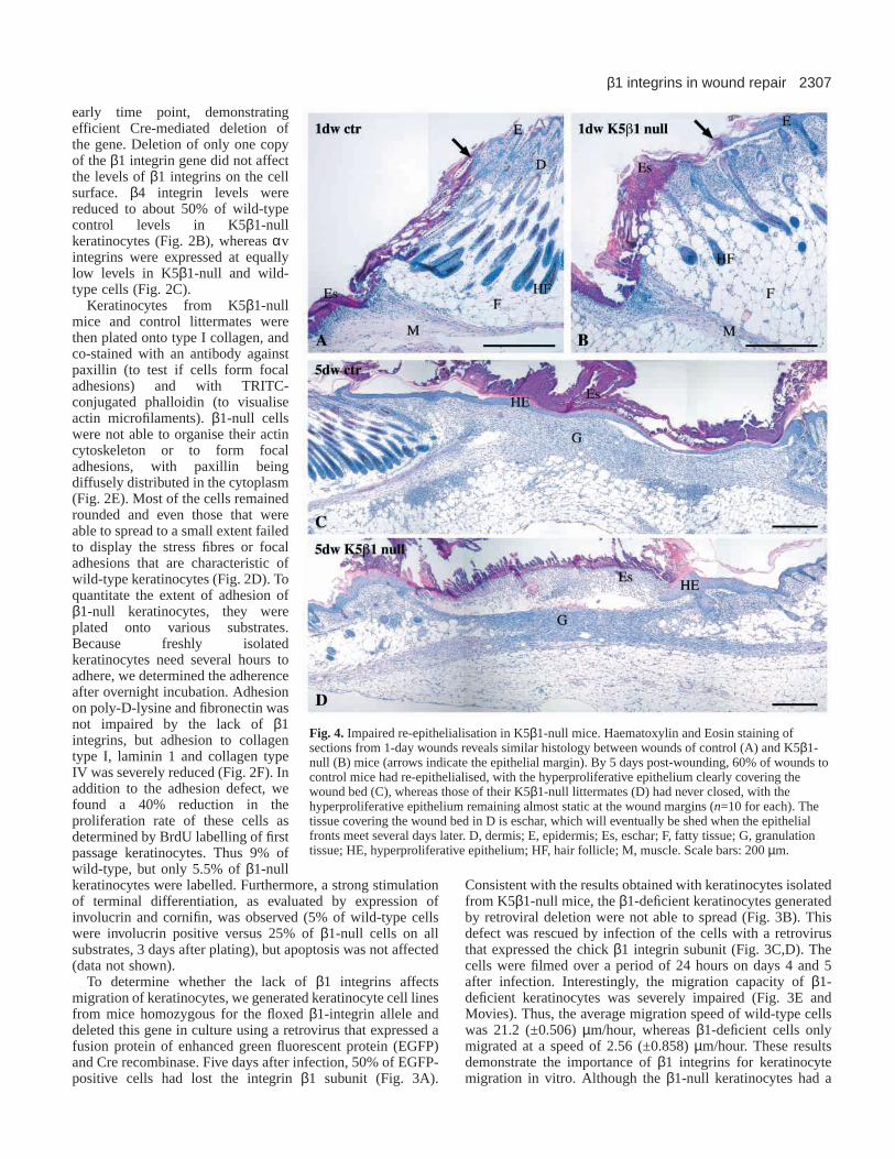
Fig. 4. Impaired re-epithelialisation in K5β1-null mice. Haematoxylin and Eosin staining ofsections from 1-day wounds reveals similar histology between wounds of control (A) and K5β1-null (B) mice (arrows indicate the epithelial margin). By 5 days post-wounding, 60% of wounds tocontrol mice had re-epithelialised, with the hyperproliferative epithelium clearly covering thewound bed (C), whereas those of their K5β1-null littermates (D) had never closed, with thehyperproliferative epithelium remaining almost static at the wound margins (n=10 for each). Thetissue covering the wound bed in D is eschar, which will eventually be shed when the epithelialfronts meet several days later. D, dermis; E, epidermis; Es, eschar; F, fatty tissue; G, granulationtissue; HE, hyperproliferative epithelium; HF, hair follicle; M, muscle. Scale bars: 200 µm.

2308
defect in spreading and migration, they were still able toundergo cell division (see Movies).
Delayed re-epithelialisation in K5 β1-null miceTo determine the importance of β1 integrins for re-epithelialisation of skin wounds, we analysed full-thicknessexcisional wounds from knockout animals and controllittermates at days 1, 2, 3, 5, 10 and 15 after injury (n≥4 foreach time point and genotype). No obvious abnormalities inwound contraction or appearance were observed during thefirst 5 days after injury, but at later time points, the loss of thescab was delayed in K5β1-null mice (not shown). At thehistological level, one-day wounds from K5β1-null miceappeared grossly similar to those from control littermates (Fig.4A,B). By 5 days post-wounding, 60% of control woundswere fully covered with a thin neoepidermis (Fig. 4C). Bycontrast, in K5β1-null mice, the epidermis at the woundmargin remained almost static, and the number of woundkeratinocytes was reduced in the mutant mice. Only a smallarea of the granulation tissue was covered by the newepidermis, whereas the remaining part was still covered byeschar. Surprisingly, the keratinocytes at the wound edge hadclearly increased in number, resulting in an overtlyhyperthickened epidermis at the wound edge (Fig. 4D; Fig.5D). Most noticeably, no clear migrating epithelial tongue wasdistinguishable. By contrast, granulation tissue formationappeared unaffected.
Cell proliferation is not reduced in early wounds ofcontrol and K5β 1-null mice To investigate if the delayed re-epithelialisation in K5β1-nullmice was due to a difference in cell proliferation, we performedBrdU labelling studies. No difference in the percentage ofBrdU-positive cells was observed in the epidermis of 1-day-oldwounds (Fig. 5A,B). At day 5 after injury, the percentage ofproliferating cells was still similar in the hyperthickened woundepidermis of control and K5β1-null mice, with 10-15% ofkeratinocytes being labelled in each case (n=5 wounds foreach). However, the distribution of the labelled cells wasaltered, with more BrdU-positive suprabasal cells being foundin the wound epidermis of K5β1-null mice (Fig. 5C,D). At day15 after injury, the epidermal keratinocytes had returned to theirnormal proliferation rate in control mice, but the epidermis wasstill hyperproliferative in K5β1-null mice (data not shown). Nodifference in cell proliferation was observed in the granulationtissue at any stage of repair (Fig. 5; data not shown).
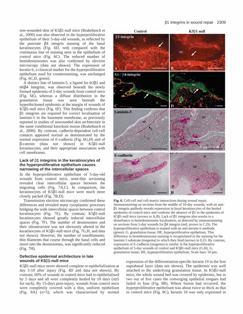
Cell-cell and cell-matrix interactions during woundrepair To confirm the lack of β1 integrins in wound keratinocytes, westained 10-day-old wound sections with an antibody againstthis integrin subunit. It was expressed in basal cells of thehyperproliferative wound epithelium in control mice (Fig. 6A),but absent in keratinocytes of K5β1-null animals (Fig. 6B).
The reduction in the number of hemidesmosomes seen in
R. Grose and others
Fig. 5. Cell proliferation in 5-day wounds. Mice were injected with BrdU 2 hours before sacrifice. Sections of 1-day (A,B) and 5-day (C,D)wounds from control mice (A,C) and K5β1-null mice (B,D) were stained with an anti-BrdU antibody (stained nuclei are indicated byarrowheads and the epithelial wound edge by arrows in A,B). There was an obvious impairment of keratinocyte proliferation in the hair folliclesof K5β1-null mice (A-D; compare asterisks in A and B). G, granulation tissue; HE, hyperproliferative epithelium. Scale bars: 200 µm.
2309β1 integrins in wound repair
non-wounded skin of K5β1-null mice (Brakebusch etal., 2000) was also observed in the hyperproliferativeepithelium of their 5-day-old wounds, as reflected bythe punctate β4 integrin staining of the basalkeratinocytes (Fig. 6D, red) compared with thecontinuous line of staining seen in the epithelium ofcontrol mice (Fig. 6C). The reduced number ofhemidesmosomes was also confirmed by electronmicroscopy (data not shown). The expression ofkeratin 6, a classical marker for the hyperproliferativeepithelium used for counterstaining, was unchanged(Fig. 6C,D, green).
A distinct line of laminin-5, a ligand for α3β1 andα6β4 integrins, was observed beneath the newlyformed epidermis of 5-day wounds from control mice(Fig. 6E), whereas a diffuse distribution in thegranulation tissue was seen beneath thehyperthickened epidermis at the margin of wounds ofK5β1-null mice (Fig. 6F). This finding confirms thatβ1 integrins are required for correct localisation oflaminin-5 in the basement membrane, as previouslyreported in studies of unwounded skin architecture inthe same conditional knockout mouse (Brakebusch etal., 2000). By contrast, cadherin-dependent cell-cellcontacts appeared normal as demonstrated by thenormal expression of E-cadherin (Fig. 6G,H) and ofβ-catenin (data not shown) in K5β1-nullkeratinocytes, and their appropriate association withcell membranes.
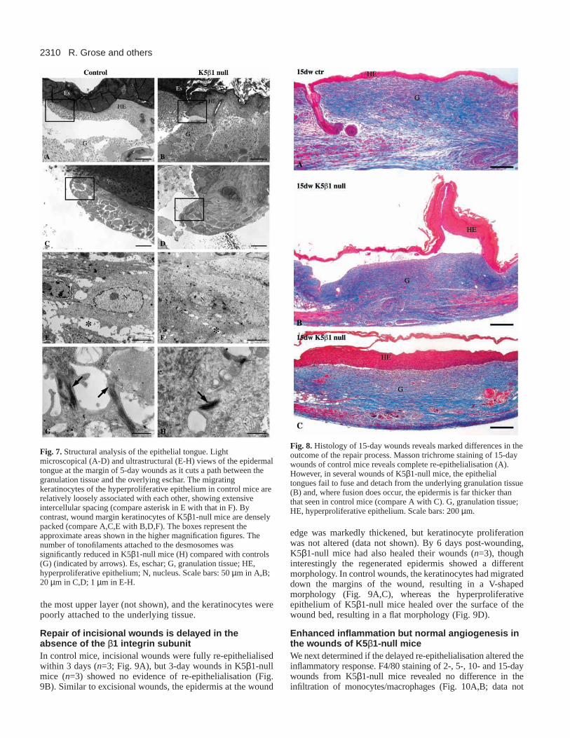
Lack of β 1 integrins in the keratinocytes ofthe hyperproliferative epithelium causesnarrowing of the intercellular spacesIn the hyperproliferative epithelium of 5-day-oldwounds from control mice, semi-thin sectioningrevealed clear intercellular spaces between themigrating cells (Fig. 7A,C). In comparison, thekeratinocytes of K5β1-null mice were much moreclosely packed (Fig. 7B,D).
Transmission electron microscopy confirmed thesedifferences and revealed many cytoplasmic processesbridging the wide intercellular spaces between controlkeratinocytes (Fig. 7E). By contrast, K5β1-nullkeratinocytes showed greatly reduced intercellularspaces (Fig. 7F). The number of desmosomes andtheir ultrastructure was not obviously altered in thekeratinocytes of K5β1-null mice (Fig. 7G,H, and datanot shown). However, the number of tonofilaments,thin filaments that course through the basal cells andinsert into the desmosomes, was significantly reduced(Fig. 7H).
Defective epidermal architecture in latewounds of K5β 1-null miceK5β1-null mice never showed complete re-epithelialisation atday 5-10 after injury (Fig. 4D and data not shown). Bycontrast, 60% of wounds in control mice had re-epithelialisedby 5 days and all were completely healed by 10 days (n≥5for each). By 15-days post-injury, wounds from control micewere completely covered with a thin, uniform epithelium(Fig. 8A) (n=5), which was characterised by normal
expression of the differentiation-specific keratin 10 in the firstsuprabasal layer (data not shown). The epidermis was wellattached to the underlying granulation tissue. In K5β1-nullmice, the whole wound bed was covered by epidermis, but intwo out of five cases the converging epithelial tongues hadfailed to fuse (Fig. 8B). Where fusion had occurred, thehyperproliferative epithelium was about twice as thick as thatin control mice (Fig. 8C), keratin 10 was only expressed in
Fig. 6. Cell-cell and cell-matrix interactions during wound repair.Immunostaining on sections from the middle of 10-day wounds, with an anti-β1 integrin antibody reveals expression in basal keratinocytes of the healedepidermis of control mice and confirms the absence of β1 in the epidermis ofK5β1-null mice (arrows in A,B). Lack of β1 integrins also results in adisturbance in hemidesmosome localisation, as detected by immunostainingon sections from 5-day wounds for β4 integrin (red; arrows in C,D). Thehyperproliferative epithelium is stained with an anti-keratin 6 antibody(green). G, granulation tissue; HE, hyperproliferative epithelium. Thisdifference in hemidesmosome staining is recapitulated in the staining for thelaminin 5 substrate (magenta) to which they bind (arrows in E,F). By contrast,expression of E-cadherin (magenta) is similar in the hyperproliferativeepithelium of 5-day wounds of control and K5β1-null mice (G,H). G,granulation tissue; HE, hyperproliferative epithelium. Scale bars: 50 µm.
2310
the most upper layer (not shown), and the keratinocytes werepoorly attached to the underlying tissue.
Repair of incisional wounds is delayed in theabsence of the β 1 integrin subunitIn control mice, incisional wounds were fully re-epithelialisedwithin 3 days (n=3; Fig. 9A), but 3-day wounds in K5β1-nullmice (n=3) showed no evidence of re-epithelialisation (Fig.9B). Similar to excisional wounds, the epidermis at the wound
edge was markedly thickened, but keratinocyte proliferationwas not altered (data not shown). By 6 days post-wounding,K5β1-null mice had also healed their wounds (n=3), thoughinterestingly the regenerated epidermis showed a differentmorphology. In control wounds, the keratinocytes had migrateddown the margins of the wound, resulting in a V-shapedmorphology (Fig. 9A,C), whereas the hyperproliferativeepithelium of K5β1-null mice healed over the surface of thewound bed, resulting in a flat morphology (Fig. 9D).
Enhanced inflammation but normal angiogenesis inthe wounds of K5β 1-null miceWe next determined if the delayed re-epithelialisation altered theinflammatory response. F4/80 staining of 2-, 5-, 10- and 15-daywounds from K5β1-null mice revealed no difference in theinfiltration of monocytes/macrophages (Fig. 10A,B; data not
R. Grose and others
Fig. 7.Structural analysis of the epithelial tongue. Lightmicroscopical (A-D) and ultrastructural (E-H) views of the epidermaltongue at the margin of 5-day wounds as it cuts a path between thegranulation tissue and the overlying eschar. The migratingkeratinocytes of the hyperproliferative epithelium in control mice arerelatively loosely associated with each other, showing extensiveintercellular spacing (compare asterisk in E with that in F). Bycontrast, wound margin keratinocytes of K5β1-null mice are denselypacked (compare A,C,E with B,D,F). The boxes represent theapproximate areas shown in the higher magnification figures. Thenumber of tonofilaments attached to the desmosomes wassignificantly reduced in K5β1-null mice (H) compared with controls(G) (indicated by arrows). Es, eschar; G, granulation tissue; HE,hyperproliferative epithelium; N, nucleus. Scale bars: 50 µm in A,B;20 µm in C,D; 1 µm in E-H.
Fig. 8.Histology of 15-day wounds reveals marked differences in theoutcome of the repair process. Masson trichrome staining of 15-daywounds of control mice reveals complete re-epithelialisation (A).However, in several wounds of K5β1-null mice, the epithelialtongues fail to fuse and detach from the underlying granulation tissue(B) and, where fusion does occur, the epidermis is far thicker thanthat seen in control mice (compare A with C). G, granulation tissue;HE, hyperproliferative epithelium. Scale bars: 200 µm.

2311β1 integrins in wound repair
shown). However, in their still exposed 5-day wounds,neutrophils were still present in large numbers throughout thegranulation tissue (Fig. 10D), although no signs of infectionwere observed. There was no obvious difference in angiogenesisas determined by immunofluorescence analysis with an antibodyagainst the endothelial-specific protein PECAM (Fig. 10E,F).
Changes in gene expression in the wounds of K5 β1-null miceTo gain insight into the molecular mechanisms underlying theobserved phenotype, we performed RNase protection assays forgenes that encode important regulators of repair. For thispurpose, we used two independent pools of total RNA, eachisolated from two wounds (1, 5 and 10 day) from each of at leastfive control and five K5β1-null mice (Fig. 11). Owing to thedevelopment of fibrosis in the K5β1-null mice, we usedunwounded skin from 10 and 20 d.p.p. wild-type and K5β1-nullmice as controls, thus enabling us to distinguish betweendevelopmental and wound-specific changes in gene expression.
Expression of the pro-inflammatory cytokines interleukin 1β(IL1β), IL1α and tumour necrosis factor α(TNFα) wasincreased in 10 and 20 d.p.p. unwounded back skin of K5β1-null mice (Fig. 11; data not shown). Pro-inflammatory cytokineexpression was further increased at days 5 and 10 post-woundingcompared with expression in wild-type animals. Similarexpression profiles were observed for IL1αand TNFα(data notshown). The levels of activin βA and keratinocyte growth factor(KGF) mRNAs were both increased during repair in K5β1-nullmice compared with controls. By contrast, expression ofvascular endothelial growth factor (VEGF), TGFβ1 and TGFβ3,as well as of connective tissue growth factor (CTGF) remainedunchanged in the absence of epidermal β1 integrins (Fig. 11;data not shown).
We also observed a slight increase in mRNA levels offibronectin, collagen α1 (I) and tenascin-C both in non-woundedskin and during wound repair of K5β1-null mice (Fig. 11 anddata not shown). Concomitant with elevated levels of mRNAsencoding matrix proteins was a significant increase in the mRNAlevels of matrix metalloproteinase 10 (MMP10; stromelysin 2)and MMP13 (collagenase 3), also both in skin and wounds ofK5β1-null mice (Fig. 11). These MMPs are expressed mainly atthe migrating edge of the epidermis in wounds of wild-typemice (Madlener et al., 1998). Taken together, these findingsdemonstrate that several major players of the wound repairprocess are aberrantly expressed in K5β1-null mice.
DISCUSSION
A series of expression studies have suggested an important roleof β1 integrins in keratinocyte migration during woundhealing, but this has not yet been experimentally proven. Theavailability of genetically engineered mice lacking this integrinsubunit in the epidermis offered the unique possibility toaddress this question.
Proliferation of β 1-null wound keratinocytes isreduced in vitro but not in vivoWithin a few hours of cutaneous injury, keratinocytes start tomigrate over the injured dermis. Subsequently, the cells behindthe migrating epithelial sheet increase their proliferation rateand constitute a pool of extra cells that replace those lostduring injury (reviewed by Martin, 1997). Thus, delayed re-epithelialisation can result from either impaired migration orproliferation. Interestingly, the results of the present studyrevealed no difference in the percentage of proliferating cells inthe hyperthickened epidermis at the wound edge in earlywounds of K5β1-null mice and even prolonged keratinocytehyperproliferation. This was unexpected, as high expression ofthe β1 integrin subunit has been shown to be a marker forproliferation competent stem cells (Jones and Watt, 1993; Joneset al., 1995). Furthermore, keratinocyte proliferation is reducedin non-wounded skin of K5β1-null mice (Brakebusch et al.,2000) and of K14β1-null mice, which revealed a similar, butmore severe, phenotype (Raghavan et al., 2000). Finally, theproliferation rate of our cultured β1-deficient keratinocytes wasalmost halved and terminal differentiation was stimulated inthese cells. These differences between the in vitro and in vivosituation could reflect the lack of dermal inflammatorycytokines and growth factors in culture, the presence of a
Fig. 9. Repair of incisional wounds is delayed K5β1-null mice.Haematoxylin and Eosin staining reveals that full-thicknessincisional wounds on the backs of 10 d.p.p. control mice had healedby day 3 post-injury (A; arrows indicate where the panniculuscarnosus has been cut), but wounds of K5β1-null mice are wide openat the same stage (B). By 6 days after wounding, wounds of bothcontrol and K5β1-null mice have healed (C,D). Es, eschar; F, fattytissue; G, granulation tissue; HE, hyperproliferative epithelium; HF,hair follicle; M, muscle. Scale bars: 200 µm in A-D.

2312
different matrix and the three-dimensional organisation in vivo.
Keratinocyte migration isimpaired in the absence of β1integrins in vitro and in vivoBecause the total number of woundkeratinocytes was reduced in themutant mice, although the percentageof proliferating cells was unchanged,reduced migration of β1-nullkeratinocytes is likely to be the reasonfor the decrease in the total number ofkeratinocytes in the wound. Thisobservation is strengthened by theobservation that the β1-nullkeratinocytes are piling up at thewound edges instead of forming athin epithelial tongue across thewound. Most importantly, thishypothesis is strongly supported bythe impaired migratory capacity ofβ1-deficient cultured keratinocytes.
Under normal circumstances,keratinocytes first migrate down overthe injured dermis where they contactfibrillar type I collagen (Pilcher et al.,1997). Based on in vivo expressionstudies and functional in vitro data, ithas been suggested that the α2β1integrin is required for this migrationover the wounded dermis (Pilcher etal., 1997; Hodivala-Dilke et al., 1998;Goldfinger et al., 1999). The resultspresented in the present study thusprovide the first in vivo evidence forthis hypothesis.
Although the onset of keratinocytemigration was retarded in K5β1-nullmice, a strongly hyperthickenedepidermis was observed at thewound edge, most probably dueto the continued keratinocytehyperproliferation in the absence ofmigration. Several days afterwounding, the keratinocytes finallymigrated over the wound bed, but theepidermis was strongly hyperplastic,poorly differentiated and poorly attached to the underlyinggranulation tissue. Most interestingly, the healed epidermis ofincisional wounds in control mice had a V-shaped morphology,whereas it was flat in K5β1-null mice, suggesting that re-epithelialisation in K5β1-null mice occurs via an alternative,compensatory mechanism (summarised in Fig. 12). Becauseβ1-deficient keratinocytes lack the principal adhesive receptorsused for ligation to dermal matrix, they must wait untilcompensatory mechanisms are upregulated. Such mechanismsare probably based on the expression of non-β1 integrins bykeratinocytes and on the presence of the corresponding ligands.The most likely candidates are αvβ5 or αvβ6, receptors for theprovisional wound matrix proteins vitronectin, fibronectin and
tenascin C (Gailit et al., 1994; Larjava et al., 1993; Zambrunoet al., 1995; Haapasalmi et al., 1996; Huang et al., 1998).Although αv integrins are weakly expressed in normal and β1-deficient cultured keratinocytes, αvβ5 and αvβ6 areupregulated in migrating keratinocytes during cutaneous woundrepair (Cavani et al., 1993; Gailit et al., 1994; Haapasalmi et al.,1996). αvβ5 on keratinocytes is characteristic of a migratoryphenotype (Adams and Watt, 1991), and αvβ6 is important forkeratinocyte migration on vitronectin, fibronectin and tenascinC in vitro (Huang et al., 1998). The period required for theupregulation of these integrins and for the deposition of theirligands in the wound bed is likely to determine the time framefor the onset of re-epithelialisation in K5β1-knockout animals.
R. Grose and others
Fig. 10.Increased inflammation but normal angiogenesis in the wounds of K5β1-null mice. F4/80staining for monocytes/macrophages (brown stain indicated by arrows in A and B) at day 2 post-wounding showed no difference. In 5-day wounds (C-F) there was a clear increase in neutrophilpresence (as detected by staining for Ly-6G – brown stain indicated by arrows in C,D) in thewounds of K5β1-null mice (D) compared with control littermates (C). No difference was observedin angiogenesis as detected by staining for PECAM1 – magenta stain indicated by arrows in E,F).Es, eschar; F, fatty tissue; G, granulation tissue; HE, hyperproliferative epithelium; HF, hairfollicle. Scale bars: 200 µm in A-D; 40 µm in E,F.

2313β1 integrins in wound repair
Fig. 11.Gene expression during the repair process.Expression levels of genes encoding key players duringwound healing were determined by RNase protectionassay on 20 µg RNA samples from non-wounded backskin from 10 and 20 d.p.p. mice plus 1, 5 and 10 daywounds from control and K5β1-null mice. 1000 cpm ofthe hybridisation probes were loaded in the lanes labelled‘probe’ and used as size markers. tRNA (20 µg) was usedas a negative control. RNA (1 µg of each) was loaded on a1% agarose gel and stained with Ethidium Bromide tocontrol for sample integrity and concentration (bottompanel). The intensity of the signals as determined byphosphorimaging is shown schematically on the right-hand side. All protection assays were repeated with aseparate pool of RNA samples from an independentskin/wound series.
1-2 days post-wounding
3-4 days post-wounding
Contro l
KEY
Keratinocyte
β1 integrin
Basement membrane
Fibroblast
Adipocyte
Dermal matrix
Wound matrix
Muscle
K5ββββ1-null
1-2 days post-wounding
3-4 days post-wounding
Epidermis
Dermis
Fat
Muscle
Fig. 12. Model for re-epithelialisation of incisional wounds by normal and β1-null keratinocytes. In skin of control mice, β1 integrins arelocalised to the basolateral surfaces of basal keratinocytes. After wounding, control keratinocytes use β1 integrins to bind to the newly exposeddermal ligands. Thus, re-epithelialisation occurs rapidly and results in a downward migration of the epidermis. In K5β1-null mice,keratinocytes are unable to recognise dermal ligands after wounding and remain static at the wound margin, although they still proliferate. Theircell-cell contacts are also tighter than those of control keratinocytes. Once the wound is filled with granulation tissue, which contains matrixmolecules recognised by non-β1 integrins, K5β1-null keratinocytes are able to migrate across the surface of the wound to complete re-epithelialisation, but they do not show the V-shaped repair morphology characteristic of control wounds.
The dependence on non-β1 integrins also provides anexplanation for the flat morphology of the healed epidermis.Thus, we propose that K5β1-null keratinocytes are not able tomigrate down the injured dermis, because of the absence ofligands for the dermal substrate, but rather wait until the woundis filled with granulation tissue that contains the ligands for non-β1 integrins.
Wound edge keratinocytes pack together moredensely in the absence of β1 integrinsAn additional difference between the wound marginkeratinocytes is that their cell-cell contacts appear tighter,when compared with the loose association in controlhyperproliferative epithelium. The normal patterns of E-cadherin and β-catenin staining that we observed suggest thatthe tight contacts are not due to a difference in the expressionor compartmentalisation of adherens junction proteins.Furthermore, electron microscopy revealed that the number ofdesmosomes was not altered. However, the number oftonofilaments attached to the desmosomes was significantlyreduced. It has been reported that the insertion of tonofilamentsinto the attachment plate is a late event in the formation ofdesmosomes in the wounded epidermis (Krawczyk andWilgram, 1973), indicating that our observation reflects adefect in the maturation process of the wound keratinocytes inthe absence of β1 integrins.
Abnormal epidermal architecture in healed woundsof K5β1 null miceRe-epithelialisation was eventually completed in K5β1-nullepidermis, but the wounds had a thicker epidermal covering,reflecting the prolonged period of keratinocytehyperproliferation and their delayed differentiation. This isunlikely to be a direct result of a lack of β1 integrins inkeratinocytes, but could be due to a prolonged proliferativesignal from cells within the granulation tissue, as suggested bythe increased expression of epithelial growth factors. Inaddition, there might be a lack of β1-dependent contactinhibition when the epithelial fronts meet (Huttenlocher et al.,1998) as supported by the epithelial morphology of some ofthe wounds where the epidermal edges had not fused and whereobviously unlimited growth of the epidermis had occurred.
Finally, K5β1-null keratinocytes failed to establish a normalbasement membrane above the newly formed granulationtissue. This finding is likely to underlie the poor attachment ofthe new epidermis to the mesenchyme and concurs with theobserved blister formation, as well as with the reduced numberof hemidesmosomes seen in non-wounded skin of K5β1-nullmice (Brakebusch et al., 2000). Blister formation was alsoobserved in mice that lack the integrin α3 subunit (DiPersio etal., 1997), suggesting that the lack of α3β1 in our mice ispredominantly responsible for the poor attachment of theepidermis. However, in contrast to K5β1-null keratinocytes,the exclusive lack of the α3 integrin subunit in this cell typedid not cause an altered expression of α6β4 in vitro or in vivo(Hodivala-Dilke et al., 1998), suggesting that the additionalloss of other β1 integrins is responsible for the reduction in thenumber of α6β4 integrins in K5β1-null keratinocytes.
Prolonged inflammation in K5 β1-null miceIn K5β1-null mice, we observed an extended window of
neutrophil presence, most likely as a result of the prolongedexposure of K5β1-null wounds to the external environment.Because re-epithelialisation is so delayed, their wounds aresusceptible to pro-inflammatory stimuli such as desiccationand mechanical stress for a longer period. As neutrophils area major source of pro-inflammatory cytokines in skin wounds(Hübner et al., 1996), the elevated number of these cells islikely to contribute to the increased expression of IL1α, IL1βand TNFαin the wounds of K5β1-null mice. These cytokinesinduce expression of many epithelial growth factors (reviewedby Werner and Smola, 2001), which could contribute to theincreased keratinocyte proliferation rate. Increased expressionof pro-inflammatory cytokines was also observed in theunwounded skin of older K5β1-null mice, but these are thoughtto be released by macrophages rather than neutrophils(Brakebusch et al., 2000). However, the expression levelsassociated with the developmental inflammation and fibrosis ofunwounded skin were significantly lower than the transientexpression levels seen during the repair process. Thus, thedevelopmental fibrosis should have a rather minor effect on therepair process in K5β1-null mice, as supported by the normalcontraction of the healing wound.
Taken together, our results reveal a strongly impairedmigratory capacity of β1-deficient keratinocytes in vitro and adramatic delay in epithelial migration during wound repair inK5β1-null mice. We thus present the first in vivo evidence insupport of findings from in vitro studies that have shown β1integrins to be key players in cell migration. However, ourresults also demonstrate that keratinocytes are not totallydependent on this integrin subunit to heal their wounds. Rather,other integrins appear to compensate at least partially for thelack of β1, leading to complete, although imperfect, re-epithelialisation.
We thank Silke Durka, Katharina Huggel and Christiane Born-Berclaz for excellent technical assistance, Dr William C. Parks forcritically reading the manuscript and for many helpful suggestions,and Dr Laurence Vindevoghel for helpful discussions. This work wassupported by the Swiss National Science Foundation (grant number31-61358.00 to S. W.), the ETH Zürich (to S. W), the AustrianBundesministerium (fellowship to C. H.), the ICRF (C. H. and F. M.W.) and the European Union (S. W., R. F. and F. M. W.).
REFERENCES
Adams, J. C. and Watt, F. M. (1991). Expression of β1, β3, β4, and β5integrins by human epidermal keratinocytes and non-differentiatingkeratinocytes.J. Cell Biol.115, 829-841.
Bloch, W., Huggel, K., Sasaki, T., Grose, R., Bugnon, P., Addicks, K.,Timpl, R. and Werner, S. (2000). The angiogenesis inhibitor endostatinimpairs blood vessel maturation during wound healing.FASEB J.14, 2373-2376.
Brakebusch, C., Hirsch, E., Potocnik, A. and Fässler, R. (1997). Geneticanalysis of β1 integrin function: confirmed, new and revised roles for acrucial family of cell adhesion molecules.J. Cell Sci. 110, 2895-2904.
Brakebusch, C., Grose, R., Quondamatteo, F., Ramirez, A., Jorcano, J. L.,Pirro, A., Svensson, M., Herken, R., Sasaki, T., Timpl, R. et al. (2000).Skin and hair follicle integrity is crucially dependent on β1 integrinexpression on keratinocytes.EMBO J.19, 3990-4003.
Cavani, A., Zambruno, G., Marconi, A., Manca, V., Marchetti, M. andGiannetti, A. (1993). Distinctive integrin expression in the newly formingepidermis during wound healing in humans.J. Invest. Dermatol. 101, 600-604.
Carroll, J. M., Romero, R. and Watt, F. M. (1995). Suprabasal integrin
2314 R. Grose and others

2315β1 integrins in wound repair
expression in the epidermis of transgenic mice results in developmentaldefects and a phenotype resembling psoriasis.Cell 83, 957-968.
Chomczynski, P. and Sacchi, N. (1987). Single-step method of RNA isolationby acid guanidinium thiocyanate-phenol-chloroform extraction.Anal.Biochem.162, 156-159.
Clark, R. A. F. (1990). Fibronectin matrix deposition and fibronectin receptorexpression in healing wounds and normal skin.J. Invest. Dermatol. 94Suppl., 128S-134S.
DiPersio, C. M., Hodivala-Dilke, K. M., Jaenisch, R., Kreidberg, J. A.,Hynes, R. O. (1997). alpha3beta1 integrin is required for normaldevelopment of the epidermal basement membrane.J. Cell Biol.137, 729-742.
Frank, S., Hübner, G., Breier, G., Longaker, M. T., Greenhalgh, D. G andWerner, S. (1995). Regulation of vascular endothelial growth factorexpression in cultured keratinocytes: Implications for normal and impairedwound healing.J. Biol. Chem.270, 12607-12613.
Gailit, J., Welch, M. P. and Clark, R. A. F. (1994). TGF-β1 stimulatesexpression of keratinocyte integrins during reepithelialisation of cutaneouswounds.J. Invest. Dermatol.103, 221-227.
Goldfinger, L. E., Hopkinson, S. B., deHart, G. W., Collawn, S.,Couchman, J. R. and Jones, J. C. R. (1999). The α3 laminin subunit, α6β4and α3β1 integrin coordinately regulate wound healing in cultured epithelialcells and in the skin.J. Cell Sci.112, 2615-2629.
Grose, R. and Werner, S. (2002). Wound healing studies in transgenic miceand knock-out mice. In Methods in Molecular Medicine: Wound Healing:Methods and Protocols. (ed. L. A. Di Pietro and A. L. Burns-Harring) (inpress).
Haapasalmi, K., Zhang, K., Tonnesen, M., Olerud, J., Sheppard, D., Salo,T., Kramer, R., Clark, R. A., Uitto, V-J. and Larjava, H. (1996).Keratinocytes in human wounds express αvβ6 integrin.J. Invest. Dermatol.106, 42-48.
Hertle, M. D., Kubler, M.-D., Leigh, I. M. and Watt, F. M. (1992). Aberrantintegrin expression during epidermal wound healing and in psoriaticepidermis.J. Clin. Invest. 89, 1892-1901.
Hodivala-Dilke, K. M., DiPersio, C. M., Kreidberg, J. A. and Hynes, R.O. (1998). Novel roles for α3β1 integrin as a regulator of cytoskeletalassembly and as a trans-dominant inhibitor of integrin receptor function inmouse keratinocytes.J. Cell Biol. 142, 1357-1369.
Huang, X., Wu, J., Spong, S. and Sheppard, D. (1998). The integrin αvβ6is critical for keratinocyte migration on both its known ligand, fibronectin,and on vitronectin.J. Cell Sci.111, 2189-2195.
Hübner, G., Brauchle, M., Smola, H., Madlener, M., Fässler, R. andWerner, S. (1996). Differential regulation of pro-inflammatory cytokinesduring normal and impaired wound healing.Cytokine8, 548-556.
Huttenlocher, A., Lakonishok, M., Kinder, M., Wu, S., Truong, T.,Knudsen, K. A. and Horwitz, A. F. (1998). Integrin and cadherin synergyregulates contact inhibition of migration and motile activity.J. Cell Biol.141, 515-526.
Jones, P. H. and Watt, F. M. (1993). Separation of human epidermal stemcells from transit amplifying cells on the basis of differences in integrinfunction and expression.Cell 73, 713-724.
Jones, P. H., Harper, S. and Watt, F. M. (1995). Stem cell patterning andfate in human epidermis.Cell 80, 83-93.
Krawczyk, W. S. and Wilgram, G. F. (1973). Hemidesmosome anddesmosome morphogenesis during epidermal wound healing.J. Ultrastruct.Res.45, 93-101.
Larjava, H., Salo, T., Haapasalmi, K., Kramer, R. H. and Heino, J. (1993).Expression of integrins and basement membrane components by woundkeratinocytes.J. Clin. Invest.92, 1425-1435.
Levy, L., Broad, S., Zhu, A. J., Carroll, J. M., Khazaal, I., Peault, B. andWatt, F. M. (1998). Optimised retroviral infection of human epidermalkeratinocytes: long-term expression of transduced integrin gene followinggrafting on to SCID mice.Gene Ther. 5, 913-922.
Madlener, M., Parks, W. C. and Werner, S. (1998). Matrixmetalloproteinases (MMPs) and their physiological inhibitors (TIMPs) aredifferentially expressed during excisional skin wound repair.Exp. Cell Res.242, 201-210.
Martin, P. (1997). Wound healing – aiming for perfect skin regeneration.Science276, 75-81.
Munz, B., Smola, H., Engelhardt, F., Bleuel, K., Brauchle, M., Lein, I.,Evans, L. W., Huylebroeck, D., Balling, R. and Werner, S. (1999).Overexpression of activin A in the skin of transgenic mice reveals newactivities of activin in epidermal morphogenesis, dermal fibrosis and woundrepair.EMBO J. 18, 5205-5215.
Palmer, E. L., Ruegg, C., Ferrando, R., Pytela, R. and Sheppard, D. (1993).Sequence and tissue distribution of the integrin alpha 9 subunit, a novelpartner of beta 1 that is widely distributed in epithelia and muscle.J. CellBiol. 123, 1289-1297.
Pilcher, B. K., Dumin, J. A., Sudbeck, B. D., Krane, S. M., Welgus, H. G.and Parks, W. C. (1997). The activity of collagenase-1 is required forkeratinocyte migration on a type I collagen matrix.J. Cell Biol. 137, 1445-1457.
Plow, E. F., Haas, T. A., Zhang, L., Loftus, J. and Smith, J. W. (2000).Ligand binding to integrins.J. Biol. Chem. 275, 21785-21788.
Raghavan, S., Bauer, C., Mundschau, G., Li, Q. and Fuchs, E. (2000).Conditional ablation of beta1 integrin in skin. Severe defects in epidermalproliferation, basement membrane formation, and hair follicle invagination.J. Cell Biol. 150, 1149-1160.
Ramirez, A., Bravo, A., Jorcano, J. L. and Vidal, M. (1994). Sequences 5`ofthe bovine keratin 5 gene direct tissue- and cell-type specific expression ofa lacZ gene in the adult and during development.Differentiation58, 53-64.
Romero, M. R., Carroll, J. M. and Watt, F. M. (1999). Analysis of culturedkeratinocytes from a transgenic mouse model of psoriasis: effects ofsuprabasal integrin expression on keratinocyte adhesion, proliferation andterminal differentiation. Exp. Dermatol. 8, 53-67.
Roper, E., Weinberg, W., Watt, F. M. and Land, H. (2001). P19ARF-independent induction of p53 and cell cycle arrest by Raf in murinekeratinocytes.EMBO Rep. 2, 145-150.
Werner, S. and Smola, H. (2001) Paracrine regulation of keratinocyteproliferation and differentiation.Trends Cell Biol.11, 143-146.
Werner, S., Smola, H., Liao, X., Longaker, M. T., Krieg, T., Hofschneider,P. H. and Williams, L. T. (1994). The function of KGF in morphogenesisof epithelium and reepithelialization of wounds.Science266, 819-822.
Yamada, K. M., Gailit, J. and Clark, R. A. F. (1996). Integrins in woundrepair. In The Molecular and Cellular Biology of Wound Repair. (ed. R. A.F. Clark), pp. 311-338. New York: Plenum Press.
Yokosaki, Y., Palmer, E. L., Prieto, A. L., Crossin, K. L., Bourdon, M. A.,Pytela, R. and Sheppard, D. (1994). The integrin alpha 9 beta 1 mediatescell attachment to a non-RGD site in the third fibronectin type III repeat oftenascin. J. Biol. Chem. 269, 26691-26696.
Zambruno, G., Marchisio, P. C., Marconi, A., Vaschieri, C., Melchiori, A.,Giannetti, A. and De Luca, M. (1995). Transforming growth factor-beta 1modulates beta 1 and beta 5 integrin receptors and induces the de novoexpression of the alpha v beta 6 heterodimer in normal human keratinocytes:implications for wound healing.J. Cell Biol.129, 853-865.
Zhu, A. J., Haase, I. and Watt, F. M. (1999). Signaling via beta1 integrinsand mitogen-activated protein kinase determines human epidermal stem cellfate in vitro.Proc. Natl. Acad. Sci. USA96, 6728-6733.