beyond the limits of microscopy: revealing the plenary...
TRANSCRIPT

http://microscopy.org/MandM/2016/
PLENARY SPEAKER Drew BerryWalter and Eliza Hall Institute of Medical Research, Melbourne, Australia
“Beyond the Limits of Microscopy: Revealing the Unseeable through Hollywood Visual Effects”
MONDAY, JULY 25, 2016Columbus Convention Center, Columbus, OH
Drew Berry is a biomedical animator who creates scientifically accurate and aesthetically rich visualisations that reveal the cellular and molecular processes for a wide range of audiences. Beginning his career as a cell biologist and microscopist Drew brings a rigorous scientific approach to each project, immersing himself in relevant research to ensure current data are represented. Since 1995, he has been a biomedical animator at the Walter and Eliza Hall Institute of Medical Research. His animations have exhibited at venues such as the Guggenheim Museum, MoMA, the Royal Institute of Great Britain and the University of Geneva. In 2010 he received a MacArthur Fellowship "Genius Grant". He has been profiled in articles in the New York Times, the New Yorker and the American Scientist. He has also received an Emmy for his contribution to a documentary on DNA.
Plenary SessionDrew Berry1
1. The Walter and Eliza Hall Institute of Medical Research,Melbourne Australia
This report describes the research and visualisation techniques used to create The Malaria Lifecycle animations. The animation required one year of full time production, including three months of research, two months of model building and seven months of animation development. The goal was to visualize the entire malaria lifecycle, accurately represented for parasite behaviour and cellular structure, founded on an extensive review of the malaria research literature. The intended audience was as broad as possible, including scientists, students, the media and the general public. Technical scientific jargon was avoided in the narration to make it more accessible to non-experts.
The Malaria Lifecycle is presented as two animations "Human Host" and "Mosquito Host”. The animations were designed as succinct as possible with a duration under 4 minutes each, which is considered to be a reasonable attention span for a motivated audience listening to technical content, and the utility of brief visuals for use in classrooms, YouTube and lecture presentation.
The animation was created on an Apple MacPro with Maya animation software. Image rendering used Pixar's Renderman, Maya's internal renderer, Mental Ray, and OpenGL hardware. Most shot sequences were made of 20-50 layers which were then composited together in After Effects for the final result.
The animation begins with the neck of a sleeping child in the early evening, somewhere in the tropics. Early evening is the primary feeding time when Anopheles typically hunts for a blood meal. The colour of the shot is tinted towards dark blue to suggest the evening, supported by the nighttime jungle noises. An attempt was made to make the skin tone racially neutral and therefore relate to as broad an audience as possible.
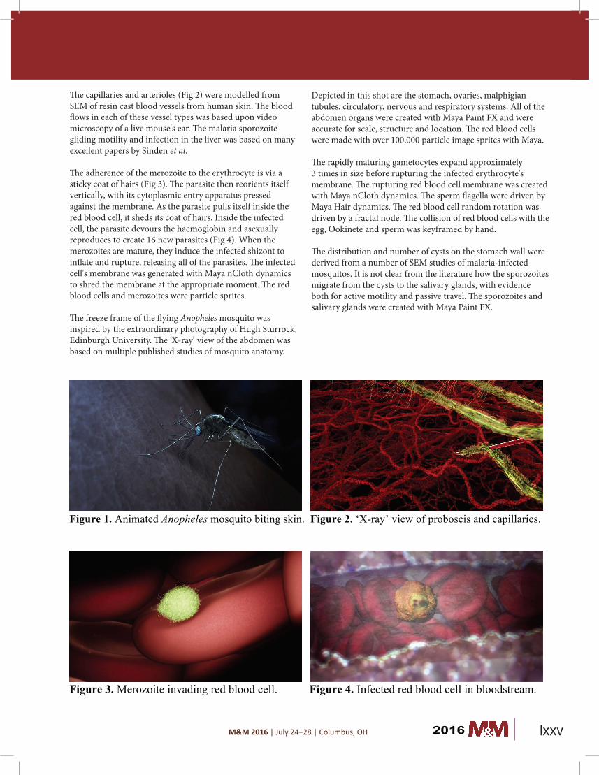
The flight, landing and stance of the Anopheles mosquito (Fig 1) were derived from published video studies of the insect'sbehaviour. The mechanics of the bite, such as the proboscisbending into the wound and the fold of the labrum, werederived from papers on the mosquito feeding action. The3D model for the mosquito required substantial detailingof textures, hairs and other features to make it resemble arealistic female Anopheles insect. The model also requiredkinematic rigging for movement. The mouthparts wereaccurately constructed with labrum and multiple mandiblestylets and maxillae that make up the proboscis tube.
Beyond the Limits of Microscopy: Revealing the Unseeable through Hollywood Visual Effects
2016lxxiv
M&M 2016 | July 24–28 | Columbus, OH
The capillaries and arterioles (Fig 2) were modelled from SEM of resin cast blood vessels from human skin. The blood flows in each of these vessel types was based upon video microscopy of a live mouse's ear. The malaria sporozoite gliding motility and infection in the liver was based on many excellent papers by Sinden et al.
The adherence of the merozoite to the erythrocyte is via a sticky coat of hairs (Fig 3). The parasite then reorients itself vertically, with its cytoplasmic entry apparatus pressed against the membrane. As the parasite pulls itself inside the red blood cell, it sheds its coat of hairs. Inside the infected cell, the parasite devours the haemoglobin and asexually reproduces to create 16 new parasites (Fig 4). When the merozoites are mature, they induce the infected shizont to inflate and rupture, releasing all of the parasites. The infected cell's membrane was generated with Maya nCloth dynamics to shred the membrane at the appropriate moment. The red blood cells and merozoites were particle sprites.
The freeze frame of the flying Anopheles mosquito was inspired by the extraordinary photography of Hugh Sturrock, Edinburgh University. The ‘X-ray’ view of the abdomen was based on multiple published studies of mosquito anatomy.
merozoites are mature, they induce the infected shizont to inflate and rupture, releasing all of theparasites. The infected cell's membrane was generated with Maya nCloth dynamics to shred themembrane at the appropriate moment. The red blood cells and merozoites were particle sprites.
The freeze frame of the flying Anopheles mosquito was inspired by the extraordinary photography of Hugh Sturrock, Edinburgh University. The ‘X-ray’ view of the abdomen was based on multiplepublished studies of mosquito anatomy. Depicted in this shot are the stomach, ovaries, malphigian tubules, circulatory, nervous and respiratory systems. All of the abdomen organs were created with Maya Paint FX and were accurate for scale, structure and location. The red blood cells were made with over 100,000 particle image sprites with Maya.
The rapidly maturing gametocytes expand approximately 3 times in size before rupturing the infected erythrocyte's membrane. The rupturing red blood cell membrane was created with Maya nClothdynamics. The sperm flagella were driven by Maya Hair dynamics. The red blood cell random rotation was driven by a fractal node. The collision of red blood cells with the egg, Ookinete and sperm waskeyframed by hand.
The distribution and number of cysts on the stomach wall were derived from a number of SEM studiesof malaria-infected mosquitos. It is not clear from the literature how the sporozoites migrate from thecysts to the salivary glands, with evidence both for active motility and passive travel. The sporozoitesand salivary glands were created with Maya Paint FX.
Figure 1. Animated Anopheles mosquito biting skin. Figure 2. ‘X-ray’ view of proboscis and capillaries.
Figure 3. Merozoite invading red blood cell. Figure 4. Infected red blood cell in bloodstream.
Depicted in this shot are the stomach, ovaries, malphigian tubules, circulatory, nervous and respiratory systems. All of the abdomen organs were created with Maya Paint FX and were accurate for scale, structure and location. The red blood cells were made with over 100,000 particle image sprites with Maya.
The rapidly maturing gametocytes expand approximately 3 times in size before rupturing the infected erythrocyte's membrane. The rupturing red blood cell membrane was created with Maya nCloth dynamics. The sperm flagella were driven by Maya Hair dynamics. The red blood cell random rotation was driven by a fractal node. The collision of red blood cells with the egg, Ookinete and sperm was keyframed by hand.
The distribution and number of cysts on the stomach wall were derived from a number of SEM studies of malaria-infected mosquitos. It is not clear from the literature how the sporozoites migrate from the cysts to the salivary glands, with evidence both for active motility and passive travel. The sporozoites and salivary glands were created with Maya Paint FX.
2016 lxxv