beyond the whole-mount phenotype: high-resolution imaging...
TRANSCRIPT

METHODS AND TECHNIQUES
Beyond the whole-mount phenotype: high-resolution imaging influorescence-based applications on zebrafishVeronika Oralova1,2, Joana T. Rosa1,3, Mieke Soenens1, Jan Willem Bek4, Andy Willaert4, Paul Eckhard Witten1
and Ann Huysseune1,*
ABSTRACTZebrafish is now widely used in biomedical research as a model forhuman diseases, but the relevance of the model depends on arigorous analysis of the phenotypes obtained. Many zebrafishdisease models, experimental techniques and manipulations takeadvantage of fluorescent reporter molecules. However, phenotypicanalysis often does not go beyond establishing overall distributionpatterns of the fluorophore in whole-mount embryos or usingvibratome or paraffin sections with poor preservation of tissuearchitecture and limited resolution. Obtaining high-resolution data offluorescent signals at the cellular level from internal structures mostlydepends on the availability of expensive imaging technology. Here,we propose a new and easily applicable protocol for embedding andsectioning of zebrafish embryos using in-house prepared glycolmethacrylate (GMA) plastic that is suited for preservation offluorescent signals (including photoactivatable fluorophores) withoutthe need for antibodies. Four main approaches are described, allinvolving imaging fluorescent signals on semithin (3 µm or less)sections. These include sectioning transgenic animals, whole-mountimmunostained embryos, cell tracking, as well as on-section enzymehistochemistry.
KEY WORDS: Zebrafish, GMA, GFP, Fluorophores,Immunofluorescence, Cell tracking, TRAP
INTRODUCTIONThe increase in zebrafish genomic resources together with moresophisticated protocols for genome editing and other tools havecontributed not only to unravel the genetic networks controllingdevelopment, but also generated zebrafish models with relevance tohuman disease (e.g. skeletal diseases, Laizé et al., 2014; Wittenet al., 2017; Gistelinck et al., 2018). To assess the mutantphenotypes, many studies use fluorophores as marker molecules,whether genetically engineered in transgenic lines, in whole-mountin situ hybridization and fluorescent in situ staining, or as afluorescent reporter in vital staining. Many of these studies, aiming
at investigating the expression or function of disease-causing genesor localization of proteins, or perform cell tracking, rely onobservations on whole-mount specimens. Obtaining cellular details,especially of structures located deep in the embryo, neverthelessoffers a substantial added value to such studies. Furthermore,studies often focus on easily accessible or superficially exposedanatomical structures, such as caudal fin rays or scales forregeneration studies (e.g. Wehner and Weidinger, 2015; Coxet al., 2018), or the thin trunk of embryos for modeling vasculardiseases (e.g. Hogan and Schulte-Merker, 2017). For a detailedphenotypic characterization, especially for internal structures, and/or structures that develop beyond the stage of complete transparencyof the embryo, such as the skeletal system, it is helpful tocomplement whole-mount techniques and standard embedding andsectioning procedures, including paraffin or vibratome sections.Advanced imaging techniques such as dual photon microscopy orlight sheet microscopy can overcome the limitations of observationson whole-mount embryos or superficially positioned structures.They also have the advantage of analysis in 3D, and enable in vivoanalysis which can readily and precisely answer a broad range ofbiological questions, including those regarding dynamic cellmovement (e.g. Liu et al., 2018). Yet, such imaging techniquesrequire expensive equipment that is not readily available to mostlabs. Laser scanning confocal microscopy (LSCM) performed atwhole-mount level can provide some information of internalstructures but has limitations when deeper structures need to beimaged (Bruneel and Witten, 2015). Chemical clearing methods areunder progress to allow deeper imaging (e.g. Watson et al., 2017),but even here, additional equipment is required. On the other hand,LSCM can be used to generate high-resolution pictures ofimmunostained sections obtained after paraffin embedding orcryotome sectioning (e.g. Schultz et al., 2018). However,fluorophores are not preserved in paraffin-embedded specimens,in contrast to cryosections. The downside of cryosections is the poorhistological preservation, especially for heterogeneous tissues suchas whole heads. Not only are anatomical structures in zebrafishsmall, cells are also smaller compared to mammalian cells. This isbecause cell size is tightly correlated to the total nuclear DNAcontent, expressed in picograms per cell nucleus. Compared tohumans (3.50 pg DNA) zebrafish cells contain about half theamount of DNA per nucleus (1.80 pg DNA) while medaka cellscontain only 1.09 pg DNA per nucleus. The small cell size requireshigher precision in imaging and histological analyses (Witten et al.,2017). Thus, while standard histological techniques provide aconvenient way to analyze tissues and cells, they need to be adaptedfor the use of fluorophores in zebrafish research.
In biological and biomedical research, different plastics(methacrylate or epoxy resins) are used in histology to obtainadequate cellular resolution. For light microscopical histology,glycol methacrylate (GMA, also termed HEMA, hydroxyethylReceived 8 February 2019; Accepted 2 May 2019
1Evolutionary Developmental Biology, Biology Department, Ghent University, 9000Ghent, Belgium. 2Institute of Animal Physiology and Genetics, Czech Academy ofSciences, 602 00 Brno2, Czech Republic. 3Center of Marine Sciences (CCMAR),University of Algarve, 8005-139 Faro, Portugal. 4Center for Medical Genetics Ghent,Ghent University, 9000 Ghent, Belgium.
*Author for correspondence ([email protected])
A.W., 0000-0002-9543-1932; P.E.W., 0000-0002-2928-5762; A.H., 0000-0001-8713-0462
This is an Open Access article distributed under the terms of the Creative Commons AttributionLicense (https://creativecommons.org/licenses/by/4.0), which permits unrestricted use,distribution and reproduction in any medium provided that the original work is properly attributed.
1
© 2019. Published by The Company of Biologists Ltd | Biology Open (2019) 8, bio042374. doi:10.1242/bio.042374
BiologyOpen
by guest on May 31, 2019http://bio.biologists.org/Downloaded from
methacrylate) (Newman and Hobot, 2001) is widely acknowledgedfor its resolution, superior to paraffin. This embedding medium notonly allows much thinner sections to be made, but also causes lessdistortion and superior cellular preservation in comparison toparaffin. GMA has a low viscosity and is therefore well suited forinfiltration, and polymerization of the yolk-rich early embryos.Commercially produced glycol methacrylate such as JB-4 or
Technovit 7100 (Kulzer, Germany) has been shown to be suitedto preserve EGFP (enhanced green fluorescent protein) labelingof transgenic whole embryos, and fluorescent signals afterimmunostaining (Sullivan-Brown et al., 2011). However, thearray of fluorescent molecules currently used in zebrafishresearch has become vast, as is the range of applications inwhich they are used, including vital staining and enzymehistochemistry.Here, we developed a new easy protocol for GMA embedding,
serial sectioning and visualization of fluorescent signals intransgenic zebrafish, as well as in immunohistochemistry, celltracking and enzyme histochemistry, with preservation offluorescent signals at the tissue and cellular level. We use a non-commercial in-house prepared GMA (glycol methacrylate) plasticresin – a medium that results in excellent preservation of tissuemorphology (Witten, 1997; Witten et al., 2001), as commercialGMA preparations present the disadvantage of not being availableeverywhere, and not allowing adjustment of its formulation in orderto meet specific needs. GMA nevertheless has a drawback of notbeing electron-beam resistant and thus cannot be used fortransmission electron microscopy (TEM) (Newman and Hobot,2001). For correlative light and electron microscopy (CLEM) withpreservation of fluorescence from genetically encoded fluorescentproteins, other acrylic resins are more suitable, such as Lowicryl(Nixon et al., 2009) or LRWhite (Bell et al., 2013). In return, GMAis more tolerant to water than either epoxy or polyester resins anddoes not require stringent conditions of dehydration. The wholeprocedure – from embryo preparation, embedding, sectioning andstaining to visualization – can be accomplished in a few days.Overall, we demonstrate that GMA embedding can be part of dailyroutine, yielding cellular details with high resolution, thus nicelycomplementing methods that rely on observations of whole-mountembryos and providing a substitute if advanced imaging technologyis not readily available.
RESULTS AND DISCUSSIONWe have previously proposed a method that allows high-resolutionimaging on sections of whole-mount in situ hybridized embryos,using the epoxy resin Epon 812 (Verstraeten et al., 2012). However,embedding in epon does not preserve fluorescent signals, and thusanother embedding medium is required. Glycol methacrylate suitsthis approach well. The use of a commercial version (JB-4) has beenproposed before to reveal signals from ISH and whole-mountimmunohistochemistry (Sullivan-Brown et al., 2011). Loizideset al. (2014) published a protocol for whole-mount staining of boneand cartilage followed by GMA embedding of the specimens fordetailed histology. Here, we expand the number of applications thatuse fluorophores, including on-section methods, using a GMAmade in-house.
Embedding and visualization of transgenic zebrafishembryosWe established a simple protocol for visualization of GFP-positiveor mCherry-positive cells after GMA embedding. Since 4% PFA isroutinely used in most zebrafish labs, we employed this fixative as a
standard. Fixed embryos can be kept in the fridge, but we noticed aloss of GFP signal strength over the course of weeks. Thus, embryosshould be processed for embedding as rapidly as possible afterfixation. Apart from a proper fixation, a critical step to avoid loss offluorescent signal is dehydration. We tested graded series of bothethanol and acetone, varying the number and length of the steps. Weobtained excellent preservation of embryos and fluorescence with agraded series of acetone (30, 50, 70, 80, 90, 100%), performed onice. According to Xiong et al. (2014), quenched GFP molecules canbe chemically reactivated to a fluorescent state by an alkaline bufferduring imaging. We tested two alkaline buffers, Na2CO3 (0.1 M,pH 11.6) and NaOH (1 mM, pH 11), but concluded that they donot offer an advantage over the use of 1×PBS (pH 7.4). A drop of1×PBS followed by coverslipping exposes the fluorescent signalwell. It has been previously reported that application of water to(ultra-thin) sections collected from animals (in casu C. elegans)embedded in GMA immediately increased fluorescence intensity by30%, and that this restoration of fluorescence suggests that a largefraction of the fluorescent proteins is maintained in a non-fluorescent, dehydrated state (Watanabe et al., 2011). Theadvantage of such a temporary mounting is that the coverslip canbe flushed away, and the section reutilized for observation, providedit is dried at room temperature (RT) and stored in the dark. It can alsobe stained with DAPI even after long storage or the DAPI stainingcan be provided as a final step of IHC staining.
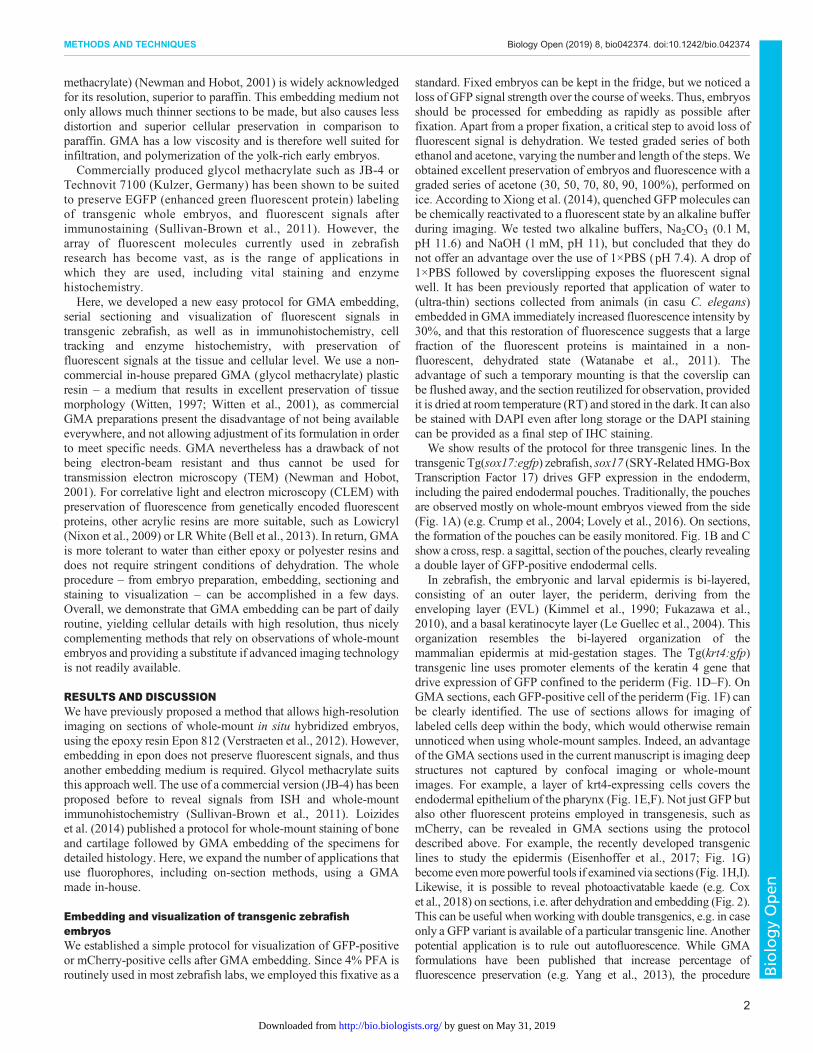
We show results of the protocol for three transgenic lines. In thetransgenic Tg(sox17:egfp) zebrafish, sox17 (SRY-RelatedHMG-BoxTranscription Factor 17) drives GFP expression in the endoderm,including the paired endodermal pouches. Traditionally, the pouchesare observed mostly on whole-mount embryos viewed from the side(Fig. 1A) (e.g. Crump et al., 2004; Lovely et al., 2016). On sections,the formation of the pouches can be easily monitored. Fig. 1B and Cshow a cross, resp. a sagittal, section of the pouches, clearly revealinga double layer of GFP-positive endodermal cells.
In zebrafish, the embryonic and larval epidermis is bi-layered,consisting of an outer layer, the periderm, deriving from theenveloping layer (EVL) (Kimmel et al., 1990; Fukazawa et al.,2010), and a basal keratinocyte layer (Le Guellec et al., 2004). Thisorganization resembles the bi-layered organization of themammalian epidermis at mid-gestation stages. The Tg(krt4:gfp)transgenic line uses promoter elements of the keratin 4 gene thatdrive expression of GFP confined to the periderm (Fig. 1D–F). OnGMA sections, each GFP-positive cell of the periderm (Fig. 1F) canbe clearly identified. The use of sections allows for imaging oflabeled cells deep within the body, which would otherwise remainunnoticed when using whole-mount samples. Indeed, an advantageof the GMA sections used in the current manuscript is imaging deepstructures not captured by confocal imaging or whole-mountimages. For example, a layer of krt4-expressing cells covers theendodermal epithelium of the pharynx (Fig. 1E,F). Not just GFP butalso other fluorescent proteins employed in transgenesis, such asmCherry, can be revealed in GMA sections using the protocoldescribed above. For example, the recently developed transgeniclines to study the epidermis (Eisenhoffer et al., 2017; Fig. 1G)become evenmore powerful tools if examined via sections (Fig. 1H,I).Likewise, it is possible to reveal photoactivatable kaede (e.g. Coxet al., 2018) on sections, i.e. after dehydration and embedding (Fig. 2).This can be useful when working with double transgenics, e.g. in caseonly a GFP variant is available of a particular transgenic line. Anotherpotential application is to rule out autofluorescence. While GMAformulations have been published that increase percentage offluorescence preservation (e.g. Yang et al., 2013), the procedure
2
METHODS AND TECHNIQUES Biology Open (2019) 8, bio042374. doi:10.1242/bio.042374
BiologyOpen
by guest on May 31, 2019http://bio.biologists.org/Downloaded from

described here is anticipated to maintain sufficient levels offluorescence for most applications.
Immunofluorescent localization of antibodiesThe elucidation of spatiotemporal patterns of protein distribution inearly embryos is a key prerequisite for understanding development.Again, many data obtained from immunostaining of embryoniczebrafish are presented on whole-mount embryos only, and an exactlocalization of the antibody is often lacking, especially for deeperlying structures.We routinely use standard procedures for whole-mount
immunohistochemistry (IHC) followed by embedding the samplesin GMA. As indicated before (Sullivan-Brown et al., 2011),
immunofluorescent techniques work on whole-mount embryosonly, not on GMA sections. For immunofluorescent localization ofantigens on plastic sections, it is recommended to use other acrylicresins such as LR White (Newman and Hobot, 2001; Luby-Phelpset al., 2003) or methyl methacrylate – that has nevertheless to beremoved from the sections prior to staining (Hand and Blythe, 2016;and references therein). Fig. 3A–C present the results ofwhole-mount anti-laminin immunofluorescent staining followedby GMA embedding and processing as described above, to visualizethe basement membranes. Laminins are large glycoproteinheterotrimers that are found as major components of basementmembranes in almost every animal tissue (Colognato andYurchenco, 2000; Ekblom et al., 2003). Basement membranes
Fig. 2. Imaging ofphotoactivatable kaede. Vertebralend plates of an adult Tg(osx:kaede) zebrafish shown using aGFP (A–C) and rhodamine (A′–C′)filter, before (A,A′), after 5 s (B,B′)and 30 s (C,C′) of exposure to awavelength of 365 nm. Note thefading of the green signal fromA to C and strengthening of thered signal from A′ to C′ in theosteoblasts lining the vertebral endplates (arrowheads). All pictureswere taken using the sameexposure time (1.6 s). Scale bar:25 µm.
Fig. 1. Imaging of transgeniczebrafish lines. (A,D,G) Whole-mount, (B,E,H) low- and (C,F,I)high-magnification (B,E,F,H,I) crosssections and (C) sagittal section.Lines in A,D,G indicate approximatelevel of sectioning in B,E,H and C,F,I. (A–C) Tg(sox17:egfp) zebrafishwith GFP-positive endodermal cellsalong the midline, extending into thepouches (arrows). Blood vesselsalso show a fluorescent signal.(D–F) Tg(krt4:gfp) zebrafish withGFP-positive periderm (arrows).Labeled cells also cover theoropharyngeal lining (arrowheads).(G–I) Et(Gal4-VP16)zc1044A;Tg(UAS-E1b:nsfB-mCherry)c264,abbreviated as GET-periderm line(GET, Gal4 enhancer trap)(Eisenhoffer et al., 2017). Theperiderm is labeled red (arrows). b,brain; e, eye; nt, notochord; ov, oticvesicle; y, yolk. Scale bars: (A,D)250 µm; (E,G,H) 100 µm; (B,C,F,I)50 µm.
3
METHODS AND TECHNIQUES Biology Open (2019) 8, bio042374. doi:10.1242/bio.042374
BiologyOpen
by guest on May 31, 2019http://bio.biologists.org/Downloaded from

play an important role in tissue development and maintenanceincluding mechanical stability, promotion of cell adhesion,migration, growth and differentiation. Laminin protein, and byextension basement membranes, are clearly identifiable below theepidermis, around the brain and in ocular structures, particularly inthe developing and mature lens (cf. Hallmann et al., 2005) (Fig. 3B).Anti-laminin also serves as a convenient marker to show boundariesbetween epithelial and mesenchymal tissues in sections (Fig. 3C).Cell proliferation is an essential process during growth and
development and often examined using BrdU. We pulse-labeled36 hpf embryos and used a standard procedure to reveal BrdU onwhole-mount embryos (Fig. 3D).Afterembedding and sectioning, thedistribution of labeled cells can be precisely mapped and if necessaryquantified (Fig. 3E). BrdU can also be administered to transgenicembryos and the fluorescent secondary antibody can be revealedsimultaneously with the fluorophore used in the transgene (Fig. 3F).In another example combining different fluorophores, we used a
pan-cytokeratin antibody on Tg(sox17:egfp) embryos (Fig. 3G–I).The latter two examples show that the fluorescence of GFP (as wellas of other fluorophores) is maintained through the whole-mountimmunolabeling procedure as well as the GMA embeddingprotocol. The GFP can also be visualized in the whole-mountembryo prior to embedding. While immunolabeling of transgenicGFP-labeled embryos is a common procedure, the resolutionobtained by processing whole embryos into sections is a clear asset.
Visualization of fluorescent dyes used for cell trackingLineage tracing and cell fate determination relies for a large part onthe use of fluorescent cell tracers. DiI is a lipophilic dye that isusually administered via injection (Fig. 4A). It is weakly fluorescentuntil incorporated into membranes and is used as long-term tracerfor neuronal and other cells. DiI can be revealed on sections afterethanol dehydration and embedding in GMA (Fig. 4B,C).
We also used vital staining with CDCFDA to label the externalsurface of the embryo (the periderm) and to establish the fate ofthese cells (Fig. 4D). CDCFDA is a non-fluorescent molecule thatdiffuses into cells and is hydrolyzed by intracellular non-specificesterases to give a fluorescent product (similar to CCFSE, used byShone and Graham, 2014). The fluorescent product accumulatesonly in those cells that have intact cell membranes (Griffith and Hay,1992); therefore, dead cells with leaky membranes are not stained.Fig. 4E and F show how labeled peridermal cells invade the embryovia the gill slits.
On-section stainingDAPI is a widespread nuclear counterstain that is often applied onwhole-mount embryos. DAPI can also easily be applied on sections(Fig. 4G–I). The result is comparable to whole-mount stainingfollowed by embedding.
Various enzymes that are involved in skeletal modeling andremodeling can be studied after GMA embedding and on-sectionimmunohistochemistry in order to map their precise spatial andtemporal pattern of activity. For example, tartrate-resistant acidphosphatase specifically marks cells responsible for bone resorption(osteoclasts) (Witten et al., 2001; Witten and Huysseune, 2009).In zebrafish, cells expressing TRAP are small in size andmononuclear, at least in early development, and therefore noteasily revealed on conventionally stained sections. We havesuccessfully employed GMA embedding and TRAP staining onsections of both larval and adult zebrafish (Witten et al., 2001;Kague et al., 2018). The reaction product can be visualized both inbright field (Fig. 4J) and under epifluorescence (Fig. 4K), andimages can be overlain (Fig. 4L). Other enzymes can be revealedafter GMA embedding such as ATPase, alkaline phosphatase oralpha naphtyl acetate esterase (Witten, 1997; Witten et al., 1999;Witten et al., 2000; Witten et al., 2001).
Fig. 3. Whole-mountimmunostaining. (A,D,G) Whole-mount, (B,E,H) low- and (C,F,I)high-magnification cross sections.Lines in A,D,G indicate approximatelevel of sectioning in B,E,H andC,F,I. (A–C) Wild-type (WT)zebrafish immunostained forlaminin. Note the basal laminadelimiting optic cup, brain,epidermis and endodermalpouches. (D–F) Tg(sox17:egfp)zebrafish pulse-labeled with BrdUand whole-mount stained with ananti-BrdU antibody. (G-I) Tg(sox17:egfp) zebrafish immunostained witha pan-cytokeratin antibody. Thewhole-mount images in D,G showthe red channel only; the GFPfluorescence is likewise visible priorto dehydration and embedding.b, brain; e, eye; ep, epidermis;oc, optic cup; p, endodermal pouch.Scale bars: (A,D,G) 250 µm; (E,H)100 µm; (B,F,I) 50 µm; (C) 25 µm.
4
METHODS AND TECHNIQUES Biology Open (2019) 8, bio042374. doi:10.1242/bio.042374
BiologyOpen
by guest on May 31, 2019http://bio.biologists.org/Downloaded from

ConclusionIn conclusion, we propose an easy-to-perform GMA embeddingmethod that preserves fluorescent signals and allows visualizationof GFP, mCherry and other fluorophores on semithin sections (3 µmor less) without the need for antibodies. The method is suited for awide range of applications including study of transgenic zebrafish ofunlimited size, immunostaining of whole-mount embryos, celltracking and on-section enzyme histochemistry of embryonic oreven adult zebrafish. In addition, tissue preservation is superior toany of the other common procedures used in histology (such asparaffin and vibratome sections) and allows a detailed study of cellsin their tissue or organ context. The method nicely complementswhole-mount methods especially if advanced imaging technologyis not readily available.
MATERIALS AND METHODSEthical statementAnimal care, experimentation and euthanasia complied with EuropeanDirective 2010/63/EU of 22 September, 2010. The experimental protocoland all animal procedures used in this study were approved by GhentUniversity (laboratory permit number LA1400452).
Zebrafish lines and collection of transgenic fishWild-type AB line and transgenic Tg(sox17:egfp) zebrafish(Danio rerio) lines (Mizoguchi et al., 2008) were obtained from thelaboratory of Dr R. Opitz (VUB, Brussels, Belgium). Tg(krt4:gfp) (Gonget al., 2002) were a gift from the laboratory of Dr M. Hammerschmidt(University of Köln, Köln, Germany). Et(Gal4-VP16)zc1044A;Tg(UAS-E1b:nsfB-mCherry)c264, abbreviated as GET-periderm line (GET, Gal4 enhancertrap) were obtained from the laboratory of Dr G. Eisenhoffer (MDAndersonCancer Center, Houston, Texas, USA) (Eisenhoffer et al., 2017). Anunpublished Tg(osx:kaede) strain was made available by Dr KennethD. Poss and Sumeet P. Singh (Duke University, Durham, NC, USA).All zebrafish were maintained and bred in accordance to Westerfield(1993). WT and transgenic embryos were kept at 28.5°C untileuthanasia by an overdose of 1% MS222 (ethyl 3-aminobenzoatemethane sulfonate, cat No.: E10521, Sigma Aldrich). They were stagedaccording to Kimmel et al. (1995), fixed in 4% paraformaldehyde(PFA) in PBS buffer (pH 7.2) for 2 h at RT, and either processedimmediately for embedding or stored in methanol at −20°Cuntil immunostaining. Adult transgenic zebrafish were fixed overnightat 4°C in 4% PFA in PBS buffer (pH 7.2), rinsed in tap water for 1 h,and decalcified with 10% EDTA in Tris buffer (100 mmol, pH 7.2)for 48 h.
Fig. 4. Cell tracking withfluorescent vital dyes and on-section histochemistry. (A–I) Celltracking. (A,D) Whole-mount, (B,E)low- and (C,F) high-magnificationcross sections. (A–C) WT zebrafishinjected with DiI (A, arrow) at28 hpf and euthanized after 48 hreveal areas of DiI distribution(arrowheads); the DiI label(yellowish dots) stands out sharplyagainst an autofluorescentbackground (B,C). (D–F) WTzebrafish vital stained withCDCFDA. Only cells exposed to thesolution (periderm) take up the stain(E, arrows). After a chase time of22 h, labeled cells can be observedinside the forming gill slit (F,arrowhead). Lines in D indicateapproximate level of sectioning inE,F. (G–I) DAPI staining (G) onsection of CDCFDA labeled embryo(H) and merged picture (I). (J–K)On-section histochemistry for theosteoclast marker TRAP, showingthe palatoquadrate and palatinebone in adult WT zebrafish, viewedin transmitted light (J), underepifluorescence (K) and theoverlay of transmitted light andepifluorecence (L). b, brain; e, eye;m, mouth; nt, notochord; ph,pharyngeal lumen. Scale bars:(A,G,H,I) 100 µm; (D) 250 µm;(B,E,F) 50 µm; (C,J,K,L) 25 µm.
5
METHODS AND TECHNIQUES Biology Open (2019) 8, bio042374. doi:10.1242/bio.042374
BiologyOpen
by guest on May 31, 2019http://bio.biologists.org/Downloaded from

ImmunofluorescenceWe used whole-mount immunofluorescent staining according to establishedprotocols to detect various antigens (laminin, pan-cytokeratin, BrdU). In allcases, WT and transgenic embryos were euthanized by an overdose of 1%MS222, fixed in 4% PFA for 10 min at RT, and then overnight at 4°C (anti-laminin staining), or just for 2 h (for anti-BrdU and anti-pan-cytokeratinstaining) followed by storage in 100% methanol at −20°C.
The protocol for whole-mount immunostaining for laminin followsO’Brien et al. (2011), with adjustment for the time of permeabilization.Following fixation, embryos were permeabilized in acetone for 10 min at–20°C (the time for permeabilization depends on the developmental stage ofthe embryos, see Table 1). The embryos were rinsed in 1×PBS (phosphate-buffered saline) (pH 7.2) and the primary antibody (anti-α-laminin, 1:100,cat. no.: L9393, Sigma Aldrich) was applied overnight at 4°C. On thefollowing day, embryos were incubated in the secondary antibody (goatanti-rabbit DyLight 488 nm, 1:200, Abcam) for 4 h at RT. Embryos wererinsed in 1×PBS. After antibody staining, embryos were rinsed in 1×PBSagain and stored in 4% PFA until processing for GMA embedding.
For proliferation studies, 36 hpf embryos were pulse-labeled for 20 min in10 mM BrdU (5-bromo-2′-deoxyuridine)/15% dimethylsulfoxide andsacrificed immediately after BrdU administration, following the protocoldescribed in Verduzco and Amatruda (2013). Whole-mount immunostainingfor BrdU was performed following this same protocol, using an anti-BrdU(mouse, 1:100, cat. no.: B2531-2ML, Sigma Aldrich) as primary antibody,and an Alexa Fluor® 594 (goat anti-mouse IgG, 1:200, Abcam) as secondaryantibody. Embryos were next rinsed and kept in 4% PFA until processing forGMA embedding.
The same protocol (Verduzco and Amatruda, 2013, from step 4 onwards)was employed for whole-mount immunostaining of keratins using a pan-cytokeratin (AE1/AE3) (mouse, Santa Cruz, 1:200) as primary antibody, andan Alexa Fluor® 594 (goat anti-mouse IgG, 1:200, Abcam) as secondaryantibody.
Negative controls were performed by omitting the primary antibody fromthe reaction mixture.
Cell trackingTwo molecules for cell tracking were used: DiI and CDCFDA. DiIis a lipophilic dye commonly used for cell fate tracing. A stock solution of5 µg/µl DiI (Invitrogen Cell Tracker CM-DiI, cat no. C-7000) was preparedby diluting 50 µg in Ethanol 1:10, and stored at−20°C. A dilution of 1:10 in0.3 M Sucrose (made up in nuclease-free water from Sigma Aldrich) wasused to inject dechorionated WT embryos of 28 hpf. For injection, ca. 5dechorionated embryos were placed on a smooth agar plate using a plasticballoon pipette. All excess water was sucked up with a plastic balloonpipette and one drop of MS222 0.001% was sprinkled on the embryos.Excess fluid was sucked away. Embryos were euthanized at appropriatetimes using an overdose of 1% MS222, fixed in 4% PFA for 24 h, andtransferred to 1×PBS with 0.02% sodium azide for storage at 4°C, untilembedding.
CDCFDA[5-(and-6)-carboxy-2′,7′-dichlorofluoresceindiacetate, succinimidylester, mixed isomers, cat. no.: 22026, AAT Bioquest, Inc.] is an isomermixture of CCFSE–[5-(and-6)-carboxy-2′,7′-dichlorofluorescein diacetate,succinimidyl ester] used by Shone and Graham (2014). It was dissolved inanhydrous DMSO (dimethylsulfoxide) to 50 mM and stored at −20°C. Aworking concentration (20, 100, 250, 500 µM) in 1×PBS was used. WTembryos varying in age between 8 and 22 hpf, as well as some older embryos(30, 40 and 56 hpf) were soaked in CDCFDA for 4 h, rinsed in eggwater andtransferred to fresh egg water. At selected time points, embryos were
euthanized by an overdose of 1%MS222, fixed in 4% PFA and stored in 4%PFA until processing for GMA embedding.
GMA embedding and sectioningWe used a GMA embedding protocol as described by Witten et al. (2001).Briefly, the zebrafish were rinsed in two changes of 1×PBS (15 min each)and dehydrated using a graded series of acetone solutions [30, 50, 70, 80, 90,100% for 15 min each (embryos) or 30, 60, 100% for 1 h each (adults)].These steps were performed on ice, sheltered from light, using a shaker.Embryos were then impregnated in several changes of fresh glycolmethacrylate monomer solution for 15 min at 4°C (two changes),followed by 60 min at 4°C. Adults were kept in the monomer for severaldays. Glycol methacrylate monomer solution consists of 80 ml (2-hydroxyethyl)-methacrylate (+200 ppm p-methoxyphenol, usually alreadyin the product, cat. no.: 17348-250ML-F, Sigma Aldrich), 12 ml ethyleneglycol monobuthyl ether (cat. no.: 537551-1L.A, Sigma Aldrich) and270 mg benzoyl peroxide (added to the solution and stirred overnight).The samples were subsequently transferred to fresh GMA monomer solutionand left for 24 h at 4°C. After a total of 24 h in the monomer, the sampleswere placed in an embedding mold (PTFE Flat embedding mold,Electron Microscopy Sciences) containing GMA with 2% catalyst(N,N-dimethylaniline 1 ml+poly-ethylene glycol-200 10 ml, Sigma Aldrich)added. A slice of polymerized GMA was placed at the bottom of each wellprior to positioning the fish in order to prevent the sample from sinking to thebottom of the well (hence side of the block). Care was taken duringpolymerization to protect the blocks from air by covering the embeddingmoldwith an oxygen barrier film trimmed to the appropriate size (ACLAR 33Cembedding film). Polymerization occurred, sheltered from light, at 4°C for24 h (embedding mold placed on crushed ice) and another 24 h at RT. TheGMA block was removed from the mold and stored at RT in the dark. Forsectioning, the block was mounted on a standard histology microtome(Microm HM360, Prosan) and routinely sectioned at 3 µm (down to 1 µm)using disposable knives (Technovit Histoblade knives, Kulzer). Sections wereplaced on a drop of demineralized water on a glass slide and air-dried at RT,sheltered from light.
On-section enzyme histochemistryFor TRAP staining, zebrafish were euthanized with an overdose of MS222,fixed in cold 100% acetone and stored at −20°C prior to dehydration andembedding, as long-term storage in PFA destroys enzyme activity. TRAPstaining was carried out on sections of GMA embedded tissues, usingnaphthol AS-TR phosphate (N-AS-TR-P, cat. no.: N6125-1G, SigmaAldrich) as substrate and hexazotized pararosaniline (PRS, Acros Organics227881000, Thermo Fisher Scientific) as color component, following theprotocol of Witten et al. (2001). After final rinsing with demineralizedwater, sections were air-dried. Controls are performed either by (a) heatingthe section at 90°C for 10 min prior to incubation, (b) incubation withoutsubstrate, (c) incubation without tartrate or (d) adding NaF (10 mmol/l) tothe incubation solution (Witten, 1997).
Nuclear counterstainingWhen required, a nuclear counterstain was applied either before or afterimaging the sections using DAPI (4′, 6-diamidino-2-phenylindole,dihydrochloride, cat. no.: D9542-1MG, Sigma Aldrich). To this end,sections were covered with a drop of DAPI (1:1000 in 1×PBS) for 15 min atRT, sheltered from light, and washed three times for 10 min with 1×PBS.Other methods of counterstaining, such as with Toluidine Blue, arenecessarily performed after imaging since these techniques destroy thefluorescent signal.
ImagingFluorescent signals were visualized by covering the sections with a drop of1×PBS, followed by coverslipping. Sections were observed with a ZeissAxioimager Z1, equipped for epifluorescence, using the following filters:GFP (Excitation BP 470/40, Beam splitter FT 495, Emission BP 525/50),Rhodamine (Excitation BP 546/12, Beam splitter FT 560, Emission BP575-640) and DAPI (Excitation G 365, Beam splitter FT 395, Emission BP
Table 1. Time for permeabilization with acetone related to thedevelopmental stage of the zebrafish embryos
Stage (hpf) Time (min)
30–38 1040–48 1550–72 2072–96 40
6
METHODS AND TECHNIQUES Biology Open (2019) 8, bio042374. doi:10.1242/bio.042374
BiologyOpen
by guest on May 31, 2019http://bio.biologists.org/Downloaded from

445/50), and photographed with a Zeiss Axiocam 503 camera using ZENsoftware (www.zeiss.com). After imaging, the coverslip was removed byflushing with demineralized water, and the section was allowed to dry forrenewed observation or for counterstaining and permanent mounting.
AcknowledgementsWe thank Dr R. Opitz (VUB, Brussels, Belgium), Dr M. Hammerschmidt (KolnUniversity, Koln, Germany), and Dr G.T. Eisenhoffer and Dr O. Ruiz (MD AndersonCancer Center, Houston Texas, USA) for sharing their transgenic fish. KennethD. Poss and Sumeet P. Singh (Duke University, Durham, NC, USA) are gratefullyacknowledged for sharing an unpublished strain. We thank A. Gillis (CambridgeUniversity, UK) for performing DiI injections on embryonic zebrafish.
Competing interestsThe authors declare no competing or financial interests.
Author contributionsConceptualization: A.H.; Methodology: V.O., J.T.R., M.S., P.E.W.; Validation: V.O.,J.T.R., J.W.B., A.W., P.E.W., A.H.;Writing - original draft: V.O., P.E.W., A.H.;Writing -review & editing: V.O., J.T.R., J.W.B., A.W., P.E.W., A.H.; Visualization: V.O., J.T.R.,J.W.B., A.W., P.E.W., A.H.; Supervision: A.H.; Project administration: A.H.; Fundingacquisition: A.H.
FundingThis work was supported by the Ghent University Research Fund(BOF24J2015001401).
ReferencesBell, K., Mitchell, S., Paultre, D., Posch, M. and Oparka, K. (2013). Correlativeimaging of fluorescent proteins in resin embedded plant material. Plant Physiol.161, 1595-1603. doi:10.1104/pp.112.212365
Bruneel, B. andWitten, P. E. (2015). Power and challenges of using zebrafish as amodel for skeletal tissue imaging. Conn. Tiss. Res. 56, 161-173. doi:10.3109/03008207.2015.1013193
Colognato, H. and Yurchenco, P. D. (2000). Form and function: the laminin family ofheterotrimers. Dev. Dyn. 218, 213-234. doi:10.1002/(SICI)1097-0177(200006)218:2<213::AID-DVDY1>3.0.CO;2-R
Cox, B. D., De Simone, A., Tornini, V. A., Singh, S. P., Di Talia, S. and Poss, K. D.(2018). In toto imaging of dynamic osteoblast behaviors in regenerating skeletalbone. Curr. Biol. 28, 3937-3947.e4. doi:10.1016/j.cub.2018.10.052
Crump, J. G., Maves, L., Lawson, N. D., Weinstein, B. M. and Kimmel, C. B.(2004). An essential role for Fgfs in endodermal pouch formation influences latercraniofacial skeletal patterning. Development 131, 5703-5716. doi:10.1242/dev.01444
Eisenhoffer, G. T., Slattum, G., Ruiz, O. E., Otsuna, H., Bryan, C. D., Lopez, J.,Wagner, D. S., Bonkowsky, J. L., Chien, C.-B., Dorsky, R. I. et al. (2017). Atoolbox to study epidermal cell types in zebrafish. J. Cell Sci. 130, 269-277. doi:10.1242/jcs.184341
Ekblom, P., Lonai, O. and Talts, J. F. (2003). Expression and biological role oflaminin-1. Matrix Biol. 22, 35-47. doi:10.1016/S0945-053X(03)00015-5
Fukazawa, C., Santiago, C., Park, K. M., Deery, W. J., de la Torre Canny, S. G.,Holterhoff, C. K. and Wagner, D. S. (2010). poky/chuk/ikkl is required fordifferentiation of the zebrafish (Danio rerio). Int. J. Dev. Biol. 48, 217-231.doi:10.1016/j.ydbio.2010.07.037
Gistelinck, C., Kwon, R. Y., Malfait, F., Symoens, S., Harris, M. P., Henke, K.,Hawkins, M. B., Fisher, S., Sips, P., Guillemyn, B. et al. (2018). Zebrafish type Icollagen mutants faithfully recapitulate human type I collagenopathies. Proc. Natl.Acad. Sci. USA 115, E8037-E8046. doi:10.1073/pnas.1722200115
Gong, Z., Ju, B.,Wang, X., He, J.,Wan, H., Sudha, P.M. and Yan, T. (2002). Greenfluorescent protein expression in germ-line transmitted transgenic zebrafish undera stratified epithelial promoter from Keratin8. Dev. Dyn. 223, 204-215. doi:10.1002/dvdy.10051
Griffith, C. M. and Hay, E. D. (1992). Epithelial-mesenchymal transformation duringpalatal fusion: carboxyfluorescein traces cells at light and electron microscopiclevels. Development 116, 1087-1099.
Hallmann, R., Horn, N., Selg, M., Wendler, O., Pausch, F. and Sorokin, L. M.(2005). Expression and function of laminins in the embryonic and maturevasculature. Physiol. Rev. 85, 979-1000. doi:10.1152/physrev.00014.2004
Hand, N. M. and Blythe, D. (2016). Plastic embedding of bone marrow trephinebiopsies for routine immunohistochemistry and diagnosis: our developments,updates and experiences over 20 years. J. Histotechnol. 39, 135-146. doi:10.1080/01478885.2016.1207912
Hogan, B. M. and Schulte-Merker, S. (2017). How to plumb a Pisces:understanding vascular development and disease using zebrafish embryos.Dev. Cell 42, 567-583. doi:10.1016/j.devcel.2017.08.015
Kague, E., Witten, P. E., Soenens, M., Campos, C. L., Lubiana, T., Fisher, S.,Hammond, C., Robson-Brown, K., Passos-Bueno, M. R. and Huysseune, A.
(2018). Zebrafish sp7 mutants show tooth cycling independent of attachment,eruption and poor differentiation of teeth. Dev. Biol. 435, 176-184. doi:10.1016/j.ydbio.2018.01.021
Kimmel, C. B., Warga, R. M. and Schilling, T. F. (1990). Origin and organization ofthe zebrafish fate map. Development 108, 581-594.
Kimmel, C. B., Ballard, W. W., Kimmel, S. R., Ullmann, B. and Schilling, T. F.(1995). Stages of embryonic development of the zebrafish. Dev. Dyn. 203,253-310. doi:10.1002/aja.1002030302
Laize, V., Gavaia, P. J. and Cancela, M. L. (2014). Fish: a suitable system to modelhuman bone disorders and discover drugs with osteogenic or osteotoxic activities.Drug Discov. Today Dis. Model. 13, 29-37. doi:10.1016/j.ddmod.2014.08.001
Le Guellec, D., Morvan-Dubois, G. and Sire, J.-Y. (2004). Skin development inbony fish with particular emphasis on collagen deposition in the dermis of thezebrafish (Danio rerio). Int. J. Dev. Biol. 48, 217-231. doi:10.1387/ijdb.15272388
Liu, T.-L., Upadhyayula, S., Milkie, D. E., Singh, V., Wang, K., Swinburne, I. A.,Mosaliganti, K. R., Collins, Z. M., Hiscock, T. W., Shea, J. et al. (2018).Observing the cell in its native state: Imaging subcellular dynamics in multicellularorganisms. Science 360, eaaq1392. doi:10.1126/science.aaq1392
Loizides, M., Georgiou, A. N., Somarakis, S., Witten, P. E. andKoumoundouros, G. (2014). A new type of lordosis and vertebral bodycompression in Gilthead sea bream, Sparus aurata L.: aetiology, anatomy andconsequences for survival. J. Fish Dis. 37, 949-957. doi:10.1111/jfd.12189
Lovely, C. B., Swartz, M. E., McCarthy, N., Norrie, J. L. and Eberhart, J. K. (2016).Bmp signaling mediates endoderm pouch morphogenesis by regulating Fgfsignaling in zebrafish. Development 143, 2000-2011. doi:10.1242/dev.129379
Luby-Phelps, K., Ning, G., Fogerty, J. andBesharse, J. C. (2003). Visualization ofidentified GFP-expressing cells by light and electron microscopy. J. Histochem.Cytochem. 51, 271-274. doi:10.1177/002215540305100301
Mizoguchi, T., Verkade, H., Heath, J. K., Kuroiwa, A. and Kikuchi, Y. (2008).Sdf1/Cxcr4 signaling controls the dorsal migration of endodermal cells duringzebrafish gastrulation. Development 135, 2521-2529. doi:10.1242/dev.020107
Newman, G. R. and Hobot, J. A. (2001). Resin Microscopy and on-SectionImmunocytochemistry. 2nd edn. Berlin, Heidelberg: Springer Verlag.
Nixon, S. J., Webb, R. I., Floetenmeyer, M., Schieber, N., Lo, H. P. and Parton,R. G. (2009). A single method for cryofixation and correlative light, electronmicroscopy and tomography of zebrafish embryos. Traffic 10, 131-136. doi:10.1111/j.1600-0854.2008.00859.x
O’Brien, G. S., Rieger, S., Wang, F., Smolen, G. A., Gonzalez, R. E., Buchan, J.and Sagasti, A. (2011). Coordinate development of skin cells and cutaneoussensory axon in zebrafish. J. Comp. Neurol. 520, 816-831. doi:10.1002/cne.22791
Schultz, L. E., Haltom, J. A., Almeida, M. P., Wierson, W. A., Solin, S. L., Weiss,T. J., Helmer, J. A., Sandquist, E. J., Shive, H. R. and McGrail, M. (2018).Epigenetic regulators Rbbp4 and Hdac1 are overexpressed in a zebrafish modelof RB1 embryonal brain tumor, and are required for neural progenitor survival andproliferation. Dis. Model. Mech. 11, dmm034124. doi:10.1242/dmm.034124
Shone, V. and Graham, A. (2014). Endodermal/ectodermal interfaces duringpharyngeal segmentation in vertebrates. J. Anat. 225, 479-491. doi:10.1111/joa.12234
Sullivan-Brown, J., Bisher, M. E. and Burdine, R. D. (2011). Embedding, serialsectioning and staining of zebrafish embryos using JB-4 resin. Nat. Protocols 6,46-55. doi:10.1038/nprot.2010.165
Verduzco, D. andAmatruda, J. F. (2013). Analysis of cell proliferation, senescenceand cell death in zebrafish embryos.Methods Cell Biol. 101. doi:10.1016/B978-0-12-387036-0.00002-5
Verstraeten, B., Sanders, E. and Huysseune, A. (2012). Whole mountimmunohistochemistry and in situ hybridization of larval and adult zebrafishdental tissues. In Odontogenesis. Methods and Protocols, Vol. 887 (ed. C.Kioussi), pp. 179-191. Meth. Molec. Biol. USA: Humana Press.
Watanabe, S., Punge, A., Hollopeter, G., Willig, K. I., Hobson, R. J., Davis, M.W.,Hell, S. W. and Jorgensen, E. M. (2011). Protein localization in electronmicrographs using fluorescence nanoscopy.Nat. Methods 8, 80-84. doi:10.1038/nmeth.1537
Watson, A. M., Rose, A. H., Gibson, G. A., Gardner, C. L., Sun, C., Reed, D. S.,Lam, L. K. M., St. Croix, C. M., Strick, P. L. et al. (2017). Ribbon scanningconfocal for high-speed high-resolution volume imaging of brain. PLoS ONE 12,e0180486. doi:10.1371/journal.pone.0180486
Wehner, D. and Weidinger, G. (2015). Signaling networks organizing regenerativegrowthof thezebrafish fin.TrendsGenet.31, 336-343.doi:10.1016/j.tig.2015.03.012
Westerfield, M. (1993). The Zebrafish Book, a Guide for the Laboratory use ofZebrafish (Danio rerio). Eugene, USA: University of Oregon press.
Witten, P. E. (1997). Enzyme histochemical characteristics of osteoblasts andmononucleated osteoclasts in a teleost fish with acellular bone (Oreochromisniloticus, Cichlidae). Cell Tiss. Res. 287, 591-599. doi:10.1007/s004410050782
Witten, P. E. and Huysseune, A. (2009). A comparative view on mechanisms andfunctions of skeletal remodelling in teleost fish, with special emphasis onosteoclasts and their function. Biol. Rev. 84, 315-346. doi:10.1111/j.1469-185X.2009.00077.x
Witten, P. E., Holliday, L. S., Delling, G. and Hall, B. K. (1999).Immunohistochemical identification of a vacuolar proton pump (V-ATPase) in
7
METHODS AND TECHNIQUES Biology Open (2019) 8, bio042374. doi:10.1242/bio.042374
BiologyOpen
by guest on May 31, 2019http://bio.biologists.org/Downloaded from

bone-resorbing cells of an advanced teleost species (Oreochromis niloticus,Teleostei, Cichlidae). J. Fish Biol. 55, 1258-1272. doi:10.1111/j.1095-8649.1999.tb02074.x
Witten, P. E., Villwock, W., Peters, N. and Hall, B. K. (2000). Bone resorption andbone remodelling in juvenile carp, Cyprinus carpio L. J. Appl. Ichthyol. 16,254-261. doi:10.1046/j.1439-0426.2000.00233.x
Witten, P. E., Hansen, A. and Hall, B. K. (2001). Features of mono andmultinucleated bone resorbing cells of the zebrafish Danio rerio and theircontribution to skeletal development, remodeling and growth. J. Morphol. 250,197-207. doi:10.1002/jmor.1065.abs
Witten, P. E., Harris, M. P., Huysseune, A. and Winkler, C. (2017). Chapter 13.Fish provide insight into skeletal diseases. Methods Cell Biol. 138, 321-346.doi:10.1016/bs.mcb.2016.09.001
Xiong, H., Zhou, Z., Zhu, M., Lv, X., Li, A., Li, S., Li, L., Yang, T., Wang, S., Yang,Z. et al. (2014). Chemical reactivation of quenched fluorescent protein moleculesenables resin-embedded fluorescence microimaging. Nat. Comm. 5, 3992.doi:10.1038/ncomms4992
Yang, Z., Hu, B., Zhang, Y., Luo, Q. andGong, H. (2013). Development of a plasticembedding method for large-volume and fluorescent-protein-expressing tissues.PLoS ONE 8, e60877. doi:10.1371/journal.pone.0060877
8
METHODS AND TECHNIQUES Biology Open (2019) 8, bio042374. doi:10.1242/bio.042374
BiologyOpen
by guest on May 31, 2019http://bio.biologists.org/Downloaded from