biliary tract disease - پزشکی...
TRANSCRIPT

Biliary Tract Disease NIKI TADAYON
GENERAL & VASCULAR SURGEON
SHOHADA TAJRISH HOSPITAL
Differential Diagnosis of RUQ pain
Gallstone disease (and its related complications)
Gastritis/duodenitis
Peptic ulcer disease/perforated peptic ulcer
Acute pancreatitis
Right lower lobe pneumonia
MI
If presenting with RUQ pain all patients should get
Blood tests
Obstruction series [CXR (to exclude perforation/pneumonia)Abdominal X-ray supine ,upwright
ECG

Biliary Tract
Part of the digestive system.
Consists of:
Intra hepatic ducts
Exta hepatic ducts
Gallbladder
Common Bile Duct

The Gallbladder
The gallbladder concentrates and stores bile.
Bile:
Secreted by the liver
Contains cholesterol, bile pigments and phospholipids
Flows from the liver, through the hepatic ducts, into the gallbladder.
Exits the gallbladder via the cystic duct.
Flows from the cystic duct into the common bile duct, into the small intestine
In the small intestine, aids digestion by breaking down fatty foods and fat-soluble vitamins
Pathogenesis
Composition of bile:
Bilirubin (by-product of haem degradation)
Cholesterol (kept soluble by bile salts and lecithin)
Bile salts/acids (cholic acid/chenodeoxycholic acid): mostly reabsorbed in terminal ileum(entero-hepatic circulation).
Lecithin (increases solubility of cholesterol)
Inorganic salts (sodium bicarbonate to keep bile alkaline to neutralise gastric acid in duodenum)
Water (makes up 97% of bile)
Gallstones – Pathophysiology
Cholesterol, ordinarily insoluble in water, comes into solution by forming vesicles with phospholipids
If ratio of cholesterol, phospholipids, and bile salts altered, cholesterol crystals may form
Gallstone formation involves a variety of factors:
Cholesterol supersaturation
Mucin hypersecretion by the gallbladder mucosa creates a viscoelastic gel that fosters nucleation.
Bile stasis
Occurs in diabetes, pregnancy, oral contraceptive use, and prolonged fasting in critically ill patients on total parenteral nutrition.
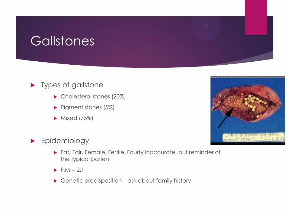
Gallstones
Types of gallstone
Cholesterol stones (20%)
Pigment stones (5%)
Mixed (75%)
Epidemiology
Fat, Fair, Female, Fertile, Fourty inaccurate, but reminder of the typical patient
F:M = 2:1
Genetic predisposition – ask about family history
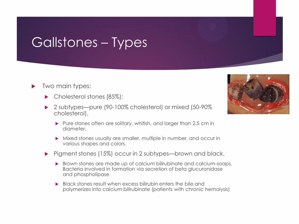
Gallstones – Types
Two main types:
Cholesterol stones (85%):
2 subtypes—pure (90-100% cholesterol) or mixed (50-90% cholesterol).
Pure stones often are solitary, whitish, and larger than 2.5 cm in diameter.
Mixed stones usually are smaller, multiple in number, and occur in various shapes and colors.
Pigment stones (15%) occur in 2 subtypes—brown and black.
Brown stones are made up of calcium bilirubinate and calcium-soaps. Bacteria involved in formation via secretion of beta glucuronidase and phospholipase
Black stones result when excess bilirubin enters the bile and polymerizes into calcium bilirubinate (patients with chronic hemolysis)

Complications of Gallstones
Biliary Colic
Acute Cholecystitis
Gallbladder Empyema
Gallbladder gangrene
Gallbladder perforation
Obstructive Jaundice
Ascending Cholangitis
Pancreatitis
Gallstone Ileus (rare)

Gallstones – Natural History
80% of patients, gallstones are clinically silent.
20% of patients develop symptoms over 15-20 years
About 1% per year.
Almost all become symptomatic before complications
develop.
Biliary-type pain due to obstruction of the bile duct
lumen.
Predictive value of other complaints (eg, intolerance to
fatty food, indigestion) too low to be clinically helpful.

Gallstones – Diverse symptoms
Abdominal pain
Aching or tightness, typically severe and located in the epigastrium
May develop suddenly, last for 15 minutes to several hours, and then resolve suddenly
Referred pain – posterior scapula or right shoulder area
Nausea and vomiting
Jaundice
Pruritus:
Itching, typically worse at night.
Fatigue
Weight loss
Miscellaneous:
Fatty food intolerance
Gas
Bloating
Dyspepsia
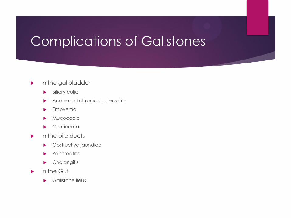
Complications of Gallstones
In the gallbladder
Biliary colic
Acute and chronic cholecystitis
Empyema
Mucocoele
Carcinoma
In the bile ducts
Obstructive jaundice
Pancreatitis
Cholangitis
In the Gut
Gallstone ileus

Which Gallstone
Complication?
Can differentiate between gallstone complications
based on:
History
Examination
Blood tests
FBC
LFT
CRP
Clotting
Amylase
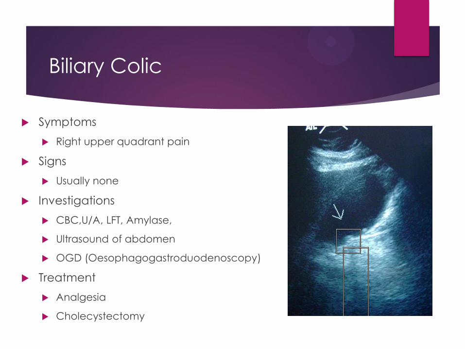
Biliary Colic
Symptoms
Right upper quadrant pain
Signs
Usually none
Investigations
CBC,U/A, LFT, Amylase,
Ultrasound of abdomen
OGD (Oesophagogastroduodenoscopy)
Treatment
Analgesia
Cholecystectomy
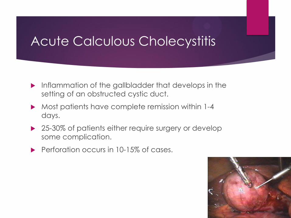
Acute Calculous Cholecystitis
Inflammation of the gallbladder that develops in the
setting of an obstructed cystic duct.
Most patients have complete remission within 1-4
days.
25-30% of patients either require surgery or develop
some complication.
Perforation occurs in 10-15% of cases.
Acute Calculous Cholecystitis
Symptoms
Right upper quadrant pain – continuous, longer duration
Signs
Fever, Local peritonism.
Murphy’s sign
Investigations
CBC, U/A, LFT, Amylase,
Ultrasound of RUQ
Thickened gallbladder wall, pericholecystic fluid and stones
OGD (Oesophagogastroduodenoscopy)
Treatment
NPO
Analgesia
Intravenous antibiotics
Cholecystectomy
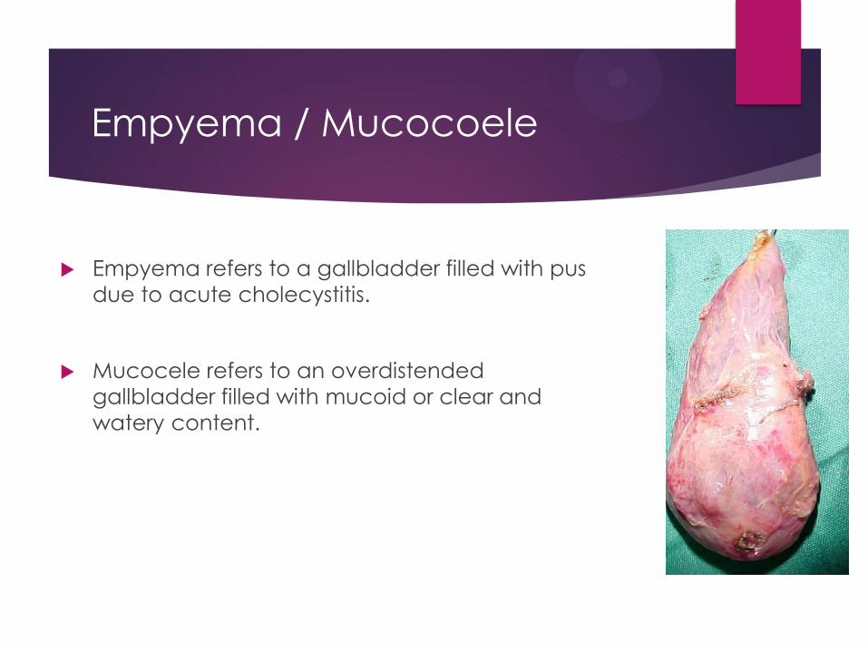
Empyema / Mucocoele
Empyema refers to a gallbladder filled with pus
due to acute cholecystitis.
Mucocele refers to an overdistended
gallbladder filled with mucoid or clear and
watery content.
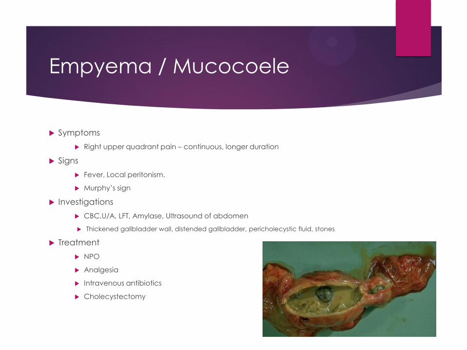
Empyema / Mucocoele
Symptoms
Right upper quadrant pain – continuous, longer duration
Signs
Fever, Local peritonism.
Murphy’s sign
Investigations
CBC,U/A, LFT, Amylase, Ultrasound of abdomen
Thickened gallbladder wall, distended gallbladder, pericholecystic fluid, stones
Treatment
NPO
Analgesia
Intravenous antibiotics
Cholecystectomy

Obstructive Jaundice
Pathogenesis:
Stone obstructing CBD (bear in mind there are other causes for obstructive jaundice) – danger is progression to ascending cholangitis.
USS
Will confirm gallstones in the gallbladder
CBD dilatation i.e. >8mm (not always!)
May visualise stone in CBD (most often does not)
MRCP
In cases where suspect stone in CBD but USS indeterminate
Treatment
Must unobstruct biliary tree with ERCP to prevent progression to ascending cholangitis.
Whilst awaiting ERCP monitor for signs of sepsis suggestive of cholangitis.
Surgical unobstruction (dilated CBD)

Obstructive Jaundice
Blockage of the biliary tree by gallstones
Symptoms
Pain, Jaundice, dark urine, acolic stools
Signs
Jaundice.
Investigations
CBC, U/A, LFT, Amylase,Hepatitis screen, Coagulation screen
Ultrasound of abdomen
MRCP
Treatment Drainage of the biliary tree
ERCP
T-Tube or choleducoenteric anastomosis
PTC
Ascending Cholangitis
Stone obstructing CBD with infection/pus proximal to the blockage
Symptoms
Charcot triad (ie, fever, right upper quadrant pain, jaundice) occurs in about 50% of cases
Signs
Sepsis (Fever, tachycardia, low BP), Jaundice.
Investigations
CBC,U/A, LFT, Amylase, Coagulation screen,Hepatits screen test
Ultrasound of abdomen
Treatment
Intravenous antibiotics,resuscitation
Drainage of biliary tract Pus must be drained* - this is done by decompressing the biliary tree
ERCP
T-TUBE
PTC

Acute Pancreatitis
Acute inflammation of pancreas and other retroperitoneal tissues.
Symptoms
Severe central abdominal pain radiating to back, vomiting
Signs
Variable – None to Sepsis (Fever, tachycardia, low BP), Jaundice, acute abdomen
Investigations
CBC,U/A,electrolytis, LFT, Amylase,
Ultrasound of abdomen
CT Pancreas
Treatment
Supportive
Analgesia
Fluid resuscitation
Pancreatic rest
Acute Pancreatitis
95% settle with above conservative management
5% who do no settle or deteriorate need CT scan to look for pancreatic necrosis
Gallstone ileus
Obstruction of the small bowel by a large gallstone
A stone ulcerates through the gallbladder into the
duodenum and causes obstruction at the terminal
ileum
Treatment
Laparotomy (will not settle with conservative
management) – enterotomy + removal of stone
Diagnosis of gallstone ileus usually made at the time
of surgery
Mirizzi Syndrome
Refers to common hepatic duct obstruction caused
by an extrinsic compression from an impacted stone
in the cystic duct
Estimated to occur in 0.7-1.4% of all
cholecystectomies
Often not recognized preoperatively, which can
lead to significant morbidity and biliary injury,
particularly with laparoscopic surgery.

Acute Acalculous Cholecystitis
Presence of an inflamed gallbladder in the absence of an obstructed cystic or common bile duct
Typically occurs in the setting of a critically ill patient (eg, severe burns, multiple traumas, lengthy postoperative care, prolonged intensive care)
Accounts for 5% of cholecystectomies
Etiology is thought to have ischemic basis, and gangrenous gallbladder may result
Increased rate of complications and mortality
An uncommon subtype known as acute emphysematous cholecystitis generally is caused by infection with clostridial organisms and occlusion of the cystic artery associated with atherosclerotic vascular disease and, often, diabetes.

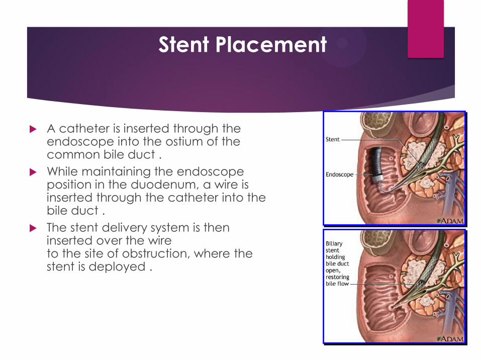
Stent Placement
The Endoscope is
positioned in the
duodenum at the
opening of the bile
duct.
Stent Placement
A catheter is inserted through the endoscope into the ostium of the common bile duct .
While maintaining the endoscope position in the duodenum, a wire is inserted through the catheter into the bile duct .
The stent delivery system is then inserted over the wire to the site of obstruction, where the stent is deployed .

PTC
For biliary stent placement using a
percutaneous approach:
A fine needle is inserted between the 4th and
5th rib on the patient’s right side
The puncture is through the liver
The needle is inserted into an intrahepatic
duct under image guidance.
Photo on file at Medtronic

Complication History Examination Blood tests Biliary Colic - Intermittent RUQ/epigastric
pain (minutes/hours) into
back or right shoulder
- N&V
-Tender RUQ
-No peritonism
-Murphy’s –
-Apyrexial, HR and BP (N)
-WCC (N) CRP (N)
- LFT (N)
Acute Cholecystitis -Constant RUQ pain into back
or right shoulder
-N&V
-Feverish
-Tender RUQ
-Periotnism RUQ
(guarding/rebound)
-Murphy’s +
-Pyrexia, HR (↑)
-WCC and CRP (↑)
-LFT (N or mildly (↑)
Empyema -Constant RUQ pain into back
or right shoulder
-N&V
-Feverish
-Tender RUQ
-Peritonism RUQ
-Murphy’s +
-Pyrexia, HR (↑), BP (↔ or ↓)
-More septic than acute
cholecystitis
-WCC and CRP (↑)
-LFT (N or mildly (↑)
Obstructive Jaundice -Yellow discolouration
-Pale stool, dark urine
-painless or assocaited with
mild RUQ pain
-Jaundiced
-Non-tender or minimally tender
RUQ
-No peritonism
-Murphy’s –
-Apyrexial, HR and BP (N)
-WCC and CRP (N)
-LFT: obstructive pattern bili
(↑), ALP (↑), GGT (↑), ALT/AST
(↔)
-INR (↔ or ↑)
Ascending Cholangitis Becks triad
-RUQ pain (constant)
-Jaundice
-Rigors
-Jaundiced
-Tender RUQ
-Peritonism RUQ
-Spiking high pyrexia (38-39)
-HR (↑), BP (↔ or ↓)
-Can develop septic shock
-WCC and CRP (↑)
-LFT : obstructive pattern bili
(↑), ALP (↑), GGT (↑), ALT/AST
(↔)
-INR (↔ or ↑)
Acute Pancreatitis -Severe upper abdominal
pain (constant) into back
-Profuse vomiting
-Tender upper abdomen
-Upper abdominal or
generalised peritonism
-Usually apyrexial, HR (↑), BP (↔
or ↓)
-WCC and CRP (↑)
-LFT: (N) if passed stone or
obstructive pattern ifstone
still in CBD
-Amylase (↑)
-INR/APTT (N) or (↑) if DIC
Gallstone Ileus - 4 cardinal features of SBO -distended tympanic abdomen
-hyperactive/tinkling bowel
sounds

Imagings for gallstone disease
USS: first line investigation in gallstone disease
Confirms presence of gallstones
Gall bladder wall thickness (if thickened suggests cholecystitis)
Biliary tree calibre (CBD/extrahepatic/intrahepatic) – if dilated suggests stone in CBD (normal CBD <8mm).
Sometimes CBD stone can be seen.
MRCP: To visualise biliary tree accurately (much more accurate than USS)
Diagnostic only but non-invasive
CT: Not first line investigation. Mainly used if suspicion of gallbladder empyema, gangrene, or perforation and in acute pancreatitis

Cholecystectomy
Asymptomatic gallstones do not require operation
Indications
A single complication of gallstones is an indication for
cholecystectomy (this includes biliary colic)
After a single complication risk of recurrent complications
is high (and some of these can be life threatening e.g.
cholangitis, pancreatitis)
Whilst awaiting laparoscopic cholecystectomy
Low fat diet
Dissolution therapy (ursodeoxycholic acid) generally
useless

Cholecystectomy
All performed laparoscopically
Advantages:
Less post-op pain
Shorter hospital stay
Quicker return to normal activities
Timing
Early
After acute cholecystitis, cholecystectomy traditionally performed after 6 weeks [not any more recommended]
After gallstone pancreatitis cholecyctectomy should be performed within 3 weeks.

Cholecystectomy when to perform?
Arguments for 6 weeks later
Laparoscopic dissection more difficult when acutely inflammed
Surgery not optimal when patient septic/dehydrated
Logistical difficulties (theatre space, lack of surgeons)
Arguments for same admission
Research suggests same admission lap chole as safe as elective chole (conversion to open maybe higher)
Waiting increases risk of further attacks/complications which can be life threatening
Risk of failure of conservative management and development of dangerous complication such as empyema, gangrene and perforation can be avoided
National guidelines state

The End QUESTIONS?