bilirubin-induced neurotoxicity and environmental impacts
TRANSCRIPT
Research Article Open Access
Volume 6 • Issue 6 • 1000414J Environ Anal Toxicol, an open access journalISSN: 2161-0525
Open AccessReview Article
Journal ofEnvironmental & Analytical Toxicology
Jour
nal o
f Env
ironmental & Analytical Toxicology
ISSN: 2161-0525
Yueh et al., J Environ Anal Toxicol 2016, 6:6DOI: 10.4172/2161-0525.1000414
Keywords: Hyperbilirubinemia; Neurotoxicity; Total serum bilirubin; UDP-glucuronosyltransferase
IntroductionNewborn hyperbilirubinemia or jaundice, characterized by elevated
levels of total serum bilirubin (TSB), is one of the most common clinical diagnoses in neonates, especially in preterm babies. Hyperbilirubinemia in neonates is caused by a combination of a burst in rapid production and then turnover of erythrocytes by the reticuloendothelial system immediately after birth and a delayed expression of hepatic UDP-glucuronosyltransferase 1A1 (UGT1A1) (Figure 1), the sole enzyme responsible for bilirubin elimination through glucuronidation conjugation [1,2]. More than 60% of otherwise healthy newborns develop hyperbilirubinemia during the first week of life with a majority of them experiencing temporary, physiological jaundice that has benign outcomes. A small portion of neonates, however, suffer dangerously high levels of unconjugated bilirubin (UCB) that are associated with bilirubin-induced neurologic dysfunction (BIND) [3]. The severe form of BIND results in encephalopathy termed kernicterus, an anatomic characteristic of yellow staining in certain regions of the brain, particularly the basal ganglia, hippocampus, cerebellum, and nuclei of the floor of the fourth ventricle [3,4]. Clinically, kernicterus is manifested as lethargy, ocular muscle paralysis, high-pitched crying, dystonia, seizures, mental retardation, and even death. The debilitating actions of kernicterus are irreversible and the symptoms associated with this condition remain permanent throughout life [5]. These infants (and adults if they survive to reach adulthood) are exposed to the risk of bilirubin encephalopathy and death [6], and survival will be dependent on lifelong phototherapy, which can span 10 – 12 hours daily. Liver transplantation is deemed the only long-lasting therapeutic alternative to cure severe forms of hyperbilirubinemia but generally requires continuous immunosuppression with substantial risks, and hyperbilirubinemia patients may undergo such a procedure only if they are in acute neurological crisis.
In preterm infants with less than 30 weeks of gestational age, the incidence of kernicterus is about 1.8 per 1000 births [7], and it is estimated that the current risk of chronic kernicterus is about one in
seven infants with TSB levels >30 mg/dL (513 µM) [8]. However, in many parts of the world, especially in low-income countries, these ratios would underestimate the incidence of kernicterus because of major risk factors that induce hemolysis such as Rhesus disease and glucose-6-phosphate dehydrogenase deficiency. South Asia and Sub-Saharan Africa have the highest incidence with prevalence estimated at 3.9 per 1000 live births [5,9], and recent global estimates found the prevalence of extreme hyperbilirubinemia (>25 mg/dL) being 4%, 32%, and 39% in Latin America, sub-Saharan Africa, and South Asia, respectively. At a country level, Nigeria was reported to have hyperbilirubinemia cases accounting for over 35% of all hospital admissions, with 9% of the newborns having developed kernicterus [9]. The inability to clear bilirubin in cases of rapid accumulation in the early phases of the neonatal window result from developmental delay or repression in expression of UGT1A1. We predict that improved understanding of the mechanisms behind this delay would become useful as a target to develop treatment approaches that would accelerate bilirubin clearance and eliminate the prospects of neurological toxicity.
We document in this review that the UGT1A1 activity is influenced by genetic polymorphism and is regulated at the transcriptional level through a number of mechanisms during the developmental stage; in addition to genetic elements of the UGT1A1 gene, we focus on the role of environmental factors in modulating UGT1A1 bilirubin conjugation
Bilirubin-Induced Neurotoxicity and Environmental Impacts on Hyperbilirubinemia DevelopmentMei-Fei Yueh*, Shujuan Chen, Nghia Nguyen and Robert H TukeyLaboratory of Environmental Toxicology, Department of Pharmacology, University of California, San Diego, La Jolla, CA 92093, United States of America
AbstractHyperbilirubinemia (also known as jaundice), caused by the accumulation of unconjugated bilirubin, is one of the
most common clinical diagnoses in both premature and term newborns. Owing to the fact that bilirubin is metabolized solely through glucuronidation by UDP-glucuronosyltransferase (UGT) 1A1, it is now known that immaturity of UGT1A1 in combination with overproduction of bilirubin during the developmental stage acts as a bottleneck to bilirubin elimination and predisposes the infant to high TBS levels. While neonatal jaundice is mostly benign, excessively high levels of serum bilirubin in a small percentage of newborns can cause acute or chronic bilirubin-induced neurologic dysfunction (BIND), potentially progressing to acute encephalopathy leading to irreversible brain damage and death. As a series of hereditary UGT1A1 mutations have been identified that are associated with UGT1A1 deficiency, new evidence has verified that developmental expression of UGT1A1 is a tightly controlled event, and both genetic polymorphisms and developmental regulation of UGT1A1 are major contributing factors determining the severity of hyperbilirubinemia and bilirubin-induced neurotoxicity. This review recapitulates the progress that has been made in recent years in understanding etiology and physiopathology of severe hyperbilirubinemia, investigating molecular mechanisms underlying bilirubin-induced encephalopathy, and searching for potential therapies for treating pathologic hyperbilirubinemia. Several animal models have been developed to make it possible to examine bilirubin-induced neurotoxicity from multiple directions. Moreover, environmental factors that may alleviate or worsen the condition of hyperbilirubinemia are discussed.
*Corresponding author: Mei-Fei Yueh, Laboratory of Environmental Toxicology, Department of Pharmacology, University of California, San Diego, La Jolla, CA 92093, United States of America, Tel: 8588221351; E-mail: [email protected]
Received October 19, 2016; Accepted October 28, 2016; Published November 01, 2016
Citation: Yueh MF, Chen S, Nguyen N, Tukey RH (2016) Bilirubin-Induced Neurotoxicity and Environmental Impacts on Hyperbilirubinemia Development. J Environ Anal Toxicol 6: 414. doi: 10.4172/2161-0525.1000414
Copyright: © 2016 Yueh MF, et al. This is an open-access article distributed under the terms of the Creative Commons Attribution License, which permits unrestricted use, distribution, and reproduction in any medium, provided the original author and source are credited.
Citation: Yueh MF, Chen S, Nguyen N, Tukey RH (2016) Bilirubin-Induced Neurotoxicity and Environmental Impacts on Hyperbilirubinemia Development. J Environ Anal Toxicol 6: 414. doi: 10.4172/2161-0525.1000414
Page 2 of 7
Volume 6 • Issue 6 • 1000414J Environ Anal Toxicol, an open access journalISSN: 2161-0525
capacity. Emphasis is also placed on recent data obtained from novel animal models that delineate cellular and molecular events occurring in the brain in response to bilirubin neurotoxicity. Finally, new evidence suggesting that bilirubin metabolism is accomplished by both hepatic and extrahepatic UGT1A1 activities is presented.
UDP-Glucuronosyltransferase 1A1 is the Primary Enzyme Involving Bilirubin Encephalopathy
Under the normal physiological conditions, bilirubin is poorly water-soluble and is therefore required to be metabolized to allow its disposition and excretion. UGTs are a family of member-bound enzymes that catalyze the conjugation of a wide array of xenobiotics and endogenous substrates with glucuronic acid [10]. Of all isoforms, only UGT1A1 has physiological relevance to metabolize bilirubin [11], which is the rate limiting step for bilirubin biliary excretion and detoxification. Clinical data and animal experiments support the fact that regardless of the factors contributing to hyperbilirubinemia, bilirubin detoxification is predominantly determined by regulatory events that control expression of the UGT1A1 gene.
Inherited mutations of the UGT1A1 gene and other contributing factors to hyperbilirubinemia
Congenital inborn errors of the UGT1A1 gene are associated with altered UGT1A1 expression and thereby reduce or completely abolish bilirubin conjugating activity. Over 40 inherited mutations in the UGT1A1 gene are associated with hyperbilirubinemia, and the degree of deficiency of UGT1A1 activity primarily determines the severity of hyperbilirubinemia and encephalopathy [10]. Gilbert syndrome is a mild form of UGT1A1 genetic polymorphism that results in a slight reduction in UGT1A1 activity [12,13], whereas Crigler-Najjar (CN) syndrome exhibits complete abolishment (type I) or severe reduction of UGT1A1 (type II) [14]. A few key mutations in the coding region and the promoter region of the UGT1A1 gene have been discovered in CN patients that are correlated with reduction or elimination of UGT1A1 activity [2,12]. Clinical data showed that untreated babies with CN type I rapidly develop high plasma levels of UCB (20-50 mg/dL), exposing them to the possibility of serious neurological damage.
Mild forms of UGT1A1 mutations result in benign jaundice;
however, when coupling other genetically determined traits, severe hyperbilirubinemia may take place. For example, babies who have hemolytic conditions caused by glucose-6-phosphate dehydrogenase deficiency and Rhesus disease may be predisposed to severe hyperbilirubinemia [5,15]. Expression of P-glycoprotein (P-gp) in the brain has also been reported to be associated with bilirubin neurotoxicity. P-gp is expressed abundantly in brain capillary endothelial cells and astrocytes of the blood-brain barriers and has the ability to transport bilirubin out of the brain across the blood-brain-barrier by acting as a membrane efflux pump [16,17]. Compared with wild type mice, Mdr1a (P-gp encoding gene) null mice had a higher brain bilirubin content, possibly by enhanced brain bilirubin influx, implying that Pgp expression in the blood-barrier plays a role in protecting the CNS against bilirubin neurotoxicity [16,17]. In addition to the aforementioned genetic factors, prematurity, concurrent illness, and interventions that impede bilirubin-albumin binding are also considered to be risk factors for severe hyperbilirubinemia [8].
Experimental models established for studying neonatal hyperbilirubinemia and regulation of UGT1A1
BIND is characterized by a wide range of neurological deficits, and the underlying molecular mechanisms are only starting to emerge as a number of animal models producing the neonatal hyperbilirubinemia condition have been developed in the past few years. The majority of hyperbilirubinemia animal models harbor UGT1A1 mutations that occur naturally or are a result of genetic manipulations.
Gunn rats: Gunn [18] discovered that mutant Wistar rats carrying a premature stop codon in the Ugt1a1 gene and exhibiting a very low UGT1A1 activity developed jaundice. These spontaneously jaundiced rats, termed Gunn rats, are deemed to be the first hyperbilirubinemia animal model, which mimics the condition of CN syndrome type 1. Since then, many studies have employed Gunn rats in combination with administering sulfadimethoxine (displacing unconjugated bilirubin from albumin) or phenylhydrazine (inducing hemolysis) to examine acute bilirubin encephalopathy [19,20].
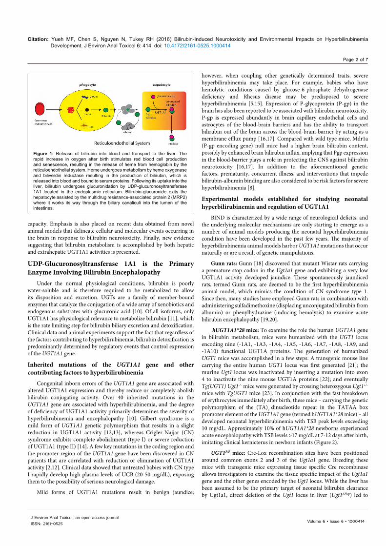
hUGT1A1*28 mice: To examine the role the human UGT1A1 gene in bilirubin metabolism, mice were humanized with the UGT1 locus encoding nine (-1A1, -1A3, -1A4, -1A5, -1A6, -1A7, -1A8, -1A9, and -1A10) functional UGT1A proteins. The generation of humanized UGT1 mice was accomplished in a few steps: A transgenic mouse line carrying the entire human UGT1 locus was first generated [21]; the murine Ugt1 locus was inactivated by inserting a mutation into exon 4 to inactivate the nine mouse UGT1A proteins [22]; and eventually Tg(UGT1) Ugt1-/- mice were generated by crossing heterozygous Ugt1+/- mice with TgUGT1 mice [23]. In conjunction with the fast breakdown of erythrocytes immediately after birth, these mice – carrying the genetic polymorphism of the (TA)7 dinucleotide repeat in the TATAA box promoter element of the UGT1A1 gene (termed hUGT1A1*28 mice) – all developed neonatal hyperbilirubinemia with TSB peak levels exceeding 10 mg/dL. Approximately 10% of hUGT1A1*28 newborns experienced acute encephalopathy with TSB levels >17 mg/dL at 7-12 days after birth, imitating clinical kernicterus in newborn infants (Figure 2).
UGT1F/F mice: Cre-Lox recombination sites have been positioned around common exons 2 and 3 of the Ugt1a1 gene. Breeding these mice with transgenic mice expressing tissue specific Cre recombinase allows investigators to examine the tissue specific impact of the Ugt1a1 gene and the other genes encoded by the Ugt1 locus. While the liver has been assumed to be the primary target of neonatal bilirubin clearance by Ugt1a1, direct deletion of the Ugt1 locus in liver (Ugt1ΔHep) led to
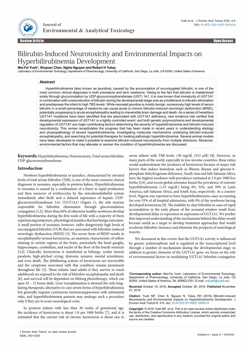
Figure 1: Release of bilirubin into blood and transport to the liver. The rapid increase in oxygen after birth stimulates red blood cell production and senescence, resulting in the release of heme from hemoglobin by the reticuloendothelial system. Heme undergoes metabolism by heme oxygenase and biliverdin reductase resulting in the production of bilirubin, which is released into blood and bound to serum proteins. Following its uptake into the liver, bilirubin undergoes glucuronidation by UDP-glucuronosyltransferase 1A1 located in the endoplasmic reticulum. Bilirubin-glucuronide exits the hepatocyte assisted by the multidrug resistance-associated protein 2 (MRP2) where it works its way through the biliary canaliculi into the lumen of the intestines.
Citation: Yueh MF, Chen S, Nguyen N, Tukey RH (2016) Bilirubin-Induced Neurotoxicity and Environmental Impacts on Hyperbilirubinemia Development. J Environ Anal Toxicol 6: 414. doi: 10.4172/2161-0525.1000414
Page 3 of 7
Volume 6 • Issue 6 • 1000414J Environ Anal Toxicol, an open access journalISSN: 2161-0525
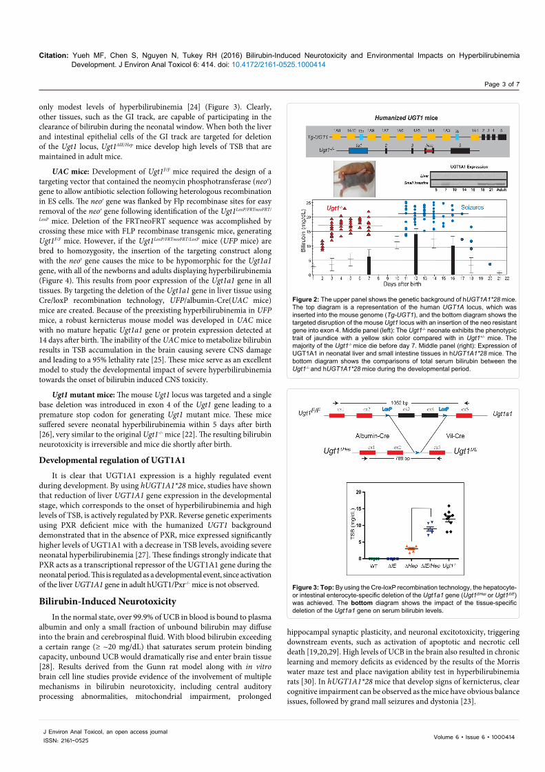
only modest levels of hyperbilirubinemia [24] (Figure 3). Clearly, other tissues, such as the GI track, are capable of participating in the clearance of bilirubin during the neonatal window. When both the liver and intestinal epithelial cells of the GI track are targeted for deletion of the Ugt1 locus, Ugt1ΔIE/Hep mice develop high levels of TSB that are maintained in adult mice.
UAC mice: Development of Ugt1F/F mice required the design of a targeting vector that contained the neomycin phosphotransferase (neor) gene to allow antibiotic selection following heterologous recombination in ES cells. The neor gene was flanked by Flp recombinase sites for easy removal of the neor gene following identification of the Ugt1LoxP/FRTneoFRT/
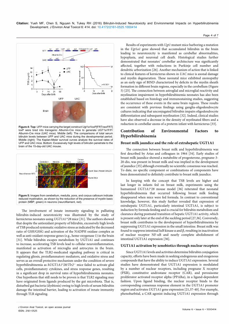
LoxP mice. Deletion of the FRTneoFRT sequence was accomplished by crossing these mice with FLP recombinase transgenic mice, generating Ugt1F/F mice. However, if the Ugt1LoxP/FRTneoFRT/LoxP mice (UFP mice) are bred to homozygosity, the insertion of the targeting construct along with the neor gene causes the mice to be hypomorphic for the Ugt1a1 gene, with all of the newborns and adults displaying hyperbilirubinemia (Figure 4). This results from poor expression of the Ugt1a1 gene in all tissues. By targeting the deletion of the Ugt1a1 gene in liver tissue using Cre/loxP recombination technology, UFP/albumin-Cre(UAC mice) mice are created. Because of the preexisting hyperbilirubinemia in UFP mice, a robust kernicterus mouse model was developed in UAC mice with no mature hepatic Ugt1a1 gene or protein expression detected at 14 days after birth. The inability of the UAC mice to metabolize bilirubin results in TSB accumulation in the brain causing severe CNS damage and leading to a 95% lethality rate [25]. These mice serve as an excellent model to study the developmental impact of severe hyperbilirubinemia towards the onset of bilirubin induced CNS toxicity.
Ugt1 mutant mice: The mouse Ugt1 locus was targeted and a single base deletion was introduced in exon 4 of the Ugt1 gene leading to a premature stop codon for generating Ugt1 mutant mice. These mice suffered severe neonatal hyperbilirubinemia within 5 days after birth [26], very similar to the original Ugt1-/- mice [22]. The resulting bilirubin neurotoxicity is irreversible and mice die shortly after birth.
Developmental regulation of UGT1A1
It is clear that UGT1A1 expression is a highly regulated event during development. By using hUGT1A1*28 mice, studies have shown that reduction of liver UGT1A1 gene expression in the developmental stage, which corresponds to the onset of hyperbilirubinemia and high levels of TSB, is actively regulated by PXR. Reverse genetic experiments using PXR deficient mice with the humanized UGT1 background demonstrated that in the absence of PXR, mice expressed significantly higher levels of UGT1A1 with a decrease in TSB levels, avoiding severe neonatal hyperbilirubinemia [27]. These findings strongly indicate that PXR acts as a transcriptional repressor of the UGT1A1 gene during the neonatal period. This is regulated as a developmental event, since activation of the liver UGT1A1 gene in adult hUGT1/Pxr-/- mice is not observed.
Bilirubin-Induced NeurotoxicityIn the normal state, over 99.9% of UCB in blood is bound to plasma
albumin and only a small fraction of unbound bilirubin may diffuse into the brain and cerebrospinal fluid. With blood bilirubin exceeding a certain range (≥ ~20 mg/dL) that saturates serum protein binding capacity, unbound UCB would dramatically rise and enter brain tissue [28]. Results derived from the Gunn rat model along with in vitro brain cell line studies provide evidence of the involvement of multiple mechanisms in bilirubin neurotoxicity, including central auditory processing abnormalities, mitochondrial impairment, prolonged
Figure 2: The upper panel shows the genetic background of hUGT1A1*28 mice. The top diagram is a representation of the human UGT1A locus, which was inserted into the mouse genome (Tg-UGT1), and the bottom diagram shows the targeted disruption of the mouse Ugt1 locus with an insertion of the neo resistant gene into exon 4. Middle panel (left): The Ugt1-/- neonate exhibits the phenotypic trait of jaundice with a yellow skin color compared with in Ugt1+/- mice. The majority of the Ugt1-/- mice die before day 7. Middle panel (right): Expression of UGT1A1 in neonatal liver and small intestine tissues in hUGT1A1*28 mice. The bottom diagram shows the comparisons of total serum bilirubin between the Ugt1-/- and hUGT1A1*28 mice during the developmental period.
Figure 3: Top: By using the Cre-loxP recombination technology, the hepatocyte- or intestinal enterocyte-specific deletion of the Ugt1a1 gene (Ugt1ΔHep or Ugt1ΔIE) was achieved. The bottom diagram shows the impact of the tissue-specific deletion of the Ugt1a1 gene on serum bilirubin levels.
hippocampal synaptic plasticity, and neuronal excitotoxicity, triggering downstream events, such as activation of apoptotic and necrotic cell death [19,20,29]. High levels of UCB in the brain also resulted in chronic learning and memory deficits as evidenced by the results of the Morris water maze test and place navigation ability test in hyperbilirubinemia rats [30]. In hUGT1A1*28 mice that develop signs of kernicterus, clear cognitive impairment can be observed as the mice have obvious balance issues, followed by grand mall seizures and dystonia [23].
Citation: Yueh MF, Chen S, Nguyen N, Tukey RH (2016) Bilirubin-Induced Neurotoxicity and Environmental Impacts on Hyperbilirubinemia Development. J Environ Anal Toxicol 6: 414. doi: 10.4172/2161-0525.1000414
Page 4 of 7
Volume 6 • Issue 6 • 1000414J Environ Anal Toxicol, an open access journalISSN: 2161-0525
Results of experiments with Ugt1 mutant mice harboring a mutation in the Ugt1a1 gene showed that accumulated bilirubin in the brain leading to neurotoxicity is manifested as cerebellar abnormalities, hypoplasia, and neuronal cell death. Histological studies further demonstrated that neonates’ cerebellar architecture was significantly affected, together with reductions in Purkinje cell number and dendritic arborization [26]. Another mechanism of action that is linked to clinical features of kernicterus shown in UAC mice is axonal damage and myelin degeneration. These neonatal mice exhibited axonopathy as an early sign of BIND characterized by deficits in the myelin sheath formation in different brain regions, especially in the cerebellum (Figure 5) [25]. The connection between astroglial and microglial reactivity and myelination impairment in hyperbilirubinemia neonates has also been established based on histology and immunostaining studies, suggesting the occurrence of these events in the same brain regions. These results are consistent with previous findings using ganglia-oligodendrocyte cultures indicating that unconjugated bilirubin impairs oligodendrocyte differentiation and subsequent myelination [32]. Indeed, clinical studies have also observed a decrease in the density of myelinated fibers and a reduction in cerebellar axons of a preterm infant with kernicterus [33].
Contribution of Environmental Factors to HyperbilirubinemiaBreast milk jaundice and the role of extrahepatic UGT1A1
The connection between breast milk and hyperbilirubinemia was first described by Arias and colleagues in 1964 [34]. Early studies of breast milk jaundice showed a metabolite of progesterone, pregnane-3-20-dio, was present in breast milk and was implied in the development of jaundice [35] although eventually no scientific consensus was reached. To date, no specific component or combinations of components have been demonstrated to definitely contribute to breast milk jaundice.
In keeping with the concept that TSB levels are higher and last longer in infants fed on breast milk, experiments using the humanized UGT1A1*28 mouse model [36] reiterated that neonatal hyperbilirubinemia that occurred following breast milk feeding disappeared when mice were fed formula. In contrast to conventional knowledge, however, this study further revealed that expression of extrahepatic UGT1A1, particularly intestinal UGT1A1, is subject to induction by formula feeding and is crucial for bilirubin metabolism and clearance during postnatal transition of hepatic UGT1A1 activity, which is present only later at the end of the suckling period [27,36]. Conversely, breast milk contributes to the development of hyperbilirubinemia by suppressing UGT1A1 expression in the small intestine. Breast milk was found to suppress intestinal IĸB kinase α and β, resulting in inactivation of nuclear receptor NF-ĸB and nearly complete abolishment of intestinal UGT1A1 expression [36].
UGT1A1 activation by xenobiotics through nuclear receptors
Since UGT1A1 levels and activities determine bilirubin conjugation capacity, efforts have been made in seeking endogenous and exogenous compounds that have the ability to induce UGT1A1 expression. Several studies have demonstrated that UGT1A1 expression is modulated by a number of nuclear receptors, including pregnane X receptor (PXR), constitutive androstane receptor (CAR), and peroxisome proliferator activated receptor alpha (PPARα), in a ligand-dependent fashion. Upon ligand binding, the nuclear receptor binds to the corresponding consensus response element in the UGT1A1 promoter region and activates UGT1A1 gene expression [21,37-40]. For example, phenobarbital, a CAR agonist inducing UGT1A1 expression through
Figure 4: Top: UFP mice carrying the target construct Ugt1a1loxP[FRTneoFRT]loxP were bred into transgenic Albumin-Cre mice to generate UGT1a1F/F/Albumin-Cre mice (UAC mice). Middle (left): The comparisons of total serum bilirubin levels between UFP and UAC mice during the developmental period. Middle (right): The Kaplan-Meier survival curves analyze the survival rates of UFP and UAC mice. Bottom: Excessively high levels of bilirubin penetrate to the brain of the 15-day-old UAC mouse.
Figure 5: Images from cerebellum, medulla, pons, and corpus callosum indicate reduced myelination, as shown by the reduction of the presence of myelin basic protein (MBP, green) in neurons (neurofilament, red).
The involvement of innate immunity signaling in palliating bilirubin-induced neurotoxicity was illustrated by the study of kernicterus neonates using UGT1A1*28 mice [31]. The authors showed that despite the antioxidant property of bilirubin, excessively high levels of TSB produced systematic oxidative stress as indicated by the decreased ratio of GSH/GSSG and activation of the NADPH oxidase complex as well as anti-oxidant response genes (e.g., heme-oxygenase 1) in the brain [31]. While bilirubin escapes metabolism by UGT1A1 and continues to increase, accelerating TSB levels lead to cellular neuroinflammation, manifested as activation of microglia and astrocytes in the brain. It appears that the TLR2-medicated signaling pathway is critical in regulating gliosis, proinflammatory mediators, and oxidative stress and serves as an overall protective mechanism under the condition of severe hyperbilirubinemia as hUGT1A1*28/Tlr2-/- mice failed to activate glial cells, proinflammatory cytokines, and stress response genes, resulting in a significant drop in survival rates of hyperbilirubinemia neonates. One hypothesis that still needs to be proven is that TLR2 signaling may have originated from ligands produced by the gut microbiota. That is, disturbed gut bacteria (dysbiosis) owing to high levels of serum bilirubin damage the intestinal barrier, leading to activation of innate immunity through TLR signaling.
Citation: Yueh MF, Chen S, Nguyen N, Tukey RH (2016) Bilirubin-Induced Neurotoxicity and Environmental Impacts on Hyperbilirubinemia Development. J Environ Anal Toxicol 6: 414. doi: 10.4172/2161-0525.1000414
Page 5 of 7
Volume 6 • Issue 6 • 1000414J Environ Anal Toxicol, an open access journalISSN: 2161-0525
interacting with a phenobarbital responsive element (PBREM), has been used clinically in conjunction with phototherapy for the treatment of severe jaundice in infants to enhance bilirubin metabolism, thus reducing the need for exchange transfusion [41,42]. PBREM involvement in controlling UGT1A1 expression is also supported by a study linking polymorphism of the UGT1A1 PBREM (T-3279G) to an increased risk of hyperbilirubinemia [43]. Glucocorticoids have also been used to treat hyperbilirubinemia: Dexamethasone-treated infants experienced lower incidence of hyperbilirubinemia than the untreated controls [44]. Studies in mice with neonatal hyperbilirubinemia indicated that PXR serves as a key regulator following glucocorticoid treatment by inducing liver UGT1A1 expression and reducing TSB levels [27].
Modulation of UGT1A1 expression by environmental chemicals
When hyperbilirubinemia neonatal mice were exposed to the environmental chemicals arsenic and cadmium, their TSB levels unexpectedly decreased, correlated with elevated levels of intestinal UGT1A1 expression with no detectable changes in expression of hepatic UGT1A1. Gene expression profiling data and biochemical studies revealed that, as potent inducers of oxidative stress, arsenic and cadmium alter the redox state of the intestines, leading to induction of UGT1A1 and a dramatic reduction in TSB levels [45]. These results suggest that modulation of intestinal UGT1A1 activity by initiating the oxidative stress signaling pathway may be an unconventional alternative to lower TSB and improve hyperbilirubinemia.
Alternative Approaches to Treat HyperbilirubinemiaHepatocyte transplantation and gene transfer therapy
As evidence indicated that only ~5% of normal UGT1A1 activity is adequate to significantly lower the plasma bilirubin concentration and eliminate the risk of kernicterus [46], alternative therapies intending to alleviate hyperbilirubinemia with persistent expression of the UGT1A1 enzyme are underway in the experimental stage. A recent study illustrated that the advantage of neonatal hepatocytes over adult hepatocytes lies in the fact that neonatal hepatocytes exhibit better engraftment and repopulation capacity after transplantation, thus resulting in better bilirubin clearance in icteric Gunn rats [47]. Significant progress has also been made by means of gene therapy in past decades using adenovirus-based or similar techniques or the correction of UGT1A1 gene defects with the site-directed gene repair approach to treat hyperbilirubinemia animals [48-51]. A gene therapy study showed that a single injection of a helper-dependent adenoviral vector expressing UGT1A1 that specifically targets the liver tissue can completely correct hereditary hyperbilirubinemia in Gunn rats with long-lasting effects and low-chronic toxicity [52].
Administration of albumin
Due to the high affinity of albumin to bilirubin, in a normal state, UCB is bound to albumin following transportation through the circulation to the liver [28]. When UCB levels exceed the capacity of albumin, free bilirubin is capable of crossing the blood-brain-barrier and accumulating in the brain. Therefore, a potential approach to prevent bilirubin accumulation in the brain is to increase bilirubin binding capacity by albumin supplementation. When hyperbilirubinemia neonatal mice carrying inherited mutations of Ugt1a1 were subject to daily albumin infusion, they were rescued from neurological damage and lethality. By increasing plasma bilirubin-binding capacity, albumin
mobilizes bilirubin from tissues to plasma and results in reduced systemic plasma bilirubin levels [53].
Regardless of efficacy of these alternative treatments, they are still in the experimental stage and clinical trials are apparently needed to evaluate the acute toxicity, immunogenic responses, and long-term safety profile before they can be applied in the market to humans.
Develop therapeutics targeted to induce UGT1A1 gene expression
The use of animal models, such as the humanized UGT1A1*28 mice, helps define the mechanisms that control neonatal hyperbilirubinemia and provides an important venue to exploit the impact of safe and therapeutic chemicals to regulate the UGT1A1 gene and lower TSB levels. These non-invasive approaches could take advantage of drug delivery directly to newborns or alternatively by lactation following drug administration to nursing mothers. In vivo studies with humanized UGT1A1*28 mice can directly exploit tissue specific contributions, such as the liver and gastrointestinal tract, that direct bilirubin clearance, while also being able to examine pharmacokinetics parameters of the inducing agents.
SummarySevere neonatal hyperbilirubinemia and acute kernicterus remain
a clinical emergency. It is clear that UGT1A1 bilirubin conjugating capacity (hence the ability to excrete bilirubin) is the primary factor determining the severity of hyperbilirubinemia along with other risk factors such as hemolysis and prematuration, and UGT1A1 levels can be regulated by environmental and dietary compounds through activation of nuclear receptors or alteration of oxidative-stress status. While the complex cascade of molecular and cellular events leading to bilirubin-induced neurotoxicity and kernicterus remains incompletely delineated, emerging evidence indicates that high levels of TSB activate innate immunity and cause myelination impairment. As we have learned more about bilirubin metabolism and neurologic injury with the advent of novel toxicology models, some of the conventional knowledge regarding hyperbilirubinemia is now being challenged: Intestinal UGT1A1 is subject to the regulation of breast milk and environmental compounds and plays a critical role during the developmental stage when expression of hepatic UGT1A1 is delayed.
Acknowledgements
The writing of this review was supported in part by Public Health Service Grants ES010337, GM086713, and GM100481 (to RHT), R21ES024818 (to SC) and R21ES023906 (to M-F Y).
References
1. Burchell B, Coughtrie M, Jackson M, Harding D, Fournel-Gigleux S, et al. (1989) Development of human liver UDP-glucuronosyltransferases. Dev Pharmacol Ther 13: 70-77.
2. Fujiwara R, Maruo Y, Chen S, Tukey RH (2015) Role of extrahepatic UDP-glucuronosyltransferase 1A1: Advances in understanding breast milk-induced neonatal hyperbilirubinemia. Toxicol Appl Pharmacol 289: 124-132.
3. Johnson L, Bhutani VK (2011) The clinical syndrome of bilirubin-induced neurologic dysfunction. In Seminars in perinatology 35: 101-113.
4. Shapiro SM, Bhutani VK, Johnson L (2006) Hyperbilirubinemia and kernicterus. Clin Perinatol 33: 387-410.
5. Bhutani VK, Zipursky A, Blencowe H, Khanna R, Sgro M, et al. (2013) Neonatal hyperbilirubinemia and Rhesus disease of the newborn: incidence and impairment estimates for 2010 at regional and global levels. Pediatr Res 74 Suppl 1: 86-100.

Citation: Yueh MF, Chen S, Nguyen N, Tukey RH (2016) Bilirubin-Induced Neurotoxicity and Environmental Impacts on Hyperbilirubinemia Development. J Environ Anal Toxicol 6: 414. doi: 10.4172/2161-0525.1000414
Page 6 of 7
Volume 6 • Issue 6 • 1000414J Environ Anal Toxicol, an open access journalISSN: 2161-0525
6. Crigler JF Jr, Najjar VA (1952) Congenital familial nonhemolytic jaundice with kernicterus; a new clinical entity. AMA Am J Dis Child 83: 259-260.
7. Morioka I, Nakamura H, Koda T, et al. (2015) Current incidence of clinical kernicterus in preterm infants in Japan. Pediatr Int 57: 494-497.
8. Bhutani VK, Johnson L (2009) Kernicterus in the 21st century: frequently asked questions. J Perinatol 29 Suppl 1: S20-24.
9. Chime HE, Egenede JA, Arute JE (2011) Prevalence of Neonatal Jaundice on Central Hospital, Warri, Delta State, Nigeria. International journal of health research 4: 123-126.
10. Tukey RH, Strassburg CP (2000) Human UDP-glucuronosyltransferases: metabolism, expression, and disease. Annu Rev Pharmacol Toxicol 40: 581-616.
11. Bosma PJ, Seppen J, Goldhoorn B, Bakker C, Oude Elferink RP, et al. (1994) Bilirubin UDP-glucuronosyltransferase 1 is the only relevant bilirubin glucuronidating isoform in man. J Biol Chem 269: 17960-17964.
12. Kadakol A, Ghosh SS, Sappal BS, Sharma G, Chowdhury JR, et al. (2000) Genetic lesions of bilirubin uridine-diphosphoglucuronate glucuronosyltransferase (UGT1A1) causing Crigler-Najjar and Gilbert syndromes: correlation of genotype to phenotype. Hum Mutat 16: 297-306.
13. Strassburg CP (2008) Pharmacogenetics of Gilbert's syndrome. Pharmacogenomics 9: 703-715.
14. Ciotti M, Obaray R, Martin MG, Owens IS (1997) Genetic defects at the UGT1 locus associated with Crigler-Najjar type I disease, including a prenatal diagnosis. American journal of medical genetics 68: 173-178.
15. Huang CS, Huang MJ, Lin MS, Yang SS, Teng HC, et al. (2005) Genetic factors related to unconjugated hyperbilirubinemia amongst adults. Pharmacogenetics and genomics 15: 43-50.
16. Watchko JF, Daood MJ, Hansen TW (1998) Brain bilirubin content is increased in P-glycoprotein-deficient transgenic null mutant mice. Pediatr Res 44: 763-766.
17. Watchko JF, Daood MJ, Mahmood B, Vats K, Hart C, et al. (2001) P-glycoprotein and bilirubin disposition. J Perinatol 21 Suppl 1: S43-47.
18. Gunn CH (1938) Hereditary acholuric jaundice in a new mutant strain of rats. Journal of Heredity 29: 137-139.
19. Shapiro SM (1988) Acute brainstem auditory evoked potential abnormalities in jaundiced Gunn rats given sulfonamide. Pediatr Res 23: 306-310.
20. Rice AC, Shapiro SM (2008) A new animal model of hemolytic hyperbilirubinemia-induced bilirubin encephalopathy (kernicterus). Pediatric research 64: 265-269.
21. Chen S, Beaton D, Nguyen N, Senekeo-Effenberger K, Brace-Sinnokrak E, et al. (2005) Tissue-specific, inducible, and hormonal control of the human UDP-glucuronosyltransferase-1 (UGT1) locus. J Biol Chem 280: 37547-37557.
22. Nguyen N, Bonzo JA, Chen S, Chouinard S, Kelner MJ, et al. (2008) Disruption of the ugt1 locus in mice resembles human Crigler-Najjar type I disease. J Biol Chem 283: 7901-7911.
23. Fujiwara R, Nguyen N, Chen S, Tukey RH (2010) Developmental hyperbilirubinemia and CNS toxicity in mice humanized with the UDP glucuronosyltransferase 1 (UGT1) locus. Proc Natl Acad Sci USA 107: 5024-5029.
24. Chen S, Yueh MF, Bigo C, Barbier O, Wang K, et al. (2013) Intestinal glucuronidation protects against chemotherapy-induced toxicity by irinotecan (CPT-11). Proc Natl Acad Sci USA 110: 19143-19148.
25. Barateiro A, Chen S, Yueh MF, Fernandes A, Domingues HS, et al. (2016) Reduced Myelination and Increased Glia Reactivity Resulting from Severe Neonatal Hyperbilirubinemia. Mol Pharmacol 89: 84-93.
26. Bortolussi G, Codarin E, Antoniali G, Vascotto C, Vodret S, et al. (2015) Impairment of enzymatic antioxidant defenses is associated with bilirubin-induced neuronal cell death in the cerebellum of Ugt1 KO mice. Cell Death Dis 6: e1739.
27. Chen S, Yueh MF, Evans RM, Tukey RH (2012) Pregnane‐x‐receptor controls hepatic glucuronidation during pregnancy and neonatal development in humanized UGT1 mice. Hepatology 56: 658-667.
28. Ostrow JD, Mukerjee P, Tiribelli C (1994) Structure and binding of unconjugated bilirubin: relevance for physiological and pathophysiological function. J Lipid Res 35: 1715-1737.
29. Chang FY, Lee CC, Huang CC, Hsu KS (2009) Unconjugated bilirubin exposure impairs hippocampal long-term synaptic plasticity. PLoS One 4: e5876.
30. Song S, Hu Y, Gu X, Si F, Hua Z (2014) A novel newborn rat kernicterus model created by injecting a bilirubin solution into the cisterna magna. PLoS One 9: e96171.
31. Yueh MF, Chen S, Nguyen N, Tukey RH (2014) Developmental onset of bilirubin-induced neurotoxicity involves Toll-like receptor 2-dependent signaling in humanized UDP-glucuronosyltransferase1 mice. J Biol Chem 289: 4699-4709.
32. Barateiro A, Miron VE, Santos SD, Relvas JB, Fernandes A, et al. (2013) Unconjugated bilirubin restricts oligodendrocyte differentiation and axonal myelination. Molecular neurobiology 47: 632-644.
33. Brites D (2012) The evolving landscape of neurotoxicity by unconjugated bilirubin: role of glial cells and inflammation. Front Pharmacol 3: 88.
34. Arias IM, Gartner LM, Seifter S, Furman M (1964) Prolonged neonatal unconjugated hyperbilirubinemia associated with breast feeding and a steroid, pregnane-3 (alpha), 20 (beta)-diol, in maternal milk that inhibits glucuronide formation in vitro. Journal of clinical investigation 43: 2037.
35. Hargreaves T, Piper RF (1971) Breast milk jaundice. Effect of inhibitory breast milk and 3 alpha, 20 abeta-pregnanediol on glucuronyl transferase. Arch Dis Child 46: 195-198.
36. Fujiwara R, Chen S, Karin M, Tukey RH () Reduced expression of UGT1A1 in intestines of humanized UGT1 mice via inactivation of NF-κB leads to hyperbilirubinemia. Gastroenterology 142: 109-118.
37. Yueh MF, Huang YH, Hiller A, Chen S, Nguyen N, et al. (2003) Involvement of the xenobiotic response element (XRE) in Ah receptor-mediated induction of human UDP-glucuronosyltransferase 1A1. Journal of Biological Chemistry 278: 15001-15006.
38. Xie W, Yeuh MF, Radominska-Pandya A, Saini SP, Negishi Y, et al. (2003) Control of steroid, heme, and carcinogen metabolism by nuclear pregnane X receptor and constitutive androstane receptor. Proceedings of the National Academy of Sciences 100: 4150-4155.
39. Senekeo-Effenberger K, Chen S, Brace-Sinnokrak E, Bonzo JA, Yueh MF, et al. (2007) Expression of the human UGT1 locus in transgenic mice by 4-chloro-6-(2, 3-xylidino)-2-pyrimidinylthioacetic acid (WY-14643) and implications on drug metabolism through peroxisome proliferator-activated receptor α activation. Drug Metabolism and Disposition 35: 419-427.
40. Yueh MF, Tukey RH (2007) Nrf2-Keap1 signaling pathway regulates human UGT1A1 expression in vitro and in transgenic UGT1 mice. J Biol Chem 282: 8749-8758.
41. Valaes T, Kipouros K, Petmezaki S, Solman M, Doxiadis SA (1980) Effectiveness and safety of prenatal phenobarbital for the prevention of neonatal jaundice. Pediatr Res 14: 947-952.
42. Murki S, Dutta S, Narang A, Sarkar U, Garewal G (2005) A randomized, triple-blind, placebo-controlled trial of prophylactic oral phenobarbital to reduce the need for phototherapy in G6PD-deficient neonates. Journal of perinatology 25: 325-330.
43. Sugatani J, Yamakawa K, Yoshinari K, Machida T, Takagi H, et al. (2002) Identification of a defect in the UGT1A1 gene promoter and its association with hyperbilirubinemia. Biochem Biophys Res Commun 292: 492-497.
44. Madarek EO, Najati N (2003) The effect of glucocorticoid therapy in preventing early neonatal complications in preterm delivery. J Perinat. Med 31: 441-443.
45. Liu M, Chen S, Yueh MF, Fujiwara R, Konopnicki C, et al. (2016) Cadmium and arsenic override NF-κB developmental regulation of the intestinal UGT1A1 gene and control of hyperbilirubinemia. Biochem Pharmacol 110-111: 37-46.
46. Fox IJ, Chowdhury JR, Kaufman SS, Goertzen TC, Chowdhury NR, et al. (1998) Treatment of the Crigler-Najjar syndrome type I with hepatocyte transplantation. N Engl J Med 338: 1422-1426.
47. Tolosa L, López S, Pareja E, Donato MT, Myara A, et al. (2015) Human neonatal hepatocyte transplantation induces long‐term rescue of unconjugated hyperbilirubinemia in the Gunn rat. Liver Transplantation 21: 801-811.
48. Li Q, Murphree SS, Willer SS, Bolli R, French BA (1998) Gene therapy with bilirubin-UDP-glucuronosyltransferase in the Gunn rat model of Crigler-Najjar syndrome type 1. Hum Gene Ther 9: 497-505.
49. Kren BT, Parashar B, Bandyopadhyay P, Chowdhury NR, Chowdhury JR, et al. (1999) Correction of the UDP-glucuronosyltransferase gene defect in the gunn
Citation: Yueh MF, Chen S, Nguyen N, Tukey RH (2016) Bilirubin-Induced Neurotoxicity and Environmental Impacts on Hyperbilirubinemia Development. J Environ Anal Toxicol 6: 414. doi: 10.4172/2161-0525.1000414
Page 7 of 7
Volume 6 • Issue 6 • 1000414J Environ Anal Toxicol, an open access journalISSN: 2161-0525
rat model of crigler-najjar syndrome type I with a chimeric oligonucleotide. Proc Natl Acad Sci USA 96: 10349-10354.
50. Roy-Chowdhury N, Kadakol A, Sappal BS, Thummala NR, Ghosh SS, et al.(2001) Gene therapy for inherited hyperbilirubinemias. J Perinatol 21 Suppl 1:S114-118.
51. Bellodi‐Privato M, Aubert D, Pichard V, Myara A, Trivin F, et al. (2005)Successful gene therapy of the Gunn rat by in vivo neonatal hepatic genetransfer using murine oncoretroviral vectors. Hepatology 42: 431-438.
52. Toietta G, Mane VP, Norona WS, Finegold MJ, Ng P, et al. (2005) Lifelongelimination of hyperbilirubinemia in the Gunn rat with a single injection ofhelper-dependent adenoviral vector. Proc Natl Acad Sci USA 102: 3930-3935.
53. Vodret S, Bortolussi G, Schreuder AB, Jašprová J, Vitek L, et al. (2015) Albumin administration prevents neurological damage and death in a mouse model ofsevere neonatal hyperbilirubinemia. Scientific reports, p: 5.