bims,pria - eric · bims,pria. tmf01 /pc02 plus postage. "" descriptors *allied...
TRANSCRIPT

DOCUMENT RESUME....,
ED 213 966 CE 031 773
TITLE The' Respiratory System [and) Instructor's Guide: TheRespiratory SyateM, Health Occupations EducationModule: Ingtructional Materials in Anatomy and .
. :
.Physiology for Pennsylvania Health,Occupations
.Programs. .
° INSTITUTION National Evaluation Systems, Inc., Aiherst, Mass. .
SPONS AGENCY Pennsylvania State Dept. of Education, Harrisburg.Bureau of Vocational and Technical Education.
PUB DATE , .Jun 79 .
A
NOTE' 40p.; For related documents see listing in note of CE. 031-758.
i
BIMS,PRIa tmF01 /pc02 Plus Postage. ""
DESCRIPTORS *Allied Hea,lth.Occupations Education; *Anatomy; ,
Behavioral Objectives; *Individualized Instruction;. .
.*Learning Activities; Learning Modules"; -MedicalVocabulary; *Physiology; Postsecondary Education;Pretests Posttests; Programed Instructional.aterials; Secondary Education; Self Evaluation
4 (Individuals); Teaching MethodsIDENTIFIERS Pennsylvania;. *Respiratory System
4
ABSTR4CT,This module on the respiratory system is one of 17
modules,.designed for individualized instruction in health occupationsedudation'programs at bop the secondary and postsecondary levels. Itis part of an eight -unit. miniseries to anatomy and physiology withinthe series of 17 modules. Folloidpg a preface which explainei to the
_student how to. use the module, fHe unit consists of apretest withaAswers, four sections'(ihrfsrmation sheets) with their goals- (e.g., .
identify organs and/or structures related to the lungs), optiongl .
activities <-e.g., discuss how cigarette smoking affects the;structures and function of the lungs), .and posttests., and ,a glossaryof terms. Topids covered in .the unit are introduction 'to theIrespiratory system, the,upper respiratory tract, the lungs, andrespiration. An accompanying inst'ructor'-'s guide contains suggestionsfor using the module and answers to the posttest. (KC)
C
9
4
A" A
9
a 3
****** **********1*****************************************************Reproductiong supplied by'EDRS 'are-the best that can be,made. *
* ,7-' . ^from the original document. *
******************,x************t*************************************,
10

..;
i
*
. I
Q
11
AIEALTH
OCCUPATIONS EDUCATION
MODULE
't.
-.
III
r
-r
a
U S DEPARTMENT OF EDUCATION
NATIO \Al INSTITWIE Or EDUCATION
. `I
-1-6" 4
THE RESPIRATORY SYSTEM
V
0 t
PERMISSION TO REPRODUCE THIS-illMATERIAL HAS BEEN GRANTED BY
/. _ 771
, .0TO THE\ CDUCAT-IONAL RESOURCESINFORMATION CENTER IERICI
,, ))44,4

14
,
.
...,
$ri
i.
.
II
.....F.I.
li
Indtructional Materials in Anatomy and Physiology .
for Pennsylvania.1-lealth Occupations Programs
47.
THE RESPIRATORY SYSTEM
IPrepared for:
"4
Research Coordinating Unit for Vocational Education'
Pennsylvania Department of Educa, ,0 Box 911Harrisburg, pennsylvania 17126°
, ..
.
National Evaluation Systems, Inc.° 30 Gatehouse Road
Amherst, Massachusetts 01002
41,
Prepared by:
4
,
9
June, 1979
-4,
.
I
%
)

(
PREFACE
An understanding of basic hums-anatorny 'and'phySiology is essential to any .
person preparing to enter a health occupation. This instructional unit is designed .
to introduce you to the Structures and functions \of the human respiratory
systemand the interrelationships of\1144jtrwoand to familiarize you with someof the terms and concepts necessary f an uricierstan,ding of 'the respiratory
system.si
This unit consists ,of.a pretest; four .modules with their optional activitiesand post-tests; and a qlosOfy of terms.
A
Begin this 'modular unit by taking the brief pretest at the frOnt-of thebooklet. The pretest is for your use on3.y._to give you an idea of what is included
in this unit, 'and to give yclu an indication of the areas within the unit to Which
you should pay special attention (perhaps by working on the optional'activities)..When .you have completed the pretest, turn to the angwera-In the back (page 29)
to check your own score. You will not be graded on the pretest.
Next, read through each of the modules (Introduction to the RespiratorySystem, Upper Respiratory Tract, The Lungs, and Resrairatiop.Lrd invesygate
any of the optional activities that may be helpful or interesting to you. Theoptional activities will help you learn more about some of the material presented.
At the endo of this unit (page 2') is a glossary which provides you with briefdefinitions of many of the terms used in the modules.
Upon comPfeiion. of each module, you sho.uld be able to demonstrate'
When you have finished a module end feel th you understand the jnformation inrundersynding ofi the material presented, by our performance on the post-test.
that module, take the post-test that follows it. Write down yoar answers on ONE
r piece of paper and pass it in to your instructor, who will give you your grade.
41
t
4

r
-2-
1,- r PRETEST
1., Which of the followingis a part of the respiratory system?
4,
A.- heartB. brainC. lungsD. liver
s
/\ 2. Which of the following is removed from the blood through respiration?
s
A. hemoglobinB. carbon dioxideC. glucoseD. erythrocytes
441
3. The;main function of tthe respiratory system is to provide the blood, with
4:.
During Inspiration, air first passes through,the:
bronchi.alveoli.
C. lungs.D. nose.
-
I
f
.
5. '-The pairs of hollow chambers surrounding the nasal are called .,/
.., .. /
,
' ,V ,....r 4. . ..

-.1 .
1'
,17
4,
6. Whichyfihe following is found in t e laryn
A. vocaLcords
0( B. bronchi "N),
. C. ¢ turbinatesD. sinuses'
"i
4
.
The structures in, the-throat that vibrate to produce'sounds' are the ,
I
:
A ,
.7
11. The `twg passageways -that split from the trachea and lead ,to the lungs' are
called - .. ,
9.. The-pritnary mus e involved in breathing is the:* .
A. .pleura. ti
,B. diaphragm.pharynx.:trachea.
, .
, .
10. Name the two ,Major types of respiration%
e
-

.
IN,TRODUCTION TO THE RESPIRM'ORY SYSTEM
4 Goals
,
r
. . .\
Upon completion of th)s module, you should be'ple to:r.. ..
. 4 .
i. Identify the-components of the respiratory system.°
2.° identify the major.tInctions tlf.therespiratory systems.
j I6 . , .*
. ...:j.sV. .,: g..4.2_MPONEJ \I T S OF TH -RESPIR,4TOR.`k-S*YSTEM,
. 1
4-..) -v The respiratory systerp- is the system of the body that we are ,probably most. .,,
4r1,7, aware of in-bur daily activities. Hard exercise, chest colds, stuffy rooms, and .
# frosty mbimings malcei,t,ua.aware of the actions of ttfe respiratcrry system and the
need for its constant and efficient funo*tiohing.
,;.)' ?
...'' Althouqh'the. hauman .12ody..tran survive for a considerable period of time_
tktithbut stime'of the materials necessary to Support life (such'as fold and-water),
It cannot'-survive for More, than a few minutes without oxygen. Deprived of a
supply ofoxit*ri, the cells' of, the-body.will'soon begin to, die.' Two systema- ih the .
Q
hUman body serve-ithe ,critical function of bringing 'oxygen to the cells: the.
oircucitory iystem; which. is the3 system, of transpor e circulatory system is
ii the subject of another' unit); and . =the respiratory,sy which provides' oxygen'
to the .circulatory system. . .
/
The respiratory system consists of twirl major divisions: conducting' organs ,.. ., .
and gas exchange organs.. Essentially, the conducting organs (upper tract) fbirn a
tube opening to the outside environment to' permit' airflow to and from the lower
area. This tuba include's the nasal cavity, pharynx, larynx; and trachea. , , ), - - ....*
.
-It k , ' k.
A .
'ff'4' ,
Gas exc.hansge occurs.in the, lungs. In theloi/erarea, 'mulTiple branchings 'of - ,
...,,. the passageways lead to the lungssancligle organs where gas exoharige'-occurs: the - ,
- 1.-
., alveoli; whigh'ar& the functional' uniti cif the respiratory sYste?n, 'AM) important , ..:,
to the itospiratcry, prpcess are the structures associated with the lungs: the .
.-',..
pleurae, 'fib'he's) 'wail and ribcage, and the ditphragrn and-niusclev'that aid in I,
linkIIIP
the mechinical process of breathing..1 .-, I" .'
rik
J
-J
-5-
a
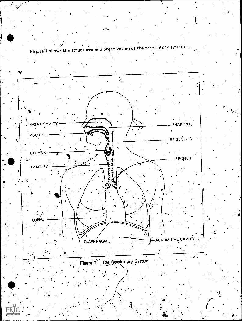
Figur'e/1 shows the structures and organization of the respiratory system. .
io?
. NASAL CAVITY
I2
MOUTH
a.
LARYNX
TRACHEA'
3
P
1n
PHARYNX.
EPIGLOTTIS .
,
"DIAPHFIAGM
.
-BRONCHI
.
ABDOMINAL CAVI rY
Figure . .:The iratqry System
.r '
°
, .
. A A
V
A 1
I
-6- ,
fUtirT.IONS OF THE RESPIRATORY SYSTEM4
Breathing is a cycle that begins with inspiration, or taking in air. Next
follows a two -way
i
exchange: oxygen passes from the lungs into the bloodstream
and carbon dioxide passes from the bloodstream into the lungs. Carbon dioxide is
a waste product of normal cell functioning (rnetabolism),and must be vented from
the body by expiration, or exhalihg. In fact, breathing is 'triggered-when casbgif
dioxide .concentration .in the blood reaches a certain level; the body breathe In
order to eliminate carbon 'dioxide as well, as to take in oxygen. The cycle of
Inspiration-expiration (reipiratory rate) is triggered about 10714 times per
minute in a resting adult and, about 20 fiThei per minute.inki child. The rate of
respiration depends largely On body demands. As:body activity increases, so does
the Utilization of 'oxygen and the production of carbon dioxide. When the trigger
level of carbon dioxide is achieved, more rapidly, breathing becomes faster and
deeper.
Because the respiratory system is, open to ,the outside environment (throLigh
the nose and mouth), it is -susceptible to particles and bacteria in Abe air.
Pollutants and microorganisms are taken in, with every, breath of air. Secretions
produCep by, the respiratory tract trap .and engulf the particles .and, pollutants;
and even .contain enzymes which destroy bacteria. !waits way the respiratory
system protects the body fiom constant -exposure to harmful particle's, while,
carrying on the process of supplying the body with"the oxygen necessary to
maintain life. 1rw
A
1' ..
r)
e
-- 7
U. -4
, t
ti
4.

ti
k
. -7-
INTRODUCTION TO THE RESPIRATORY SYSTEM
.Post -Test 1
41?
alszmai,;,
_ .
1, M'atct, the parts of thIS diagram (indicated by latte'rs, A-E) to the games of
the organs. (One of the letters will mat be usect.) c "-.
a.
C.
'Pt
trachga
larynx
bronchus
lung
I
.6

2: The two,cnajor divisions of the resOirEitory system are:
A. conductitjg organs and gas exchange organs.
. B. the pharynx and the larynx.C. - organs' of support and organs of movemer,D.° the trachea and -the bronchi:
,.,
I0.
3. In the lower respiratory tract, gases are_exc'hanged between the lungs and
bloodstream in the:
A. diaphragm.B. trachea.C. pleurae.a alveoli.
tr
4. Which of the following Is ND-ra component of- the. respiratoiy system?.
.A. esophagusB. pharynxC. larynx.D. Basal cavity
1
I'
5. The upper respizatory tract has the ability to:-.
A. exchange gases with the blood.
B.. transport oxygen to the brain.
C. purify air.that is inhaled.-
D. produce oxygen for the lung13- ,
,,,. .
6. . Breathing is directly triggered by high ofitbONA)xide concentrations in the:or. . .
. A. nasal cavity.B. lungs.-'C. ,blOod.D. muscles.
'
4
O
a
.
5'
6'
-Goals:
4'
-9-
UPPER RESPIRATORY TRACT,
1.
Upon completion of this module,' you should be able to:
1. ,Identify the-structures of the nose. -
2. Describe the functions of the nose and its related structures.'
3.* Identify the structures of the pharynx, laryn'x, and trachea;
4. Describe thefunctions of the pharynx, larynx, and trachea':
49:
I
*, i
4 e
4.
Air enters the body through the upper respiratory tract, which includes the
nose, 'pharynx, larypx, trachea, and bronchi.' Each of these organs is uniquely
eqUipp4d to help provide the lungs'with properly conditioned air ,and to maintain-
. the flow of air through the system. '
THE NOSE,
The e is the first organ of the respiratory system. Its external openings,
the nostrils.Or nares, lead backulard i4tb 'the nasal oavIties, which. ar separated
by.a wall of cartilage .wd bone railed the septum. On' the side walls of e nasal
cavities,arehe turbinarei, spiral 'passagewayswhich hang like "sagging s elves"
in the 'patik;.of incoming .air. Hollow chaeribers^withip the faCial bones called
Sin ses surround and open into the nasal cavities.' The sinuses and .walls of the
nese CavitieS are lined with mdcOus membranes containing cilia, which are,,-
microscopic. hair-like Structures (7tre surface of the mucous cells. These cilia
are constantly in moton; they beat 'back and forth'aboot 10-12 times per second..
Ciliated mucous membrane extends through the *entire upper respiratqsry tract(
and,, as will be discussed,, has an important functiOn.-
I' 1 .
1
In addition to providing the initial openings to the outside air, the nose has
other functioris. Nerve .endings in the roof of, the nasal cavities are responsible
for the.senee of Smell, or olfaction. OlfaOtion Can serve a protective fune-tion by
yarning the body of air possibly unsuitable or 'dangerous to breathe.
.-
.
. A.
12
9
A
S.
s.
rd.
-10-
4
L
. .
In another' 'functions- the nasal cavities acras a sort of "air conditioner":
they condition the air going to the lungs. The temperature and humidity, of the.
air in the environment vary a great deal as compared to the fairly constant
conditions within the body; inspired air could :dry outor freeze the delicate lung
tissue were it not for the fact that the air must first, pass the turbinates and
thticous membranes. As air entering the body passes over and' through t,thes
Structures, it. becoMes.owai-med and moistened almost to the level of the
temperature and humidity. inside the lungs.
Also, the 'nasal cavities filter and clean-the air going into the .respiratiSry
system., Mucus a thick and sticky fluid produced f336 the mucous membranes,
traps incoming particles. If the respiratory tract 'were' not lined with cilja, the
mucus would quickly become, covered with trapped material and thus lose its
moisfening'r(ind protective prbperties. But the constant, waving motion of the
cilia Moves -the Mucus to the pharynx where, together with he trapped foreign
matter, it it swalledand later disposed of by the stomach. In the rest 'of the
upper respiratoimp tract, the cilia carry mucus and the filter -out substances
toward the pharynx constantly, like a downward escalator.
THE PHARYNX
The'pharynx is the 'throat, a cavity extending frornb.ehind the nasal cavities
down to the larynx. Becausg,it is an area Common to both. -the digestive and
respheatory systems, the phitir, is ideal for disposing of the 'particle-filled
mucus sent to it by the ciliar esca ator. The pharynx also completes the -task 'of
warming and moistening the inspired air, which vial begun in the nose. .Ar: the
base of the throat the pharynx. opens to the larynx, pr voicebox; at. the back of
the neck it leads to the esoph s, the tube which carries food to the stomach.
RYNX r
rigid,: box-like structure made of sturdy plates of cdrtilage, the larynx
houses and protects the vocal cords. We can speak and- make 'Sounds because of
our vocal .cords; which die-Theririgility to contract, combined with the cPritrolled
"exhaling of air, to--produce different vibrations that result in 'Sound. , The 'vocal
cords also guard the opening to the'trachea, 4a large tube of the upper respiratory
tract., Between the cords is a slit called the, qlottik which leads into the
trachea. Should anything intended for .the esophagus food or water)
accidentally enter4 the jarynx," the glottis can close shut to seal off the lower
respiratocytract. Also,. the epiglottis, an elastic flap of tissue, folds over /the
glottis 'to .seal it off when food or liquid enters the pharynx; In this, way the
larynx protects the respiratory tract from baking in ariything-except air;

ro
THE TRACHEA
The last passageway of the. upper respiratory 'tract -is the trachea, or .
"Windpipe," a short tube aboUt five inches long and one inch 'in diameter which
links the pharynx and the lungs. It is made of about 20 C-shaped rings of
cartilage, stacked one on top of another and joined'together by tough connective
tissue. Cartilage provides rigidity'arid protection which allows the passageWaY to
remain open reglirdless of the position of the head and neck. Although. the
C-shaped cartilage is rigid, the back of the trachea, where it Ilea against the
esophagus, is soft. This ,allows for expansion of the esophagus when food is
passiqg through.
The' trachea is theseniliof the upper respirat?fryyact. It leadq to the lungs,
.where the actual respiration takes place.
figure 2 shows the structures of the upper respiratory tract..Oi
NASAL TURBINATES
iV
NASAL CAVITY
e
GLOTTIS
NASOPHARYNX
TRACHEA
Figure 2. The Upper Respiratory Passages

Optional Activitieswe
, .
Look at the structure of the cilia Under a microscope, or view slides of
cilia.::. %
. ..,
:4
ft, Discuss what:actually happegs when fOod "goes down the wrong wa.'1 g
no
s,,FinCout about the emergency procedures and techniques to apply :when
someone is choking.
c
0
i
4

.,
-13-
UPPER RESPIRATORY T1RACT
s- oPost-Test
1 .
r
1. The Flair -like structures.within the mucous membranes of the nose_are called
0
2. A major function of the nose is to:
A.. effect gas exchange..B. mdisten air.C. produce blood cells.
ID. protect the sinuses.
3. The sense of smell is also known as:1
A. olfaction.B.. turbinate.
cilia.D. guqation.
4. Mucus is prodiked by mucous membranes in the:
A. lungs.B. alveoli.C. sinuses..D. esophagus.
O.{

o
St
:14-
5. Structures of the nose serve a protective function by: ".
A. preventing food froM entering the trachea. -
13: warming air entering the body.'
lc. removing poison&us substanceCfroni.the blood.
D. preventing carbon dioxide'from entering the lungs.
t, \
6. The nose Is divided 4ntd two nostrils- by a:
A. bronchiole.B. septum.C. cavity.D-. sinus.
4
4
f
7. ,which of the following is NOT a conducting organ of the respiratory systeni?
,. ,
A. pharynx ., B. trachea
C.- alveolus.D. larynx
8.. The passageway that connects the nasal cavity and the larynx is the
1.-
9: The trachea is a protective structure because it is Made up of:
A. bones and fatty tissue.B. C-shaped rings of cartilage.Cr blood vessels and ligaments.D. tendons and muscle..
4

10. The structures that constantly carry mucus and foreign particles toward'
4 the pharynx ate the:
A. elves.B. bronchi.C. alveoli.D. cilia.
11. What is the name of the slit that open, into the tracheli?-
12. The function of the epiglottis le to:
-- 'A. remove carbon dioxide froth the blood.
B. produce mucus.C. prevent food from entering the trachea..)
o D. protect the 'vocal cords.
4
18.
10

A
Goals:
I
-16-
THE LUNGS
<4.
Upon completion of this module, you shoUld be able to:
, .
1. Identify the structure of the lungs.
2. ',Identify organs and/or structUres related to the lungs.
. .
STRUCTURE OF THE LUNGS,.
4111 .,
Only the ',mechanical process of conducting air takes place. in theUpper
respiratory Vet. Actual respiration, the exchange of gases, takes place, in the.
lungs, w7fictife ideally structured,ftO fulfill this function.
66 '
. P
s .) 4 .
,.
<4
The two lungs entirely fill the chest cavity. Shaped like large. blunt cones,
they point upward past the cravictf collarbones) and are broad at their bastes-)t ,
which rest on the upper ,surface o i the 'dieRhreom at about the level of the
. seventh rib. Because of the position of the liver, the base of the right lung rests.
higher in the chest than the base of the left lung. Short and broad, the right lung.
is divided into 'three lobes. The left lung is divided into two lobes and is longer
and thinner, to allow for the space taken up by the heart...
_ -_,
, The two main bronchi, which split oil from the trachea, co nnect the upper
respiratory tract to the lungs. About ont-half inch in diameter, ttiese bronchi
resemble the trachea in that they too are 'made of rings of cartilage. As the
bronchi continue to branch.off into the {tinge, the passageways divide into smaller
and smaller passageways so that the -entire bronchial system resembles a free
(upside-down). The next" branches of this tree are. the . bronchioles, tiny
passageways that reach deep into the lungs to .bring -air to every possible area.
As the tubes jivide further and. get smaller, they become the terminal ,
bronchioles. Here the walls of the bronchioles lose their cartilage and, become' -
th nner, until the-walls are simply a Single!layer of tissue. The smallest twige,,,9f
the bronchial trees are .the respiratory bronchioles, which lead directly to the
functional units of the respiratory system- -the alveoli .(see Figure 3 on the next
page).
19
ti

Figure 3. The Terminal Structures of the Respiratory System .
Alveoli aremicroscopic bubbles of very thin tissue clustered around the
respiratory bronchioles. Each cluster,, or, alveolar sac, is made up of rnarty,.
individual alveoli. Because they are clustered, millions of alveoli with extremely
thin walls can fit inside the lungs to provide the lungs %hith a vas inner surfaCe
'area. This great amount of thin-walled surface area-. permits 'diffusion, gas
exchange betWeen the lungs Sand the bloodstream; to take place efficiently.
Because of the thinness of the alveolar- walls, the lungs could not support
t,hemeelves or maintain their shape were it not for thepleurae. These are the
membranes covering the outside of the lungs and lining the chest 'well. The
visceral pleura is. always in contact with the lung tissue and rests agbinst the
parietal pleura, which lines .the thoracic (chest) cavity. and diaphragm. .Between
these two "membranes iss a thin film 'of an Oil-like substance which binds them
firmly together. The rigid chest wall pulls the parietal pleura outward; the
parietal pleura pulls on thetwisceral pleUra, which in turn pulls on the rung-tissue.
This constant pull keeps_ the thin-Walled lungs from folding up and collapsing.
Pleural fluid also lubricates the lungs so. that 'they can move in relation to the
chest- wall- with very little friction.- .

Optional Activities%
' "'
le
7
Listen to the sounds ofobreathing witty a stethoscope. /Nee they the same
all Oyer the chest and bark?' Why. or why-not? et.
-Discyss how cigarette smoking affecta the structures and function of the
lungs. ,Q
S.
a
4.
5'
ry
,.34
4,4 41
F.
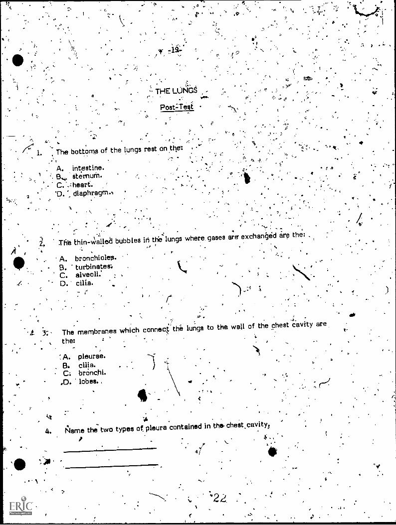
" THE LUNGSe- .
Post :-Test
4'.. -'
The bottomp of the lungs rest on the:.
. A. intestine.demur:11.
C. ,a
2 D. diaphragm.*
2, ,-
,, -..
1 . t. : ... .
2, The Ehin-walled bubbles in the'lungs where gases are exchan§ed ire the: .
'4,,, , - .
,
1
"
A
;-
A
A. bronchioles.B. turbinate&C. alveoli: ,
D. cilia.I
,,
-4
4 3,
)- I
3:- The membranes which connect r the lungs to the wall of the chest cavity are
the:
A. pleurae.B. cilia.
brcinc.hi.,D. lobes. ,
4..
c-
4,
4. Name the two types of pleura contained in the. chest.cavity:
. ,\.
47t
A
a
.1
`

01.
ok'
-20-
5., A cluster of microscopic alveoli is called an alveolar
0
-t
V.
4
4-. -
O
F
.1
Se
'I
J

40
ys Alb
I
-21-
RESPIRATION
Goals:
:Upon completion o this module, you shouW be.able to:
4at,e
1. .Identify the types of respiration.
.2. IdentAiy and describe the processes involved in respiration.
s
TYPES OF RESPIRATIONa.
C
o
Respiration, or gas exchange between the blood and body tissues, occurs in
two areati: between' the lungs and the pulmonary blood supply, and betweenthe .
bloodstream and the cells of the body.
Internal' respiration is the e)ichantyk of gases between- the blood and body.
cells. (This process is described in the unit _bit the circulatciry systerne External-respiration is the exchange of gases between the blood and the lungs--a process
which, first requires -that oxygen- be .ptesent' In the Lungs., The active process of
getting air into`the lungs.is the function of the muscles of resAiration.
PROCESES-OF RESPIRATION , k
The diaphragm is the primary muscle of respiration. It is attached to thebaies of the lun%s and the lower edge of the ribCage., When the .diaphragin is
relaxesi, as it is when not involved. in inspiration, it bulges upward into the chest
cpvity. . When it cont-reets; the diaphragm shortens and flattens out, Jiulling
downward. Because the lungs are attached to tht diaphragm by the pleurae, they
too are pulled' downward, and their Interior space increases. This expansion
causes the pressure in the lungs to drop below atcapspheric pressure (the pressure
on the air in the outside environment). As a result, outside air is forced into theexpanded lungs through the upper respiratory tract (inspiration).
*I'
ay
24
A

:22-. t
7-Th ".. ..
.
in the breathing process,:the ribcage- .vorks in mtich !-.Fe sloie way as thediaphragth. The ribs are attached to the spine and -breastbone in such a way. that
thy)/ are 'capable of swiveli .ng up and, out; muscles called 'r.tercor.tal muscles
cause this swiveling motion iluriag inspiration. .The effect of thM motion is 'toenlarge the diameter of the ribedge, which expands the entire chest wall. As the
chest wall expands, the pleurae cause the lungs to expand with it. Again, the
expansion of the lungs causes -a drop in internal pressure and the outside air flews
'into thelungs. . ;
,. . . .
The Itrength of contractiOn of the diaphragm' and the intercostal muscleschanges the amount of air taken in with .:each breath 4Juring inspiration. In quiet
breathing, the diaphragm flattens out by Only an inch or so, but In heavy
breathing it can drop more than three inches. The- heaving chest ...,f ,an athlete
who has just run a race . is an example of greater-than-normal chest (Abei
therefore lung) expansions, 9k,htcf-: 'causes inspiration of greater-than-normal
amounts of ai-r., '= - ' ..;
.,
c, The arabunt of gir inhaled and the'ri exhaled with each breath is callid thetidal volume, because it ebbs and, flows: Average resting tidal volume is about
500 cubic centimeters (cc), or about a pint, of air moved by the action of the
respiratory muscles. When the need for air is greater, as in stress or exercise,
the muscles cause greater ,chest expansion and the tidal volume increasesproportionately. There is aMays a supply of air left in the lungs which cannot be
exhaled. This reserve II .called residual volurne; it help.; prevent. the thift-wallapasSageways of the lungs from, 'collapsing.. It is a substantial volumeabout q.00
cc, or over a quart. The. vikal capacitysof the lungs isthe maximum amount..of
ir that can be forcibly inhaled and exhaled in one breath
The end , of the respiratory cycle is expiration, which eliminates carbondioxide from the blood and the body. Expiration, is netar ally a passive process)
the diaphragm' aria intercostal muscles relax arid the ribcage 2.1ci:Chest, cavity
lose their expansion. Thai lungs, which are elastic ,in nature, return to theirunstretched size. The waste air is forced out of the lungs and bark throthh the
upper respiratory tract to the outside environment.. The speed wsth which 11.r.
respkatory cycle occurs determines respiratory rate. A normal resting aifufi
usually has a respiratdry rate of about 10 to 14 breaths (cycles) per minute.-
In summary, ,the respiratory system takes in air; WEII InS, r.ir,,Itens, and
filters, it; transports it to the systems functional units, the :11VP0i!; "upplies the
blood with oxygen; and removes carboh dioxide ftert-the body.
.
a
25

e
\.23-
Optional Activitiks
,
..Find out.hqw to construct-a model of thec est which .demonstrates the i
Action of ..the.-respi7ratorylinuscles on -the. 16h the resulting flow" of
. ,,....,4.
air. .*- .
.
.
Measure your respiratory rate before and after exercise. What causes )-
the changes? I
,2:-
.4.11
.
Try to complete the/word maze on the following. page. It uses 'some of ; -N,,,
the terms that you learne this unit: 1 SP . /-
. IP
0
O
;
4
0.
C
t-
re.
0,

= ,q
a
'f
,
,
' .P", -
E P I R AGlitiGXF-0I0liEN A 0 Y S UN I S. BR EA ICSDil. J 0IQRYNXNEAU''I
4SINVK SUELPOPHPA10 ApGEM'ANH IMS,CHL,I'VRH,K TODGTOBVAR Y S
HURWX'PL AR YNXR AWD'E E A F I ZRIAROWTG'fillALGOJI,,K ZCLC.(1EM_AORP,MEPOSTROCOVA'ICY . A M S - T. U I Z U 'F ISM E -II X L
L. S E T R ,L EO-C*OLBNTI AU R W S. I .T T 0 L ,G I P E:1( 0 C
N o Ate CTDSP_JV'A,QUAW 0
.4
-24*-
WORD MAZE
N.
GOOsIAPARUELVEOL',VSti,IIR X ILISEIDIY8M
0
- 97
..
Ay'
...
Find the following terms in this maze by circling the words. The9' may appear
frontwards or backward*, vertically, horizontally, or diagonally.
.
...
4
4
a
,.I.
.
alveoli la-rYnx respiration
cilia, . lungs . trachea
4
'diaphragni pleura , .vocal cordsO.
a/
,
epiglottis,\
. 41
. .r
.
4
t
A.
,
2 7 °'r d

I-25-
RESPIRATION,
Post-Test
1. Tfi upply of air that always remains in the lungs is 'called
.
2. The muscles which elevate the ribs in breathing are the:
A. pectorals..B. turbinates.'C. cilia.D. intercostals.
. .
0
. 3. The exalange of gases between the lungs and the blood is called:
A. external respiration.B. oxygen transport.C. osmosis.D. capillary action.
- -
4. During breathing, outside air is forced into the lung
A. a drop in external pressure.B. an increase in-residual air.
C. a pressure drop in the chest cavity.D. an increase in tidal volume.
`28
cause of:
,I

P 44
-26-0
94
5. The maximum amount of air that can be Inhaled and exhaled in tone breath
is called:
A. tidal volume.B. vital capacity.C-., inspiratdry reserve volume.D. . residual volume,
a. e
2
- 6. A normal, resting adult breathes about:.,.
.
A. 5-9 times per minute.B. 10-14 times per, minute.C. 15-19 times per minute.D. 20-24 times per minute.
/0
0
*
.
; sX-
C
I o .
,
a

a-
alveolar sac: aclustee of alveoli.
-27-
GLOSSARY
alveolus (pl. alveoli): a microscopic bubble within the lungs wher/gas exchange
takes*op
bronchiole: a smaller division of the bronchi.
bronchus pl. bronchi): one of the two tubes that branch off the trachea and into
the lungs.
che'st cavity: the cavity in which the heart and lungs are located.
cilia: hair-like projections from the upper respiratory tract. 'I.,
ti
a
..diaphragm: the primary muscle of respiration; lo6ted between the chest and
abdomen. 41*diffusion: the rang of materials (especially gases) though the motion of,
molecules.
epiglottis: an elastic fold of cartilage which guards the opening of the trachea.
expiration: the tict of moving airout of the lungs; exhalation.
N- external respiration: the exchange of gases between the lungs and the pulmonary
blood supply.
glottis: the slit-like opening of the trachea,
'inspiration: the act of taking air into the lungs;-breathing in.
Intercostal muscles: muscles which move the ribs in breathing.V
iternal respiration: the exchange of gases between the blood and body tissues.,
larynx: the respiratory organ responsible for voice production.
nasal cavity: the hollow internal structure from the nose to the pharynx.
nostrils: external, openings to the nasal cavity.
'olfaction: sense if smell.
30 : O

-28- .
pharynx: the organ between the oral cavity and the larynx, shared by both
respiratOry ands digestive systems.
pleurae (sing. pleura): membranesattached to the exterior of the lungs and
connecting them to the walls of the chest cavity.
pulmonary: of or relating to the lungs.
residual volume: the supply of- air which always remains in the lungs.
resiMat oriithe exchange of gases.
respiratory bronchioles: the final subdivision of the bronchial tree; lead directly
to the alveoli.
sepeurn; a wall which separates the nasal cavity.
sinuses: hollow chambers surrounding the nasal cavity.
terminal bronchioles: the last division of the bronchioles whose sole functio
gas conduction.tidal volume: the amount of air moved in and out of the lungs in a normal breath.
0. trachea: the tube-which transports air between the larynx and the bronchi.
turbinates: areas within the nasal cavities.
upper respiratory tract: the respiratory passageway from the-nostrils to the
bronchi.
vital .Capacity: the maximum amount of air that can be moved through the
respiratory, system in one breath.
VQC 81cottds: fibrous bands suspended across the larynx which"vibrate to produce
speech. *,
0
a' 31
v.
,
.,
N
.
1
IP
,
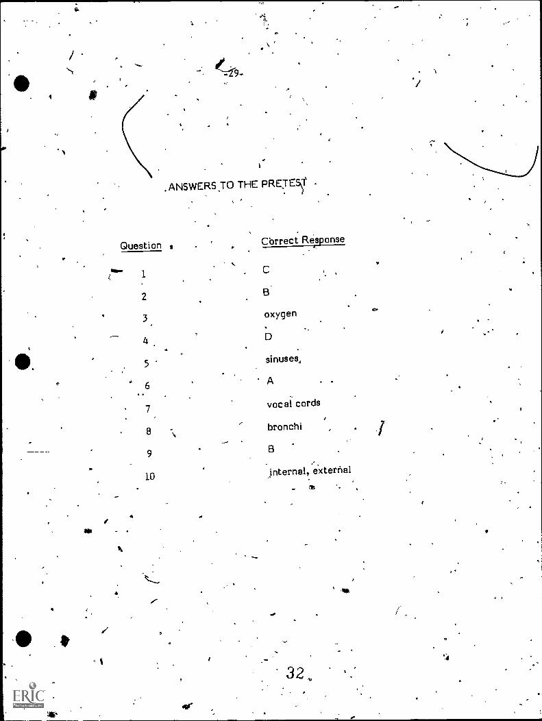
.ANSWERS30 THE PRE:rEy ., .
.1.
I
Question , Cbrrect Response
az,
law1
2
3
4,
5
.6
. .
7
8
9
10
, C
B
oxygen
D
sinuses,
A .
vocal cords
- bronchi
B
internal, external
eair .
i,
/e"
- t ,
Oar
- as,.
32 Az
MI
...
4.4

-N
/11fALTH
OCCUPATIONS EDUCATION
MODULE
ee
INSTRUCTOR'S GUIDE-
' HE. RESPIRATORY SYSTEM33
N.
Instructional Materials in Anato ny and.Physiologyfor Pennsylvania Health Qccupations Progiiims$.
INSTRUCTOR'S GUIDE:
THE RESPIRATORY SYSTEM
Prepared for:
Research' Coordinating Unit for Vocrftional EducationPennsylvania Department of Education
Box 911Harrisburg, Pennsylvania 17126
lS
. Prepared by:
National Evaluation Systems, inc.30 Gatehouse Road
Amherst, Massachusetts 01002
Ti
June,' 19794 .
34:
le
,...
a
J

O
INTRODUCTION
- These instructional modular units have been 'developed for the Pennsylvania
Department of 'Education for use, in vocational education programs.. They were
designed"' on the assumption that a 'basic underst,andiqg of human anatomy and
physiology is essential to any person preparing tb enter a hearth care occupation
such as practical. nyrsing, nursing assistant, medical assistant, emergency (medical technician, or dental assistant. Each of these modular units. will cover,
the most important aspects of one of the major systems of ,the human, body. In
the first four units the following systems will be covered: circulatory system,
respiratory system, musculoskeletal system, and digestive system.
This Instructor's 'Guide is designed to provide suggestions to you on-how. to
use a modular unit most effectively in your instruction. These recommendations,
however, do not represent the only way to use these units: you may' be able to
devise more beneficial uses4or\ttle materials.
THE MODULAR UNITS
.
Each modular unit( is rijade up of several components: ,a pretest, four to
seven instructional modules with corresponding post-tests, optional activities for
the students, and a glossary of terms used in.the unit., Each of these components
has a specific purpose and is organized in a specific way, as Will.be exptined in
° the following sections::
.
Pretest
. After reading* ,,the preface, which is simply an introduction to these
instructional units, a student Albriking through a modular unit should first take the
pretest. As its name implies, this test is designed to be taken by the student
before beginning work on the materials contained in the unit. Its purpose is
twofold: (1)'to stimulate interest in the ,modular unit by giving the student a
a:preview of the topics covered, and (2) to provide a means Of self-diagnosis so the
student may identify, based on performance on the pretest, those areas of the

0
modular unit which may requir special :attention and extra effort ortthe part ofthe student. After selecting arC;a9swer to eactOf the pretest gkiestions, thestudent should turn to the back of .the - modular ur;1.- and check the correct,answers. If the student /answers incorreetly on a numbp,,pf guestiOns dealingwith a particular subject, then the student should -pay clo*r attenliOn to themodule on that subject. i,.43
Instructional Modules
This modular unit is composes of four separate but closely related modules,including: Introductiori to thb Respiratory System,.UPper Respiratory Tract; TheLungs, and Respiration. 'After, taking the pretest and checking the 'answers, thtstudent should re,ad through and study each of the instructional modules. For thestudent's benefit, each module begins with a .statement of the goals, orobjectives,, that a student 'should have mastered upon completion of thatartic lar module. The level of achievement of these goal's is measured by the
9 dent performance on the corresponding.post-test. The-language level andcon of each module is aimed toward the student seeking an introductlan to°the,compbnents, structures, and functions and the' basic terminology, rest ired foran understanding of the respiratory system.
.4
Optional Activities0
Following many modules a re optional activities intended to P'rOvide thestudent with ari opportunity to pursue the content of the module at a min-depth level. Many of these activities may require teacher participatioh,feast itt obtaining and preparing' additional 'materials fOrthe.student to Utilize.
'In addition to tie optional activities available, to the.students, you aychoose to provide further information'to the students by teaching a brief uh onthe common disorders of the respiratory. system. Discussion of these diso dershas not been included. in the texts because a basic knowledge of the operstructure and function of the human body in a 'healthy individual seems ore-,appropriate. for the purposes of an introductory progrIm. If you do, choose odiscuss common disorders, the most effective, approach may be one in which youuse disorders to illustrate what can go wrong in the body, as a means of
. clarifying the students' understanding t.f, how the body work's when functioningproperly.
. You ;nay also wish to provide studentri with the names lif books or articlesas suggested feadiogs to further their uhderstanding of a particular area.

C
4,
C
-3-
Glossary.
After the last of the modules in the unit is a glossary. Thii is not intended
to be a cemprehenIce` glossary to' be used by the student as a dictionary.Rather, it includes the basic ferms.used in the unit which are necessary to anunderstanding of the system covere'd. Thosq words' which appear. in the"modules
and have been defined in the text are not always defined in tAe glossary. Someof these particular terms have been used in the module because they, are,_
essehtial but. difficult terms needed to explain the content taught in the unit.-The student should use the glossary to review the vocabulary essential to the unit.
before taking the postztists. A °
Post-Tests 7,
°
The post-tests are the final assessment of, a student's understanding _of_ the
,material presented in each module. They consist of multiple-choice and
open-ended questions designed to measure a student's mastery Of the goals(objectives) stated at the beginning of each module. Each of the questions hasbeep written to measure an aspect of the 'drills and/or knowledge that a student
may 'be expected,to, acquire as a result of working through a particular module.When a student has finisheol studying a module, has pursued any chosen optional
activities, and has reviewed the vocabulary in the glossary, the student should
take the poit-test that follows the module.
SCORING THE POST-TESTS4 . ,
As previously mentioned, the purpose of the post -tests is to measurewhether or_ not a student has mastered the objectives (goals) stated at the-beginning of each module. Due to the differing lengths of the post-tests, thevariety of ways in wAich 'teachers may choose to utilize these modules, and
discrepancies among studgnts' previous exposure to the subject matter, it is flat
practical to set a standard cut-off score on each of the tests that would indicate v.
mastery- (30 the objectives. Rather,' teachers. are asked to use their professional
rudgment in individualcases to determine if a student's performance on a
,post -test indicates that he or she has mastered the objectives/ stated for that
module. In making this determination, you should consider at least all of .thefollowing factors:

. /
"--4-
,, (1) How long is each post-test?
(2) How much information is included in each module and how c plex is
the information, relative to other modules?
)
(3) Has the student been exposed, to the kind of curricular material
before Thetis, has the student been taught the basics of this system
of the body before?. ..1.,..,
(4) Should the, entire 'class be requred to achieve a certain score in order
'to pass, or shoUld each student be 'Considered indivklually? (This
depends ors how Ind with whom you use this module as ,instructional
material.)-,
..,. (5)' Should the student be graded pass7fail on eachpostItesti.e., onmastery of each module - -or on the unit as a whole?
To facilitate the scoring of pose-tests, each student will record his or her
answers to all the post-tests on one 'separate sheet ofjaper. You should mark
each answer correct or 'incorrect, then give the student'a "pass" or "fail" on each
module, or on the unit as a whole.
Because of the subject matter, responses to open-ended questions may vary
slightly from those listed -below, but these responses may also be acceptable..
Again, "in these cases instructors are asked to use their- professional, judgment to
determine if a response is correct. .
Use the following list of answers to questions on the post-tests to grade
your students' papers. .
i
vo
F,
38 G
..
4
fa
b
;C
a 4 .
-. .
N.
...
/ -5-
*iP
-.-,-..
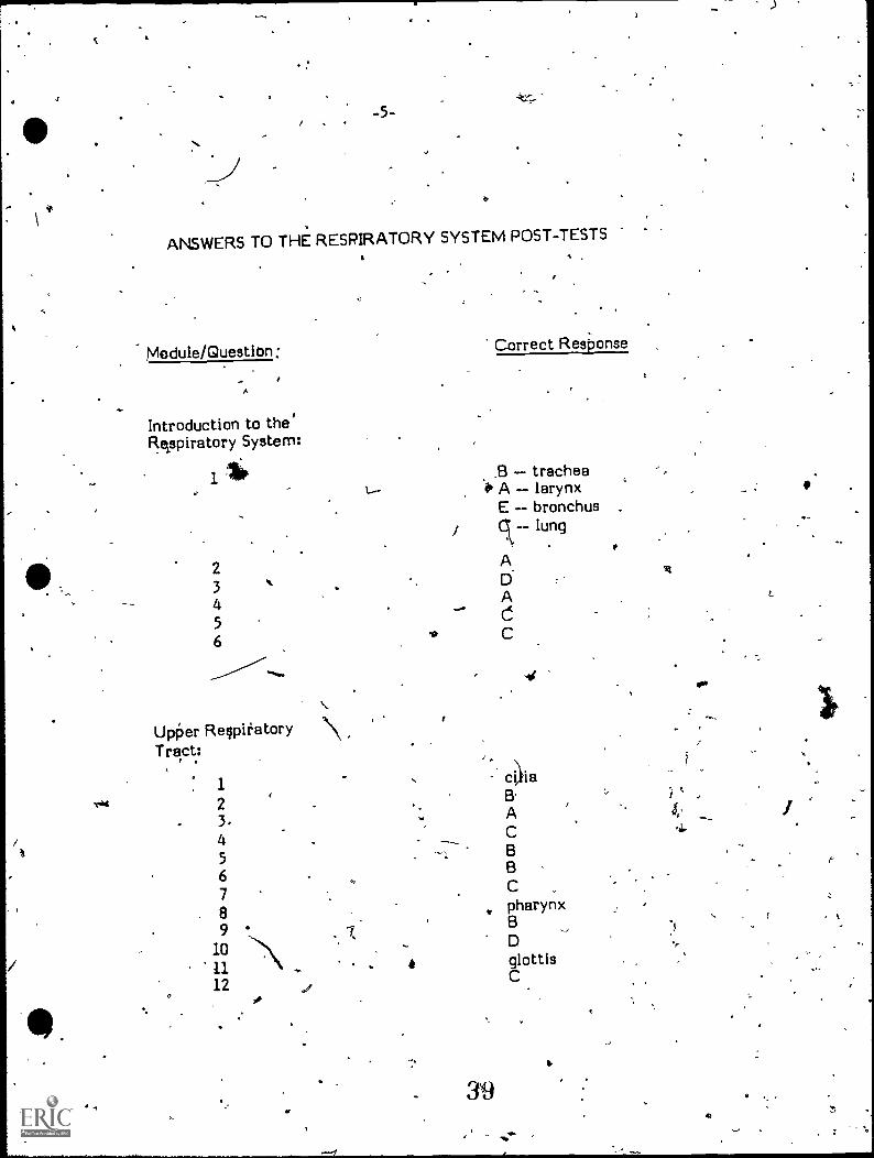
ANSWERS TO THE RESPIRATORY SYSTEM POST-TESTS
Module/Question: Correct Response
Introduction to the'Reppiratory System:
11*. t..-
23 ,. .
45
6it.
Upper RespiratoryTract:
'.\
1.c.hd 2 .
3./t
45 --.L
6789
/ 10'11
12..
,B -- trachea.0 A larynx
E bronchus
i gc-- lung
ADA
C
4
c4iaB-
ACBBC _
pharynx
, D
. glottisC
I
., 30.
16
a
v
F
,.. ( 1
43
a .....
.
I..

4
.Module/Question
The Lings:
1
2345
vv.
9"
-6-
4,
OP
Correct Response
DCAparietal, visceral`sac
Respiration:
1 ,
, residual volume
2et D
3 .' . A*4 "41* C
5 B
6 B
A
r-
ti