binding modes of quinuclidinium esters to
TRANSCRIPT
* Author to whom correspondence should be addressed. (E-mail: [email protected])
CROATICA CHEMICA ACTA CCACAA, ISSN 0011-1643, e-ISSN 1334-417X
Croat. Chem. Acta 85 (1) (2012) 77–83. http://dx.doi.org/10.5562/cca2060
Original Scientific Article
Binding Modes of Quinuclidinium Esters to Butyrylcholinesterase
Ines Primožič,* Tomica Hrenar, and Srđanka Tomić
Department of Chemistry, Faculty of Science, University of Zagreb, Horvatovac 102A, HR-10 000 Zagreb, Croatia
RECEIVED FEBRUARY 22, 2012; REVISED MARCH 26, 2012; ACCEPTED APRIL 18, 2012
Abstract. The orientations of chiral quinuclidin-3-ol esters and benzoylcholine in the active site of horse butyrylcholinesterase have been investigated by flexible ligand docking. Change of the esters' acyl moiety as well as the substituent at the quinuclidinium nitrogen atom affected the activity and stereoselectivity of the biotransformations. Analysis of interactions in the active site revealed the most important binding pat-terns for enantiomers, which define their reactivity. Calculated Gibbs energies of binding obtained by mo-lecular docking simulations were well correlated to the experimentally determined binding affinities of the investigated chiral esters. (doi: 10.5562/cca2060)
Keywords: butyrylcholinesterase, quinuclidinium esters, BChE-ester complexes, docking studies
INTRODUCTION
Butyrylcholinesterase (BChE, EC 3.1.1.8, also called cholinesterase, pseudocholinesterase or nonspecific cholinesterase) is a nonspecific ester hydrolyzing en-zyme with function in cholinergic neurotransmission and probably involved in some other nervous system activities.1 It is present in nearly all body fluids and tissues in vertebrates and can hydrolyze various esters of choline and other compounds, as well as some or-ganophosphorus compounds. Furthermore, BChE was used in the prophylaxis of hazardous organophosphorus poisoning (pesticides, insecticides and chemical warfare agents).2 It is believed that BChE is involved in the pathological development of Alzheimer's disease and BChE inhibitors have been used to raise acetylcholine levels and accordingly delay symptoms of the illness.3 Because of this and other significant pharmacological and toxicological functions it is important to understand and study interactions of small molecules in the active site of BChE.
The specific structure of the active site of choli-nesterases has been revealed by the resolved crystal structures.4,5 Like in other serine hydrolases, BChE catalysed hydrolysis is carried out by a catalytic triad Ser200, Glu327 and His440 in the active centre of the protein.6 The imidazole ring of histidine transfers pro-tons between serine and glutamate. Initially, an en-zyme–substrate complex is formed. An ester acyl–enzyme intermediate is produced as a result of a nu-cleophilic attack of the serine hydroxyl group on the
acyl group. Finally, a water molecule deacylates the enzyme (Scheme 1).
Derivatives of quinuclidin-3-ol are well known compounds with various pharmacological activities and are often the main or supportive part of the active sub-stance.7,8 In our previous work,9,10 chiral esters of quinuclidin-3-ol and benzoylcholine were subjected to enzymatic hydrolysis in the presence of BChE11,12 and the kinetics of BChE-catalyzed hydrolyses were deter-mined. The hydrolysis of quaternary quinuclidine esters proved to be highly enantioselective. Kinetic studies have shown that (R)-enantiomers of all tested quinu-clidinium esters are to a large extent better substrates of the enzyme than (S)-enantiomers. On the other hand, (S)-enantiomers showed higher affinity toward BChE (3–60 μM). In order to explain experimental data con-
Scheme 1.
E OH +O
R'RO
k1
k-1
E OH O
R'
RO
k2ROH
E O
R'
O
k3
H2OE OH +
O
R'HO
78 I. Primožič et al., Binding Modes of Quinuclidinium Esters to BChE
Croat. Chem. Acta 85 (2012) 77.
cerning reactivity of enantiomers, we applied flexible ligand docking protocol, as implemented in AutoDock 4.2.2.13 To determine structural characteristics of Michaelis complexes, 11 compounds (Figure 1) were docked into the active site of the homology structural model of BChE.14
EXPERIMENTAL
The coordinates of BChE were obtained from homology modeling.14 The enzyme was prepared for docking us-ing the Autodock Tools15 (ADT). Polar hydrogens were added and a Gasteiger partial charge was assigned to each atom.
Geometries of all ligands were optimized using the B3LYP functional16,17 and the 6-31G(d) basis set. All quantum chemical calculations were performed using the Gaussian 09 package.18 Non-polar hydrogens were removed from ligands and Gasteiger partial charges were assigned to all atoms using ADT. Tor-sional motion around single bonds was allowed (Figure 1).
Docking studies were performed by using the AutoDock 4.2.2 suite of programs.13,19 AutoDock re-quires a precalculated electrostatic grid map for each
atom type present in the substrate molecule. These elec-trostatic maps were calculated using the AutoGrid part of the suite with 0.2 Å spacing between grid points and
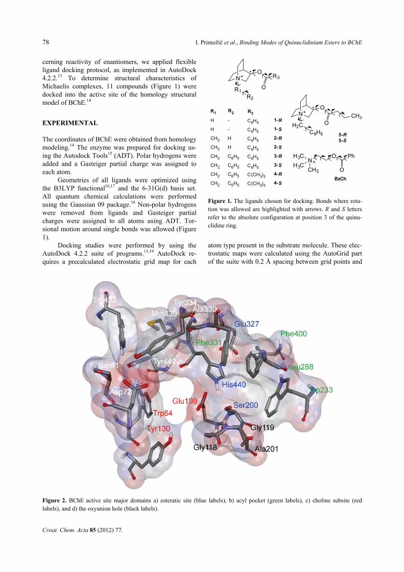
Figure 2. BChE active site major domains a) esteratic site (blue labels), b) acyl pocket (green labels), c) choline subsite (redlabels), and d) the oxyanion hole (black labels).
Figure 1. The ligands chosen for docking. Bonds where rota-tion was allowed are highlighted with arrows. R and S letters refer to the absolute configuration at position 3 of the quinu-clidine ring.
N O
NH3C
CH3H3C
BzCh
R3
OR1
O Ph
O
R1
H
H
CH2
CH2
CH2
CH2
CH2
CH2
1-R
1-S
2-R
2-S
3-R
3-S
4-R
4-S
R2
-
-
H
H
C6H5
C6H5
C6H5
C6H5
R2
C6H5
C6H5
C6H5
C6H5
C6H5
C6H5
C(CH3)3
C(CH3)3
R3 N O
OH2CC6H5
CH3
5-R5-S
I. Primožič et al., Binding Modes of Quinuclidinium Esters to BChE 79
Croat. Chem. Acta 85 (2012) 77.
the centre of the grid was placed in the γ-oxygen atom of catalytic Ser200. Dimensions of the active site box were set to 20 Å × 20 Å × 20 Å thus ensuring appropri-ate size of the ligand-accessible space. Consistencies of electrostatic maps were ascertained by checking maxi-mum and minimum values of van der Waals energies and electrostatic potentials for each calculated grid map.
Flexible ligand docking was performed for 10 new compounds (5 enantiomeric pairs) as well as for ben-zoylcholine. Docking calculations were carried out using the Lamarckian genetic algorithm and all parame-ters were the same for each docking run. We used ini-tially a population of 5000 random individuals, a maxi-mum number of 2×10⁷ energy evaluations, a maximum number of generations of 1×10⁶, elitism value of 1, mutation rate of 0.02 and crossover rate of 0.08. For the local search, the pseudo-Solis and Wets method was used with a maximum of 10000 iterations per local search, the probability of performing a local search on an individual in the same population was 0.06, the maximum number of consecutive successes or failures before changing the size of local search space was 4 in both cases, initial size of local search space to sample was 1.0 and its lower bound was 0.01. To ensure the validity of results, the docking procedure for each sub-strate consisted of 100 independent docking runs. The resulting positions were clustered according to an r.m.s.
criterion of 0.5 Å. Obtained structures were analyzed visually and evaluated based on their interactions with the amino acids within the active site of the enzyme.
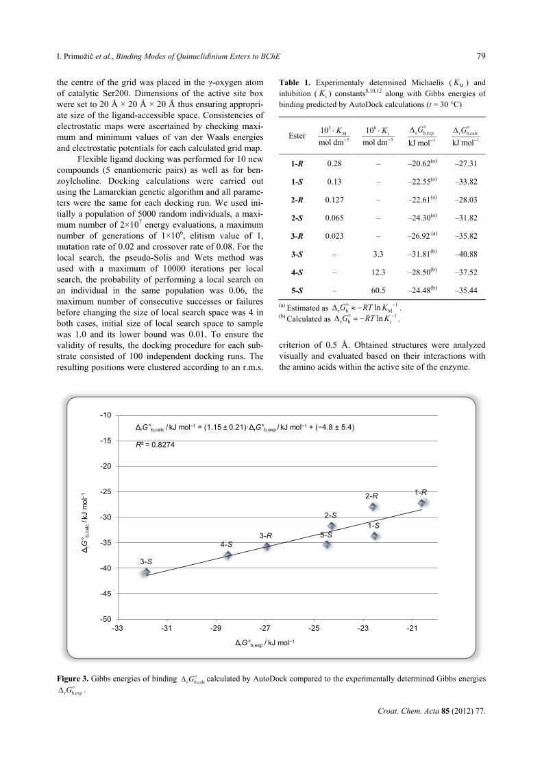
Table 1. Experimentaly determined Michaelis ( MK ) and inhibition ( iK ) constants8,10,12 along with Gibbs energies of binding predicted by AutoDock calculations (t = 30 °C)
Ester 3
M3
10mol dm
K−
⋅
6i
3
10mol dm
K−
⋅ r b,exp
1kJ molG
−
∆
r b,calc1kJ mol
G−
∆
1-R 0.28 – –20.62(a) –27.31
1-S 0.13 – –22.55(a) –33.82
2-R 0.127 – –22.61(a) –28.03
2-S 0.065 – –24.30(a) –31.82
3-R 0.023 – –26.92 (a) –35.82
3-S – 3.3 –31.81(b) –40.88
4-S – 12.3 –28.50(b) –37.52
5-S – 60.5 –24.48(b) –35.44
(a) Estimated as 1r b MlnG RT K −∆ ≈ − .
(b) Calculated as 1r b ilnG RT K −∆ = − .
Figure 3. Gibbs energies of binding r b,calcG∆ calculated by AutoDock compared to the experimentally determined Gibbs energies
r b,expG∆ .
1-R
1-S
2-R
2-S
3-R
3-S
4-S5-S
-50
-45
-40
-35
-30
-25
-20
-15
-10
-33 -31 -29 -27 -25 -23 -21
Δ rG
° b,c
alc
/kJ
mol
−1
ΔrG°b,exp / kJ mol−1
ΔrG°b,calc / kJ mol−1 = (1.15 ± 0.21)·ΔrG°b,exp / kJ mol−1 + (−4.8 ± 5.4)
R² = 0.8274

80 I. Primožič et al., Binding Modes of Quinuclidinium Esters to BChE
Croat. Chem. Acta 85 (2012) 77.
RESULTS AND DISCUSSION
In order to explain and rationalize experimentally ob-tained data, enantiomers of five different qui-nuclidinium esters (1–5) and benzoylcholine (BzCh) were docked into the active site of BChE by utilizing the AutoDock program. The BChE active site gorge14
(Figure 2) consists of several major regions: a) esteratic site containing the active serine as a part of catalytic triad (Ser, His, Glu), b) acyl pocket - a hydrophobic region which accommodates the acyl group of an ester, c) choline subsite for the recognition of the substrate’s quaternary ammonium group and d) the oxyanion hole formed by the main chain
Figure 4. Substrate-BChE complexes derived from docking study. BChE active site is represented by some structurally importantamino acids. Nonpolar hydrogen atoms are omitted for clarity. The entrance to the active site gorge is from below. (S)-enantiomer(carbon atoms white), (R)-enantiomer (carbon atoms orange): A) BzCh, B) 1; C) 2; D) 3; E) 4; F) 5. Selected interatomic dis-tances are given in Table 2.
I. Primožič et al., Binding Modes of Quinuclidinium Esters to BChE 81
Croat. Chem. Acta 85 (2012) 77.
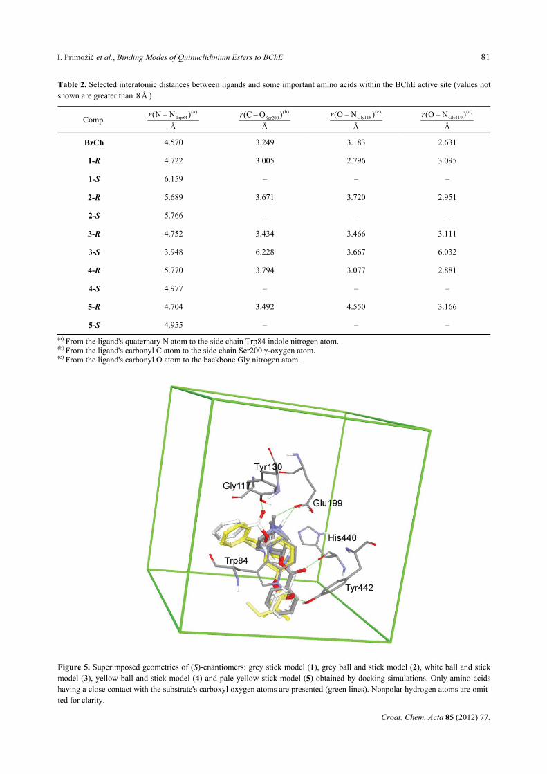
Table 2. Selected interatomic distances between ligands and some important amino acids within the BChE active site (values not shown are greater than 8 Å )
Comp. (a )
Trp84(N – N )
Å
r (b)Ser200(C – O )
Å
r
(c)Gly118(O – N )
Å
r
(c)Gly119(O – N )
Å
r
BzCh 4.570 3.249 3.183 2.631
1-R 4.722 3.005 2.796 3.095
1-S 6.159 – – –
2-R 5.689 3.671 3.720 2.951
2-S 5.766 – – –
3-R 4.752 3.434 3.466 3.111
3-S 3.948 6.228 3.667 6.032
4-R 5.770 3.794 3.077 2.881
4-S 4.977 – – –
5-R 4.704 3.492 4.550 3.166
5-S 4.955 – – –
(a) From the ligand's quaternary N atom to the side chain Trp84 indole nitrogen atom. (b) From the ligand's carbonyl C atom to the side chain Ser200 γ-oxygen atom. (c) From the ligand's carbonyl O atom to the backbone Gly nitrogen atom.
Figure 5. Superimposed geometries of (S)-enantiomers: grey stick model (1), grey ball and stick model (2), white ball and stick model (3), yellow ball and stick model (4) and pale yellow stick model (5) obtained by docking simulations. Only amino acids having a close contact with the substrate's carboxyl oxygen atoms are presented (green lines). Nonpolar hydrogen atoms are omit-ted for clarity.
82 I. Primožič et al., Binding Modes of Quinuclidinium Esters to BChE
Croat. Chem. Acta 85 (2012) 77.
N–H dipoles interacting with the carbonyl oxygen atom of the substrate.
Several clusters of binding orientations for all sub-strates were obtained. Since (S)-enantiomers were much better inhibitors than the substrates of the enzyme, clus-ters with the lowest absolute energy values for (S)-enantiomers were compared with the energies of the correctly placed geometries of (R)-enantiomers (rather good substrates) for the reaction to occur. The correct position for hydrolysis to occur was assumed to be the one with the acyl moiety in the acyl pocket and the ester carboxyl group in the vicinity of catalytic Ser hydroxyl group. In Table 1, the binding energies predicted by AutoDock in conjunction with the kinetic data compiled from the literature are shown.
The calculated docking energies for quinuclidin-ium esters were well correlated to the experimentally obtained values (R2=0.8274) as shown in Figure 3. Even better correlation is found within the series of only (R)-enantiomers of compounds 1–3 (R2=0.9445) or only (S)-enantiomers of compounds 1–5 (R2=0.8314). The good quantitative agreement between computed and experi-mental values confirmed the correctness of the chosen docking method and analyses applied.
Substrate-BChE complexes derived from docking studies for quinuclidinium compounds 1–5 and ben-zoylcholine are shown in Figure 4. Analyses of the docked geometries for each pair of enantiomers re-vealed trends in binding associated with the stereochem-istry of substrates. (R)-enantiomers of all compounds and benzoylcholine are bound productively: the car-bonyl group is found in the vicinity of hydroxyl oxygen atom of Ser200, forming two H-bonds with the back-bone Gly118 and Gly119 amide group (Table 2). The quaternary quinuclidinium group of all enantiomers is positioned in the choline binding site and its orientation depends on the bulkiness of the substituents at the quinuclidinium nitrogen atom. Thus, compounds 1 and 2 with the smallest substituents (protonated and methyl substituted (S)-quinuclidinium derivatives respectively) are bound in a similar way and are positioned in the vicinity of the Glu199 side chain: (a strong H-bond of 1-S (2.6 Å) and 2-S (2.8 Å)). On the other hand, N-benzyl substituted compounds cannot have similar stabilization due to the present bulky aromatic moiety. Therefore, two different modes of binding for (S)-N-benzyl enanti-omers are found - one for the esters of aliphatic acids (pivalic 4 and butyric 5) and a different one for the best inhibitor, (S)-ester of benzoic acid 3 (Figure 5). In 4-S and 5-S complexes with BChE, carboxyl oxygen atom is pointing away from the catalytic triad and establishes strong H-bonds with the Tyr442 side chain hydroxyl group and the Trp84 indole amino group (2.9 Å). The benzyl moiety is positioned in the hydrophobic region near the Trp84 backbone and in the vicinity of the
Tyr130 side chain. The aromatic ring of the acyl part of 3-S is also stabilized in this domain. The carboxyl group of 3-S has two H-bonds as well: the Tyr130 side chain hydroxyl group (2.7 Å) and the Gly117 backbone amide (3.3 Å). The additional favourable interactions of two aromatic parts of 3-S may be the reason for the best inhibitory power of this compound. These results sup-ported our previous findings based on experimental data and quantum-chemical calculations11,12 that (S)-enantiomers compared to (R)- have stronger interactions within the active site of BChE which successfully com-pete with productive binding leading to the tetrahedral intermediate and subsequent hydrolysis. CONCLUSION
Differences, similarities and relations among enanti-omers of selected quinuclidinium esters were studied with molecular docking simulations in the active site of BChE to determine the origins of observed activity and stereoselectivity of the enzyme. The calculated docking energies were well correlated with the experimental values and supported the experimental data for binding affinities of chiral quinuclidinium esters. This study demonstrated that flexible ligand docking is appropriate and useful tool for studying BChE binding specificity of quinuclidinium compounds.
Acknowledgements. We wish to thank Professor Israel Silman of the Weizmann Institute, Rehovot, Israel for providing coor-dinates of the theoretical structure of human BChE. This work was supported by the Croatian Ministry of Science, Education and Sports, Research Project No. 119-1191344-3121.
REFERENCES
1. S. Darvesh, D. A. Hopkins, and C. Geula, Nature Rev. Neurosci. 4 (2003) 131–138.
2. J. Bajgar, J. Med. Chem. Def. 1 (2004) 1–16. 3. E. Giacobini, Drugs Aging 18 (2001) 891–898. 4. Y. Nicolet, O. Lockridge, P. Masson, J. C. Fontecilla-Camps,
and F. Nachon, J. Biol. Chem. 278 (2003) 41141–41147. 5. J. P. Colletier, D. Fournier, H. M. Greenblatt, J. Stojan, J. L.
Sussman, G. Zaccai, I. Silman, and M. Weik, EMBO J. 25 (2006) 2746–2756.
6. P. Taylor and Z. Radić, Annu. Rev. Pharmacol. Toxicol. 34 (1994) 281–320.
7. M. D. Mashkovsky, L. N. Yakhontov, M. E. Kaminka, and E. E. Mikhlina, Prog. Drug Res. 27 (1983) 9–61.
8. I. Primožič, T. Hrenar, S. Tomić, and Z. Meić J. Phys. Org. Chem. 58 (2002) 608–614.
9. I. Primožič, M. Bolant, A. Ramić, and S. Tomić, Molecules 17 (2012) 786–795.
10. I. Primožič and S. Tomić, Croat. Chem. Acta 84 (2011) 245–249. 11. I. Primožič, T. Hrenar, S. Tomić, and Z. Meić, Croat. Chem.
Acta 76 (2003) 93–99. 12. I. Primožič, T. Hrenar, S. Tomić, and Z. Meić, Eur. J. Org.
Chem. (2003) 295–301.
I. Primožič et al., Binding Modes of Quinuclidinium Esters to BChE 83
Croat. Chem. Acta 85 (2012) 77.
13. G. M. Morris, D. S. Goodsell, R. S. Halliday, R. Huey, W. E. Hart, R. K. Belew, and A. J. Olson, J. Comput. Chem. 19 (1998) 1639–1662.
14. M. Harel, J. L. Sussman, E. Krejci, S. Bon, P. Chanal, J. Massoulie, and I. Silman, Proc. Natl. Acad. Sci. USA 89 (1992) 10827–10831.
15. M. F. Sanner, J. Mol. Graphics Mod. 17 (1999) 57–61. 16. A. D. Becke, J. Chem. Phys. 98 (1993) 5648–5652. 17. C. Lee, W.Yang, and R. G. Parr, Phys. Rev. B 37 (1988) 785–
789. 18. Gaussian 09, Revision A.02, M. J. Frisch, G. W. Trucks, H. B.
Schlegel, G. E. Scuseria, M. A. Robb, J. R. Cheeseman, G. Scalmani, V. Barone, B. Mennucci, G. A. Petersson, H. Nakatsuji, M. Caricato, X. Li, H. P. Hratchian, A. F. Izmaylov, J. Bloino, G. Zheng, J. L. Sonnenberg, M. Hada, M. Ehara, K.
Toyota, R. Fukuda, J. Hasegawa, M. Ishida, T. Nakajima, Y. Honda, O. Kitao, H. Nakai, T. Vreven, J. A. Montgomery, Jr., J. E. Peralta, F. Ogliaro, M. Bearpark, J. J. Heyd, E. Brothers, K. N. Kudin, V. N. Staroverov, R. Kobayashi, J. Normand, K. Raghavachari, A. Rendell, J. C. Burant, S. S. Iyengar, J. Tomasi, M. Cossi, N. Rega, J. M. Millam, M. Klene, J. E. Knox, J. B. Cross, V. Bakken, C. Adamo, J. Jaramillo, R. Gomperts, R. E. Stratmann, O. Yazyev, A. J. Austin, R. Cammi, C. Pomelli, J. W. Ochterski, R. L. Martin, K. Morokuma, V. G. Zakrzewski, G. A. Voth, P. Salvador, J. J. Dannenberg, S. Dapprich, A. D. Daniels, O. Farkas, J. B. Foresman, J. V. Ortiz, J. Cioslowski, and D. J. Fox, Gaussian, Inc., Wallingford CT, 2009.
19. R. Huey, G. M. Morris, A. J. Olson, and D. S. Goodsell, J. Comput. Chem. 28 (2007) 1145–1152.