binding of flavivirus nonstructural protein ns1 to c4b binding
TRANSCRIPT

of February 1, 2018.This information is current as
Complement ActivationNS1 to C4b Binding Protein Modulates Binding of Flavivirus Nonstructural Protein
Anna M. Blom, Michael S. Diamond and John P. AtkinsonPanisadee Avirutnan, Richard E. Hauhart, Pawit Somnuke,
ol.1100750http://www.jimmunol.org/content/early/2011/06/03/jimmun
published online 3 June 2011J Immunol
MaterialSupplementary
0.DC1http://www.jimmunol.org/content/suppl/2011/06/06/jimmunol.110075
average*
4 weeks from acceptance to publicationSpeedy Publication! •
Every submission reviewed by practicing scientistsNo Triage! •
from submission to initial decisionRapid Reviews! 30 days* •
?The JIWhy
Subscriptionhttp://jimmunol.org/subscription
is online at: The Journal of ImmunologyInformation about subscribing to
Permissionshttp://www.aai.org/About/Publications/JI/copyright.htmlSubmit copyright permission requests at:
Email Alertshttp://jimmunol.org/alertsReceive free email-alerts when new articles cite this article. Sign up at:
Print ISSN: 0022-1767 Online ISSN: 1550-6606. Immunologists, Inc. All rights reserved.Copyright © 2011 by The American Association of1451 Rockville Pike, Suite 650, Rockville, MD 20852The American Association of Immunologists, Inc.,
is published twice each month byThe Journal of Immunology
by guest on February 1, 2018http://w
ww
.jimm
unol.org/D
ownloaded from
by guest on February 1, 2018
http://ww
w.jim
munol.org/
Dow
nloaded from
The Journal of Immunology
Binding of Flavivirus Nonstructural Protein NS1 to C4bBinding Protein Modulates Complement Activation
Panisadee Avirutnan,*,† Richard E. Hauhart,* Pawit Somnuke,*,‡ Anna M. Blom,x
Michael S. Diamond,*,{,‖ and John P. Atkinson*,{,‖
The complement system plays a pivotal protective role in the innate immune response to many pathogens including flaviviruses.
Flavivirus nonstructural protein 1 (NS1) is a secreted nonstructural glycoprotein that accumulates in plasma to high levels and is
displayed on the surface of infected cells but absent from viral particles. Previous work has defined an immune evasion role of
flavivirus NS1 in limiting complement activation by forming a complex with C1s and C4 to promote cleavage of C4 to C4b. In
this study, we demonstrate a second mechanism, also involving C4 and its active fragment C4b, by which NS1 antagonizes com-
plement activation. Dengue,West Nile, or yellow fever virus NS1 directly associatedwith C4b binding protein (C4BP), a complement
regulatory plasma protein that attenuates the classical and lectin pathways. Soluble NS1 recruited C4BP to inactivate C4b in so-
lution and on the plasma membrane. Mapping studies revealed that the interaction sites of NS1 on C4BP partially overlap with the
C4b binding sites. Together, these studies further define the immune evasion potential of NS1 in reducing the functional capacity of
C4 in complement activation and control of flavivirus infection. The Journal of Immunology, 2011, 187: 000–000.
The complement system is a key component of the earlyinnate immune response to pathogens. Three activationpathways, the classical (CP), lectin, and alternative path-
ways (AP), may become engaged to control invading microorga-nisms. Each complement activation pathway is initiated by adistinct set of recognition molecules and converges at the cleavageof C3 to C3a and C3b. The CP is initiated by the binding ofC1q upon IgM or IgG engaging Ag. The lectin pathway is triggeredby the binding of mannose-binding lectin or ficolins to definedsugar moieties on microbial surfaces. Both pathways activate C4
and C2, resulting in generation of the common C3 convertaseC4b2a that cleaves C3 to C3b. The AP is constitutively activatedby spontaneous hydrolysis of C3, resulting in generation of C3b,which deposits on pathogen surfaces. Association of factor B withC3b enables the serine protease factor D to cleave factor B intoBb, which then forms the C3bBb AP C3 convertase. The binding ofC3b to C4b2a and C3bBb convertases converts them into theCP (C4b2a3b) and AP (C3bBb3b) C5 convertases, respectively.Cleavage of C5 promotes formation of the membrane attackcomplex C5b-9 that lyses pathogens or infected cells. Beyondits lytic capacity, complement protects against viral infections bypriming adaptive B and T cell responses, triggering leukocytechemotaxis through the release of anaphylatoxins (C3a and C5a),and opsonizing viruses for phagocytosis and destruction by mac-rophages (reviewed in Refs. 1, 2).In response to these protective functions, many viral patho-
gens have evolved evasion strategies to limit complement-mediatedcontrol, including the display of surface proteins that bind the Fcdomain of Abs to prevent C1q-dependent complement activation,secretion of viral proteins that mimic or recruit host complementregulators, expression of viral proteins that degrade key comple-ment components, direct incorporation of host complementnegative regulatory proteins onto the virion surface, and upregu-lation of complement regulatory proteins on the surface of infectedcells (reviewed in Refs. 1–3).C4b binding protein (C4BP) is the primary fluid-phase regula-
tor of the CP and lectin pathways. The 570-kDa glycoprotein hasa large spider-like structure and is composed of seven identical70-kDa a-chains and one 45-kDa b-chain that are linked by di-sulfide bonds at their C-terminal domains (4). C4BP is synthe-sized and secreted primarily by hepatocytes and accumulates tohigh levels (150–300 mg/ml) in human plasma (5). Similar toother regulators of complement activation for which genes areencoded on the long arm of chromosome 1, the a- and b-chains ofC4BP contain eight and three complement control protein (CCP)modules, respectively (6). C4BP regulates complement activationby interacting with the noncatalytic subunit C4b of the CP andlectin pathway C3 and C5 convertases; it inhibits convertaseassembly, enhances convertase decay, and has cofactor activity
*Department of Medicine, Washington University School of Medicine, St. Louis,MO 63110; †Dengue Hemorrhagic Fever Research Unit, Office for Research andDevelopment, Faculty of Medicine Siriraj Hospital, Mahidol University, Bangkok10700, Thailand; ‡Department of Immunology, Faculty of Medicine Siriraj Hospital,Mahidol University, Bangkok 10700, Thailand; xDepartment of Laboratory MedicineMalmo, Lund University, 20502 Malmo, Sweden; {Department of Pathology andImmunology, Washington University School of Medicine, St. Louis, MO 63110;and ‖Department of Molecular Microbiology, Washington University School of Med-icine, St. Louis, MO 63110
Received for publication March 14, 2011. Accepted for publication May 3, 2011.
This work was supported by the Midwest Regional Centers for Excellence for Bio-defense and Emerging Infectious Disease Research (U54-AI057160 to M.S.D. andJ.P.A.), a Mahidol University grant (to P.A.), and the National Institutes of Health(P30 AR48335 to the Rheumatic Diseases Core Center). P.S. is supported by anM.D.-Ph.D. training fellowship from the Medical Scholars Program, Mahidol Uni-versity. P.A. has been supported by National Institutes of Health postdoctoral traininggrants from the Divisions of Dermatology and Rheumatology in the Department ofMedicine, Washington University School of Medicine.
Address correspondence and reprint requests to Dr. Panisadee Avirutnan at the currentaddress: Dengue Hemorrhagic Fever Research Unit, 12th Floor, AdulyadejvikromBuilding, Faculty of Medicine, Siriraj Hospital, Prannok Road, Bangkoknoi, Bangkok10700, Thailand and Prof. John P. Atkinson, Department of Medicine, WashingtonUniversity School ofMedicine, 660 South Euclid Avenue, Campus Box 8045, St. Louis,MO 63110. E-mail addresses: [email protected] and [email protected]
The online version of this article contains supplemental material.
Abbreviations used in this article: AP, alternative pathway; BHK, baby hamsterkidney fibroblast; C4BP, C4b binding protein; C4BP-dp HS, C4BP-depleted humanserum; CCP, complement control protein; CP, classical pathway; DENV, denguevirus; DENV2-Rep, DENV-2 subgenomic replicon; DVB, dextrose veronal bufferedsaline; NHS, normal human serum; NS1, nonstructural protein 1; proC1s, C1s pro-enzyme; rWT, recombinant wild-type; WNV, West Nile virus; WNV-Rep, WNVsubgenomic replicon; YFV, yellow fever virus; YFV-Rep, YFV subgenomic replicon.
Copyright� 2011 by The American Association of Immunologists, Inc. 0022-1767/11/$16.00
www.jimmunol.org/cgi/doi/10.4049/jimmunol.1100750
Published June 3, 2011, doi:10.4049/jimmunol.1100750 by guest on February 1, 2018
http://ww
w.jim
munol.org/
Dow
nloaded from
for the serine protease factor I, which cleaves and thereby inac-tivates fluid-phase and cell-bound C4b (7–10).Although C4BP is a plasma complement inhibitor, several
studies have shown that it also binds to the surface of cells. Bindingof C4BP to CD40 on B lymphocytes induces proliferation andactivation (11). The complex of C4BP/CD154/CD40 promotessurvival of epithelial cells by inhibiting apoptosis (12). Interactionof the a-chain of C4BP with heparin-sulfate proteoglycans inconjunction with CD91 induces cellular uptake of C4BP (13).C4BP can act as a bridging molecule that facilitates the up-take of adenoviruses by liver cells through the interaction withcell-surface heparin-sulfate proteoglycans (14). C4BP binds tocertain types of malignant cell lines via its a-chain and canact as a cofactor for factor I to inactivate C4b on the cell sur-face (15). Finally, C4BP in concert with protein S binds to phos-phatidylserine exposed on the outer membrane of apoptotic andnecrotic cells to inhibit DNA release and complement-mediatedanaphylatoxin generation and lysis (16, 17).Flaviviruses are single-stranded positive-sense enveloped RNA
viruses that cause extensive global morbidity and mortality in hu-mans and include insect-transmitted viruses such as dengue virus(DENV), West Nile virus (WNV), yellow fever virus (YFV), Jap-anese encephalitis, and tick-borne encephalitis viruses. The 11-kbflavivirus RNA genome encodes a polyprotein that is cleavedby viral and host proteases to generate three structural (capsid,membrane, and envelope) and seven nonstructural proteins in-cluding nonstructural protein 1 (NS1), NS2A, NS2B, NS3, NS4A,NS4B, and NS5 (18). Flavivirus NS1 is a 48-kDa nonstructuralglycoprotein that is absent from the virion. NS1 is an essential geneas it is required for efficient viral RNA replication (19–21). Ininfected mammalian cells, NS1 is synthesized as a soluble mono-mer, dimerizes after posttranslational modification in the lumenof the endoplasmic reticulum, and is transported to the cell sur-face and accumulates extracellularly as a hexamer (22–25). SolubleNS1 also binds back to the plasma membrane of cells throughinteractions with specific sulfated glycosaminoglycans (26).DENV NS1 has been implicated in the pathogenesis of severe
infections (dengue hemorrhagic fever/dengue shock syndrome), al-though the mechanism remains uncertain. High levels of NS1 aredetected in the serum of DENV-infected patients and correlate withsevere disease (27, 28). NS1 has been proposed to facilitate im-mune complex formation (28), elicit autoantibodies that react withplatelet and extracellular matrix proteins (29–31), damage en-dothelial cells via Ab-dependent complement-mediated cytolysis (32),and directly enhance infection (33). Anti-flavivirus NS1 Abs pro-tect mice against lethal virus challenge (34–45) and restrict viralspread by complement-mediated lysis of infected cells (46). Flavi-virus NS1 also exhibits immune-evasion functions. WNV NS1 atte-nuates the AP of complement activation by binding the comple-ment regulatory protein factor H (47). Recently, DENV, WNV,and YFV NS1 were shown to antagonize the CP and lectin pathwaysby forming a tripartite complex between C4–NS1–C1s/C1s pro-enzyme (proC1s), which promotes degradation of native C4 (48).In this study, we describe a second mechanism by which fla-
vivirus NS1 antagonizes the CP and lectin pathways of comple-ment activation through a direct interaction with C4BP. SolubleNS1 recruits C4BP to inactivate C4b in solution and on theplasma membrane of cells.
Materials and MethodsPurified NS1, complement proteins, and sera
DENV, WNV, and YFV NS1 were produced and isolated from thesupernatants of baby hamster kidney fibroblast (BHK) cells that stablypropagate DENV-2 (BHK-DENV-2 subgenomic replicon [DENV2-Rep]),
WNV (BHK-WNV subgenomic replicon [WNV-Rep]), or YFV (BHK-YFV subgenomic replicon [YFV-Rep]) subgenomic replicons, respec-tively (48). A two-step purification of flavivirus NS1 was performed usingimmunoaffinity and size-exclusion chromatography as described pre-viously (26, 48). Purified recombinant wild-type (rWT) C4BP or mutantslacking individual CCP domains were generated and purified as previouslydescribed (8). All other purified human complement proteins were pur-chased from Complement Technologies. Normal human serum (NHS) andC4BP-deficient serum were prepared as previously reported (49).
NS1 and C4BP binding ELISA
Maxi-Sorp microtiter plates (Nunc) were adsorbed with 50 ml purifiedC4BP or BSA (5 mg/ml in PBS) at 4˚C overnight. After five washes withPBS, nonspecific binding sites were blocked with 150 ml 2% heat-inactivated BSA in PBS for 1.5 h at 37˚C followed by five washes withPBS. Purified DENV, WNV, and YFV NS1 (50 ml diluted in PBS con-taining 0.1% BSA) at specified concentrations were added to each well andincubated for 2 h at room temperature. Plates were then washed five timeswith PBS containing 0.05% Tween 20 followed by a 1-h incubation with 1mg/ml DENV NS1-specific polyclonal Ab, an anti-WNV NS1 mAb(3NS1) (34), or a 1:700 dilution of ascites fluid containing YFV-specificanti-NS1 mAb (4E3) (43). After washing, biotinylated goat anti-mouseIgG (1 mg/ml; Sigma-Aldrich) and HRP-conjugated streptavidin (1 mg/ml; Vector Laboratories) were added sequentially at room temperature.After six final washes with PBS, signal was developed by adding 50 mltetramethylbenzidine substrate (DakoCytomation) and 25 ml 2 N H2SO4
stop solution to each well. The OD at 450 nm was determined by a 96-wellplate reader (Genio Pro; Tecan Instruments). In some experiments, purifiedNS1, anti-NS1 specific Abs, biotinylated goat anti-mouse IgG, and HRP-conjugated streptavidin were diluted in 25 mM phosphate buffer (pH 7.4)containing 75, 150, 300, 600, or 1200 mM NaCl and 0.1% BSA. Plateswere washed with phosphate buffer (25 mM) (pH 7.4) containing 75, 150,300, 600, or 1200 mM NaCl and 0.05% Tween 20.
Coimmunoprecipitation of NS1 and C4BP
Serum-free supernatants containing NS1 from BHK-DENV2-Rep or BHK-WNV-Rep cells (1 ml, clarified at 20,0003 g, 10 min) were incubated withpurified human C4BP (15 mg), NHS, or C4BP-depleted human serum(C4BP-dp HS) (70 ml) at 4˚C overnight and then precleared with normalmouse IgG (2 mg; Santa Cruz Biotechnology) and protein A Sepharose 4B(40 ml 50% slurry; Zymed) for 3 h at 4˚C followed by immunoprecipitationwith anti-DENV NS1 mAb (2G6)-Sepharose or anti-WNV NS1 mAb(9NS1)-Sepharose (20 ml 50% slurry) at 4˚C overnight. After six washeswith 1 ml DMEM containing 0.05% Tween 20, bound proteins were elutedin SDS reducing (with 5% [v/v] 2-ME) or nonreducing sample buffer andseparated by 4–12% gradient SDS-PAGE (NuPAGE; Invitrogen). Westernblotting was performed using a rabbit anti-human C4BP polyclonal Ab(Quidel) followed by a 1:5000 dilution of HRP-conjugated donkey anti-rabbit IgG (GE Healthcare).
NS1 binding to C4BP deletion mutants
Purified rWT C4BP or mutants lacking individual CCP domains (DCCP1,DCCP2, DCCP3, DCCP4, DCCP5, DCCP6, DCCP7, and DCCP8) (8) (1mg) were separated by 4% SDS-PAGE or 12% SDS-PAGE without or with5% (v/v) 2-ME, respectively, followed by silver staining using a Proteo-SilverSilver Stain Kit (Sigma-Aldrich) to assess purity and quality of therecombinant proteins. Maxi-Sorp microtiter plates (Nunc) were coatedwith 20 mg/ml purified rWT C4BP or mutants lacking an individual CCPdomain or BSA at 4˚C overnight. Nonspecific binding sites were blockedwith 2% BSA in PBS for 1.5 h at 37˚C. After five washes with 0.05%Tween 20 in PBS, 50 ml clarified (20,000 3 g, 10 min) serum-free cellsupernatant from BHK-DENV2-Rep or BHK-WNV-Rep cells was addedto each well and incubated for 1 h at room temperature. Bound NS1 wasdetected as outlined above. In parallel, additional wells coated with wild-type and mutant C4BP were incubated with a 1:5000 dilution of rabbitanti-C4BP polyclonal Ab followed by HRP-conjugated donkey anti-rabbitIgG (1:5000) prior to addition of tetramethylbenzidine substrate.
C4b cofactor activity assay
After NS1-C4BP coimmunoprecipitation. These assays were performedin low-salt buffer (25 mM phosphate buffer [pH 7.4] and 25 mM NaCl).Biotinylated C4b (25 ng) and factor I (50 ng) were mixed, and immuno-precipitates from 2G6 anti-DENV NS1 or 9NS1 anti-WNV NS1-Sepharose4B were added in a total of 60 ml and incubated at 37˚C for 15 min.Reducing SDS sample buffer was added, C4b fragments were separated ona 4–12% gradient SDS-PAGE, and Western blot analysis was performed.
2 FLAVIVIRUS NS1 INTERACTION WITH C4BP
by guest on February 1, 2018http://w
ww
.jimm
unol.org/D
ownloaded from
In some experiments, unlabeled C4b (300 ng) was used instead of bio-tinylated C4b, and Western blot of C4b fragments was performed usinga 1:2000 dilution of anti-C4d mAb (Quidel) followed by a 1:5000 dilutionof HRP-conjugated anti-mouse IgG.
On the cell surface. Serum-free supernatants from control BHK or BHK-DENV2-Rep cells (200 ml) were incubated with C4BP (1.25 or 2.5 ml/ml) for 1 h on ice. Adherent BHK cells were detached after incubationwith PBS supplemented with 8 mM EDTA and 10% FBS. Cells (1 3 106)in suspension were added to the C4BP solution (6 NS1) and incubated for1 h on ice. After three washes with 1 ml DMEM at 4˚C, cells were washedwith 1 ml dextrose veronal buffered saline (DVB; 2.5 mM veronal bufferpH 7.35, 25 mM NaCl, and 240 mM glucose) at 4˚C. C4b (400 ng) andfactor I (200 ng) diluted in DVB were added to cell pellets in a totalvolume of 50 ml. After a 15-min incubation at 37˚C, cells were pelleted(3000 3 g), and supernatants were mixed with SDS sample buffer sup-plemented with 2-ME (5% v/v) and subjected to 4–12% gradient SDS-PAGE. C4b fragmentation was analyzed by Western blot using an anti-human C4d mAb as outlined above. In some experiments, after incubationwith C4BP in the absence or presence of DENV NS1, BHK cells werewashed three times to remove unbound C4BP followed by a 15-min in-cubation at 37˚C with an equivalent volume of DVB as performed in thecofactor experiments above, but without addition of factor I and C4b.Subsequently, the supernatants and cells were separately subjected toa cofactor assay as described above.
Recruitment of C4BP by soluble NS1 to the cell surface
Adherent BHK cells were detached as described above. Employing thesecells (1 3 106/condition), in one set of experiments, purified DENV NS1or BSA (30 mg/ml) was mixed with various concentrations of C4BP andimmediately added to the cell suspension. In the other, supernatants fromBHK or BHK-DENV2-Rep cells (200 ml) [the latter containing NS1 (26)]were mixed with C4BP and the cells. After a 1-h incubation on ice, thecells were washed three times with 1 ml DMEM at 4˚C. Surface-boundNS1 and C4BP were detected after adding mouse anti-DENV NS1 2G6
mAb (50) or mouse anti-human C4BP MK 104 mAb (51) followed by 4mg/ml Alexa Fluor 647-conjugated goat anti-mouse IgG (Invitrogen) and0.5 mg/ml propidium iodide (Molecular Probes) to exclude dead cells. Insome experiments, BHK cells were incubated first with purified DENVNS1 or BSA (10 mg/ml) or serum-free supernatants from BHK or BHK-DENV2-Rep cells (200 ml) for 1 h on ice. After three washes, the cellswere incubated with various concentrations of C4BP for 1 h on ice fol-lowed by three washes with DMEM at 4˚C. Subsequently, bound NS1 andC4BP were detected as described above. The levels of surface NS1 andC4BP were assessed by flow cytometry (FACSCalibur; BD Biosciences),and data were processed with FlowJo software (Tree Star).
Statistical analysis
Data sets were compared by a two-tailed, unpaired t test. Multiple com-parisons were performed using an ANOVA test using Prism software(GraphPad). Statistical significance was achieved when p values were,0.05.
ResultsDENV, WNV, and YFV NS1 directly interact with C4BP
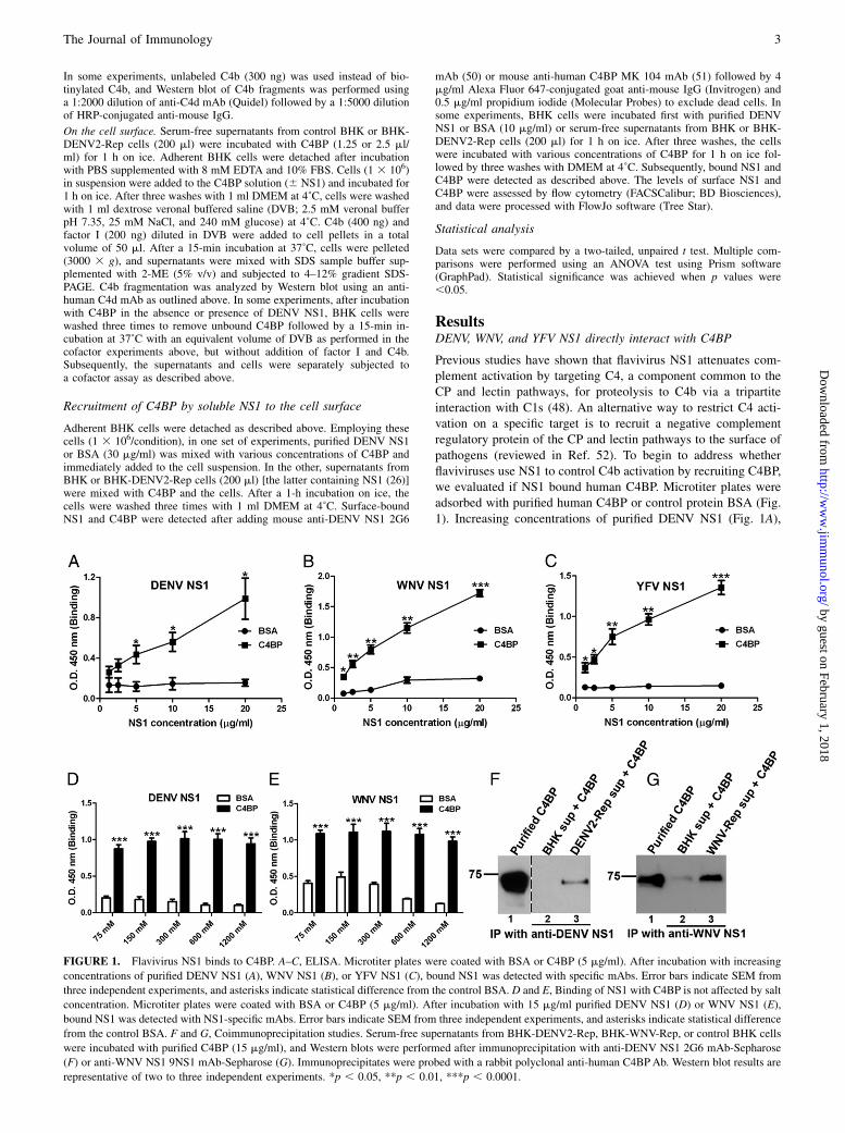
Previous studies have shown that flavivirus NS1 attenuates com-plement activation by targeting C4, a component common to theCP and lectin pathways, for proteolysis to C4b via a tripartiteinteraction with C1s (48). An alternative way to restrict C4 acti-vation on a specific target is to recruit a negative complementregulatory protein of the CP and lectin pathways to the surface ofpathogens (reviewed in Ref. 52). To begin to address whetherflaviviruses use NS1 to control C4b activation by recruiting C4BP,we evaluated if NS1 bound human C4BP. Microtiter plates wereadsorbed with purified human C4BP or control protein BSA (Fig.1). Increasing concentrations of purified DENV NS1 (Fig. 1A),
FIGURE 1. Flavivirus NS1 binds to C4BP. A–C, ELISA. Microtiter plates were coated with BSA or C4BP (5 mg/ml). After incubation with increasing
concentrations of purified DENV NS1 (A), WNV NS1 (B), or YFV NS1 (C), bound NS1 was detected with specific mAbs. Error bars indicate SEM from
three independent experiments, and asterisks indicate statistical difference from the control BSA. D and E, Binding of NS1 with C4BP is not affected by salt
concentration. Microtiter plates were coated with BSA or C4BP (5 mg/ml). After incubation with 15 mg/ml purified DENV NS1 (D) or WNV NS1 (E),
bound NS1 was detected with NS1-specific mAbs. Error bars indicate SEM from three independent experiments, and asterisks indicate statistical difference
from the control BSA. F and G, Coimmunoprecipitation studies. Serum-free supernatants from BHK-DENV2-Rep, BHK-WNV-Rep, or control BHK cells
were incubated with purified C4BP (15 mg/ml), and Western blots were performed after immunoprecipitation with anti-DENV NS1 2G6 mAb-Sepharose
(F) or anti-WNV NS1 9NS1 mAb-Sepharose (G). Immunoprecipitates were probed with a rabbit polyclonal anti-human C4BPAb. Western blot results are
representative of two to three independent experiments. *p , 0.05, **p , 0.01, ***p , 0.0001.
The Journal of Immunology 3
by guest on February 1, 2018http://w
ww
.jimm
unol.org/D
ownloaded from
WNV NS1 (Fig. 1B), or YFV NS1 (Fig. 1C) were added to C4BP-or BSA-coated wells, and bound NS1 was detected with specificmAbs. A dose-dependent interaction between all three NS1 andC4BP was identified. Increasing ionic strength of the buffer didnot appreciably affect the NS1–C4BP interaction, suggestinga nonionic interaction between C4BP and DENV or WNV NS1(Fig. 1D, 1E). Coimmunoprecipitation experiments confirmed theinteraction between NS1 and C4BP (Fig. 1F, 1G).To determine the region(s) of C4BP that interact(s) with NS1,
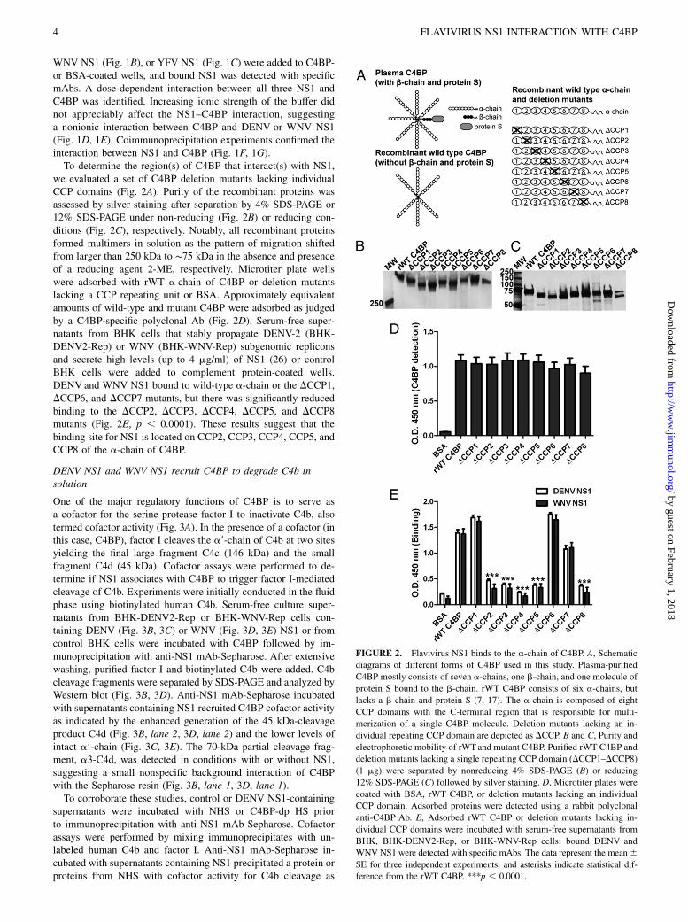
we evaluated a set of C4BP deletion mutants lacking individualCCP domains (Fig. 2A). Purity of the recombinant proteins wasassessed by silver staining after separation by 4% SDS-PAGE or12% SDS-PAGE under non-reducing (Fig. 2B) or reducing con-ditions (Fig. 2C), respectively. Notably, all recombinant proteinsformed multimers in solution as the pattern of migration shiftedfrom larger than 250 kDa to ∼75 kDa in the absence and presenceof a reducing agent 2-ME, respectively. Microtiter plate wellswere adsorbed with rWT a-chain of C4BP or deletion mutantslacking a CCP repeating unit or BSA. Approximately equivalentamounts of wild-type and mutant C4BP were adsorbed as judgedby a C4BP-specific polyclonal Ab (Fig. 2D). Serum-free super-natants from BHK cells that stably propagate DENV-2 (BHK-DENV2-Rep) or WNV (BHK-WNV-Rep) subgenomic repliconsand secrete high levels (up to 4 mg/ml) of NS1 (26) or controlBHK cells were added to complement protein-coated wells.DENV and WNV NS1 bound to wild-type a-chain or the DCCP1,DCCP6, and DCCP7 mutants, but there was significantly reducedbinding to the DCCP2, DCCP3, DCCP4, DCCP5, and DCCP8mutants (Fig. 2E, p , 0.0001). These results suggest that thebinding site for NS1 is located on CCP2, CCP3, CCP4, CCP5, andCCP8 of the a-chain of C4BP.
DENV NS1 and WNV NS1 recruit C4BP to degrade C4b insolution
One of the major regulatory functions of C4BP is to serve asa cofactor for the serine protease factor I to inactivate C4b, alsotermed cofactor activity (Fig. 3A). In the presence of a cofactor (inthis case, C4BP), factor I cleaves the a9-chain of C4b at two sitesyielding the final large fragment C4c (146 kDa) and the smallfragment C4d (45 kDa). Cofactor assays were performed to de-termine if NS1 associates with C4BP to trigger factor I-mediatedcleavage of C4b. Experiments were initially conducted in the fluidphase using biotinylated human C4b. Serum-free culture super-natants from BHK-DENV2-Rep or BHK-WNV-Rep cells con-taining DENV (Fig. 3B, 3C) or WNV (Fig. 3D, 3E) NS1 or fromcontrol BHK cells were incubated with C4BP followed by im-munoprecipitation with anti-NS1 mAb-Sepharose. After extensivewashing, purified factor I and biotinylated C4b were added. C4bcleavage fragments were separated by SDS-PAGE and analyzed byWestern blot (Fig. 3B, 3D). Anti-NS1 mAb-Sepharose incubatedwith supernatants containing NS1 recruited C4BP cofactor activityas indicated by the enhanced generation of the 45 kDa-cleavageproduct C4d (Fig. 3B, lane 2, 3D, lane 2) and the lower levels ofintact a9-chain (Fig. 3C, 3E). The 70-kDa partial cleavage frag-ment, a3-C4d, was detected in conditions with or without NS1,suggesting a small nonspecific background interaction of C4BPwith the Sepharose resin (Fig. 3B, lane 1, 3D, lane 1).
To corroborate these studies, control or DENV NS1-containingsupernatants were incubated with NHS or C4BP-dp HS priorto immunoprecipitation with anti-NS1 mAb-Sepharose. Cofactorassays were performed by mixing immunoprecipitates with un-labeled human C4b and factor I. Anti-NS1 mAb-Sepharose in-cubated with supernatants containing NS1 precipitated a protein orproteins from NHS with cofactor activity for C4b cleavage as
FIGURE 2. Flavivirus NS1 binds to the a-chain of C4BP. A, Schematic
diagrams of different forms of C4BP used in this study. Plasma-purified
C4BP mostly consists of seven a-chains, one b-chain, and one molecule of
protein S bound to the b-chain. rWT C4BP consists of six a-chains, but
lacks a b-chain and protein S (7, 17). The a-chain is composed of eight
CCP domains with the C-terminal region that is responsible for multi-
merization of a single C4BP molecule. Deletion mutants lacking an in-
dividual repeating CCP domain are depicted as DCCP. B and C, Purity and
electrophoretic mobility of rWTand mutant C4BP. Purified rWT C4BP and
deletion mutants lacking a single repeating CCP domain (DCCP1–DCCP8)
(1 mg) were separated by nonreducing 4% SDS-PAGE (B) or reducing
12% SDS-PAGE (C) followed by silver staining. D, Microtiter plates were
coated with BSA, rWT C4BP, or deletion mutants lacking an individual
CCP domain. Adsorbed proteins were detected using a rabbit polyclonal
anti-C4BP Ab. E, Adsorbed rWT C4BP or deletion mutants lacking in-
dividual CCP domains were incubated with serum-free supernatants from
BHK, BHK-DENV2-Rep, or BHK-WNV-Rep cells; bound DENV and
WNV NS1 were detected with specific mAbs. The data represent the mean6SE for three independent experiments, and asterisks indicate statistical dif-
ference from the rWT C4BP. ***p , 0.0001.
4 FLAVIVIRUS NS1 INTERACTION WITH C4BP
by guest on February 1, 2018http://w
ww
.jimm
unol.org/D
ownloaded from
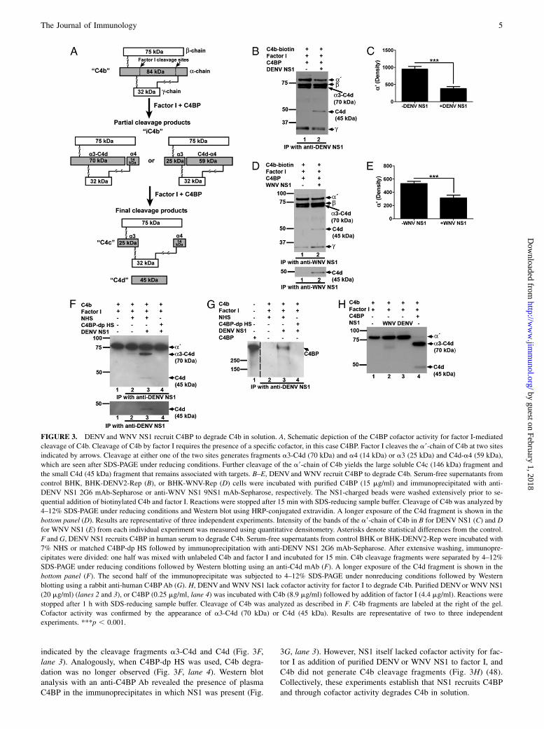
indicated by the cleavage fragments a3-C4d and C4d (Fig. 3F,lane 3). Analogously, when C4BP-dp HS was used, C4b degra-dation was no longer observed (Fig. 3F, lane 4). Western blotanalysis with an anti-C4BP Ab revealed the presence of plasmaC4BP in the immunoprecipitates in which NS1 was present (Fig.
3G, lane 3). However, NS1 itself lacked cofactor activity for fac-tor I as addition of purified DENV or WNV NS1 to factor I, andC4b did not generate C4b cleavage fragments (Fig. 3H) (48).Collectively, these experiments establish that NS1 recruits C4BPand through cofactor activity degrades C4b in solution.
FIGURE 3. DENV and WNV NS1 recruit C4BP to degrade C4b in solution. A, Schematic depiction of the C4BP cofactor activity for factor I-mediated
cleavage of C4b. Cleavage of C4b by factor I requires the presence of a specific cofactor, in this case C4BP. Factor I cleaves the a9-chain of C4b at two sites
indicated by arrows. Cleavage at either one of the two sites generates fragments a3-C4d (70 kDa) and a4 (14 kDa) or a3 (25 kDa) and C4d-a4 (59 kDa),
which are seen after SDS-PAGE under reducing conditions. Further cleavage of the a9-chain of C4b yields the large soluble C4c (146 kDa) fragment and
the small C4d (45 kDa) fragment that remains associated with targets. B–E, DENV and WNV recruit C4BP to degrade C4b. Serum-free supernatants from
control BHK, BHK-DENV2-Rep (B), or BHK-WNV-Rep (D) cells were incubated with purified C4BP (15 mg/ml) and immunoprecipitated with anti-
DENV NS1 2G6 mAb-Sepharose or anti-WNV NS1 9NS1 mAb-Sepharose, respectively. The NS1-charged beads were washed extensively prior to se-
quential addition of biotinylated C4b and factor I. Reactions were stopped after 15 min with SDS-reducing sample buffer. Cleavage of C4b was analyzed by
4–12% SDS-PAGE under reducing conditions and Western blot using HRP-conjugated extravidin. A longer exposure of the C4d fragment is shown in the
bottom panel (D). Results are representative of three independent experiments. Intensity of the bands of the a9-chain of C4b in B for DENV NS1 (C) and D
for WNV NS1 (E) from each individual experiment was measured using quantitative densitometry. Asterisks denote statistical differences from the control.
F and G, DENV NS1 recruits C4BP in human serum to degrade C4b. Serum-free supernatants from control BHK or BHK-DENV2-Rep were incubated with
7% NHS or matched C4BP-dp HS followed by immunoprecipitation with anti-DENV NS1 2G6 mAb-Sepharose. After extensive washing, immunopre-
cipitates were divided: one half was mixed with unlabeled C4b and factor I and incubated for 15 min. C4b cleavage fragments were separated by 4–12%
SDS-PAGE under reducing conditions followed by Western blotting using an anti-C4d mAb (F). A longer exposure of the C4d fragment is shown in the
bottom panel (F). The second half of the immunoprecipitate was subjected to 4–12% SDS-PAGE under nonreducing conditions followed by Western
blotting using a rabbit anti-human C4BPAb (G). H, DENVand WNV NS1 lack cofactor activity for factor I to degrade C4b. Purified DENVor WNV NS1
(20 mg/ml) (lanes 2 and 3), or C4BP (0.25 mg/ml, lane 4) was incubated with C4b (8.9 mg/ml) followed by addition of factor I (4.4 mg/ml). Reactions were
stopped after 1 h with SDS-reducing sample buffer. Cleavage of C4b was analyzed as described in F. C4b fragments are labeled at the right of the gel.
Cofactor activity was confirmed by the appearance of a3-C4d (70 kDa) or C4d (45 kDa). Results are representative of two to three independent
experiments. ***p , 0.001.
The Journal of Immunology 5
by guest on February 1, 2018http://w
ww
.jimm
unol.org/D
ownloaded from
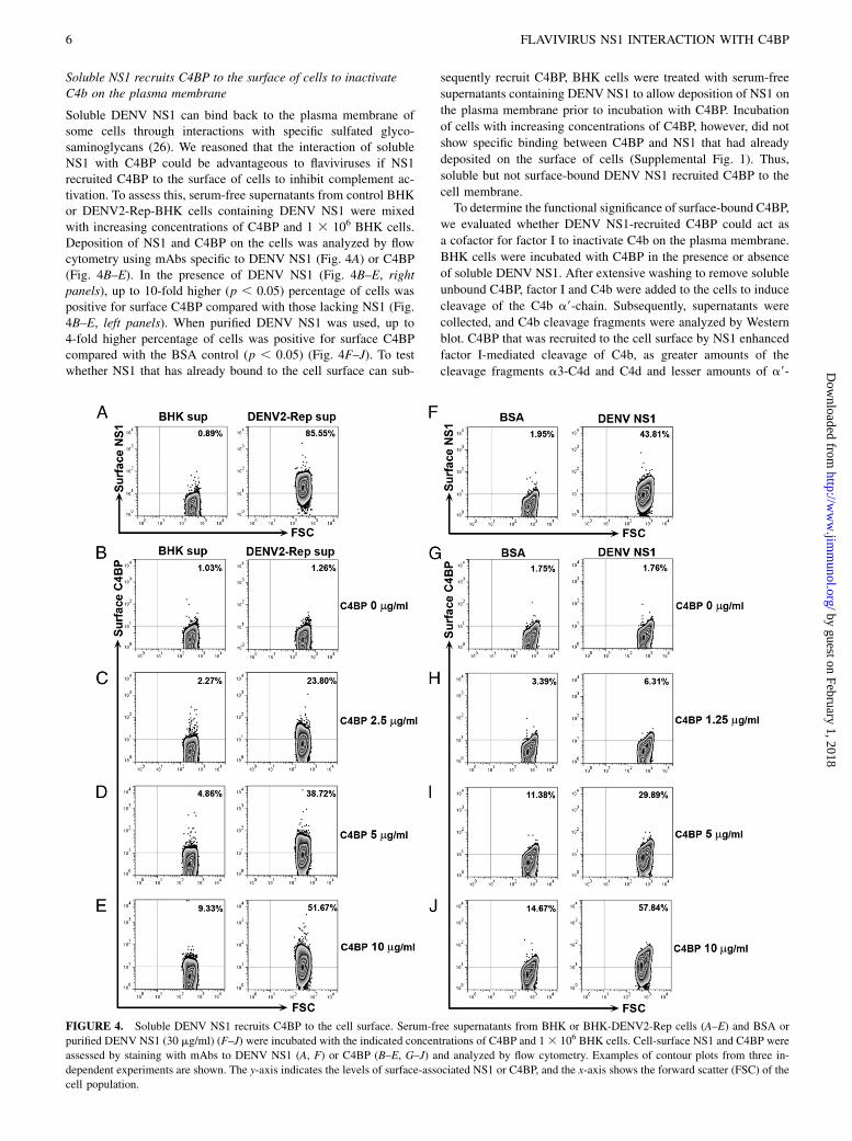
Soluble NS1 recruits C4BP to the surface of cells to inactivateC4b on the plasma membrane
Soluble DENV NS1 can bind back to the plasma membrane ofsome cells through interactions with specific sulfated glyco-saminoglycans (26). We reasoned that the interaction of solubleNS1 with C4BP could be advantageous to flaviviruses if NS1recruited C4BP to the surface of cells to inhibit complement ac-tivation. To assess this, serum-free supernatants from control BHKor DENV2-Rep-BHK cells containing DENV NS1 were mixedwith increasing concentrations of C4BP and 1 3 106 BHK cells.Deposition of NS1 and C4BP on the cells was analyzed by flowcytometry using mAbs specific to DENV NS1 (Fig. 4A) or C4BP(Fig. 4B–E). In the presence of DENV NS1 (Fig. 4B–E, rightpanels), up to 10-fold higher (p , 0.05) percentage of cells waspositive for surface C4BP compared with those lacking NS1 (Fig.4B–E, left panels). When purified DENV NS1 was used, up to4-fold higher percentage of cells was positive for surface C4BPcompared with the BSA control (p , 0.05) (Fig. 4F–J). To testwhether NS1 that has already bound to the cell surface can sub-
sequently recruit C4BP, BHK cells were treated with serum-free
supernatants containing DENV NS1 to allow deposition of NS1 on
the plasma membrane prior to incubation with C4BP. Incubation
of cells with increasing concentrations of C4BP, however, did not
show specific binding between C4BP and NS1 that had already
deposited on the surface of cells (Supplemental Fig. 1). Thus,
soluble but not surface-bound DENV NS1 recruited C4BP to the
cell membrane.To determine the functional significance of surface-bound C4BP,
we evaluated whether DENV NS1-recruited C4BP could act as
a cofactor for factor I to inactivate C4b on the plasma membrane.
BHK cells were incubated with C4BP in the presence or absence
of soluble DENV NS1. After extensive washing to remove soluble
unbound C4BP, factor I and C4b were added to the cells to induce
cleavage of the C4b a9-chain. Subsequently, supernatants were
collected, and C4b cleavage fragments were analyzed by Western
blot. C4BP that was recruited to the cell surface by NS1 enhanced
factor I-mediated cleavage of C4b, as greater amounts of the
cleavage fragments a3-C4d and C4d and lesser amounts of a9-
FIGURE 4. Soluble DENV NS1 recruits C4BP to the cell surface. Serum-free supernatants from BHK or BHK-DENV2-Rep cells (A–E) and BSA or
purified DENV NS1 (30 mg/ml) (F–J) were incubated with the indicated concentrations of C4BP and 13 106 BHK cells. Cell-surface NS1 and C4BP were
assessed by staining with mAbs to DENV NS1 (A, F) or C4BP (B–E, G–J) and analyzed by flow cytometry. Examples of contour plots from three in-
dependent experiments are shown. The y-axis indicates the levels of surface-associated NS1 or C4BP, and the x-axis shows the forward scatter (FSC) of the
cell population.
6 FLAVIVIRUS NS1 INTERACTION WITH C4BP
by guest on February 1, 2018http://w
ww
.jimm
unol.org/D
ownloaded from
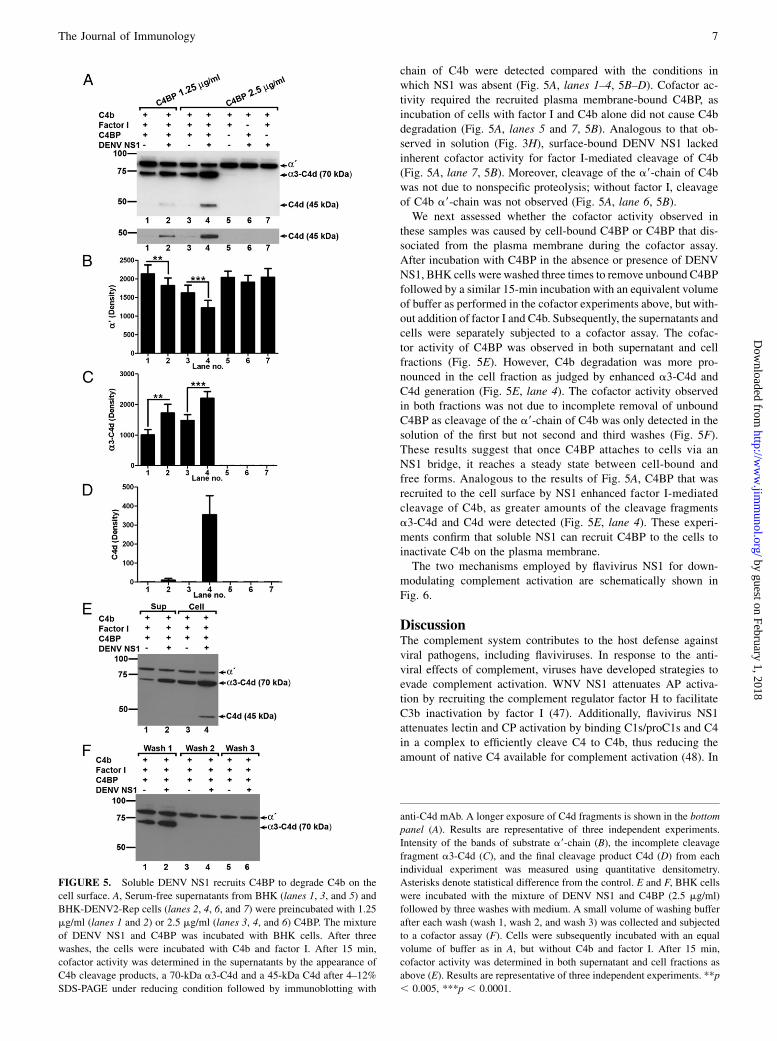
chain of C4b were detected compared with the conditions inwhich NS1 was absent (Fig. 5A, lanes 1–4, 5B–D). Cofactor ac-tivity required the recruited plasma membrane-bound C4BP, asincubation of cells with factor I and C4b alone did not cause C4bdegradation (Fig. 5A, lanes 5 and 7, 5B). Analogous to that ob-served in solution (Fig. 3H), surface-bound DENV NS1 lackedinherent cofactor activity for factor I-mediated cleavage of C4b(Fig. 5A, lane 7, 5B). Moreover, cleavage of the a9-chain of C4bwas not due to nonspecific proteolysis; without factor I, cleavageof C4b a9-chain was not observed (Fig. 5A, lane 6, 5B).We next assessed whether the cofactor activity observed in
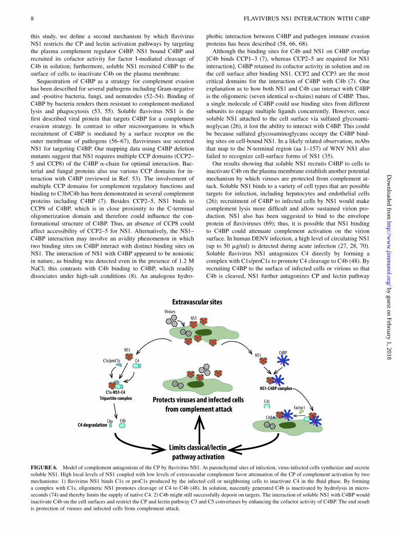
these samples was caused by cell-bound C4BP or C4BP that dis-sociated from the plasma membrane during the cofactor assay.After incubation with C4BP in the absence or presence of DENVNS1, BHK cells were washed three times to remove unbound C4BPfollowed by a similar 15-min incubation with an equivalent volumeof buffer as performed in the cofactor experiments above, but with-out addition of factor I and C4b. Subsequently, the supernatants andcells were separately subjected to a cofactor assay. The cofac-tor activity of C4BP was observed in both supernatant and cellfractions (Fig. 5E). However, C4b degradation was more pro-nounced in the cell fraction as judged by enhanced a3-C4d andC4d generation (Fig. 5E, lane 4). The cofactor activity observedin both fractions was not due to incomplete removal of unboundC4BP as cleavage of the a9-chain of C4b was only detected in thesolution of the first but not second and third washes (Fig. 5F).These results suggest that once C4BP attaches to cells via anNS1 bridge, it reaches a steady state between cell-bound andfree forms. Analogous to the results of Fig. 5A, C4BP that wasrecruited to the cell surface by NS1 enhanced factor I-mediatedcleavage of C4b, as greater amounts of the cleavage fragmentsa3-C4d and C4d were detected (Fig. 5E, lane 4). These experi-ments confirm that soluble NS1 can recruit C4BP to the cells toinactivate C4b on the plasma membrane.The two mechanisms employed by flavivirus NS1 for down-
modulating complement activation are schematically shown inFig. 6.
DiscussionThe complement system contributes to the host defense againstviral pathogens, including flaviviruses. In response to the anti-viral effects of complement, viruses have developed strategies toevade complement activation. WNV NS1 attenuates AP activa-tion by recruiting the complement regulator factor H to facilitateC3b inactivation by factor I (47). Additionally, flavivirus NS1attenuates lectin and CP activation by binding C1s/proC1s and C4in a complex to efficiently cleave C4 to C4b, thus reducing theamount of native C4 available for complement activation (48). In
FIGURE 5. Soluble DENV NS1 recruits C4BP to degrade C4b on the
cell surface. A, Serum-free supernatants from BHK (lanes 1, 3, and 5) and
BHK-DENV2-Rep cells (lanes 2, 4, 6, and 7) were preincubated with 1.25
mg/ml (lanes 1 and 2) or 2.5 mg/ml (lanes 3, 4, and 6) C4BP. The mixture
of DENV NS1 and C4BP was incubated with BHK cells. After three
washes, the cells were incubated with C4b and factor I. After 15 min,
cofactor activity was determined in the supernatants by the appearance of
C4b cleavage products, a 70-kDa a3-C4d and a 45-kDa C4d after 4–12%
SDS-PAGE under reducing condition followed by immunoblotting with
anti-C4d mAb. A longer exposure of C4d fragments is shown in the bottom
panel (A). Results are representative of three independent experiments.
Intensity of the bands of substrate a9-chain (B), the incomplete cleavage
fragment a3-C4d (C), and the final cleavage product C4d (D) from each
individual experiment was measured using quantitative densitometry.
Asterisks denote statistical difference from the control. E and F, BHK cells
were incubated with the mixture of DENV NS1 and C4BP (2.5 mg/ml)
followed by three washes with medium. A small volume of washing buffer
after each wash (wash 1, wash 2, and wash 3) was collected and subjected
to a cofactor assay (F). Cells were subsequently incubated with an equal
volume of buffer as in A, but without C4b and factor I. After 15 min,
cofactor activity was determined in both supernatant and cell fractions as
above (E). Results are representative of three independent experiments. **p
, 0.005, ***p , 0.0001.
The Journal of Immunology 7
by guest on February 1, 2018http://w
ww
.jimm
unol.org/D
ownloaded from
this study, we define a second mechanism by which flavivirusNS1 restricts the CP and lectin activation pathways by targetingthe plasma complement regulator C4BP. NS1 bound C4BP andrecruited its cofactor activity for factor I-mediated cleavage ofC4b in solution; furthermore, soluble NS1 recruited C4BP to thesurface of cells to inactivate C4b on the plasma membrane.Sequestration of C4BP as a strategy for complement evasion
has been described for several pathogens including Gram-negativeand -positive bacteria, fungi, and nematodes (52–54). Binding ofC4BP by bacteria renders them resistant to complement-mediatedlysis and phagocytosis (53, 55). Soluble flavivirus NS1 is thefirst described viral protein that targets C4BP for a complementevasion strategy. In contrast to other microorganisms in whichrecruitment of C4BP is mediated by a surface receptor on theouter membrane of pathogens (56–67), flaviviruses use secretedNS1 for targeting C4BP. Our mapping data using C4BP deletionmutants suggest that NS1 requires multiple CCP domains (CCP2–5 and CCP8) of the C4BP a-chain for optimal interaction. Bac-terial and fungal proteins also use various CCP domains for in-teraction with C4BP (reviewed in Ref. 53). The involvement ofmultiple CCP domains for complement regulatory functions andbinding to C3b/C4b has been demonstrated in several complementproteins including C4BP (7). Besides CCP2–5, NS1 binds toCCP8 of C4BP, which is in close proximity to the C-terminaloligomerization domain and therefore could influence the con-formational structure of C4BP. Thus, an absence of CCP8 couldaffect accessibility of CCP2–5 for NS1. Alternatively, the NS1–C4BP interaction may involve an avidity phenomenon in whichtwo binding sites on C4BP interact with distinct binding sites onNS1. The interaction of NS1 with C4BP appeared to be nonionicin nature, as binding was detected even in the presence of 1.2 MNaCl; this contrasts with C4b binding to C4BP, which readilydissociates under high-salt conditions (8). An analogous hydro-
phobic interaction between C4BP and pathogen immune evasionproteins has been described (58, 66, 68).Although the binding sites for C4b and NS1 on C4BP overlap
[C4b binds CCP1–3 (7), whereas CCP2–5 are required for NS1interaction], C4BP retained its cofactor activity in solution and onthe cell surface after binding NS1. CCP2 and CCP3 are the mostcritical domains for the interaction of C4BP with C4b (7). Oneexplanation as to how both NS1 and C4b can interact with C4BPis the oligomeric (seven identical a-chains) nature of C4BP. Thus,a single molecule of C4BP could use binding sites from differentsubunits to engage multiple ligands concurrently. However, oncesoluble NS1 attached to the cell surface via sulfated glycosami-noglycan (26), it lost the ability to interact with C4BP. This couldbe because sulfated glycosaminoglycans occupy the C4BP bind-ing sites on cell-bound NS1. In a likely related observation, mAbsthat map to the N-terminal region (aa 1–157) of WNV NS1 alsofailed to recognize cell-surface forms of NS1 (35).Our results showing that soluble NS1 recruits C4BP to cells to
inactivate C4b on the plasma membrane establish another potentialmechanism by which viruses are protected from complement at-tack. Soluble NS1 binds to a variety of cell types that are possibletargets for infection, including hepatocytes and endothelial cells(26); recruitment of C4BP to infected cells by NS1 would makecomplement lysis more difficult and allow sustained virion pro-duction. NS1 also has been suggested to bind to the envelopeprotein of flaviviruses (69); thus, it is possible that NS1 bindingto C4BP could attenuate complement activation on the virionsurface. In human DENV infection, a high level of circulating NS1(up to 50 mg/ml) is detected during acute infection (27, 28, 70).Soluble flavivirus NS1 antagonizes C4 directly by forming acomplex with C1s/proC1s to promote C4 cleavage to C4b (48). Byrecruiting C4BP to the surface of infected cells or virions so thatC4b is cleaved, NS1 further antagonizes CP and lectin pathway
FIGURE 6. Model of complement antagonism of the CP by flavivirus NS1. At parenchymal sites of infection, virus-infected cells synthesize and secrete
soluble NS1. High local levels of NS1 coupled with low levels of extravascular complement favor attenuation of the CP of complement activation by two
mechanisms: 1) flavivirus NS1 binds C1s or proC1s produced by the infected cell or neighboring cells to inactivate C4 in the fluid phase. By forming
a complex with C1s, oligomeric NS1 promotes cleavage of C4 to C4b (48). In solution, nascently generated C4b is inactivated by hydrolysis in micro-
seconds (74) and thereby limits the supply of native C4. 2) C4b might still successfully deposit on targets. The interaction of soluble NS1 with C4BP would
inactivate C4b on the cell surfaces and restrict the CP and lectin pathway C3 and C5 convertases by enhancing the cofactor activity of C4BP. The end result
is protection of viruses and infected cells from complement attack.
8 FLAVIVIRUS NS1 INTERACTION WITH C4BP
by guest on February 1, 2018http://w
ww
.jimm
unol.org/D
ownloaded from
activation. Overall, these data point to the importance of the CP/lectin pathway in controlling flavivirus infection. Indeed, micelacking CP/lectin pathway components are more susceptible tolethal WNV infection (71, 72), and recognition of N-linkedglycans on the envelope protein by mannose-binding lectin ac-celerates the clearance of DENV from plasma (73).In summary, our studies define a second mechanism by which
NS1 inhibits the activation of the CP and lectin pathways. Inperipheral tissues where the majority of flavivirus infection oc-curs, higher local concentrations of NS1 coupled with lowerlevels of extravascular complement could efficiently attenuatecomplement activation (Fig. 6). Flaviviruses may use NS1 to bindproC1s produced from infected or neighboring cells to inactivateC4 in the fluid phase. By forming a complex with C1s, oligomericNS1 promotes cleavage of C4 to C4b. In solution, nascently gen-erated C4b is inactivated rapidly by hydrolysis. However, onceC4b is deposited on the surface of targets (e.g., virions or in-fected cells), the interaction of NS1 with C4BP inactivates C4band restricts classical and lectin C3 and C5 convertase activities.In this manner, NS1 enables flavivirus to evade complementcontrol and facilitate dissemination.
AcknowledgmentsWe thank R. Kuhn for the BHK-YFV-Rep cells, C. Puttikhunt and
W. Kasinrerk for providing the anti-DENV NS1 mAbs, J. Schlesinger
for the anti-YFV NS1 mAb, F. Bergstrom for purification of the C4BP
mutants, and the Rheumatic Diseases Core Center for NS1 purification.
DisclosuresThe authors have no financial conflicts of interest.
References1. Avirutnan, P., E. Mehlhop, and M. S. Diamond. 2008. Complement and its role
in protection and pathogenesis of flavivirus infections. Vaccine 26(Suppl 8):I100–I107.
2. Stoermer, K. A., and T. E. Morrison. 2011. Complement and viral pathogenesis.Virology 411: 362–373.
3. Lambris, J. D., D. Ricklin, and B. V. Geisbrecht. 2008. Complement evasion byhuman pathogens. Nat. Rev. Microbiol. 6: 132–142.
4. Dahlback, B., C. A. Smith, and H. J. Muller-Eberhard. 1983. Visualization ofhuman C4b-binding protein and its complexes with vitamin K-dependent proteinS and complement protein C4b. Proc. Natl. Acad. Sci. USA 80: 3461–3465.
5. de Cordoba, S. R., O. C. Garcia, and P. Sanchez-Corral. 2000. C4b-bindingprotein. In The complement facts book. B. J. Morley, and M. J. Walport, eds.Academic Press, London, p. 161–167.
6. Rodriguez de Cordoba, S., P. Sanchez-Corral, and J. Rey-Campos. 1991.Structure of the gene coding for the alpha polypeptide chain of the humancomplement component C4b-binding protein. J. Exp. Med. 173: 1073–1082.
7. Blom, A. M., L. Kask, and B. Dahlback. 2001. Structural requirements for thecomplement regulatory activities of C4BP. J. Biol. Chem. 276: 27136–27144.
8. Blom, A. M., J. Webb, B. O. Villoutreix, and B. Dahlback. 1999. A cluster ofpositively charged amino acids in the C4BP alpha-chain is crucial for C4bbinding and factor I cofactor function. J. Biol. Chem. 274: 19237–19245.
9. Fujita, T., and V. Nussenzweig. 1979. The role of C4-binding protein and beta1H in proteolysis of C4b and C3b. J. Exp. Med. 150: 267–276.
10. Fujita, T., and N. Tamura. 1983. Interaction of C4-binding protein with cell-bound C4b. A quantitative analysis of binding and the role of C4-binding proteinin proteolysis of cell-bound C4b. J. Exp. Med. 157: 1239–1251.
11. Brodeur, S. R., F. Angelini, L. B. Bacharier, A. M. Blom, E. Mizoguchi,H. Fujiwara, A. Plebani, L. D. Notarangelo, B. Dahlback, E. Tsitsikov, andR. S. Geha. 2003. C4b-binding protein (C4BP) activates B cells through theCD40 receptor. Immunity 18: 837–848.
12. Williams, K. T., S. P. Young, A. Negus, L. S. Young, D. H. Adams, andS. C. Afford. 2007. C4b binding protein binds to CD154 preventing CD40mediated cholangiocyte apoptosis: a novel link between complement and epi-thelial cell survival. PLoS ONE 2: e159.
13. Spijkers, P. P., C. V. Denis, A. M. Blom, and P. J. Lenting. 2008. Cellular uptakeof C4b-binding protein is mediated by heparan sulfate proteoglycans and CD91/LDL receptor-related protein. Eur. J. Immunol. 38: 809–817.
14. Shayakhmetov, D. M., A. Gaggar, S. Ni, Z. Y. Li, and A. Lieber. 2005. Ade-novirus binding to blood factors results in liver cell infection and hepatotoxicity.J. Virol. 79: 7478–7491.
15. Holmberg, M. T., A. M. Blom, and S. Meri. 2001. Regulation of complementclassical pathway by association of C4b-binding protein to the surfaces ofSK-OV-3 and Caov-3 ovarian adenocarcinoma cells. J. Immunol. 167: 935–939.
16. Trouw, L. A., A. A. Bengtsson, K. A. Gelderman, B. Dahlback, G. Sturfelt, andA. M. Blom. 2007. C4b-binding protein and factor H compensate for the loss ofmembrane-bound complement inhibitors to protect apoptotic cells against ex-cessive complement attack. J. Biol. Chem. 282: 28540–28548.
17. Trouw, L. A., S. C. Nilsson, I. Goncalves, G. Landberg, and A. M. Blom. 2005.C4b-binding protein binds to necrotic cells and DNA, limiting DNA release andinhibiting complement activation. J. Exp. Med. 201: 1937–1948.
18. Lindenbach, B. D., H.-J. Thiel, and C. Rice. 2007. Flaviviridae: the viruses andtheir replication. In Field’s Virology, 5th ed. D. M. Knipe, and P. M. Howley, eds.Lippincott Williams & Wilkins, Philadelphia. p. 1101–1152.
19. Khromykh, A. A., P. L. Sedlak, K. J. Guyatt, R. A. Hall, and E. G. Westaway.1999. Efficient trans-complementation of the flavivirus kunjin NS5 protein butnot of the NS1 protein requires its coexpression with other components of theviral replicase. J. Virol. 73: 10272–10280.
20. Lindenbach, B. D., and C. M. Rice. 1997. trans-Complementation of yel-low fever virus NS1 reveals a role in early RNA replication. J. Virol. 71: 9608–9617.
21. Mackenzie, J. M., M. K. Jones, and P. R. Young. 1996. Immunolocalization ofthe dengue virus nonstructural glycoprotein NS1 suggests a role in viral RNAreplication. Virology 220: 232–240.
22. Flamand, M., F. Megret, M. Mathieu, J. Lepault, F. A. Rey, and V. Deubel. 1999.Dengue virus type 1 nonstructural glycoprotein NS1 is secreted from mammaliancells as a soluble hexamer in a glycosylation-dependent fashion. J. Virol. 73:6104–6110.
23. Crooks, A. J., J. M. Lee, L. M. Easterbrook, A. V. Timofeev, andJ. R. Stephenson. 1994. The NS1 protein of tick-borne encephalitis virus formsmultimeric species upon secretion from the host cell. J. Gen. Virol. 75: 3453–3460.
24. Winkler, G., S. E. Maxwell, C. Ruemmler, and V. Stollar. 1989. Newly syn-thesized dengue-2 virus nonstructural protein NS1 is a soluble protein butbecomes partially hydrophobic and membrane-associated after dimerization.Virology 171: 302–305.
25. Winkler, G., V. B. Randolph, G. R. Cleaves, T. E. Ryan, and V. Stollar. 1988.Evidence that the mature form of the flavivirus nonstructural protein NS1 isa dimer. Virology 162: 187–196.
26. Avirutnan, P., L. Zhang, N. Punyadee, A. Manuyakorn, C. Puttikhunt,W. Kasinrerk, P. Malasit, J. P. Atkinson, and M. S. Diamond. 2007. Secreted NS1of dengue virus attaches to the surface of cells via interactions with heparansulfate and chondroitin sulfate E. PLoS Pathog. 3: e183.
27. Libraty, D. H., P. R. Young, D. Pickering, T. P. Endy, S. Kalayanarooj, S. Green,D. W. Vaughn, A. Nisalak, F. A. Ennis, and A. L. Rothman. 2002. High circu-lating levels of the dengue virus nonstructural protein NS1 early in dengue ill-ness correlate with the development of dengue hemorrhagic fever. J. Infect. Dis.186: 1165–1168.
28. Avirutnan, P., N. Punyadee, S. Noisakran, C. Komoltri, S. Thiemmeca,K. Auethavornanan, A. Jairungsri, R. Kanlaya, N. Tangthawornchaikul,C. Puttikhunt, et al. 2006. Vascular leakage in severe dengue virus infections:a potential role for the nonstructural viral protein NS1 and complement. J. Infect.Dis. 193: 1078–1088.
29. Chang, H. H., H. F. Shyu, Y. M. Wang, D. S. Sun, R. H. Shyu, S. S. Tang, andY. S. Huang. 2002. Facilitation of cell adhesion by immobilized dengue viralnonstructural protein 1 (NS1): arginine-glycine-aspartic acid structural mimicrywithin the dengue viral NS1 antigen. J. Infect. Dis. 186: 743–751.
30. Falconar, A. K. 1997. The dengue virus nonstructural-1 protein (NS1) generatesantibodies to common epitopes on human blood clotting, integrin/adhesin pro-teins and binds to human endothelial cells: potential implications in haemor-rhagic fever pathogenesis. Arch. Virol. 142: 897–916.
31. Sun, D. S., C. C. King, H. S. Huang, Y. L. Shih, C. C. Lee, W. J. Tsai, C. C. Yu,and H. H. Chang. 2007. Antiplatelet autoantibodies elicited by dengue virus non-structural protein 1 cause thrombocytopenia and mortality in mice. J. Thromb.Haemost. 5: 2291–2299.
32. Lin, C. F., H. Y. Lei, A. L. Shiau, C. C. Liu, H. S. Liu, T. M. Yeh, S. H. Chen, andY. S. Lin. 2003. Antibodies from dengue patient sera cross-react with endothelialcells and induce damage. J. Med. Virol. 69: 82–90.
33. Alcon-LePoder, S., M. T. Drouet, P. Roux, M. P. Frenkiel, M. Arborio,A. M. Durand-Schneider, M. Maurice, I. Le Blanc, J. Gruenberg, andM. Flamand. 2005. The secreted form of dengue virus nonstructural protein NS1is endocytosed by hepatocytes and accumulates in late endosomes: implicationsfor viral infectivity. J. Virol. 79: 11403–11411.
34. Chung, K. M., G. E. Nybakken, B. S. Thompson, M. J. Engle, A. Marri,D. H. Fremont, and M. S. Diamond. 2006. Antibodies against West Nile Virusnonstructural protein NS1 prevent lethal infection through Fc gamma receptor-dependent and -independent mechanisms. J. Virol. 80: 1340–1351.
35. Chung, K. M., B. S. Thompson, D. H. Fremont, and M. S. Diamond. 2007.Antibody recognition of cell surface-associated NS1 triggers Fc-gammareceptor-mediated phagocytosis and clearance of West Nile Virus-infectedcells. J. Virol. 81: 9551–9555.
36. Despres, P., J. Dietrich, M. Girard, and M. Bouloy. 1991. Recombinant bacu-loviruses expressing yellow fever virus E and NS1 proteins elicit protectiveimmunity in mice. J. Gen. Virol. 72: 2811–2816.
37. Falgout, B., M. Bray, J. J. Schlesinger, and C. J. Lai. 1990. Immunization of micewith recombinant vaccinia virus expressing authentic dengue virus nonstructural
The Journal of Immunology 9
by guest on February 1, 2018http://w
ww
.jimm
unol.org/D
ownloaded from
protein NS1 protects against lethal dengue virus encephalitis. J. Virol. 64: 4356–4363.
38. Gould, E. A., A. Buckley, A. D. Barrett, and N. Cammack. 1986. Neutralizing(54K) and non-neutralizing (54K and 48K) monoclonal antibodies againststructural and non-structural yellow fever virus proteins confer immunity inmice. J. Gen. Virol. 67: 591–595.
39. Henchal, E. A., L. S. Henchal, and J. J. Schlesinger. 1988. Synergistic inter-actions of anti-NS1 monoclonal antibodies protect passively immunized micefrom lethal challenge with dengue 2 virus. J. Gen. Virol. 69: 2101–2107.
40. Jacobs, S. C., J. R. Stephenson, and G. W. Wilkinson. 1992. High-level ex-pression of the tick-borne encephalitis virus NS1 protein by using an adenovirus-based vector: protection elicited in a murine model. J. Virol. 66: 2086–2095.
41. Jacobs, S. C., J. R. Stephenson, and G. W. Wilkinson. 1994. Protection elicitedby a replication-defective adenovirus vector expressing the tick-borne enceph-alitis virus non-structural glycoprotein NS1. J. Gen. Virol. 75: 2399–2402.
42. Schlesinger, J. J., M. W. Brandriss, C. B. Cropp, and T. P. Monath. 1986. Pro-tection against yellow fever in monkeys by immunization with yellow fever virusnonstructural protein NS1. J. Virol. 60: 1153–1155.
43. Schlesinger, J. J., M. W. Brandriss, and E. E. Walsh. 1985. Protection against17D yellow fever encephalitis in mice by passive transfer of monoclonal anti-bodies to the nonstructural glycoprotein gp48 and by active immunization withgp48. J. Immunol. 135: 2805–2809.
44. Schlesinger, J. J., M. W. Brandriss, and E. E. Walsh. 1987. Protection of miceagainst dengue 2 virus encephalitis by immunization with the dengue 2 virusnon-structural glycoprotein NS1. J. Gen. Virol. 68: 853–857.
45. Schlesinger, J. J., M. Foltzer, and S. Chapman. 1993. The Fc portion of antibodyto yellow fever virus NS1 is a determinant of protection against YF encephalitisin mice. Virology 192: 132–141.
46. Krishna, V. D., M. Rangappa, and V. Satchidanandam. 2009. Virus-specificcytolytic antibodies to nonstructural protein 1 of Japanese encephalitis viruseffect reduction of virus output from infected cells. J. Virol. 83: 4766–4777.
47. Chung, K. M., M. K. Liszewski, G. Nybakken, A. E. Davis, R. R. Townsend,D. H. Fremont, J. P. Atkinson, and M. S. Diamond. 2006. West Nile virusnonstructural protein NS1 inhibits complement activation by binding the regu-latory protein factor H. Proc. Natl. Acad. Sci. USA 103: 19111–19116.
48. Avirutnan, P., A. Fuchs, R. E. Hauhart, P. Somnuke, S. Youn, M. S. Diamond,and J. P. Atkinson. 2010. Antagonism of the complement component C4 byflavivirus nonstructural protein NS1. J. Exp. Med. 207: 793–806.
49. Kask, L., L. A. Trouw, B. Dahlback, and A. M. Blom. 2004. The C4b-bindingprotein-protein S complex inhibits the phagocytosis of apoptotic cells. J. Biol.Chem. 279: 23869–23873.
50. Puttikhunt, C., W. Kasinrerk, S. Srisa-ad, T. Duangchinda, W. Silakate,S. Moonsom, N. Sittisombut, and P. Malasit. 2003. Production of anti-dengueNS1monoclonal antibodies by DNA immunization. J. Virol. Methods 109: 55–61.
51. Hardig, Y., A. Hillarp, and B. Dahlback. 1997. The amino-terminal module ofthe C4b-binding protein alpha-chain is crucial for C4b binding and factor I-cofactor function. Biochem. J. 323: 469–475.
52. Blom, A. M., B. O. Villoutreix, and B. Dahlback. 2004. Complement inhibitorC4b-binding protein-friend or foe in the innate immune system? Mol. Immunol.40: 1333–1346.
53. Blom, A. M., and S. Ram. 2008. Contribution of interactions between comple-ment inhibitor C4b-binding protein and pathogens to their ability to establishinfection with particular emphasis on Neisseria gonorrhoeae. Vaccine 26(Suppl8): I49–I55.
54. Haapasalo, K., T. Meri, and T. S. Jokiranta. 2009. Loa loa Microfilariae evadecomplement attack in vivo by acquiring regulatory proteins from host plasma.Infect. Immun. 77: 3886–3893.
55. Carlsson, F., K. Berggard, M. Stalhammar-Carlemalm, and G. Lindahl. 2003.Evasion of phagocytosis through cooperation between two ligand-bindingregions in Streptococcus pyogenes M protein. J. Exp. Med. 198: 1057–1068.
56. Accardo, P., P. Sanchez-Corral, O. Criado, E. Garcıa, and S. Rodrıguez deCordoba. 1996. Binding of human complement component C4b-binding protein(C4BP) to Streptococcus pyogenes involves the C4b-binding site. J. Immunol.157: 4935–4939.
57. Barbosa, A. S., D. Monaris, L. B. Silva, Z. M. Morais, S. A. Vasconcellos,A. M. Cianciarullo, L. Isaac, and P. A. Abreu. 2010. Functional characterizationof LcpA, a surface-exposed protein of Leptospira spp. that binds the humancomplement regulator C4BP. Infect. Immun. 78: 3207–3216.
58. Blom, A. M., K. Berggard, J. H. Webb, G. Lindahl, B. O. Villoutreix, andB. Dahlback. 2000. Human C4b-binding protein has overlapping, but not iden-tical, binding sites for C4b and streptococcal M proteins. J. Immunol. 164: 5328–5336.
59. Blom, A. M., A. Rytkonen, P. Vasquez, G. Lindahl, B. Dahlback, andA. B. Jonsson. 2001. A novel interaction between type IV pili of Neisseriagonorrhoeae and the human complement regulator C4B-binding protein. J.Immunol. 166: 6764–6770.
60. Grosskinsky, S., M. Schott, C. Brenner, S. J. Cutler, M. M. Simon, andR. Wallich. 2010. Human complement regulators C4b-binding protein and C1esterase inhibitor interact with a novel outer surface protein of Borrelia recur-rentis. PLoS Negl. Trop. Dis. 4: e698.
61. Johnsson, E., A. Thern, B. Dahlback, L. O. Heden, M. Wikstrom, andG. Lindahl. 1996. A highly variable region in members of the streptococcal Mprotein family binds the human complement regulator C4BP. J. Immunol. 157:3021–3029.
62. Kamdar, H. V., K. B. Rowley, D. Clements, and S. S. Patil. 1991. Pseudomonassyringae pv. phaseolicola genomic clones harboring heterologous DNAsequences suppress the same phaseolotoxin-deficient mutants. J. Bacteriol. 173:1073–1079.
63. Morfeldt, E., K. Berggard, J. Persson, T. Drakenberg, E. Johnsson, E. Lindahl,S. Linse, and G. Lindahl. 2001. Isolated hypervariable regions derived fromstreptococcal M proteins specifically bind human C4b-binding protein: impli-cations for antigenic variation. J. Immunol. 167: 3870–3877.
64. Nordstrom, T., A. M. Blom, A. Forsgren, and K. Riesbeck. 2004. The emergingpathogen Moraxella catarrhalis interacts with complement inhibitor C4b bind-ing protein through ubiquitous surface proteins A1 and A2. J. Immunol. 173:4598–4606.
65. Pietikainen, J., T. Meri, A. M. Blom, and S. Meri. 2010. Binding of the com-plement inhibitor C4b-binding protein to Lyme disease Borreliae.Mol. Immunol.47: 1299–1305.
66. Ram, S., M. Cullinane, A. M. Blom, S. Gulati, D. P. McQuillen, B. G. Monks,C. O’Connell, R. Boden, C. Elkins, M. K. Pangburn, et al. 2001. Binding of C4b-binding protein to porin: a molecular mechanism of serum resistance of Neis-seria gonorrhoeae. J. Exp. Med. 193: 281–295.
67. Weiser, J. N., and E. C. Gotschlich. 1991. Outer membrane protein A (OmpA)contributes to serum resistance and pathogenicity of Escherichia coli K-1. Infect.Immun. 59: 2252–2258.
68. Prasadarao, N. V., A. M. Blom, B. O. Villoutreix, and L. C. Linsangan. 2002. Anovel interaction of outer membrane protein A with C4b binding proteinmediates serum resistance of Escherichia coli K1. J. Immunol. 169: 6352–6360.
69. Blitvich, B. J., J. S. Mackenzie, R. J. Coelen, M. J. Howard, and R. A. Hall.1995. A novel complex formed between the flavivirus E and NS1 proteins:analysis of its structure and function. Arch. Virol. 140: 145–156.
70. Alcon, S., A. Talarmin, M. Debruyne, A. Falconar, V. Deubel, and M. Flamand.2002. Enzyme-linked immunosorbent assay specific to Dengue virus type 1nonstructural protein NS1 reveals circulation of the antigen in the blood duringthe acute phase of disease in patients experiencing primary or secondaryinfections. J. Clin. Microbiol. 40: 376–381.
71. Fuchs, A., T. Y. Lin, D. W. Beasley, C. M. Stover, W. J. Schwaeble,T. C. Pierson, and M. S. Diamond. 2010. Direct complement restriction of fla-vivirus infection requires glycan recognition by mannose-binding lectin. CellHost Microbe 8: 186–195.
72. Mehlhop, E., and M. S. Diamond. 2006. Protective immune responses againstWest Nile virus are primed by distinct complement activation pathways. J. Exp.Med. 203: 1371–1381.
73. Fuchs, A., A. K. Pinto, W. J. Schwaeble, and M. S. Diamond. 2011. The lectinpathway of complement activation contributes to protection from West Nile virusinfection. Virology 412: 101–109.
74. Morgan, B. P. 1990. Complement: Clinical Aspects and Relevance to Disease.Academic Press, London.
10 FLAVIVIRUS NS1 INTERACTION WITH C4BP
by guest on February 1, 2018http://w
ww
.jimm
unol.org/D
ownloaded from