binding specificity of serum amyloid
TRANSCRIPT
B I N D I N G S P E C I F I C I T Y O F S E R U M A M Y L O I D P
C O M P O N E N T F O R T H E P Y R U V A T E A C E T A L O F
G A L A C T O S E
BY C. R. K. HIND, P. M. COLLINS, D. RENN, R. B. COOK, D. CASPI, MARILYN L. BALTZ, AND M. B. PEPYS
From the Immunological Medicine Unit, Department of Medicine, Royal Postgraduate Medical School, Hammersmith Hospital, London W12 OHS, Englanc# the Department of Chemistry,
Birkbeck College, University of London, London WC1E 7HX, England; and the Marine Colloids Division, FMC Corporation, Rockland, Maine
Serum amyloid P component (SAP) 1 is a normal plasma glycoprotein composed of non-covalently associated subunits a r ranged with cyclic pentameric symmetry in a disk-like configuration. It is a member o f the pentaxin family o f proteins, which includes C-reactive prote in (CRP), the classical acute phase reactant, and the homologues o f CRP and SAP that exist in lower animals (1, 2). Although its biological function is unknown, there are a number of observations indicating that SAP is likely to be o f both physiological and pathophysiological importance. First, there are proteins that closely resemble human SAP in terms o f appearance in the electron microscope, subunit composition, and amino acid sequence in the sera o f all vertebrates in which they have been sought (3-5). Second, a protein that is immunochemical ly indistinguishable f rom SAP is a normal integral con- sti tuent o f the human glomerular basement membrane (6) and o f the peripheral microfibrillar mantle o f elastic fibers th roughou t the body (7). Thi rd , both in man and lower animals SAP is laid down as amyloid P component (AP), a minor but significant const i tuent o f all forms o f amyloid deposits (8) with the possible exception o f the intracerebral plaques in human Alzheimer 's disease and senile dementia (9).
In addit ion to the stable evolut ionary conservation o f s t ructure in SAP of
This work was supported by Medical Research Council Programme grant 979/51. A preliminary account of these findings was presented at the Medical Research Society meeting, 7 July 1983 (C. R. K. Hind et al. 1983. Clin. Sci. (Lond.). 65:17p). C. R. K. Hind is the recipient of a research fellowship from the Medical Research Council. Correspondence should be addressed to M. B. Pepys at the Immunological Medicine Unit, Department of Medicine, Royal Postgraduate Medical School, Ham- mersmith Hospital, Du Cane Road, London W12 0HS.
1 Abbreviations used in this paper: [a]D temp, optical rotation; AP, amyloid P component; BSA, bovine serum albumin; pmax, frequency of infra-red absorption band; C4bp, C4-binding protein; CRP, C- reactive protein; cyclic acetal MOBDG, methyl 4,6-O-(1-carboxyethylidene)-fl-D-galactopyranoside; ~i, chemical shifts; Fn, fibronectin; J, coupling constants; NHS, normal human serum; NMR, nuclear magnetic resonance; non-cyclic acetal MOBDG, methyl (4 or 6)-O-[(R,S)-I '-carboxy-1 '-methoxy-eth- yl]4~-D-galactopyranoside; PAGE, polyacrylamide gel electrophoresis; p.p.m., parts per million; Rf, spot mobility on t.l.c.; SAP, serum amyloid P component; signal multiplicities: s, singlets; d, doublets; t, triplets; m, multiplets; t.l.c., thin layer chromatography; TMS, tetramethylsilane; Tris-saline-Ca, 0.01 M Tris-buffered 0.138 M NaCI containing 0.002 M CaCI~ and 0.1% wt/vol NaNs at pH 8.0; Tris-saline°EDTA, 0.01 M Tris-buffered 0.138 M NaCI containing 0.01 M EDTA at pH 8.0.
1058 J. ExP. MED. © The Rockefeller University Press • 0022-1007/84/04/1058/12 $1.00 Volume 159 April 1984 1058-1069
Dow
nloaded from http://rupress.org/jem
/article-pdf/159/4/1058/1094287/1058.pdf by guest on 05 January 2022
HIND ET AL. 1059
different species, these proteins all share the capacity for specific calcium- dependent ligand binding. This property was first discovered with respect to agarose (3, 10, 11) although earlier work on a then unidentified plasma glyco- protein (12), which was later shown to be SAP (13), had demonstrated its affinity for calcium ions. It was then established that SAP binds to amyloid fibrils in a calcium-dependent interaction that probably underlies the in vivo deposition of AP (14). Furthermore, in the presence of calcium, aggregated, but not native, human SAP selectively binds fibronectin (Fn) and C4-binding protein (C4bp) from normal human serum (15). All these interactions are highly specific and it seems likely that ligand binding by SAP participates in its in vivo function. Identification of the chemical nature of the ligand(s) should therefore help to explain the biological role of SAP and may also contribute to structural charac- terization of the protein itself.
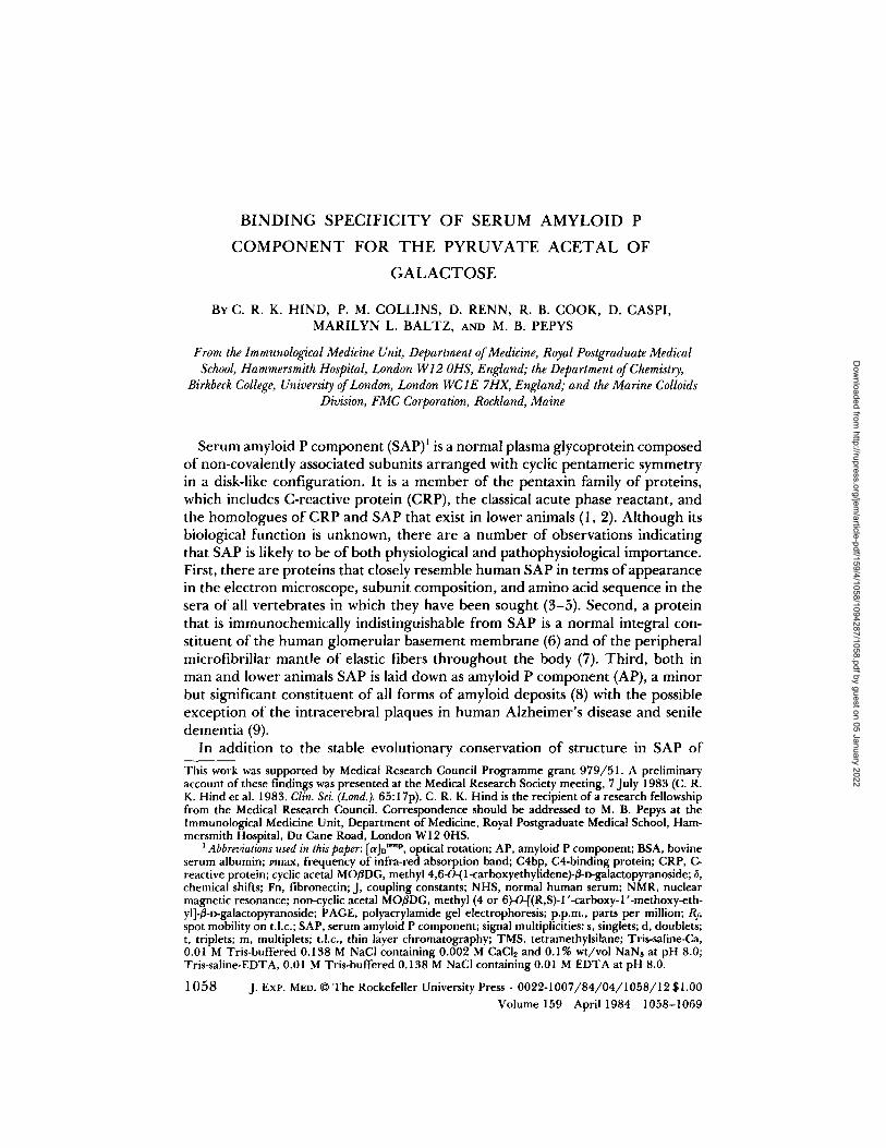
Although it is most improbable that binding of SAP to agarose is itself related to a physiological function of the protein, it has greatly facilitated the isolation and characterization of SAP from man and other animals and provides a model system suitable for chemical studies. Agarose is a linear galactan hydrocolloid derived from marine algae and composed of repeating units of agarobiose (1,3- 1inked ~-D-galactopyranose and 1,4-1inked 3,6-anhydro-a-L-galactopyranose) to- gether with trace amounts of sulfate and pyruvate, the latter as the 4,6-pyruvate acetal of/3-D-galactopyranose (16, 17) (Fig. 1). There are large differences in the amounts of SAP that bind to beaded agarose gels from different sources (14), suggesting that the actual ligand for SAP in agarose may be a variable trace constituent of the polysaccharide. We report here that binding of SAP to gels prepared from a range of agaroses of known composition correlates closely with their pyruvate content, but not at all with the amount of sulfate present. Methylation of the carboxylic acid of the pyruvate residues abolishes the binding of SAP. We have therefore synthesized a pyruvate acetal at the 4,6-positions of galactose, the form in which it exists in agarose, and have shown that it inhibits the calcium-dependent binding of SAP to agarose, to amyloid fibrils, to fibro- nectin, and to C4-binding protein. The availability of this chemically defined low molecular weight ligand should aid further work on SAP.
Materials and Methods
Agarose Agaroses of known pyruvate and sulfate content derived from Gelidium and Gracilaria
species (kindly provided by FMC Corporation, Rockland, ME) were used to prepare a 4% wt/vol gel in 0.01 M Tris-buffered 0.138 M NaCI containing 0.002 M CaCI2 and 0.1% wt/vol NaN3 at pH 8.0 (Tris-saline-Ca). The gels were fragmented into beads by sequential
o:Z ol. %o/"0 ..
FIGURE 1. Agarobiose showing the position of pyruvic acid as the acetal 4,6-O-(1-carboxy- ethylidene)-/3-D-galactopyranose.
Dow
nloaded from http://rupress.org/jem
/article-pdf/159/4/1058/1094287/1058.pdf by guest on 05 January 2022
1060 BINDING SPECIFICITY OF SERUM AMYLOID P COMPONENT
passage through 710-gm and 80-gm sieves (Endecotts Ltd., London, England), and were stored in suspension in Tris-saline-Ca at 4°C.
Diazomethane-treated Aga rose Pyruvate-rich (0.25% wt/wt) agarose beads (4 ml packed volume) were added to 30 ml
of an ethereal solution of diazomethane (18) that had been diluted 1:1 vol/voi in methanol, and stirred for 60 rain at room temperature. After removal of the supernatant, the beads were washed with distilled water and then resuspended in Tris-saline-Ca. Control solvent- treated beads were mixed with identical volumes of ether/methanol (1:1 vol/vol) alone.
Chemicals Methyl/3-D-galactopyranoside, methyl pyruvate, sodium pyruvate, and D-galactose were
obtained from Sigma Chemical Co. Ltd., Poole, Dorset, England; ion exchange resin (IR 120 H +) and organic solvents from BDH Ltd., Dagenham, Essex, England and 10% palladium on charcoal from Johnson Matthey Chemicals Ltd., Royston, Herts, England.
Chemical Techniques all-Nuclear magnetic resonance (NMR) spectra were usually measured in CDCI3 with
a JNM-FX200 Fourier transform NMR spectrometer (Jeol Ltd., Tokyo, Japan) or a WH- 400 spectrometer (Bruker Ltd., Karlsruhe, Federal Republic of Germany) with tetra- methyisilane (TMS) as internal standard. Natural-abundance ~SC-NMR spectra were measured in CDC13 with JNM-FX60 or FX200 instruments (Jeol Ltd.) operating at 15 or 50 MHz, respectively. The 1H-NMR spectra are presented as follows: chemical shifts (6) in parts per million (p.p.m.) downfield from TMS; signal multiplicities (s, d, t, m, for singlets, doublets, triplets, muitiplets); signal intensities (e.g. 3H); coupling constants in Hz (e.g. Jl.~ 3,0 Hz); signal assignment (e.g. H-2). For the 'SC-NMR spectra the chemical shift is reported followed, where possible, by the signal assignment.
Optical rotations ([aID temp) were measured in chloroform with an AA-10 polarimeter (Optical Activity Ltd., Huntingdon, Cambridgeshire, England). Infrared spectra were measured (3'm~x recorded in cm -l) with a 597 spectrometer (Perkin Elmer Ltd., Beacons- field, Bucks, England) on samples smeared on potassium bromide disks.
Column chromatography was carried out on silica gel 230-400 mesh (Merck Kieselgel 9385, BDH Ltd.). For thin layer chromatography (TLC) 0.25-ram films of silica gel (Merck Kieseigel 60 F2s4, BDH Ltd.) were used, and the spots were located with an ethanol 1% sulfuric acid spray reagent. The mobility of the spot relative to the mobility of the solvent front (Rf) was determined (19). The eluting solvents were dried over molecular sieves 4A (BDH Ltd.).
Synthesis of acetals of Pyruvic Acid Derived from Methyl 13-D-Galactopyranoside Preparation of starting materials: Methyl pyruvate dimethyl acetal (compound I) was
prepared by heating methyl pyruvate under reflux with methanol in the presence of IR 120H + ion exchange resin. Methyl 2,3-di-O-benzyl-/3-D-galactopyranoside (compound II) was prepared by standard procedures from methyl 4,6-O-benzylidene-B-D-galactopyrano- side (20).
Condensation of methyl 2,3-di-O-benzyl-/3-D-galactopyranoside with methyl pyruvate: Compound II (1 g) was dissolved in compound I (9 ml) at 20°C. The solution was cooled to 0°C and concentrated sulfuric acid (0.1 ml) added with stirring. The solution was maintained at 20°C under reduced pressure (0.1 mm Hg) for 4 h when TLC (40:60 vol/vol petroleum ether) showed several intense spots in the range R l 0.1-0.5 and a little unchanged compound II (R: 0.0). The acid was then neutralized by stirring the solution with an excess of anhydrous sodium hydrogen carbonate (NaHCO3) added in portions during 3 h. Dichloromethane (1 1) was then added and the solution filtered. The filtrate was washed with aqueous NaHCOs and, after drying, evaporated to give a crude residue that was separated by column chromatography on silica gel with toluene, toluene/ethyl acetate (9:2 vol/vol) as an increasingly polar elutant. The major product was methyl 2,3-di-O- benzyl-4,6-O-[(R)-l-methoxycarbonylethylidene]-~-D-galactopyranoside (compound III)
Dow
nloaded from http://rupress.org/jem
/article-pdf/159/4/1058/1094287/1058.pdf by guest on 05 January 2022
HIND ET AL. 1 0 6 1
(642 rag); 1H-NMR (200 MHz) data: 4.17 (d, IH, J~.2 7.5 Hz, H-l), 3.52 (s, 3H, aglycone OCH3), 3.78 (s, 3H, CO~CH3), 1.58 (s, 3H, CH3C), 7.1-7.4 (m, 10H) and 4.65-4.92 (m, 4H) (2 × CH2C6Hs), plus signals for six galactose protons. In a similar, larger scale experiment using 5 g of compound II, a new major product was isolated (2.3 g) by chromatography using toluene, toluene/ethyl acetate (9:2 vol/vol), ethyl acetate as increasingly polar elutants. It has been tentatively identified as methyl 2,3-di-O-benzyl-(4 or 6)-O-(R,S-l'-methoxycarbonyl-l'-methoxy-ethyl)-3-~galactopyranoside (compound IV). Details of the 1H and lSC-NMR results may be obtained from the authors.
Hydrogenolysis of methyl 2,3-di-O-benzyl-B-o-galactopyranoside acetals of methyl pyruvate: Com- pound III (642 mg) in ethyl acetate (60 ml) containing 10% palladium on charcoal (150 mg) was shaken for 2 h under 2.5 atmospheres of hydrogen. The catalyst was filtered off, and the filtrate evaporated to give methyl 4,6-0-[(R)-1-methoxycarbonylethylidene]-3-D- galactopyranoside (compound V) (415 mg), 1H-NMR (200 MHz) data: 3.80 (s, 3H, COnCHs), 3.56 (s, 3H, aglycone OCH3), 1.63 (s, 3H, CCH3). The mixed, non-cyclic acetal (compound IV) (228 mg) was similarly hydrogenolyzed to give methyl (4 or 6)-0-[(R,S)- l'-methoxycarbonyl-l'-methoxy-ethyl]-3-r~galactopyranoside (compound VI) (141 mg).
I 13 Details of the H and C-NMR results may be obtained from the authors. Saponification of methyl 3-D-galactopyranoside acetals of methyl pyruvate: The cyclic acetai
methyl ester (compound V) (225 mg) was placed in 1.0 M aqueous sodium hydroxide (0.4 ml) at 23 °C for 24 h~ and then the pH adjusted to 7.4 with 1.0 M hydrochloric acid. This solution of the sodium salt of the acetal was used without further characterization. The mixed non-cyclic acetal methyl ester (compound VI) (141 mg) was similarly treated.
Proteins Isolated pure SAP (21) and Fn (22) were prepared from normal human serum (NHS)
and citrated human plasma, respectively, according to established methods. Isolated pure C4bp (23) was the generous gift of Dr. V. Nussenzweig, New York University, New York. Bovine serum albumin (BSA) was obtained from Sigma Chemical Co. Ltd.
Serum Fresh NHS was obtained by allowing venous blood from laboratory volunteers to clot
at room temperature for 2-4 h before centrifugation. In some experiments, serum from a single donor was used, in others different sera were pooled. Acute phase mouse serum was obtained by exsanguination of MRL mice 48 h after subcutaneous injection of 0.5 ml 2% wt/voi AgNO3 solution. Pooled normal plaice serum (Pleuronectes platessa L.) was kindly provided by Dr. Thelma C. Fletcher, NERC, Institute of Marine Biochemistry, Aberdeen.
Antisera Monospecific antisera to human SAP and Fn, and to mouse and plaice SAP were raised
by immunization of sheep and rabbits with the isolated pure proteins (24, 25).
Assay of SAP and Fn Specific protein concentrations were measured by electroimmunoassay (26) using
standards of the isolated pure proteins.
PAGE Gradient PAGE analysis of native proteins was performed in 4-30% gradient gels
(Pharmacia, Uppsala, Sweden), run exactly according to the manufacturer's instructions (15).
Immobilized Proteins Human SAP was coupled to CNBr-activated Sepharose 4B (Pharmacia) (SAP-Sepharose,
10 mg/ml) in pH 9.0 carbonate buffer precisely as described elsewhere (15). Control BSA-Sepharose beads (10 mg/ml) were prepared as before (15).
Dow
nloaded from http://rupress.org/jem
/article-pdf/159/4/1058/1094287/1058.pdf by guest on 05 January 2022
1062 B I N D I N G S P E C I F I C I T Y O F S E R U M A M Y L O I D P C O M P O N E N T
Isolation of Amyloid Fibrils Fibrils from the spleen of a patient with reactive systemic (AA) amyloidosis were isolated
(27) and lyophilized from distilled water.
Binding of SAP to Agarose Beads and to Amyloid Fibrils Known amounts of agarose beads and, in different experiments, of amyloid fibrils were
equilibrated with Tris-saline-Ca and then tested for SAP binding from serum exactly as described elsewhere (14). Elution with the chemicals under test was attempted using solutions in Tris-saline-Ca. In other experiments these solutions were added to serum before it was offered to either pyruvate-rich agarose beads (0.25% wt/wt) or fibrils. Tris- saline-EDTA was used as a control for complete elution or inhibition of binding of SAP.
Binding of Fn and C4bp by Immobilized Human SAP The experimental procedure for demonstration of binding of Fn and C4bp from NHS
by immobilized SAP has been described previously (15). Solutions in Tris-saline-Ca of the chemicals under test were used to either elute bound Fn and C4bp or to inhibit their uptake. Eiuates in Tris-saline-EDTA from SAP-Sepharose columns were tested by gradient PAGE and by specific electroimmunoassay for Fn.
Resu l t s
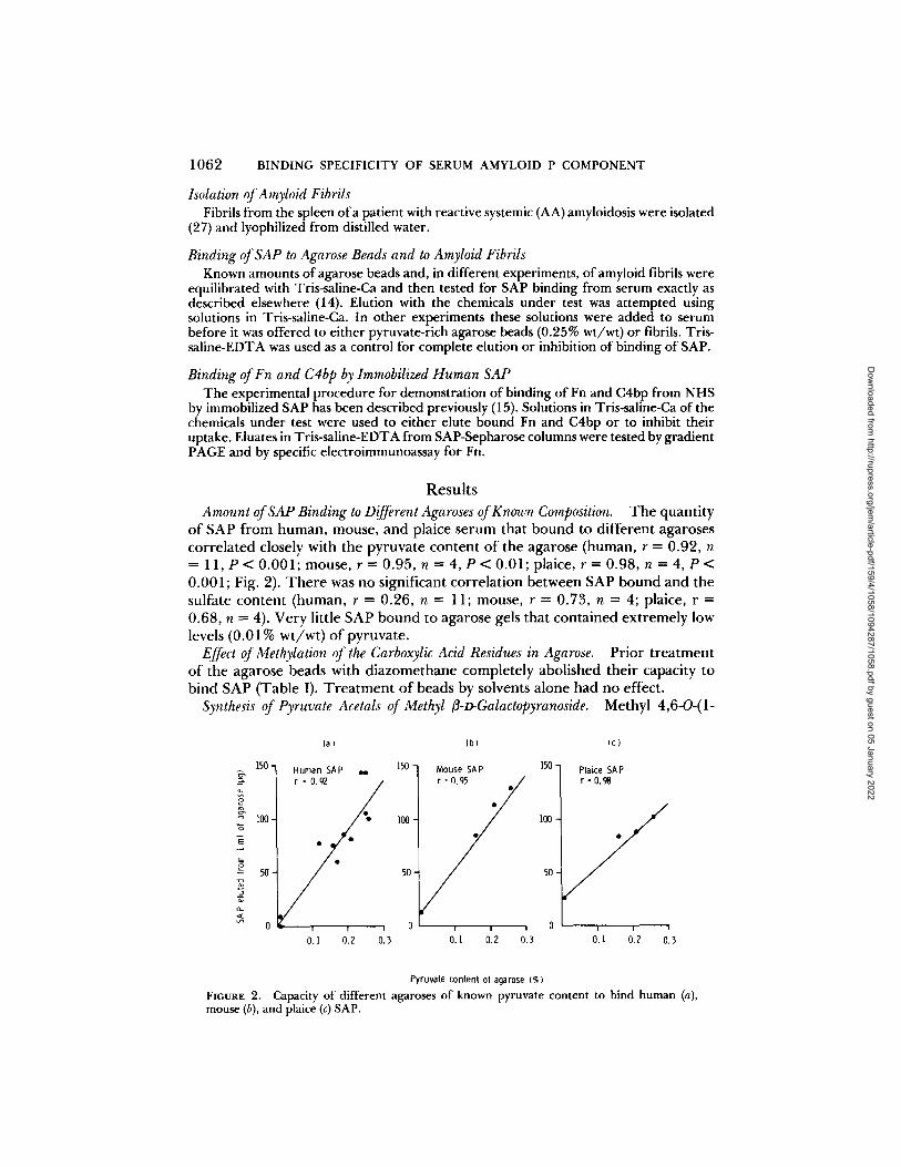
Amount of SAP Binding to Different Agaroses of Known Composition. T h e quanti ty o f SAP f rom human, mouse, and plaice serum that bound to different agaroses corre la ted closely with the pyruva te content o f the agarose (human, r = 0.92, n -- 11, P < 0.001; mouse, r = 0.95, n - 4, P < 0.01; plaice, r = 0.98, n = 4, P < 0.001; Fig. 2). T h e r e was no significant correlat ion between SAP bound and the sulfate content (human, r -- 0.26, n = 11; mouse, r = 0.73, n = 4; plaice, r = 0.68, n = 4). Very little SAP bound to agarose gels that contained ex t remely low levels (0.01% wt /wt ) o f pyruvate .
Effect of Methylation of the Carboxylic Acid Residues in Agarose. Prior t r ea tmen t o f the agarose beads with d iazomethane complete ly abolished their capacity to bind SAP (Table I). T r e a t m e n t o f beads by solvents alone had no effect.
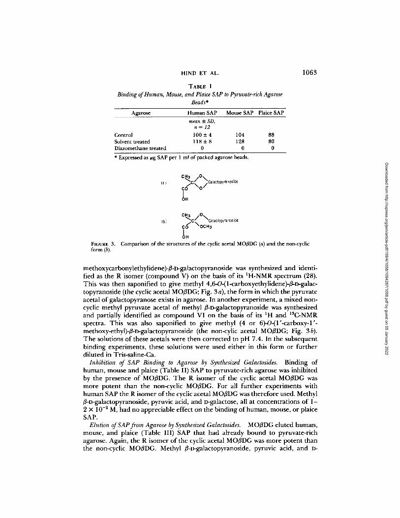
Synthesis of Pyruvate Acetals of Methyl 13-o-Galactopyranoside. Methyl 4,6-0-(1-
o_
(a) (b) 150 - 150 -
100-
Human SAP w r = 0.92 /
/ - / - 150 - Mouse SAP
]00 : "
50
100
50
(c)
Plaice SAP r = o.g8
/ / i i , 0 i i , 0 i i ,
0. l 0.2 0.3 0.1 0.2 0.3 0.1 0.2 0.3
Pyruvate content of agarose (%) FIGURE 2. C a p a c i t y o f d i f f e r e n t a g a r o s e s o f k n o w n p y r u v a t e c o n t e n t to b i n d h u m a n (a), m o u s e (b), a n d p la ice (c) SAP.
Dow
nloaded from http://rupress.org/jem
/article-pdf/159/4/1058/1094287/1058.pdf by guest on 05 January 2022
HIND ET AL.
TABLE I
Binding of Human, Mouse, and Plaice SAP to Pyruvate-rich Agarose Beads*
Agarose Human SAP Mouse SAP Plaice SAP
mean + SD, n = 1 2
Control 100 _+ 4 104 88 Solvent treated 118 -4- 8 128 80 Diazomethane treated 0 0 0
* Expressed as #g SAP per 1 ml of packed agarose beads.
1063
FIGURE 3. form (b).
(a) c , ~ / o \
/ .C ~,. /Galactopyra noside c O - - 0 " I OH
c ~ / o \ ( b ) -C. Ga lactopyra nos kle
c ~ " ~OCH3 I OH
Comparison of the structures of the cyclic acetal MO3DG (a) and the non-cyclic
methoxycarbonylethylidene)-/3-o-galactopyranoside was synthesized and identi- fied as the R isomer (compound V) on the basis of its IH-NMR spectrum (28). This was then saponified to give methyl 4,6-O-(1-carboxyethylidene)-/3-o-galac- topyranoside (the cyclic acetal MO/SDG; Fig. 3 a), the form in which the pyruvate acetal of galactopyranose exists in agarose. In another experiment, a mixed non- cyclic methyl pyruvate acetal of methyl /~-D-galactopyranoside was synthesized and partially identified as compound VI on the basis of its IH and 13C-NMR spectra. This was also saponified to give methyl (4 or 6)-O-(1 '-carboxy-1 '- methoxy-ethyl)-/3-I)-galactopyranoside (the non-cylic acetal MO/3DG; Fig. 3b). The solutions of these acetals were then corrected to pH 7.4. In the subsequent binding experiments, these solutions were used either in this form or further diluted in Tris-saline-Ca.
Inhibition of SAP Binding to Agarose by Synthesized Galactosides. Binding of human, mouse and plaice (Table II) SAP to pyruvate-rich agarose was inhibited by the presence of MO/3DG. The R isomer of the cyclic acetal MO/3DG was more potent than the non-cyclic MO/3DG. For all further experiments with human SAP the R isomer of the cyclic acetal MO/3DG was therefore used. Methyl /~-D-galactopyranoside, pyruvic acid, and D-galactose, all at concentrations of 1- 2 × 10 -2 M, had no appreciable effect on the binding of human, mouse, or plaice SAP.
Elution of SAP from Agarose by Synthesized Galactosides. MO/3DG eluted human, mouse, and plaice (Table III) SAP that had already bound to pyruvate-rich agarose. Again, the R isomer of the cyclic acetal MO/3DG was more potent than the non-cyclic MO/~DG. Methyl /3-o-galactopyranoside, pyruvic acid, and D-
Dow
nloaded from http://rupress.org/jem
/article-pdf/159/4/1058/1094287/1058.pdf by guest on 05 January 2022
1064 BINDING SPECIFICITY OF SERUM AMYLOID P COMPONENT
TABLE II Inhibition of Binding of SAP to Agarose by MO/3DG
Inhibition of SAP Chemical Concentration binding
%
Human MOflDG, R isomer 7.63 x 10 -3 M 95
7.63 × 10 -4 M 60 7.63 × 10 -~ M 0
Non-cyclic MOBDG 6.82 X 10 -3 M 52 6.82 × 10 -4 M 0
MO~DG, R isomer Mouse Plaice
5 X 10 -3 M 100 73 5 X 10 -4 M 60 15 5× 10-SM 0 0
TABLE III Elution of SAP from Agarose by MO[3DG
Chemical Concentration SAP eluted %
Human Tris-saline-EDTA control MOI3DG, R isomer
Non-cyclic MOI3DG
100 7.75 X 10 -3 M 91 7.75 X 10 -4 M 66 7.75 X 10 -5 M 0 2.04 × 10 -~ M 66 2.04 × 10 -5 M 42 2.04 × 10 -4 M 0
Tris-saline-EDTA control MOI3DG, R isomer
Mouse Plaice 100 100
3.8 × 10 -~ M 100 100 5.4 × 10 -3 M 74 21 5.4 X 10 -4 M 20 0 5.4 × 10 -5 M 0 0
galactose, all at 1 -2 × 10 -2 M, did no t e lu te h u m a n , mouse , or plaice SAP that had a l ready b o u n d to agarose.
Effect of the Cyclic acetal MOI3DG on Binding of Fn and C4bp by saP-Sepha- rose. W h e n a d d e d to fresh N H S be fo re m i x i n g with SAP-Sepharose , the R i somer o f the cyclic acetal MO/3DG i nh i b i t e d the b i n d i n g o f bo th Fn a n d C4bp. T h i s was shown by i m m u n o a s s a y o f Fn (Tab le IV) a n d g r a d i e n t P A G E analysis o f the s u b s e q u e n t T r i s - s a l i n e - E D T A e lua te f rom the SAP-Sepharose . In o t he r e x p e r i m e n t s , the R i somer of the cyclic acetal MO/3DG e lu ted Fn (Tab le V) a n d C 4 b p that were a l ready b o u n d by SAP-Sepharose , the g r a d i e n t P A G E prof i le o f these e luates b e i n g ident ica l to tha t o f the T r i s - s a l i n e - E D T A eluates (15).
Effect of the Cyclic Acetal MOI3DG on Human SAP Binding to Amyloid Fibrils. W h e n inc reas ing a m o u n t s o f the R i somer of the cyclic acetal MO/3DG were a d d e d to N H S be fo re b e i n g o f fe red to A A amylo id fibrils, the re was a progress ive
Dow
nloaded from http://rupress.org/jem
/article-pdf/159/4/1058/1094287/1058.pdf by guest on 05 January 2022
HIND ET AL.
TABLE IV Effect of the Cyclic Acetal MO[3DG on the Capacity of SAP-Sepharose to
Bind Fn
Total Fn eluted*
BSA- SAP-Seph- Sepha-
arose rose
ug Control:
Tris-saline-Ca 45 0 MOflDG, R isomer (7.66 × 10 -s M) 0 0
* NHS was offered to SAP-Sepharose in the presence of MOflDG, or Tris- saline-Ca as control, and after washing with Tris-saline-Ca was eluted with Tris-saline-EDTA.
TABLE V
Elution of Fn from SAP-Sepharose by the Cyclic Acetal MO[3DG
MOflDG, R isomer concentration Fn eluted: percent of
amount eluted by Tris-saline-EDTA
1.15 x 10 -2 M 100 2.23 × 10 -s M 85 1.15 x 10 -s M 85
2.3 × 10 -4 M 67
1065
TABLE VI Effect of the Cyclic Acetal MOI3DG on the Binding of Human SAP to
Amyloid Fibrils
Human SAP eluted with MOflDG, R isomer concentration Tris-saline-EDTA %
0 100 5.5 x 10 -5 M 78 5.5 x 10 -4 M 56 5.5 × 10 -3 M 50
r e d u c t i o n in t h e a m o u n t o f S A P b o u n d ( T a b l e VI) . In o t h e r e x p e r i m e n t s , t h e R i s o m e r o f t he cyclic ace ta l M O f l D G e l u t e d h u m a n S A P tha t h a d a l r e a d y b o u n d to t he A A a m y l o i d f ibr i ls ( T a b l e VII ) . N o e lu t i on was o b s e r v e d wi th m e t h y l fl-D- g a l a c t o p y r a n o s i d e , p y r u v i c ac id , o r D-galac tose a t c o n c e n t r a t i o n s b e t w e e n 10 -4 a n d 2 × 10 - 2 M .
D i s c u s s i o n
T h e p r e s e n t resu l t s show c lea r ly t ha t t h e a m o u n t o f S A P tha t b inds to a g a r o s e gels is d i r e c t l y r e l a t e d to t h e i r p y r u v a t e c o n t e n t . S A P scarce ly b i n d s a t all to gels t ha t e i t h e r have b e e n p r e p a r e d f r o m p y r u v a t e - f r e e a g a r o s e o r in wh ich the c a r b o x y l i c m o i e t y o f t h e p y r u v a t e has b e e n m e t h y l a t e d by t r e a t m e n t wi th
Dow
nloaded from http://rupress.org/jem
/article-pdf/159/4/1058/1094287/1058.pdf by guest on 05 January 2022
1066 B IN D IN G SPECIFICITY OF SERUM AMYLOID P C O M P O N E N T
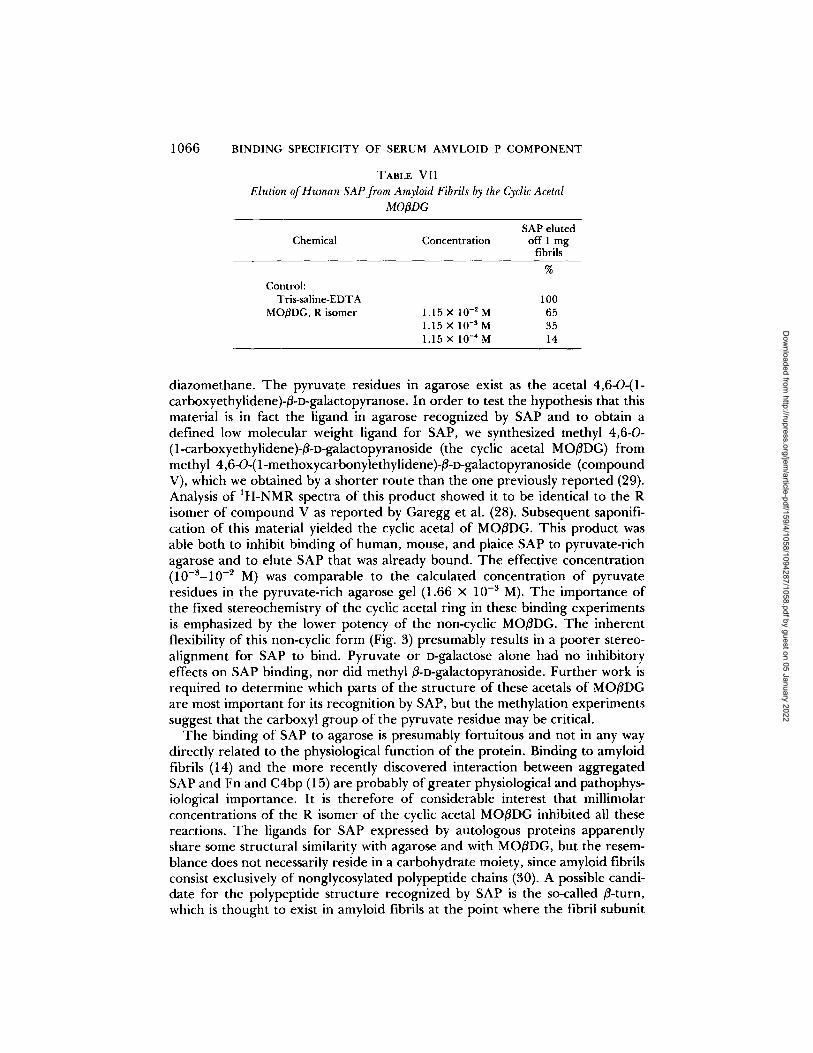
TABLE VII Elution of Human SAP from Amyloid Fibrils by the Cyclic Acetal
MOODG
SAP eluted Chemical Concentration off 1 mg
fibrils %
Control: Tris-saline-EDTA
MOBDG, R isomer 100
1.15 × 10 -2 M 65 1.15 X 10 -3 M 35 1.15 × 10 -4 M 14
diazomethane. The pyruvate residues in agarose exist as the acetal 4,6-O-(1- carboxyethylidene)-13-D-galactopyranose. In order to test the hypothesis that this material is in fact the ligand in agarose recognized by SAP and to obtain a defined low molecular weight ligand for SAP, we synthesized methyl 4,6-0- (1-carboxyethylidene)-li-o-galactopyranoside (the cyclic acetal MO/3DG) from methyl 4,6-O-(1-methoxycarbonylethylidene)-/i-D-galactopyranoside (compound V), which we obtained by a shorter route than the one previously reported (29). Analysis of 1H-NMR spectra of this product showed it to be identical to the R isomer of compound V as reported by Garegg et al. (28). Subsequent saponifi- cation of this material yielded the cyclic acetal of MOIiDG. This product was able both to inhibit binding of human, mouse, and plaice SAP to pyruvate-rich agarose and to elute SAP that was already bound. The effective concentration (10-3-10 -2 M) was comparable to the calculated concentration of pyruvate residues in the pyruvate-rich agarose gel (1.66 × 10 -3 M). The importance of the fixed stereochemistry of the cyclic acetal ring in these binding experiments is emphasized by the lower potency of the non-cyclic MOI3DG. The inherent flexibility of this non-cyclic form (Fig. 3) presumably results in a poorer stereo- alignment for SAP to bind. Pyruvate or D-galactose alone had no inhibitory effects on SAP binding, nor did methyl/3-D-galactopyranoside. Further work is required to determine which parts of the structure of these acetals of MO/3DG are most important for its recognition by SAP, but the methylation experiments suggest that the carboxyl group of the pyruvate residue may be critical.
The binding of SAP to agarose is presumably fortuitous and not in any way directly related to the physiological function of the protein. Binding to amyloid fibrils (14) and the more recently discovered interaction between aggregated SAP and Fn and C4bp (15) are probably of greater physiological and pathophys- ioiogical importance. It is therefore of considerable interest that millimolar concentrations of the R isomer of the cyclic acetal MOIiDG inhibited all these reactions. The ligands for SAP expressed by autologous proteins apparently share some structural similarity with agarose and with MO/iDG, but the resem- blance does not necessarily reside in a carbohydrate moiety, since amyloid fibrils consist exclusively of nonglycosylated polypeptide chains (30). A possible candi- date for the polypeptide structure recognized by SAP is the so-called 13-turn, which is thought to exist in amyloid fibrils at the point where the fibril subunit
Dow
nloaded from http://rupress.org/jem
/article-pdf/159/4/1058/1094287/1058.pdf by guest on 05 January 2022
HIND ET AL. 1067
proteins, whether of AA, AL, prealbumin, or other origin, fold back on them- selves to form the anti-parallel/3-pleated sheets that characterize amyloid (31). The 13-turn forms when there are many glycine residues appropriately distributed among bulky hydrophobic L-amino acids, and it is a 10-atom hydrogen-bonded ring inserted into the polypeptide backbone so that the end peptide moiety has its peptide acyl oxygen directed nearly perpendicular to the plane of the other atoms (32). This is an exposed position in which it is capable of binding cations, and sequences that form B-turns bind calcium ions with high selectivity over other metallic cations (32). Amyloid fibrils are known to bind calcium (33) as does elastin, the first protein in which 13-turns were described (32). It is therefore notable that in man normal elastic fibers throughout the body bear SAP on their surface (7). Both Fn and C4bp bind calcium ions and it is possible that their structures also contain 13-turns.
The functional significance of the existence throughout vertebrate evolution of a plasma protein equipped under certain circumstances to recognize and bind /3-turns in other proteins a n d / o r the pyruvate acetal of galactose in carbohydrates of autologous or extrinsic origin remains obscure. However, identification of MO/3DG as a low molecular weight ligand for SAP should assist investigation of this question. Fur thermore the complete amino acid sequences of both human (34) and mouse SAP (35) have recently been elucidated and it should be possible to use MOI3DG to localize the binding sequence in SAP.
SUITlmary
Serum amyloid P component (SAP) is a normal plasma protein that is of interest because of its presence in amyloid deposits, its presence in normal human glomerular basement membrane, and its stable evolutionary conservation. It has calcium-dependent ligand-binding specificity for amyloid fibrils, fibronectin (Fn), C4-binding protein (C4bp), and agarose. Although the binding to agarose, a linear galactan hydrocolloid derived from some marine algae, is unlikely per se to be related to the physiological function of SAP, it does provide a model system in which to explore the precise ligand requirements of SAP. We report here that the amount of SAP from human, mouse, and plaice (Pleuronectes platessa L.) serum able to bind to agarose from different sources reflect precisely their pyruvate content. Methylation with diazomethane of the carboxyl groups in the pyruvate moiety of agarose completely abolishes SAP binding to agarose.
The pyruvate in agarose exists as the 4,6-pyruvate acetal of 13-D-galactopy- ranose. We have therefore synthesized this galactoside, using a novel procedure, established its structure by analysis of its nuclear magnetic resonance spectra, and shown that it completely inhibits all known calcium-dependent binding reactions of SAP. The R isomer of the cyclic acetal, methyl 4,6-O-(1-carboxy- ethylidene)-13-D-gaiactopyranoside (MOI3DG) was effective at millimolar concen- tration and was more potent than its noncyclic analogue, while pyruvate, D- galactose, and methyl/3-D-galactopyranoside were without effect.
The autologous protein ligands of SAP presumably, therefore express a structural determinant(s) that stereochemically resembles MOI3DG. Availability of this specific, well-characterized, low molecular weight ligand for SAP should facilitate further investigation of the function of SAP and its role in physiological
Dow
nloaded from http://rupress.org/jem
/article-pdf/159/4/1058/1094287/1058.pdf by guest on 05 January 2022
1068 BINDING SPECIFICITY OF SERUM AMYLOID P COMPONENT
and pathophysiological processes.
We wish to thank Dr. F. Farnia for assistance in the galactoside synthesis.
Received for publication 20 October 1983 and in revised form 20 December 1983.
References 1. Pepys, M. B., and M. L. Baltz. 1983. Acute phase proteins with special reference to
G-reactive protein and related proteins (pentaxins) and serum amyloid A protein. Adv. Immunol. 34:141.
2. Kushner, I., J. E. Volanakis, and H. Gewurz, editors. 1982. C-reactive protein and the plasma protein response to tissue injury. Ann. N.Y. Acad. Sci. Volume 398.
3. Pepys, M. B., A. C. Dash, T. C. Fletcher, N. Richardson, E. A. Munn, and A. Feinstein. 1978. Analogues in other mammals and in fish of human plasma proteins, C-reactive protein and amyloid P component. Nature (Lond.). 273:168.
4. Baltz, M. L., F. C. de Beer, A. Feinstein, E. A. Munn, C. P. Milstein, T. C. Fletcher, J. F. March, J. Taylor, C. Bruton, J. R. Clamp, A. J. S. Davies, and M. B. Pepys. 1982. Phylogenetic aspects of C-reactive protein and related proteins. Ann. N. K Acad. Sci. 389:49.
5. Anderson, J. K., J. A. Taylor, M. B. Pepys, C. J. Bruton, and J. E. Mole. 1983. Comparative studies on the primary structures of amyloid P component and C- reactive protein homologues in mouse, rat and plaice. Immunobiology. 164:204.
6. Dyck, R. F., C. M. Lockwood, M. Kershaw, N. McHugh, V. C. Duance, M. L. Baltz, and M. B. Pepys. 1980. Amyloid P component is a constituent of normal human glomerular basement membrane. J. Exp. Med. 152:1162.
7. Breathnach, S. M., S. M. Melrose, B. Bhogal, F. C. de Beer, R. F. Dyck, G. Tennent, M. M. Black, and M. B. Pepys. 1981. Amyloid P component is located on elastic fibre microfibrils in normal human tissue. Nature (Lond.). 293:652.
8. Pepys, M. B., M. L. Baltz, F. C. de Beer, R. F. Dyck, S. Holford, S. M. Breathnach, M. M. Black, C. R. Tribe, D. J. Evans, and A. Feinstein. 1982. Biology of serum amyloid P component. Ann. N.E Acad. Sci. 389:286.
9. Westermark, P., T. Shirahama, M. Skinner, A. Brun, R. Cameron, and A. S. Cohen. 1982. Immunohistochemical evidence for the lack of amyloid P component in some intracerebral amyloids. Lab. Invest. 46:457.
10. Pepys, M. B., A. C. Dash, and M. J. Ashley. 1977. Isolation of C-reactive protein by affinity chromatography. Clin. Exp. Immunol. 30:32.
11. Pepys, M. B., A. C. Dash, E. A. Munn, A. Feinstein, M. Skinner, A. S. Cohen, H. Gewurtz, A. P. Osmand, and R. H. Painter. 1977. Isolation of amyloid P component (protein AP) from normal serum as a calcium-dependent binding protein. Lancet. 1:1029.
12. Haupt, H., N. Heimburger, T. Kranz, and S. Baudner. 1972. Humanserumproteine mit hoher Affinitat zu Carboxymethyl-cellulose, III. Physikalisch-chemische und Immunologische charakterisierung eines Metallbindenden 9,5S-al-Glykoproteins (CM-Protein III). Hoppe-Seyler's Z. Physiol. Chem. 353:1841.
13. Binette, P., M. Binette, and E. Calkins. 1974. The isolation and identification of the P-component of normal human plasma proteins. Biochem. J. 143:253.
14. Pepys, M. B., R. F. Dyck, F. C. de Beer, M. Skinner, and A. S. Cohen. 1979. Binding of serum amyloid P-component (SAP) by amyloid fibrils. Clin. Exp. Immunol. 38:284.
15. de Beer, F. C., M. L. Baltz, S. Holford, A. Feinstein, and M. B. Pepys. 1981. Fibronectin and C4-binding protein are selectively bound by aggregated amyloid P component.J. Exp. Med. 154:1134.
Dow
nloaded from http://rupress.org/jem
/article-pdf/159/4/1058/1094287/1058.pdf by guest on 05 January 2022
HIND ET AL. 1069
16. Hirase, S. 1957. Studies on the chemical constitution of agar-agar. Bull. Chem. Soc. Jpn. 30:68.
17. Guiseley, K. B. 1968. Seaweed colloids. In Kirk-Othmer Encyclopedia of Chemical Technology. John Wiley & Sons, New York. 17:763.
18. Black, T. H. 1983. The preparation and reactions of diazomethane. Aldrichimica Acta. 16:3.
19. Touchstone, J. C., and M. F. Dobbins. 1978. Practice of thin layer chromatography. John Wiley & Sons, New York. p. 11.
20. Paulsen, H., and D. Schnell. 1981. Synthese der trisaccharid-sequenz a-D-GIcNAc- (1----~4)-3-D-GaI-(1---~4)-D-GIcNAc aus blutgruppenaktiven Substanzen. Chem. Ber. 114:333.
21. de Beer F. C., and M. B. Pepys. 1982. Isolation of human C-reactive protein and serum amyloid P component. J. Immunol. Methods. 50:17.
22. Vuento, M., and A. Vaheri. 1979. Purification of fibronectin from human plasma by affinity chromatography under non-denaturing conditions. Biochem. J. 183:331.
23. Scharfstein, J., A. Ferreira, I. Gigli, and V. Nussenzweig. 1978. Human C4-binding protein. I. Isolation and characterization.J. Exp. Med. 148:207.
24. Pepys, M. B. 1979. Isolation of serum amyloid P-component (protein SAP) in the mouse. Immunology. 37:637.
25. Pepys, M. B., F. C. de Beer, C. P. Milstein, J. F. March, A. Feinstein, N. Butress, J. R. Clamp, J. Taylor, C. Bruton, and T. C. Fletcher. 1982. C-reactive protein and serum amyloid P-component in the plaice (Pleuronectes Platessa L.), a marine teleost, are homologous with their human counterparts. Biochim. Biophys. Acta. 704:123.
26. Laurell, C. B. 1972. Electroimmunoassay. Scand. J. Clin. Lab. Invest. 29(124):21. 27. Pras, M., M. Schubert, D. Zucker-Franklin, A. Rimon, and E. C. Franklin. 1968. The
characterization of soluble amyloid prepared in water. J. Clin. Invest. 47:924. 28. Garegg, P.J., B. Lindberg, and I. Kvarnstrom. 1979. Preparation and n.m.r, studies
of pyruvic acid and related acetals of pyranosides: configuration at the acetal carbon atoms. Carbohydr. Res. 77:71.
29. Gorin, P. A. J., and T. Ishikawa. 1967. Configuration of pyruvic acid ketals, 4,6-0- linked to o-galactose units, in bacterial and algal polysaccharides. Can. J. Chem. 45:521.
30. Gienner, G. G. 1980. Amyloid deposits and amyloidosis: the 3-fibrilloses. N. Engl. J. Med. 302:1283, 1333.
31. Cooper, J. H. 1983. A histochemical construct of the amyloid fibril. In Amyloidosis E.A.R.S.C.R. Tribe and P. A. Bacon, editors. John Wright and Sons, Ltd., Bristol, England. p. 31.
32. Urry, D. W. 1974. Studies on the conformation and interaction of elastin. In Arterial Mesenchyme and Arteriosclerosis. W. D. Wagner and T. B. Clarkson, editors. Plenum Press, New York. Adv. Exp. Med. Biol. 43:211.
33. Kula, R. W., W. K. Engel, and B. R. Line. 1977. Scanning for soft tissue amyloid. Lancet. I:92.
34. Anderson, J. K., and J. E. Mole. 1982. Large scale isolation and partial primary structure of human plasma amyloid P-component. Ann. N.Y. Acad. Sci. 389:216.
35. Taylor, J. A. 1983. The primary structure of proteins of the pentaxin family. PhD thesis. University of London.
Dow
nloaded from http://rupress.org/jem
/article-pdf/159/4/1058/1094287/1058.pdf by guest on 05 January 2022