biochimica et biophysica acta - fudan...
TRANSCRIPT

Biochimica et Biophysica Acta 1803 (2010) 1396–1408
Contents lists available at ScienceDirect
Biochimica et Biophysica Acta
j ourna l homepage: www.e lsev ie r.com/ locate /bbamcr
Sigma-1 receptors amplify dopamine D1 receptor signaling at presynaptic sites in theprelimbic cortex
Yingmei Fu, Yanyan Zhao 1, Wenjie Luan, Lian-Yian Dong, Yi Dong, Bin Lai, Yanhua Zhu, Ping Zheng ⁎State Key Laboratory of Medical Neurobiology, Shanghai Medical College and Institutes of Brain Science, Fudan University, Shanghai, China
⁎ Corresponding author. State Key Laboratory ofUniversity Shanghai Medical College, 138 Yixueyuan RoRepublic of China. Tel.: +86 21 54237437; fax: +86 21
E-mail address: [email protected] (P. Zheng).1 Co-first author.
0167-4889/$ – see front matter © 2010 Elsevier B.V. Adoi:10.1016/j.bbamcr.2010.08.005
a b s t r a c t
a r t i c l e i n f oArticle history:Received 11 June 2010Received in revised form 28 July 2010Accepted 16 August 2010Available online 20 August 2010
Keywords:Sigma-1 receptorsSynaptosomesPrelimbic cortexD1 receptorscAMP-dependent protein kinaseCa2+ concentration
Sigma-1 receptors are highly expressed in the brain. The downstream signaling mechanisms associated withthe sigma-1 receptor activation have been shown to involve the activation of protein kinase C (PKC), thecontrol of Ca2 homoeostasis and the regulation of voltage- and ligand-gated ion channels. But few studiesexamined the regulatory effect of sigma-1 receptors on metabotropic receptor signaling. The present paperstudied the regulatory effect of sigma-1 receptors on the signaling of dopamine D1 receptors, one ofmetabotropic receptors, by examining the effect of sigma-1 receptor agonists on the D1 receptor agonist-induced cAMP-dependent protein kinase (PKA) activation at presynaptic sites using the synaptosomes fromthe prelimbic cortex. The results showed that sigma-1 receptor agonists alone had no effects on the PKAactivity, but could amplify the D1 receptor agonist-induced PKA activation. The sigma-1 receptor agonist alsoamplified the membrane-permeable analog of cAMP- and the adenylyl cyclase (AC) activator-induced PKAactivation, but did not on the D1 receptor agonist-induced AC activation. The conventional PKC (cPKC),especially the PKCβI, and the extracellular Ca2+ influx through L-type Ca2+ channels might play key roles inthe amplifying effect of the sigma-1 receptor agonists. The activation of PKC by sigma-1 receptor agonistswas the upstream event of the increase in the intrasynaptosomal Ca2+ concentration. These results suggestthat sigma-1 receptors may amplify the D1 receptor agonist-induced PKA activation by sigma-1 receptors -cPKC (especially the PKCβI) - L-type Ca2+ channels - Ca2+ - AC and/or cAMP signaling pathway.
Medical Neurobiology, Fudanad, Shanghai, 200032, People's64174579.
ll rights reserved.
© 2010 Elsevier B.V. All rights reserved.
1. Introduction
Sigma receptors, recognized as unique receptors, are widelydistributed in the mammalian brain. Sigma receptors have beenknown to be implicated in numerous physiological and pathophys-iological processes such as learning andmemory, schizophrenia, drug-seeking behaviors and psychostimulant-induced behavioral sensiti-zation [1]. However, many important questions involving thedownstream signaling pathways of sigma receptors remain to bestudied.
Sigma receptors contain two subtypes, sigma-1 and sigma-2receptors. The sigma-1 receptor has been cloned and its predictedamino acid sequence suggests two transmembrane domains, demon-strating that it does not correspond to a traditional G-protein-coupledreceptor [2]. One of the earliest questions about the downstreamsignaling mechanisms associated with sigma-1 receptor activationinvolved their possible coupling to G-proteins [3]. This issue has beenstudied with different experimental approaches, but the results are
contradictory. Some sigma-1 agonists seemed to act through G-proteins, whereas others did not [4,5]. Although the coupling ofsigma-1 receptors to G-proteins remains controversial, the down-stream signaling mechanisms of the sigma-1 receptors have beenshown to involve the activation of protein kinase C (PKC), the controlof Ca2 homoeostasis and the regulation of voltage- and ligand-gatedion channels such as Ca2+ channels, transient outward K+ channels,Ca2+-dependent K+ channels and NMDA-gated ion channels [6–8]. Inaddition, studies by Hayashi and Su et al showed that the sigma-1receptors could exist on lipid rafts at the endoplasmic reticulum (ER)membrane and modulate intracellular Ca2+ mobilization at the ERwhen translocated from lipid droplets on the ER after stimulated byagonists [3,9].
In our previous study, when we examined the mechanism of theeffect of neurosteroid dehydroepiandrosterone sulphate (DHEAS) onpresynaptic glutamate release in the prelimbic cortex, we found thatthe sigma-1 receptor antagonist partially blocked the effect of DHEAS,but the dopamine D1 receptor antagonist completely blocked theeffect of DHEAS [10]. This result suggests that it is through D1receptors that sigma-1 receptors produce the promoting effect onglutamate release because D1 receptor antagonist can completelyblock this effect. In addition, some behavioral evidence suggested thatsigma-1 receptors might induce behavioral changes via their actionson D1 receptor [11]. However, the direct evidence supporting that

1397Y. Fu et al. / Biochimica et Biophysica Acta 1803 (2010) 1396–1408
sigma-1 receptors can amplify D1 receptor signaling is still lacking. Inthe present paper, we studied the regulatory effect of sigma-1receptors on the signaling of dopamine D1 receptors by examining theeffect of sigma-1 receptor agonists on the D1 receptor agonist-induced cAMP-dependent protein kinase (PKA) activation at presyn-aptic sites using the synaptosomes from the prelimbic cortex. Inaddition, since considerable morphological and functional evidencehave shown that synaptosomes are sealed particles that containvesicles, viable mitochondria and all the components necessary tostore, release and retain neurotransmitters [12], we also furtherinvestigate the mechanism underlying the amplifying effect of sigma-1 receptor agonists on the D1 receptor agonist-induced PKA activationusing pharmacological approaches combined with the measure of theintrasynaptosomal Ca2+ concentration.
2. Materials and methods
2.1. Synaptosome preparation
Male Sprague-Dawley rats (200-240 g) were anesthetized withchloral hydrate (400 mg/kg, i.p.). All experimental proceduresconformed to Fudan University as well as international guidelineson the ethical use of animals and all efforts were made to minimizethe number of animals used and their suffering. Synaptosomes wereprepared as described previously [13]. The prelimbic cortex wasdissected and homogenized in 0.32 M sucrose solution at 4 °C usingthe Art-Miccra D-8 tissue grinder with a motor-driven pestle rotatingat 900 rpm. The homogenate was centrifuged at 3000×g for 3 min at4 °C. The supernatant (S1) was centrifuged at 14500×g for 12 min at4 °C. The pellet (P2) was resuspended and loaded onto Percollgradients consisting three steps of 23, 10 and 3% Percoll in 0.32 Msucrose additionally containing 1 mM EDTA and 250 μM DTT. Thegradients were centrifuged at 32500×g for 6.5 min at 4 °C. Synapto-somes were harvested from the interface between the 23 and 10%Percoll layers and washed in Hanks' balanced salt solution (HBSS)containing 140 mM NaCl, 5 mM KCl, 5 mM NaHCO3, 1 mM MgCl2,1.2 mM Na2HPO4, 10 mM glucose and 20 mM HEPES, pH 7.4. Washedsynaptosomes were centrifuged at 27000×g for 15 min at 4 °C. Theprotein concentration of the synaptosomes was determined by themethod of Lowry et.al [14] with the bovine serum albumin as thestandard protein.
2.2. Assessment of PKA activity
The activity of PKA was assessed with the PepTag Non-RadioactivePKA Assay Kits from Promega. The assay was based on the change inthe net charge of the fluorescent PKA substrate before and afterphosphorylation. This change in the net charge of the substrateallowed the phosphorylated and nonphosphorylated version of thesubstrate to be rapidly separated on an agarose gel at neutral pH. ThePKA activity was quantified based on the intensity of the fluorescenceof the phosphorylated peptide and no internal standardwas used [15].
2.3. Assessment of AC activity
The assessment of AC activity was performed using cAMP-GloAssay (Promega) following the protocol of the manufacturer [16].Briefly, the synaptosomes in each well of the assay plate were treatedwith 1X phosphate-buffered saline (PBS) containing phosphodiester-ase inhibitors IBMX and Ro20-1724 to prevent the degradation ofcAMP. Accumulation of cAMP is used as an index of the AC activity.The cAMP is detected using the detection solution provided in the kitafter cell lysis. The luminescence was measured using a MDSSpectraMax L (Molecular Devices Corporation). A standard curvewas performed in parallel for each experiment using serial dilutions ofcAMP provided with the kit.
2.4. Western blotting
Immunoblot analysis of sigma-1 receptors was performed onsynaptosomes. The synaptosomal pellets were dissolved with2×Laemmli sample buffer (100 mM Tris-HCl, 20% glycerol, 4% SDS,pH 6.8) at 4 °C for 10 min [17]. The lysate was centrifuged at12,000 rpm for 10 min at 4 °C [18]. Protein concentrations weredetermined using a BCA kit (Pierce Chemicals, Rockford, IL). Thesamples were treated with the SDS sample buffer at 95 °C for 5 min,loaded on a 10% SDS polyacrylamide gel and blotted to a PVDFmembrane. Each blot was incubated with a normal rabbit IgG (1:200,Invitrogen) or a rabbit anti-sigma-1 receptor antibody (1:200,Invitrogen; 1:500, from Hayashi and Su) or a monoclonal anti-α-tubulin antibody (1:4000, Santa Cruz Biotechnology). The horseradishperoxidase development system as previously described [19] wasused for immunodetection. The immunoreactivity of the sigma-1receptors was normalized to that of α-tubulin. Each experiment wasrepeated at least five times.
Immunoblot analysis of cPKCβI was performed on cytosolic andmembrane fractions of synaptosomes prepared according to methodsdescribed previously [20,21]. Briefly, synaptosomal pellets weretransferred immediately to ice-cold homogenization buffer (20 mMTris-HCl, pH 7.5, 2 mM EDTA, 2 mM EGTA, 2 mM DTT, 50 μg/mlaprotinin, 48 μg/ml leupeptin, 5 μMpepstatin A, 1 mMphenylmethyl-sulfonyl fluoride, 0.1 mM sodium vanadate, and 50 mMNaF) and thenlysed by sonication and centrifuged at 100,000×g for 1 h. Thesupernatant was designated to the cytosolic fraction. The pelletswere incubated on ice for 10 min in homogenization buffer containing0.3% Triton X-100 followed by centrifugation at 100,000×g for 30 minto obtain the supernatant called the membrane fraction. Proteinconcentrations were determined using a Bradford assay kit (Bio-RadLaboratories; Hercules, USA). Equal amounts (30 μg) of protein fromdifferent experimental groups were loaded on a 8% SDS polyacryl-amide gel, and blotted to a PVDF membrane, incubated with Rabbitpolyclonal cPKCβI antibody (1:1000; Abcam, Cambridge, UK). In orderto normalize for protein loading, we used a β-actin antibody (1:4000;Santa Cruz Biotechnology, Inc., Santa Cruz, Bergheimer, Germany).The proportion of membrane-bound cPKCβI was expressed as theamount in membranes (Amembrane) relative to the total cellularamount, according to Amembrane/(Amembrane+Acytosol).
2.5. Measurement of the intrasynaptosomal Ca2+ concentration
Purified synaptosomes were loaded with the acetoxymethyl esterderivative of the calcium-sensitive fluorescent dye Fluo-3 AM(5.0 μM) for 30 min at 37 °C [22]. After being washed by centrifuga-tion, the synaptosomes were resuspended in HBSS containing 1.0 mMCa2+ and 0.5 mMMg2+. Aliquotes (200 μl) of synaptosomal sampleswere put into the 96 well plates with a final protein concentration200-250 μg/ml. The fluorescence intensity was measured at 527 nmin response to 485 nm excitation with Thermo Scientific FluoroskanAscent equipped with fluorescent analysis system. The Ca2+ concen-tration is expressed as the fluorescence ratiometric ΔF/F0%, whichreflects a relative, not an absolute, measurement of the free Ca2+
concentration [23]. ΔF/F0%=(F-F0)/F0×100, where F is the fluores-cence intensity at each time point and F0 is the fluorescence intensityof the “rest” level given by the average of fluorescence intensitiesbefore adding different reagent.
2.6. Drugs
Ethyleneglycol-bis(β-aminoethyl ether) N,N,N',N'- tetraacetic acid(EGTA), 2-(4-morpholinoethyl-1)-phenylcyclohexane-1-carboxylatehydrochloride (PRE084), verapamil hydrochloride, nimodipine, cheler-ythrine, N-[2-(p-bromocinnamylamino)-ethyl]-5-isoquinolinesulfona-mide dihydrochloride (H89), phorbol 12,13-dibutyrate (PDBu), AC915,

1398 Y. Fu et al. / Biochimica et Biophysica Acta 1803 (2010) 1396–1408
caffeine, (±)-1-phenyl-2,3,4,5-tetrahydro-(1H)-3-benzazepine-7,8-diol hydrobromide (SKF38393),α-(4-fluorophenyl)-4-(S-fluoro-2-pyr-imidinyl)-1-piperazinebutanol (BMY14802), cadmium chloride, rottle-rin, 8-(4-chlorophenylthio)-adenosine-3',5'-cyclic monophosphate(CPT-cAMP), 3-isobutyl-1-methylxanthine (IBMX), 4-(3-butoxy-4-methoxybenzyl) imidazoline-2-one (Ro20-1724) and ω-conotoxinGVIA were purchased from Sigma. (+)-N-allylnormetazocine(SKF10047), thapsigargin, dizocilpine (MK801) and 5,6-bis[(4-fluor-ophenyl)amino]-1H-isoindole-1,3(2H)-dione (CGP53353) were fromTocris Bioscience. ω-Agatoxin IVA was from Alomone. Fluo-3 AM andpluronic F-127 were purchased from Molecular Probes. Non-radioac-tive PKA Assay Kit, non-radioactive AC assay Kit and non-radioactivePKC assay Kit were from Promega Corporation. Percoll was purchasedfrom Amersham Biosciences Corporation. cPKCβI antibody was fromAbcam. β-actin antibody was from Santa Cruz Biotechnology. Com-mercial sigma-1 antibody was from Invitrogen. The self-made sigma-1receptor antibody was from Dr. Teruo Hayashi's lab. Other reagents inAR grades were products of Shanghai Chemical Plant. PRE084, EGTA,verapamil, H89, SKF10047, ω-Agatoxin IVA, ω-conotoxin GVIA,cadmium chloride, rottlerin, CPT-cAMP, SKF38393, BMY14802 andMK-801 were dissolved in ddH2O and others were dissolved in DMSO.When DMSO was used as the vehicle, drugs were initially dissolved in100% DMSO and then diluted into HBSS at a final DMSO concentrationof 0.1%, which, we confirmed, had no detectable effects on theparameters we observed.
2.7. Off-line data analysis
Numerical data were expressed as mean±SEM (standard error ofmeans). Statistical significancewas determined using Student's pairedt-test for comparisons between two groups or ANOVA followed byStudent–Newman–Keuls test for comparisons among three or moregroups. In all cases n refers to the number of samples (differentsamples come from different animals) studied.
3. Results
3.1. Sigma-1 receptor agonists alone have no effects on PKA activity, butthey can amplify the D1 receptor agonist-induced PKA activation
As shown in the left panel of Fig. 1A, the sigma-1 receptor agonistPRE084 [24] alone had no effects on the PKA activity (n=5, PN0.05),but it could significantly increase the effect of the D1 receptor agonistSKF38393 on the PKA activity in the synaptosomes from the prelimbiccortex. The PKA activity was increased by 30.5±7.3% (n=5, Pb0.05)at 4 min after SKF38393 (10 μM) alone, but after the synaptosomeswere pretreated with PRE084 (10 μM) for 2 min, the PKA activityinduced by SKF38393 was increased by 60.6±10.3% (n=5, Pb0.05,compared to the SKF38393 alone group). We also observed theinfluence of another sigma-1 receptor agonist SKF10047 [25] on theeffect of SKF38393. In the SKF38393 alone group, SKF38393 wasapplied at 4 min before the measurement of PKA activity. In theSKF10047+SKF38393 group, the synaptosomes were pretreatedwith SKF10047 for 2 min before SKF38393 and then SKF38393 wasapplied at 4 min before the measurement of PKA activity. The resultshowed that SKF10047 alone had no effects on the PKA activity (n=4,PN0.05, the right panel of Fig. 1B), but they could potentiate the effectof SKF38393 on the PKA activity (n=4, the right panel of Fig. 1B). ThePKA activity was enhanced by 19.1±2.9% (n=4, Pb0.05) bySKF38393 (10 μM) alone, but in the presence of SKF10047, the PKAactivity induced by SKF38393 (10 μM) was increased by 58.6±18.2%(n=4, Pb0.05, compared to the SKF38393 alone group). Moreover,the potentiating effect of PRE084 on the effect of SKF38393 could beabolished by the sigma-1 receptor antagonist BMY14802 [26] (Fig. 1C,n=4). In addition, we examined the expression of the sigma-1receptors in the synaptosomes from the prelimbic cortex. The result
showed that the sigma-1 receptors were detected in the synapto-somes from the prelimbic cortex as a 25-kDa band inWestern blotting(Fig. 1D). The commercial sigma-1 receptor antibody and the self-made sigma-1 receptor antibody from Dr. Teruo Hayashi showed asimilar result. We also examined the expression of the sigma-1receptors in the kidney tissues (Fig. 1D) as a control and obtained asimilar result to that reported by Hayashi and Su [27]. Meanwhile, thesynaptosomes incubatedwith the normal rabbit IgG showed no bands(Fig. 1D).
To explore the possible sites of the action of the sigma-1 receptoragonist in the D1-PKA signal transduction pathway, we observed theinfluence of the sigma-1 receptor agonist on the effect of themembrane-permeable analog of cAMP (CPT-cAMP) on the PKA ac-tivity. As shown in Fig. 2A, the PKA activity was enhanced by 75.0±12.2% (n=4, Pb0.05) by CPT-cAMP (1 μM) alone, but in the presenceof PRE084 (10 μM), the PKA activity after CPT-cAMP was increased by162.0±16.1% (n=4, Pb0.05, compared to the CPT-cAMP alonegroup). Also, the sigma-1 receptor agonist PRE084 could potentiatethe effect of the adenylyl cyclase (AC) activator forskolin on the PKAactivity (Fig. 2B). The PKA activity was enhanced by 36.1±7.8%(n=4, Pb0.05) by forskolin (20 μM) alone, but in the presence ofPRE084 (10 μM), the PKA activity was increased by 64.8±9.7% (n=4,Pb0.05, compared to the forskolin alone group). However, PRE084did not have the potentiating effect on the D1 receptor agonistSKF38393-induced AC activation (Fig. 2C). The AC activity wasenhanced by 19.3±4.7% (n=8, Pb0.05) by SKF38393 (10 μM)alone and the AC activity was still increased by 18.8±2.9% bySKF38393 (10 μM) in the presence of PRE084 (10 μM) (n=8, Pb0.05,compared to control; PN0.05, compared to SKF38393 group).
3.2. PKC plays an important role in the amplifying effect of the sigma-1receptor agonist on the D1 receptor agonist-induced PKA activation
To test the role of PKC in the amplifying effect of the sigma-1receptor agonist on the D1 receptor agonist-induced PKA activation,we observed the influence of the wide-spectrum PKC inhibitorchelerythrine [28] on the amplifying effect of the sigma-1 receptoragonist. The result showed that chelerythrine alone had no effects(n=4), but the amplifying effect of PRE084 on the SKF38393-evokedPKA activation disappeared in the presence of chelerythrine (Fig. 3A,n=5). This result suggests that the activation of PKC by PRE084 mayplay an important role in the amplifying effect of the sigma-1 receptoragonist on the D1 receptor agonist-induced PKA activation. Thisstatement is further supported by our result that the PKC activatorPDBu can mimick the amplifying effect of the sigma-1 receptoragonist on the D1 receptor agonist-induced PKA activation. The PKAactivity was enhanced by 37.2±18.1% (n=4, Pb0.05) at 4 min afterSKF38393 (10 μM) alone, but after the synaptosomes were pretreatedwith PDBu (1 μM) for 2 min, the PKA activity induced by SKF38393was increased by 52.9±22.3% (n=4, Pb0.05, compared to theSKF38393 alone group).
To explore which isozymes of PKC were responsible for theamplifying effect of the sigma-1 receptor agonist on the D1 receptoragonist-induced PKA activation, we observed the influence of theselective PKC isozyme inhibitors on the effect of PRE084. We firstobserved the influence of rottlerin, a PKC inhibitor that was selectivefor the conventional PKC (cPKC) (including α, βI, βII and γ subtypes)at 30-42 μM [29], on the effect of PRE084. As shown in Fig. 3B, in thepresence of rottlerin (40 μM), the amplifying effect of PRE084 on theSKF38393-evoked PKA activation disappeared. Rottlerin alone had noeffects on the PKA activity (n=4, PN0.05). Next, we examined theinfluence of the cPKCβ-specific inhibitor CG53353 (the IC50 value forcPKCβ, including cPKCβI and cPKCβII, is 3.8 μM and the IC50 value forcPKCβII is 0.41 μM) [30] on the effect of PRE084. First, we used 4 μMCGP53353 to inhibit both cPKCβI and cPKCβII. The result showed thatin the presence of 4 μM CGP53353, the amplifying effect of PRE084 on
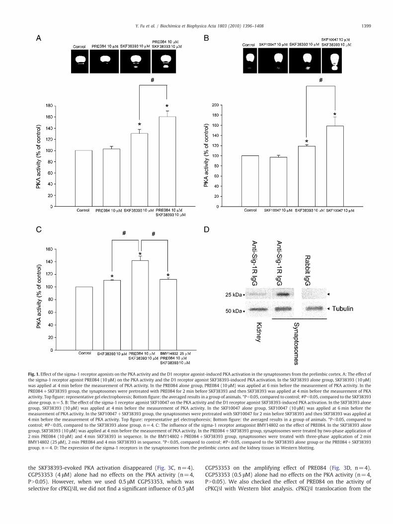
Fig. 1. Effect of the sigma-1 receptor agonists on the PKA activity and the D1 receptor agonist-induced PKA activation in the synaptosomes from the prelimbic cortex. A: The effect ofthe sigma-1 receptor agonist PRE084 (10 μM) on the PKA activity and the D1 receptor agonist SKF38393-induced PKA activation. In the SKF38393 alone group, SKF38393 (10 μM)was applied at 4 min before the measurement of PKA activity. In the PRE084 alone group, PRE084 (10 μM) was applied at 6 min before the measurement of PKA activity. In thePRE084+SKF38393 group, the synaptosomes were pretreated with PRE084 for 2 min before SKF38393 and then SKF38393 was applied at 4 min before the measurement of PKAactivity. Top figure: representative gel electrophoresis; Bottom figure: the averaged results in a group of animals. *Pb0.05, compared to control; #Pb0.05, compared to the SKF38393alone group. n=5. B: The effect of the sigma-1 receptor agonist SKF10047 on the PKA activity and the D1 receptor agonist SKF38393-induced PKA activation. In the SKF38393 alonegroup, SKF38393 (10 μM) was applied at 4 min before the measurement of PKA activity. In the SKF10047 alone group, SKF10047 (10 μM) was applied at 6 min before themeasurement of PKA activity. In the SKF10047+SKF38393 group, the synaptosomes were pretreated with SKF10047 for 2 min before SKF38393 and then SKF38393 was applied at4 min before the measurement of PKA activity. Top figure: representative gel electrophoresis; Bottom figure: the averaged results in a group of animals. *Pb0.05, compared tocontrol; #Pb0.05, compared to the SKF38393 alone group. n=4. C: The influence of the sigma-1 receptor antagonist BMY14802 on the effect of PRE084. In the SKF38393 alonegroup, SKF38393 (10 μM) was applied at 4 min before the measurement of PKA activity. In the PRE084+SKF38393 group, synaptosomes were treated by two-phase application of2 min PRE084 (10 μM) and 4 min SKF38393 in sequence. In the BMY14802+PRE084+SKF38393 group, synaptosomes were treated with three-phase application of 2 minBMY14802 (25 μM), 2 min PRE084 and 4 min SKF38393 in sequence. *Pb0.05, compared to control; #Pb0.05, compared to the SKF38393 alone group or the PRE084+SKF38393group. n=4. D: The expression of the sigma-1 receptors in the synaptosomes from the prelimbic cortex and the kidney tissues in Western blotting.
1399Y. Fu et al. / Biochimica et Biophysica Acta 1803 (2010) 1396–1408
the SKF38393-evoked PKA activation disappeared (Fig. 3C, n=4).CGP53353 (4 μM) alone had no effects on the PKA activity (n=4,PN0.05). However, when we used 0.5 μM CGP53353, which wasselective for cPKCβII, we did not find a significant influence of 0.5 μM
CGP53353 on the amplifying effect of PRE084 (Fig. 3D, n=4).CGP53353 (0.5 μM) alone had no effects on the PKA activity (n=4,PN0.05). We also checked the effect of PRE084 on the activity ofcPKCβI with Western blot analysis. cPKCβI translocation from the

1400 Y. Fu et al. / Biochimica et Biophysica Acta 1803 (2010) 1396–1408
cytosolic to membrane fraction in synaptosomes was used to assessthe relative amount of activated (membrane-bound) cPKCβI, an assaythat was described previously [31]. cPKCβI activation is associated
with translocation of the protein from the cytosol to cellular mem-branes. Fig. 4A showed representative immunoblots of the cPKCβI inboth cytosolic and membrane fractions of synaptosomes before and10 min after addition of PRE084 (10 μM) and the PKC activator PDBu(1 μM). Calculation of the percentage of cPKCβI in the membranefraction revealed that PRE084 increased membrane-bound cPKCβI by16.4% (Fig. 4B, n=7, Pb0.05), indicating that PRE084 stimulated thecPKCβI translocation. PDBuhad a similar effect to that of PRE084on thecPKCβI translocation (Fig. 4, n=7, Pb0.05).
3.3. Intrasynaptosomal Ca2+ plays an important role in the amplifyingeffect of the sigma-1 receptor agonist on the D1 receptor agonist-inducedPKA activation
We observed the influence of the removal of extracellular Ca2+ bythe Ca2+-free medium containing EGTA [32] on the amplifying effectof the sigma-1 receptor agonist on the D1 receptor agonist-inducedPKA activation. The result showed that the Ca2+-free mediumcontaining EGTA (1 mM) alone had no effects on the PKA activity(n=4), but it could abolish the amplifying effect of PRE084 on theSKF38393-induced PKA activation (Fig. 5A, n=4).
We also examined the influence of the voltage-gated Ca2+ channelblocker cadmium [33] on the amplifying effect of the sigma-1 receptoragonist. The result showed that cadmium (100 μM) alone had noeffects (n=4), but in the presence of cadmium, the amplifying effectof PRE084 disappeared (Fig. 5B, n=4). To determine whether L-typeCa2+ channels were involved in the amplifying effect of PRE084, weobserved the influence of the L-type Ca2+ channel blocker nimodipine[34] on the effect of PRE084. The results showed that nimodipine(10 μM) alone had no effects on the PKA activity (n=4, PN0.05), butit could significantly inhibit the amplifying effect of PRE084 on theSKF38393-evoked PKA activation (n=4, Fig. 5C).
3.4. Activation of PKC by sigma-1 receptor agonists is the upstream eventof the increase in the intrasynaptosomal Ca2+ concentration
To study the relationship between the activation of PKC and theincrease in the intrasynaptosomal Ca2+ concentration in the ampli-fying effect of the sigma-1 receptor agonist on the D1 receptoragonist-induced PKA activation, we observed the influence of the PKCinhibitor chelerythrine on the sigma-1 agonist-induced increase inthe intrasynaptosomal Ca2+ concentration. The result showed that inthe presence of chelerythrine, the effect of PRE084 on the intrasy-naptosomal Ca2+ concentration disappeared (Fig. 6A). The averagedincrease percentage of the Ca2+ concentration after PRE084 (10 μM)alone was 24.2±1.7% (n=6, Pb0.05, compared to control), but in the
Fig. 2. Influence of the sigma-1 receptor agonist on the cAMP- and the AC activator-induced PKA activation and the effect of the sigma-1 receptor agonist on the D1receptor agonist-induced AC activation in the synaptosomes from the prelimbic cortex.A: The influence of the sigma-1 receptor agonist PRE084 on the membrane-permeableanalog of cAMP (CPT-cAMP)-induced PKA activation. In the cAMP alone group, cAMP(1 μM) was applied at 4 min before the measurement of PKA activity. In the PRE084alone group, PRE084 (10 μM) was applied at 6 min before the measurement of PKAactivity. In the PRE084+cAMP group, synaptosomes were treated by two-phaseapplication of 2 min PRE084 and 4 min cAMP in sequence. *Pb0.05, compared tocontrol; #Pb0.05, compared to the CPT-cAMP alone group. n=4. B: The influence ofthe sigma-1 receptor agonist PRE084 on the AC activator forskolin -induced PKAactivation. In the forskolin alone group, forskolin (20 μM) was applied at 4 min beforethe measurement of PKA activity. In the PRE084 alone group, PRE084 (10 μM) wasapplied at 6 min before the measurement of PKA activity. In the PRE084+forskolingroup, synaptosomes were treated by two-phase application of 2 min PRE084 and4 min forskolin in sequence. *Pb0.05, compared to control; #Pb0.05, compared to theforskolin alone group. n=4. C: The effect of the sigma-1 receptor agonist PRE084 on theD1 receptor agonist SKF38393-induced AC activation. In the SKF38393 alone group,SKF38393 (10 μM) was applied at 4 min before the measurement of AC activity. In thePRE084 alone group, PRE084 (10 μM) was applied at 6 min before the measurement ofAC activity. In the PRE084+SKF38393 group, synaptosomes were treated by two-phase application of 2 min PRE084 and 4 min SKF38393 in sequence. *Pb0.05,compared to control. n=8.

Fig. 3. Influence of PKC inhibitors on the amplifying effect of the sigma-1 receptor agonist on the D1 receptor agonist-induced PKA activation in the synaptosomes from the prelimbiccortex. A: Influence of the wide-spectrum PKC inhibitor chelerythrine on the amplifying effect of the sigma-1 receptor agonist PRE084. In the SKF38393 alone group, SKF38393(10 μM) was applied at 4 min before the measurement of PKA activity. In the PRE084+SKF38393 group, synaptosomes were treated by two-phase application of 2 min PRE084(10 μM) and 4 min SKF38393 in sequence. In the chelerythrine+PRE084+SKF38393 group, synaptosomes were treated with three-phase application of 2 min chelerythrine(2.5 μM), 2 min PRE084 and 4 min SKF38393 in sequence. *Pb0.05, compared to control; #Pb0.05, compared to the SKF38393 alone group or the PRE084+SKF38393 group. n=4.B: Influence of the cPKC inhibitor rottlerin on the amplifying effect of the sigma-1 receptor agonist PRE084. In the SKF38393 alone group, SKF38393 (10 μM) was applied at 4 minbefore the measurement of PKA activity. In the PRE084+SKF38393 group, synaptosomes were treated by two-phase application of 2 min PRE084 (10 μM) and 4 min SKF38393 insequence. In the rottlerin+PRE084+SKF38393 group, synaptosomes were treated with three-phase application of 2 min rottlerin (40 μM), 2 min PRE084 and 4 min SKF38393 insequence. *Pb0.05, compared to control; #Pb0.05, compared to the SKF38393 alone group or the PRE084+SKF38393 group. n=4. C: Influence of the cPKCβ inhibitor CGP53353 onthe amplifying effect of the sigma-1 receptor agonist PRE084. In the SKF38393 alone group, SKF38393 (10 μM) was applied at 4 min before the measurement of PKA activity. In thePRE084+SKF38393 group, synaptosomes were treated by two-phase application of 2 min PRE084 (10 μM) and 4 min SKF38393 in sequence. In the CGP53353+PRE084+SKF38393 group, synaptosomes were treated with three-phase application of 2 min CGP53353 (4 μM), 2 min PRE084 and 4 min SKF38393 in sequence. *Pb0.05, compared tocontrol; #Pb0.05, compared to the SKF38393 alone group or the PRE084+SKF38393 group. n=4. D: Influence of the cPKCβII inhibitor CGP53353 (0.5 μM) on the amplifying effectof the sigma-1 receptor agonist PRE084. In the SKF38393 alone group, SKF38393 (10 μM) was applied at 4 min before the measurement of PKA activity. In the PRE084+SKF38393group, synaptosomes were treated by two-phase application of 2 min PRE084 (10 μM) and 4 min SKF38393 in sequence. In the CGP53353+PRE084+SKF38393 group,synaptosomes were treated with three-phase application of 2 min CGP53353 (0.5 μM), 2 min PRE084 and 4 min SKF38393 in sequence. *Pb0.05, compared to control; #Pb0.05,compared to the SKF38393 alone group or the PRE084+SKF38393 group. n=4.
1401Y. Fu et al. / Biochimica et Biophysica Acta 1803 (2010) 1396–1408
presence of chelerythrine (2.5 μM), the percentage after PRE084 was4.1±1.5 % (n=6, PN0.05, compared to control). Moreover, beingconsistent with the results of the above PKC isozyme experimentsinvolving the mechanism of the amplifying effect of the sigma-1receptor agonist on the D1 receptor agonist-induced PKA activation,both the cPKC inhibitor rottlerin (Fig. 6B, n=6) and the cPKCβ
inhibitor CG53353 (4 μM) (Fig. 6C, n=6) could also block the effect ofPRE084 on the intrasynaptosomal Ca2+ concentration. In addition, theL-type Ca2+ channel blockers nimodipine (Fig. 7A, n=6) andverapamil [35] (Fig. 7B, n=6) could also abolish the effect ofPRE084 on the intrasynaptosomal Ca2+ concentration. However, wedid not find a significant influence of the N-type Ca2+ channel blocker

Fig. 4. Effect of PRE084 on the activity of cPKCβI using Western blot analysis in thesynaptosomes from the prelimbic cortex. cPKCβI translocation from the cytosolic tomembrane fraction in synaptosomes was used to assess the relative amount ofactivated (membrane-bound) cPKCβI. A: Representative immunoblots of the cPKCβI inboth cytosolic and membrane fractions of synaptosomes before and 10 min afteraddition of PRE084 (10 μM) and PDBu (1 μM). B: Calculation of the percentage ofcPKCβI in the membrane fraction before and 10 min after addition of PRE084 (10 μM)and PDBu (1 μM). *Pb0.05, compared to control. n=7.
1402 Y. Fu et al. / Biochimica et Biophysica Acta 1803 (2010) 1396–1408
ω-contotoxin GVIA [36] (Fig. 7C, n=6) and the P/Q-type Ca2+
channel blocker ω-agatoxin IVA [37] (Fig. 7D, n=6) on the effect ofPRE084 on the intrasynaptosomal Ca2+ concentration. We also didnot find a significant influence of the NMDA receptor antagonist
Fig. 5. Influence of the extracellular Ca2+ chelator, the voltage-gated Ca2+ channelblocker and the L-type Ca2+ channel blocker on the amplifying effect of the sigma-1receptor agonist on the D1 receptor agonist-induced PKA activation in the synapto-somes from the prelimbic cortex. A: Influence of the removal of the extracellular Ca2+
by the Ca2+-free medium containing EGTA on the amplifying effect of the sigma-1receptor agonist PRE084. In the SKF38393 alone group, SKF38393 (10 μM) was appliedat 4 min before the measurement of PKA activity. In the PRE084+SKF38393 group,synaptosomes were treated by two-phase application of 2 min PRE084 (10 μM) and4 min SKF38393 in sequence. In the Ca2+-free medium containing EGTA+PRE084+SKF38393 group, synaptosomes were treated with three-phase application of 2 minCa2+-free medium containing EGTA (1 mM), 2 min PRE084 and 4 min SKF38393 insequence. *Pb0.05, compared to control; #Pb0.05, compared to the SKF38393 alonegroup or the PRE084+SKF38393 group. n=4. B: Influence of the voltage-gated Ca2+
channel blocker cadmium on the amplifying effect of the sigma-1 receptor agonistPRE084. In the SKF38393 alone group, SKF38393 (10 μM) was applied at 4 min beforethe measurement of PKA activity. In the PRE084+SKF38393 group, synaptosomeswere treated by two-phase application of 2 min PRE084 (10 μM) and 4 min SKF38393in sequence. In the cadmium+PRE084+SKF38393 group, synaptosomes were treatedwith three-phase application of 2 min cadmium (100 μM), 2 min PRE084 and 4 minSKF38393 in sequence. *Pb0.05, compared to control; #Pb0.05, compared to theSKF38393 alone group or the PRE084+SKF38393 group. n=4. C: Influence of the L-type Ca2+ channel blocker nimodipine on the amplifying effect of the sigma-1 receptoragonist PRE084. In the SKF38393 alone group, SKF38393 (10 μM) was applied at 4 minbefore the measurement of PKA activity. In the PRE084+SKF38393 group, synapto-somes were treated by two-phase application of 2 min PRE084 (10 μM) and 4 minSKF38393 in sequence. In the nimodipine+PRE084+SKF38393 group, synaptosomeswere treated with three-phase application of 2 min nimodipine (10 μM), 2 min PRE084and 4 min SKF38393 in sequence. *Pb0.05, compared to control; #Pb0.05, compared tothe SKF38393 alone group or the PRE084+SKF38393 group. n=4.

Fig. 6. Influence of PKC inhibitors on the effect of the sigma-1 receptor agonist on the intrasynaptosomal Ca2+ concentration in the synaptosomes from the prelimbic cortex.A: Influence of the wide-spectrum PKC inhibitor chelerythrine on the effect of the sigma-1 receptor agonist PRE084 on the intrasynaptosomal Ca2+ concentration. In the PRE084alone group, PRE084 (10 μM) was applied at 4 min before the measurement of Ca2+ concentration. In the chelerythrine+PRE084 group, synaptosomes were treated by two-phaseapplication of 2 min chelerythrine (2.5 μM) and 4 min PRE084 in sequence. *Pb0.05, compared to control; #Pb0.05, compared to the PRE084 alone group. n=6. B: Influence of thecPKC inhibitor rottlerin on the effect of the sigma-1 receptor agonist PRE084 on the intrasynaptosomal Ca2+ concentration. In the PRE084 alone group, PRE084 (10 μM) was appliedat 4 min before the measurement of Ca2+ concentration. In the rottlerin+PRE084 group, synaptosomes were treated by two-phase application of 2 min rottlerin (40 μM) and 4 minPRE084 in sequence. *Pb0.05, compared to control; #Pb0.05, compared to the PRE084 alone group. n=6. C: Influence of the cPKCβ inhibitor CGP53353 (4 μM) on the effect of thesigma-1 receptor agonist PRE084 on the intrasynaptosomal Ca2+ concentration. In the PRE084 alone group, PRE084 (10 μM) was applied at 4 min before the measurement of Ca2+
concentration. In the CGP53353+PRE084 group, synaptosomes were treated by two-phase application of 2 min CGP53353 (4 μM) and 4 min PRE084 in sequence. *Pb0.05,compared to control; #Pb0.05, compared to the PRE084 alone group. n=6.
1403Y. Fu et al. / Biochimica et Biophysica Acta 1803 (2010) 1396–1408

1404 Y. Fu et al. / Biochimica et Biophysica Acta 1803 (2010) 1396–1408
MK801 [38] on the effect of PRE084 on the intrasynaptosomal Ca2+
concentration (Fig. 7E, n=6). In addition, we also checked the role ofintracellular Ca2+ stores in the sigma-1 receptor-mediated intrasy-naptosomal Ca2+ increase. The result showed that thapsigargin,which depleted all intracellular Ca2+ stores by inhibiting theendosomal Ca2+-ATPase activity [39], had no significant influenceon the effect of PRE084. PRE084 (10 μM) still increased the intra-synaptosomal Ca2+ concentration by 22.6±3.8% (n=6, Pb0.05) inthe presence of thapsigargin (25 μM), showing no significant
difference from that (22.9±3.8%, n=6) of the PRE084 alone group(PN0.05).
4. Discussion
One of the main findings of the present study is that sigma-1receptor agonists can amplify the D1 receptor agonist-induced PKAactivation at presynaptic sites in the prelimbic cortex. To the best of

Fig. 7. Influence of the L-type Ca2+ channel blockers, the N-type Ca2+ channel blocker, the P/Q-type Ca2+ channel blocker and the NMDA receptor antagonist on the effect of thesigma-1 receptor agonist on the intrasynaptosomal Ca2+ concentration in the synaptosomes from the prelimbic cortex. A: The influence of the L-type Ca2+ channel blockernimodipine on the effect of the sigma-1 receptor agonist PRE084 on the intrasynaptosomal Ca2+ concentration. In the PRE084 alone group, PRE084 (10 μM) was applied at 4 minbefore the measurement of Ca2+ concentration. In the nimodipine+PRE084 group, synaptosomes were treated by two-phase application of 2 min nimodipine (10 μM) and 4 minPRE084 in sequence. *Pb0.05, compared to control; #Pb0.05, compared to the PRE084 alone group. n=6. B: The influence of the L-type Ca2+ channel blocker verapamil on the effectof the sigma-1 receptor agonist PRE084 on the intrasynaptosomal Ca2+ concentration. In the PRE084 alone group, PRE084 (10 μM) was applied at 4 min before the measurement ofCa2+ concentration. In the verapamil+PRE084 group, synaptosomes were treated by two-phase application of 2 min verapamil (50 μM) and 4 min PRE084 in sequence. *Pb0.05,compared to control; #Pb0.05, compared to the PRE084 alone group. n=6. C: The influence of the N-type Ca2+ channel blocker ω-contotoxin GVIA on the effect of the sigma-1receptor agonist PRE084 on the intrasynaptosomal Ca2+ concentration. In the PRE084 alone group, PRE084 (10 μM) was applied at 4 min before the measurement of Ca2+
concentration. In the ω-contotoxin GVIA+PRE084 group, synaptosomes were treated by two-phase application of 2 min ω-contotoxin GVIA (0.5 μM) and 4 min PRE084 insequence. *Pb0.05, compared to control. n=6. D: The influence of the P/Q-type Ca2+ channel blocker ω-agatoxin IVA on the effect of the sigma-1 receptor agonist PRE084 on theintrasynaptosomal Ca2+ concentration. In the PRE084 alone group, PRE084 (10 μM) was applied at 4 min before the measurement of Ca2+ concentration. In the ω-agatoxin IVA+PRE084 group, synaptosomes were treated by two-phase application of 2 min ω-agatoxin IVA (0.5 μM) and 4 min PRE084 in sequence. *Pb0.05, compared to control. n=6. E: Theinfluence of the NMDA receptor antagonist MK801 on the effect of the sigma-1 receptor agonist PRE084 on the intrasynaptosomal Ca2+ concentration. In the PRE084 alone group,PRE084 (10 μM)was applied at 4 min before the measurement of Ca2+ concentration. In the MK801+PRE084 group, synaptosomes were treated by two-phase application of 2 minMK801 (20 μM) and 4 min PRE084 in sequence. *Pb0.05, compared to control. n=6.
1405Y. Fu et al. / Biochimica et Biophysica Acta 1803 (2010) 1396–1408
our knowledge, this is the first report involving the regulation of D1receptor signaling by sigma-1 receptors.
The concentration (10 μM) of PRE084 used here to produce theeffect was larger than the Ki value (44 nM) of PRE084 [40], but thiseffect should be specific to the sigma-1 receptors. The evidencesupporting this statement was that (1) the specific sigma-1 receptorantagonist BMY14802 could completely abolish the effect of PRE084;(2) in the literature, although the Ki value of PRE084 for the sigma-1receptors was in the nanomolar range, the IC50 values of PRE084 forproducing effects via the activation of the sigma-1 receptors weregenerally in the micromoles range. For example, the IC50 value ofPRE084 for the inhibition of the ASIC1a-induced [Ca2+]i increase was13.7 μM [41] and for the protection of human retinal cells againstoxidative stress was 10 μM [42].
Sigma-1 receptors have been known to exist at postsynaptic sites.In addition, a number of evidence suggested that sigma-1 receptorsmight also exist at presynaptic sites. The evidence supporting theexistence of sigma-1 receptors at presynaptic sites was that (1) thefunctional experiments in brain slices [43–45] and synaptosomes (apreparation of presynaptic terminals) [46] showed that the activationof sigma-1 receptors could modulate presynaptic neurotransmitterrelease; (2) radioligand binding experiments showed that selectivesigma-1 receptor agonists could bind with synaptosomes with highaffinity in a competitive manner [47]. Moreover, the present studyshowed the existence of sigma-1 receptors in the synaptosomes byWestern blotting.
The signaling pathway between D1 receptors and PKA involves thecoupling to Gs-protein, the activation of AC and the action of cAMP on

1406 Y. Fu et al. / Biochimica et Biophysica Acta 1803 (2010) 1396–1408
PKA. We explored the possible sites of the action of the sigma-1receptors in this pathway. First, we used CPT-cAMP, a membrane-permeable analog of cAMP, to generate the cAMP-induced PKAactivation and then observed the influence of the sigma-1 receptoragonist on this activation. The result showed that the sigma-1receptor agonist could potentiate the effect of CPT-cAMP on PKA.Then, we used forskolin, an activator of AC, to generate the AC/cAMP-induced PKA activation and observed the influence of the sigma-1receptor agonist on this activation. The result showed that the sigma-1 receptor agonist could also potentiate this activation. However, wedid not find that the sigma-1 receptor agonist had the potentiatingeffect on the D1 receptor agonist-induced AC activation. These resultssuggest that the sigma-1 receptor agonist may amplify D1 receptorsignaling via amplifying the action of cAMP on PKA.
It has been known that the downstream pathway of the sigma-1receptors involves the activation of PKC and the enhancement ofintracellular Ca2+ concentration. Therefore, it is possible that PKC andintracellular Ca2+, as important downstream signaling molecules ofsigma-1 receptors, play an important role in the amplifying effect of thesigma-1 receptor agonist on the D1 receptor agonist-induced PKAactivation. This hypothesis has been confirmedby our results.Moreover,this result provides evidence supporting that sigma-1 receptors mayamplify D1 receptor signaling via PKC and intracellular Ca2+.
Noteworthily,whenwe removed theextracellular Ca2+by the Ca2+-free medium containing EGTA, the activity of PKA returned to thecontrol levels but cadmium and nimodipine only inhibited theamplifying effect achieved by PRE084. This phenomenon suggestspossible involvement of another Ca2+ source in the D1 receptor-inducedPKAactivation. In this aspect, it's possible that this another Ca2+
source may be from D-1 receptor-induced intracellular Ca2+ releasebecause it is shown that the activation of D1 receptors can result in a risein intracellular Ca2+ through both extracellular influx and release fromintracellular compartments [48].
PKC family includes at least 12 isozymes, which are classified intothree major groups [49]. The first of these groups is the conventionalPKC (cPKC), which includes α, βI, βII and γ types. The second group,called the novel PKC (nPKC), includes δ, ε, η and θ type. The thirdgroup is the atypical PKC (aPKC), including ζ and λ type. The PKCisozymes are distributed in various tissues. Among them, cPKC is theone localized at presynaptic terminals [50]. So it is possible that cPKCplays an important role in the amplifying effect of the sigma-1receptor agonist on the D1 receptor agonist-induced PKA activation.This hypothesis was confirmed by our result that the cPKC selectiveinhibitor rottlerin could abolish the effect of the sigma-1 receptoragonist. Moreover, CG53353 at 4 μM, which inhibited both PKCβII andPKCβI of cPKC, could also abolish the effect of the sigma-1 receptoragonist. However, CGP53353 at 0.5 μM, which was selective forPKCβII, had no influence. These results suggest that the cPKC,especially the PKCβI, may play a key role in the effect of theamplifying effect of the sigma-1 receptor agonist on the D1 receptoragonist-induced PKA activation.
About the source of Ca2+ involving the amplifying effect of thesigma-1 receptor agonist on the D1 receptor agonist-induced PKAactivation, it may involve the promotion of the extracellular Ca2+
influx or the mobilization of the intracellular Ca2+ store. In thepresent paper, we studied the role of the extracellular Ca2+ in theeffect of the sigma-1 receptor agonist. The result showed that afterremoving the extracellular Ca2+ by the Ca2+-free medium containingEGTA, the amplifying effect of the sigma-1 receptor agonist dis-appeared. Moreover, the voltage-gated Ca2+ channel blocker cadmi-um and the L-type Ca2+ channel blocker nimodipine could abolish theamplifying effect of the sigma-1 receptor agonist, suggesting that theextracellular Ca2+ influx through the L-type Ca2+ channels mightmediate the amplifying effect of the sigma-1 receptor agonist.
About the relationship between the activation of PKC and theincrease in the intrasynaptosomal Ca2+ concentration in the ampli-
fying effect of the sigma-1 receptor agonist on theD1 receptor agonist-induced PKA activation, our results suggested that the activation ofPKC by sigma-1 receptor agonists might be the upstream event of theincrease in the intrasynaptosomal Ca2+ concentration. Interestingly,based on our results, it appeared that the sigma-1 receptors couldmodulate the resting-state L-type Ca2+ channels, which then led to anincrease in the intrasynaptosomal Ca2+ concentration. This statementwas consistent with the evidence: (1) there were spontaneous L-typeCa2+ channel activities at the rest membrane potentials [51] and theseactivities contributed to the genesis of spontaneous Ca2+ sparks in thecytoplasm [52]; (2) some active substances, such as ganglioside andphorbol ester (the PKC activator), could increase the Ca2+ influxthrough the modulation of these activities [53,54]. However, howsigma-1 receptors modulate the resting-state L-type Ca2+ channelsremains to be studied. In addition, previous studies in NG-108 andSHSY5Y cell lines showed that the sigma-1 receptor agonists enhancedthe intracellular Ca2+ by the mobilization of the intracellular Ca2+
store [55–57]. However, this mechanism might not be involved in theeffect of the sigma-1 receptor agonists on the intrasynaptosomal Ca2+
concentration in the prelimbic cortex because our result showed thatthe intracellular Ca2+ store depleter hadno significant influence on theeffect of the sigma-1 receptor agonist. Moreover, the present resultthat the removal of the extracellular Ca2+ by the Ca2+-free mediumcontaining EGTA could abolish the effect of the sigma-1 receptoragonist also supported that themechanism of the effect of the sigma-1receptor agonist on the intrasynaptosomal Ca2+ concentration mightinvolve the promotion of the extracellular Ca2+ influx, rather than themobilization of the intracellular Ca2+ store.
Subcellular distribution of sigma-1 receptors has been studiedwith radioligand binding in subcellular fractions, and more recentlywith immunochemical methods. These studies found that sigma-1receptors were abundant in endoplasmic reticulum (ER), nuclear,mitochondrial and synaptic membranes (synaptosomes) [3]. Howev-er, it appears that the functions of sigma-1 receptors in differentsubcellular fractions are different. For example, recent studies showthat the sigma-1 receptors in the ER, as a Ca2+-sensitive and ligand-operated receptor chaperone at mitochondrion-associated ER mem-brane (MAM), can lead to a prolonged Ca2+ signaling into themitochondria via IP3 receptors, and thus regulating ER-mitochondrialinterorganellar Ca2+ signaling and cell survival [58]; the present studyshows that one function of the sigma-1 receptors at presynaptic sites(synaptosomes) may amplify the D1 receptor agonist-induced PKAactivation by the sigma-1 receptors-cPKC (especially the PKCβI)- L-type Ca2+ channels-Ca2+-AC and/or cAMP signaling pathway (Fig. 8).It is noteworthy that the synaptosomal preparation may becontaminated with ER or mitochondrion where sigma-1 receptorsare highly concentrated in a specialized area known as the MAM. Butthis contamination may not contribute to the amplifying effect of thesigma-1 receptor agonist on the D1 receptor agonist-induced PKAactivation because these contaminated ER or mitochondria are in theextrasynaptosomal space.
Significance – It has been known that the dopaminergic projectionfrom the ventral tegmental area to the prelimbic cortex plays a majorrole in cognition and neuropsychiatric processes. In particular, theinvolvement of prefrontal D1 dopamine receptors in the behavioralsensitization to psychostimulants has received much attention. Theactivation of D1 receptors in the prelimbic cortex stimulatedpsychostimulant-induced motor activity and increased behavioralsensitization to psychostimulants [59–62]. Thus, the present findingthat the sigma-1 receptors can amplify the presynaptic dopamine D1receptor signaling in the prelimbic cortex is of significance forunderstanding the neuronal basis of the behavioral sensitizationinduced by sigma-1 receptors. In addition, the present finding thatthe activation of sigma-1 receptors amplifies the D1 receptor-inducedPKA activation via cPKC (especially the PKCβI) - L-type Ca2+ channels -Ca2+ - AC and/or cAMP signaling pathway is of important significance

Fig. 8. Schematic representation of the possible mechanism underlying the amplifyingeffect of the sigma-1 receptor agonist on theD1 receptor agonist-induced PKA activationin the synaptosomes from the prelimbic cortex. The sigma-1 receptor in the presynapticsite may amplify the D1 receptor agonist-induced PKA activation by sigma-1 receptors -cPKC (especially the PKCβI) - L-type Ca2+ channels - Ca2+ - AC and/or cAMP signalingpathway.
1407Y. Fu et al. / Biochimica et Biophysica Acta 1803 (2010) 1396–1408
for revealing the downstream signal transduction pathway of sigma-1receptors.
Acknowledgements
Thisworkwas supported byProject 31070932, 30900424, 30670653and 30821002 of Foundation of National Natural Science of China, theNational Programof Basic Research sponsoredby theMinistry of Scienceand Technology of China (2009CB52201) and Project B119 of ShanghaiLeading Academic Discipline. We are grateful to Dr Teruo Hayashi forproviding the sigma-1 receptor antibody.
References
[1] T. Maurice, T.P. Su, The pharmacology of sigma-1 receptors, Pharmacol. Ther. 124(2009) 195.
[2] J. Mei, G.W. Pasternak, Molecular cloning and pharmacological characterization ofthe rat sigma1 receptor, Biochem. Pharmacol. 62 (2001) 349.
[3] E.J. Cobos, J.M. Entrena, F.R. Nieto, C.M. Cendan, P.E. Del, Pharmacology andtherapeutic potential of sigma(1) receptor ligands, Curr. Neuropharmacol. 6(2008) 344.
[4] J.E. Bermack, G. Debonnel, The role of sigma receptors in depression, J. Pharmacol.Sci. 97 (2005) 317.
[5] F.P. Monnet, G. Debonnel, R. Bergeron, B. Gronier, M.C. de, The effects of sigmaligands and of neuropeptide Y on N-methyl-D-aspartate-induced neuronalactivation of CA3 dorsal hippocampus neurones are differentially affected bypertussin toxin, Br. J. Pharmacol. 112 (1994) 709.
[6] F.P. Monnet, M.P. Morin-Surun, J. Leger, L. Combettes, Protein kinase C-dependentpotentiation of intracellular calcium influx by sigma1 receptor agonists in rathippocampal neurons, J. Pharmacol. Exp. Ther. 307 (2003) 705.
[7] M.P. Morin-Surun, T. Collin, M. avit-Saubie, E.E. Baulieu, F.P. Monnet, Intracellularsigma1 receptor modulates phospholipase C and protein kinase C activities in thebrainstem, Proc. Natl. Acad. Sci. U. S. A 96 (1999) 8196.
[8] T. Hayashi, T. Su, The sigma receptor: evolution of the concept in neuropsycho-pharmacology, Curr. Neuropharmacol. 3 (2005) 267.
[9] T. Hayashi, T.P. Su, Sigma-1 receptors (sigma(1) binding sites) form raft-likemicrodomains and target lipid droplets on the endoplasmic reticulum: roles inendoplasmic reticulum lipid compartmentalization and export, J. Pharmacol. Exp.Ther. 306 (2003) 718.
[10] L.Y. Dong, Z.X. Cheng, Y.M. Fu, Z.M. Wang, Y.H. Zhu, J.L. Sun, Y. Dong, P. Zheng,Neurosteroid dehydroepiandrosterone sulfate enhances spontaneous glutamaterelease in rat prelimbic cortex through activation of dopamine D1 and sigma-1receptor, Neuropharmacology 52 (2007) 966.
[11] H. Kamei, T. Kameyama, T. Nabeshima, (+)-SKF-10, 047 and dextromethorphanameliorate conditioned fear stress via dopaminergic systems linked to phenytoin-regulated sigma 1 sites, Eur. J. Pharmacol. 309 (1996) 149.
[12] A.I. Breukel, E. Besselsen, W.E. Ghijsen, Synaptosomes. A model system to studyrelease of multiple classes of neurotransmitters, Methods Mol. Biol. 72 (1997) 33.
[13] Y. Dong, Y.M. Fu, J.L. Sun, Y.H. Zhu, F.Y. Sun, P. Zheng, Neurosteroid enhancesglutamate release in rat prelimbic cortex via activation of alpha1-adrenergic andsigma1 receptors, Cell. Mol. Life Sci. 62 (2005) 1003.
[14] O.H. Lowry, N.J. Rosebrough, A.L. Farr, R.J. Randall, Protein measurement with theFolin phenol reagent, J. Biol. Chem. 193 (1951) 265.
[15] L.G. Lou, G. Pei, Modulation of protein kinase C and cAMP-dependent proteinkinase by delta-opioid, Biochem. Biophys. Res. Commun. 236 (1997) 626.
[16] R. Golla, R. Seethala, A homogeneous enzyme fragment complementation cyclicAMP screen for GPCR agonists, J. Biomol. Screen. 7 (2002) 515.
[17] G. Cormaci, T. Mori, T. Hayashi, T.P. Su, Protein kinase A activation down-regulates, whereas extracellular signal-regulated kinase activation up-regulatessigma-1 receptors in B-104 cells: Implication for neuroplasticity, J. Pharmacol.Exp. Ther. 320 (2007) 202.
[18] T. Hayashi, T.P. Su, Sigma-1 receptor chaperones at the ER-mitochondrioninterface regulate Ca(2+) signaling and cell survival, Cell 131 (2007) 596.
[19] R.A. Moore, H. Nguyen, J. Galceran, I.N. Pessah, P.D. Allen, A transgenic myogeniccell line lacking ryanodine receptor protein for homologous expression studies:reconstitution of Ry1R protein and function, J. Cell Biol. 140 (1998) 843.
[20] H.M. Korchak, L.B. Dorsey, H. Li, D. Mackie, L.E. Kilpatrick, Selective roles for alpha-PKC in positive signaling for O-(2) generation and calcium mobilization but notelastase release in differentiated HL60 cells, Biochim. Biophys. Acta 1773 (2007)440.
[21] V.O. Rybin, A. Sabri, J. Short, J.C. Braz, J.D. Molkentin, S.F. Steinberg, Cross-regulation of novel protein kinase C (PKC) isoform function in cardiomyocytes.Role of PKC epsilon in activation loop phosphorylations and PKC delta inhydrophobic motif phosphorylations, J. Biol. Chem. 278 (2003) 14555.
[22] J.P. Kao, A.T. Harootunian, R.Y. Tsien, Photochemically generated cytosolic calciumpulses and their detection by fluo-3, J. Biol. Chem. 264 (1989) 8179.
[23] R. ZhuGe, R.A. Tuft, K.E. Fogarty, K. Bellve, F.S. Fay, J.V. Walsh Jr., The influence ofsarcoplasmic reticulum Ca2+ concentration on Ca2+ sparks and spontaneoustransient outward currents in single smooth muscle cells, J. Gen. Physiol. 113 (1999)215.
[24] T.P. Su, X.Z. Wu, E.J. Cone, K. Shukla, T.M. Gund, A.L. Dodge, D.W. Parish, Sigmacompounds derived from phencyclidine: identification of PRE-084, a new,selective sigma ligand, J. Pharmacol. Exp. Ther. 259 (1991) 543.
[25] K.T. Tchedre, R.Q. Huang, A. Dibas, R.R. Krishnamoorthy, G.H. Dillon, T. Yorio,Sigma-1 receptor regulation of voltage-gated calcium channels involves a directinteraction, Invest Ophthalmol. Vis. Sci. 49 (2008) 4993.
[26] H. Ujike, S. Kuroda, S. Otsuki, sigma Receptor antagonists block the developmentof sensitization to cocaine, Eur. J. Pharmacol. 296 (1996) 123.
[27] T. Hayashi, T.P. Su, Sigma-1 receptor chaperones at the ER-mitochondrioninterface regulate Ca(2+) signaling and cell survival, Cell 131 (2007) 596.
[28] S.J. Chmura, E. Nodzenski, R.R. Weichselbaum, J. Quintans, Protein kinase Cinhibition induces apoptosis and ceramide production through activation of aneutral sphingomyelinase, Cancer Res. 56 (1996) 2711.
[29] M. Gschwendt, H.J. Muller, K. Kielbassa, R. Zang, W. Kittstein, G. Rincke, F. Marks,Rottlerin, a novel protein kinase inhibitor, Biochem. Biophys. Res. Commun. 199(1994) 93.
[30] S.W. Lee, H.B. Kwak, W.J. Chung, H. Cheong, H.H. Kim, Z.H. Lee, Participation ofprotein kinase C beta in osteoclast differentiation and function, Bone 32 (2003)217.
[31] D. Schechtman, D. Mochly-Rosen, Isozyme-specific inhibitors and activators ofprotein kinase C, Methods Enzymol. 345 (2002) 470.
[32] S.M. Rawls, J.F. McGinty, D.M. Terrian, Presynaptic kappa-opioid and muscarinicreceptors inhibit the calcium-dependent component of evoked glutamate releasefrom striatal synaptosomes, J. Neurochem. 73 (1999) 1058.
[33] T. Oba,M.Yamaguchi, S.Wang, J.D. Johnson,Modulation of theCa2+channel voltagesensor and excitation-contraction coupling by silver, Biophys. J. 63 (1992) 1416.
[34] K. Jun, E.S. Piedras-Renteria, S.M. Smith, D.B. Wheeler, S.B. Lee, T.G. Lee, H. Chin, M.E. Adams, R.H. Scheller, R.W. Tsien, H.S. Shin, Ablation of P/Q-type Ca(2+)channel currents, altered synaptic transmission, and progressive ataxia in micelacking the alpha(1A)-subunit, Proc. Natl. Acad. Sci. U. S. A 96 (1999) 15245.
[35] G. Aicardi, P.A. Schwartzkroin, Suppression of epileptiform burst discharges inCA3 neurons of rat hippocampal slices by the organic calcium channel blocker,verapamil, Exp. Brain Res. 81 (1990) 288.
[36] B.M. Olivera, G.P. Miljanich, J. Ramachandran, M.E. Adams, Calcium channeldiversity and neurotransmitter release: the omega-conotoxins and omega-agatoxins, Annu. Rev. Biochem. 63 (1994) 823.
[37] I.M. Mintz, M.E. Adams, B.P. Bean, P-type calcium channels in rat central andperipheral neurons, Neuron 9 (1992) 85.
[38] M.L. Mayer, G.L. Westbrook, L. Vyklicky Jr., Sites of antagonist action on N-methyl-D-aspartic acid receptors studied using fluctuation analysis and a rapid perfusiontechnique, J. Neurophysiol. 60 (1988) 645.
[39] O. Thastrup, P.J. Cullen, B.K. Drobak, M.R. Hanley, A.P. Dawson, Thapsigargin, atumor promoter, discharges intracellular Ca2+ stores by specific inhibition ofthe endoplasmic reticulum Ca2(+)-ATPase, Proc. Natl. Acad. Sci. U. S. A 87(1990) 2466.
[40] S.N. Calderon, S. Izenwasser, B. Heller, J.S. Gutkind, M.V. Mattson, T.P. Su, A.H.Newman, Novel 1-phenylcycloalkanecarboxylic acid derivatives are potent andselective sigma 1 ligands, J. Med. Chem. 37 (1994) 2285.
[41] Y. Herrera, C. Katnik, J.D. Rodriguez, A.A. Hall, A. Willing, K.R. Pennypacker, J.Cuevas, sigma-1 receptor modulation of acid-sensing ion channel a (ASIC1a) andASIC1a-induced Ca2+ influx in rat cortical neurons, J. Pharmacol. Exp. Ther. 327(2008) 491.
[42] C. Bucolo, F. Drago, L.R. Lin, V.N. Reddy, Sigma receptor ligands protect humanretinal cells against oxidative stress, NeuroReport 17 (2006) 287.

1408 Y. Fu et al. / Biochimica et Biophysica Acta 1803 (2010) 1396–1408
[43] L. Chen, M. Sokabe, Presynaptic modulation of synaptic transmission by pregnen-olone sulfate as studied by optical recordings, J. Neurophysiol. 94 (2005) 4131.
[44] D.A. Meyer, M. Carta, L.D. Partridge, D.F. Covey, C.F. Valenzuela, Neurosteroidsenhance spontaneous glutamate release in hippocampal neurons. Possible role ofmetabotropic sigma1-like receptors, J. Biol. Chem. 277 (2002) 28725.
[45] Y. Dong, Y.M. Fu, J.L. Sun, Y.H. Zhu, F.Y. Sun, P. Zheng, Neurosteroid enhancesglutamate release in rat prelimbic cortex via activation of alpha1-adrenergic andsigma1 receptors, Cell. Mol. Life Sci. 62 (2005) 1003.
[46] B. Garrone, M. Magnani, M. Pinza, L. Polenzani, Effects of trazodone onneurotransmitter release from rat mossy fibre cerebellar synaptosomes, Eur. J.Pharmacol. 400 (2000) 35.
[47] A. Klouz, J.P. Tillement, M.F. Boussard, M.Wierzbicki, V. Berezowski, R. Cecchelli, S.Labidalle, B. Onteniente, D. Morin, [3H]BHDP as a novel and selective ligand forsigma1 receptors in liver mitochondria and brain synaptosomes of the rat, FEBSLett. 553 (2003) 157.
[48] T.S. Tang, I. Bezprozvanny, Dopamine receptor-mediated Ca(2+) signaling instriatal medium spiny neurons, J. Biol. Chem. 279 (2004) 42082.
[49] K.J. Way, E. Chou, G.L. King, Identification of PKC-isoform-specific biologicalactions using pharmacological approaches, Trends Pharmacol. Sci. 21 (2000) 181.
[50] W.S. Liu, C.A. Heckman, The sevenfold way of PKC regulation, Cell. Signal. 10(1998) 529.
[51] J.F. Fiekers, L.M. Konopka, Spontaneous transients of [Ca2+]i depend on externalcalcium and the activation of L-type voltage-gated calcium channels in a clonalpituitary cell line (AtT-20) of cultured mouse corticotropes, Cell Calcium 19(1996) 327.
[52] Z. Guangqin, F. Yu, Y. Dongmei, H. Xuemei, B. Shuhua, T. Yiqun, E.G. Lakatta, W.Caihong, C. Heping, Contribution of spontaneous L-type Ca2+ channel activationto the genesis of Ca2+ sparks in resting cardiac myocytes, Sci. China C Life Sci. 47(2004) 31.
[53] R.O. Carlson, D. Masco, G. Brooker, S. Spiegel, Endogenous ganglioside GM1modulates L-type calcium channel activity in N18 neuroblastoma cells, J. Neurosci.14 (1994) 2272.
[54] A. Dosemeci, R.S. Dhallan, N.M. Cohen, W.J. Lederer, T.B. Rogers, Phorbol esterincreases calcium current and simulates the effects of angiotensin II on culturedneonatal rat heart myocytes, Circ. Res. 62 (1988) 347.
[55] T. Hayashi, T. Maurice, T.P. Su, Ca(2+) signaling via sigma(1)-receptors: novelregulatory mechanism affecting intracellular Ca(2+) concentration, J. Pharmacol.Exp. Ther. 293 (2000) 788.
[56] T. Hayashi, T.P. Su, Regulating ankyrin dynamics: Roles of sigma-1 receptors, Proc.Natl. Acad. Sci. U. S. A 98 (2001) 491.
[57] W. Hong, S.J. Nuwayhid, L.L. Werling, Modulation of bradykinin-induced calciumchanges in SH-SY5Y cells by neurosteroids and sigma receptor ligands via a sharedmechanism, Synapse 54 (2004) 102.
[58] T. Hayashi, T.P. Su, Sigma-1 receptor chaperones at the ER-mitochondrioninterface regulate Ca(2+) signaling and cell survival, Cell 131 (2007) 596.
[59] P. Vezina, G. Blanc, J. Glowinski, J.P. Tassin, Opposed Behavioural Outputs ofIncreased Dopamine Transmission in Prefrontocortical and Subcortical Areas:A Role for the Cortical D-1 Dopamine Receptor, Eur. J. Neurosci. 3 (1991)1001.
[60] R.A. Radcliffe, V.G. Erwin, Alterations in locomotor activity after microinjections ofGBR-12909, selective dopamine antagonists or neurotensin into the medialprefrontal cortex, J. Pharmacol. Exp. Ther. 277 (1996) 1467.
[61] B.A. Sorg, N. Li, W.R. Wu, Dopamine D1 receptor activation in the medialprefrontal cortex prevents the expression of cocaine sensitization, J. Pharmacol.Exp. Ther. 297 (2001) 501.
[62] B.A. Sorg, N. Li,W.Wu, T.M. Bailie, Activation of dopamineD1 receptors in themedialprefrontal cortex produces bidirectional effects on cocaine-induced locomotoractivity in rats: effects of repeated stress, Neuroscience 127 (2004) 187.