biocompatibility of sputtered nitinol thin film...
TRANSCRIPT
ACQUANDAS GmbH . Kaiserstraße 2 . 24143 Kiel.
MAIL [email protected] WEB acquandas.com TEL +49 (0) 431 880-62 11 FAX +49 (0) 431 880-62 03
Technical White Paper:
Biocompatibility of Sputtered Nitinol Thin Films
Nickel–titanium shape memory alloys (Nitinol) exhibit great mechanical and chemical properties which
make them attractive candidate materials for various types of biomedical applications (e.g. staples for
compression osteosynthesis, hip endoprosthesis and acetabular cups with integrated self-expanding NiTi
elements)1. These alloys demonstrate good deformability that is associated with their superelastic behavior,
a mechanically imposed strain in order of a few percent can be reversibly recovered after unloading. Besides
these tremendous mechanical properties, NiTi is well suited for applications in the medical field since the
material is known for its excellent biocompatibility1, and a high number of FDA approved permanent
implants made from Nitinol exist.
Acquandas NiTi thin film technology is a novel fabrication route for Nitinol devices which allows the
fabrication of complex geometrical structures with micrometer precision from materials with high cyclic
mechanical stability2. During the deposition process, the material goes through the gas phase, a fact that has
an impact on microstructural features of the deposited Nitinol material: In contrast to standard Nitinol
sputtered material lacks oxide and carbide inclusions, and exhibits therefore excellent mechanical fatigue
and corrosion properties.
For determining the biological safety of Nitinol the corrosion of binary NiTi itself has been the subject of
many studies3,4,5,6
. For use as implant material a smooth surfaces with a homogeneous, defect-free
corrosion-resistant titanium oxide is desirable and leads to low corrosion rates and low Ni release as well as
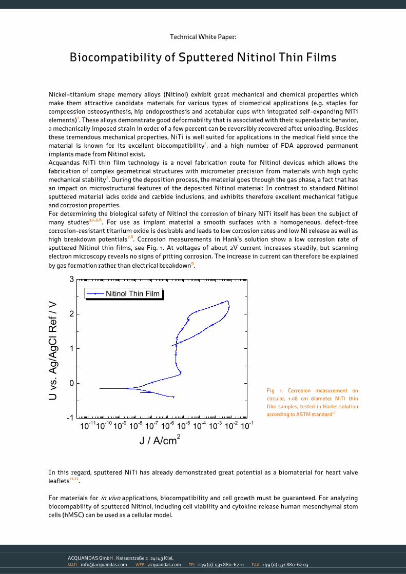
high breakdown potentials7,8. Corrosion measurements in Hank’s solution show a low corrosion rate of
sputtered Nitinol thin films, see Fig. 1. At voltages of about 2V current increases steadily, but scanning
electron microscopy reveals no signs of pitting corrosion. The increase in current can therefore be explained
by gas formation rather than electrical breakdown9.
Fig 1. Corrosion measurement on
circular, 1.08 cm diameter NiTi thin
film samples, tested in Hanks solution
according to ASTM standard10
In this regard, sputtered NiTi has already demonstrated great potential as a biomaterial for heart valve
leaflets11,12
.
For materials for in vivo applications, biocompatibility and cell growth must be guaranteed. For analyzing
biocompability of sputtered Nitinol, including cell viability and cytokine release human mesenchymal stem
cells (hMSC) can be used as a cellular model.
10-11
10-10
10-9
10-8
10-7
10-6
10-5
10-4
10-3
10-2
10-1
-1
0
1
2
3
U v
s. A
g/A
gC
l R
ef / V
J / A/cm2
Nitinol Thin Film
ACQUANDAS GmbH . Kaiserstraße 2 . 24143 Kiel.
MAIL [email protected] WEB acquandas.com TEL +49 (0) 431 880-62 11 FAX +49 (0) 431 880-62 03
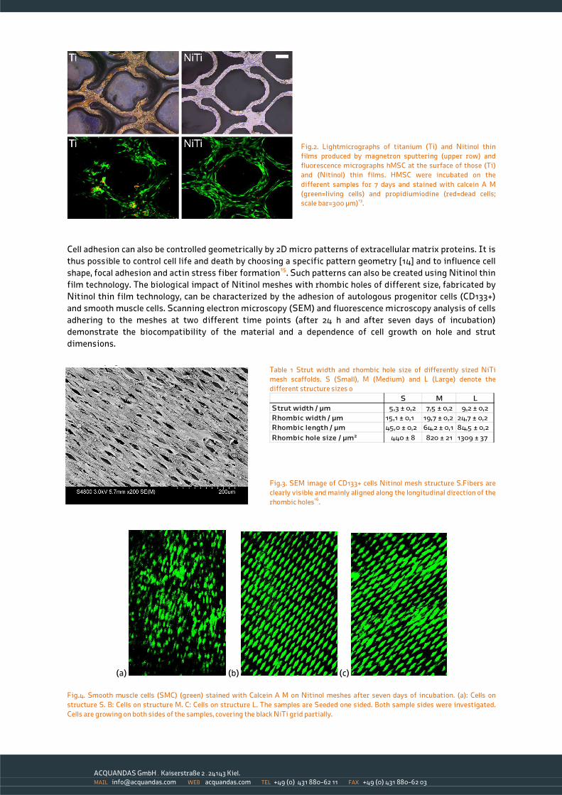
Fig.2. Lightmicrographs of titanium (Ti) and Nitinol thin
films produced by magnetron sputtering (upper row) and
fluorescence micrographs hMSC at the surface of those (Ti)
and (Nitinol) thin films. HMSC were incubated on the
different samples for 7 days and stained with calcein A M
(green=living cells) and propidiumiodine (red=dead cells;
scale bar=300 µm)13.
Cell adhesion can also be controlled geometrically by 2D micro patterns of extracellular matrix proteins. It is
thus possible to control cell life and death by choosing a specific pattern geometry [14] and to influence cell
shape, focal adhesion and actin stress fiber formation15. Such patterns can also be created using Nitinol thin
film technology. The biological impact of Nitinol meshes with rhombic holes of different size, fabricated by
Nitinol thin film technology, can be characterized by the adhesion of autologous progenitor cells (CD133+)
and smooth muscle cells. Scanning electron microscopy (SEM) and fluorescence microscopy analysis of cells
adhering to the meshes at two different time points (after 24 h and after seven days of incubation)
demonstrate the biocompatibility of the material and a dependence of cell growth on hole and strut
dimensions.
Table 1 Strut width and rhombic hole size of differently sized NiTi
mesh scaffolds. S (Small), M (Medium) and L (Large) denote the
different structure sizes o
S M L
Strut width / µm 5,3 ± 0,2 7,5 ± 0,2 9,2 ± 0,2
Rhombic width / µm 15,1 ± 0,1 19,7 ± 0,2 24,7 ± 0,2
Rhombic length / µm 45,0 ± 0,2 64,2 ± 0,1 84,5 ± 0,2
Rhombic hole size / µm2 440 ± 8 820 ± 21 1309 ± 37
Fig.3. SEM image of CD133+ cells Nitinol mesh structure S.Fibers are
clearly visible and mainly aligned along the longitudinal direction of the
rhombic holes16.
(a) (b) (c)
Fig.4. Smooth muscle cells (SMC) (green) stained with Calcein A M on Nitinol meshes after seven days of incubation. (a): Cells on
structure S. B: Cells on structure M. C: Cells on structure L. The samples are Seeded one sided. Both sample sides were investigated.
Cells are growing on both sides of the samples, covering the black NiTi grid partially.

ACQUANDAS GmbH . Kaiserstraße 2 . 24143 Kiel.
MAIL [email protected] WEB acquandas.com TEL +49 (0) 431 880-62 11 FAX +49 (0) 431 880-62 03
NiTi thin film meshes are promising biomaterials for the fabrication of mechanically and geometrically well
defined, free-standing tissue engineered implants. The effective mechanical properties of the scaffold can
be adapted to implant requirements by varying the hole and strut size and the macroscopic dimensions of
the Nitinol film. Nitinol thin film meshes are highly biocompatible and cell adhesion can be controlled by the
size of the rhombic holes in the mesh.
The suitability of Nitinol scaffolds for biological applications in general is proved. The inflammatory
response shows a lower TNF-α level for tissue enclosed meshes than for bare NiTi meshes, and furthermore,
the superior biocompatibility of NiTi compared to stainless steel was confirmed.
About ACQUANDAS GmbH: ACQUANDAS GmbH is a technology company that supplies thin film components to the healthcare
industry – in particular to medical device OEMs – and other industrial markets, such as the automotive and consumer electronics
industries. ACQUANDAS is located in Kiel, Germany.
Based on state-of-the-art microsystem technology processes, we fabricate an entirely new generation of metallic components for
applications in medical devices and many other products. The combination of properties that our devices have is unique: miniaturized
structures with high geometrical complexity, integrated micro-electrode arrays, increased radiopacity, high feature resolution,
excellent biocompatibility and improved mechanical properties!
We look forward to working with you…
1 Clinical Applications of Shape Memory Alloys Based on NiTi as Implant Materials – Possibilities in Trauma and Orthopaedic Surgery,
S.A. Esenwein, D. Bogdanski, M. Köller, L. Krone, M. Epple, G. Muhr, SMST Proc. (2006) 837–844. 2 Comparison of the fatigue performance of commercially produced samples versus sputter-deposited NiTi , G. Siekmeyer, A. Schüßler,
R. Lima de Miranda, E. Quandt, , J. Mater. Eng. Perform. 23 (7) (2014) 2437–2445. 3 Surface, corrosion and biocompatibility aspects of nitinol as an implant material, S.A Shabalovskaya. Biomed Mater Eng (2002) 12:69–
109 4 Biocompatibility of nickel-titanium shape memory metal and its corrosion behavior in human cell cultures, J. Ryhänen, E. Niemi, W.
Serlo, E. Niemelä, P. Sandvik, H. Pernu, T. Salo. J Biomed Mater Res (1997)35(4):451–457 5 Critical overview of nitinol surfaces and their modifications for medical applications, S.A. Shabalovskaya, J. Anderegg, J. van
Humbeeck. Acta Biomater (2008) 4(3):447–467 6 Cytotoxic, allergic and genotoxic activity of a nickel–titanium alloy, D.J. Wever, A.G. Veldhuizen, M.M. Sanders, J.M. Schakenraad, J.R.
van Horn. Biomaterial (1997) 18(16):1115–1120 7 Assessing the corrosion behaviour of nitinol for minimally-invasive device design, R. Venugopalan, C. Trepanier. Minim Invasive Ther
Allied Technol (2000) 9(2):67–74 8 Comparative corrosion performance of black oxide, sandblasted, and fine-drawn nitinol wires in potentiodynamic and potentiostatic
tests: effects of chemical etching and electropolishing, S.A. Shabalovskaya. J Biomed Mater Res B (2004) 69(2):223–231 9 Method for Fabricating Miniaturized NiTi Self-Expandable Thin Film Devices with Increased Radiopacity, C. Bechtold, R. Lima de
Miranda, C. Chluba, C. Zamponi, E. Quandt, Shap. Mem. Superelasticity (2016)2:391-398 10 Standard test method for conducting cyclic potentiodynamic polarization measurements to determine the corrosion susceptibility of
small implant devices, ASTM F2129-08 (2001) 11 A thin film nitinol heart valve, L. Stepan, D. Levi, G. Carman, , J. Biomech. Eng. 127 (6) (2005) 915–918.
12 Fabrication and evaluation of nitinol thin film heart valves, K. Loger, R. Lima de Miranda, A. Engel, M. Marczynski-Bühlow, G. Lutter, E.
Quandt, Cardiovasc. Eng. Technol. 5 (4) (2014) 308–316. 13 The biocompatibility and mechanical properties of cylindrical NiTi thin films produced ba magnetron sputtering, T. Habijan, R. Lima
de Miranda, C. Zamponi, E. Quandt, C. Greulich, T.A. Schildhauer, M. Köller, , Mater. Sci. Eng. C 32 (2012) 2523-2528. 14 Geometric control of cell life and death, C. Chen, M. Mrksich, S. Huang, G.Whitesides, D. Ingber, , Science 276 (1425) (1997) 1425–1428.
15 Cell distribution of stress fibres in response to the geometry of the adhesive environment, M. Théry, A. Pépin, E. Dressaire, Y. Chen, M.
Bornens, , Cell Motil. Cytoskeleton 63 (6) (2006) 341–355. 16 Cell adhesion on NiTi thin film sputtered-deposited meshes, K. Loger, A. Engel, J. Haupt, Q. Li, R. Lima de Miranda, E. Quandt, G. Lutter,
C. Selhuber-Unkel, , Mater. Sci. Eng C 59 (2016) 611-616.