bioenergetics of the archaea · chaeal bioenergetics by a critical state-of-the-art report and to...
TRANSCRIPT

MICROBIOLOGY AND MOLECULAR BIOLOGY REVIEWS,1092-2172/99/$04.0010
Sept. 1999, p. 570–620 Vol. 63, No. 3
Copyright © 1999, American Society for Microbiology. All Rights Reserved.
Bioenergetics of the ArchaeaGUNTER SCHAFER,1* MARTIN ENGELHARD,2 AND VOLKER MULLER3
Institut fur Biochemie, Medizinische Universitat zu Lubeck, Lubeck,1 Max Planck Institut fur Molekulare Physiologie,Dortmund,2 and Lehrstuhl fur Mikrobiologie, Ludwigs-Maximilia Universitat, Munich,3 Germany
INTRODUCTION .......................................................................................................................................................571CHEMIOSMOSIS IN ARCHAEA .............................................................................................................................572ENERGETICS OF METHANOGENESIS...............................................................................................................573
Proton Motive Electron Transport Chains in Methanogens............................................................................574Components of the Electron Transport Chain ...................................................................................................575
Heterodisulfide reductase ..................................................................................................................................575Hydrogenases.......................................................................................................................................................576F420 dehydrogenase.............................................................................................................................................576
Membrane-Integral Electron Carriers.................................................................................................................577Possible Mechanisms of DmH1 Formation Coupled to Electron Transport Reactions ................................577Sodium Bioenergetics of Methanogenesis ...........................................................................................................578ATP Synthesis in Methanogens ............................................................................................................................579Bioenergetics of the Acetyl-CoA Pathway in Archaea and Bacteria: Differences and Similarities...............579
ENERGETICS OF RESPIRATION .........................................................................................................................579Aerobiosis and Other Respiration Forms in Archaea........................................................................................579Components of Aerobic Electron Transfer..........................................................................................................580
Membrane-residing quinone reductases..........................................................................................................581(i) NADH dehydrogenases .............................................................................................................................581(ii) SDHs and a novel complex II ................................................................................................................581
Membrane-integral quinol-oxidizing complexes .............................................................................................582(i) The SoxABCD complex.............................................................................................................................582(ii) The SoxM complex...................................................................................................................................583(iii) Other archaeal terminal oxidases ........................................................................................................583
Mobile electron carriers, hemes, and small metal proteins .........................................................................585(i) Quinones.....................................................................................................................................................585(ii) Hemes ........................................................................................................................................................585(iii) Ferredoxins ..............................................................................................................................................586(iv) Rieske iron-sulfur proteins ....................................................................................................................587(v) Small copper proteins ..............................................................................................................................587
Organization of Archaeal Respiratory Chains ...................................................................................................588Oxygen respiration..............................................................................................................................................588Alternate types of respiration ...........................................................................................................................589
Proton Pathways in Terminal Oxidases of Archaea ...........................................................................................590LIGHT-DRIVEN ENERGETICS ..............................................................................................................................592
General Overview....................................................................................................................................................592BR .............................................................................................................................................................................592
Structure of BR...................................................................................................................................................592Mechanism of proton pumping.........................................................................................................................594
HR.............................................................................................................................................................................595Structure of HR ..................................................................................................................................................595Mechanism of chloride pumping ......................................................................................................................596
SRs ............................................................................................................................................................................597SRI ........................................................................................................................................................................597SRII.......................................................................................................................................................................597
Proton Transport in Archaeal Rhodopsins: a Common Property of Ion Pumps and Photoreceptors.......598Phototaxis and Chemotaxis ...................................................................................................................................598
Signal transduction chain..................................................................................................................................599Flagella and the flagellar motor .......................................................................................................................600
SECONDARY ENERGY CONVERTERS................................................................................................................600The Family of ATPases ..........................................................................................................................................600The ATPases of Sulfolobus .....................................................................................................................................601
* Corresponding author. Mailing address: Institut fur Biochemie,Medizinische Universitat zu Lubeck, 23538 Lubeck, Germany. Phone:49 451 500-4060. Fax: 49 451 500-4068. E-mail: [email protected].
570
on May 21, 2020 by guest
http://mm
br.asm.org/
Dow
nloaded from

The Halobacterial ATPases ...................................................................................................................................601The ATPases of Methanogens...............................................................................................................................602
Cellular function of the A1Ao ATPase from methanogens............................................................................602Features of ATPases from methanogens .........................................................................................................603
Genetic Organization of Known A1Ao ATPases..................................................................................................603Properties and Functions of the Polypeptides Involved in ATPase Function and Assembly ......................604A Structural Model.................................................................................................................................................605
The novel features of archaeal ATP synthases...............................................................................................606Auxiliary Energy Transducers...............................................................................................................................606
The unique structure of archaeal adenylate kinases .....................................................................................607Archaeal PPases..................................................................................................................................................607
CONCLUSIONS AND PERSPECTIVES.................................................................................................................608ACKNOWLEDGMENTS ...........................................................................................................................................608ADDENDUM IN PROOF..........................................................................................................................................608REFERENCES ............................................................................................................................................................608
INTRODUCTION
Ever since archaea have been studied, their ability to thrivein unusual habitats under extremely harsh conditions has stim-ulated interest in the molecular mechanisms that confer heatstability on proteins at temperatures above 100°C, tolerance ofextreme pH values and salt concentrations, and unique meta-bolic functions not found in bacteria, such as methanogenesisor rhodopsin-linked energy and signal transduction. Actually,ever since Archaea was identified as a third evolutionary king-dom (606–608) presumably located relatively near the hypo-thetical root of the evolutionary tree, it has been speculatedthat the structural organization and metabolic pathways ofarchaea might reflect more ancestral organisms whose essen-tial properties differ from those of bacteria and eucarya. In thisregard, one has to realize with respect to biological energyconservation that all existing forms of life rely on the universalprinciple of chemiosmotic energy transduction (366, 367),which in phylogenetic terms should have evolved very early. Infact, the origin of cellular life must have been connected withthe permanent manifestation of mechanisms allowing thetransduction of energy between exergonic and endergonic pro-cesses and with the development of transitory and long-termenergy stores.
The definition of Archaea as a separate domain of organismswas based on the comparative analysis of 16S rRNA sequences(405), which led to a result different from classical taxonomy.The term Archaea reflects an earlier idea that these organismsdescended from life forms that existed prior to the division intothe bacterial and eukaryal domains. However, based on thesequences of universally present proteins (176), Archaea hasbeen placed on the branch also leading to Eucarya. A featurethat distinguishes Archaea from Bacteria is the structure ofarchaeal ribosomes (435), which in halophiles were first rec-ognized to contain acidic rather than basic proteins (42). Inaddition, the transcriptional machinery is unique as to thestructure of DNA-dependent RNA polymerases (133, 626).With respect to subunit structure, a closer relationship to eu-karyotes than to bacteria was found (300). Another featuredistinguishing Archaea from Bacteria is the specific composi-tion of archaeal surface layers (266), which do not containpeptidoglycans. Their glycoprotein surface layers can formquasicrystalline structures (97, 360) that are firmly attached tothe plasma membrane, thus leaving practically no periplasmicspace (39).
Nevertheless, although archaea are located on a distinctevolutionary branch as depicted in Fig. 1, they represent, withregard to their primary energy-transducing mechanisms, a veryheterogeneous domain comprising chemolithoautotrophic as
well as organotrophic species. In addition to obligate anaer-obes such as the methanogens, a second group that performsvarious types of aerobic or anaerobic respiration can be dis-tinguished. Further, for some halobacteria we have to considerarchaeal phototrophic energy transformation in addition torespiratory mechanisms.
In contrast to this diversity, archaeal membrane structuresreveal a comparatively homogeneous phenotype, significantlydifferent from that of other prokaryotes. Diether and tetraetherlipids are uniformly used as building blocks for archaeal plasmamembranes (266). Obviously, the low ion permeability of mem-branes formed from these bipolar monolayer-forming lipids(136, 571) contributes significantly to the stability of chemios-motic charge separation in archaea, particularly at high tem-peratures and/or at extremely low pH values.
Interestingly, neither oxygenic nor anoxygenic “green” pho-tosynthesis has been found in the archaeal kingdom. This latterobservation served as an argument in favor of the respiration-first hypothesis, suggesting that the formation of the basicstructure of terminal oxidase complexes preceded the occur-rence of chlorophyll-based water-splitting and charge-separat-ing systems (93, 94).
It is the aim of this review to introduce the reader to ar-chaeal bioenergetics by a critical state-of-the-art report and todemonstrate similarities and distinguishing features by con-trast to bacterial and eucaryal systems. It will not be possible inall cases to give an unambiguous answer to the question ofwhat is typical or genuine for the archaeal domain, because,especially within respiratory electron transport, we will find anumber of chimeric functional complexes. In fact, one has toassume that during early evolution, i.e., prior to the divisioninto three urkingdoms, the barriers against lateral gene trans-fer were much lower than they are now (605).
Of the various bioenergetic mechanisms in archaeal organ-isms, the present review focuses specifically on the primaryenergy conservation that involves membrane-residing chemi-osmotic processes. Therefore, purely fermentative energytransduction by substrate-level phosphorylation as well as sec-ondary active-transport systems for solutes will not be dis-cussed. As an exception among secondary energy transducers,the ATP synthase complexes will be dealt with because theyapparently possess a unique and ubiquitously conserved mech-anism, irrespective of the primary energy converter which pro-vides the electrochemical ion gradient to be used as the drivingforce for high-energy bond formation during ADP phosphor-ylation.
Another limitation is the great diversity of the archaeal do-main, much greater than that suggested by Fig. 1. What wepresently know about diversity within the archaeal domain is
VOL. 63, 1999 BIOENERGETICS OF THE ARCHAEA 571
on May 21, 2020 by guest
http://mm
br.asm.org/
Dow
nloaded from

probably only the tip of the iceberg. By means of molecularstudies based on rRNA-directed probes, new archaea are con-stantly being discovered not only in deep-sea vents or solfataricfields (37, 89, 379) but also in mesophilic and even low-tem-perature environments. Unfortunately, only a few of theseisolates or new species identified by DNA hybridization willprove amenable to cultivation. Thus, the diversity of bioener-getic systems may well exceed the number of classes reviewedin this comprehensive study.
CHEMIOSMOSIS IN ARCHAEA
Primary energy conservation by membrane-residing systemsis characterized by the formation of an electrochemical poten-tial of hydrogen ions or sodium ions. According to the work ofMitchell (367), the free energy stored in this gradient is de-scribed by equations 1 and 2 for the proton motive force:
DmH1 5 RT z ln([H1
i]/[H1o]) 1 F z DC (1)
Dp 5 DmH1/F 5 DC 2 ZDpH (2)
The primary pumps may be driven by redox systems, by methyltransfer reactions as in methanogenesis, or by light as in pho-tophosphorylating halobacteria.
As illustrated by some representative examples below,whole-cell experiments with various genera of Archaea haveproven that ATP synthesis is driven, according to the chemi-
osmotic theory, at the expense of such ion gradients. Theseexperiments were of significance because minimal systems suchas inverted plasma membrane vesicles or spheroplasts whichare easily prepared from several bacterial organisms are essen-tially inaccessible in the case of Archaea. The rigid structureand extremely tight adhesion or interdigitation of the glyco-protein cell walls covering archaeal plasma membranes (39, 98,181, 270, 514) represent an invincible obstacle. For the samereason, the preparation of intact complexes of energy-trans-ducing membrane proteins is quite difficult. In addition, otherfactors are frequently responsible for the failure to purify cat-alytically active complexes, such as ATP synthase or terminaloxidases. Such factors can be the absence of the high pHgradient to which membrane proteins are exposed in vivo,extreme salt concentrations, hypersensitivity toward oxygen,and cold dissociation even at room temperature. Also, thedetermination of energetic parameters such as DpH or DC bydirect monitoring or by distribution of diffusible molecularprobes is very limited at ambient pH values below 3, the phys-iological environmental pH for many extreme acidophiles.
Extremely acidophilic organisms, including the archaeonThermoplasma, were shown to create an inverted membranepotential (30, 364) in order to prevent acidification of thecytosol by influx of H1 at the prevailing DpH. This does notgenerally apply to all acidophiles, however. The membranepotential of thermoacidophilic archaea such as Sulfolobus maybe rather low (approximately 30 mV), and most of the proton
FIG. 1. Phylogenetic tree. The scheme demonstrates the division into Crenarchaeota and Euryarchaeota and shows the position of the major archaeal genera. Thetree was redrawn according to references 88, 175, and 608. Stars denote archaeal species for which specific bioenergetic information has been found.
572 SCHAFER ET AL. MICROBIOL. MOL. BIOL. REV.
on May 21, 2020 by guest
http://mm
br.asm.org/
Dow
nloaded from

motive force is maintained by a large pH gradient of .3 (335,370, 467). Chemiosmotic H1 cycling (370) with H1/O ratios of3 and a strict correlation of proton motive force (Dp) withcellular ATP levels could be established for Sulfolobus. Dissi-pation of Dp by protonophores caused an immediate collapseof ATP synthesis; at Dp > 0, a persisting residual DpH wascounterbalanced by an inverted membrane potential, in whichthe inside was positive (335). In the same experiments, externalproton pulses that lowered the pH from 6.1 to 3.4 produced anincrease of Dp from 294 to 2170 mV with a concomitant riseof intracellular ATP. For experimental reasons, the reporteddata was determined at 45°C at an ambient pH of 3.5 and thusmay assume slightly different values at the optimal growthtemperature of the cells. A review of strategies to cope withextremely low pH values is given in reference 465.
With Halobacterium halobium, the coupling of either a light-or a respiration-induced electrochemical proton gradient withintracellular ATP has been established (361–363). Interest-ingly, by cation counter transport considerable energy can bestored in the form of a potassium gradient also. Photophos-phorylation is a backup system under oxygen limitation inextremely halophilic archaea. This is corroborated by recentstudies of the haloalkaliphile Natronobacterium pharaonis(604) demonstrating full recovery of Dp under oxygen-limitingconditions during illumination. Actually, in these latter ar-chaea the main contribution to the proton motive force ismade by the membrane potential of DC 5 2225 to 2280 mV,and DpH is influenced only marginally by oxygen limitation.Under these conditions, the high membrane potential is gen-erated by an outwardly directed chloride gradient produced bythe light-activated chloride pump halorhodopsin (HR); it isinsensitive to protonophores and uncouplers and can even beincreased by the Cl2/OH2 exchanger triphenyltin (604).
The membrane potential can contribute approximately 90%to the proton motive force (56) in methanogenic archaea also,as shown with Methanosarcina barkeri. Evidence for H1- andNa1-mediated chemiosmotic energy transduction in methano-gens has been compiled previously (124); thereby, the sodiumand proton gradients may be linked by Na1-H1 antiporters(382). A methanogenic strain, Go1 (now classified as Methano-sarcina mazei), is the only known case in which the successfulpreparation of archaeal vesicular membrane systems has pro-vided a useful experimental model for the study of energytransduction (58, 59).
The coexistence of proton- and sodium ion-coupled energyconverters in anaerobes as well as the branching of electrontransport pathways in aerobic archaea is difficult to resolvebecause these organisms either lack or are insensitive to thesite-specific inhibitors known to function in bacteria or euca-rya. In addition, genetic systems for directed mutagenesis orgene disruption in archaea have scarcely been developed or areunavailable.
The scheme of Fig. 2 illustrates the generation of ion gradi-ents by primary pumps and their utilization by secondary pro-cesses. In the following sections, molecular properties of theknown functional complexes are discussed separately formethanogenic, respiring, or photophosphorylating archaea; itshould be noted, however, that current complete genomeprojects have predicted the existence of additional functionalcomplexes which have not yet been verified at the protein ormRNA level.
ENERGETICS OF METHANOGENESIS
Methanogens are a phylogenetically diverse but nutritionallyrather uniform group of strictly anaerobic archaea. They are
able to grow by the conversion of a small number of com-pounds to methane. This rather simple pathway is not coupledto substrate-level phosphorylation but, instead, to the genera-tion of ion gradients across the membrane that are used todrive the synthesis of ATP. Interestingly, the pathway of meth-ane formation is coupled to the simultaneous generation ofprimary gradients of both protons and sodium ions. Althoughmethanogens are nutritionally rather similar and employ iden-tical pathways, they differ significantly with respect to the com-ponents involved in the proton motive electron transport chainand, therefore, most likely employ different mechanisms togenerate the proton gradient. For example, methylotrophicmethanogens, such as M. mazei Go1, contain cytochromes,whereas hydrogenotrophic methanogens, such as Methanobac-
FIG. 2. Primary energy-transducing processes and coupling principles inmembrane bioenergetics. The top scheme illustrates the processes found inarchaea that contribute to the formation of either proton or sodium ion poten-tials across the plasma membrane. Details are discussed throughout this review.The bottom schemes illustrate three mechanisms by which an ion gradient can beproduced: (a) chemical charge separation (only electrons are transferred throughthe membrane); (b) a mobile membrane-integral cofactor like the quinones ormethanophenazine functioning as proton transporter (examples are bc1 com-plexes); and (c) redox-driven pumps like cyt c oxidase. All schemes are drawn foran H1/e2 ratio of 1. Scheme d illustrates the proton-driven ATP synthase of theFoF1 or A1Ao type as an example for a secondary energy transducer. D, electrondonor; Ac, electron acceptor.
VOL. 63, 1999 BIOENERGETICS OF THE ARCHAEA 573
on May 21, 2020 by guest
http://mm
br.asm.org/
Dow
nloaded from

terium thermoautotrophicum, do not. Since most of our currentknowledge derives from studies using methylotrophic meth-anogens, in particular M. barkeri and M. mazei, this sectionfocuses on these organisms. For a more thorough discussion ofthe pathways and the biochemistry of methanogenesis, thereader is referred to recent reviews (58, 124, 381, 561).
Proton Motive Electron Transport Chains in Methanogens
Central to all pathways of methane formation is the inter-mediate methyl coenzyme M (2-methylthioethanesulfonate;CoM), the ultimate precursor of methane (Fig. 3). It is reduc-tively demethylated by the methyl-CoM reductase with elec-trons derived from reduced CoB (7-mercaptoheptanoylthreo-nine phosphate), to give rise to methane and a heterodisulfideof CoM and CoB (CoM-S-S-CoB; henceforth referred to asthe heterodisulfide), in a reaction involving the cofactor F430(reaction 6 in Fig. 3). To complete the cycle, the heterodisul-fide is reduced by the heterodisulfide reductase complex (re-action 7 in Fig. 3); this reaction is most important in terms ofenergy conservation (561). The heterodisulfide reductase ismembrane bound and operates as the final limb of severalmembrane-bound electron transport chains (124). Dependingon the substrate, the electron donor used is different. Hydro-
genase is employed during growth on H2 plus CO2, CO dehy-drogenase (or reduced ferredoxin:heterodisulfide oxidoreduc-tase) is used during growth on acetate, and F420 dehydrogenase(F420, a 59-deazaflavin, is the universal electron carrier in meth-anogens) and formylmethanofuran (formyl-MF) dehydroge-nase are used during growth on methyl group-containing C1substrates.
H2-dependent reduction of the heterodisulfide, as catalyzedby inverted vesicles of M. mazei Go1, was accompanied by H1
translocation into the lumen of the vesicles (Fig. 4). Protono-phores inhibited ATP formation but stimulated electron trans-port, i.e., heterodisulfide reduction. Electron transport andATP synthesis were inhibited by the ATPase inhibitor N,N9-dicyclohexylcarbodiimide (DCCD), but inhibition was relievedby the addition of protonophores. These effects are clearlyreminiscent of respiratory control as observed in mitochondriaand can be taken as evidence that the DmH1 generated drivesthe synthesis of ATP from ADP and Pi. Washed everted ves-icles exhibited stringent coupling between heterodisulfide re-duction and ATP synthesis, with maximal stoichiometries of1H1 translocated/e2 and 1ATP synthesized/4e2 (120).
The F420H2-dependent heterodisulfide reduction was alsoshown to drive proton translocation into the lumen of evertedvesicles of M. mazei Go1, resulting in the generation of a DmH1
FIG. 3. Pathways of methanogenesis. Reactions involved in energy conservation are boxed. The reduction of methyl-CoM (reactions 6 and 7) is common to allmethanogenic substrates. During methane formation from H2 plus CO2, reactions 1 to 5 proceed in the direction of CO2 reduction. The methyl groups of methanoland acetate enter the central pathway at the level of H4MPT. During methanogenesis from methanol, one-fourth of the methanol is oxidized to CO2 by the reversalof reactions 1 to 5; the six reducing equivalents gained are used to reduce 3 mol of methanol to methane. During methanogenesis from acetate, the carboxyl group isoxidized to CO2 and the electrons gained are used to reduce the methyl group to acetate. F420, oxidized form of coenzyme F420; F420H2, reduced form of F420; HS-CoM,CoM (2-mercaptoethanesulfonate); HS-CoB, CoB (7-mercaptoheptanoylthreonine phosphate); CoM-S-S-CoB, heterodisulfide of HS-CoM and HS-CoB. Enzymes: 1,formyl-MF dehydrogenase; 2, formyl-MF:H4MPT formyltransferase and methenyl-H4MPT cyclohydrolase; 3, F420-dependent methylene-H4MPT dehydrogenase; 4,F420-dependent methylene-H4MPT reductase; 5, methyl-H4MPT:CoM-methyltransferase; 6, methyl-CoM reductase; 7, heterodisulfide reductase system (differentelectron donor systems are indicated).
574 SCHAFER ET AL. MICROBIOL. MOL. BIOL. REV.
on May 21, 2020 by guest
http://mm
br.asm.org/
Dow
nloaded from

(Fig. 5) Protonophores stimulated the heterodisulfide reduc-tion but prevented DmH1 formation and ATP synthesis. TheATP synthase inhibitor DCCD decreased the rate of F420H2-dependent heterodisulfide reduction. The reversal of thisDCCD-mediated inhibition by protonophores and the stimu-
lation of the F420H2-dependent heterodisulfide reduction byADP indicate stringent coupling between electron transportand ATP synthesis. The F420H2-dependent heterodisulfide re-ductase system displayed stoichiometries of 1 H1 translo-cated/e2 and 0.8 ATP synthesized/4e2 (122).
Evidence that the conversion of CO to CO2 and H2 (DG89 5220 kJ/mol) by resting cells of M. barkeri is coupled to thesynthesis of ATP has been presented (70, 71). The cleavage ofacetyl-CoA as catalyzed by carbon monoxide dehydrogenaseyields enzyme-bound CO and an enzyme-bound methyl group(150, 308). The latter is transferred via a corrinoid protein totetrahydromethanopterin (H4MPT). Enzyme-bound CO un-dergoes ferredoxin-dependent oxidation to carbon dioxide,catalyzed by carbon monoxide dehydrogenase (151, 560). In areconstituted system consisting of purified CO dehydrogenase,heterodisulfide reductase, and ferredoxin, CO oxidation wascoupled to heterodisulfide reduction. However, the rate ofheterodisulfide reduction was increased 10-fold by addition ofmembranes, indicating a membrane-bound electron transportchain from ferredoxin to the heterodisulfide (Fig. 6) (420, 512).
Methyl group oxidation proceeds via the reversal of CO2reduction (reactions 1 to 5 of Fig. 3 in the oxidative direction).There are indications that formyl-MF oxidation is accompa-nied by the generation of an electrochemical ion potentialacross the membrane, either protons or sodium ions (262, 603).The formyl-MF-dependent heterodisulfide reduction is associ-ated with a large DG89 of 258 kJ/mol. In contrast, DG89 of theelectron transfer from F420H2 to the heterodisulfide is consid-erably smaller (229 kJ/mol). Since the physiological electronacceptor employed in the oxidation of formyl-MF to CO2 isunknown, the DG89 of the formyl-MF oxidation cannot becalculated. However, the midpoint potential at pH 7 (Em,7) ofthe CO2–formyl-MF couple of 2500 mV indicates that a low-potential electron carrier can be reduced (47).
Components of the Electron Transport Chain
Since little is known about the formyl-MF-dependent het-erodisulfide reduction, only the F420, the H2-, and the CO-dependent systems will be considered here.
Heterodisulfide reductase. The reaction catalyzed by theheterodisulfide reductase resembles a polysulfide reductioncatalyzed by some bacteria and archaea. The S-S bonds ofpolysulfide can be reduced by H2 as external electron donor,and this reaction is coupled with energy conservation (480).
Heterodisulfide reductase was first purified from H2-CO2-grown M. thermoautotrophicum. It contained three subunitswith apparent molecular masses of 80 (HdrA), 36 (HdrB), and21 (HdrC) kDa and (per mol of heterotrimer) approximately 1mol of flavin adenine dinucleotide (FAD), 20 mol of nonhemeiron, and 20 mol of acid-labile sulfur (205, 508). The encodinggenes have been cloned and sequenced (206). Sequence com-parisons indicated that HdrA harbors four [4Fe-4S] clustersand binds FAD. HdrC is considered to be an electron carrierprotein with two [4Fe-4S] clusters and a short stretch of hy-drophobic amino acids that could anchor the complex to themembrane. Interestingly, HdrB is similar to subunit C of thesuccinate dehydrogenase (SDH) of Acidianus ambivalens andSulfolobus acidocaldarius.
From membranes of acetate-grown M. barkeri, a heterodi-sulfide reductase complex which also contained the electrondonor, the F420-nonreactive hydrogenase, was purified. Thiscomplex contained nine subunits of 46, 39, 28, 25, 23, 21, 20,16, and 15 kDa, three of which are subunits of the F420-non-reactive hydrogenase. The monomeric heterodisulfide reduc-tase contained 0.7 mol of cytochrome b (cyt b) and 18 mol of
FIG. 4. Tentative scheme of electron flow and proton translocation duringheterodisulfide reduction with H2 as electron donor. This reaction sequence ispart of methanogenesis from H2-CO2. This scheme is valid for methylotrophicmethanogens only, for hydrogenotrophic methanogens do not contain cyto-chromes and the presence of methanophenazine (MP) has not been verified. Theheterodisulfide reductase is not indicated to be a proton pump, but this cannotbe ruled out a priori. This scheme is based on the experimentally derivedstoichiometry of 3 to 4 H1 translocated/methyl group reduced. P, periplasm;CM, cytoplasmic membrane; C, cytoplasm. For explanations, see the text.
FIG. 5. Tentative scheme of electron flow and proton translocation duringheterodisulfide reduction with F420H2 as electron donor. This reaction sequenceis part of methanogenesis from methanol, methylamines, and formate. Thisscheme is valid for methylotrophic methanogens only (see the legend to Fig. 4).F420, coenzyme F420; MP, methanophenazine; P, periplasm; CM, cytoplasmicmembrane; C, cytoplasm. For explanations, see the text.
VOL. 63, 1999 BIOENERGETICS OF THE ARCHAEA 575
on May 21, 2020 by guest
http://mm
br.asm.org/
Dow
nloaded from

nonheme iron and acid-labile sulfur. The 23-kDa subunit car-ried cyt b (211).
Heterodisulfide reductase itself was purified from mem-branes of methanol-grown M. barkeri, and the encoding geneswere cloned and sequenced (212, 296). The reductase wascomposed of only two subunits with apparent molecularmasses of 46 (HdrD) and 23 (HdrE) kDa. The enzyme con-tained 0.6 mol of cyt b and 20 mol of nonheme iron andacid-labile sulfur per mol of heterodimer. Biochemical andmolecular data revealed that HdrE is a b-type cytochrome withfive potentially membrane-spanning helices. HdrD containstwo [4Fe-4S] clusters, and its N and C termini are similar toHdrC and HdrB from M. thermoautotrophicum, respectively,indicating that HdrD of M. barkeri and HdrC and HdrB ofM. thermoautotrophicum are functionally equivalent. Althoughsmall amounts of FAD were found in the heterodisulfide re-ductase from M. barkeri, it was shown later that heterodisulfidereduction did not depend on FAD. Moreover, no FAD bindingsite was found in the deduced amino acid sequence of theenzyme (296), which is in contrast to the enzyme from hydrog-enotrophic methanogens.
From membranes of acetate-grown Methanosarcina ther-mophila, a two-subunit heterodisulfide reductase (53 and 27kDa) was isolated. The small subunit contained 2 mol of cyto-chrome; the large subunit contained two distinct [Fe4S4]21/11
clusters. One heme is a high-spin heme with a midpoint po-tential of 223 mV, whereas the low-spin heme has a midpointpotential of 2180 mV. The midpoint potentials for the twoclusters are 2100 and 2400 mV (512).
Hydrogenases. The hydrogenases are the entry point forelectrons derived from molecular hydrogen. Of the four typesof hydrogenases isolated from methanogens to date, one wasclearly shown to be involved in energy conservation. The F420-reactive hydrogenase reacts with F420 and viologen dyes,whereas the F420-nonreactive hydrogenase reacts with viologen
dyes only. The latter enzyme is therefore often referred to asmethyl viologen-reactive hydrogenase. The function of theF420-reactive hydrogenase in energy metabolism is still a mat-ter for debate (14, 78, 338, 385). On the other hand, there isclear evidence that the F420-nonreactive hydrogenase is theelectron donor for a membrane-bound electron transportchain. In methylotrophic methanogens, the F420-nonreactivehydrogenase is found in the particulate fraction. The enzymeas purified from M. mazei Go1 was composed of only twosubunits containing redox-active Ni and iron-sulfur clusters(123). A molecular analysis revealed that M. mazei Go1 con-tains two operons encoding isoenzymes designated vho forviologen-reactive hydrogenase 1 and vht for viologen-reactivehydrogenase 2. Both operons encode the structural subunits ofthe hydrogenase (VhoG, VhtG, VhoA, and VhtA) and a genecoding for cyt b (VhoC and VhtC); this indicates that these bcytochromes are the natural electron acceptors of the twoF420-nonreactive isoenzymes. The small subunit contains aleader peptide, which suggests that the catalytic part of theenzyme faces the periplasm (121). Interestingly, the C terminiof the two b cytochromes are not homologous, indicating thatthey interact with different proteins. Indeed, Northern blotanalysis revealed that the expression of the isoenzymes is sub-strate dependent. vho was apparently constitutively expressed,whereas vht was expressed only during growth on H2-CO2 ormethanol (119). Therefore, it was speculated that the vho geneproducts are part of the heterodisulfide reductase systemwhereas the vht gene products are involved in electron flow toand from CO2 in the course of the formyl-MF dehydrogenasereaction (124).
F420 dehydrogenase. The F420H2 dehydrogenase is the entrypoint for the electrons derived from F420H2 oxidation. Theenzyme was first isolated from Methanolobus tindarius aftersolubilization from membranes with detergents (182). The ap-parent molecular mass of the native enzyme was 120 kDa; it
FIG. 6. Tentative scheme of electron flow and proton translocation coupled to heterodisulfide reduction with CO as electron donor. This reaction sequence is partof methanogenesis from acetate. The presence of methanophenazine (MP) in acetate-grown cells has not been verified. Fd, ferredoxin; P, periplasm; CM, cytoplasmicmembrane; C, cytoplasm. For explanations, see the text.
576 SCHAFER ET AL. MICROBIOL. MOL. BIOL. REV.
on May 21, 2020 by guest
http://mm
br.asm.org/
Dow
nloaded from

consisted of five different subunits of 45, 41, 22, 18, and 17 kDa.The enzyme contained 16 mol of nonheme iron and 16 mol ofacid-labile sulfur per mol, but flavin was not detected. Thegene encoding the 40-kDa subunit (ffdB) was cloned (600).Sequence analysis, primer extension, and reverse transcription-PCR indicated that ffdB is part of an operon harboring threeadditional genes (ffdA, ffdC, and ffdD). FfdA is similar to theF420-dependent methylene-H4MPT reductase. The first 90amino acids of FfdB are similar to numerous ferredoxins, sug-gesting the likely presence of at least two iron-sulfur centers.FfdC and FfdD are similar to proteins of unknown function ofMethanococcus jannaschii and Archaeoglobus fulgidus. FfdDappears to be very hydrophobic and is likely to be the mem-brane anchor. Recently, F420 dehydrogenases were purifiedfrom M. mazei Go1 and the sulfate-reducing archaeon A. fulgi-dus (3, 297). Flavin was detected in both enzymes. Therefore,it is likely that flavin is also present in the enzyme from M.tindarius but lost during purification.
Membrane-Integral Electron Carriers
With respect to their membrane-integral electron carriers,and thus probably with respect to the mechanism of protontranslocation, methanogens can be divided into two groups:the methylotrophic organisms, in which a variety of b- andc-type cytochromes were found, and the hydrogenotrophicmethanogens, which are devoid of cytochromes (260, 295). Inhydrogenotrophic methanogens, the situation is far from set-tled; polyferredoxins described above and a recently describedflavoprotein encoded by the gene fpaA (394) are the onlyelectron carriers identified so far.
In methylotrophic methanogens, there are several lines ofevidence for the involvement of cytochromes in electron trans-port from the F420-nonreactive hydrogenase to the heterodis-ulfide in methylotrophic methanogens. First, membranes ofacetate-grown cells catalyze an H2-dependent reduction of cy-tochromes (275, 560), and second, hdrE (designated cyt b2) ispart of the heterodisulfide reductase operon and was expressedduring growth on H2-CO2 (296). The vho operon encoding cytb1 along with the structural subunits of the F420-nonreactivehydrogenase was also expressed during growth on H2-CO2(119). Therefore, an electron flow from the F420-nonreactivehydrogenase via cyt b1 and b2 to the heterodisulfide can beenvisaged (Fig. 4).
Experiments performed with M. mazei Go1 strongly suggestthat one or several cytochromes also participate in electrontransport from F420H2 to the heterodisulfide (264). Mem-branes of M. mazei Go1 contain two b- and two c-type cyto-chromes with midpoint potentials (Em,7) of 2135 and 2240mV (b-type cytochromes) and 2140 and 2230 mV (c-typecytochromes). The cytochromes were reduced by F420H2 andoxidized by the heterodisulfide at high rates. Addition of theheterodisulfide to reduced cytochromes and subsequent low-temperature spectroscopy showed the oxidation of cyt b564.This indicates the involvement of cytochromes in electrontransport from F420 via cyt b to the heterodisulfide (Fig. 5).
A different class of membrane-bound electron carriers wasdiscovered recently (1). Membranes of methanogens do notcontain typical quinones found in bacteria or aerobic archaea.However, extraction of membranes from methanol-grownM. mazei Go1 with isooctane yielded a fraction containing aredox-active, low-molecular-weight compound identified as aphenazine derivative, methanophenazine. The structure andreactivity of methanophenazine are given in Fig. 7. Methano-phenazine is reduced by F420 dehydrogenase or hydrogenase,and reduced methanophenazine then reduces the heterodisul-
fide (2, 40). In a reconstituted system consisting of purifiedF420 dehydrogenase and heterodisulfide reductase, methano-phenazine mediated the electron transfer from F420 to theheterodisulfide (40). Methanophenazine was isolated frommethanol-grown M. mazei Go1, but it is probably also involvedin electron transport to the heterodisulfide from other donors,i.e., formyl-MF and CO. The most interesting question,whether methanophenazine is also present in hydrogenotro-phic methanogens, remains to be solved.
Possible Mechanisms of DmH1 Formation Coupled toElectron Transport Reactions
In methylotrophic methanogens, the F420-nonreactive hy-drogenase is localized in the periplasm, as inferred from itsleader sequence and its homology to membrane-bound, cyto-chrome-containing bacterial hydrogenases. Formation of aproton potential can be easily envisaged, because the uptake ofH2 and transfer of electrons to an electron acceptor would leadto the liberation of scalar protons on the outside of the cyto-plasmic membrane. However, the H1/CH4 stoichiometry of 3to 4 (measured in M. barkeri during methanogenesis frommethanol-H2 [57]) cannot be accounted for by scalar protonsonly. That leaves us with the question of the nature of thevectorial proton pump. Electron flow from F420H2 to meth-anophenazine, as well as from the reduced methanophenazineto the heterodisulfide, is coupled to proton translocation, in-dicating the presence of two coupling sites (2). The F420H2dehydrogenase and the H1-translocating bacterial NADH de-hydrogenase have in common a complex structure and thepresence of flavins and iron-sulfur centers. Therefore, it istempting to speculate that the F420H2 dehydrogenase, like theNADH dehydrogenase, is a proton pump. It is not knownwhether the heterodisulfide reductase itself is a proton pump.The genes encoding the hydrogenase, the heterodisulfide re-ductase, and part of the F420 dehydrogenase are known, butthe similarities of the deduced proteins to subunits of NADHdehydrogenases or cytochrome oxidases are too low to allowidentification of polypeptides involved in proton transport.
With the discovery of methanophenazine, another possibilityhas emerged. By analogy with the ubiquinone cycle in the bc1complex, it is likely that electron transfer from cyt b1 to meth-
FIG. 7. Structure and reactivity of methanophenazine, a membrane-integralelectron and hydrogen carrier of methanogens.
VOL. 63, 1999 BIOENERGETICS OF THE ARCHAEA 577
on May 21, 2020 by guest
http://mm
br.asm.org/
Dow
nloaded from
anophenazine is coupled to proton uptake from the cytoplasm.The reduced methanophenazine then donates its electrons tocyt b2, and the protons are liberated into the periplasm. TheH1/e2 stoichiometry of such a mechanism would be fixed at1H1/e2.
Sodium Bioenergetics of Methanogenesis
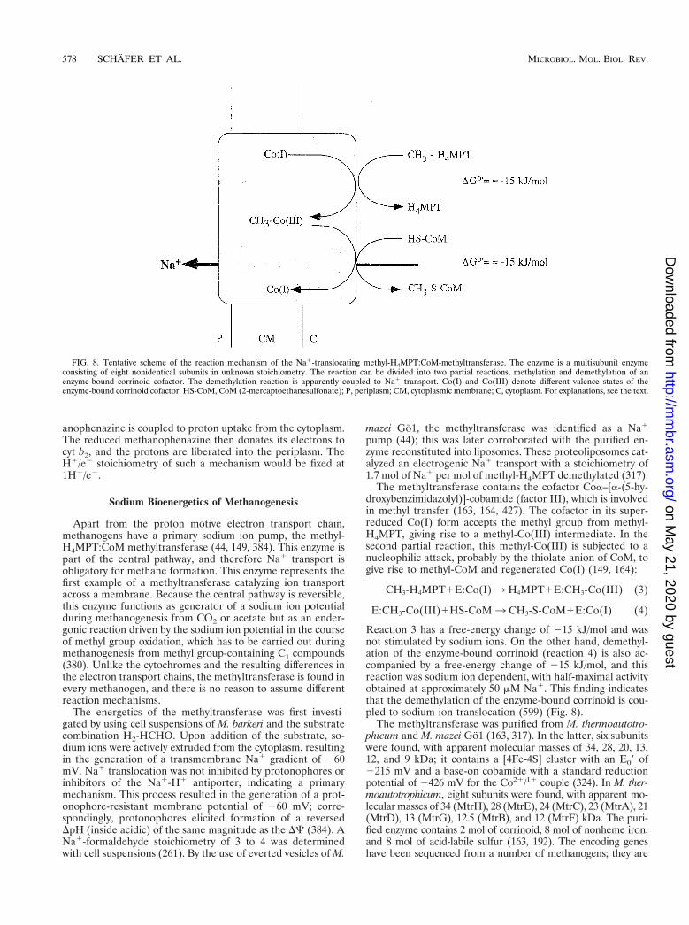
Apart from the proton motive electron transport chain,methanogens have a primary sodium ion pump, the methyl-H4MPT:CoM methyltransferase (44, 149, 384). This enzyme ispart of the central pathway, and therefore Na1 transport isobligatory for methane formation. This enzyme represents thefirst example of a methyltransferase catalyzing ion transportacross a membrane. Because the central pathway is reversible,this enzyme functions as generator of a sodium ion potentialduring methanogenesis from CO2 or acetate but as an ender-gonic reaction driven by the sodium ion potential in the courseof methyl group oxidation, which has to be carried out duringmethanogenesis from methyl group-containing C1 compounds(380). Unlike the cytochromes and the resulting differences inthe electron transport chains, the methyltransferase is found inevery methanogen, and there is no reason to assume differentreaction mechanisms.
The energetics of the methyltransferase was first investi-gated by using cell suspensions of M. barkeri and the substratecombination H2-HCHO. Upon addition of the substrate, so-dium ions were actively extruded from the cytoplasm, resultingin the generation of a transmembrane Na1 gradient of 260mV. Na1 translocation was not inhibited by protonophores orinhibitors of the Na1-H1 antiporter, indicating a primarymechanism. This process resulted in the generation of a prot-onophore-resistant membrane potential of 260 mV; corre-spondingly, protonophores elicited formation of a reversedDpH (inside acidic) of the same magnitude as the DC (384). ANa1-formaldehyde stoichiometry of 3 to 4 was determinedwith cell suspensions (261). By the use of everted vesicles of M.
mazei Go1, the methyltransferase was identified as a Na1
pump (44); this was later corroborated with the purified en-zyme reconstituted into liposomes. These proteoliposomes cat-alyzed an electrogenic Na1 transport with a stoichiometry of1.7 mol of Na1 per mol of methyl-H4MPT demethylated (317).
The methyltransferase contains the cofactor Coa–[a-(5-hy-droxybenzimidazolyl)]-cobamide (factor III), which is involvedin methyl transfer (163, 164, 427). The cofactor in its super-reduced Co(I) form accepts the methyl group from methyl-H4MPT, giving rise to a methyl-Co(III) intermediate. In thesecond partial reaction, this methyl-Co(III) is subjected to anucleophilic attack, probably by the thiolate anion of CoM, togive rise to methyl-CoM and regenerated Co(I) (149, 164):
CH3-H4MPT1E:Co(I)3 H4MPT1E:CH3-Co(III) (3)
E:CH3-Co(III)1HS-CoM3 CH3-S-CoM1E:Co(I) (4)
Reaction 3 has a free-energy change of 215 kJ/mol and wasnot stimulated by sodium ions. On the other hand, demethyl-ation of the enzyme-bound corrinoid (reaction 4) is also ac-companied by a free-energy change of 215 kJ/mol, and thisreaction was sodium ion dependent, with half-maximal activityobtained at approximately 50 mM Na1. This finding indicatesthat the demethylation of the enzyme-bound corrinoid is cou-pled to sodium ion translocation (599) (Fig. 8).
The methyltransferase was purified from M. thermoautotro-phicum and M. mazei Go1 (163, 317). In the latter, six subunitswere found, with apparent molecular masses of 34, 28, 20, 13,12, and 9 kDa; it contains a [4Fe-4S] cluster with an E09 of2215 mV and a base-on cobamide with a standard reductionpotential of 2426 mV for the Co21/11 couple (324). In M. ther-moautotrophicum, eight subunits were found, with apparent mo-lecular masses of 34 (MtrH), 28 (MtrE), 24 (MtrC), 23 (MtrA), 21(MtrD), 13 (MtrG), 12.5 (MtrB), and 12 (MtrF) kDa. The puri-fied enzyme contains 2 mol of corrinoid, 8 mol of nonheme iron,and 8 mol of acid-labile sulfur (163, 192). The encoding geneshave been sequenced from a number of methanogens; they are
FIG. 8. Tentative scheme of the reaction mechanism of the Na1-translocating methyl-H4MPT:CoM-methyltransferase. The enzyme is a multisubunit enzymeconsisting of eight nonidentical subunits in unknown stoichiometry. The reaction can be divided into two partial reactions, methylation and demethylation of anenzyme-bound corrinoid cofactor. The demethylation reaction is apparently coupled to Na1 transport. Co(I) and Co(III) denote different valence states of theenzyme-bound corrinoid cofactor. HS-CoM, CoM (2-mercaptoethanesulfonate); P, periplasm; CM, cytoplasmic membrane; C, cytoplasm. For explanations, see the text.
578 SCHAFER ET AL. MICROBIOL. MOL. BIOL. REV.
on May 21, 2020 by guest
http://mm
br.asm.org/
Dow
nloaded from

organized in an operon in the order mtrEDCBAFGH. Hydro-phobicity plots indicate that all of the subunits except MtrAand MtrH are hydrophobic and potentially membrane bound.Very recently, the membrane localization of MtrD was con-firmed experimentally for M. mazei Go1, M. thermoautotrophi-cum, and M. jannaschii (456). This subunit may be directlyinvolved in Na1 transport (318).
MtrA was overexpressed, purified from Escherichia coli, andsuccessfully reconstituted with cobalamin. Electron paramag-netic resonance (EPR) spectroscopic studies indicate that thecobalamin is in the base-off form and that the axial ligand is ahistidine residue of MtrA (191). From this observation, a hy-pothetical mechanism was formulated for coupling the methyltransfer reaction to ion transport via a long-range conforma-tional change in the protein (191). It is known that cob(II)alaminand cob(III)alamin, but not cob(I)alamin, carry an axial ligand.Methylation of cob(I)alamin gives rise to a methylcob(III)alamin,which is then able to ligate the histidine residue; demethylationleads to a reversal of this reaction. It is easily conceivable thatbinding and dissociation of the histidine residue with the cor-rinoid lead to a conformational change in the hydrophilic partof the enzyme. This change is then transmitted to the mem-brane-bound subunits, giving rise to Na1 transport. Work onthe structure and function of this interesting enzyme is justemerging but is apparently well on its way.
ATP Synthesis in Methanogens
Methanogens are the only microorganisms known to pro-duce two primary ion gradients, DmNa1 and DmH1, at the sametime. They are, therefore, confronted with the problem ofcoupling both ion gradients to the synthesis of ATP (124). Howthis is achieved is still a matter for debate. There have beenconflicting reports regarding DmNa1-driven ATP synthesis inthe hydrogenotrophic archaeon M. thermoautotrophicum. Smi-gan and coworkers (515, 516) had indications of a Na1-ATPasealong with a H1-ATPase, whereas Kaesler and Schonheit fa-vored a mechanism in which the DmNa1 established by themethyltransferase reaction is converted to a secondary protongradient that then drives synthesis of ATP via a H1-translo-cating A1Ao ATPase (262). The latter hypothesis is supportedby the finding that the genomes of the hydrogenotrophic meth-anogens M. jannaschii and M. thermoautotrophicum containgenes that encode the A1Ao ATPase but lack those of F1FoATPase (87, 517). On the other hand, differential inhibitorstudies indicated the simultaneous presence of both A1Ao andF1Fo ATP synthases in M. mazei Go1 (43). The A1Ao enzymemay be coupled to H1 transport, whereas the F1Fo ATPasemay be Na1 coupled. However, no F1Fo ATPase could bepurified from M. mazei Go1, nor have the encoding genes beendetected. In another organism, M. barkeri MS, a gene clusterencoding an F1Fo ATPase has been identified in addition tothe archaeal A1Ao ATPase genes (549); however, the deducedg subunit is very unusual and presumably nonfunctional, andno gene encoding subunit d was found. Since an mRNA tran-script could not be detected in cells grown on methanol, it isdoubtful that the F1Fo-like genes are expressed in M. barkeri(312). The presence of both F1Fo and A1Ao was also proposedfor halobacteria (227, 230). Most likely, the F1Fo ATPasegenes definitely present at least in M. barkeri MS have arisenfrom horizontal gene transfer. In line with this argument is thediscovery of V1Vo ATPases in bacteria (433, 620). However,the presence of the F1Fo ATPases in Methanosarcina speciesstill has to be proven biochemically, and, if present, their con-tribution to energy metabolism has to be clarified. Apparently,the mechanism for DmNa1-driven ATP synthesis differs among
the methanogens. The structure and function of the A1AoATPases are discussed below (“Secondary Energy Converters”).
Bioenergetics of the Acetyl-CoA Pathway in Archaea andBacteria: Differences and Similarities
One of the major differences between the anaerobic bacteriaand the archaea that employ the acetyl-CoA pathway is the waythat CO2 is activated. In bacteria, this requires the action offormate dehydrogenase and formyltetrahydrofolate synthase,at the expense of ATP hydrolysis. In the reverse reaction,oxidation of formyltetrahydrofolate is coupled to ATP synthe-sis by substrate-level phosphorylation (320). In methanogens,the low redox potential of the [CO2 1 MF]/[formyl-MF] cou-ple of approximately 2500 mV (47) is overcome, not by ATPhydrolysis, but by a reversed electron flow driven by the trans-membrane ion (H1 or Na1) potential (262).
Whereas all methanogens tested so far require Na1 forgrowth and methane formation (422, 423), homoacetogens canbe divided into two groups with respect to their energy metab-olism, the proton organisms and the sodium ion organisms. Inthe latter, an as yet unidentified primary sodium ion pump isoperative. Since these organisms have membrane-bound cor-rinoids, it is speculated that the methyltransferase is thesodium ion pump (382). If this is the case, this would allowa study of the evolution of Na1-translocating methyltrans-ferases.
The Na1 gradient established in methanogens is coupled toATP synthesis, but the mechanisms involved are still contro-versial and may differ among the various methanogens. Inhomoacetogens, H1-translocating ATPases are found in pro-ton organisms (112, 113), but a Na1-translocating F1Fo ATPsynthase was found in the Na1-dependent homoacetogen Ace-tobacterium woodii (214, 439). The finding of Na1-ATPases inhomoacetogens strengthens the assumption that Na1-ATPasesare also present in methanogens.
ENERGETICS OF RESPIRATION
Aerobiosis and Other Respiration Forms in Archaea
Obligate aerobes are relatively uncommon among the ar-chaea. Given the phylogenetic position of Archaea, this mayreflect the prevalence of anaerobic energy-transducing reac-tions at early stages of evolution; likewise, all organismsbranching off at the bottom of the phylogenetic tree are hy-perthermophiles, in conformity with the assumption that lifeoriginated in hot environments (6, 540).
Table 1 gives an overview of the archaea that grow obligatelyor facultatively with oxygen or other high-potential terminalelectron acceptors. Only those for which sufficient data is avail-able are included. The genus Acidianus displays obligatechemolithoautotrophic growth with CO2 as the sole carbonsource. Most other species are facultative or obligate hetero-trophs. The autotrophic growth of S. acidocaldarius with sulfuras electron donor, as reported for the original isolates (82), hasto be questioned since the deposited type strains (DSM 639and ATCC 33909) are incapable of such growth.
Members of the genus Sulfolobus are obligate aerobes. In-terestingly, some Sulfolobus isolates were found to reduce fer-ric ions or molybdate as terminal acceptors under low oxygentension (80, 83). In addition, Sulfolobus and Acidianus strainshave been shown to grow aerobically by the oxidation of mo-lecular hydrogen (Knallgas reaction) (236) at low oxygen con-centrations (0.2 to 0.5%).
Alternatively, Acidianus species can derive energy from the
VOL. 63, 1999 BIOENERGETICS OF THE ARCHAEA 579
on May 21, 2020 by guest
http://mm
br.asm.org/
Dow
nloaded from

reduction of sulfur by molecular hydrogen under anoxic con-ditions. This sulfur respiration appears to be the preferentialenergy source: Acidianus is thus classified as a facultative aerobe.
With the exception of Pyrobaculum, the extreme thermo-philes in Table 1 are also extreme acidophiles (optimal growthat pH 1 to 3.5), which imposes a bioenergetic challenge re-garding the maintenance of a nearly neutral cytosol. In con-trast, the members of the aerobic order Halobacteriales areeither neutrophilic or alkaliphilic. However, only halobacteriacan synthesize purple membranes and use light as an addi-tional energy source. It has been proposed that respiration wasthe primary energy-transducing mechanism of halobacteriaand that the light-driven ion pumps might reflect later adap-tations to low oxygen tension (513), an inevitable consequenceof the extremely high salinity of their natural habitat.
Not only oxygen reduction but also various forms of anaer-obic respiration have been reported as energy sources for ar-chaea. Table 2 summarizes the free-energy changes of therespiratory redox systems used as primary energy sources inarchaea. Haloferax mediterranei, Haloferax denitrificans, andHaloferax volcanii are capable of reducing nitrate (as terminalacceptor) to nitrogen (407, 566, 567); several halobacterialnitrate reductases have been described (16, 50, 229). The hy-perthermophile Pyrobaculum aerophilum also reduces nitrateunder anoxic conditions (586). Moreover, some members ofthe halobacterial family perform fumarate respiration (406) orgrow fermentatively on arginine (193). Purely anaerobic respi-ration was reported for the genera Thermoproteus, Pyrodictium,Desulfurococcus, Archaeoglobus, and Thermodiscus (7, 483,540).
In contrast to oxygen respiration, none of the alternate elec-tron transport systems has yet been elucidated in detail at thelevel of genes or proteins.
Components of Aerobic Electron Transfer
The paradigm derived from mitochondrial respiratorychains and from studies of purple bacteria suggests the pres-ence of four major complexes for optimal energy conservation.In this scheme, complex I acts as an energy-transducingNADH dehydrogenase on the low-potential side, and complexII serves as SDH; both are Q reductases. Reduced quinonesare reoxidized by complex III, the so-called bc1 complex, whichtransfers electrons to complex IV, the terminal oxidase, via cytc. In contrast to the classical concept, membrane-integral ar-chaeal electron transfer complexes connected by mobile carri-ers can be fused to supercomplexes and, in some cases, haveunusual compositions. Both membrane-integral redox com-
TABLE 1. Overview of growth conditions and energy sources for aerobic archaea
Species Aerobiosisa Growthb Tmax(°C)c
pHranged e2 acceptor(s) e2 donor(s)e End product(s)
Sulfolobus acidocaldarius o 85 1–5 O2 Organic [H], cellSulfolobus solfataricus o h 87 3–5 O2 Sugars, amino acids H2O; H2SO4; CO2Sulfolobus shibatae o (f) h 86 3–5 O2; (Fe31, Mo41) H2S/[H2]Sulfolobus metallicus o f/(f) 75 ? O2 Sulfidic ores; S22/S0 H2SO4Metallosphaera sedula o a 80 1–4.5 O2 Sulfidic ores/S22/S0 H2SO4Acidianus infernus f a 95 1.5–4 O2/S0 Sulfidic ores/S22/S0/H2 H2SO4; H2SAcidianus brierleyi o (f) a 75 1.5–4 O2/S0 S22/S0 H2SO4; H2SAcidianus ambivalens f a 95 1–4 O2/S0 S22/S0 H2SO4; H2SStygiolobus azoricus an a 89 1.5–5 S0 H2 H2SPyrobaculum aerophilum f h 103 5.8–9 O2/NO3
2 Peptone/yeast/H2 H2O; CO2?; N2?Pyrobaculum islandicum an f/(h) 103 5–7 S0/S22 H2/cell/peptone H2SPyrobaculum organotrophicum an h 104 5–7 S0/S22/S2O3
2 Yeast/peptone H2SThermoplasma acidophilum o h 65 0.5–4 O2 Yeast/sugars H2O; CO2Thermoplasma volcanium o h 65 0.5–4 O2/S0 Yeast/sugars H2O; CO2Picrophilus oshimae o h 60 0.5–2.2 O2 Yeast 1 sugar H2O; CO2Picrophilus torridus o h 60 0.5–2.2 O2 Yeast 1 sugar H2O; CO2Halobacterium salinarum o/hn h m n O2 Cell/yeast H2O; CO2Halobacterium saccharovorum o/hn h m n O2 Cell/yeast H2O; CO2Haloferax mediterranei o/hn h m n O2 Cell/yeast H2O; CO2Haloferax volcanii o (f) h m n O2/NO3
2 Cell/yeast H2O; N2; CO2Haloferax denitrificans f h m n NO3
2/O2 Cell/yeast N2; H2O; CO2Natronobacterium pharaonis o/hn h 45 7.5–9.5 O2 Amino acids/carbonic acids H2O; CO2
a o, obligate aerobe; f, facultative respiration with acceptors other than oxygen; an, obligate anaerobe; hn, auxiliary phototrophic energy conservation.b h, heterotrophic; a, autotrophic; f, facultatively heterotrophic.c Maximum temperature at which growth occurs. m, mesophilic.d n, neutrophilic.e cell, cell extract; yeast, yeast extract.
TABLE 2. Standard free energy changes of aerobic and anaerobicrespiratory reactions identified as energy sources
for growth of archaeaa
Reaction Redox system 2DG°9(kJ/mol)
1 H2 1 1/2O2 3 H2O 236.62 2S0 1 2H2O 1 3O2 3 2H2SO4 1,014.03 2FeS2 1 2H2O 1 7O2 3 2FeSO4 1 2H2SO4 1,498.94 [H]2-X 1 1/2O2 3 H2O 1 X
(X 5 NADH2, QH2 succinate, etc.)219.0
5 S0 1 2[H] 3 H2S 33.56 SO4
22 1 8[H] 1 2H1 3 H2S 1 4H2O 151.77 NO3
2 1 8[H] 1 2H1 3 NH41 1 3H2O 598.7
8 2NO 1 2H1 1 2e2 3 N2O 1 H2O 305.99 N2O 1 2H1 1 2e2 3 N2 1 H2O 341.1
a The free energy values were calculated from redox potentials or the energiesof formation as described in reference 562.
580 SCHAFER ET AL. MICROBIOL. MOL. BIOL. REV.
on May 21, 2020 by guest
http://mm
br.asm.org/
Dow
nloaded from

plexes and mobile electron carriers are discussed in the fol-lowing sections.
Membrane-residing quinone reductases. NADH dehydro-genases (complex I) and SDHs are the major reductants ofquinones in all respiratory chains. Two types of the formerdehydrogenase are known, NDH-I and NDH-II; only NDH-Itypes are capable of H1 or Na1 pumping. Whereas mem-brane-associated NDH-II activities and NADH-dependentrespiration have been found in archaea, nothing equivalent toan energy-transducing complex I has been detected.
(i) NADH dehydrogenases. Older reports on the character-ization and partial purification of NADH dehydrogenase ac-tivities from halobacteria are reviewed in reference 226. Theseactivities were usually measured with redox dyes as electronacceptors; however, inhibition by the quinone analog 2-heptyl-4-hydroxyquinoline-N-oxide has been interpreted to indicatethe in vivo transfer of electrons to the quinone pool (302).Sulfolobus membranes oxidize NADH with low activity in acyanide-sensitive reaction (21, 592). None of these activitieswas sensitive to rotenone, amytal, piericidine, or antimycin.From the inhibition of cell respiration by acridone carbonicacid derivatives (402), it was concluded that S. acidocaldariushas an NDH-II type enzyme (463). This activity is only looselyassociated with the membrane.
An NADH:acceptor oxidoreductase from the cytosol of Sul-folobus sp. strain 7 has been purified as a dimeric, water-soluble, 95-kDa protein with two molecules of FAD/moleculeof protein (595); it may represent the membrane-peripheralfraction of a larger complex, because there was virtually noactivity with caldariella quinone (Qcal) as acceptor. This hasnot yet been confirmed. Interestingly, a similar enzyme hasbeen isolated from aerobically grown A. ambivalens and char-acterized (177). The monomeric, 76-kDa protein containsFAD and has NADH:acceptor oxidoreductase activity. How-ever, from EPR spectroscopic studies on its interaction withferredoxin, the authors conclude that the protein might func-tion in vivo as an NADH:ferredoxin oxidoreductase.
Numerous inhibitors of respiratory electron transport inmembranes of Halobacterium salinarum have been tested, con-firming that this NADH-oxidizing activity is also due to anNDH-II type enzyme which is not involved in energy conser-vation (531). In contrast to Sulfolobus, the membrane-boundenzyme is capable of transferring reducing equivalents to horseheart cyt c as experimental acceptor.
Sequence data is not available for any of these NADHdehydrogenases from halobacteria or members of the Sul-folobales. The ongoing genome sequencing projects may helpto clarify whether the genes for an energy-conserving NDH-Icomplex are completely absent from archaea. Surprisingly,open reading frames in the genome of the obligate anaerobeA. fulgidus could be attributed by similarity to genes of NADHdehydrogenase (subunit 1) and of cytochrome oxidase (281).Similar attributions have been made for genes from M. ther-moautotrophicum (517), M. jannaschii (87), or P. aerophilum(153). However, these alignments are only fragments of theanalogous bacterial or eucaryal genes, respectively; moreover,they usually concern only a single polypeptide. Furthermore,there is no indication of the presence of any of the remaining(up to 14) polypeptides, e.g., the iron-sulfur proteins, requiredfor completion of a functional complex I.
In conclusion, attempts to isolate an archaeal integratedNADH:quinone reductase complex have failed so far. Cumu-lative evidence for the absence of a complex I analog in mem-branes from Sulfolobus and H. salinarum was mainly derivedfrom three observations: (i) none of the characteristic iron-sulfur centers of complex I-type NADH dehydrogenases could
be detected by EPR spectroscopy (309, 310), (ii) no sensitivityto complex I inhibitors could be demonstrated, and (iii) hy-bridization with NDH-I-directed DNA probes (against NDH-I-encoding genes of Paracoccus denitrificans) failed.
(ii) SDHs and a novel complex II. Succinate-stimulated res-piration and SDH activities with redox dyes as electron accep-tors have been reported for several aerobic archaeal species(10, 20, 477, 592).
An unusually high concentration of SDH was found in mem-branes of Thermoplasma acidophilum (20); this allowed directcharacterization of the three iron-sulfur clusters, S1 [2Fe-2S],S2 [4Fe-4S], and S3 [3Fe-4S], with redox potentials of 160 and168 mV for clusters S3 and S1, respectively. The S2 cluster wasindirectly detected by spin-spin interaction. The physiologicalsignificance of the high SDH activity in T. acidophilum remainsunexplained. The enzyme complex has not been purified tohomogeneity.
Well-characterized preparations were obtained from thethermoacidophiles S. acidocaldarius (371) and Sulfolobus sp.strain 7 (250) and from the haloalkaliphile N. pharaonis (477).Complete genetic analyses of archaeal SDH operons are pres-ently available for only S. acidocaldarius and N. pharaonis (254,477). Figure 9 shows a comparison of gene arrangements inarchaeal SDH operons, with reference to E. coli as a bacterialrepresentative. While the order of the genes varies, the basicpolypeptide composition is in agreement with the known com-plex II (SDH-fumarate reductase) (184, 207). The catalyticcore consists of a larger (approximately 65 kDa) FAD-contain-ing subunit and a smaller polypeptide (26 to 37 kDa) that bearsthree FeS clusters. This is accompanied by two smaller sub-units which usually function as a membrane anchor. One ofthese may host a heme B. Its participation in electron transportis not entirely certain; it may also have structural functions(184). Heme B was found in archaeal SDH complexes fromT. acidophilum (29) and N. pharaonis (477) but not in thosefrom members of the Sulfolobales.
Two complex II preparations from Sulfolobus (250, 294)deserve special attention. In contrast to other archaeal SDHcomplexes, that from Sulfolobus sp. strain 7 was reported toreduce Qcal readily; succinate respiration could be restored inliposomes by reconstitution of the preparation with a terminalQ-oxidizing complex (250). This is the only direct evidence thatin Sulfolobus reducing equivalents are transferred from succi-nate to terminal oxidases via CoQ.
This observation is in obvious contrast to the unusual prop-erties of a novel complex II from S. acidocaldarius (254, 371).The purified complex is inactive with Qcal. In isolated mem-branes, electron transfer from succinate to respiratory RieskeFeS proteins is evident from EPR spectroscopy (471), but theexact electron path is unclear. The involvement of a radicalspecies was indicated by the strong inhibitory interaction withthe free radicals of 2,3,5,6-tetrachlorobenzoquinone (371). Thecomplex dissociates easily from the membrane. Genetic anal-ysis of the SDH operon (254) suggests that the two smallpolypeptides are essentially hydrophilic; indeed, none of thepolypeptides contained a clear membrane-spanning a helix. Inaddition, subunit B, which hosts the FeS clusters in all SDHsand fumarate reductases (184), is likely to bear in S. acidocal-darius a second [4Fe-4S] cluster instead of the regular [3Fe-4S]cluster S3. This might be indicative of a different electronpathway that eventually involves thiols, because subunit C alsoexhibits a novel and unusual sequence pattern containing arepetitive, cysteine-rich motif, CX31–37CCX26–35CX2C. Thismotif has also been detected in a hypothetical protein se-quence of Synechocystis and in the heterodisulfide reductase ofM. thermoautotrophicum (206). Its functional significance is not
VOL. 63, 1999 BIOENERGETICS OF THE ARCHAEA 581
on May 21, 2020 by guest
http://mm
br.asm.org/
Dow
nloaded from

understood, but it may explain the failure of S. acidocaldariusSDH to directly reduce Qcal in vitro. The recent finding inmethanogen membranes of a phenazine isoprenoid (1, 40)which functions as an intermediate electron carrier has encour-aged the search for an equivalent in aerobic archaea. It maybridge the gap in electron transfer from SDH in S. acidocal-darius to Qcal. A dual function of the complex also appearspossible: it may function as a fumarate reductase under au-totrophic growth conditions, with a hitherto unknown sourceof reducing equivalents, analogous to a thiol-driven fumaratereductase of M. thermoautotrophicum (206). The properties ofthe two well-characterized SDH complexes are compiled inTable 3.
Membrane-integral quinol-oxidizing complexes. Quinol ox-idation in aerobic respiration is usually accomplished by inte-gral membrane-protein complexes acting sequentially as re-dox-driven proton pumps. The identification and mechanismsof the respective archaeal proton pumps are still under debate.Detailed investigations have been reported for just a few ex-tremely thermoacidophilic archaea and two halobacterial spe-cies. The latter appear to possess unbranched quinol-oxidizingrespiratory chains, with complexes essentially similar to thosefrom mitochondria or purple bacteria. In contrast, unusual andnovel supercomplexes from members of the Sulfolobales havebeen isolated and characterized.
Membrane-bound quinol-oxidizing systems contain multiplehemoproteins detectable by difference spectroscopy in visi-ble light. Indeed, archaebacterial membranes were shown toreveal composite (reduced-oxidized) difference spectra by su-perimposing various cytochrome spectra. Table 4 gives a com-prehensive listing of cytochromes from aerobically growing
archaea, together with their redox potentials, heme types, andtentative functions.
(i) The SoxABCD complex. Figure 10 illustrates schemati-cally the composition of a novel terminal oxidase complex,SoxABCD, discovered in S. acidocaldarius (92, 329). It wasfound to combine features of what are usually separate respi-ratory complexes III and IV, although its cyt aa3 subunit(SoxB) could be isolated as a functional quinol oxidase byharsh detergent treatment (23). The integrated complex istranscribed from an operon encoding four polypeptides, des-ignated SoxA to -D. Of these, SoxB represents a subunit Iequivalent of typical heme-Cu oxidases; its binuclear centercontains two hemes A and one CuB, which have been wellcharacterized by optical, EPR, and resonance-Raman spec-troscopy (168, 173, 224). SoxA, like subunit II in the quinoloxidase of E. coli (339, 388), lacks the typical mixed-valencebinuclear CuA ligands of cyt c oxidases. In total, the complexcontains four hemes AS. The two additional hemes are thereaction centers of SoxC; judged by its primary and secondarystructural elements, it represents a cyt b homolog and can beidentified spectroscopically as cyt a587-I. When examined inintact membranes, the complex reveals CO binding kineticssimilar to those of mitochondrial cyt c oxidase; the cyt a587 andcyt aa3 centers exhibit coordinated redox behavior on the mil-lisecond time scale (172). Interestingly, with tetramethyl-p-phenylenediamine (TMPD) as electron donor, the turnover ofthe integrated complex (1,300 s21) is about threefold higherthan that of the purified subunit I equivalent, SoxB. The smallhydrophobic subunit, SoxD, has no redox centers.
The presence of a diheme cyt b analog is reminiscent of acomplex III equivalent, although there is no Rieske-type iron-
FIG. 9. Gene organization within archaeal SDH operons. Numbers indicate the calculated molecular masses (in kilodaltons) of the gene products. In all cases,subunit A is the flavin-containing dehydrogenase polypeptide; subunit B refers to the iron-sulfur protein in complex II or fumarate reductase, respectively. The columnson the right indicate the type of quinone used as terminal electron acceptor in vivo and the number of heme B molecules present in the complex. MQ, menaquinone;TQ, Thermoplasma quinone, CQ, caldariella quinone. tmh, putative transmembrane helices. E.coli, E. coli (256); B.sub., B. subtilis (353); T.ac., T. acidophilum (29); S.ac., S.acidocaldarius (254) (accession no. Y09041); N.ph., N. pharaonis (accession no. Y07709). The assignment of the open reading frames was done by analyzing sequencesimilarities. From Thermoplasma, only a partially sequenced operon has been reported; ORF-1 has homology to sdhB and frdB; ORF-2 is a putative diheme cyt b.
582 SCHAFER ET AL. MICROBIOL. MOL. BIOL. REV.
on May 21, 2020 by guest
http://mm
br.asm.org/
Dow
nloaded from

sulfur protein in SoxABCD. On that basis, the possibility of aproton-pumping Q cycle analogous to quinol:cyt c reductaseshas been proposed: the function of cyt c1 is replaced by directelectron transfer to the low-spin cyt a in the terminal oxidasesegment, SoxB (92).
The SoxABCD complex, the only example known from aer-obic archaea, was reconstituted into liposomes and shown toact as an electrogenic proton pump by a yet unknown mecha-nism (175). By applying an artificial electron-donating systemthat does not produce scalar protons outside the vesicles,H1/e2 ratios of .1 were measured. This would be in agree-ment with the proposed Q cycle. Details of this mechanism arediscussed below (“Proton Pathways in Terminal Oxidases ofArchaea”).
(ii) The SoxM complex. Even larger than SoxABCD, theso-called SoxM complex of S. acidocaldarius comprises in itspurified state seven functional redox centers involving a totalof 10 metal binding sites (92, 327). Figure 11 shows a sketch ofthe complex, including the redox potentials of its components.All subunits are encoded by a gene cluster, which suggestscoordinated transcription of a supercomplex composed of twofunctional substructures. One complex is made up of the geneproducts of soxH and soxM. SoxM is a ba3-type heme-Cu ter-minal oxidase with a bands at 562 and 605 nm; the polypeptide
has homologies to a fusion protein of subunits I and III oftypical cyt c oxidases. Subunit II (SoxH) is supplied by thesecond gene cluster and, in contrast to quinol oxidases, pro-vides all the ligands for the formation of a CuA mixed-valenceredox center. At this site, electrons may be provided by thepolypeptide SoxE (sulfocyanine), a homolog to blue copperproteins similar to rusticyanine (91). This mobile electron car-rier with low membrane affinity is thought to serve as electronacceptor for a bc1-analogous part of the complex composed ofcyt a587-II, another cyt b analog and product of the soxG gene,and the Rieske FeS protein, SoxF.
It is important to note that two Rieske FeS proteins havebeen identified in S. acidocaldarius (487). Both have beencharacterized, sequenced, and expressed heterologically in E.coli (26, 489, 490). Whereas Rieske II (SoxF) has been clearlyidentified as a component of the SoxM supercomplex, RieskeI (SoxL) (488) has not been attributed to any of the knownrespiratory pathways and is assumed to belong to a novelcomplex of the quinol-oxidizing respiratory system which hasnot yet been characterized at the protein level (491). BothRieske proteins are expressed constitutively in Sulfolobus, asare the genes of both complexes, SoxABCD and SoxM.
Nevertheless, the electron transport activity of the isolatedSoxM complex is extremely low (327). Neither Qcal nor artifi-cial donors produced a significant turnover. This is most likelydue to the lack of sulfocyanine as an intermediate carrier,which is obviously lost during membrane preparation and hasso far been identified only on a genetic basis, not chemically. Itis likely to be present in vivo because transcription into a singleRNA has been demonstrated (286).
(iii) Other archaeal terminal oxidases. A similar quinol-oxidizing supercomplex has been reported for Sulfolobus sp.strain 7 (246). However, primary sequence data and gene or-ganization are unknown. This supercomplex contains at leastfour different heme centers and also a Rieske FeS protein(gxyz 5 2.02, 1.89, and 1.79). The attribution of redox potentialsto individual heme centers appears very preliminary and un-certain in terms of the proposed electron transport pathwaysand proton-pumping mechanisms. The supercomplex acts as acaldariella quinol oxidase (246) and was functionally reconsti-tuted into detergent micelles together with complex II prepa-rations from the same Sulfolobus strain to serve as quinonereductase (250). A Q-cycle-like mechanism for electron trans-port and proton translocation has been proposed. However,the typical Q-cycle inhibitors myxothiazole and stigmatellin didnot inhibit electron transport activity. Moreover, the Rieskeprotein would not function in a Q cycle with a single-hemeb-type cytochrome (b562; E0 5 146 mV), while cyt a583 may wellassume a cyt c-like function with E0 5 1270 mV. A c-typecytochrome or a blue copper protein was not detected in thepreparation. These obvious differences from the SoxM com-plex of S. acidocaldarius necessitate the complete sequencingof the genes in order to disclose the real number of hemecenters involved.
The quinol-oxidizing complex of A. ambivalens (24) has thesimplest structure in terms of redox centers. It is composed ofsix subunits encoded by two separate operons (430), both ofwhich are present in duplicate in the A. ambivalens genome.Only one polypeptide (the doxB gene product) has redox cen-ters, which identify it as a heme-Cu oxidase. This 64.9-kDapolypeptide contains two hemes AS as cyt a and a3, respec-tively; the latter is associated with one Cu in the binuclearcenter. Functional assignments of the other subunits (20.4,18.8, 38, 7.1, and 7.2 kDa) could not be made because they lackany detectable similarity to known membrane proteins. How-ever, all polypeptides have several putative transmembrane
TABLE 3. Succinate:acceptor oxidoreductasesa
Parameter S. acidocaldarius(DSM 639)
Sulfolobus sp.strain 7
No. of subunits 4 4Mr (103) 138 148Mr subunits (103) 66, 31, 28, 12.8 66, 37, 33, 12Genes sdhABCD NA
Flavin (nmol/mg) 4.6 5.6lmax (nm) 455 (ox-red) 445Fe (nmol/mg) 102 83S0 (nmol/mg) 150 —Cyt b None None
Km (succinate) (mM) 1.4 0.28Km (DCPIP) (mM) 65.4 89Km (Qcal) (mM) — 60
Vmax (mmol/min/mg)b 7.8 (55°C) 13 (50°C)— 14 (Q1)— 1.8 (Qcal)
Turnover (s21) 154 (81°C) —
EPR(S-3) oxidized g 5 2.02 g 5 2.02
g 5 2.08 (satell.) —(S-1) succinate reduced gz 5 2.05 gz 5 2.03
gy 5 1.935 gy 5 1.94gx 5 1.904 gx 5 1.90
pH optimum 6.5 6.5–6.8Ea (kJ/mol) 59–64 —Ki
Malonate (mM) 3.1 —Oxaloacetate (mM) 0.28 —TCBQc (mM) 1.5 —
a Comparison of structural and kinetic data for the only two archaeal SDHpreparations (complex II). Data were taken from references 250, 369, and 371.NA, not available; Qcal, caldariella quinone; Q1, ubiquinol-1; (satell.), satelliteband; —, not tested or not reported.
b Activity with artificial electron acceptors phenazine methosulfate (PMS) and2,6-dichlorophenolindophenol (DCPIP).
c TCBQ, tetrachlorobenzoquinone.
VOL. 63, 1999 BIOENERGETICS OF THE ARCHAEA 583
on May 21, 2020 by guest
http://mm
br.asm.org/
Dow
nloaded from

helices; deviating from typical heme-Cu oxidases, the catalyticpolypeptide has 14 instead of 12 membrane-spanning helices,with the additional pair at the C-terminal end.
The integrated complex oxidizes Qcal with high activity andis completely inhibited by cyanide; it is also highly sensitive toinhibition by quinolone derivatives such as 2-methyl-3-decyl-quinolone (356). Preparations which have lost the 45-kDa sub-unit display significant TMPD oxidase activity, which is negli-gible in the intact complex (430).
This single complex represents the entire respiratory systemof A. ambivalens. Its redox centers have been characterized aslow- and high-spin hemes by EPR spectroscopy (24), withredox potentials recently actualized as 1215 and 1415 mV,respectively, displaying a redox-Bohr effect with a pKa of 5.4(171). The noncompetitive inhibition by quinolones suggeststwo quinone-quinol binding sites (356), one of which mightserve as an intermediate electron storage site as revealed alsoby the persistent appearance of a stable radical in the enzyme.This has been supported by rapid kinetic studies suggestingthat a tightly bound quinone might mimic the function of theCuA site of cyt c oxidases (171), thus forming a fourth redoxcenter.
Most halobacterial terminal oxidases barely survive standardpurification procedures: therefore, none of the isolated com-
plexes have been functionally characterized. An aa3-type oxi-dase has been isolated from H. salinarum as an inactive com-plex (158), and its amino acid sequence is now available (117).In H. salinarum, a spectral feature was reported (531), indicat-ing the likely presence of a cytochrome bd analog (408). Aterminal oxidase complex of N. pharaonis (349, 477) was firstisolated as a catalytically incompetent protein fraction. Thoughits function is not well characterized, it is assumed to act as acyt c oxidase, and its structure has been deduced from thecompletely sequenced operon.
The oxidase complex of N. pharaonis is spectroscopicallycharacterized as a ba3-type oxidase with two major polypep-tides of 36 and 40 kDa. In subunit I, the six heme and CuBligands were identified at the canonically conserved positions.In membranes, the potentials of the heme centers were deter-mined as 1268 mV (cyt b) and 1358 mV (cyt a3). Subunit IIprovides all the ligands to form the mixed-valence CuA center(348) and thus may function physiologically either as a cyt coxidase or as the oxidase for halocyanine (see above). Infor-mation on a complex III equivalent is rudimentary, although acyt bc fraction could be isolated from membrane solubilizates(477). At least, supercomplexes of the kind found in ther-moacidophilic archaea are absent in N. pharaonis.
For a comparison of the compositions of archaeal quinol-
TABLE 4. Properties of cytochromes from thermoacidophilic aerobic archaeaa
Cytochrome type and source Molecular mass(kDa) Gene(s) Cofactor(s) E0
(mV) Function(s) Reference(s)
b558/566, S. acidocaldarius (DSM 639) 66 (PAGE) cbsA, cbsB 1 heme B .300 Highly glycosylated; unknownfunction; exposed toperiplasm
45, 22055 (deglyc.)50.5 (DNA)
b562, S. acidocaldarius (DSM 639) 45 (PAGE) soxM 1 heme B ND Alternate terminal ba3oxidase complex
327, 32887.1 (DNA) 1 heme As
1 Cu
b563, Sulfolobus sp. strain 7 37 (PAGE) NA 1 heme B 146 Component of respiratorycomplex
246, 247
a587 I, S. acidocaldarius (DSM 639) 39/64b (PAGE) soxC 2 heme AS 210 Component of terminaloxidase SoxABCD
92, 173, 17562.8 (DNA) 270
a587 II, S. acidocaldarius (DSM 639) 37 (PAGE) soxG 2 heme AS 20 Component of SoxMalternative heme-Cuoxidase
92, 32756.7 (DNA) 100
a583, Sulfolobus sp. strain 7 Unknown NA 1 heme AS 270 Terminal oxidase complex; cytc function assumed
246 247,250
aa3, S. acidocaldarius (DSM 639) 38 (PAGE) soxB 2 heme AS 220 Subunit I of heme-Cu quinoloxidase SoxABCD
22, 23, 32957.9 (DNA) 1 Cu 365
aa3, Sulfolobus sp. strain 7 40 (PAGE) NA 2 heme AS 117 Putative subunit I of terminaloxidase complex (c oxidaseactivity)
2471 Cu 325
aa3, A. ambivalens 40 (PAGE) doxB 2 heme AS 235 Subunit I of terminal quinoloxidase
24
65 (DNA) 1 Cu 330
b562/aa3, S. solfataricus (DSM 1616) ND NA Heme B ND Unresolved components; CO-reactive respiratory complex
594ND Heme AS
b558/562, T. acidophilum 18 (PAGE) Orf-2 2 heme B 75 cyt b of putative fumaratedehydrogenase
1622150
a Abbreviations: deglyc., deglycosylated protein; AS, A-type heme as described for Fig. 14; NA, no genetic data available; ND, not determined; PAGE, Mr determinedby PAGE; DNA, molecular mass derived from gene sequence. Further details may be extracted from the references given.
b Molecular mass determined by PAGE in presence of 1% i-butanol.
584 SCHAFER ET AL. MICROBIOL. MOL. BIOL. REV.
on May 21, 2020 by guest
http://mm
br.asm.org/
Dow
nloaded from

oxidizing complexes, Fig. 12 illustrates the organization of therespective operons.
Mobile electron carriers, hemes, and small metal proteins.Mobile electron carriers include membrane-integral as well asmembrane-associated and soluble redox systems. For example,the Rieske iron-sulfur proteins are anchored in the membraneby a single membrane-spanning polypeptide chain, but theirfunctional center is located within a mobile domain that shut-tles between different electron-accepting sites (610, 625). Smallblue copper proteins are associated with the membrane sur-
face, as are c-type cytochromes, while ferredoxins are solublein the aqueous phase. In contrast, the quinones are strictlymembrane-embedded, lipid-soluble, redox mediators. All ofthese components, including the soluble compounds, are nec-essary to constitute functional respiratory electron transportsystems. The typical archaeal examples are discussed belowtogether with the specific hemes of archaeal cytochromes.
(i) Quinones. Electron flow between dehydrogenases andcytochrome components of the respiratory system is mediatedby quinones. The structures of archaeal quinones are depictedin Fig. 13. Halobacteria contain menaquinones with unsatur-ated isoprenoid side chains, predominantly MK-8 (104, 106,107), whereas partially or fully reduced side chains are found inother archaea (Archaeoglobus, Thermoproteus, and Pyrobacu-lum). T. acidophilum contains thermoplasma quinone that isoxidized 10 times faster than is reduced Q10 (105, 234).
Sulfolobus and Acidianus have unique quinones that arederivatives of benzo-[b]-thiophen-4,7-quinone. The novel cal-dariella quinone (126) was detected in all members of theSulfolobales as a major constituent, making up approximately60% of the total quinones (106), which corresponds to 0.28 to0.38% of cell dry weight. Three variants were detected, ofwhich the tricyclic quinone (2-polyprenyl-benzo[1,2-b;4,5-b9]dithiophene-4,8-quinone) is present only in traces (392, 573).In Sulfolobus solfataricus, the ratio between Sulfolobus quinoneand caldariella quinone (Qcal) was reported to vary consider-ably with growth temperature (392), which shifts the relativecontent of Qcal between 60 and 99% of the total quinone. In A.ambivalens, the ratio between Qcal and Sulfolobus quinone isreversed by transition from aerobic to anaerobic growth. Thefunction of quinone in anaerobically growing cells has not beenstudied so far.
Electrochemical properties were reported only for caldari-ella quinone (QcalH2), extracted from S. acidocaldarius (DSM639) (23). In vitro, QcalH2 acts as a substrate of the isolated cytaa3 of Sulfolobus; in vivo, it is the reductant of the SoxABCDcomplex and presumably also of the SoxM supercomplex (327)(see below). Stabilized in Triton micelles at an ambient pH of6.5 and at 25°C, it has a half-reduction potential of 1100 to1106 mV. The reduced-minus-oxidized difference spectrumshows a minimum at 351 nm, a maximum at 325 nm, and anisosbestic point at 341 nm (e351–341 5 1.77 3 103 M21 cm21).At temperatures above 60°C and at pH . 6.5, considerableautoxidation occurs.
(ii) Hemes. Prenylated hemes, the typical cofactors of ter-minal heme-copper oxidases, have been analyzed for a varietyof archaeal membranes (325, 332). Neither unmodified hemeA nor heme O (protoheme IX) is present in terminal oxidasesfrom archaea. Instead, several novel prenylated hemes havebeen identified as depicted in Fig. 14. The substituents atposition 8 are identical to those in the usual heme A or O(formyl or methyl, respectively), but the oxidation state and thelength of the isoprenoid side chain at position 2 are altered. Inall cases, the farnesyl residue is extended by one isoprene unit.The increased hydrophobicity of the geranylgeranyl chain maybe relevant to the stabilization of the heme cofactor in oxidasesof extreme thermophiles. It has been proposed (332) that thelarge pool of the precursor, geranylgeranyl-pyrophosphate,might affect the preferential formation of these new hemes. Asdiscovered by resonance-Raman spectroscopy, unusual profilesof vibrational modes result from the immediate environmentof the chromophore in the enzyme-bound state but not fromaltered porphyrin substituents (see below and references 167,168, and 210).
Heme AS was found in the aa3 and ba3 oxidases of Sulfolo-bus, Acidianus, Halobacterium, and Natronobacterium; Thermo-
FIG. 10. Composition of the SoxABCD terminal oxidase of S. acidocaldarius.The shaded areas represent the membrane. Each box represents a single con-stituent polypeptide with its type of redox center and the redox potential mea-sured at pH 6.5. The dashed oval insert illustrates the proposed location of thebinding site for caldariella quinone at an interface between subunit II (SoxA) andthe cyt b analog SoxC. Further details are given in the text.
FIG. 11. Composition of the respiratory supercomplex SoxM. Each box ofthe scheme represents a constituent polypeptide together with its redox centersand the respective redox potentials (where known). The location of the bluecopper protein sulfocyanine is hypothetical, as a possible link between the partialcomplexes of SoxG-SoxF and SoxM-SoxH. For details, see the text.
VOL. 63, 1999 BIOENERGETICS OF THE ARCHAEA 585
on May 21, 2020 by guest
http://mm
br.asm.org/
Dow
nloaded from

plasma has heme OT, while Pyrobaculum has two similar hemeOP species as well as heme AS. T. acidophilum may also containheme D (332). Surprisingly, Thermus thermophilus is the onlyrepresentative of the bacterial domain that also contains hemeAS (typical for archaea) in both terminal oxidases (caa3 and ba3).
Based on spectroscopic analysis (187, 264, 301, 477), oneconcludes that heme B is ubiquitous in archaea, whereas hemeC has been found in methanogens and halophiles but is appar-ently absent from thermoacidophiles. There have been contra-dictory reports on T. acidophilum. The presence of heme C (46,233, 502) has been demonstrated, whereas recent heme compo-
sition studies of archaeal cytochromes detected no heme C inmembranes of either T. acidophilum or P. aerophilum (332).
(iii) Ferredoxins. In sulfur-metabolizing archaea, an abun-dance of ferredoxins has been observed (559). Their high ther-mostability (27) may not only indicate the use of iron-sulfurclusters as primordial redox systems in early evolution (589)but also reflect their advantage over NAD1 as a primary hy-drogen-transducing coenzyme at high temperatures. In fact,archaeal ferredoxins have functionally replaced pyridine nucle-otides in several key enzymes such as 2-oxoacid dehydroge-nases (60, 276, 621), aldehyde dehydrogenase (166, 377), orglyceraldehyde dehydrogenase (486). Although ferredoxins arenot directly involved in primary energy conservation, they as-sume a key role in channeling hydrogen to NADH as the actualrespiratory substrate. Ferredoxins have the advantage of pro-viding reducing equivalents at a potential negative enough todrive hydrogenase-catalyzed reactions (E0 5 2450 mV),NAD(P)1 reduction (E0 5 2320 mV), or the reduction ofsulfur (E0 5 2270 mV). In addition, a ferredoxin (E0 5 2342mV) has been purified from N. pharaonis (477). The primarystructures of several archaeal ferredoxins are known (276, 365,425); the three-dimensional structure of one of the Sulfolobusferredoxins (156) reveals a Zn21 binding site in addition to the[3Fe-4S] and the [4Fe-4S] clusters. Recent genetic and se-quencing data (558) suggests that multiple genes in membersof the Sulfolobales may code for highly similar ferredoxins.Despite the presence of an additional metal center, no differ-ences in thermostability have been found. Thus, the hypothesisthat the ferredoxins differ with respect to electron acceptorspecificity may be considered. Both ferredoxins from A. am-bivalens have been cloned and expressed in E. coli (253), which
FIG. 12. Gene organization of archaeal terminal oxidases. The operon structure is compared to those of E. coli and P. denitrificans. Numbers within the boxes referto the molecular masses (in kilodaltons) of the gene products; arrows indicate the direction of transcription. The assignment of genes to subunits of the oxidasecomplexes is given below the boxes; the darkly shaded box in all cases signifies subunit I, identifying the complex as a member of the heme-Cu oxidase family. In thecase of A. ambivalens, the two operons are located far apart on the genome, whereas the soxM locus of S. acidocaldarius forms a close gene cluster. Data were compiledfrom references 92, 99, 327, 329, 348, 430, 443, 470, and 471 and/or the EMBL data bank. su, subunit.
FIG. 13. Structures of respiratory quinones isolated from membranes ofthermoacidophilic and halophilic archaea.
586 SCHAFER ET AL. MICROBIOL. MOL. BIOL. REV.
on May 21, 2020 by guest
http://mm
br.asm.org/
Dow
nloaded from

should allow this hypothesis to be tested in the near future.Physicochemical and EPR spectroscopic properties of archaealferredoxins from hyperthermophiles are discussed in refer-ences 248 and 559. A more detailed analysis of redox-linkedprotonation equilibria was presented for the Sulfolobus ferre-doxin (79). A [3Fe-4S] cluster with a reduction potential of2275 mV shows uptake of one proton with a pKa of 5.8 upona one-electron reduction; the similarity to the intracellular pHof S. acidocaldarius may indicate a physiological role of thisreversible protonation, which may influence the electron trans-fer properties of this ferredoxin. The [4Fe-4S] cluster has aredox potential of 2529 mV at pH 6.4.
The ferredoxin-reoxidizing iron flavoprotein (IFP) isolatedfrom Sulfolobus sp. strain 7 may be important for hydrogenflow in oxidative bioenergetic reactions (249). The dimer of thethree-subunit protein contains two flavin mononucleotide cen-ters (E0 5 257 mV) and two [2Fe-2S] centers (E0 5 2260mV). A possible electron pathway, ferredoxin f IFP[2Fe-2S]f FMN f X, has been proposed. Whether X represents anentry point into a respiratory chain is speculative. The isolatedIFP does not react with pyridine nucleotides (249) and may bea fragment of a yet-unresolved new electron pathway in ar-chaeal bioenergetics. As mentioned previously, a 76-kDa fla-voprotein from A. ambivalens may act as an NADH:ferredoxinoxidoreductase and therefore also play an important role in thebioenergetics of hydrogen flow (177).
(iv) Rieske iron-sulfur proteins. Rieske FeS proteins arecharacteristic components of complex III in respiratory chains(so-called bc1 complexes) and also of the structurally relatedb6f complexes in photosynthetic electron transport. A commonfeature of respiratory Rieske proteins is the Cys-Cys-His-Hisligation of the iron atoms in the [2Fe-2S] cluster, which showsa high redox potential and two typical pK values (for a review,see reference 574). Their function is to accept one electronfrom a reduced quinone (QH2) which is channeled to the nexthigher acceptor, while the other electron is transferred to alow-potential heme of a b-type cytochrome as a quinone re-ductant in the Q cycle (575).
Archaeal Rieske proteins have been detected by EPR spec-troscopy only in Sulfolobus and H. salinarum (19, 531). TwoRieske proteins were isolated from S. acidocaldarius, and theirsequences were determined (487).
One Rieske protein, the product of the soxL gene, has apronounced ubiquinol:cyt c reductase activity (488) and only29% similarity to the SoxF Rieske protein (component of theSoxM complex; see above). Both exhibit the typical bis-Cys/
bis-His ligation (in contrast to ferredoxin-type FeS clusters)but reveal an insertion between the cluster binding sites muchlarger than that of any other Rieske-type FeS protein (487).Nevertheless, in membranes, their EPR signals (gxyz 5 2.031,1.89, and 1.725) and the redox potential (E0 5 2325 mV)identify these archaeal iron-sulfur proteins as typical respira-tory Rieske proteins; two redox-dependent pKa values (6.2 and8.5) can be titrated (25). Both proteins have been expressed inE. coli in a truncated form without the membrane-anchoring Nterminus (490). The recombinant SoxF Rieske protein hasbeen crystallized (69).
A reading frame in the fully sequenced genome of P. aero-philum was identified as coding for a Rieske protein, which wasthen produced as a recombinant protein in E. coli (491). Theproduct has all the functional properties of a respiratory Rieskeprotein.
Nevertheless, since representatives of Sulfolobales do notcontain c-type cytochromes, classical bc1 complexes are absent,although the association of these [2Fe-2S] proteins with cyt bor cyt b homologs suggests an essentially similar function. Con-sequently, it seems appropriate to define cyt b-Rieske com-plexes as the actual core of complex III analogs, irrespective ofthe low-potential electron acceptor.
A novel type of iron cluster, which may act as a functionalanalog of Rieske iron-sulfur proteins, was found in Sulfolobusmetallicus (601). The characteristic EPR signals, which couldbe detected in intact cells and in membrane preparations,suggest a spin-coupled cluster with at least two iron atoms; inthe ground state, it has a spin of S 5 1/2. The center has a highredox potential of 1350 mV and is located in succinate-reduc-ible membranes. Interestingly, the presence of a typical Rieskecenter could also be detected by EPR spectroscopy in thisorganism.
(v) Small copper proteins. Blue copper proteins, which mayfunctionally substitute for cyt c, have been detected in archaea.Halocyanine was isolated from membranes of N. pharaonis asa 15.5-kDa protein by treatment with mild detergents (473).The soluble protein contains one Cu21 ion per mol and has abroad absorption band at 600 nm. EPR spectra revealed aclose resemblance to plastocyanine, suggesting a type I copperligation with two His, one Met, and one Cys. This is in line withprimary sequence data, which also predicts an N-terminalmembrane anchor (350). The Cu binding site, located at theC-terminal end, has a high degree of sequence identity withother small copper proteins (446a). The potentials for bluecopper proteins range from 1183 to 1680 mV (551), whereas
FIG. 14. Structures of hemes found in archaeal terminal oxidases. The hemes are assigned according to references 332 and 336.
VOL. 63, 1999 BIOENERGETICS OF THE ARCHAEA 587
on May 21, 2020 by guest
http://mm
br.asm.org/
Dow
nloaded from

the redox potential of 1183 mV at neutral pH is well suited toassume a cyt c-like function in respiratory electron transport.The redox potential changes from 1333 mV at pH 4 to 1119mV at pH 10: there are three pK values within this range,which are presumably associated with three histidine residuesthat interact with the redox site. Fourier transform infrared(FTIR) spectra point to a special CA O peptide group thatserves as a fifth ligand to copper. Upon oxidation at a low pH,the uptake of a proton by a carboxyl moiety of Glu or Asp wasinferred from strong bands at 1,398 and 1,557 cm21: this re-action can be written as H1f Cured 1 R-COO2f CuOX 1R-COOH (81). The pH dependence of the midpoint potentialemerging from FTIR spectral changes was not seen in reso-nance-Raman spectroscopy. A pronounced similarity to thecoordination geometry of azurin was determined. The proteinbands strongly suggest a predominant b-sheet structure for thisnew copper protein.
Sulfocyanine was first predicted as a putative blue copperprotein in S. acidocaldarius from the sequence of the soxE geneof the SoxM complex (92). Northern blotting revealed that thegene is transcribed, but the protein has never been isolatedfrom the native organism. Recently, it could be expressed in E.coli in amounts which will allow its biophysical characterization(286). This is quite significant because sulfocyanine is thoughtto be an essential intermediate in electron transport throughthe SoxM complex of S. acidocaldarius and is thus expected torestore activity to the isolated supercomplex. Membrane bind-ing of sulfocyanine is apparently very weak, resulting in its lossduring purification of Sulfolobus plasma membranes.
Organization of Archaeal Respiratory Chains
Oxygen respiration. Spectroscopic investigation of respira-tory pigments in membranes suggested that archaeal respira-tory chains are quite similar to those of bacteria or mitochon-dria; however, the discovery of supercomplexes and of unusualassociations between cytochromes revealed a number of spe-cific archaeal features which are described below. A full set ofthe energy-transducing respiratory complexes I to IV has neverbeen found in archaea.
However, individual respiratory catalysts and fairly completerespiratory chains have been characterized so far only in Sul-folobus, Acidianus, and some Halobacterium spp. Their elec-tron transport systems were mainly identified by absorptionspectroscopy of intact cells, of membranes, or of detergent-solubilized complexes (21, 157, 186, 326, 409, 593). The se-quence of electron transport was derived from redox titrationsof absorption bands assigned to specific cytochromes or tomobile electron carriers with the aid of standard redox poten-tials (E0).
As mentioned above (“Components of Aerobic ElectronTransfer”), no equivalent to complex I (the proton-pumpingNADH:CoQ reductase) has been found. The strongest evi-dence for this was the absence of the characteristic iron-sulfurcenters of complex I-type NADH dehydrogenases, usually de-tectable by EPR spectroscopy (309, 310, 585).
SDH activities, i.e., complex II equivalents, were found in allorganisms from Table 1 (20, 29, 179, 477) that have beeninvestigated, and several enzymes have been partially or com-pletely purified (250, 369). The presence of this enzyme doesnot necessarily indicate a role within the respiratory system butrather its metabolic function in the citric acid cycle. Recall thatmuch of this cycle operates anaerobically (504) and that it caneven operate in reverse for CO2 fixation (267, 499).
The occurrence of a complex III, usually organized as a bc1complex, is a crucial issue in aerobic archaea. These bc1 com-
plexes are quinol:cyt c oxidoreductases, which typically containRieske iron-sulfur proteins (576). Such proteins have beenunequivocally detected only in S. acidocaldarius (26, 487),P. aerophilum (491), and H. salinarum (531). In other organ-isms, a firmly bound c-type cytochrome serves as the usualelectron acceptor. Since no c-type cytochromes are present inmembers of the Sulfolobales, however, the association of theseiron-sulfur proteins and their electron acceptors has to bedifferent from that of known bc1 complexes. In other cases,such as N. pharaonis, b- and c-type cytochromes were detectedbut an associated Rieske iron-sulfur protein was not. It seemsappropriate to define cyt b-FeS protein complexes with theoption of variable or alternate electron acceptors as possibleequivalents to complex III in archaea. Hence, the absence of aclassical complex III suggests that energy conservation by pro-ton pumping in this part of the archaeal respiratory chain mayalso be accomplished by novel complexes, if it occurs at all.
Terminal oxidases (complex IV equivalents) are ubiqui-tously present in aerobic archaea, comprising quinol oxidasesand cytochrome oxidases of the aa3 or ba3 type (as describedabove and in references 325, 430, 470, and 471). The actualterminal oxidase polypeptides are, in several cases, constituentparts of the novel archaeal supercomplexes. Although the ter-minal section of archaeal respiratory systems has to be consid-ered an energy-conserving segment, it may function either bychemical charge separation or as an electrogenic vectorial pro-ton pump; this is discussed in the following section.
Figure 15 presents tentative schemes for respiratory electronflux for S. acidocaldarius and A. ambivalens. The split electrontransport chain of S. acidocaldarius was postulated earlier (21,466) from spectra of CO-reduced membrane extracts; onebranch consists of the SoxABCD complex. An electron path-way has been envisaged for SoxABCD (109, 325), Qcal f cyta587 f cyt aa3 f O2, possibly involving a proton motive Qcycle (575). cyt a587 (SoxG) would, in this mechanism, functionlike the b cytochromes in bc1 complexes. Another possiblefunction would be that of an electron store for the transitionfrom two-electron to one-electron transport reactions in res-piration, i.e., as a feeder to the terminal aa3-type oxidase.Notably, however, the isolated polypeptide SoxB (as the actualheme-Cu oxidase) also has quinol-oxidizing activity.
The supercomplex SoxM may also function as a quinol oxi-dase but is apparently composed of two sequential segmentsbridged by the blue copper protein, sulfocyanine. Presumably,the latter has a redox potential similar to that of halocyanine(81). Thus, this supercomplex may represent a genetic as wellas a functional fusion of an analog to complex III with aba3-type terminal oxidase utilizing sulfocyanine instead of cytc. In fact, the presence of a putative CuA binding site in itssubunit II has been deduced from the DNA-derived sequence(92). The respective protein could be expressed in E. coli, andCu21 can be correctly inserted (286). The association of theRieske iron-sulfur protein (SoxF) with cyt a587 (SoxG) andsulfocyanine (SoxE) would mimic a bc1 complex. Actually, acatalytically active form of the complex SoxM has not beenobtained yet.
A third QH2-oxidizing pathway, involving the alternateRieske FeS protein SoxL of S. acidocaldarius (487, 488), can bepostulated on a genetic basis because the respective gene clus-ter also encodes a novel b-type cytochrome. This suggests thecoordinated expression of an additional cyt b-Rieske complexhitherto unidentified on the protein level (491a). Actually, thepredicted b-type cytochrome, a product of the newly discov-ered soxN gene, may again host heme AS and therefore not beeasily detectable under the huge absorption peak (587 nm) thatdominates the visible spectrum of the reduced membranes.
588 SCHAFER ET AL. MICROBIOL. MOL. BIOL. REV.
on May 21, 2020 by guest
http://mm
br.asm.org/
Dow
nloaded from

A similar but unbranched electron pathway was proposedfor the supercomplex of Sulfolobus sp. strain 7 (an S. solfatari-cus strain) (247). The authors suggested the following electronpathway: Qcal f cyt b562 f Rieske FeS f cyt a583 f cyt aa3f O2. In this proposal, cyt a583 assumes a cyt c-like functionwhereas cyt b562 is associated in a complex III-like mechanismwith a Rieske iron-sulfur protein (246, 247). The available datais insufficient, however, to confirm this hypothesis; in particu-lar, the lack of primary structural data on the components hasprevented the proof that cyt a583 in S. solfataricus really is asingle-heme cytochrome, as has been postulated.
A. ambivalens has the simplest respiratory system ever de-tected. Its terminal oxidase functions as a quinol oxidase and isthe only cytochrome identified in the plasma membrane. How-ever, the reduction of Qcal during aerobic growth of the obli-gate chemoautotrophic sulfur oxidizer is enigmatic. A sulfuroxygenase-reductase with a complicated reaction mechanismhas been described (282, 283). The enzyme catalyzes a partialdisproportionation of sulfur into oxidized and reduced specieswith a broken stoichiometry. It is certain that electrons flow toQcal in the oxidative branch of the reaction, but the nature ofthe reductases and the possible intermediates involved areunknown. There is no indication of alternate electron pathwayswith oxygen as electron acceptor.
There is little detailed functional information on respiratorychains of halobacteria. Catalytically intact respiratory com-plexes have not been isolated. Spectroscopic investigations ofH. salinarum (H. halobium) membranes have revealed thepresence of b-type cytochromes, which may possibly be in-volved in a bc1-like complex (178, 187). A partial purificationhas been described (186) along with c-type cytochromes andeven an aa3-type terminal oxidase (157, 159, 160). The gene forsubunit I of the latter has been sequenced, clearly placing theenzyme in the heme-Cu oxidase family (117).
A full set of electron transport components, purified fromN. pharaonis and characterized spectroscopically (477), may bearranged according to their redox potentials into a respiratorychain. It includes b- and c-type cytochromes, the blue copperprotein halocyanine (E0 5 1128 mV) (350, 473), and a ba3-
type terminal oxidase, which has been sequenced (349). Elec-trons enter the respiratory chain via SDH and/or NADH oxi-dase. The reducing equivalents may be transported via a cyt bccomplex to halocyanine, which is presumably reoxidized by theterminal ba3-type oxidase. An FeS protein that is absent fromthe preparation would be required to restore the proposed bccomplex to a fully active complex III equivalent. In addition,on the basis of reported redox potentials, the transfer of elec-trons from succinate to this complex would require energyunder standard conditions, a discrepancy which remains to beexplained. Table 5 summarizes the properties of componentsconstituting the respiratory chain of N. pharaonis (477).
Alternate types of respiration. Anaerobic respiratory en-zymes such as nitrate reductases have been described forarchaea elsewhere (50, 406, 566, 586) and referred to brieflyabove (“Aerobiosis and Other Respiration Forms in Ar-chaea”). Complete electron transport pathways and energyconservation sites with electron acceptors other than oxygenhave not yet been worked out. However, a purified sulfur-reducing membrane-protein complex from Pyrococcus abyssi(129), comprising hydrogenase and sulfur reductase activities,is a candidate for energy conservation by sulfur respiration.The organism gains energy by reduction of elemental sulfurwith H2 to H2S. The Triton-solubilized complex of approxi-mately 520 kDa contains nine polypeptides ranging in molec-ular mass from 24 to 82 kDa. The presence of acid-labile sulfurindicates the participation of iron-sulfur centers; in addition,nickel, copper, and a b-type cytochrome were identified asfunctional components. The orientation in the membrane ofoxidative and reductive partial reactions, i.e., hydrogenase ac-tivity and sulfur reductase activity, is not known. However, ascheme (Fig. 16) illustrates the postulated transduction of en-ergy from hydrogen oxidation into a proton motive force, onthe assumption that sulfur reduction and hydrogen oxidationoccur on opposite sides of the membrane. The critical stepwould be the electrogenic transport of negative charge fromoutside to inside. Although this is only a hypothesis, it encour-ages us to examine the isolated complex in reconstituted mem-branes with an eye to the orientation of its activities. If hydro-
FIG. 15. Schematic representation of the electron transport chains of S. acidocaldarius and A. ambivalens. The brackets encompass components coded for by a singlegene cluster or operon. Brackets with dashed lines signify functional units which may operate as proton pumps. The position of sulfocyanine in the upper scheme ishypothetical at present. The presence of CuA is inferred from the binding motif in subunit II of SoxM. The question mark in the A. ambivalens chain underscores thelack of information concerning the path of electrons from sulfur oxidation to caldariella quinone.
VOL. 63, 1999 BIOENERGETICS OF THE ARCHAEA 589
on May 21, 2020 by guest
http://mm
br.asm.org/
Dow
nloaded from
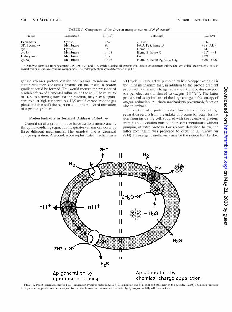
genase releases protons outside the plasma membrane andsulfur reduction consumes protons on the inside, a protongradient could be formed. This would require the presence ofa soluble form of elemental sulfur inside the cell. The volatilityof H2S, as a driving force for the reaction, may play a signifi-cant role; at high temperatures, H2S would escape into the gasphase and thus shift the reaction equilibrium toward formationof a proton gradient.
Proton Pathways in Terminal Oxidases of ArchaeaGeneration of a proton motive force across a membrane by
the quinol-oxidizing segment of respiratory chains can occur bythree different mechanisms. The simplest one is chemicalcharge separation. A second, more sophisticated mechanism is
a Q cycle. Finally, active pumping by heme-copper oxidases isthe third mechanism that, in addition to the proton gradientproduced by chemical charge separation, translocates one pro-ton per electron transferred to oxygen (1H1/e2). The latterprocess makes optimal use of the large change in free energy ofoxygen reduction. All three mechanisms presumably functionalso in archaea.
Generation of a proton motive force via chemical chargeseparation results from the uptake of protons for water forma-tion from inside the cell, coupled with the release of protonsfrom quinol oxidation outside the plasma membrane, withoutpumping of extra protons. For reasons described below, thelatter mechanism was proposed to occur in A. ambivalens(234). Its energetic inefficiency may be the reason for the slow
FIG. 16. Possible mechanisms for DmH1 generation by sulfur reduction. (Left) H2 oxidation and S0 reduction both occur on the outside. (Right) The redox reactions
take place on opposite sides with respect to the membrane. For details, see the text. Hy, hydrogenase; SR, sulfur reductase.
TABLE 5. Components of the electron transport system of N. pharaonisa
Protein Localization Mr (103) Cofactor(s) E0 (mV)
Ferredoxin Cytosol 15.2 2Fe-2S 2342SDH complex Membrane 90 FAD, FeS, heme B 18 (FAD)cyt c Cytosol 75 Heme C 2142cyt bc Membrane 14, 18 Heme B, heme C 2117, 244Halocyanine Membrane 15.4 Cu 1128cyt ba3 Membrane 40, 36 Heme B, heme AS, CuA, CuB 1268, 1358
a Data was compiled from references 349, 350, 473, and 477, which describe all experimental details on electrochemistry and UV-visible spectroscopic data ofsolubilized or membrane-residing components. The redox potentials were determined at pH 8.
590 SCHAFER ET AL. MICROBIOL. MOL. BIOL. REV.
on May 21, 2020 by guest
http://mm
br.asm.org/
Dow
nloaded from

aerobic growth of this archaeon, despite rapid oxygen turnoverand sulfur consumption.
Proton pumping by means of a Q cycle has been proposedfor the SoxABCD complex (329) and was demonstrated exper-imentally when the complex was coreconstituted in liposomestogether with the SoxL-Rieske protein, which allows the lipo-somes to be energized by cyt c (175). This artificial systemmade use of the fact that reduced cyt c can deliver electrons tothe Rieske protein, and the latter, via coreconstituted endog-enous caldariella quinone, reduces the cyt a587 of the SoxABCDcomplex. Thus, the quinol oxidase is energized from the out-side of the liposomes by cyt c. The procedure circumvents twoserious obstacles: first, reduced caldariella quinone (the spe-cific substrate of the complex) cannot be added in aqueoussolution, and second, the random orientation of reconstitutedoxidase also excludes an energization by TMPD as reductant.The latter is an effective substrate for the solubilized complex(174) but, due to its significant membrane permeability, wouldreduce both orientations of the complex and prevent the de-termination of H1/e2 ratios. The interpretation of the resultsof these sophisticated experiments was that the SoxABCDcomplex performs a Q cycle, assuming that the SoxB subunit ofthe complex is not itself a proton pump. The reasons are thesame as those for the A. ambivalens oxidase, explained belowon the basis of primary sequence data.
Do archaeal heme-copper oxidases provide the structuralprerequisites for the third mechanism, i.e., proton pumping bythe heme- and copper-containing polypeptide of terminal oxi-dases? To answer this question, the following argument mustbe considered. From extensive mutational studies of terminaloxidases from E. coli, Rhodobacter sphaeroides (146, 161, 563,564), and P. denitrificans (215, 426) and especially from thethree-dimensional structure of cyt c oxidases from P. denitrifi-cans (251) and mitochondria (578, 579), it has been concludedthat different pathways exist for protons consumed during ox-ygen reduction and those that are pumped. Two channels insubunit I, designated the K channel (involving an invariantly
conserved Lys residue) and the D channel (involving a con-served Asp residue) are characterized by two distinct sequencemotifs in transmembrane helix XII and in the helix II-III loopof heme-Cu oxidases. In other respects, these share a strictlyconserved topology of their heme and CuB ligands (90, 165) intransmembrane helices II-X and VI-VII, respectively. In addi-tion, a strictly conserved Glu residue (Glu-274 in P. denitrifi-cans) at the end of the proton uptake pathway through the Dchannel is definitely required for pumping. Recent FTIR stud-ies and molecular modelling underline the significance of theglutamate residue within a strongly apolar environment as acritical proton gate, presumably involving a chain of fixed watermolecules (431, 445). Thus, besides the specific -N-(X)10-D-(X)6-N-motif of the helix II-III loop of the K channel, theglutamate in the -HPE- motif in helix VI was declared indis-pensable for proton pumping (165).
Both motifs are missing in several archaeal terminal oxidasecomplexes and in that of the eubacterium T. thermophilus,strongly suggesting that these complexes are incapable of pro-ton pumping. They contrast with oxidases from P. aerophilum,S. acidocaldarius (ba3 [SoxM]), and H. salinarum. Accordingly,the terminal quinol oxidase of A. ambivalens has been desig-nated as nonpumping but capable of generating a proton mo-tive force by chemical charge separation. Similarly, the SoxBsubunit of the SoxABCD complex from S. acidocaldarius andthe aa3 oxidase from N. pharaonis would not be competent asproton pumps. Figure 17 depicts the relevant amino acidsparticipating in the formation of the D and K channels insubunit I of the compared terminal oxidases. Earlier reports ofthe generation of a proton motive force by purified SoxB re-constituted into liposomes and capable of oxidizing TMPD-ascorbate (174) can easily be explained by chemical chargeseparation without electrogenic pumping. Therefore, the dem-onstration of proton pumping (with H1/e2 .1) by the inte-grated SoxABCD complex (175) described above suggests thatthe operation of a Q cycle is most likely the mechanism ofproton translocation.
FIG. 17. Amino acid residues of subunit I involved in proton pumping by terminal oxidases. The boxed area encloses the archaeal terminal oxidases which arecompared to the bacterial ones from T. thermophilus and P. denitrificans; the latter serves as a reference enzyme with known three-dimensional structure. Column 1identifies the positions of the critical amino acids within individual transmembrane helices or the helix II-III connecting loop; 1 and 2 denote the presence and absence,respectively, of a specific residue in the enzymes; letters and numbers in parentheses describe positions and types of the replacement amino acids in the archaealorganisms. All oxidases which lack the motif in loop II-III are considered to be incapable of pumping protons by the mechanism employed by cyt c oxidase. Detailsand conclusions from this analysis are described in the text. “a,” numbering according to P. denitrificans; “b” refers to entire motif (-Nx10Dx6N-). P.den., P. denitrificans;H.sal., H. salinarum; S.ac., S. acidocaldarius; A.amb., A. ambivalens; N.pha., N. pharonis; T.th., T. thermophilus.
VOL. 63, 1999 BIOENERGETICS OF THE ARCHAEA 591
on May 21, 2020 by guest
http://mm
br.asm.org/
Dow
nloaded from

Whether the inability of an oxidase to pump protons can bededuced simply from structural comparisons remains to bedemonstrated experimentally in each individual case. The re-constituted cyt c oxidase ba3 from T. thermophilus appears topump protons with low stoichiometry (0.5H1/e2) when ener-gized with a photoactivated ruthenium II complex (269). Thisresult makes the above conclusions questionable and, in fact,strongly suggests the possibility of alternate proton pathways.Assuming that the SoxABCD complex can perform a Q cycleand, in addition, that the SoxB subunit would pump protons,this archaeal oxidase complex should be able to achieve H1/e2
ratios of .2, which has not been observed. Irrespective ofthese arguments, the results with the T. thermophilus oxidaseconfirm the necessity of the strictly conserved K channel forthe redox reaction, while possibly alternate pathways to the Dchannel may exist but have not been resolved structurally.
Archaea, like bacteria, can possess simultaneously bothtypes of terminal oxidases (e.g., S. acidocaldarius and T. ther-mophilus), which may or may not contain the sequence motifsdiscussed above. Therefore, terminal oxidases may haveevolved by an early gene duplication of subunit I, prior to thedivision into the archaeal and bacterial domains (608). Thephysiological meaning of the coexistence of two oxidases is notyet understood. For example, greatly differing oxygen affinitieswould permit adaptation to varying micro-oxygenic conditions,as in Bradyrhizobium japonicum (429). Another possibilitywould be the preferential operation of specific oxidases atcertain pH values. Since no data is available, these possibilitiesare moot pending further investigation.
LIGHT-DRIVEN ENERGETICS
General Overview
In the early 1970s, the so-called purple membrane from H.salinarum (55) was found to contain only a single protein (401).It was later demonstrated that a retinal chromophore attachedto this protein gives rise to the purple color. Because of theobvious resemblance to vertebrate rhodopsins, the authorsnamed this retinylidene protein bacteriorhodopsin (BR). Itwas also immediately clear that, upon light activation, a protonis transferred across the membrane from the inside of the cellto the extracellular medium. The proton potential generatedthereby is utilized by H. salinarum for the synthesis of ATP(432).
During the elucidation of the proton-pumping mechanism ofBR and the physiology of H. salinarum, three other bacterialrhodopsins were discovered (an extensive progress report onthe photophysics and photochemistry of retinal proteins is pre-sented in reference 413). These pigments are functionally quitedistinct. HR, like BR an ion pump, is an inwardly directedchloride pump. The two other bacterial rhodopsins turned outto be photoreceptors that trigger the photophobic and/or pho-toattractive response of bacteria. All four bacterial rhodopsinsare strikingly similar with respect to structure, the retinal chro-mophore and its attachment site, and the light-activated reac-tion cycle.
The four pigments are membrane proteins with an approx-imate molecular mass of 26 kDa (see reference 503 for acomparison of their amino acid sequences). In each case, thepolypeptide chain folds into seven membrane-spanning helices(indexed helices A to G) with the C terminus positioned on thecytoplasmic side. The retinal chromophore is attached in theall-trans configuration to a Lys residue via a protonated Schiffbase in the C-terminal helix G. The structural model of BR(216) revealed the locations in the sequence that are involved
in the chromophore binding site. This was confirmed by acomparison of the amino acid sequences of all archaeal rho-dopsins known at the time that exhibited distinct homologies atthose particular positions (503, 519).
In a direct comparison of amino acid sequences of rho-dopsins from different archaeal species, clusters of conservedresidues are found in helices C and D, containing three Trpresidues, which enclose the retinal chromophore. The Lys res-idue of helix G, which marks the retinal attachment site, and anAsp residue situated one helical turn upstream are conservedthroughout. This aspartic acid residue is part of the counterioncomplex of the protonated Schiff base. Another interestingstretch of homologies was detected on helix C, where an Argresidue also contributes to the Schiff base environment.
Upon light excitation, all four microbial rhodopsins undergocyclic reaction sequences. During the photocycle, the physio-logical response, either vectorial ion transport or initiation ofthe signal transduction chain, is activated. The reversible mo-lecular events associated with this include the isomerization ofthe retinal chromophore from all-trans to 13-cis, the deproto-nation of the Schiff base (HR is an exception), and confor-mational changes of the protein backbone. Protonation-de-protonation reactions of internal aspartyl carboxyl groups arefunctionally also quite important (see below). All these light-triggered thermal reactions have to be reversed because, oncea cycle is completed, the protein regains its original state.
The photocycle has been studied in great detail for BR andless stringently for the other three pigments. The general pic-ture emerging from these investigations can be summarized asfollows: the intermediates of the BR photocycle are named inalphabetical order, starting with J for the earliest reactionproduct. J is followed by five other intermediates, K, L, M, N,and O. These letters were chosen by analogy to the reactionsequence observed in visual pigments, with Lumi and Metaidentified as physiologically important states (323, 542).
When the photocycles of the other pigments were comparedto those from BR, it became apparent that they follow a com-mon scheme with some variations, which depend on the initialstate and the nature of the counterion complex (see below).The long-lived intermediate M, which is formed on the micro-second time scale, is physiologically quite relevant. In the caseof the sensory rhodopsins (SRs), it represents the signalingstate (615, 617), whereas in BR, M separates the proton re-lease pathway from the proton uptake steps. The chloridepump HR is an exception because in the naturally occurringphotocycle an intermediate with a deprotonated Schiff base (atypical property of the M state) is missing.
The ion pumps return to the original state on the millisecondtime scale. The SRs are slower, requiring about 100 ms. Thereason for the difference is likely to be related to the functionsof these proteins. Whereas the efficiency of the ion pumps isdependent on a high turnover rate, the SRs have to remain inthe signaling state long enough to relay the signal to cytoplas-mic proteins.
BR
Structure of BR. The observation that BR forms a hexagonaltwo-dimensional lattice within the purple membrane (55) fa-cilitated further analysis of its structure. A low-resolution mapof the electron-scattering density of the purple membrane re-vealed seven peaks, which can be attributed to seven helices ofBR that are oriented almost perpendicularly to the membranesurface (217). From the primary structure of BR (277, 414), itbecame evident that the protein is a chain of 248 amino acids,which folds into a bundle of seven helices with short nonhelical
592 SCHAFER ET AL. MICROBIOL. MOL. BIOL. REV.
on May 21, 2020 by guest
http://mm
br.asm.org/
Dow
nloaded from
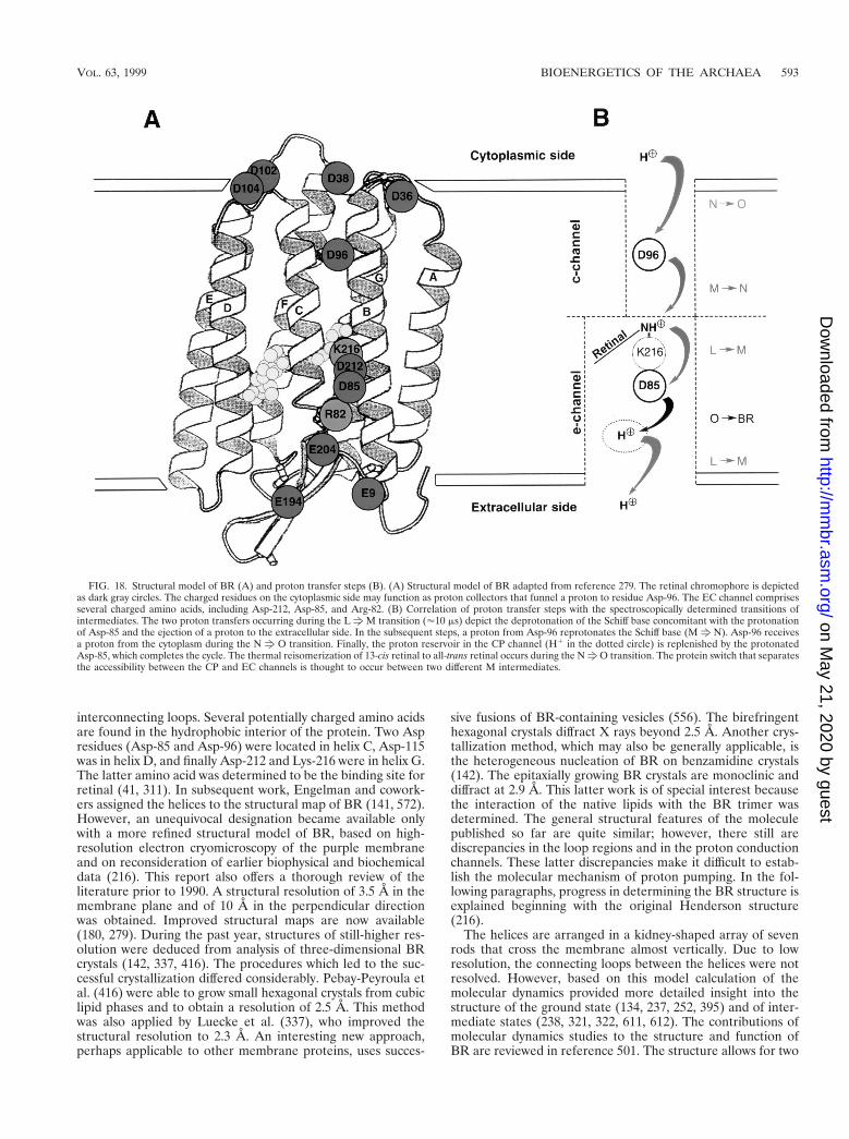
interconnecting loops. Several potentially charged amino acidsare found in the hydrophobic interior of the protein. Two Aspresidues (Asp-85 and Asp-96) were located in helix C, Asp-115was in helix D, and finally Asp-212 and Lys-216 were in helix G.The latter amino acid was determined to be the binding site forretinal (41, 311). In subsequent work, Engelman and cowork-ers assigned the helices to the structural map of BR (141, 572).However, an unequivocal designation became available onlywith a more refined structural model of BR, based on high-resolution electron cryomicroscopy of the purple membraneand on reconsideration of earlier biophysical and biochemicaldata (216). This report also offers a thorough review of theliterature prior to 1990. A structural resolution of 3.5 Å in themembrane plane and of 10 Å in the perpendicular directionwas obtained. Improved structural maps are now available(180, 279). During the past year, structures of still-higher res-olution were deduced from analysis of three-dimensional BRcrystals (142, 337, 416). The procedures which led to the suc-cessful crystallization differed considerably. Pebay-Peyroula etal. (416) were able to grow small hexagonal crystals from cubiclipid phases and to obtain a resolution of 2.5 Å. This methodwas also applied by Luecke et al. (337), who improved thestructural resolution to 2.3 Å. An interesting new approach,perhaps applicable to other membrane proteins, uses succes-
sive fusions of BR-containing vesicles (556). The birefringenthexagonal crystals diffract X rays beyond 2.5 Å. Another crys-tallization method, which may also be generally applicable, isthe heterogeneous nucleation of BR on benzamidine crystals(142). The epitaxially growing BR crystals are monoclinic anddiffract at 2.9 Å. This latter work is of special interest becausethe interaction of the native lipids with the BR trimer wasdetermined. The general structural features of the moleculepublished so far are quite similar; however, there still arediscrepancies in the loop regions and in the proton conductionchannels. These latter discrepancies make it difficult to estab-lish the molecular mechanism of proton pumping. In the fol-lowing paragraphs, progress in determining the BR structure isexplained beginning with the original Henderson structure(216).
The helices are arranged in a kidney-shaped array of sevenrods that cross the membrane almost vertically. Due to lowresolution, the connecting loops between the helices were notresolved. However, based on this model calculation of themolecular dynamics provided more detailed insight into thestructure of the ground state (134, 237, 252, 395) and of inter-mediate states (238, 321, 322, 611, 612). The contributions ofmolecular dynamics studies to the structure and function ofBR are reviewed in reference 501. The structure allows for two
FIG. 18. Structural model of BR (A) and proton transfer steps (B). (A) Structural model of BR adapted from reference 279. The retinal chromophore is depictedas dark gray circles. The charged residues on the cytoplasmic side may function as proton collectors that funnel a proton to residue Asp-96. The EC channel comprisesseveral charged amino acids, including Asp-212, Asp-85, and Arg-82. (B) Correlation of proton transfer steps with the spectroscopically determined transitions ofintermediates. The two proton transfers occurring during the LfM transition ('10 ms) depict the deprotonation of the Schiff base concomitant with the protonationof Asp-85 and the ejection of a proton to the extracellular side. In the subsequent steps, a proton from Asp-96 reprotonates the Schiff base (Mf N). Asp-96 receivesa proton from the cytoplasm during the N f O transition. Finally, the proton reservoir in the CP channel (H1 in the dotted circle) is replenished by the protonatedAsp-85, which completes the cycle. The thermal reisomerization of 13-cis retinal to all-trans retinal occurs during the NfO transition. The protein switch that separatesthe accessibility between the CP and EC channels is thought to occur between two different M intermediates.
VOL. 63, 1999 BIOENERGETICS OF THE ARCHAEA 593
on May 21, 2020 by guest
http://mm
br.asm.org/
Dow
nloaded from

half-channels that connect the cytoplasm and the extracellularside with the Schiff base environment (Fig. 18). A hydrophobicpore with a smaller diameter opens to the cytoplasm. Thischannel contains only a few cavities that might harbor watermolecules (142, 337, 416). The other, broader channel con-nects the interior of the protein with the extracellular side.Retinal is located at the interface between the two channels,with the proton of the Schiff base facing the extracellular chan-nel (EC channel). In their original report, Henderson et al.(216) proposed that these channels constitute the path of theprotons across the membrane; this has now been corroboratedby the higher-resolution structures. FTIR experiments demon-strated that Asp residues within the hydrophobic interior of theprotein are proton donor and acceptor sites (135, 137, 511).These sites have been assigned to specific Asp residues bysite-directed mutagenesis studies (76, 170) and by solid-statenuclear magnetic resonance (NMR) studies of [4-13C]Asp-la-beled BR (138, 139, 354, 355). These experiments showed thatAsp-85 and Asp-212, which are located in the EC channel, aredeprotonated in the resting state. The protonated carboxylgroup of Asp-96 (354), a component of the cytoplasmic chan-nel (CP channel), donates its proton to the deprotonated Schiffbase during the Mf N transition (170). Asp-115, not directlylocated in the cytoplasmic pore, is also protonated (354). ThepKs of these two residues were determined to be greater than11 (354, 552). Further investigations of the structural proper-ties of the CP channel using FTIR spectroscopic techniques incombination with site-directed mutagenesis of BR have pro-vided evidence for a hydrogen-bonded chain that includes Asp-96, Thr-46, Val-49, Pro-50, Ala-53, and water molecules (183,216, 613, 614). The existence of water in this part of the mol-ecule has also been suggested by structural analysis (142, 337).During proton uptake, this hydrogen-bonded chain apparentlybridges the carboxyl group of Asp-96 with the Schiff base.
The EC channel constitutes the proton release pathway,broader and more hydrophilic than the CP channel. There areseveral charged amino acids (Asp-212, Asp-85, Arg-82, Glu-194, and Glu-204) and two Tyr residues (Tyr-57 and Tyr-185)that contribute to the interior of the channel and to the envi-ronment of the Schiff base. Asp-85 has a special role as theprimary proton acceptor. The positive charge of the Schiff baseis neutralized by the charged residues mentioned above, whichform a diffuse counterion complex (115, 235). Structural mod-els of the two channels, including the pattern of hydrogenbonds, were provided in recent papers on the structure of BR.Since various aspects of these proposals still differ, the molec-ular mechanism of proton pumping will remain the subject offurther experiments.
Five helices (B, C, E, F, and G) form the environment of theretinal chromophore. Retinal is tilted relative to the mem-brane normal, with the ionone ring pointing away from thecytoplasm. The configuration and the position of retinal withinthe BR molecule have been determined by a variety of bio-physical methods, including solid-state NMR spectroscopy,neutron diffraction, and resonance-Raman and FTIR spectros-copy (reviewed in reference 413; see also reference 372 for amore recent publication). The retinal chromophore is locatednear the center of the protein and assumes an all-trans config-uration. The isomerization around the 6s bond that connectsthe ionone ring with the unsaturated hydrocarbon chain is inthe trans conformation, which allows for the chromophore tobe planar (108, 352). This observation has important implica-tions for the wavelength regulation in BR and may also applyto the other rhodopsins. Protonated Schiff bases usually absorbat approximately 460 nm; BR has an absorption maximum at568 nm. The difference calculated in wave numbers is called
the opsin shift because the specific interaction of the proteinwith the chromophore leads to this bathochromic displacementof the absorption maximum. Solid-state NMR experiments(235) and the spectroscopic properties of BR reconstitutedwith retinal analogs (as well as similar experiments with modelsubstances [319]) led to an explanation of the opsin shift.According to this model, two effects, i.e., the C6-C7 single-bondconfiguration and the weak counterion strength (115), influ-ence each other synergistically and are sufficient to explain thelarge opsin shift of BR (235). The data from NMR spectros-copy and the analysis of chromophore models clearly indicatethat the interaction of the protonated Schiff base with thecounterion is weak. The NMR data also suggests that thiscounterion cannot be a point charge, e.g., a single, negativecarboxylate group from an aspartic acid residue, but must be acomplex consisting of charged groups in the neighborhood ofthe Schiff base. This picture has been substantiated by recentdata on the structure of BR.
Mechanism of proton pumping. The mechanism of vectorialproton transfer was elucidated by analysis of the photocycle.After absorption of light, the all-trans retinal Schiff baseisomerizes to a 13-cis conformation. The first subpicosecondevent (500 fs) in the primary photoreaction is the formation ofthe J intermediate (128, 245, 397, 509). It is quite striking thatthis photoisomerization is so rapid. The protein apparentlyprovides a vacuum-like environment in which the ultrafastreaction can occur. With a quantum yield of approximately 0.6,this process is quite efficient (568). K, the next intermediate inthe reaction sequence, is formed within 3 ps (130, 397, 424,428, 509). As determined by FTIR spectroscopy, the proteinresponds only slightly to this altered situation, which results ina strained, distorted chromophore (75, 144). The chromophorerelaxes during the transition from K to L, which is reached onthe microsecond time scale. In L, the conditions for the releaseof the proton to the extracellular side are established. Duringthe L f M transition, the proton of the Schiff base is trans-ferred to Asp-85. Concomitantly, another proton is released tothe bulk phase. Recent data, although still controversial (142,337, 436), indicates that this proton originates from Glu-204(440, 479), the pKa of which decreases upon protonation ofAsp-85 (31, 440). During the lifetime of the M intermediate,which has an absorption maximum at 410 nm, the transitionbetween the extracellular and the cytoplasmic accessibility ofthe Schiff base must take place (Fig. 18). The nature of thistransition is not known, but conformational changes of theprotein are probably responsible (198, 285, 546). The Schiffbase is then reprotonated by Asp-96, forming the N interme-diate (219, 520). The reisomerization of the retinal chro-mophore from the 13-cis to the all-trans configuration firstoccurs in the next step of the reaction sequence, in which theO intermediate is formed. At the same time, Asp-96 receives aproton from Asp-38, which has obtained a proton from thecytoplasm (444). The O intermediate absorbs at 620 nm. Thisredshift of the absorption maximum may be due to the still-protonated Asp-85. A similar redshift is observed for the so-called blue membrane (278), for which protonation of Asp-85could also be demonstrated (355). In the last step of the pho-tocycle, Asp-85 loses its proton. Since all steps of the photo-cycle need to be reversed in order to regain the original state(195), it is also necessary that access to the Schiff base returnsto the extracellular side. That the first transition does occurand that it occurs between the M and N states has been dem-onstrated in voltage-clamp experiments (386). In this work, BRwas expressed in the plasma membrane of oocytes (387), andthe voltage-dependent photocurrent was measured. Double-flash experiments with blue light (which photoconverts the M
594 SCHAFER ET AL. MICROBIOL. MOL. BIOL. REV.
on May 21, 2020 by guest
http://mm
br.asm.org/
Dow
nloaded from

intermediate directly back to the initial ground state) at pump-inhibiting potentials provide the first experimental data on Mstates with different accessibilities, thus supporting the idea ofa switching event between two different forms of M.
Several other experiments have attempted to identify theswitch at the molecular level. For example, amide I and amideII vibrational modes have been interpreted as indicative ofconformational changes of the protein backbone (for example,see reference 218). Time-resolved EPR measurements directlyshowed differences in mobility at defined positions of the BRmolecule (446, 539). A more direct approach has been chosenby a few groups, who used X-ray (263, 285, 389), electron (218,545, 546, 587), and neutron diffraction (116) studies to char-acterize structural changes occurring at different stages of thephotocycle. These experiments show that helices F and G arepredominantly affected during the lifetimes of the M and Nintermediates, interpreted as an increase in the structural or-der of helix G and an outward tilting of the cytoplasmic part ofhelix F. This latter finding is of significance because it bears ona possible mechanism of signal transfer from SR to its trans-ducer (see below).
The photocycle has been the subject of numerous reports.The original model of Lozier et al. (323), with the sequence BRf K f L f M f N f O f BR, is now generally accepted,though the existence of the N intermediate has sometimesbeen neglected. The kinetic data of the photocycle can besatisfactorily approximated with seven exponentials (101), in-dicating that at least seven distinct intermediates must exist; ofthese, only the five spectrally distinguishable species K to Ohave been identified. Therefore, the simple kinetic scheme BRf Kf LfMf Nf Of BR has had to be modified; backreactions, side reactions, new intermediates, or even parallelphotocycles have been introduced. The most controversial partof the photocycle involves the M f N f O transition, forwhich two (and as many as five) M intermediates have beenproposed, as have also back reactions such as MN N and/or NN O (for a review, see reference 306). In an effort to reconcilethe published data with their own measurements, Chizhov etal. (101) argued for a scheme of irreversible sequential transi-tions between quasiequilibria of intermediates (Fig. 19). Thequasiequilibria are controlled by rate-limiting dynamics of theprotein and/or proton transfer steps. P3 and P4 both representL540 N M410 equilibria and are distinguished by the relativepercentages of the two components L540 and M410, which shiftfrom 20% M410 to 70% M410. The irreversible transition of(L540 N M410) f M410 may be considered the molecularswitch proposed in references 195 and 528 and verified by thework of Nagel et al. (386), in which the access to the Schiff basechanges from the extracellular side to the CP channel. In thesecond part of the photocycle, a proton from the CP channelreprotonates the Schiff base. These steps are also characterizedby quasiequilibria (P6 and P7) that are quite sensitive to the pHof the buffer and the temperature. This model implies that theion-translocating steps are not rate limiting. Apparently, theconformational changes of the protein and/or the isomeriza-tion of the retinal chromophore determines the time course ofthe vectorial proton transfer.
It should be noted that Lanyi and coworkers have proposeda mechanism for the proton pump (84). According to thislocal-access model, the retinal Schiff base retains connectivityto both the EC and the CP channels throughout the photo-cycle. Vectoriality is obtained by a shift in the pKa values of thegroups in the proton conduction chains. In wild-type BR, aninitial increase of the pKa of Asp-85 facilitates proton transferfrom the Schiff base. Conversely, the subsequent decrease of
the pKa of Asp-96 allows the rapid reprotonation of the Schiffbase.
HR
Structure of HR. HR, the second light-driven ion pump ofH. salinarum (for a recent comprehensive review, see reference398), has now been found in other halophilic archaebacteria(411, 519). First described and named by Mukohata and col-leagues (347), HR has an absorption maximum at 576 nm andwas first sequenced by Blanck and Oesterhelt (53). Upon lightexcitation, chloride ions are transferred from the medium intothe cell (35, 65, 496, 534). The ion selectivity of the transportis not restricted to chloride; bromide, iodide, and nitrate arealso transported (203, 208, 496). Compared to that of BR, theabsorption maximum of HR has a longer wavelength.
The amino acid sequences of several HRs reveal structuresquite similar to those of BR, including the omnipresent mainmotif of seven helical transmembrane chains. Further struc-tural information was gained through electron cryomicroscopyof membrane sheets. The projection map of HR at 7-Å reso-lution was very similar to that of BR at the same resolution(199, 200). These structural similarities match the close resem-blance of the primary sequences of the two pigments. Oneinteresting difference is found on the cytoplasmic side of thehelices B, C, F, and G. As mentioned above, these helices formthe CP channel. In HR, the cross-sectional area is larger thanin BR, indicating a pore wider than would be required totransport halides. The EC channels, of similar dimensions inBR and HR, would easily accommodate chloride anions asindicated by functional studies (102, 152).
Most of the conserved residues are associated with the ret-inal binding site. Compared to BR, interesting differences arefound in positions of functional importance for proton trans-location. Asp-96 (numbered according to the BR sequence) isreplaced by an Ala residue, and Asp-85 is replaced by a Thr
FIG. 19. Scheme of the BR photocycle based on a sequential irreversiblemodel according to reference 101. P0 to P7 denote the spectroscopically definedintermediates. The four states P3, P4, P7, and P6 are quasiequilibria between Mand L, M and N, and N and O. The decay rates (at 20°C) of the intermediates aredepicted between the wings.
VOL. 63, 1999 BIOENERGETICS OF THE ARCHAEA 595
on May 21, 2020 by guest
http://mm
br.asm.org/
Dow
nloaded from

residue. This latter substitution is of particular interest becausea negative charge is removed from the counterion complex,thereby creating an anion binding site (410, 474). Indeed, chlo-ride binds to a mutant of BR in which Asp-85 is replaced byThr. This mutated BR pumps chloride actively upon light ex-citation (461).
The putative binding of anions has been studied extensively.Ogurusu et al. (403) concluded that HR has a single bindingsite, because the absorption spectrum of HR is dependent onthe chloride concentration. Further spectroscopic analyses ofthe binding of different anions by HR in Na2SO4 (which itselfdoes not bind to the protein) indicate that anions occupy twodifferent sites (305, 498). These two sites may participate in theuptake and release of chloride during transport. However, newFTIR spectroscopic results indicate only a single binding site(597). The discrepancies between this view and the proposedtwo sites may be resolved by assuming a one-site, two-statemodel (597).
As already mentioned, the replacement of an Asp residue inBR by a Thr residue creates an anion binding site. Data fromresonance-Raman spectroscopy of HR supports the idea thatchloride binds near the Schiff base (127, 169, 341, 415); it wasconcluded that the positive Schiff base cation is weakly hydro-gen bonded to a complex counterion made up of chloride,Asp-238, and Arg-108 (18). Braiman and coworkers demon-strated that the CAN stretching vibration is affected by thetype of anion, indicating a binding site near the Schiff base(597). Furthermore, vibrational modes from the Arg residuewere also perturbed (77). In elegant experiments, these modeswere assigned to Arg-108; it was also shown that the guani-dinium ion can mimic the Arg residue in the Arg108Gln mu-tant (450). The similarity between the HR and BR counterioncomplexes is quite evident from the observation of a so-calledacid purple form of BR (125, 152), the creation of an anionbinding site in the BR mutant Asp96Thr, and the observationthat HR from N. pharaonis (132, 304) forms a blue pigmentupon anion removal (474). Even the proton-pumping capabil-ity of BR could be reestablished in HR from N. pharaonis(582).
N. pharaonis, a haloalkaliphilic bacterium from the WadiNatrun of Upper Egypt, expresses two rhodopsin-like pig-ments, HR (pHR) and the photophobic receptor-SR II (pSRII)(52). The blue form of pHR is spectroscopically quite similarto the cation-depleted so-called blue membrane of BR. Re-moval of a negative charge from the counterion complex of theparent state leads to this bathochromic shift. In BR, the pro-tonation or neutralization of Asp-85 (355, 412, 547) removesthe negative charge, whereas in pHR the anion can be ex-tracted by size-exclusion chromatography (474). The purplecoloration of pHR can be restored by adding anions, useful inexamining the effect of specific anions on spectroscopic prop-erties and photocycle kinetics (474). All monovalent anionssuch as halides, azide, or nitrate are able to bind to the protein.The anion binding kinetics are compatible with a single bindingsite (102), thus confirming the interpretation of Walter andBraiman (597) for HR. It should be noted that azide-reconsti-tuted pHR displays a photocycle very similar to that of BR,including an M-like intermediate. Indeed, transport studiesclearly demonstrated that this modified pHR acts as a protonpump (582).
The chromophore of HR, all-trans retinal, binds via a pro-tonated Schiff base to Lys-242 on helix G. The pK of the Schiffbase varies from 7.4 to 8.9, depending on the anion present(474, 498, 535). It is noteworthy that light adaptation does notlead to a 100% all-trans configuration of the retinal chro-mophore in HR, as observed for BR (265, 306, 584, 628). This
somewhat complicates an analysis of the photocycle because ofthe contribution of the transport-inactive cis cycle.
Mechanism of chloride pumping. The functional and phys-iological properties of HR have been investigated intensively(reviewed in reference 398). The chromophore adopts an all-trans configuration, including a C6-C7 trans single bond (38).After absorption of light, the electronically excited state re-laxes within the picosecond time scale to a redshifted photo-product with maximal absorption at 600 nm (HR600). Thequantum yield of this reaction, about 0.3, is much lower thanthat determined for BR (268, 400). The primary photoreactioninvolves an all-trans313-cis isomerization of retinal (127, 154,202, 303, 399), and according to the FTIR difference spectrumbetween HR600 and HR, this is quite similar to the K-BRdifference spectrum (448). The following sequence of eventsinvolves at least two additional intermediates, which absorb at520 nm (HR520) and 640 nm (HR640). These intermediatesabsorb above 500 nm and must therefore possess a protonatedSchiff base. Unlike the BR photocycle, the functionally activeHR photocycle does not include the M intermediate, althoughthis is observed in a side reaction which is catalyzed by azide(209). The photocycle has been analyzed with transient-absorption spectroscopy, resonance-Raman spectroscopy, andFTIR spectroscopy (reviewed in references 15 and 398).
A structural analysis of the retinal chromophore by reso-nance-Raman spectroscopy (127, 154) revealed that HR520 andHR640 are both characterized by a 13-cis, 15-anti-retinylideneSchiff base. However, recent data from FTIR experiments us-ing HR from N. pharaonis indicated an all-trans configurationof the retinal chromophore (583).
Until now, there has been no comprehensive model of thephotocycle which explains the effects of temperature, anion,and pH upon the reaction kinetics. In a recent proposal, Varoand coworkers (85) ascribe HR640 to the so-called cis cycle,which originates from the photoactivation of (13-cis retinal)-HR. On the other hand, the HR640 intermediate is clearly seenin HR from N. pharaonis (474, 583). By analogy to the modelof the BR photocycle (101), the HR photocycle may also rep-resent a sequence of irreversible steps between quasiequilibriaof spectroscopically identified species (100). Closely related tothe problem of the photocycle model is that of the steps ofchloride uptake and release. Based on the dependence of thekinetics on the chloride concentration, it has been concludedthat chloride is released during the lifetime of HR520 and takenup during the reformation of the ground state (307, 399, 400,627). Electrical measurements by the black lipid membranetechnique placed the ion movements within the same timeranges. However, it is possible that the actual ion-translocatingsteps are considerably faster than the apparent rate constants.If this is the case, the observed rate constants may reflect onlyrate-limiting changes in protein conformation or retinalisomerization. In new experiments, Chizhov et al. (100a)showed that this accessibility switch between the CP and theEC channels involves two different HR640 species, which rap-idly equilibrate with HR520 species. The unsettled questions ofthe photocycle of HR and the mechanism of chloride transferacross the membrane require the combined efforts of time-resolution techniques (e.g., visual absorption and FTIR spec-troscopy).
HR can be converted into a proton pump in a two-photonprocess (36). Green light alone causes the transfer of chlorideions into the cell. Additional blue light, which excites theHR410 formed by the side reaction mentioned above, convertsHR into a proton pump in the direction opposite the one inBR, i.e., protons are pumped into the cell; this process can beenhanced by the addition of azide. Similar effects have been
596 SCHAFER ET AL. MICROBIOL. MOL. BIOL. REV.
on May 21, 2020 by guest
http://mm
br.asm.org/
Dow
nloaded from

observed for BR mutants in which Asp-85 is replaced by aneutral amino acid such as Thr or Asn (569).
HR from N. pharaonis behaves quite differently. The pho-tocycle of pHR in the presence of azide is quite similar to theBR photocycle with a pronounced transient concentration ofan M-like intermediate (474). This observation prompted Varoet al. (582) to analyze the ion specificity of the one-photon-activated transport, revealing that this modified HR acts as aperfect proton pump in which protons are transferred into theextracellular medium. This result is in line with reports that theAsp85Thr BR mutant can function as a chloride pump (461)and that chloride transport activities are restored in the so-called acid purple membrane, which is obtained at very low pHvalues (125).
SRs
The phototactic activity of the archaeon H. salinarum ismediated by two photoreceptors that have a close structuralrelationship to the ion pumps BR and HR (for recent reviewson the photochemistry and function of microbial SRs, seereferences 232 and 530). The archaea are attracted to light of.520 nm and repelled by light of ,500 nm. The response to aphotoattractant is mediated by SRI (62, 204). In a two-photonreaction, this receptor can also trigger a negative response tonear-UV light (527). Moreover, repellent light of ,500 nm inwavelength is recognized by a second pigment, SRII, whichabsorbs at 490 nm. SRI and SRII are expressed under differentconditions. The biosynthesis of SRI, like that of the two ionpumps, is induced by anoxic conditions. SRII is produced con-stitutively by the cell, suggesting that SRII appeared quite earlyin the evolution of retinal pigments (554), which thus protectedthe respiring and fermenting halobacteria from exposure tophotooxidative conditions. By contrast, SRI enables thehalobacteria to locate light conditions optimal for the opera-tion of the two ion pumps, BR and HR, without being harmedby UV light.
Biochemical data (521, 523) and the sequence determinationof SRI and SRII revealed sequences, upstream of both the sopIand sopII gene loci, that correspond to the halobacterial trans-ducer of rhodopsin (Htr) (503, 619, 623). HtrI and HtrII showdistinct similarities to the bacterial chemoreceptors, especiallyin the signal domain region and in the flanking methylation-demethylation sites. Apparently, archaea have developed in-formation transfer chains that possess extended structural sim-ilarities to those of bacteria.
SRI. SRI, first described in 1982 by Bogomolni and Spudich(62), was isolated (482) and subsequently sequenced (54). Itcontains an all-trans, 6s-trans protonated Schiff base (155)which is attached to a Lys residue on helix G (Fig. 18). Thechromophore-binding site accepts only all-trans retinal and not13-cis retinal, as was also observed for BR (616). In compari-son to the other archaeal rhodopsins, the absorption maximumof SRI is shifted down to 587 nm. This bathochromic shift wasexplained by the protonation of the side chain carboxyl groupof Asp-76 (437, 438), which essentially results in the removal ofa negative charge from the Schiff base counterion complex.The deprotonated pigment absorbs at 550 nm; a pK of 7.2 hasbeen determined for this transition (64). A similar effect isobserved for BR, in which the protonation of Asp-85 leads tothe so-called blue membrane (278), and also for HR from N.pharaonis, in which the removal of the chloride anion (474)leads to chromophores absorbing at 600 nm. It should be notedthat the pK of Asp-76 rises to 8.5 upon binding of the halobac-terial transducer of rhodopsin I (HtrI) (64), an observation
that may be significant for the mechanism of signal transduc-tion (see below).
The photocycle of SRI closely resembles that of BR (63).Upon light excitation, a redshifted intermediate (S610) is ob-tained and then decays to an L-like intermediate (S560) withinapproximately 90 ms. The next intermediate (S373), which isformed within 300 ms, is characterized by an unprotonatedSchiff base and a 13-cis configuration of the retinal chro-mophore (196, 577, 616). It constitutes the physiologically ac-tive state that triggers positive phototaxis (343, 615). It slowlydecays, returning to the original ground state within approxi-mately 800 ms. These kinetic data imply a high transient con-centration of S373, which makes an additional two-photon pro-cess possible. This photoactivated branch of the photocycleleads to reversal of the flagellar motor, thus enabling the bac-teria to avoid harmful UV light (527). It is certainly a strikingfeature of SRI to allow a positive response in a one-photonprocess and a negative response in a two-photon reaction; theaspartate receptor (Tar), among others, also displays this dualcapacity (9).
It has already been mentioned that Asp-85 and Asp-96 playa crucial role in the mechanism of the BR proton pump. InSRI, the corresponding sites are Asp-76 and Tyr-87. As out-lined above, in SRI Asp-76 is protonated. The protonation ofthe corresponding amino acid in BR (Asp-85) leads to a non-functional protein. Mutations of Asp-96 to neutral amino acidresidues impair the proton pump activity, which can be re-stored by the addition of azide (570). This data suggests thatSRI can also function as a proton pump. Indeed, during lightexcitation, a vectorial proton transport directed to the outsideof envelope vesicles was observed (64). The action spectrum ofthe proton pump activity has a maximum at 540 to 550 nm, notat 587 nm as in the parent SRI. This shift toward the blueregion has been explained by the presence of a deprotonatedAsp-76. Apparently, protonation of Asp-76 does not lead tofunctional (proton pump) activity, as found with the acid blueform of BR. The binding of HtrI to SRI apparently abolishesthe ability to pump protons (summarized in references 524 and525). In contrast to these results, Haupts et al. (195) providedevidence that the HtrI-SRI complex also pumps protons. Thequestion whether the proton pump activity observed for SRIand its transducer is required for signal transduction has notyet been answered. In this context, it should be noted that theAsp76Asn mutant is fully functional in phototaxis (437).
SRII. The fourth retinal protein of H. salinarum, SRII orphoborhodopsin, was first described by Takahashi et al. (553).This receptor had eluded a thorough analysis due to its verylow concentration in the cell, coupled with its instability at lowionic strength and in the presence of certain detergents. Al-though a partial purification was accomplished (475), informa-tion about SRII mostly was restricted to spectroscopic data andphysiological properties (346, 617). The absorption maximumat 490 nm with a shoulder at 460 nm suggests a planar structureof the retinal chromophore (554). The photocycle of SRII, likethat of BR and SRI, includes an intermediate with a blue-shifted absorption maximum characteristic of a deprotonatedSchiff base (241, 475).
The difficulties encountered in isolating SRII from H. sali-narum stimulated a search for SRII-like pigments in otherspecies. Such a blue-light receptor was detected in N. pharaonis(52) and characterized spectroscopically (225, 239, 240, 368).This SR from N. pharaonis (pSRII) is functionally identical toSRII but stable under low salt concentrations (476, 478), al-lowing its purification and amino acid sequencing (503). Thislatter work showed that pSRII and pHtrII are transcribed as asingle transcript, indicating the existence of a pSRII-pHtrII
VOL. 63, 1999 BIOENERGETICS OF THE ARCHAEA 597
on May 21, 2020 by guest
http://mm
br.asm.org/
Dow
nloaded from

protein complex similar to SRI-HtrI. Comparison of the aminoacid sequences revealed strong homologies to the other ar-chaeal proteins, particularly SRI. Calculations of the phyloge-netic relationship of 22 amino acid sequences in archaeal rho-dopsins and correlation with functional properties haverevealed four members of this family. (i) Proton pumps arecharacterized by Asp-85 and Asp-96 (numbering according tothe BR sequence); (ii) in chloride pumps, these sites are oc-cupied by Thr and Ala residues, respectively; (iii) in SRI, theAsp residue in position 85 is conserved whereas Asp-96 isreplaced by an aromatic residue; and (iv) in the distinct addi-tional member, pSRII, the indicator positions 85 and 96 arehomologous to those of SRI, but Met-118 is replaced by a Valresidue. The amino acid sequence of SRII from H. salinarum,recently analyzed (623), is also quite similar to that of pSRII.
The photoreaction of pSRII from N. pharaonis has beenstudied by several groups (103, 225, 239, 240, 368, 476). Likethat of SRII, the pSRII photocycle contains K-, L-, M-, andO-like species. The K- and M-like intermediates have beenanalyzed by FTIR difference spectroscopy (140), indicatingthat considerable conformational movements of pSRII alreadytake place at the stage of the early K-like intermediate. Thesechanges are more intense than those described for SRI (437).Concomitant with the deprotonation of the Schiff base, a car-boxyl group which may arise from Asp-75 is protonated. Theprotonation reaction is pH independent over the pH rangefrom 5 to 8, in contrast to the situation in SRI, in which a pKof 7.2 for the corresponding residue Asp-76 was observed (64).Unpublished data from measurements with black lipid mem-branes indicates that pSRII is an outwardly directed protonpump (492).
The extensive homologies of the archaeal transducers withthose from the chemotactic signal transduction chain describedfor E. coli raise questions about the phylogenetic origin of thetransducers and their receptors. Retinal protein structures areshared by archaea and eucarya with regard to the photochem-istry. However, the 11-cis/trans isomerization found in severalvertebrate rhodopsins differs from the trans/13-cis thermo-reversible photoexcitation in the archaeal rhodopsins, as dothe coupled proton reactions. The transducers in many repre-sentative eucarya are G proteins, whereas archaeal transducersresemble bacterial chemoreceptors-transducers. Recent stud-ies analyzing transcription factors suggest that the archaea mayhave evolved after the bacteria branched from the main stem(449). If this is true, rhodopsins would have appeared afterBacteria branched from the evolutionary tree that leads toEucarya. The organisms of the archaeal branch could havecombined proteins of the bacterial signal transduction chainwith the newly developed photoreceptors.
Proton Transport in Archaeal Rhodopsins: a CommonProperty of Ion Pumps and Photoreceptors
Present knowledge about archaeal rhodopsins warrants thedevelopment of general models combining ion transport andsensory transduction. The Oesterhelt group proposed the so-called isomerization-switch-transfer (IST) model (195, 197),which reconciles the different one- and two-photon ion trans-locations in BR, HR, and SRI. The authors proposed that theselectivity of BR for protons and of HR for chloride ions canbe switched to chloride ions and protons, respectively. Sincethe overall reaction sequence of the archaeal rhodopsins leadsback to the original ground state, it is cyclic in nature, and thethree different functional elements must occur at least twice toreset the system. The photochemical or thermal isomerizationof retinal has to be reversed in order to restore the original
state. The same holds true for the switch that modulates theaccess to the CP or periplasmic channels and also the chargetransfer. The model postulates that ion transfer (T) andchange of accessibility (S) are kinetically independent; both areinduced by the isomerization of retinal (I). It is noteworthythat all ion transfer steps observed so far in archaeal rho-dopsins can be explained by the IST model. To illustrate themodel, we describe proton transport in BR.
After photoisomerization of BR (I*) (the asterisk denotesphotoisomerization), a proton is transferred from the Schiffbase to Asp-85 (T); this step resembles the L f M transition.The accessibility of the Schiff base is then changed from the ECto the CP channel (S). A proton from the cytoplasm is nowable to reprotonate the Schiff base via Asp-96 (T), and retinalisomerizes thermally from a 13-cis to an all-trans configuration(I). In the final step, the Schiff base regains access to the ECchannel (S), resulting in the overall sequence I*-T-S-T-I-S. Fora more detailed description of the IST model, the reader isreferred to reference 197.
In the other (constrained-relaxed [CR]) model, proposed bySpudich and Lanyi (528), ion transport and signal transductionare explained by two conformations of the archaeal rho-dopsins. In the relaxed state (R), the Schiff base has access tothe CP channel, whereas in the constrained conformation (C),the accessibility is reversed and the Schiff base is connected tothe EC channel. According to this proposal, BR retains aconstrained conformation in its unphotolyzed state. Upon lightexcitation, a volume increase (500) indicates the transition tothe relaxed state (263, 271) and the change of accessibility fromthe EC to the CP channel. The back reaction to the groundstate returns the protein to its constrained form. In contrast toBR, HR in the unphotolyzed state adopts a relaxed conforma-tion, which switches upon light excitation to the constrainedform and therefore causes chloride transport into the cyto-plasm. The authors note that this assumption is supported bythe projection map of HR (200), which resembles that of therelaxed BR.
For the unphotolyzed state of SRI, a mixture of the twoconformations is assumed, representing the attractant-repel-lent functions of the protein. The one- or two-photon processshifts the state to the relaxed or constrained conformation,respectively, thus causing the organism to be attracted or re-pelled. Recently, the CR model has been reviewed and ex-plained in more detail by Spudich (526).
The IST and the CR models certainly do not exclude eachother. They provide working hypotheses to inspire new exper-iments. However, for a more detailed understanding of themechanisms of ion transport and signal transduction, high-resolution structural data of the four pigments and their pho-tocycle intermediates will be critical. Unfortunately, this infor-mation is presently not available.
Phototaxis and Chemotaxis
The phototactic and chemotactic behavior of archaeal spe-cies has been studied chiefly for H. salinarum. Quite early,methylation-demethylation reactions were shown to be in-volved in the phototaxis of H. salinarum (396, 484). The sub-strate involved is Htr, a 94-kDa protein (523). Subsequently,the amino acid sequence of HtrI revealed close similarities tothe chemotactic receptors (145, 619). Additional informationabout the coupling of the SRs with the chemotactic signaltransduction chain in bacteria was gained from the sequenceanalysis of the genes for pSRII (503) and SRII (623). Biochem-ical data indicated that SR and Htr form tightly bound proteincomplexes (292). HtrI associates with SRI as a dimer (624),
598 SCHAFER ET AL. MICROBIOL. MOL. BIOL. REV.
on May 21, 2020 by guest
http://mm
br.asm.org/
Dow
nloaded from

and the same stoichiometry is assumed for the SRII-HtrIIcomplex. The photochemistries of SRI and SRII are alteredupon binding to their respective transducers (462, 522). Thesetransducer-SR complexes provide intriguing model systems forthe elucidation of signal transduction across membranes. In-deed, functional and physiological analysis of the interaction ofSRI with its transducer, HtrI, has already provided consider-able insight into the signal transduction mechanism. A schemeof the signal transduction chain taken from reference 526 isshown in Fig. 20.
An analysis of the phototaxis of SR and Htr mutants showedthat protonatable residues in the Htr-SR contact loop areessential for the interaction of HtrI and SRI and influence thephotochemistry and function of SRI (258). The minimal frag-ment of HtrI able to carry out a wild-type, pH-insensitive SRIphotocycle consists of residues 1 to 147, including the twotransmembrane helices (TM1 and TM2) and a short cytoplas-mic tail. The authors proposed that the HtrI cytoplasmic re-gion extending from the C-terminal end of TM2 to residue 147forms part of an electrostatic network with SRI, which alsoinfluences the Schiff base-counterion complex. The alterationof this network by the photochemical reaction cycle shifts HtrIback and forth between attractant and repellent conforma-tions. The equilibrium between these conformations is sensi-tive to the extracellular pH and to mutations in HtrI.
The experimental removal of the cytoplasmic domain ofHtrI, which directly follows the two transmembrane helices,interrupted signal transduction (291). The authors thereforeproposed that this domain, which is absent from bacterial
transducer homologs, serves to activate the downstream sig-naling chain. Removal of both the signaling domain and themethylation sites resulted in similar findings (618). However, adirect signaling contact of the SRs with the transmembranehelices of their corresponding transducers cannot be ruled out.
Signal transduction chain. The discovery of a bacterial sig-nal transduction component in H. salinarum prompted thesearch for the other members of the halobacterial signal relaychain. In bacteria, the receptors for the external stimuli are themethyl-accepting chemotaxis proteins (MCPs), e.g., the aspar-tate or serine receptor, Tar or Tsr, respectively (for a review ofbacterial chemotaxis, see reference 201). MCPs have periplas-mic (ligand) binding domains, two transmembrane helices, anda cytoplasmic loop that contains the signaling and the flankingmethylation sites involved in the adaptive response of the bac-teria. The signaling domain interacts with the first component,the autophosphorylating histidinyl kinase, CheA, of the so-called two-component system (reviewed by Stock et al. [541]).CheA phosphorylates the response regulator, CheY, whichundergoes an increase in affinity for the switch of the flagellarmotor, thus controlling the reversal frequency. The adaptiveresponse is mediated by CheR and CheB, which function asmethylation and demethylation enzymes, respectively.
The archaeal signal transduction chain has close similaritiesto that of bacterial chemotaxis (11, 12, 396, 484, 521). Asoutlined above, the transducers HtrI and HtrII resemble thecytoplasmic domains of the MCPs. Recently, the amino acidsequences of other soluble and membrane transducer proteinsfrom H. salinarum have been genetically determined (451,
FIG. 20. The four rhodopsins of H. salinarum. The two ion pumps, BR and HR, convert light energy into a proton and a chloride gradient, respectively, which areutilized by the cell for its energy-requiring steps, e.g., ATP synthesis, metabolism, and/or ion transport. The two other retinylidene pigments, SRI and SRII, functionas photoreceptors, which direct the bacteria to optimal light conditions. The external stimuli, light and chemoeffectors, activate their corresponding receptors, i.e., theSRs, SRI and SRII, or the chemoreceptors. The SRs form complexes with their homodimeric transducers, HtrI and HtrII. After the absorption of light, the signal istransferred to the cytoplasmic domain of the transducers, which consists of three parts. Directly following the two membrane-spanning helices are small domains thatare specific for the Htrs. The methylation sites downstream are involved in processes of adaptation to constant stimuli. Finally, the signaling domain interacts with aHis kinase (CheA), which then transfers this information via the phosphoregulator (CheY) to the flagellar motor switch. (The figure is reproduced from reference 526with the kind permission of J. L. Spudich.)
VOL. 63, 1999 BIOENERGETICS OF THE ARCHAEA 599
on May 21, 2020 by guest
http://mm
br.asm.org/
Dow
nloaded from

622). These proteins belong to three subfamilies (622); (i) abacterial type of chemotaxis transducer with two transmem-brane helices connecting a periplasmic and a cytoplasmic do-main, (ii) a protein with a single cytoplasmic domain connectedto two or more transmembrane helices, and (iii) a protein witha soluble cytoplasmic domain. Furthermore, a cytoplasmic ar-ginine receptor (Car) has been identified (544). It is interestingto note that HtrII possesses, in contrast to pHtrII, an extraperiplasmic loop (623). These receptor molecules must ac-count for the ability of H. salinarum cells to respond to externalstimuli. Halobacteria are attracted by several substances (e.g.,glucose, sodium acetate, histidine, asparagine, and leucine)and repelled by chemicals such as sodium phenolate (373, 485).The cells are also attracted by oxygen (543). Furthermore, itwas shown that BR mediates an attractant response (48, 49).
The similarities of the transducers of halobacterial photo-taxis to the bacterial MCPs (313) prompted the search forcytosolic chemotactic proteins. Rudolph and Oesterhelt clonedand sequenced proteins with considerable sequence identities(30 to 35%) to the bacterial CheA (452) and CheB and CheY(454). They belong to an archaeal che operon, which includescheA, cheB, cheY, and the novel cheJ (453). The function ofCheJ is presently unknown. The extensive functional and struc-tural similarities between the bacterial and archaeal systemssuggest that the switching process will likewise be similar (453).Both cases can be kinetically described by a four-state model(298, 344).
Evidence for another component of the signal transductionsystem was found when fumarate “rescued” an H. salinarummutant that was unable to reverse its direction of swimming(346). Furthermore, biochemical evidence for a membrane-bound fumarate binding protein was presented (345). How-ever, the relationship among this factor, the CheA-CheY signaltransduction chain, and the motor switch has yet to be deter-mined.
Flagella and the flagellar motor. The motility of halobacte-ria is derived from the rotation of the flagellar bundles drivenby a biochemical motor. In the absence of external stimuli,halobacteria switch spontaneously from counterclockwise toclockwise rotation of the flagellar motor. This, together withBrownian movement, results in the random distribution of theorganisms in their environment (342). A sudden increase of anattractant (e.g., orange light) blocks the switch for a few sec-onds, which ultimately results in movement oriented towardthe light source. Conversely, an increased repellent concentra-tion or a decreased attractant concentration produces shorterswitching periods, allowing the bacteria to escape unfavorableconditions. The different signals sensed by the halobacterialcell are integrated by the signal relay and then transferred tothe motor switch. Continuous input of a signal leads to adap-tation to these conditions, the mechanism of which has beenanalyzed (343). On the other hand, deadaptation can occur byaltering the strength of the signal input (222, 223, 529).
The right-handed flagellar bundle of H. salinarum is insertedinto one pole or both of the rod-shaped cell. Interestingly, theflagella do not unbundle during a change in the rotationalorientation (13); this contrasts with E. coli cells, whose flagellarbundles fly apart in the clockwise rotational mode (340). Theflagella of H. salinarum have been isolated; they consist ofseveral proteins that form three major bands by sodium dode-cyl sulfate-polyacrylamide gel electrophoresis (PAGE) (13).The assembly of halobacterial flagellar structures has recentlybeen studied in greater detail (288, 289, 507, 557), and archaealflagellar structures and assembly have been reviewed (143).
SECONDARY ENERGY CONVERTERS
Energy stored in electrochemical gradients of ions acrossplasma membranes is utilized for various secondary processes,such as the synthesis of ATP. Recent molecular and biochem-ical data revealed the unique structure and function of ar-chaeal ATPases. Archaeal adenylate kinases and cytosolic py-rophosphatases (PPases) also are very different from theirbacterial and eucaryal counterparts. The former enzyme cata-lyzes the equilibration of cellular energy charge at the nucle-otide level, while the latter enzyme provides a thermodynamicsink to remove the pyrophosphate produced in several sub-strate activation equilibria.
The Family of ATPasesA single family of ATPases embraces the complex enzymes
designated F1Fo, V1Vo (vacuolar), and A1Ao (archaeal). TheP-type ATPases (417–419) belong to another family, distin-guished in structure, mechanism, and evolutionary origin. F-type ATPases can reversibly link the equilibrium of ATP hy-drolysis (ATP synthesis) to the flux of cations (H1 or Na1)through the membrane channel (Fo), whereas V-type ATPases,under physiological conditions, act only as ATP-driven ionpumps. Judged by their subunit composition and primary se-quences, the archaeal enzymes are more closely related to theV-type ATPases, but they function as ATP synthases, i.e., likethe F1Fo ATPases, at least in methanogens, halophiles, andS. acidocaldarius (464, 468, 472). Some hyperthermophilic ar-chaea are strictly fermentative; an ATP hydrolase would besufficient to satisfy the cellular requirements of such organisms,but the enzymes in question have not yet been investigated.
ATPases are integral membrane complexes with a periph-eral protein moiety representing the catalytic headpiece (F1,A1, and V1). This is connected to an ion channel (Fo, Ao, andVo) by a stalk that operates as a conformational energy trans-ducer (for reviews, see references 73, 147, 417, 421, 505, 506,and 598). The recent elucidation of the high-resolution, three-dimensional structure of the mitochondrial F1 domain (4, 5,581) and the demonstration that it functions as a molecularrotary engine (393, 459) have established a master image forthe whole family of ATP synthases. The electrochemical iongradient across the membrane drives the rotation of a mem-brane subunit assembly. The rotational energy is mechanicallytransferred to the catalytic sites in F1 to liberate preformedATP. Concurrently, ADP and Pi are bound and occluded atanother site (for details, the reader is referred to references259 and 280). For every four protons translocated, the rotormoves by 120° inside the stator (the headpiece of ATP syn-thase) and one molecule of ATP is synthesized (194, 460). Thestalk linking Fo to F1 is composed of two structural elements:one functions as part of the rotor (subunit g in F-type ATPases)extending into the center of F1; the other functions as part ofthe stator and holds the nonmobile domains of Fo and F1 inplace (61, 72, 601).
Archaeal ATPases from Sulfolobales members, methano-gens, and halophiles have been isolated and studied. The num-ber of polypeptides found in solubilized ATPases varied fromtwo to seven (226, 228, 468, 533), but recent molecular studiesindicate that 9 to 10 genes are required for ATPase function-assembly (457, 517, 602). The heterogeneity in the prepara-tions is probably the result of differences in isolation conditionsand in the physicochemical properties of the cytoplasmic mem-brane. In the case of extreme thermophiles, the protein com-plexes may be cold labile and/or certain polypeptides mayescape solubilization. The isolation of an intact ATP synthasecomplex from archaeal plasma membranes has not yet been
600 SCHAFER ET AL. MICROBIOL. MOL. BIOL. REV.
on May 21, 2020 by guest
http://mm
br.asm.org/
Dow
nloaded from

accomplished. Although many details of the molecular struc-ture of the membrane domain of archaeal ATP synthases re-main unknown, it is possible to propose a structural modelbased on the gene sequences.
The ATPases of Sulfolobus
Isolated plasma membranes of Sulfolobus (DSM 639) exhibitATPase activities with two widely separated pH optima, 2.5 and6.5 (333, 591, 592). The acid ATPase activity is actually due toa nonspecific PPase (an ectoenzyme), whereas the ATPaseactivity at pH 6.5 reflects that of the ATP synthase complex.
Both membrane-bound and solubilized ATPases of S. aci-docaldarius (330, 334) are activated by sulfite and, in its pres-ence, display a pH optimum of 6.3; the ATPase is insensitive toazide, oligomycin, bafilomycin, or vanadate but is inhibited bynitrate, p-HO-mercuribenzoate, mersalyl, and NBD-Cl. TheATPase can be released from the plasma membrane by pyro-phosphate treatment as a 380-kDa particle containing foursubunits of 65, 51, 20, and 12 kDa (331). The molecular prop-erties of the solubilized ATPase from S. acidocaldarius (DSM639) are summarized in Table 6. It has six nucleotide bindingsites: three high-affinity sites and three very low affinity sites(468), reminiscent of typical F1 ATPases (for reviews, see ref-erences 73, 109, 110, 147, and 421). Photoaffinity labeling with2-N3-ATP irreversibly inhibits the enzyme. The analog servesas a substrate (Km 5 400 mM) and competes with ATP forbinding. Both subunits, A and B, are labeled with a preferencefor the A polypeptide. Titration of the enzyme with 2-N3-ATPindicates (by extrapolation) that inhibition is complete whenthree sites have been blocked, but the reagent can bind co-valently to all six (68, 468, 469).
The finding of six nucleotide binding sites is in agreementwith the subunit stoichiometry (331) of A3B3 for the largepolypeptides, which is supported also by the appearance of theSulfolobus ATPase in high-resolution electron micrographs(472). The large subunits show the pseudohexagonal arrange-ment of six peripheral globular masses typical of all ATPases,reflecting the alternating arrangement of a/b-subunit pairs ex-emplified by the chloroplast enzyme (565).
Solubilized ATPases with essentially similar properties havealso been reported for two other Sulfolobus strains, Sulfolobussp. strain 7 (591) and S. solfataricus 98/2 (231). Besides, A.ambivalens was recently shown to possess a membrane-boundATPase of the same kind (213).
The Halobacterial ATPases
ATP synthesis in cells (362, 533) or membrane vesicles (374,375) of halobacteria can be driven by light, respiration, orartificially induced pH gradients. ATP synthesis and also ATPhydrolysis by isolated membranes are all sensitive to DCCD(293), suggesting that they are mediated by a proton-translo-cating ATPase.
Four species have been analyzed for ATPases: H. salinarum,Halobacterium saccharovorum, H. volcanii, and H. mediterranei.A complete ATP synthase complex could not be isolated fromany of these organisms. The existence of a typical DCCDbinding proteolipid has been demonstrated by [14C]DCCDlabeling, but there have been contradictory reports of its ap-parent molecular mass: 9.7 (538), 14 (111), or 42 (287) kDa.Sequence data, available for only H. salinarum, clearly demon-strates an 8-kDa proteolipid with two transmembrane helices.
Kinetic studies of the ATPase from H. saccharovorum (493,494) suggested nonlinear kinetics and inhibition by ADP (asalso observed with F1 ATPase). Tight binding of ADP and Pihas been demonstrated, but the total number of nucleotidesites has not been determined for any of the halobacterialenzymes. A common feature of the enzymes from H. salinarumand H. volcanii is the preference for Mn21 over Mg21 as anactivating ion (51, 111, 390). The catalytic properties and in-hibitor responses of halobacterial ATPases are compared inTable 7; additional details are reviewed below.
H. saccharovorum (ATCC 29253) ATPase, solubilized byoctylglucoside as a 350-kDa complex, is composed of polypep-tides with apparent molecular masses of 87, 60, 29, and 20 kDa(228, 533). As in many other halophilic proteins, an excess ofacidic amino acids is present in polypeptides A and B, probablyan adaptation to the high salt concentration of their environ-ment (66). The enzyme is inhibited by N-ethylmaleimide(NEM) and NBD-Cl (a typical covalent inhibitor of F1 ATPases).Nucleotides protect the enzyme from inhibition by NEM andNBD-Cl. NBD-Cl binds preferentially to the 60-kDa polypep-tide, whereas NEM labels the 87-kDa subunit (548).
A cryptic, detergent-inducible ATPase activity from mem-branes of H. saccharovorum (M6) exhibits strong inhibition byADP (293). The preparation is activated by dithiothreitol andcontains polypeptides with molecular masses of 98, 71, 31, and22 kDa (67, 497). Direct photolabeling with [a-32P]ATP re-vealed the presence of a high-affinity site on subunit A (98kDa) with a Kd of 0.6 mM, indicating that A is the catalytic
TABLE 6. Molecular and catalytic properties of A1 ATPase from S. acidocaldariusa
Property Value
Molecular mass (kDa) of A1 preparation ..................................................................................380Subunits and stoichiometry ..........................................................................................................A3B3GDApparent molecular mass of polypeptides (kDa) .....................................................................65 (A), 51 (B), 20 (G), 12 (D)Substrate specificity .......................................................................................................................ATP . GTP . ITP . CTP . UTPRelative sp act (%) .......................................................................................................................100b, 77, 72, 37, 31Apparent Km (Mg-ATP) (mM)....................................................................................................180–200Activating cations ..........................................................................................................................Mg21, Mn21
Activating anion.............................................................................................................................SulfitepH optimum...................................................................................................................................6.25 (sulfite present)Temp optimum (°C)......................................................................................................................75–80Mg-ADP binding (mM) ................................................................................................................Kd1 (70°C), 0.2; Kd1 (70°C), 1.8Activation energy (kJ/mol)...........................................................................................................67Inhibitors ........................................................................................................................................Nitrate, NBD-Cl, erythrosin B, p-HO-mercuribenzoateInsensitive toward: ........................................................................................................................N3
2, o-vanadate, oligomycin, bafilomycin A1
a The A1 moiety of ATP synthase from S. acidocaldarius is the best-characterized example from thermoacidophilic crenarchaeota. Experimental details ondetermination of the data summarized above are derived from references 468 and 469.
b 100% corresponds to 33 U/mg.
VOL. 63, 1999 BIOENERGETICS OF THE ARCHAEA 601
on May 21, 2020 by guest
http://mm
br.asm.org/
Dow
nloaded from

subunit. A noncatalytic site was postulated for subunit B (71kDa) (67). The stoichiometry of sites and subunits was notdetermined. A reaction mechanism analogous to that of F1ATPase was inferred from the nonlinear kinetics of ATP hy-drolysis and from the inhibition by tight binding of ADP andMg21 in the absence of Pi (493). Development of this inhibi-tion is turnover dependent (transition into a slow state). LikeF-type ATPases, this enzyme is inhibited by azide. In addition,a second binding site for Mg21 separate from the catalytic sitehas been proposed (494). Occupation of the second site isthought to stimulate both the reaction rate and affinity at thecatalytic site.
The H. salinarum ATPase in its membrane-bound state isinhibited by NEM, NBD-Cl, and NO3
2 but not by azide (376).Treatment with alkaline EDTA releases a 320-kDa complexfrom the membrane; the complex contains two polypeptides of86 and 64 kDa and retains the inhibitor profile (390). In addi-tion to ATP, the enzyme hydrolyzes ITP, GTP, and CTP;sulfite and sulfate strongly stimulate activity; and Mn21 is threetimes more effective than Mg21 in stimulating activity. ForATP, a Km of 1.4 mM was determined; the Ki for ADP (com-petitive) was 80 mM.
H. volcanii and H. mediterranei are closely related to eachother but distantly related to the genus Halobacterium (536,538). The membrane-bound enzymes from both species havemaximal activities at pH 9 and 60°C; both ATPases requirehigh salt concentrations (2 and 3.5 M, respectively) and are
inhibited by free Mn21 ions (111). Isolated ATPase from H.mediterranei is inhibited by azide, nitrate, NBD-Cl, and NEM(very weakly). DCCD did not inhibit the membrane-boundATPase, but it labeled a 14-kDa polypeptide of the detergent-solubilized enzyme; apparent molecular masses of the putativesubunits in this preparation were 54, 49, 22, 14, 12, and 7.5 kDa(111). The ATPase from H. volcanii is specific for ATP (Km 50.24 mM for Mn-ATP or 1 mM for Mg-ATP; corrected forthe dissociation equilibria at the applied concentrations ofMn21 and Mg21) (537). The soluble enzyme is sensitive toNEM but not to azide, bafilomycin, NBD-Cl, or nitrate. Theinsensitivity toward nitrate correlates with the ability of H.volcanii to grow with nitrate as terminal electron acceptor(16, 50).
The ATPases of Methanogens
Cellular function of the A1Ao ATPase from methanogens.A1Ao ATPases have been purified from various methanogens,but there is no direct experimental proof that the archaealATPase is indeed an ATP synthase. However, there is over-whelming circumstantial evidence in favor of this assumption(383). Furthermore, recently published genome sequences oftwo methanogens, M. jannaschii (87) and M. thermoautotrophi-cum (517), revealed the presence of genes encoding the A1AoATPase but no genes encoding an F1Fo ATPase. Since iongradient-driven phosphorylation is the only way for these or-
TABLE 7. Characteristics of soluble A1 ATPases from halophilic archaeaa
CharacteristicResult or value for the following species:
H. saccharovorum H. salinarum H. volcanii H. mediterranei
Molecular mass (kDa) 350 320 420b NDNo. of subunit polypeptides 4 .2 4 4–7Sequenced genes None 2 (A, B) A–E NoneKm [ATP] (mM) 2.9 1.4 0.2 [Mn], 1.2 [Mg] 5Ki [ADP] (mM) 25–50 80 2.5 3 103 NDpH optimum 8.5–10 5.8 9 (60°C) 9 (60°C)Azide inhibition No/(yes)c No No YesNitrate inhibition Yes Yes No (yes)e YesActivating anion(s) Cl2; SO3
2 SO42 NR NR
NEM inhibition Yes (A)d Yes Yes YesNBD-Cl inhibition Yes (B) Yes No YesInhibitory cation ND ND Mn21 Mn21
a Data were compiled from references 111, 376, 493–495, 532, and 536; for comparison, detailed data on enzymatic properties of ATPases from Sulfolobus arecontained in Table 6 and in reference 464. ND, not determined; NR, not reported.
b Molecular mass calculated assuming an A3B3GDE stoichiometry.c Different in membrane-bound and solibilized states.d Indicates mainly labeled polypeptide.e Conflicting reports.
TABLE 8. Comparison of basic properties of ATPases from methanogenic archaeaa
Organism No. of polypeptides (kDa) No. of identified genes Coupling ion Inhibitors Molecular mass ofproteolipid (kDa) Reference
M. barkeri 2–6 (420) 2 H1 DCCD, NO32 6 244
M. thermophila 5 (540) ND H1 DCCD, NO32 (?) 242
M. tindarius 4 (445) ND H1 DCCD, NO32 5.5 481
M. mazei 6 10 H1 DCCD, NO32 7 602
M. thermoautotrophicum 9–10b H1 NR 15.6b 290M. jannaschii 9–10b ? ? 21.3 87M. voltae 3 NA1 (?) DCCD, NO3
2 ND 96
a Compared to that for Sulfolobus and Halobacterium, knowledge of the catalytic properties is still rudimentary. Inhibitory function of DCCD refers to themembrane-bound ATPase activities. ND, not determined; NR, not reported.
b Data extracted from genome sequences.
602 SCHAFER ET AL. MICROBIOL. MOL. BIOL. REV.
on May 21, 2020 by guest
http://mm
br.asm.org/
Dow
nloaded from

ganisms to synthesize ATP, this is the most conclusive evidencethat the A1Ao ATPase does function as an ATP synthase invivo.
Features of ATPases from methanogens. The H1-translo-cating ATPase from M. mazei is the best-investigated meth-anogen ATPase, so it is described in detail. Information onother ATPases from methanogens is fragmentary, and the in-tegrity of the preparations is variable, often deviating from thepolypeptide compositions predicted by the DNA sequences. Inaddition, the reported inhibitor sensitivities have been deter-mined under nonuniform conditions, with either the mem-brane-bound or the solubilized forms. Kinetic data (Km, Kd,and Ki values) and titrations of the number of nucleotide bindingsites are not available. Apparently, the low efficiency of solu-bilization and the poor stability of the entirely intact ATPasecomplex have hampered detailed functional investigations.
Table 8 summarizes some fundamental properties of ATPasesfrom methanogens, either determined on isolated proteins orderived from genetic information.
An active ATPase consisting of only the two subunits, A (63kDa) and B (50 kDa), was purified from M. barkeri (243).Extraction of the membranes with octylglucoside allowed theisolation of a more complex, DCCD-sensitive form of thisATPase, with additional subunits of 40, 27, 23, and 6 kDa(244). The 6-kDa polypeptide was identified as the DCCDbinding, membrane-integral subunit.
The marine methanogen M. tindarius has a membrane ATPasewhich is composed of four polypeptides (481) and probably
resembles the hydrophilic headpiece of an ATP synthase com-plex; this ATPase is strongly inhibited by nitrate, a typicalinhibitor of vacuolar ATPases. Neither this nor any of theother ATPases from methanogens was sensitive to azide, atypical inhibitor of F-type ATPases.
The acetoclastic methanogen Methanothrix thermophila hasan A-type ATPase that was extracted by detergents; it wasstimulated sixfold by sulfite and had a temperature optimum of70°C (242). DCCD strongly inhibited the activity of both themembrane-bound and the soluble forms. N-terminal sequenc-ing of the five constituent polypeptides of 67 (A), 52 (B), 37,28, and 22 kDa revealed high homology to other members ofthe archaeal ATPase family. The same applies to the threepolypeptides (80, 55, and 25 kDa) of a fragmentary ATP syn-thase isolated from Methanococcus voltae (96), which is as-sumed to perform DmNa1-driven ATP synthesis.
Genetic Organization of Known A1Ao ATPases
Genes encoding A1Ao ATPases have to date been clonedfrom three methanogens, M. jannaschii (87), M. thermoautotro-phicum DH (517), and M. mazei (457, 602) and also from thearchaea A. fulgidus (281) and Pyrococcus horikoshii (273, 274).Some of the genes encoding the A1Ao ATPase from the hy-perthermophile S. acidocaldarius (118), from Desulfurococcussp. (510), and the halophile H. volcanii (537, 538) are alsoknown.
As can be seen from Fig. 21, the overall genetic organization
FIG. 21. Structure of A1Ao ATPase-encoding genes in archaea. Genes encoding similar polypeptides are indicated by the same pattern. Genes encodinghydrophobic polypeptides are marked by asterisks. It is suggested but not supported by experimental data that MT0952 and MJ0614 are related to ATPase functionor assembly. For gene-polypeptide correspondence, see the text.
VOL. 63, 1999 BIOENERGETICS OF THE ARCHAEA 603
on May 21, 2020 by guest
http://mm
br.asm.org/
Dow
nloaded from

of the A1Ao ATPase genes in the three methanogens, and alsoin A. fulgidus and P. horikoshii, is almost identical. A geneencoding a hydrophilic polypeptide is followed by two genesencoding hydrophobic subunits. The remaining genes encodehydrophilic subunits. The last gene in the aha operon of M.mazei, ahaG, has a homolog at the same position in the V1VoATPase operon from Enterococcus hirae (ntpH) (555) and inthe A1Ao ATPase operon from S. acidocaldarius (atpE) (118),but there are no unequivocal homologs in other representa-tives of the Archaea. Therefore, it is still an open questionwhether ahaG is related to ATPase function or assembly.
The entire aha gene cluster of M. mazei is transcribed as asingle 9-kb message. Upstream of ahaH is an AT-rich regionwhich contains two potential archaeal promoter sequences.Interestingly, there is an additional message of 0.64 kb thatcovers only ahaK, the gene encoding the proteolipid (457).This is of particular importance since the proteolipid is presentin 9 to 12 copies per enzyme molecule, and therefore theorganisms have had to develop mechanisms to ensure en-hanced synthesis of the proteolipid relative to others. In E. coli,the ATPase-encoding genes form a polycistronic operon, andthe high level of synthesis of the proteolipid is achieved byenhancement of translation (351). The proteolipid-encodinggene of M. mazei is also part of a polycistronic message, but notranslational enhancer motif is present. In this case, the rela-tively high copy number of the proteolipid is apparentlyachieved entirely by the additional transcription of the prote-olipid-encoding gene.
The ATPase genes of M. jannaschii are organized into twoclusters which are separated by 334 kbp (Fig. 21). This shouldbe kept in mind when deducing the minimal number of sub-units required for ATPase function and assembly in archaea fromgene sequences. Apparently, only a single cluster of ATPase
genes has been found in S. acidocaldarius and H. volcanii. Atpresent, we can assume for archaea that 9 to 10 genes arerelated to ATPase function and assembly.
Properties and Functions of the Polypeptides Involved inATPase Function and Assembly
The information given below is based on the enzymes frommethanogens (457, 602), unless otherwise stated.
Subunit H (Mr, 12,000) is hydrophilic and highly charged.Database searches have revealed no homologs in V1Vo andF1Fo ATPases.
Subunit I is very similar to subunit a of V1Vo ATPases butranges in Mr from 72,000 to 76,000. It has a hydrophilic N-terminal domain with a structure rich in a helices, resemblingsubunit b of F1Fo ATPases. Interestingly, the similarity of thehydrophilic domain of subunit b of F1Fo ATPases to the hy-drophilic domain of subunit I is 22 to 31%. The hydrophobic Cterminus of subunit I is predicted to have seven transmem-brane helices; similarity to subunit a of F1Fo ATPases is below20%. The approximately 30% difference in apparent molecularmass between subunit I and subunit a of V1Vo ATPases isexplained by an extreme shortage of hydrophilic loops connect-ing the transmembrane helices.
Subunit K is synonymous with proteolipid. Proteolipids fromseveral archaeal organisms have been purified and character-ized and shown to have an Mr of 8,000 and two transmembranehelices. This size, which is similar to that of the proteolipidfrom F1Fo ATPases, was always held to explain the F1Fo-likeproperties of the A1Ao ATPases, i.e., their function as ATPsynthases. However, the recently published genome sequencesof methanogens indicate, at least on the DNA level, the pres-ence of proteolipids with four (M. thermoautotrophicum DH)
FIG. 22. Gene-polypeptide correspondence in proteolipids from methanoarchaea. The proteolipid-encoding gene of M. mazei (atpK) codes for an 8-kDa polypep-tide with two transmembrane helices connected by a loop. It is suggested to fold in the membrane like a hairpin. Helix 2 contains the proton-translocating residue. Theproteolipid-encoding genes (atpK) of M. thermoautotrophicum and M. jannaschii arose by duplication and triplication, respectively, of an ancestral gene with subsequentfusion of the genes. The proteolipids from M. thermoautotrophicum and M. jannaschii are predicted to have two and three predicted hairpins, respectively. Theduplicated proteolipid from M. thermoautotrophicum contains the proton-translocating residue in both hairpins, but the triplicated form of M. jannaschii lacks it in thefirst of three hairpins.
604 SCHAFER ET AL. MICROBIOL. MOL. BIOL. REV.
on May 21, 2020 by guest
http://mm
br.asm.org/
Dow
nloaded from

and six (M. jannaschii) transmembrane helices, originating bygene multiplication with subsequent fusion of the genes (Fig.22). For M. jannaschii, this was verified by matrix-assisted laserdesorption ionization mass spectrometry with the purified pro-tein (455). The active carboxylate is conserved in helices twoand four of the M. thermoautotrophicum proteolipid, but in theM. jannaschii proteolipid it is conserved in only helices fourand six, not helix two. The ATP synthases of M. thermoautotro-phicum and M. jannaschii are the first ones known to haveproteolipids with four and six transmembrane helices. Mostimportant, the proteolipid from M. jannaschii has only twoactive carboxylates per six transmembrane helices, a matter ofparticular importance for the coupling of ion transport to ATPsynthesis. The proteolipids from representatives of Archaea aresimilar to those of V1Vo ATPases from representatives ofBacteria or Eucarya. A leader sequence is present in H. salina-rum and S. acidocaldarius.
Subunit E (Mr 5 20,400 to 22,900) is similar to subunit E ofV1Vo ATPases, and according to its size, it could well be thehomolog of subunit d of F1Fo ATPases.
Subunit C (Mr 5 41,300 to 42,400) is similar to subunit d ofV1Vo ATPases. Apparently, there is no homologous polypep-tide in F1Fo ATPases. Subunit d of V1Vo ATPases is a hydro-philic polypeptide, but it copurifies with the Vo domain and istherefore regarded as a Vo polypeptide. Currently, there is noinformation on the localization of subunit C in A1Ao ATPases.
Subunit F is a hydrophilic polypeptide with an apparentmolecular mass of 10.8 to 11.8 kDa, which suggests that it is thehomolog of subunit F of V1Vo ATPases and subunit ε of F1FoATPases.
Subunit A (Mr 5 63,800 to 66,400) is similar to subunit A ofV1Vo ATPases. On the other hand, 25 to 30% of the residuesare identical with those of subunit b of the F1Fo ATPase of E.coli, indicating that these two subunits have a common evolu-
tionary precursor. Subunit A contains the Walker motifs A andB (596), which are part of the nucleotide binding domain,indicating that it is the catalytic subunit. All of the residues in-volved in nucleotide binding in the mitochondrial F1Fo ATPaseare conserved in subunit A of A1Ao ATPase, indicating a com-mon mechanism for nucleotide binding.
Subunit B (Mr 5 50,600 to 51,900) is closely related tosubunit B from V1Vo ATPases and similar to subunit a fromF1Fo ATPase, but sequence analysis and experimental datasuggest that subunit B is noncatalytic. The same is apparentlytrue for subunit B from V1Vo ATPases.
Subunit D is similar to subunit D of V1Vo ATPase. Twenty-two percent of the residues of subunit D from M. mazei areidentical to subunit g of F1Fo ATPases; the N and C terminiare well conserved. These regions are predicted to be a-helical,a striking homology to subunit g of F1Fo ATPases. Thus, AhaDand the corresponding subunits in V1Vo and A1Ao ATPasesmight be homologs of subunit g of F1Fo ATPases.
AhaG (Mr 5 6,135) is a hydrophilic polypeptide. Databasesearches have not revealed homologs in V1Vo and F1Fo ATPases.It is unknown whether the gene product is related to ATPasefunction and assembly.
A Structural Model
It is evident from the genetic data that the A1Ao ATPase iscomposed of at least 9 to 10 nonidentical subunits. A structuralmodel is presented in Fig. 23. A1Ao ATPases, like F1Fo andV1Vo ATPases, are composed of a head and a base connectedby a stalk. The traditional stalk is assumed to be part of therotor, whereas a second stalk, visualized in the V1Vo ATPaseof Clostridium fervidus (61), is the stator of the rotatory en-zyme. The dimensions of the domains and the complex in its
FIG. 23. Hypothetical structure of the A1Ao ATPase from M. mazei. The model is based on experimental data and by analogy to F1Fo and V1Vo ATPases. It is notclear whether AhaG and AhaH are indeed part of the structure. The localization of AhaC, AhaD, AhaE, AhaF, AhaG, and AhaH is speculative. Note that this modeldoes not apply to all methanoarchaeal ATPases, for the number of proteolipid monomers is likely to differ, depending on the size of the monomer. Furthermore, nohomologs of AhaG can be identified with certainty in other archaea.
VOL. 63, 1999 BIOENERGETICS OF THE ARCHAEA 605
on May 21, 2020 by guest
http://mm
br.asm.org/
Dow
nloaded from

entirety are comparable to those of F1Fo and V1Vo ATPases.The subunit composition of each domain is still speculative.
The headpiece A1 is composed of subunits A and B; electronmicroscopy suggests that three copies of each are arrangedalternately around a central mass. In F1Fo ATPases, this cen-tral mass is formed by subunit g, which plays an important rolein transmitting energy from the membrane domain to the cat-alytic domain. Subunit D of A1Ao is assumed to be the ho-molog of g (i.e., part of the rotor). Subunits E and F are likelyto be the homologs of subunits d and ε of F1Fo ATPases and,therefore, part of the stator. Nothing is known about the func-tion of subunit C, except that its homolog from V1Vo ATPasescopurifies with the membrane domain. Subunit C could be partof the stalk and associated with the membrane domain.
The membrane domain Ao is composed of two subunits, theproteolipid and subunit I, and it is likely that both subunits areinvolved in ion transport. In the membrane, the 8-kDa prote-olipid from E. coli folds like a hairpin; it forms two transmem-brane helices and contains one protonatable group (in helixtwo) that catalyzes H1 transport. Twelve copies of the 8-kDaproteolipid are present per F1Fo ATPase molecule (257), ar-ranged in a ringlike structure packed front to back in themembrane (148). By analogy to F1Fo ATPases, it is assumedthat the proteolipid of archaea also has a ringlike structurewith a constant number of 24 helices per enzyme. This wouldrequire 12 copies of the two-helix proteolipid from M. mazei, 6copies of the four-helix proteolipid from M. thermoautotrophi-cum, and 4 copies of the six-helix proteolipid from M. jan-naschii.
It was always thought that the inability of the V1Vo ATPasesto synthesize ATP was due to their 16-kDa proteolipid. How-ever, recent findings of duplicated and triplicated versions ofproteolipids in A1Ao ATP synthases have made it clear that thesize of the proteolipid is not what determines whether a givenenzyme can catalyze ATP synthesis in addition to hydrolysis.The important factor is the number of protonatable groups perenzyme, which (based on a constant number of 24 helices perenzyme) is 8 in M. jannaschii, 12 in all other A1Ao and F1FoATPases presently known, and 6 in V1Vo ATPases. Assumingthree catalytic centers per enzyme, this would lead to a stoi-chiometry of 2 H1/ATP in V1Vo ATPase and 4 H1/ATP inF1Fo ATPase and almost all A1Ao ATPases, but 2.7 H1/ATPin the A1Ao ATPase of M. jannaschii. Since the enzyme fromM. jannaschii is an ATP synthase, a stoichiometry of 2.7 H1/ATP is apparently sufficient for ATP synthesis. The stoichiom-etry of 2 H1/ATP observed in V1Vo ATPase is too low tosustain ATP synthesis, but it does allow the generation of alarge proton gradient across the membrane.
Subunit I is a two-domain subunit; it is very tempting tospeculate that the two domains have distinct functions which,in F1Fo ATPases, are associated with two separate polypep-tides, subunits a and b. The hydrophobic domains of subunit aof V1Vo and of subunit a of F1Fo ATPases are involved in H1
translocation (114, 148, 314, 315). The relatively large anda-helical hydrophilic domain of subunit I could serve as thestator in A1Ao ATPases.
The novel features of archaeal ATP synthases. The samestructure, i.e., two domains connected by a stalk, is found inATPases from all three branches of the evolutionary tree, andit is assumed that all ATPases arose from a common ancestor(176, 391, 550). The major polypeptides (A and B) originatedby the duplication of a single ancestral gene. In the line leadingto the A1Ao and V1Vo ATPases, a number of deletions andinsertions occurred. These changes had two consequences: onesubunit of the A3B3 core particle lost catalytic activity, and the
catalytic subunit was enlarged relative to the noncatalytic one(the reverse is the case for F1Fo ATPases).
One hypothesis states that the divergence of the A1Ao ATPasesand the V1Vo ATPases took place by a duplication and sub-sequent fusion of the genes encoding the proteolipid. Thiswould explain the apparent inability of V1Vo ATPases tosynthesize ATP. However, multiplied proteolipids have beenfound in ATP synthases from Archaea. Interestingly, themultiplied genes were fused in M. thermoautotrophicum andM. jannaschii but not in A. fulgidus, which apparently repre-sents an intermediate in evolution (Fig. 22). Very recently,duplications and triplications of the two-helix proteolipid fromE. coli were generated by molecular methods. Interestingly,transformants were able to grow on succinate, thus demon-strating that these F1Fo ATPases with multiplied proteolipidshad retained the ability to synthesize ATP (257). Taken to-gether, these experiments supply clear evidence that the abilityof a given enzyme to synthesize ATP is not determined simplyby the size of the proteolipid per se, but rather by structureconservation and by the capacity of each hairpin to participatein ion translocation. In the duplicated four-helix proteolipidsof V1Vo ATPases, hairpin 1 is apparently degenerate, havinglost its protonatable group and the capability to pump protons;perhaps for that reason, V1Vo ATPases are unable to synthe-size ATP.
It is difficult to decide whether the discovery of multipliedproteolipids in archaeal organisms calls into question the hy-pothesis that the divergence of A1Ao from V1Vo took place byduplication of the proteolipid-encoding gene. However, itshould be kept in mind that most archaeal proteolipids have amolecular mass of approximately 8 kDa, and only a few haveundergone multiplication. Recently, it was proposed that du-plication of the eucaryal and that of the archaeal proteolipidgenes were independent events (221). Multiplication of a pro-teolipid-encoding gene (with and without subsequent fusion ofthe multiplied genes) was also found in the operon encodingthe Na1-F1Fo ATPase of the gram-positive bacterium A. woo-dii (434). The evolution of ATPases is complicated by thepossibility of a horizontal gene transfer, which could explainthe occurrence of multiplied proteolipids found in some A1AoATPases. The occurrence of V1Vo ATPases in some bacterialspecies, as well as the coexistence of A1Ao and F1Fo ATPasegenes in M. barkeri, has also been interpreted as the result ofhorizontal gene transfer (404). What could be the reason formultiplication of the proteolipid-encoding gene, which is ap-parently found in all domains of life? The copy number ofproteolipids is 4 to 12 times higher than that of the othersubunits. As long as the proteolipid-encoding gene is part of apolycistronic message (as is apparently the case in all pro-karyotes), only a few mechanisms for enhanced synthesis of theproteolipid are conceivable: first, enhancement of translationas observed for E. coli (351); second, separate, additional tran-scription of the proteolipid-encoding gene only, as observedfor M. mazei; and third, multiplication of the proteolipid-en-coding gene and insertion of the copies into the same polycis-tronic message, as observed for A. woodii. In any case, furtherregulation via degradation of mRNA is conceivable.
Auxiliary Energy Transducers
Two soluble enzymes involved in bioenergetics are funda-mental to the life of all organisms: adenylate kinases andPPases. Archaeal examples of these enzymes have recentlybeen characterized and crystallized, revealing unusual featuresunique to the archaea.
Regardless of the source of ATP, whether it is generated by
606 SCHAFER ET AL. MICROBIOL. MOL. BIOL. REV.
on May 21, 2020 by guest
http://mm
br.asm.org/
Dow
nloaded from

fermentation or by chemiosmotic processes, adenylate kinaseoperation functions as an energy buffer within the adeninenucleotide pool (28). This is facilitated by its low specificity fornucleotide triphosphates as phosphate donors. Deletion of thegene for inorganic PPase is lethal, as shown in E. coli (95),because the hydrolytic cleavage of pyrophosphate does nottake place. This reaction serves as a thermodynamic sink fornumerous activation and polymerization reactions that liberateinorganic pyrophosphate.
The unique structure of archaeal adenylate kinases. Ade-nylate kinases from Sulfolobus (272, 299), Methanococcus (87,458), and H. halobium (518) have been described elsewhere.According to genetically derived sequences, the latter adenyl-ate kinase belongs to the long type found in many eukaryotes;the former two enzymes are members of the short type, ap-parently forming a novel family typical of archaea (188). Thishas been confirmed by the high-resolution, three-dimensionalstructure of the Sulfolobus enzyme (588), which turns out to betrimeric (in contrast to the monomeric adenylate kinases frombacterial and eucaryal sources). The four methanococcal en-zymes have a high degree of homology (about 80%), suggest-ing that the oligomeric structure is characteristic of the wholefamily. Trimerization is mediated by hydrophobic interactionsand salt bridges between a helices in the core of the oligomerand by hydrogen bonds between a projecting extra loop (notfound in any other adenylate kinase) and a b strand of theneighboring monomer. The canonical P-loop motif of adenyl-ate kinases in the pyrophosphate binding fold, GxPGxGKGT(131, 378), is conserved, with two significant deviations: thereplacement of the underlined glycine residue by a hydroxy-lated residue of serine (Sulfolobus) or threonine (methano-gens) and the absence of the adjacent lysine residue in se-quences from methanogens. This lysine residue represents anessential residue in the phosphate binding region and may becompensated for by another basic residue from outside theP-loop domain in adenylate kinases from methanogens.
Trimerization is considered to contribute essentially to theextreme thermostability (86) of the adenylate kinase fromS. acidocaldarius.
The enzyme from Sulfolobus is the only archaeal examplethat has been functionally characterized after purification tohomogeneity (299). The catalytic trimer has a temperatureoptimum of 90°C; the pH optimum of 5.5 to 6.0 reflects thecytosolic pH of Sulfolobus. It is absolutely specific for AMP asphosphate acceptor, with nearly identical apparent Km valuesof 0.6 3 1023 to 0.7 3 1023 M for ATP, ADP, and AMP.In contrast to the mammalian enzymes, its sensitivity to theinhibitor diadenosine-5,59-pentaphosphate is extremely low(50% inhibitory concentration .300 mM).
A phylogenetic dendrogram including all nucleotide mono-
phosphate kinases known clearly reveals that archaeal adenyl-ate kinases form a distinct branch, with the pyrimidine nucle-otide monophosphate kinases as their closest relatives.
Archaeal PPases. Inorganic pyrophosphate as a possible pri-mordial energy source has been discussed in a number ofreviews (32–34). It serves as an energy donor in various micro-bial reactions (185, 609); an archaeal example is the phospho-fructokinase from Thermoproteus tenax (8). In addition to itsrole as an inorganic energy donor, pyrophosphate (when con-verted to higher polyphosphates) may serve also as a phos-phate store in low-salinity habitats. On the other hand, hydro-lytic removal of cytosolic pyrophosphate is fundamental for theshift of the reaction equilibria toward product formation, forexample during DNA replication or transcription and manyother pyrophosphate-liberating processes.
Mg21-dependent cytosolic inorganic PPases from methano-gens (255, 447, 580) and thermoacidophiles (17, 357, 358, 441,442, 590), but not from halobacteria, have been characterized.The discovery of membrane-bound ecto-PPases in members ofthe Sulfolobales was surprising. The fact that bacitracin inhibitsboth the PPase of S. acidocaldarius and cell growth (358, 359)suggests that this enzyme acts as a dolichol PPase and is in-volved in the glycosylation of cell wall components. DolicholPPases do not require divalent cations for activity and cleavepyrophosphate bonds with low specificity (including those ofATP and ADP); this explains the second pH optimum at pH2.5 to 3.0 for membrane-bound ATPase activities in Sulfolobus(333, 592). By contrast, the PPase from M. thermoautotrophi-cum (DSM 3590) requires Mg21 but is further activated whenFe21, Ni21, or Zn21 ions are also present. Table 9 summarizesthe properties of archaeal PPases.
A number of sequences have been derived from the ge-nomes of M. jannaschii (87) and A. fulgidus (281), but surpris-ingly, homologous genes were not identified, even though en-zymatic assays clearly revealed the presence of a cytosolicPPase with a temperature optimum of 85°C in cell extracts ofthe former (190). Very recent data suggests, however, that anovel species of inorganic PPases exists in Methanococcus andArchaeoglobus, a species which is homologous to the enzymefrom Bacillus subtilis (190, 239) and unrelated to the classicalcytosolic PPases discussed above.
The extent of knowledge on cytosolic archaeal PPases varies.The enzyme from Methanothrix soehngenii is composed of twodifferent polypeptides, a clear exception to the rule, because allother cytosolic PPases are homo-oligomers. Several enzymeswere previously described as tetrameric, e.g., those from Sul-folobus (357, 590). However, crystallization and determinationof the high-resolution three-dimensional structure of PPasefrom S. acidocaldarius (316), together with sedimentation anal-ysis, revealed a hexameric structure. Thus, it is likely that all
TABLE 9. Properties of soluble and membrane-bound archaeal PPasesa
Organism Location Mass(kDa)
Oligomericstatec
pHoptimum
Temp opti-mum (°C)
Genesequenced
Km(mM) Inhibitor(s) Activator Refer-
ence(s)
S. acidocaldarium Cytosolb 19 Hex 6.20 85 Yes ? F2, methylglyoxal Mg21 357Sulfolobus sp. strain 7 Cytosol 21 ? 6.50 ? No ND ND Mg21 590T. acidophilum Cytosol 21 Hex ? 85 Yes 7 F2 Mg21 441, 442M. thermoautotrophicum Cytosol 25 Hex 7.70 70 No 160 F2, Mn21 Mg21 580M. soehngenii Cytosol 35, 33 139 8.00 ND no 100 ND Mg21 255
M. thermoautotrophicum Membrane 31 ND 8.00 60 No ND ND Fe, Ni, Zn21 447S. acidocaldarius Membrane 17 70 3.00 ND No ? Bacitracin Noneb 358Sulfolobus sp. strain 7 Membrane 19 35 2.50 ND No ND ND None 17
a The data are compiled from the references cited in the rightmost column. ND, not determined.b In solubilized form, this enzyme requires a partially purified anionic membrane lipid fraction for maximal activation (358).c If the oligomeric state is not known, the apparent molecular mass of the solubilized enzyme is given. Hex, hexameric.
VOL. 63, 1999 BIOENERGETICS OF THE ARCHAEA 607
on May 21, 2020 by guest
http://mm
br.asm.org/
Dow
nloaded from

hyperthermophilic archaeal PPases are hexameric. The tightpacking of the oligomer may cause irregular migration in gel-exclusion chromatography. Interestingly, this phenomenon re-sembles the situation with adenylate kinase and superoxidedismutase from Sulfolobus (284, 588).
PPase from S. acidocaldarius is the example best investigated(189) with respect to thermostability. In the presence of Mg21
the midpoint temperature of thermal unfolding is 98°C; withMn21 it shifts to above 100°C. Binding of a single bivalentcation per monomer is sufficient to enhance thermostability.Moreover, X-ray crystallography has shown that the number ofintersubunit contacts and of intramolecular hydrogen bonds isalso higher than those in mesophilic PPases. The overall fold-ing is surprisingly similar to that of other prokaryotic PPases(316). Fourteen amino acid residues involved in the binding ofsubstrates and metal ions and in catalysis are invariantly con-served in all known sequences of homo-oligomeric PPases,although the degree of identity varies from 13 to 83%. Incontrast to adenylate kinases, there appears to be no class ofPPases typical of Archaea. It may be the case that archaealPPases retain the folding pattern of an ancestral enzyme andalso the spatial position of catalytic residues, while toleratingnumerous mutations that did not affect these parameters.
CONCLUSIONS AND PERSPECTIVES
The study of archaeal bioenergetics has made a number ofunexpected discoveries. These include the detection of newcoenzyme factors in methanogenesis, new energy-conservingpathways, the unusual composition of archaeal respiratorycomplexes (which may include novel mechanisms of protonpumping), and novel light-induced signaling mechanisms (notto mention rhodopsin-dependent energy conservation in mem-bers of the Halobacteriales). In addition, investigation of thearchaea in general has contributed largely to the elucidationof microbial evolution, protein phylogeny, and bioenergeticmechanisms.
Nevertheless, a number of interesting phenomena still eludeexperimental access. For example, the mechanisms of protonpumping by heterodisulfide reductase and other archaealmembrane protein complexes remain to be understood at themolecular level. Similarly, the function of the cysteine-richsubunit of Sulfolobus SDH in electron transport is entirelyenigmatic, as are the primary energy-transducing mechanismsin archaeal sulfur metabolism. Another example is the highlyglycosylated cyt b which is exposed on the outer surface of theS. acidocaldarius plasma membrane. Its function is unknownbut may afford new insight into the periplasmic redox systemsof members of the Sulfolobales. The participation of multipleisoforms of ferredoxins in archaeal redox chemistry opens theprospect of metabolic compartmentation by donor-acceptorspecificity. Many other unresolved details of bioenergetics andrelated metabolic reactions could be listed. Actually, the factthat less than 40% of the reading frames identified in variousarchaeal genomes can presently be attributed to known pro-teins illustrates that much further research work will be nec-essary, shifting the focus of biochemistry from pure genomicsto protein biochemistry.
The answers to many of the open questions will grow fromthe development of procedures for the genetic transformationof archaea. Thus far, transformation has been successful onlywith Halobacterium and Haloferax, and genetic tools for meth-anogens are available to only a limited extent; no practicalprocedures are available for members of the Sulfolobales.However, only such procedures will allow us to geneticallydisrupt individual members of parallel electron transport path-
ways and work out the function of each limb. Likewise, it willbe necessary to construct overproducers in order to overcomethe problem of accessibility of certain archaeal membrane pro-tein complexes, either for in vitro reconstitution or for crystal-lization. The major challenge in this respect is the developmentof stable shuttle vectors which replicate in both E. coli (forexample) and specific archaeal hosts and which tolerate ex-treme temperatures or ionic strength within the latter. Besidestheir importance for the further elucidation of archaeal bioen-ergetics, such systems will open a wide field of applicationsincluding the biotechnological use of hyperthermophiles as cellfactories for production of extremely thermostable, halotoler-ant, or specifically glycosylated archaeal proteins.
ACKNOWLEDGMENTS
We are most grateful to J. Kraus for his help with computer graphicsand the management of the reference data library, to H. Lorenz and A.Scholz for secretarial assistance, and to many colleagues and friendswho provided recent results prior to publication as well as stimulatingdiscussions and criticism. We are grateful to L. Rogers-Blaut for lan-guage editing.
For continuous support of our research by generous grants, weacknowledge the support of the Deutsche Forschungsgemeinschaft.
ADDENDUM IN PROOF
After the manuscript had been accepted for publication,additional evidence for the mechanisms of energy conservationby sulfur respiration was reported (R. Hedderich, O. Klimmek,A. Kroger, R. Dirmeier, M. Keller, and K. O. Stetter, FEMSMicrobiol. Rev. 22:353–381, 1999).
REFERENCES
1. Abken, H.-J., M. Tietze, J. Brodersen, S. Baumer, U. Beifuss, and U.Deppenmeier. 1998. Isolation and characterization of methanophenazineand function of phenazines in membrane-bound electron transport of Meth-anosarcina mazei Go1. J. Bacteriol. 180:2027–2032.
2. Abken, H. J., S. Baumer, J. Brodersen, E. Murakami, S. W. Ragsdale, G.Gottschalk, and U. Deppenmeier. 1998. Membrane-bound electron trans-port and H1 translocation in Methanosarcina barkeri Go1. Biospectrum,special issue, March, p. 38.
3. Abken, H. J., and U. Deppenmeier. 1997. Purification and properties of anF420H2 dehydrogenase from Methanosarcina mazei Go1. FEMS Microbiol.Lett. 154:231–237.
4. Abrahams, J. P., S. K. Buchanan, M. J. Van Raaij, I. M. Fearnley, A. G. W.Leslie, and J. E. Walker. 1996. The structure of bovine F1-ATPase com-plexed with the peptide antibiotic efrapeptin. Proc. Natl. Acad. Sci. USA93:9420–9424.
5. Abrahams, J. P., A. G. W. Leslie, R. Lutter, and J. E. Walker. 1994.Structure at 2.8 Å resolution of F1-ATPase from bovine heart mitochon-dria. Nature 370:621–628.
6. Achenbach-Richter, L., R. Gupta, K. O. Stetter, and C. R. Woese. 1987.Were the original eubacteria thermophiles? Syst. Appl. Microbiol. 9:34–39.
7. Adams, M. W. W. 1993. Enzymes and proteins from organisms that grownear and above 100°C. Annu. Rev. Microbiol. 47:627–658.
8. Adelroth, P., R. B. Gennis, and P. Brzezinski. 1998. Role of the pathwaythrough K(I-362) in proton transfer in cytochrome c oxidase fromRhodobacter sphaeroides. Biochemistry 37:2470–2476.
9. Adler, J. 1975. Chemotaxis in bacteria. Annu. Rev. Biochem. 44:341–356.10. Aitken, D. M., and A. D. Brown. 1969. Citrate and gloxylate cycles in the
halophile, Halobacterium salinarum. Biochim. Biophys. Acta 177:351–354.11. Alam, M., and G. L. Hazelbauer. 1991. Structural features of methyl-
accepting taxis proteins conserved between archaebacteria and eubacteriarevealed by antigenic cross-reaction. J. Bacteriol. 173:5837–5842.
12. Alam, M., M. Lebert, D. Oesterhelt, and G. L. Hazelbauer. 1989. Methyl-accepting taxis proteins in Halobacterium halobium. EMBO J. 8:631–639.
13. Alam, M., and D. Oesterhelt. 1984. Morphology function and isolation ofhalobacterial flagella. J. Mol. Biol. 176:459–475.
14. Alex, L. A., J. N. Reeve, W. H. Orme-Johnson, and C. T. Walsh. 1990.Cloning, sequence determination, and expression of the genes encoding thesubunits of the nickel-containing 8-hydroxy-5-deazaflavin reducing hydro-genase from Methanobacterium thermoautotrophicum strain DH. Biochem-istry 24:7237–7244.
15. Althaus, T., W. Eisfeld, R. Lohrmann, and M. Stockburger. 1995. Appli-
608 SCHAFER ET AL. MICROBIOL. MOL. BIOL. REV.
on May 21, 2020 by guest
http://mm
br.asm.org/
Dow
nloaded from

cation of raman spectroscopy to retinal proteins. Isr. J. Chem. 35:227–251.16. Alvarez-Ossorio, M. C., F. J. G. Muriana, F. F. de la Rossa, and A. M.
Relimpio. 1992. Purification and characterization of a nitrate reductasefrom the halophilic archaebacterium Haloferax mediterranei. Z. Natur-forsch. 47c:670–676.
17. Amano, T., T. Wakagi, and T. Oshima. 1993. An ecto-enzyme from Sul-folobus acidocaldarius strain 7 which catalyzes hydrolysis of inorganic py-rophosphate, ATP, and ADP: purification and characterization. J. Bio-chem. (Tokyo) 114:329–333.
18. Ames, J. B., J. Raap, J. Lugtenburg, and R. A. Mathies. 1992. ResonanceRaman study of halorhodopsin photocycle kinetics, chromophore structure,and chloride-pumping mechanism. Biochemistry 31:12546–12554.
19. Anemuller, S. 1997. Personal communication.20. Anemuller, S., T. Hettmann, R. Moll, M. Teixeira, and G. Schafer. 1995.
EPR characterization of an archaeal succinate dehydrogenase in the mem-brane bound state. Eur. J. Biochem. 232:563–568.
21. Anemuller, S., M. Lubben, and G. Schafer. 1985. The respiratory system ofSulfolobus acidocaldarius, a thermoacidophilic archaebacterium. FEBSLett. 193:83–87.
22. Anemuller, S., and G. Schafer. 1989. Cytochrome aa3 from the thermoaci-dophilic archaebacterium Sulfolobus acidocaldarius. FEBS Lett. 244:451–455.
23. Anemuller, S., and G. Schafer. 1990. Cytochrome aa3 from Sulfolobusacidocaldarius. Eur. J. Biochem. 191:297–305.
24. Anemuller, S., C. L. Schmidt, I. Pacheco, G. Schafer, and M. Teixeira. 1994.A cytochrome aa3-type quinol oxidase from Desulfurolobus ambivalens, themost acidophilic archaeon. FEMS Microbiol. Lett. 117:275–280.
25. Anemuller, S., C. L. Schmidt, G. Schafer, E. Bill, A. X. Trautwein, and M.Teixeira. 1994. Evidence for a two proton dependent redox equilibrium inan archaeal Reiske iron-sulfur cluster. Biochem. Biophys. Res. Commun.202:252–257.
26. Anemuller, S., C. L. Schmidt, G. Schafer, and M. Teixeira. 1993. Evidencefor a Rieske-type FeS center in the thermoacidophilic archaebacteriumSulfolobus acidocaldarius. FEBS Lett. 318:61–64.
27. Aono, S., F. O. Bryant, and M. W. W. Adams. 1989. A novel and remark-ably thermostable ferredoxin from the hyperthermophilic archaebacteriumPyrococcus furiosus. J. Bacteriol. 171:3433–3439.
28. Atkinson, D. E. 1968. The energy charge of the adenylate pool as a regu-latory parameter. Interaction with feedback modifiers. Biochemistry 7:4030–4034.
29. Bach, M., H. Reilander, P. Gartner, F. Lottspeich, and H. Michel. 1993.Nucleotide sequence of a putative succinate dehydrogenase operon in Ther-moplasma acidophilum. Biochim. Biophys. Acta 1174:103–107.
30. Bakker, E. P. 1991. The role of alkali-cation transport in energy coupling ofneutrophilic and acidophilic Bacteria: an assessment of methods and con-cepts. FEMS Microbiol. Rev. 75:319–334.
31. Balashov, S. P., R. Govindjee, E. S. Imasheva, S. Misra, T. G. Ebrey, Y.Feng, R. K. Crouch, and D. R. Menick. 1995. The two pKa’s of aspartate-85and control of thermal isomerization and proton release in the arginine-82to lysine mutant of bacteriorhodopsin. Biochemistry 34:8820–8834.
32. Baltscheffsky, H., and M. Baltscheffsky. 1995. Energy-rich phosphate com-pounds and the origin of life, p. 191–199. In P. Poglazov (ed.), Evolutionarybiochemistry. ANKO, Moscow, Russia.
33. Baltscheffsky, H., M. Lundin, C. Luxemburg, and P. Nyren. 1986. Inorganicpyrophosphate and the molecular evolution of biological energy coupling.Chem. Scr. 26B:259–262.
34. Baltscheffsky, M., and H. Baltscheffsky. 1992. Inorganic pyrophosphate andinorganic pyrophosphatases. Mol. Mechan. Bioenerg. 14:331–348.
35. Bamberg, E., P. Hegemann, and D. Oesterhelt. 1984. The chromoprotein ofhalorhodopsin is the light-driven electrogenic chloride pump in Halobacte-rium halobium. Biochemistry 23:6216–6221.
36. Bamberg, E., J. Tittor, and D. Oesterhelt. 1993. Light-driven proton orchloride pumping by halorhodopsin. Proc. Natl. Acad. Sci. USA 90:639–643.
37. Barns, S. M., C. F. Delwiche, J. D. Palmer, and N. R. Pace. 1996. Perspec-tives on archaeal diversity, thermophily and monophyly from environmentalrRNA sequences. Proc. Natl. Acad. Sci. USA 93:9188–9193.
38. Baselt, D. R., S. P. Fodor, R. van der Steen, J. Lugtenburg, R. A. Bogo-molni, and R. A. Mathies. 1989. Halorhodopsin and sensory rhodopsincontain a C6-C7 s-trans retinal chromophore. Biophys. J. 55:193–196.
39. Baumeister, W., and G. Lembcke. 1992. Structural features of archaebac-terial cell envelopes. J. Bioenerg. Biomembr. 24:567–575.
40. Baumer, S., E. Murakami, J. Brodersen, G. Gottschalk, S. W. Ragsdale,and U. Deppenmeier. 1998. The F420H2:heterodisulfide oxidoreductasesystem from Methanosarcina species: 2-hydroxyphenazine mediates elec-tron transfer from F420H2 dehydrogenase to heterodisulfide reductase.FEBS Lett. 428:295–298.
41. Bayley, H., K. S. Huang, R. Radhakrishnan, A. H. Ross, Y. Takagaki, andH. G. Khorana. 1981. Site of attachment of retinal in bacteriorhodopsin.Proc. Natl. Acad. Sci. USA 78:2225–2229.
42. Bayley, S. T., and D. J. Kushner. 1974. The ribosomes of the extremelyhalophilic bacterium, Halobacterium cutirubrum. J. Mol. Biol. 9:654–669.
43. Becher, B., and V. Muller. 1994. DmNa1 drives the synthesis of ATP via anDmNa1-translocating F1F0-ATP synthase in membrane vesicles of the ar-
chaeon Methanosarcina mazei Go1. J. Bacteriol. 176:2543–2550.44. Becher, B., V. Muller, and G. Gottschalk. 1992. N5-Methyl-tetrahydrometh-
anopterin:coenzyme M methyltransferase of Methanosarcina strain Go1 isan Na1-translocating membrane protein. J. Bacteriol. 174:7656–7660.
45. Becker, M., and G. Schafer. 1991. Purification and spectral characterizationof a b-type cytochrome from the plasma membrane of the archaebacteriumSulfolobus acidocaldarius. FEBS Lett. 291:331–335.
46. Belly, R. T., B. B. Bohlool, and T. D. Brock. 1973. The genus Thermoplasma.Ann. N. Y. Acad. Sci. 225:94–107.
47. Bertram, P. A., and R. K. Thauer. 1994. Thermodynamics of the formyl-methanofuran dehydrogenase reaction in Methanobacterium thermoautotro-phicum. Eur. J. Biochem. 226:811–818.
48. Bibikov, S. I., R. N. Grishanin, A. D. Kaulen, W. Marwan, D. Oesterhelt,and V. P. Skulachev. 1993. Bacteriorhodopsin is involved in halobacterialphotoreception. Proc. Natl. Acad. Sci. USA 90:9446–9450.
49. Bibikov, S. I., R. N. Grishanin, W. Marwan, D. Oesterhelt, and V. P.Skulachev. 1991. The proton pump bacteriorhodopsin is a photoreceptorfor signal transduction in Halobacterium halobium. FEBS Lett. 295:223–226.
50. Bickel-Sandkotter, S., and M. Ufer. 1995. Properties of a dissimilatorynitrate reductase from the halophilic archaeon Haloferax volcanii. Z.Naturforsch. 50c:365–372.
51. Bickel-Sandkotter, S., V. Wagner, and D. Schumann. 1998. ATP-synthesisin archaea: structure-function relations of the halobacterial A-ATPase.Photosynth. Res. 57:335–345.
52. Bivin, D. B., and W. Stoeckenius. 1986. Photoactive retinal pigments inhaloalkaliphilic bacteria. J. Gen. Microbiol. 132:2167–2177.
53. Blanck, A., and D. Oesterhelt. 1987. The halo-opsin gene. II: Sequenceprimary structure of halorhodopsin and comparison with bacteriorhodop-sin. EMBO J. 6:265–273.
54. Blanck, A., D. Oesterhelt, E. Ferrando, E. S. Schegk, and F. Lottspeich.1989. Primary structure of sensory rhodopsin I, a prokaryotic photorecep-tor. EMBO J. 8:3963–3971.
55. Blaurock, A. E., and W. Stoeckenius. 1971. Structure of the purple mem-brane. Nat. New Biol. 233:152–155.
56. Blaut, M., and G. Gottschalk. 1984. Coupling of ATP synthesis and meth-ane formation from methanol and molecular hydrogen in Methanosarcinabarkeri. Eur. J. Biochem. 141:217–222.
57. Blaut, M., V. Muller, and G. Gottschalk. 1987. Proton translocation cou-pled to methanogenesis from methanol 1 hydrogen in Methanosarcinabarkeri. FEBS Lett. 215:53–57.
58. Blaut, M., V. Muller, and G. Gottschalk. 1992. Energetics of methanogen-esis studied in vesicular systems. J. Bioenerg. Biomembr. 24:529–546.
59. Blaut, M., S. Peinemann, U. Deppenmeier, and G. Gottschalk. 1990. En-ergy transduction in vesicles of the methanogenic strain Go1. FEMS Mi-crobiol. Rev. 87:367–372.
60. Bock, A.-K., J. Kunow, J. Glasemacher, and P. Schonheit. 1996. Catalyticproperties, molecular composition and sequence alignments of pyruvate:ferredoxin oxidoreductase from the methanogenic archaeon Methanosar-cina barkeri (strain Fusaro). Eur. J. Biochem. 237:35–44.
61. Boekema, E. J., T. Ubbink-Kok, J. S. Lolkema, A. Brisson, and W. N.Konings. 1997. Visualization of a peripheral stalk in V-type ATPase: evi-dence for the stator structure essential to rotational catalysis. Proc. Natl.Acad. Sci. USA 94:14291–14293.
62. Bogomolni, R. A., and J. L. Spudich. 1982. Identification of a third rho-dopsin-like pigment in phototactic Halobacterium halobium. Proc. Natl.Acad. Sci. USA 79:6250–6254.
63. Bogomolni, R. A., and J. L. Spudich. 1987. The photochemical reactions ofbacterial sensory rhodopsin-I. Biophys. J. 52:1071–1075.
64. Bogomolni, R. A., W. Stoeckenius, I. Szundi, E. Perozo, K. D. Olson, andJ. L. Spudich. 1994. Removal of transducer HtrI allows electrogenic protontranslocation by sensory rhodopsin I. Proc. Natl. Acad. Sci. USA 91:10188–10192.
65. Bogomolni, R. A., M. E. Taylor, and W. Stoeckenius. 1984. Reconstitutionof purified halorhodopsin. Proc. Natl. Acad. Sci. USA 81:5408–5411.
66. Bohm, G., and R. Jaenicke. 1994. A structure-based model for the halo-philic adaptation of dihydrofolate reductase from Halobacterium volcanii.Protein Eng. 7:213–230.
67. Bonet, M. L., and B. Schobert. 1992. The catalytic site is located on subunitI of the ATPase from Halobacterium saccharovorum—a direct photoaffinitylabeling study. Eur. J. Biochem. 207:369–376.
68. Bonisch, H. 1992. Bindungsstudien mit dem photoreaktiven Substratanalo-gen 2-azido-ATP an der ATPase aus Sulfolobus acidocaldarius, p. 1–38.Dipl. thesis. University of Lubeck, Lubeck, Germany.
69. Bonisch, H. E. 1998. Personal communication.70. Bott, M., B. Eikmanns, and R. K. Thauer. 1986. Coupling of carbon mon-
oxide oxidation to CO2 and H2 with the phosphorylation of ADP in acetate-grown Methanosarcina barkeri. Eur. J. Biochem. 159:393–398.
71. Bott, M., and R. K. Thauer. 1989. Proton translocation coupled to theoxidation of carbon monoxide to CO2 and H2 in Methanosarcina barkeri.Eur. J. Biochem. 179:469–472.
72. Bottcher, B., L. Schwarz, and P. Graber. 1998. Direct indication for theexistence of a double stalk in CF0F1. J. Mol. Biol. 281:757–762.
VOL. 63, 1999 BIOENERGETICS OF THE ARCHAEA 609
on May 21, 2020 by guest
http://mm
br.asm.org/
Dow
nloaded from

73. Boyer, P. D. 1993. The binding change mechanism for ATP synthase—someprobabilities and possibilities. Biochim. Biophys. Acta 1140:215–250.
74. Boyer, P. D. 1998. Energy, life, and ATP. Biosci. Rep. 18:97–117.75. Braiman, M. S., and R. A. Mathies. 1982. Resonance Raman spectra of
bacteriorhodopsins: primary photoproduct evidence for a distorted 13 cisretinal chromophore. Proc. Natl. Acad. Sci. USA 79:403–407.
76. Braiman, M. S., T. Mogi, T. Marti, L. J. Stern, H. G. Khorana, and K. J.Rothschild. 1988. Vibrational spectroscopy of bacteriorhodopsin mutants:light-driven proton transport involves protonation changes of aspartic acidresidues 85, 96, and 212. Biochemistry 27:8516–8520.
77. Braiman, M. S., T. J. Walter, and D. M. Briercheck. 1994. Infrared spec-troscopic detection of light-induced change in chloride-arginine interactionin halorhodopsin. Biochemistry 33:1629–1635.
78. Braks, I., M. Hoppert, S. Roge, and F. Mayer. 1994. Structural aspects andimmunolocalization of the F420-reducing and non-F420-reducing hydroge-nases from Methanobacterium thermoautotrophicum Marburg. J. Bacteriol.176:7677–7687.
79. Breton, J. L., J. L. C. Duff, J. N. Butt, F. A. Armstrong, S. J. George, Y.Petillot, E. Forest, G. Schafer, and A. J. Thomson. 1995. Identification ofthe iron-sulfur clusters in a ferredoxin from the archaeon Sulfolobus acido-caldarius—evidence for a reduced [3Fe-4S] cluster with pH-dependent elec-tronic properties. Eur. J. Biochem. 233:937–946.
80. Brierley, C. L., and J. A. Brierley. 1982. Anaerobic reduction of molybde-num by Sulfolobus species. Zentbl. Bakteriol. Hyg. Abt. 1 Orig. C 3:289–294.
81. Brischwein, M., B. Scharf, M. Engelhard, and W. Mantele. 1993. Analysisof the redox reaction of an archaeal copper protein, halocyanin, by elec-trochemistry and FTIR spectroscopy. Biochemistry 32:13710–13717.
82. Brock, T. D., K. M. Brock, R. T. Belly, and R. L. Weiss. 1972. Sulfolobus,new genus of sulfur-oxidizing bacteria living at low pH and high tempera-ture. Arch. Microbiol. 84:54–68.
83. Brock, T. D., and J. Gustafson. 1976. Ferric ion reduction by sulfur- andiron-oxidizing bacteria. Appl. Environ. Microbiol. 32:567–571.
84. Brown, L. S., A. K. Dioumaev, R. Needleman, and J. K. Lanyi. 1998.Local-access model for proton transfer in bacteriorhodopsin. Biochemistry37:3982–3993.
85. Brown, L. S., G. Varo, M. Hatanaka, J. Sasaki, H. Kandori, A. Maeda, N.Friedman, M. Sheves, R. Needleman, and J. K. Lanyi. 1995. The complexextracellular domain regulates the deprotonation and reprotonation of theretinal schiff base during the bacteriorhodopsin photocycle. Biochemistry34:12903–12911.
86. Buchanan, S. K., and J. E. Walker. 1996. Large-scale chromatographicpurification of F1F0-ATPase and complex I from bovine heart mitochon-dria. Biochem. J. 318:343–349.
87. Bult, C. J., O. White, G. J. Olsen, L. X. Zhou, R. D. Fleischmann, G. G.Sutton, J. A. Blake, L. M. FitzGerald, R. A. Clayton, J. D. Gocayne, A. R.Kerlavage, B. A. Dougherty, J. F. Tomb, M. D. Adams, C. I. Reich, R.Overbeek, E. F. Kirkness, K. G. Weinstock, J. M. Merrick, A. Glodek, J. L.Scott, N. S. M. Geoghagen, J. F. Weidman, J. L. Fuhrmann, D. Nguyen,T. R. Utterback, J. M. Kelley, J. D. Peterson, W. P. Sadow, M. C. Hanna,M. C. Cotton, K. M. Roberts, A. M. Hurst, B. P. Kaine, M. Borodovsky,H.-P. Klenk, C. M. Fraser, H. O. Smith, C. R. Woese, and J. C. Venter.1996. Complete genome sequence of the methanogenic archaeon, Meth-anococcus jannaschii. Science 273:1058–1073.
88. Burggraf, S., H. Huber, and K. O. Stetter. 1997. Reclassification of thecrenarchaeal orders and families in accordance with 16S rRNA sequencedata. Int. J. Syst. Bacteriol. 47:657–660.
89. Burggraf, S., T. Mayer, R. Amann, S. Schadhauser, C. R. Woese, and K. O.Stetter. 1994. Identifying members of the domain Archaea with rRNA-targeted oligonucleotide probes. Appl. Environ. Microbiol. 60:3112–3119.
90. Calhoun, M. W., J. W. Thomas, and R. B. Gennis. 1994. The cytochromeoxidase superfamily of redox-driven proton pumps. Trends Biochem. Sci.19:325–330.
91. Casimiro, D. R., A. Toy-Palmer, R. C. Blake II, and H. J. Dyson. 1995. Genesynthesis, high-level expression, and mutagenesis of Thiobacillus ferrooxi-dans rusticyanin: his 85 is a ligand to the blue copper center. Biochemistry34:6640–6648.
92. Castresana, J., M. Lubben, and M. Saraste. 1995. New archaebacterialgenes coding for redox proteins: implications for the evolution of aerobicmetabolism. J. Mol. Biol. 250:202–210.
93. Castresana, J., M. Lubben, M. Saraste, and D. G. Higgins. 1994. Evolutionof cytochrome oxidase, an enzyme older than atmospheric oxygen. EMBOJ. 13:2516–2525.
94. Castresana, J., and M. Saraste. 1995. Evolution of energetic metabolism:the respiration-early hypothesis. Trends Biochem. Sci. 20:443–448.
95. Chen, J., A. Brevet, M. Froman, F. Leveque, J.-M. Schmitter, S. Blanquet,and P. Plateau. 1990. Pyrophosphatase is essential to the growth of Esch-erichia coli. J. Bacteriol. 172:5686–5689.
96. Chen, W., and J. Konisky. 1993. Characterization of a membrane-associ-ated ATPase from Methanococcus voltae, a methanogenic member of thearchaea. J. Bacteriol. 175:5677–5682.
97. Cheong, G.-W., R. Guckenberger, K.-H. Fuchs, H. Gross, and W. Baumeis-ter. 1993. The structure of the surface layer of Methanoplanus limicola
obtained by a combined electron microscopy and scanning tunneling mi-croscopy approach. J. Struct. Biol. 111:125–134.
98. Cheong, G. W., D. Typke, and W. Baumeister. 1996. Projection structure ofthe surface layer of Methanoplanus limicola at 10Å resolution obtained byelectron cryomicroscopy. J. Struct. Biol. 117:138–144.
99. Chepuri, V., L. Lemieux, D. C.-T. Au, and R. B. Gennis. 1990. The sequenceof the cyo operon indicates substantial structural similarities between thecytochrome o ubiquinol oxidase of Escherichia coli and the aa3-type familyof cytochrome c oxidases. J. Biol. Chem. 265:11185–11192.
100. Chizhov, I. 1998. Personal communication.100a.Chizhov, I., et al. Unpublished data.101. Chizhov, I., D. S. Chernavskii, M. Engelhard, K. H. Muller, B. V. Zubov,
and B. Hess. 1996. Spectrally silent transitions in the bacteriorhodopsinphotocycle. Biophys. J. 71:2329–2345.
102. Chizhov, I., M. Geeves, B. Scharf, B. Hess, and M. Engelhard. 1995. Anionbinding to halorhodopsin from Natronobacterium pharaonis. Biophys. J. 68:A441.
103. Chizhov, I., G. Schmies, R. Seidel, J. R. Sydor, B. Luttenberg, and M.Engelhard. 1998. The photophobic receptor from Natronobacterium phara-onis—temperature and pH dependencies of the photocycle of sensory rho-dopsin II. Biophys. J. 75:999–1009.
104. Collins, M. D. 1985. Analysis of isoprenoid quinones. Methods Microbiol.18:329–366.
105. Collins, M. D. 1985. Structure of Thermoplasma quinone from Thermo-plasma acidophilum. FEMS Microbiol. Lett. 28:21–23.
106. Collins, M. D., and T. A. Langworthy. 1983. Respiratory quinone compo-sition of some acidophilic bacteria. Syst. Appl. Microbiol. 4:295–304.
107. Collins, M. D., H. N. M. Ross, B. J. Tindall, and W. D. Grant. 1981.Distribution of isoprenoid quinones in halophilic bacteria. J. Appl. Bacte-riol. 50:559–565.
108. Copie, V., A. E. McDermott, K. Beshah, J. C. Williams, M. Spijker-Assink,R. Gebhard, J. Lugtenburg, J. Herzfeld, and R. G. Griffin. 1994. Deuteriumsolid-state nuclear magnetic resonance studies of methyl group dynamics inbacteriorhodopsin and retinal model compounds: evidence for a 6-s-transchromophore in the protein. Biochemistry 33:3280–3286.
109. Cross, R. L. 1992. The reaction mechanism of FoF1-ATP synthases. Mol.Mech. Bioenerg. 13:317–330.
110. Cross, R. L. 1994. Our primary source of ATP. Nature 370:594–595.111. Dane, M., K. Steinert, K. Esser, S. Bickel-Sandkotter, and F. Rodriguez-
Valera. 1992. Properties of the plasma membrane ATPase of the halophilicarchaebacteria Haloferax mediterranei and Haloferax volcanii. Z. Natur-forsch. 47c:835–844.
112. Das, A., D. M. Ivey, and L. G. Ljungdahl. 1997. Purification and reconsti-tution into proteoliposomes of the F1F0 ATP synthase from the obligatelyanaerobic gram-positive bacterium Clostridium thermoautotrophicum. J. Bac-teriol. 179:1714–1720.
113. Das, A., and L. G. Ljungdahl. 1997. Composition and primary structure ofthe F1F0 ATP synthase from the obligately anaerobic bacterium Clostridiumthermoaceticum. J. Bacteriol. 179:3746–3755.
114. Deckers-Hebestreit, G., and K. Altendorf. 1996. The F0F1-type ATP syn-thases of bacteria: structure and function of the F0 complex. Annu. Rev.Microbiol. 50:791–824.
115. De Groot, H. J. M., G. S. Harbison, J. Herzfeld, and R. G. Griffin. 1989.Nuclear magnetic resonance study of the schiff base in bacteriorhodopsin:counterion effects on the 15N shift anisotropy. Biochemistry 28:3346–3353.
116. Dencher, N. A., D. Dresselhaus, G. Zaccai, and G. Buldt. 1989. Structuralchanges in bacteriorhodopsin during proton translocation revealed by neu-tron diffraction. Proc. Natl. Acad. Sci. USA 86:7876–7879.
117. Denda, K., T. Fujiwara, M. Seki, M. Yoshida, Y. Fukumori, and T. Ya-manaka. 1991. Molecular cloning of the cytochrome aa3 gene from thearchaeon (archaebacterium) Halobacterium halobium. Biochem. Biophys.Res. Commun. 181:316–322.
118. Denda, K., J. Konishi, K. Hajiro, T. Oshima, T. Date, and M. Yoshida.1990. Structure of an ATPase operon of an acidothermophilic archaebac-terium, Sulfolobus acidocaldarius. J. Biol. Chem. 265:21509–21513.
119. Deppenmeier, U. 1995. Different structure and expression of the operonsencoding the membrane-bound hydrogenases from Methanosarcina mazeiGo1. Arch. Microbiol. 164:370–376.
120. Deppenmeier, U., M. Blaut, and G. Gottschalk. 1991. H2:heterodisulfideoxidoreductase, a second energy-conserving system in the methanogenicstrain Go1. Arch. Microbiol. 155:272–277.
121. Deppenmeier, U., M. Blaut, S. Lentes, C. Herzberg, and G. Gottschalk.1995. Analysis of the vhoGAC and vhtGAC operons from Methanosarcinamazei strain Go1, both encoding a membrane-bound hydrogenase and acytochrome b. Eur. J. Biochem. 227:261–269.
122. Deppenmeier, U., M. Blaut, A. Mahlmann, and G. Gottschalk. 1990. Re-duced coenzyme F420:heterodisulfide oxidoreductase, a proton-translocat-ing redox system in methanogenic bacteria. Proc. Natl. Acad. Sci. USA 87:9449–9453.
123. Deppenmeier, U., M. Blaut, B. Schmidt, and G. Gottschalk. 1992. Purifi-cation and properties of a F420-nonreactive, membrane-bound hydroge-nase from Methanosarcina strain Go1. Arch. Microbiol. 157:505–511.
610 SCHAFER ET AL. MICROBIOL. MOL. BIOL. REV.
on May 21, 2020 by guest
http://mm
br.asm.org/
Dow
nloaded from

124. Deppenmeier, U., V. Muller, and G. Gottschalk. 1996. Pathways of energyconservation in methanogenic archaea. Arch. Microbiol. 165:149–163.
125. Der, A., S. Szaraz, R. Toth-Boconadi, Z. Tokaji, L. Keszthelyi, and W.Stoeckenius. 1991. Alternative translocation of protons and halide ions bybacteriorhodopsin. Proc. Natl. Acad. Sci. USA 88:4751–4755.
126. De Rosa, M., S. De Rosa, A. Gambacorta, L. Minale, R. H. Thomson, andR. D. Worthington. 1977. Caldariella quinone, a unique benzo-b-thiophen-4,7-quinone from Caldariella acidophila, an extremely thermophilic andacidophilic bacterium. J. Chem. Soc. Perkin. Trans. 1:653–657.
127. Diller, R., M. Stockburger, D. Oesterhelt, and J. Tittor. 1987. ResonanceRaman study of intermediates of the halorhodopsin photocycle. FEBS Lett.217:297–304.
128. Dinur, U., B. Honig, and M. Ottolenghi. 1981. Analysis of primary photo-chemical processes in bacteriorhodopsin. Photochem. Photobiol. 33:523–527.
129. Dirmeier, R., M. Keller, G. Frey, H. Huber, and K. O. Stetter. 1998.Purification and properties of an extremely thermostable membrane-boundsulfur-reducing complex from the hyperthermophilic Pyrodictium abyssi.Eur. J. Biochem. 252:486–491.
130. Dobler, J., W. Zinth, W. Kaiser, and D. Oesterhelt. 1988. Excited-statereaction dynamics of bacteriorhodopsin studied by femtosecond spectros-copy. Chem. Phys. Lett. 144:215–220.
131. Dreusicke, D., and G. E. Schulz. 1986. The glycine-rich loop of adenylatekinase forms a giant anion hole. FEBS Lett. 208:301–304.
132. Duschl, A., J. K. Lanyi, and L. Zimanyi. 1990. Properties and photochem-istry of a halorhodopsin from the haloalkalophile, Natronobacterium phara-onis. J. Biol. Chem. 265:1261–1267.
133. Edgell, D. R., H.-P. Klenk, and W. F. Doolittle. 1997. Gene duplications inevolution of archaeal family B DNA polymerases. J. Bacteriol. 179:2632–2640.
134. Edholm, O., O. Berger, and F. Jahnig. 1995. Structure and fluctuations ofbacteriorhodopsin in the purple membrane: a molecular dynamics study.J. Mol. Biol. 250:94–111.
135. Eisenstein, L., S. L. Lin, G. Dollinger, K. Odashima, W. D. Ding, and K.Nakanishi. 1987. FTIR difference studies on apoproteins; protonationstates of aspartic and glutamic acid residues during the photocycle ofbacteriorhodopsin. J. Am. Chem. Soc. 109:6860–6862.
136. Elferink, M. G. L., J. G. De Wit, A. J. M. Driessen, and W. N. Konings.1994. Stability and proton-permeability of liposomes composed of archaealtetraether lipids. Biochim. Biophys. Acta 1193:247–254.
137. Engelhard, M., K. Gerwert, B. Hess, W. Kreutz, and F. Siebert. 1985.Light-driven protonation changes of internal aspartic acids of bacteriorho-dopsin: an investigation by static and time-resolved infrared differencespectroscopy using [4-13C] aspartic acid labeling purple membrane. Bio-chemistry 24:400–407.
138. Engelhard, M., B. Hess, D. Emeis, G. Metz, W. Kreutz, and F. Siebert.1989. Magic angle sample spinning 13C nuclear magnetic resonance ofisotopically labeled bacteriorhodopsin. Biochemistry 28:3967–3975.
139. Engelhard, M., B. Hess, G. Metz, W. Kreutz, F. Siebert, J. Soppa, and D.Oesterhelt. 1990. High resolution 13C-solid state NMR of bacteriorhodop-sin: assignment of specific aspartic acids and structural implications ofsingle site mutations. Eur. Biophys. J. 18:17–24.
140. Engelhard, M., B. Scharf, and F. Siebert. 1996. Protonation changes duringthe photocycle of sensory rhodopsin II from Natronobacterium pharaonis.FEBS Lett. 395:195–198.
141. Engelman, D. M., R. Henderson, A. D. McLachlan, and B. A. Wallace. 1980.Path of the polypeptide in bacteriorhodopsin. Proc. Natl. Acad. Sci. USA77:2023–2027.
142. Essen, L. O., R. Siegert, W. D. Lehmann, and D. Oesterhelt. 1998. Lipidpatches in membrane protein oligomers—crystal structure of the bacteri-orhodopsin-lipid complex. Proc. Natl. Acad. Sci. USA 95:11673–11678.
143. Faguy, D. M., K. F. Jarrell, J. Kuzio, and M. L. Kalmokoff. 1994. Molecularanalysis of archael flagellins: similarity to the type IV pilin-transport super-family widespread in bacteria. Can. J. Microbiol. 40:67–71.
144. Fahmy, K., F. Siebert, M. F. Großjean, and P. Tavan. 1989. Photoisomer-ization in bacteriorhodopsin studied by FTIR, linear dichroism and photo-selection experiments combined with quantum chemical theoretical analy-sis. J. Mol. Struct. 214:257–288.
145. Ferrando-May, E., M. Krah, W. Marwan, and D. Oesterhelt. 1993. Themethyl-accepting transducer protein Htrl is functionally associated with thephotoreceptor sensory rhodopsin I in the archaeon Halobacterium salina-rum. EMBO J. 12:2999–3005.
146. Fetter, J. R., J. Qian, J. Shapleigh, J. W. Thomas, A. Garcia-Horsman, E.Schmidt, J. P. Hosler, G. T. Babcock, R. B. Gennis, and S. Ferguson-Miller.1995. Possible proton relay pathways in cytochrome c oxidase. Proc. Natl.Acad. Sci. USA 92:1604–1608.
147. Fillingame, R. H., M. E. Girvin, D. Fraga, and Y. Zhang. 1993. Correlationsof structure and function in H1 translocating subunit c of F1F0 ATPsynthase. Ann. N. Y. Acad. Sci. 671:323–334.
148. Fillingame, R. H., P. C. Jones, W. Jiang, F. I. Valiyaveetil, and O. Y.Dmitriev. 1998. Subunit organization and structure in the F0 sector ofEscherichia coli F1F0 ATP synthase. Biochim. Biophys. Acta 1365:135–142.
149. Fischer, R., P. Gartner, A. Yeliseev, and R. K. Thauer. 1992. N5-Methyl-tetrahydromethanopterin:coenzyme M methyltransferase in methanogenic
archaebacteria is a membrane protein. Arch. Microbiol. 158:208–217.150. Fischer, R., and R. K. Thauer. 1989. Methyltetrahydromethanopterin as an
intermediate in methanogenesis from acetate in Methanosarcina barkeri.Arch. Microbiol. 151:459–465.
151. Fischer, R., and R. K. Thauer. 1990. Ferredoxin-dependent methane for-mation from acetate in cell extracts of Methanosarcina barkeri (strain MS).FEBS Lett. 269:368–372.
152. Fischer, U., and D. Oesterhelt. 1979. Chromophore equilibria in bacterio-rhodopsin. Biophys. J. 26:211–230.
153. Fitz-Gibbon, S., A. J. Choi, J. H. Miller, K. O. Stetter, M. I. Simon, R.Swanson, and U. Kim. 1997. A fosmid-based genomic map and identifica-tion of 474 genes of the hyperthermophilic archaeon Pyrobaculum aerophi-lum. Extremophiles 1:36–51.
154. Fodor, S. P. A., R. A. Bogomolni, and R. A. Mathies. 1987. Structure of theretinal chromophore in the hRL intermediate of halorhodopsin from res-onance Raman spectroscopy. Biochemistry 26:6775–6778.
155. Fodor, S. P. A., R. Gebhard, J. Lugtenburg, R. A. Bogomolni, and R. A.Mathies. 1989. Structure of the retinal chromophore in sensory rhodopsinI from resonance Raman spectroscopy. J. Biol. Chem. 264:18280–18283.
156. Fujii, T., Y. Hata, T. Wakagi, N. Tanaka, and T. Oshima. 1996. Novelzinc-binding centre in thermoacidophilic archaeal ferredoxins. Nat. Struct.Biol. 3:834–837.
157. Fujiwara, T., Y. Fukumori, and T. Yamanaka. 1987. aa3-type cytochrome coxidase occurs in Halobacterium halobium and its activity is inhibited byhigher concentrations of salt. Plant Cell Physiol. 28:29–36.
158. Fujiwara, T., Y. Fukumori, and T. Yamanaka. 1989. Purification and prop-erties of Halobacterium halobium “cytochrome aa3” which lacks CuA andCuB. J. Biochem. 105:287–292.
159. Fujiwara, T., Y. Fukumori, and T. Yamanaka. 1990. The separation be-tween cytochrome a and cytochrome a3 in the absolute spectrum, p. 155–159. In C. H. Kim and T. Ozawa (ed.), Bioenergetics. Plenum Press, NewYork, N.Y.
160. Fujiwara, T., Y. Fukumori, and T. Yamanaka. 1993. Halobacterium halo-bium cytochrome b-558 and cytochrome b-562: purification and some prop-erties. J. Biochem. (Tokyo) 113:48–54.
161. Garcia-Horsman, J. A., A. Puustinen, R. B. Gennis, and M. Wikstrom.1995. Proton transfer in cytochrome bo3 ubiquinol oxidase of Escherichiacoli: second-site mutations in subunit I that restore proton pumping in themutant Asp1353Asn. Biochemistry 34:4428–4433.
162. Gartner, P. 1991. Characterization of a quinole-oxidase activity in crudeextracts of Thermoplasma acidophilum and isolation of an 18-kDa cyto-chrome. Eur. J. Biochem. 200:215–222.
163. Gartner, P., A. Ecker, R. Fischer, D. Linder, G. Fuchs, and R. K. Thauer.1993. Purification and properties of N5-methyltetrahydromethanopterin:coenzyme M methyltransferase from Methanobacterium thermoautotrophi-cum. Eur. J. Biochem. 213:537–545.
164. Gartner, P., D. S. Weiss, U. Harms, and R. K. Thauer. 1994. N5-methyl-tetrahydromethanopterin:coenzyme M methyltransferase from Methano-bacterium thermoautotrophicum. Catalytic mechanism and sodium ion de-pendence. Eur. J. Biochem. 226:465–472.
165. Gennis, R. B., and S. Ferguson-Miller. 1996. Protein structure: proton-pumping oxidases. Curr. Biol. 6:36–38.
166. George, G. N., R. C. Prince, S. Mukund, and M. W. W. Adams. 1992.Aldehyde ferredoxin oxidoreductase from the hyperthermophilic archae-bacterium Pyrococcus furiosus contains a tungsten oxo-thiolate center.J. Am. Chem. Soc. 114:3521–3523.
167. Gerscher, S., S. Dopner, P. Hildebrandt, M. Gleissner, and G. Schafer.1995. Resonance Raman spectroscopic study of the terminal oxidase com-plex of Sulfolobus acidocaldarius. J. Inorg. Biochem. 59:283.
168. Gerscher, S., S. Dopner, P. Hildebrandt, M. Gleissner, and G. Schafer.1996. Resonance Raman spectroscopy of the integral quinol oxidase com-plex of Sulfolobus acidocaldarius. Biochemistry 35:12796–12803.
169. Gerscher, S., M. Mylrajan, P. Hildebrandt, M. H. Baron, R. Muller, and M.Engelhard. 1997. Chromophore-anion interactions in halorhodopsin fromNatronobacterium pharaonis probed by time-resolved resonance ramanspectroscopy. Biochemistry 36:11012–11020.
170. Gerwert, K., B. Hess, J. Soppa, and D. Oesterhelt. 1989. Role of aspar-tate-96 in proton translocation by bacteriorhodopsin. Proc. Natl. Acad. Sci.USA 86:4943–4947.
171. Giuffre, A., C. Gomes, G. Antonini, E. D’Itri, M. Teixeira, and M. Brunori.1997. Functional properties of the quinol oxidase from Acidianus ambiv-alens and the possible role of its electron donor: studies in the membrane-integrated and purified enzyme. Eur. J. Biochem. 250:383–388.
172. Giuffre, A., G. Antonini, M. Brunori, E. D’Itri, F. Malatesta, F. Nicoletti, S.Anemuller, M. Gleissner, and G. Schafer. 1994. Sulfolobus acidocaldariusterminal oxidase. A kinetic investigation and its structural interpretation.J. Biol. Chem. 269:31006–31011.
173. Gleissner, M. 1996. Der terminale oxidase Komplex aus Sulfolobus acido-caldarius, p. 1–119. Ph.D. thesis. University of Lubeck, Lubeck, Germany.
174. Gleissner, M., M. G. L. Elferink, A. J. M. Driessen, W. N. Konings, S.Anemuller, and G. Schafer. 1994. Generation of proton-motive force by an
VOL. 63, 1999 BIOENERGETICS OF THE ARCHAEA 611
on May 21, 2020 by guest
http://mm
br.asm.org/
Dow
nloaded from

archaeal terminal quinol oxidase from Sulfolobus acidocaldarius. Eur.J. Biochem. 224:983–990.
175. Gleissner, M., U. Kaiser, E. Antonopuolos, and G. Schafer. 1997. Thearchaeal SoxABCD-complex is a proton pump in Sulfolobus acidocaldarius.J. Biol. Chem. 272:8417–8426.
176. Gogarten, J. P., and L. Taiz. 1992. Evolution of proton pumping ATPases:rooting the tree of life. Photosynth. Res. 33:137–146.
177. Gomes, C. M., and M. Teixeira. 1998. The NADH oxidase from the ther-moacidophilic archaea Acidianus ambivalens: isolation and physicochemicalcharacterisation. Biochem. Biophys. Res. Commun. 243:412–415.
178. Gradin, C. H., and A. Colmsjo. 1987. Oxidation-reduction potentials andabsorption spectra of two b-type cytochromes from the halophilic archae-bacterium, Halobacterium halobium. Arch. Biochem. Biophys. 256:515–522.
179. Gradin, C. H., L. Hederstedt, and H. Baltscheffsky. 1985. Soluble succinatedehydrogenase from the halophilic archaebacterium, Halobacterium halo-bium. Arch. Biochem. Biophys. 239:200–205.
180. Grigorieff, N., T. A. Ceska, K. H. Downing, J. M. Baldwin, and R. Hender-son. 1996. Electron-crystallographic refinement of the structure of bacte-riorhodopsin. J. Mol. Biol. 259:393–421.
181. Grogan, D. W. 1996. Organization and interactions of cell envelope proteinsof the extreme thermoacidophile Sulfolobus acidocaldarius. Can. J. Micro-biol. 42:1163–1171.
182. Haase, P., U. Deppenmeier, M. Blaut, and G. Gottschalk. 1992. Purificationand characterization of F420H2-dehydrogenase from Methanolobus tin-darius. Eur. J. Biochem. 203:527–531.
183. Haberkorn, R. A., J. Herzfeld, and R. G. Griffin. 1978. High resolution 31Pand 13C nuclear magnetic resonance spectra of unsonicated model mem-branes. J. Am. Chem. Soc. 100:1296–1298.
184. Hagerhall, C. 1997. Succinate: quinone oxidoreductases—variations on aconserved theme. Biochim. Biophys. Acta 1320:107–141.
185. Halestrap, A. P. 1989. The regulation of the matrix volume of mammalianmitochondria in vivo and in vitro and its role in the control of mitochondrialmetabolism. Biochim. Biophys. Acta 973:355–382.
186. Hallberg Gradin, C., and H. Baltscheffsky. 1981. Solubilization and sepa-ration of two b-type cytochromes from a carotenoid mutant of Halobacte-rium halobium. FEBS Lett. 125:201–204.
187. Hallberg Gradin, C., and A. Colmsjo. 1989. Four different b-type cyto-chromes in the halophilic archaebacterium, Halobacterium halobium. Bio-chim. Biophys. Acta 272:130–136.
188. Haney, P. J., H. Berk, D. Lynn, and J. Konisky. 1997. The adenylate kinasegenes of M. voltae, M. thermolithotrophicus, M. jannaschii, and M. igneusdefine a new family of adenylate kinases. Gene 185:239–244.
189. Hansen, T., V.-M. Leppanen, A. Goldman, K. Brandenburg, and G. Scha-fer. 1999. The extreme thermostable pyrophosphatase from Sulfolobus aci-docaldarius: enzymatic and comparative biophysical characterization. Arch.Biochem. Biophys. 363:135–147.
190. Hansen, T., and G. Schafer. 1997. Personal communication.191. Harms, U., and R. K. Thauer. 1996. The corrinoid-containing 23-kDa
subunit MtrA of the energy-conserving N5-methyltetrahydromethanop-terin:coenzyme M methyltransferase complex from Methanobacterium ther-moautotrophicum—EOR spectroscopic evidence for a histidine residue as acobalt ligand of the cobamide. Eur. J. Biochem. 241:149–154.
192. Harms, U., D. S. Weiss, P. Gartner, D. Linder, and R. K. Thauer. 1995. Theenergy conserving N5-methyltetrahydromethanopterin:coenzyme M meth-yltransferase complex from Methanobacterium thermoautotrophicum is com-posed of eight different subunits. Eur. J. Biochem. 228:640–648.
193. Hartmann, R., H. D. Sickinger, and D. Oesterhelt. 1980. Anaerobic growthof halobacteria. Proc. Natl. Acad. Sci. USA 77:3821–3825.
194. Hasler, K., S. Engelbrecht, and W. Junge. 1998. Three-stepped rotation ofsubunits gamma and ε in single molecules of F-ATPase as revealed bypolarized, confocal fluorometry. FEBS Lett. 426:301–304.
195. Haupts, U., E. Bamberg, and D. Oesterhelt. 1996. Different modes ofproton translocation by sensory rhodopsin I. EMBO J. 15:1834–1841.
196. Haupts, U., W. Eisfeld, M. Stockburger, and D. Oesterhelt. 1994. Sensoryrhodopsin I photocycle intermediate SRI380 contains 13-cis retinal boundvia an unprotonated schiff base. FEBS Lett. 356:25–29.
197. Haupts, U., J. Tittor, E. Bamberg, and D. Oesterhelt. 1997. General con-cept for ion translocation by halobacterial retinal proteins—the isomeriza-tion/switch/transfer (IST) model. Biochemistry 36:2–7.
198. Hauss, T., G. Buldt, M. P. Heyn, and N. A. Dencher. 1994. Light-inducedisomerization causes an increase in the chromophore tilt in the M inter-mediate of bacteriorhodopsin: a neutron diffraction study. Proc. Natl. Acad.Sci. USA 91:11854–11858.
199. Havelka, W. A., R. Henderson, J. A. W. Heymann, and D. Oesterhelt. 1993.Projection structure of halorhodopsin from Halobacterium halobium at 6 Aresolution obtained by electron cryo-microscopy. J. Mol. Biol. 234:837–846.
200. Havelka, W. A., R. Henderson, and D. Oesterhelt. 1995. Three-dimensionalstructure of halorhodopsin at 7 Å resolution. J. Mol. Biol. 247:726–738.
201. Hazelbauer, G. L., R. Yaghmai, G. G. Burrows, J. W. Baumgartner, D. P.Dutton, and D. G. Morgan. 1990. Transducers: transmembrane receptorproteins involved in the bacterial chemotaxis signal transduction pathway.Symp. Soc. Gen. Microbiol. 46:107–134.
202. Hazemoto, N., N. Kamo, and Y. Kobatake. 1984. Suggestion of existence oftwo forms of halorhodospin in alkaline solution. Biochem. Biophys. Res.Commun. 118:502–507.
203. Hazemoto, N., N. Kamo, Y. Kobatake, M. Tsuda, and Y. Terayama. 1984.Effect of salt on photocycle and ion-pumping of halorhodopsin and thirdrhodopsinlike pigment of Halobacterium halobium. Biophys. J. 45:1073–1077.
204. Hazemoto, N., N. Kamo, Y. Terayama, Y. Kobatake, and M. Tsuda. 1983.Photochemistry of two rhodopsinlike pigments in bacteriorhodopsin-freemutant of Halobacterium halobium. Biophys. J. 44:59–64.
205. Hedderich, R., A. Berkessel, and R. K. Thauer. 1990. Purification andproperties of heterodisulfide reductase from Methanobacterium thermoauto-trophicum (strain Marburg). Eur. J. Biochem. 193:255–261.
206. Hedderich, R., J. Koch, D. Linder, and R. K. Thauer. 1994. The hetero-disulfide reductase from Methanobacterium thermoautotrophicum containssequence motifs characteristic of pyridine-nucleotide dependent thiore-doxin reductases. Eur. J. Biochem. 225:253–261.
207. Hederstedt, L., and T. Ohnishi. 1992. Progress in succinate: quinone oxi-doreductase research. Mol. Mech. Bioenerg. 7:163–198.
208. Hegemann, P., D. Oesterhelt, and E. Bamberg. 1985. The transport activityof the light-driven chloride pump halorhodopsin is regulated by green andblue light. Biochim. Biophys. Acta 819:195–205.
209. Hegemann, P., D. Oesterhelt, and M. Steiner. 1985. The photocycle of thechloride pump halorhodopsin. I. Azide-catalyzed deprotonation of thechromophore is a side reaction of photocycle intermediates inactivating thepump. EMBO J. 4:2347–2350.
210. Heibel, G. E., P. Anzenbacher, P. Hildebrandt, and G. Schafer. 1993.Unusual heme structure in cytochrome aa3 from Sulfolobus acidocaldarius:a resonance Raman investigation. Biochemistry 32:10878–10884.
211. Heiden, S., R. Hedderich, E. Setzke, and R. K. Thauer. 1993. Purification ofa cytochrome b containing H2-heterodisulfide oxidoreductase complexfrom membranes of Methanosarcina barkeri. Eur. J. Biochem. 213:529–535.
212. Heiden, S., R. Hedderich, E. Setzke, and R. K. Thauer. 1994. Purification ofa two-subunit cytochrome b-containing heterodisulfide reductase frommethanol-grown Methanosarcina barkeri. Eur. J. Biochem. 221:855–861.
213. Heinrich, M. E. 1999. Personal communication.214. Heise, R., V. Muller, and G. Gottschalk. 1993. Acetogenesis and ATP
synthesis in Acetobacterium woodii are coupled via a transmembrane pri-mary sodium gradient. FEMS Microbiol. Lett. 112:261–268.
215. Hellwig, P., J. Behr, C. Ostermeier, O. M. H. Richter, U. Pfitzner, A.Odenwald, B. Ludwig, H. Michel, and W. Mantele. 1998. Involvement ofglutamic acid 278 in the redox reaction of the cytochrome c oxidase fromParacoccus denitrificans investigated by FTIR spectroscopy. Biochemistry37:7390–7399.
216. Henderson, R., J. M. Baldwin, T. A. Ceska, F. Zemlin, E. Beckmann, andK. H. Downing. 1990. Model for the structure of bacteriorhodopsin basedon high-resolution electron cryo-microscopy. J. Mol. Biol. 213:899–929.
217. Henderson, R., and P. N. T. Unwin. 1975. Three-dimensional model ofpurple membrane obtained by electron microscopy. Nature 257:28–32.
218. Hendrickson, F. M., F. Burkard, and R. M. Glaeser. 1998. Structural char-acterization of the L-to-M transition of the bacteriorhodopsin photocycle.Biophys. J. 75:1446–1454.
219. Hessling, B., G. Souvignier, and K. Gerwert. 1993. A model-independentapproach to assigning bacteriorhodopsin’s intramolecular reactions to pho-tocycle intermediates. Biophys. J. 65:1929–1941.
220. Hettmann, T., C. L. Schmidt, S. Anemuller, U. Zahringer, H. Moll, A.Petersen, and G. Schafer. 1998. Cytochrome b-558/566 from the archaeonSulfolobus acidocaldarius: a novel, highly glycosylated, membrane boundB-type hemoprotein. J. Biol. Chem. 273:12032–12040.
221. Hilario, E., and J. P. Gogarten. 1998. The prokaryote-to-eukaryote transi-tion reflected in the evolution of the V/F/A-ATPase catalytic and proteo-lipid subunits. J. Mol. Evol. 46:703–715.
222. Hildebrand, E., and N. A. Dencher. 1975. Two photosystems controllingbehavioural responses in Halobacterium; phobic response; motility. Nature257:46–49.
223. Hildebrand, E., and A. Schimz. 1985. Behavioral pattern and its sensorycontrol in Halobacterium halobium, p. 129–142. In M. Eisenbach and M.Balaban (ed.), Sensing and response in microorganisms. Elsevier, Amster-dam, The Netherlands.
224. Hildebrandt, P., G. Heibel, S. Anemuller, and G. Schafer. 1991. ResonanceRaman study of cytochrome aa3 from Sulfolobus acidocaldarius. FEBS Lett.283:131–134.
225. Hirayama, J., Y. Imamoto, Y. Shichida, N. Kamo, H. Tomioka, and T.Yoshizawa. 1992. Photocycle of phoborhodopsin from haloalkaliphilic bac-terium (Natronobacterium pharaonis) studied by low-temperature spectro-photometry. Biochemistry 31:2093–2098.
226. Hochstein, L. I. 1993. The membrane bound enzymes in archaea, p. 297–323. In M. Kates, D. J. Kushner, and A. T. Matheson (ed.), The biochem-istry of archaea. Elsevier, Amsterdam, The Netherlands.
227. Hochstein, L. I. 1996. Is the Paracoccus halodenitrificans ATPase a chimericenzyme. FEMS Microbiol. Lett. 140:55–60.
228. Hochstein, L. I., H. Kristjansson, and W. Altekar. 1987. The purificationand subunit structure of a membrane-bound ATPase from the archaebac-
612 SCHAFER ET AL. MICROBIOL. MOL. BIOL. REV.
on May 21, 2020 by guest
http://mm
br.asm.org/
Dow
nloaded from

terium Halobacterium saccharovorum. Biochem. Biophys. Res. Commun.147:295–300.
229. Hochstein, L. I., and F. Lang. 1991. Purification and properties of a dis-similatory nitrate reductase from Haloferax denitrificans. Arch. Biochem.Biophys. 288:380–385.
230. Hochstein, L. I., and D. Lawson. 1993. Is ATP synthesized by a vacuolar-ATPase in the extremely halophilic Bacteria. Experientia 49:1059–1063.
231. Hochstein, L. I., and H. Stan-Lotter. 1992. Purification and properties of anATPase from Sulfolobus solfataricus. Arch. Biochem. Biophys. 295:153–160.
232. Hoff, W. D., K. H. Jung, and J. L. Spudich. 1997. Molecular mechanism ofphotosignaling by archaeal sensory rhodopsins. Annu. Rev. Biophys. Bio-mol. Struct. 26:223–258.
233. Hollander, R. 1978. The cytochromes of Thermoplasma acidophilum.J. Gen. Microbiol. 108:165–167.
234. Hollander, R., G. Wolf, and W. Mannheim. 1977. Lipoquinones of somebacteria and mycoplasmas, with considerations on their functional signifi-cance. Antonie Leeuwenhoek 43:177–185.
235. Hu, J., R. G. Griffin, and J. Herzfeld. 1994. Synergy in the spectral tuningof retinal pigments: complete accounting of the opsin shift in bacteriorho-dopsin. Proc. Natl. Acad. Sci. USA 91:8880–8884.
236. Huber, G., E. Drobner, H. Huber, and K. O. Stetter. 1992. Growth byaerobic oxidation of molecular hydrogen in archaea—a metabolic propertyso far unknown for this domain. Syst. Appl. Microbiol. 15:502–504.
237. Humphrey, W., I. Logunov, K. Schulten, and M. Sheves. 1994. Moleculardynamics study of bacteriorhodopsin and artificial pigments. Biochemistry33:3668–3678.
238. Humphrey, W., D. Xu, M. Sheves, and K. Schulten. 1995. Molecular dy-namics study of the early intermediates in the bacteriorhodopsin photo-cycle. J. Phys. Chem. 99:14549–14560.
239. Imamoto, Y., Y. Shichida, J. Hirayama, H. Tomioka, N. Kamo, and T.Yoshizawa. 1992. Nanosecond laser photolysis of phoborhodopsin: fromNatronobacterium pharaonis appearance of KL and L intermediates in thephotocycle at room temperature. Photochem. Photobiol. 56:1129–1134.
240. Imamoto, Y., Y. Shichida, J. Hirayama, H. Tomioka, N. Kamo, and T.Yoshizawa. 1992. Chromophore configuration of pharaonis phoborhodop-sin and its isomerization on photon absorption. Biochemistry 31:2523–2528.
241. Imamoto, Y., Y. Shichida, T. Yoshizawa, H. Tomioka, T. Takahashi, K.Fujikawa, N. Kamo, and Y. Kobatake. 1991. Photoreaction cycle of phobor-hodopsin studied by low-temperature spectrophotometry. Biochemistry 30:7416–7424.
242. Inatomi, K.-I., Y. Kamagata, and K. Nakamura. 1993. Membrane ATPasefrom the aceticlastic methanogen Methanothrix thermophila. J. Bacteriol.175:80–84.
243. Inatomi, K. I. 1986. Characterization and purification of the membrane-bound ATPase of the archaebacterium Methanosarcina barkeri. J. Bacteriol.167:837–841.
244. Inatomi, K. I., M. Maeda, and M. Futai. 1989. Dicyclohexylcarbodiimide-binding protein is a subunit of the Methanosarcina barkeri ATPase complex.Biochem. Biophys. Res. Commun. 162:1585–1590.
245. Ippen, E. P., and C. V. Shank. 1978. Subpicosecond spectroscopy of bac-teriorhodopsin. Science 200:1279–1281.
246. Iwasaki, T., K. Matsuura, and T. Oshima. 1995. Resolution of the aerobicrespiratory system of the thermoacidophilic archaeon, Sulfolobus sp. strain7. 1. The archaeal, terminal oxidase supercomplex is a functional fusion ofrespiratory complexes III and IV with no c-type cytochromes. J. Biol. Chem.270:30881–30892.
247. Iwasaki, T., T. Wakagi, Y. Isogai, T. Iizuka, and T. Oshima. 1995. Resolu-tion of the aerobic respiratory system of the thermoacidophilic archaeon,Sulfolobus sp. strain 7.2. Characterization of the archaeal terminal oxidasesubcomplexes and implication for the intramolecular electron transfer.J. Biol. Chem. 270:30893–30901.
248. Iwasaki, T., T. Wakagi, Y. Isogai, K. Tanaka, T. Iizuka, and T. Oshima.1994. Functional and evolutionary implications of a [3Fe-4S] cluster of thedicluster-type ferredoxin from the thermoacidophilic archaeon, Sulfolobussp. strain 7. J. Biol. Chem. 269:29444–29450.
249. Iwasaki, T., T. Wakagi, and T. Oshima. 1995. Ferredoxin-dependent redoxsystem of a thermoacidophilic archaeon, Sulfolobus sp. strain 7. Purificationand characterization of a novel reduced ferredoxin-reoxidizing iron-sulfurflavoprotein. J. Biol. Chem. 270:17878–17883.
250. Iwasaki, T., T. Wakagi, and T. Oshima. 1995. Resolution of the aerobicrespiratory system of the thermoacidophilic archaeon, Sulfolobus sp. strain7. 3. The archaeal novel respiratory complex II (succinate:caldariellaqui-none oxidoreductase complex) inherently lacks heme group. J. Biol. Chem.270:30902–30908.
251. Iwata, S., C. Ostermeier, B. Ludwig, and H. Michel. 1995. Structure at 2.8Å resolution of cytochrome c oxidase from Paracoccus denitrificans. Nature376:660–669.
252. Jahnig, F., and O. Edholm. 1992. Modeling of the structure of bacterio-rhodopsin. A molecular dynamics study. J. Mol. Biol. 226:837–850.
253. Jannsen, S., and G. Schafer. 1999. Personal communication.254. Jannsen, S., G. Schafer, and R. Moll. 1997. A succinate dehydrogenase with
novel structure and properties from the hyperthermophilic archaeon Sul-
folobus acidocaldarius: genetic and biophysical characterization. J. Bacte-riol. 179:5560–5569.
255. Jetten, M. S. M., T. J. Fluit, A. J. M. Stams, and A. J. B. Zehnder. 1992. Afluoride-insensitive inorganic pyrophosphatase isolated from Methanothrixsoehngenii. Arch. Microbiol. 157:284–289.
256. Jones, H. M., and R. P. Gunsalus. 1985. Transcription of the Escherichiacoli fumarate reductase genes (frdABCD) and their coordinate regulationby oxygen, nitrate, and fumarate. J. Bacteriol. 164:1100–1109.
257. Jones, P. C., and R. H. Fillingame. 1998. Genetic fusions of subunit-c in theFo sector of H1 transporting ATP synthase. Functional dimers and trimersand determination of stoichiometry by cross-linking. J. Biol. Chem. 273:29701–29705.
258. Jung, K. H., and J. L. Spudich. 1996. Protonatable residues at the cyto-plasmic end of transmembrane helix-2 in the signal transducer HtrI controlphotochemistry and function of sensory rhodopsin I. Proc. Natl. Acad. Sci.USA 93:6557–6561.
259. Junge, W., H. Lill, and S. Engelbrecht. 1997. ATP synthase: an electro-chemical transducer with rotary mechanics. Trends Biochem. Sci. 22:420–423.
260. Jussofie, A., and G. Gottschalk. 1986. Further studies on the distribution ofcytochromes in methanogenic bacteria. FEMS Microbiol. Lett. 22:15–18.
261. Kaesler, B., and P. Schonheit. 1989. The sodium cycle in methanogenesis.CO2 reduction to the formaldehyde level in methanogenic bacteria is drivenby a primary electrochemical potential of Na1 generated by formaldehydereduction to CH4. Eur. J. Biochem. 186:309–316.
262. Kaesler, B., and P. Schonheit. 1989. The role of sodium ions in methano-genesis. Formaldehyde oxidation to CO2 and 2 H2 in methanogenic bacte-ria is coupled with primary electrogenic Na1 translocation at a stoichiom-etry of 2—3 Na1/CO2. Eur. J. Biochem. 184:223–232.
263. Kamikubo, H., M. Kataoka, G. Varo, T. Oka, F. Tokunaga, R. Needleman,and J. K. Lanyi. 1996. Structure of the N intermediate of bacteriorhodopsinrevealed by x-ray diffraction. Proc. Natl. Acad. Sci. USA 93:1386–1390.
264. Kamlage, B., and M. Blaut. 1992. Characterization of cytochromes fromMethanosarcina strain Go1 and their involvement in electron transportduring growth on methanol. J. Bacteriol. 174:3921–3927.
265. Kamo, N., N. Hazemoto, Y. Kobatake, and Y. Mukohata. 1985. Light anddark adaptation of halorhodopsin. Arch. Biochem. Biophys. 238:90–96.
266. Kandler, O., and H. Konig. 1993. Cell envelopes of archaea: structure andchemistry, p. 223–259. In M. Kates, D. J. Kushner, and A. T. Matheson(ed.), The biochemistry of archaea. Elsiever, Amsterdam, The Netherlands.
267. Kandler, O., and K. O. Stetter. 1981. Evidence for autotrophic CO2 assim-ilation in Sulfolobus brierleyi via a reductive carboxylic acid pathway. Zentbl.Bakteriol. Hyg. Abt 1 Orig. C 2:111–121.
268. Kandori, H., K. Yoshihara, H. Tomioka, and H. Sasabe. 1992. Primaryphotochemical events in halorhodopsin studied by subpicosecond time-resolved spectroscopy. J. Phys. Chem. 96:6066–6071.
269. Kannt, A., T. Soulimane, G. Buse, A. Becker, E. Bamberg, and H. Michel.1998. Electrical current generation and proton pumping catalyzed by theba3-type cytochrome c oxidase from Thermus thermophilus. FEBS Lett. 434:17–22.
270. Karrasch, S., R. Hegerl, J. H. Hoh, W. Baumeister, and A. Engel. 1994.Atomic force microscopy produces faithful high-resolution images of pro-tein surfaces in an aqueous environment. Proc. Natl. Acad. Sci. USA 91:836–838.
271. Kataoka, M., H. Kamikubo, F. Tokunaga, L. S. Brown, Y. Yamazaki, A.Maeda, M. Sheves, R. Needleman, and J. K. Lanyi. 1994. Energy couplingin an ion pump. The reprotonation switch of bacteriorhodopsin. J. Mol.Biol. 243:621–638.
272. Kath, T., R. Schmid, and G. Schafer. 1993. Identification, cloning, andexpression of the gene for adenylate kinase from the thermoacidophilicarchaebacterium Sulfolobus acidocaldarius. Arch. Biochem. Biophys. 307:405–410.
273. Kawarabayasi, Y., M. Sawada, Y. Horikawa, Y. Haikawa, Y. Hino, S.Yamamoto, M. Sekine, S. Baba, H. Kosugi, A. Hosoyama, Y. Nagai, M.Sakai, K. Ogura, H. Otsuka, M. Nakazawa, M. Takamiya, Y. Ohfuku, T.Funahashi, T. Tanaka, Y. Kudoh, J. Yamazaki, N. Kushida, A. Oguchi, K.Aoki, and H. Kikuchi. 1998. Complete sequence and gene organization ofthe genome of a hyperthermophilic archaebacterium, Pyrococcus horikoshiiOT3. DNA Res. 5(Suppl.):147–155.
274. Kawarabayasi, Y., M. Sawada, Y. Horikawa, Y. Haikawa, Y. Hino, S.Yamamoto, M. Sekine, S. Baba, H. Kosugi, A. Hosoyama, Y. Nagai, M.Sakai, K. Ogura, H. Otsuka, M. Nakazawa, M. Takamiya, Y. Ohfuku, T.Funahashi, T. Tanaka, Y. Kudoh, J. Yamazaki, N. Kushida, A. Oguchi, K.Aoki, and H. Kikuchi. 1998. Complete sequence and organization of thegenome of a hyperthermophilic archaebacterium, Pyrococcus horikoshii.DNA Res. 5:55–76.
275. Kemner, J. M., J. A. Krzycki, R. C. Prince, and J. G. Zeikus. 1987. Spec-troscopic and enzymatic evidence for membrane-bound electron transportcarriers and hydrogenase and their relation to cytochrome b function inMethanosarcina barkeri. FEMS Lett. 48:267–272.
276. Kerscher, L., S. Nowitzki, and D. Oesterhelt. 1982. Thermoacidophilicarchaebacteria contain bacterial-type ferredoxins acting as electron accep-tors of 2-oxoacid:ferredoxin oxidoreductases. Eur. J. Biochem. 128:223–230.
VOL. 63, 1999 BIOENERGETICS OF THE ARCHAEA 613
on May 21, 2020 by guest
http://mm
br.asm.org/
Dow
nloaded from

277. Khorana, H. G., G. E. Gerber, W. C. Herlihy, C. P. Gray, R. J. Anderegg,K. Nihei, and K. Biemann. 1979. Amino acid sequence of bacteriorhodop-sin. Proc. Natl. Acad. Sci. USA 76:5046–5050.
278. Kimura, Y., A. Ikegami, and W. Stoeckenius. 1984. Salt and pH-dependentchanges of the purple membrane absorption spectrum. Photochem. Photo-biol. 40:641–646.
279. Kimura, Y., D. G. Vassylyev, A. Miyazawa, A. Kidera, M. Matsushima, K.Mitsuoka, K. Murata, T. Hirai, and Y. Fujiyoshi. 1997. Surface of bacte-riorhodopsin revealed by high-resolution electron crystallography. Nature389:206–211.
280. Kinosita, K., Jr., R. Yasuda, H. Noji, S. Ishiwata, and M. Yoshida. 1998.F1-ATPase: a rotary motor made of a single molecule. Cell 93:21–24.
281. Klenk, H.-P., R. A. Clayton, J. F. Tomb, O. White, K. E. Nelson, K. A.Ketchum, R. J. Dodson, M. Gwinn, E. K. Hickey, J. D. Peterson, D. L.Richardson, A. R. Kerlavage, D. E. Graham, N. C. Kyrpides, R. D. Fleisch-mann, J. Quackenbush, N. H. Lee, G. G. Sutton, S. Gill, E. F. Kirkness,B. A. Dougherty, K. McKenney, M. D. Adams, and B. Loftus. 1997. Thecomplete genome sequence of the hyperthermophilic, sulphate-reducingarchaeon Archaeoglobus fulgidus. Nature 390:364–370.
282. Kletzin, A. 1989. Coupled enzymatic production of sulfite, thiosulfate, andhydrogen sulfide from sulfur: purification and properties of a sulfur oxy-genase reductase from the facultatively anaerobic archaebacterium Desul-furolobus ambivalens. J. Bacteriol. 171:1638–1643.
283. Kletzin, A. 1994. Sulfur oxidation and reduction in archaea: sulfur oxygen-ase/-reductase and hydrogenases from the extremely thermophilic and fac-ultatively anaerobic archaeon Desulfurolobus ambivalens. Syst. Appl. Mi-crobiol. 16:534–543.
284. Knapp, S., S. Kardinahl, N. Hellgren, G. Tibbelin, G. Schafer, and R.Ladenstein. 1999. Refined crystal structure of superoxide dismutase fromthe hyperthermophilic archaeon Sulfolobus acidocaldarius at 2.2 A resolu-tion. J. Mol. Biol. 285:689–702.
285. Koch, M. H. J., N. A. Dencher, D. Oesterhelt, H.-J. Plohn, G. Rapp, and G.Buldt. 1991. Time-resolved X-ray diffraction study of structural changesassociated with the photocycle of bacteriorhodopsin. EMBO J. 10:521–526.
286. Komorowski, L. 1999. Personal communication.287. Konishi, T., and N. Murakami. 1984. Detection of two DCCD binding
components in the envelope membrane of H. halobium. FEBS Lett. 169:283–286.
288. Kostyukova, A. S., Y. Y. Polosina, M. G. Pyatibratov, E. I. Tiktopulo, andO. V. Fedorov. 1994. Possible participation of chaperones in archaebacterialflagella assembly. Dokl. Akad. Nauk 339:544–546. (In Russian.)
289. Kostyukova, A. S., M. G. Pyatibratov, V. Y. Tarasov, G. M. Gongadze, andO. V. Fedorov. 1996. Structural and functional peculiarities of the archaealapparatus of motility connected with extremal environment. Biofizika 41:164–168. (In Russian.)
290. Kovbasnjuk, O. N., Y. N. Antonenko, and L. S. Yaguzhinsky. 1991. Protondissociation from nigericin at the membrane-water interface, the rate-lim-iting step of K1/H1 exchange on the bilayer lipid membrane. FEBS Lett.289:176–178.
291. Krah, M., W. Marwan, and D. Oesterhelt. 1994. A cytoplasmic domain isrequired for the functional interaction of SRI and HtrI in archaeal signaltransduction. FEBS Lett. 353:301–304.
292. Krah, M., W. Marwan, A. Vermeglio, and D. Oesterhelt. 1994. Phototaxis ofHalobacterium salinarum requires a signalling complex of sensory rhodop-sin I and its methyl-accepting transducer HtrI. EMBO J. 13:2150–2155.
293. Kristjansson, H., and L. I. Hochstein. 1985. Dicyclohexylcarbodiimide-sensitive ATPase in Halobacterium saccharovorum. Arch. Biochem. Bio-phys. 241:590–595.
294. Krulwich, T. A., and A. A. Guffanti. 1989. Alkalophilic bacteria. Annu. Rev.Microbiol. 43:435–463.
295. Kuhn, W., and G. Gottschalk. 1983. Characterization of the cytochromesoccurring in Methanosarcina species. Eur. J. Biochem. 135:89–94.
296. Kunkel, A., M. Vaupel, S. Heim, R. K. Thauer, and R. Hedderich. 1997.Heterodisulfide reductase from methanol-grown cells of Methanosarcinabarkeri is not a flavoenzyme. Eur. J. Biochem. 244:226–234.
297. Kunow, J., D. Linder, K. O. Stetter, and R. K. Thauer. 1994. F420H2:quinone oxidoreductase from Archaeoglobus fulgidus—characterization of amembrane-bound multisubunit complex containing FAD and iron-sulfurclusters. Eur. J. Biochem. 223:503–511.
298. Kuo, S. C., and D. E. Koshland, Jr. 1989. Multiple kinetic states for theflagellar motor switch. J. Bacteriol. 171:6279–6287.
299. Lacher, K., and G. Schafer. 1993. Archaebacterial adenylate kinase fromthe thermoacidophile Sulfolobus acidocaldarius: purification, characteriza-tion, and partial sequence. Arch. Biochem. Biophys. 302:391–397.
300. Langer, D., J. Hain, P. Thuriaux, and W. Zillig. 1995. Transcription inarchaea: similarity to that in eucarya. Proc. Natl. Acad. Sci. USA 92:5768–5772.
301. Lanyi, J. K. 1968. Studies on the electron transport chain of extremelyhalophilic Bacteria. I. Spectrophotometric identification of the cytochromesof Halobacterium cutirubrum. Arch. Biochem. Biophys. 128:716–724.
302. Lanyi, J. K. 1969. Studies of the electron transport chain of extremelyhalophilic bacteria. II. Salt dependence of reduced diphosphopyridine nu-
cleotide oxidase. J. Biol. Chem. 244:2864–2869.303. Lanyi, J. K. 1984. Light-dependent trans to cis isomerization of the retinal
in halorhodopsin. FEBS Lett. 2:337–342.304. Lanyi, J. K., A. Duschl, G. W. Hatfield, K. May, and D. Oesterhelt. 1990.
The primary structure of halorhodopsin from Natronobacterium pharaonis.J. Biol. Chem. 265:1253–1260.
305. Lanyi, J. K., A. Duschl, G. Varo, and L. Zimanyi. 1990. Anion binding to thechloride pump, halorhodopsin, and its implications for the transport mech-anism. FEBS Lett. 265:1–6.
306. Lanyi, J. K., and G. Varo. 1995. The photocycles of bacteriorhodopsin. Isr.J. Chem. 35:365–385.
307. Lanyi, J. K., and V. Vodyanoy. 1986. Flash spectroscopic studies of thekinetics of the halorhodopsin photocycle. Biochemistry 25:1465–1470.
308. Laufer, K., B. Eikmanns, U. Frimmer, and R. K. Thauer. 1987. Methano-genesis from acetate by Methanosarcina barkeri: catalysis of acetate forma-tion by methyl iodide, CO2 and H2 by the enzyme system involved. Z.Naturforsch. 42:360–372.
309. Leif, H., V. D. Sled, T. Ohnishi, H. Weiss, and T. Friedrich. 1995. Isolationand characterization of the proton-translocating NADH:ubiquinone oxi-doreductase from Escherichia coli. Eur. J. Biochem. 230:538–548.
310. Leif, H., U. Weidner, A. Berger, V. Spehr, M. Braun, P. Van Heek, T.Friedrich, T. Ohnishi, and H. Weiss. 1993. Escherichia coli NADH dehy-drogenase I, a minimal form of the mitochondrial complex I. Biochem. Soc.Trans. 21:998–1001.
311. Lemke, H. D., and D. Oesterhelt. 1981. Lysine 216 is a binding site of theretinyl moiety in bacteriorhodopsin. FEBS Lett. 128:255–260.
312. Lemker, T., and V. Muller. 1998. Unpublished data.313. Le Moual, H., and D. E. Koshland. 1996. Molecular evolution of the
c-terminal cytoplasmic domain of a superfamily of bacterial receptors in-volved in taxis. J. Mol. Biol. 265:568–585.
314. Leng, X. H., M. F. Manolson, and M. Forgac. 1996. Site-directed mutagen-esis of the 100 kDa subunit (Vph1p) of the yeast vacuolar H1 ATPase.J. Biol. Chem. 271:22487–22493.
315. Leng, X. H., M. F. Manolson, and M. Forgac. 1998. Function of the COOHterminal domain of Vph1p in activity and asesmbly of the yeast V-ATPase.J. Biol. Chem. 273:6717–6723.
316. Leppanen, V.-M., H. Nummelin, T. Hansen, G. Schafer, and A. Goldman.1999. Refined crystal structure and basis of thermostability in Sulfolobusacidocaldarius inorganic pyrophosphatase. Protein Sci. 8:1218–1231.
317. Lienard, T., B. Becher, M. Marschall, S. Bowien, and G. Gottschalk. 1996.Sodium ion translocation by N5-methyltetrahydromethanopterin:coenzymeM methyltransferase from Methanosarcina mazei GU1 reconstituted inether lipid liposomes. Eur. J. Biochem. 239:857–864.
318. Lienard, T., and G. Gottschalk. 1998. Cloning, sequencing and expressionof the genes encoding the sodium translocating N5-methyltetrahydrometh-anopterin:coenzyme M methyltransferase of the methylotrophic archaeonMethanosarcina mazei Go1. FEBS Lett. 425:204–208.
319. Livnah, N., and M. Sheves. 1993. The schiff base bond configuration inbacteriorhodopsin and in model compounds. Biochemistry 32:7223–7228.
320. Ljungdahl, L. G. 1986. The autotrophic pathway of acetate synthesis inacetogenic bacteria. Annu. Rev. Microbiol. 40:415–450.
321. Logunov, I., W. Humphrey, K. Schulten, and M. Sheves. 1995. Moleculardynamics study of the 13-cis form (bR548) of bacteriorhodopsin and itsphotocycle. Biophys. J. 68:1270–1282.
322. Logunov, I., and K. Schulten. 1996. Quantum chemistry-molecular dynam-ics study of the dark-adaptation process in bacteriorhodopsin. J. Am. Chem.Soc. 118:9727–9735.
323. Lozier, R. H., R. A. Bogomolni, and W. Stoeckenius. 1975. Bacteriorhodop-sin: a light-driven proton pump in Halobacterium halobium. Biophys. J. 15:955–962.
324. Lu, W.-P., B. Becher, G. Gottschalk, and S. W. Ragsdale. 1995. Electronparamagnetic resonance spectroscopic and electrochemical characteriza-tion of the partially purified N5-methyltetrahydromethanopterin:coenzymeM methyltransferase from Methanosarcina mazei Go1. J. Bacteriol. 177:2245–2250.
325. Lubben, M. 1995. Cytochromes of archaeal electron transfer chains. Bio-chim. Biophys. Acta 1229:1–22.
326. Lubben, M., S. Anemuller, and G. Schafer. 1986. Investigation of thebioenergetic system of Sulfolobus acidocaldarius DSM 639. Syst. Appl.Microbiol. 7:425–426.
327. Lubben, M., S. Arnaud, J. Castresana, A. Warne, S. P. J. Albracht, and M.Saraste. 1994. A second terminal oxidase in Sulfolobus acidocaldarius. Eur.J. Biochem. 224:151–159.
328. Lubben, M., J. Castresana, and A. Warne. 1994. Terminal oxidases ofSulfolobus: genes and proteins. Syst. Appl. Microbiol. 16:556–559.
329. Lubben, M., B. Kolmerer, and M. Saraste. 1992. An archaebacterial ter-minal oxidase combines core structures of two mitochondrial respiratorycomplexes. EMBO J. 11:805–812.
330. Lubben, M., H. Lunsdorf, and G. Schafer. 1987. The plasma membraneassociated ATPase of the thermoacidophilic archaebacterium Sulfolobusacidocaldarius: purification and immunological relationships to F1-ATPases.Eur. J. Biochem. 167:211–219.
614 SCHAFER ET AL. MICROBIOL. MOL. BIOL. REV.
on May 21, 2020 by guest
http://mm
br.asm.org/
Dow
nloaded from

331. Lubben, M., H. Lunsdorf, and G. Schafer. 1988. Archaebacterial ATPase:studies on subunit composition and quaternary structure of the F1-analo-gous ATPase from Sulfolobus acidocaldarius. Biol. Chem. Hoppe-Seyler369:1259–1266.
332. Lubben, M., and K. Morand. 1994. Novel prenylated hemes as cofactors ofcytochrome oxidases. J. Biol. Chem. 269:21473–21479.
333. Lubben, M., and G. Schafer. 1985. A plasma membrane associated aden-osine triphosphatase of heterotrophically grown Sulfolobus acidocaldarius.EBEC Short Rep. 3:336–337.
334. Lubben, M., and G. Schafer. 1987. A plasma membrane associated ATPasefrom the thermoacidophilic archaebacterium Sulfolobus acidocaldarius(DSM 639). Eur. J. Biochem. 164:533–540.
335. Lubben, M., and G. Schafer. 1989. Chemiosmotic energy conversion of thearchaebacterial thermoacidophile Sulfolobus acidocaldarius: oxidativephosphorylation and the presence of an F0-related N,N9-dicyclohexylcar-bodiimide-binding proteolipid. J. Bacteriol. 171:6106–6116.
336. Lubben, M., A. Warne, S. P. J. Albracht, and M. Saraste. 1994. The purifiedSoxABCD quinol oxidase complex of Sulfolobus acidocaldarius contains anovel haem. Mol. Microbiol. 13:327–335.
337. Luecke, H., H. T. Richter, and J. K. Lanyi. 1998. Proton transfer pathwaysin bacteriorhodopsin at 2.3 Angstrom resolution. Science 280:1934–1937.
338. Lunsdorf, H., M. Niedrig, and K. Fiebig. 1991. Immunocytochemical local-ization of the coenzyme F420-reducing hydrogenase in Methanosarcinabarkeri Fusaro. J. Bacteriol. 173:978–984.
339. Ma, J. X., A. Katsonouri, and R. B. Gennis. 1997. Subunit II of the cyto-chrome bo3 ubiquinol oxidase from Escherichia coli is a lipoprotein. Bio-chemistry 36:11298–11303.
340. MacNab, R. M., and M. K. Ornston. 1977. Normal-to-curly flagellar tran-sitions and their role in bacterial tumbling. Stabilization of an alternativequaternary structure by mechanical force. J. Mol. Biol. 112:1–30.
341. Maeda, A., T. Ogurusu, and T. Yoshizawa. 1985. Resonance Raman studyon binding of chloride to the chromophore of halorhodopsin. Biochemistry24:2517–2521.
342. Marwan, W., M. Alam, and D. Oesterhelt. 1991. Rotation and switching ofthe flagellar motor assembly in Halobacterium halobium. J. Bacteriol. 173:1971–1977.
343. Marwan, W., S. I. Bibikov, M. Montrone, and D. Oesterhelt. 1995. Mech-anism of photosensory adaptation in Halobacterium salinarum. J. Mol. Biol.246:493–499.
344. Marwan, W., and D. Oesterhelt. 1987. Signal formation in the halobacterialphotophobic response mediated by a fourth retinal protein/P480. J. Mol.Biol. 195:333–342.
345. Marwan, W., and D. Oesterhelt. 1991. Light-induced release of the switchfactor during photophobic responses of Halobacterium halobium. Naturwis-senschaften 78:127–129.
346. Marwan, W., W. Schafer, and D. Oesterhelt. 1990. Signal transduction inHalobacterium depends on fumarate. EMBO J. 9:355–362.
347. Matsubo-Yagi, A., and Y. Mukohata. 1980. ATP synthesis linked to light-dependent proton uptake in a red mutant strain of Halobacterium lackingbacteriorhodopsin. Arch. Biochem. Biophys. 199:297–303.
348. Mattar, S., and M. Engelhard. 1996. Primary structure of cytochrome ba3from the haloalkaliphilic archaeon Natronobacterium pharaonis; phyloge-netic and functional implications. EBEC Short Rep. 9:85.
349. Mattar, S., and M. Engelhard. 1997. Cytochrome ba3 from Natronobacte-rium pharaonis—an archaeal four-subunit cytochrome-c-type oxidase. Eur.J. Biochem. 250:332–341.
350. Mattar, S., B. Scharf, S. B. H. Kent, K. Rodewald, D. Oesterhelt, and M.Engelhard. 1994. The primary structure of halocyanin, an archaeal bluecopper protein, predicts a lipid anchor for membrane fixation. J. Biol.Chem. 269:14939–14945.
351. McCarthy, J. E. G., H. U. Schairer, and W. Sebald. 1985. Translationalinitiation frequency of atp genes from Escherichia coli: identification of anintercistronic sequence that enhances translation. EMBO J. 4:519–526.
352. McDermott, A. E., F. Creuzet, R. Gebhard, K. van der Hoef, M. H. Levitt,J. Herzfeld, J. Lugtenburg, and R. G. Griffin. 1994. Determination ofinternuclear distances and the orientation of functional groups by solid-state NMR: rotational resonance study of the conformation of retinal inbacteriorhodopsin. Biochemistry 33:6129–6136.
353. Melin, L., K. Magnusson, and L. Rutberg. 1987. Identification of the pro-moter of the Bacillus subtilis sdh operon. J. Bacteriol. 169:3232–3236.
354. Metz, G., F. Siebert, and M. Engelhard. 1992. High-resolution solid state13C NMR of bacteriorhodopsin: characterization of [4-13C]Asp resonances.Biochemistry 31:455–462.
355. Metz, G., F. Siebert, and M. Engelhard. 1992. Asp85 is the only internalaspartic acid that gets protonated in the M intermediate and the purple-to-blue transition of bacteriorhodopsin: a solid-state 13C CP-MAS NMRinvestigation. FEBS Lett. 303:237–241.
356. Meunier, B., S. A. Madgwick, E. Reil, W. Oettmeier, and P. R. Rich. 1995.New inhibitors of the quinol oxidation sites of bacterial cytochromes bo andbd. Biochemistry 34:1076–1083.
357. Meyer, W., R. Moll, T. Kath, and G. Schafer. 1995. Purification, cloning,and sequencing of archaebacterial pyrophosphatase from the extreme ther-
moacidophile Sulfolobus acidocaldarius. Arch. Biochem. Biophys. 319:149–156.
358. Meyer, W., and G. Schafer. 1992. Characterization and purification of amembrane-bound archaebacterial pyrophosphatase from Sulfolobus acido-caldarius. Eur. J. Biochem. 207:741–746.
359. Meyer, W., and G. Schafer. 1993. Personal communication.360. Michel, H., D. C. Neugebauer, and D. Oesterhelt. 1980. The 2D-crystalline
cell wall of Sulfolobus acidocaldarius: structure, solubilization and reassem-bly, p. 27–35. In W. Baumeister and W. Vogell (ed.), Electron microscopyat molecular dimensions. Springer-Verlag, Berlin, Germany.
361. Michel, H., and D. Oesterhelt. 1980. Electrochemical proton gradientacross the cell membrane of Halobacterium halobium: comparison of thelight-induced increase with the increase of intracellular ATP under steadystate illumination. Biochemistry 19:4615–4619.
362. Michel, H., and D. Oesterhelt. 1980. Electrochemical proton gradientacross the cell membrane of Halobacterium halobium: effect of DCCD,relation to intracellular ATP, ADP and phosphate concentration, and in-fluence of the potassium gradient. Biochemistry 19:4607–4614.
363. Michel, H., and D. Oesterhelt. 1976. Light-induced changes of the pHgradient and the membrane potential in Halobacterium halobium. FEBSLett. 2:175–178.
364. Michels, M., and E. P. Bakker. 1985. Generation of a large, protonophore-sensitive proton motive force and pH difference in the acidophilic bacteriaThermoplasma acidophilum and Bacillus acidocaldarius. J. Bacteriol. 161:231–237.
365. Minami, Y., S. Wakabayashi, K. Wada, H. Matsubara, L. Kerscher, and D.Oesterhelt. 1985. Amino acid sequence of a ferredoxin from thermoacido-philic archaebacterium, Sulfolobus acidocaldarius. Presence of an N6-mono-methyllysine and phyletic consideration of archaebacteria. J. Biochem. 97:745–753.
366. Mitchell, P. 1961. Coupling of phosphorylation to electron and hydrogentransfer by a chemi-osmotic type of mechanism. Nature 191:144–148.
367. Mitchell, P. 1966. Chemiosmotic coupling in oxidative and photosyntheticphosphorylation. Glynn, Bodmin, Cornwall, United Kingdom.
368. Miyazaki, M., J. Hirayama, M. Hayakawa, and N. Kamo. 1992. Flashphotolysis study on pharaonis phoborhodopsin from a haloalkaliphilic bac-terium (Natronobacterium pharaonis). Biochim. Biophys. Acta 1140:22–29.
369. Moll, R. 1991. Isolierung und Charakterisierung des Succinat Dehydroge-nase Komplexes aus Sulfolobus acidocaldarius. Ph.D. thesis. University ofLubeck, Lubeck, Germany.
370. Moll, R., and G. Schafer. 1988. Chemiosmotic H1 cycling across the plasmamembrane of the thermoacidophilic archaebacterium Sulfolobus acidocal-darius. FEBS Lett. 232:359–363.
371. Moll, R., and G. Schafer. 1991. Purification and characterisation of anarchaebacterial succinate dehydrogenase complex from the plasma mem-brane of the thermoacidophile Sulfolobus acidocaldarius. Eur. J. Biochem.201:593–600.
372. Moltke, S., A. A. Nevzorov, N. Sakai, I. Wallat, C. Job, K. Nakanishi, M. P.Heyn, and M. F. Brown. 1998. Chromophore orientation in bacteriorho-dopsin determined from the angular dependence of deuterium nuclearmagnetic resonance spectra of oriented purple membranes. Biochemistry34:11821–11835.
373. Moracci, M., A. La Volpe, J. F. Pulitzer, M. Rossi, and M. Ciaramella.1992. Expression of the thermostable b-galactosidase gene from the archae-bacterium Sulfolobus solfataricus in Saccharomyces cerevisiae and charac-terization of a new inducible promoter for heterologous expression. J. Bac-teriol. 174:873–882.
374. Mukohata, Y., M. Isoyama, and A. Fuke. 1986. ATP synthesis in cellenvelope vesicles of Halobacterium halobium driven by membrane potentialand/or base-acid transition. J. Biochem. 99:1–8.
375. Mukohata, Y., and M. Yoshida. 1987. Activation and inhibition of ATPasesynthesis in cell envelope vesicles of Halobacterium halobium. J. Biochem.101:311–318.
376. Mukohata, Y., and M. Yoshida. 1987. The H1-translocating ATP synthasein Halobacterium halobium differs from F0F1-ATPase/synthase. J. Bio-chem. 102:797–802.
377. Mukund, S., and M. W. W. Adams. 1993. Characterization of a noveltungsten-containing formaldehyde ferredoxin oxidoreductase from the hy-perthermophilic archaeon, Thermococcus litoralis. A role for tungsten inpeptide catabolism. J. Biol. Chem. 268:13592–13600.
378. Muller, C. W., and G. E. Schulz. 1988. Structure of the complex of adenyl-ate kinase from Escherichia coli with the inhibitor P1,P5-di(adenosine-59-)pentaphosphate. J. Mol. Biol. 202:909–912.
379. Muller, E. C., and B. Wittmann-Liebold. 1997. Phylogenetic relationship oforganisms obtained by ribosomal protein comparison. Cell. Mol. Life Sci.53:34–50.
380. Muller, V., M. Blaut, and G. Gottschalk. 1988. The transmembrane elec-trochemical gradient of Na1 as driving force for methanol oxidation inMethanosarcina barkeri. Eur. J. Biochem. 172:601–606.
381. Muller, V., M. Blaut, and G. Gottschalk. 1993. Bioenergetics of methano-genesis, p. 360–406. In J. G. Ferry (ed.), Methanogenesis. Chapman andHall, New York, N.Y.
VOL. 63, 1999 BIOENERGETICS OF THE ARCHAEA 615
on May 21, 2020 by guest
http://mm
br.asm.org/
Dow
nloaded from

382. Muller, V., and G. Gottschalk. 1994. The sodium ion cycle in acetogenicand methanogenic bacteria; generation and utilization of a primary elec-trochemical sodium ion gradient, p. 127–156. In H. L. Drake (ed.), Aceto-genesis. Chapman and Hall, New York, N.Y.
383. Muller, V., C. Ruppert, and T. Lemker. 1999. Structure and function of theA0A1 ATPases from methanogenic Archaea. J. Bioenerg. Biomembr. 31:15–27.
384. Muller, V., C. Winner, and G. Gottschalk. 1988. Electron transport-drivensodium extrusion during methanogenesis from formaldehyde 1 H2 byMethanosarcina barkeri. Eur. J. Biochem. 178:519–525.
385. Muth, E. 1988. Localization of the F420-reducing hydrogenase in Methano-coccus voltae cells by immunogold technique. Arch. Microbiol. 150:205–207.
386. Nagel, G., B. Kelety, B. Mockel, G. Buldt, and E. Bamberg. 1998. Voltagedependence of proton pumping by bacteriorhodopsin is regulated by thevoltage-sensitive ratio of M-1 to M-2. Biophys. J. 74:403–412.
387. Nagel, G., B. Mockel, G. Buldt, and E. Bamberg. 1995. Functional expres-sion of bacteriorhodopsin in oocytes allows direct measurement of voltagedependence of light induced H1 pumping. FEBS Lett. 377:263–266.
388. Nakamura, H., K. Saiki, T. Mogi, and Y. Anraku. 1997. Assignment andfunctional roles of the cyoABCDE gene products required for the Esche-richia coli bo-type quinol oxidase. J. Biochem. (Tokyo) 122:415–421.
389. Nakasako, M., M. Kataoka, Y. Amemiya, and F. Tokunaga. 1991. Crystal-lographic characterization by X-ray diffraction of the M-intermediate fromthe photo-cycle of bacteriorhodopsin at room temperature. FEBS Lett. 292:73–75.
390. Nanba, T., and Y. Mukohata. 1987. A membrane-bound ATPase fromHalobacterium halobium: purification and characterization. J. Biochem.102:591–598.
391. Nelson, N. 1992. Evolution of organellar proton-ATPases. Biochim. Bio-phys. Acta 1100:109–124.
392. Nicolaus, B., A. Trincone, L. Lama, G. Palmieri, and A. Gambacorta. 1992.Quinone composition in Sulfolobus acidocaldarius grown under differentconditions. Syst. Appl. Microbiol. 15:18–20.
393. Noji, H., R. Yasuda, M. Yoshida, and K. Kinosita, Jr. 1997. Direct obser-vation of the rotation of F1-ATPase. Nature 386:299–302.
394. Nolling, J., M. Ishii, J. Koch, T. D. Pihl, J. N. Reeve, R. K. Thauer, and R.Hedderich. 1995. Characterization of a 45-kDa flavoprotein and evidencefor a rubredoxin, two proteins that could participate in electron transportfrom H2 to CO2 in methanogenesis in Methanobacterium thermoautotrophi-cum. Eur. J. Biochem. 231:628–638.
395. Nonella, M., A. Windemuth, and K. Schulten. 1991. Structure of bacterio-rhodopsin and in situ isomerization of retinal: a molecular dynamics study.Photochem. Photobiol. 54:937–948.
396. Nordmann, B., M. R. Lebert, M. Alam, S. Nitz, H. Kollmannsberger, D.Oesterhelt, and G. L. Hazelbauer. 1994. Identification of volatile forms ofmethyl groups released by Halobacterium salinarum. J. Biol. Chem. 269:16449–16454.
397. Nuss, M. C., W. Zinth, W. Kaiser, E. Koelling, and D. Oesterhelt. 1985.Femtosecond spectroscopy of the first events of the photochemical cycle inbacteriorhodopsin. Chem. Phys. Lett. 117:1–7.
398. Oesterhelt, D. 1995. Structure and function of halorhodopsin. Isr. J. Chem.35:475–494.
399. Oesterhelt, D., P. Hegemann, P. Tavan, and K. Schulten. 1986. Trans-cisisomerization of retinal and a mechanism for ion translocation in halorho-dopsin. Eur. Biophys. J. 14:123–129.
400. Oesterhelt, D., P. Hegemann, and J. Tittor. 1985. The photocycle of thechloride pump halorhodopsin II: quantum yields and a kinetic model.EMBO J. 4:2351–2356.
401. Oesterhelt, D., and W. Stoeckenius. 1971. Rhodopsin-like protein from thepurple membrane of Halobacterium halobium. Nature 233:149–152.
402. Oettmeier, W., K. Massin, M. Soll, and E. Reil. 1994. Acridones andquinolones as inhibitors of ubiquinone functions in the mitochondrial re-spiratory chain. Biochem. Soc. Trans. 22:213–216.
403. Ogurusu, T., A. Maeda, N. Sasaki, and T. Yoshizawa. 1982. Effects ofchloride on the absorption spectrum and photoreactions of halorhodopsin.Biochim. Biophys. Acta 682:446–451.
404. Olendzenski, L. E., E. Hilario, and J. P. Gogarten. 1998. Horizontal genetransfer and fusing lines of descent: the archaebacteria—a chimera?, p.63–74. In M. Syvanen, and C. I. Kado (ed.), Horizontal gene transfer.Chapman & Hall, New York, N.Y.
405. Olsen, G. J., N. R. Pace, M. Nuele, B. P. Kaine, R. Gupta, and C. R. Woese.1985. Sequence of the 16S rRNA gene from the thermoacidophilic archae-bacterium Sulfolobus solfataricus and its evolutionary implications. J. Mol.Evol. 22:301–307.
406. Oren, A. 1991. Anaerobic growth of halophilic archaebacteria by reductionof fumarate. J. Gen. Microbiol. 137:1387–1390.
407. Oren, A. 1994. The ecology of the extremely halophilic archaea. FEMSMicrobiol. Rev. 13:415–440.
408. Osborne, J. P., and R. B. Gennis. 1999. Sequence analysis of cytochrome bdoxidase suggests a revised topology for subunit I. Biochim. Biophys. Acta1410:32–50.
409. Oshima, T., M. Ohba, and T. Wakagi. 1984. Some biochemical properties
of an acido-thermophilic archaebacterium. Origins Life 14:665–669.410. Otomo, J. 1995. Anion selectivity and pumping mechanism of halorhodop-
sin. Biophys. Chem. 56:137–141.411. Otomo, J., H. Tomioka, and H. Sasabe. 1992. Bacterial rhodopsins of newly
isolated halobacteria. J. Gen. Microbiol. 138:1027–1037.412. Otto, H., T. Marti, M. Holz, T. Mogi, L. J. Stern, F. Engel, H. G. Khorana,
and M. P. Heyn. 1990. Substitution of amino acids Asp-85, Asp-212, andArg-82 in bacteriorhodopsin affects the proton release phase of the pumpand the pK of the schiff base. Proc. Natl. Acad. Sci. USA 87:1018–1022.
413. Ottolenghi, M., and M. Sheves. 1995. Photophysics and photochemistry ofretinal proteins, p. 3–4. In H. Levanon (ed.), Laser Pages Publishing Ltd.,Jerusalem, Israel.
414. Ovchinnikov, Y. A., N. G. Abdulaev, M. Y. Feigina, A. V. Kiselev, and N. A.Lobanov. 1979. The structural basis of the functioning of bacteriorhodop-sin: an overview. FEBS Lett. 100:219–224.
415. Pande, C., J. K. Lanyi, and R. H. Callender. 1989. Effects of various anionson the Raman spectrum of halorhodopsin. Biophys. J. 55:425–431.
416. Pebay-Peyroula, E., G. Rummel, J. P. Rosenbusch, and E. M. Landau.1997. X-ray structure of bacteriorhodopsin at 2.5 Angstroms from microc-rystals grown in lipidic cubic phases. Science 277:1676–1681.
417. Pedersen, P. L., and L. M. Amzel. 1993. ATP synthases. Structure, reactioncenter, mechanism, and regulation of one of nature’s most unique ma-chines. J. Biol. Chem. 268:9937–9940.
418. Pedersen, P. L., and E. Carafoli. 1987. Ion motive ATPases. I. Ubiquity,properties, and significance to cell function. Trends Biochem. Sci. 12:146–150.
419. Pedersen, P. L., and E. Carafoli. 1987. Ion motive ATPases. II. Energycoupling and work output. Trends Biochem. Sci. 12:186–189.
420. Peer, C. W., M. H. Painter, M. E. Rasche, and J. G. Ferry. 1994. Charac-terization of a CO:heterodisulfide oxidoreductase system from acetate-grown Methanosarcina thermophila. J. Bacteriol. 176:6974–6979.
421. Penefsky, H. S., and R. L. Cross. 1991. Structure and mechanism of F0F1type ATP synthase and ATPases. Adv. Enzymol. 64:173–214.
422. Perski, H. J., J. Moll, and R. K. Thauer. 1981. Sodium dependence ofgrowth and methane formation in Methanobacterium thermoautotrophicum.Arch. Microbiol. 130:319–321.
423. Perski, H. J., P. Schonheit, and R. K. Thauer. 1982. Sodium dependence ofmethane formation in methanogenic Bacteria. FEBS Lett. 143:323–326.
424. Petrich, J. W., J. Breton, J. L. Martin, and A. Antonetti. 1987. Femtosecondabsorption spectroscopy of the light-adapted and dark-adapted bacterio-rhodopsin. Chem. Phys. Lett. 137:369–375.
425. Pfeifer, F., J. Griffig, and D. Oesterhelt. 1993. The fdx gene encoding the(2Fe-2S)ferredoxin of Halobacterium salinarum (H. halobium). Mol. Gen.Genet. 239:66–71.
426. Pfitzner, U., A. Odenwald, T. Ostermann, L. Weingard, B. Ludwig, andO. M. H. Richter. 1998. Cytochrome c oxidase (heme aa3) from Paracoccusdenitrificans: analysis of mutations in putative proton channels of subunitI. J. Bioenerg. Biomembr. 30:89–97.
427. Poirot, C. M., S. W. Kengen, E. Valk, J. T. Keltjens, C. Van der Drift, andG. D. Vogels. 1987. Formation of methylcoenzyme M from formaldehyde bycell free extracts of Methanobacterium thermoautotrophicum. Evidence forthe involvement of a corrinoid-containing methyltransferase. FEMS Micro-biol. Lett. 40:7–13.
428. Polland, H. J., M. A. Franz, W. Zinth, W. Kaiser, E. Koelling, and D.Oesterhelt. 1986. Early picosecond events in the photocycle of bacteriorho-dopsin. Biophys. J. 49:651–662.
429. Preisig, O., R. Zufferey, L. Thony-Meyer, C. A. Appleby, and H. Hennecke.1996. A high-affinity cbb3-type cytochrome oxidase terminates the symbio-sis-specific respiratory chain of Bradyrhizobium japonicum. J. Bacteriol. 178:1532–1538.
430. Purschke, W., C. L. Schmidt, A. Petersen, S. Anemuller, and G. Schafer.1997. The terminal quinol oxidase of the hyperthermophilic archaeon De-sulfurolobus ambivalens exhibits unusual subunit structure and gene orga-nization. J. Bacteriol. 179:1344–1353.
431. Puustinen, A., J. A. Bailey, R. B. Dyer, S. L. Mecklenburg, M. Wikstrom,and W. H. Woodruff. 1997. Fourier transform infrared evidence for con-nectivity between CuB and glutamic acid 286 in cytochrome bo3 fromEscherichia coli. Biochemistry 36:13195–13200.
432. Racker, E., and W. Stoeckenius. 1974. Reconstitution of purple membranevesicles catalyzing light driven proton uptake and adenosine triphosphateformation. J. Biol. Chem. 249:662–663.
433. Radax, C., O. Sigurdson, G. O. Hreggvidson, N. Aichinger, C. Gruber, J. K.Kristjansson, and H. Stan-Lotter. 1998. F- and V-type ATPases in thegenus Thermus and related species. Syst. Appl. Microbiol. 21:12–22.
434. Rahlfs, S., and V. Muller. 1999. Personal communication.435. Ramirez, C., A. K. E. Kopke, D. C. Yang, T. Boeckh, and A. T. Matheson.
1993. The structure, function and evolution of archaeal ribosomes, p. 439–466. In M. Kates, D. J. Kushner, and A. T. Matheson (ed.), Biochemistry ofArchaea. Elsevier, Amsterdam, The Netherlands.
436. Rammelsberg, R., G. Huhn, M. Lubben, and K. Gerwert. 1998. Bacterior-hodopsin’s intramolecular proton-release pathway consists of a hydrogen-bonded network. Biochemistry 37:5001–5009.
616 SCHAFER ET AL. MICROBIOL. MOL. BIOL. REV.
on May 21, 2020 by guest
http://mm
br.asm.org/
Dow
nloaded from

437. Rath, P., K. D. Olson, J. L. Spudich, and K. J. Rothschild. 1994. The schiffbase counterion of bacteriorhodopsin is protonated in sensory rhodopsin I:spectroscopic and functional characterization of the mutated proteinsD76N and D76A. Biochemistry 33:5600–5606.
438. Rath, P., E. Spudich, D. D. Neal, J. L. Spudich, and K. J. Rothschild. 1996.Asp76 is the schiff base counterion and proton acceptor in the proton-translocating form of sensory rhodopsin I. Biochemistry 35:6690–6696.
439. Reidlinger, J., and V. Muller. 1994. Purification of ATP-synthase fromAcetobacterium woodii and identification as a Na1 translocating F1F0-typeenzyme. Eur. J. Biochem. 223:275–283.
440. Richter, H. T., L. S. Brown, R. Needleman, and J. K. Lanyi. 1996. A linkageof the pKa’s of asp-85 and glu-204 forms part of the reprotonation switch ofbacteriorhodopsin. Biochemistry 35:4054–4062.
441. Richter, O.-M. H., and G. Schafer. 1992. Cloning and sequencing of thegene for the cytoplasmic inorganic pyrophosphatase from the thermoaci-dophilic archaebacterium Thermoplasma acidophilum. Eur. J. Biochem.209:351–355.
442. Richter, O.-M. H., and G. Schafer. 1992. Purification and enzymic charac-terization of the cytoplasmic pyrophosphatase from the thermoacidophilicarchaebacterium Thermoplasma acidophilum. Eur. J. Biochem. 209:343–349.
443. Richter, O.-M. H., J. Tao, A. Turba, and B. Ludwig. 1994. A cytochrome ba3functions as a quinol oxidase in Paracoccus denitrificans. Purification, clon-ing, and sequence comparison. J. Biol. Chem. 269:23079–23086.
444. Riesle, J., D. Oesterhelt, N. A. Dencher, and J. Heberle. 1996. D38 is anessential part of the proton translocation pathway in bacteriorhodopsin.Biochemistry 35:6635–6643.
445. Riistama, S., G. Hummer, A. Puustinen, R. B. Dyer, W. H. Woodruff, andM. Wikstrom. 1997. Bound water in the proton translocation mechanism ofthe haem-copper oxidases. FEBS Lett. 414:275–280.
446. Rink, T., J. Riesle, D. Oesterhelt, K. Gerwert, and H. J. Steinhoff. 1997.Spin-labeling studies of the conformational changes in the vicinity of D36,D38, T46, and E161 of bacteriorhodopsin during the photocycle. Biophys.J. 72:983–993.
446a.Ronk, M., J. E. Shively, E. A. Shute, and R. C. Blake. 1991. Amino acidsequence of the blue copper protein rusticyanin from Thiobacillus ferrooxi-dans. Biochemistry 30:9435–9442.
447. Roth, R., and R. Bachofen. 1994. Characterization and partial purificationof an ATPase and inorganic pyrophosphatase of the archaebacterium Meth-anobacterium thermoautotrophicum. Biochim. Biophys. Acta 1201:271–283.
448. Rothschild, K. J., O. Bousche, M. S. Braiman, C. A. Hasselbacher, and J. L.Spudich. 1988. Fourier transform infrared study of the halorhodopsin chlo-ride pump. Biochemistry 27:2420–2424.
449. Rowlands, T., P. Baumann, and S. P. Jackson. 1994. The TATA-bindingprotein: a general transcription factor in eukaryotes and archaebacteria.Science 264:1326–1329.
450. Rudiger, M., U. Haupts, K. Gerwert, and D. Oesterhelt. 1995. Chemicalreconstitution of a chloride pump inactivated by a single point mutation.EMBO J. 14:1599–1606.
451. Rudolph, J., B. Nordmann, K. F. Storch, H. Gruenberg, K. Rodewald, andD. Oesterhelt. 1996. A family of halobacterial transducer proteins. FEMSMicrobiol. Lett. 139:161–168.
452. Rudolph, J., and D. Oesterhelt. 1995. Chemotaxis and phototaxis require aCheA histidine kinase in the archaeon Halobacterium salinarum. EMBO J.14:667–673.
453. Rudolph, J., and D. Oesterhelt. 1996. Deletion analysis of the che operonin the archaeon Halobacterium salinarum. J. Mol. Biol. 258:548–554.
454. Rudolph, J., N. Tolliday, C. Schmitt, S. C. Schuster, and D. Oesterhelt.1995. Phosphorylation in halobacterial signal transduction. EMBO J. 14:4249–4257.
455. Ruppert, C., H. Kavermann, S. Wimmers, R. Schmid, J. Kellermann, F.Lottspeich, H. Huber, K. O. Stetter, and V. Muller. The proteolipid of theA1A0 ATP synthase from Methanococcus jannaschii has six transmembranehelices but only two proton-translocating carboxyl groups. J. Biol. Chem., inpress.
456. Ruppert, C., and V. Muller. 1998. Unpublished data.457. Ruppert, C., S. Wimmers, T. Lemker, and V. Muller. 1998. The A1A0
ATPase from Methanosarcina mazei: cloning of the 59 end of the aha op-eron encoding the membrane domain and expression of the proteolipid ina membrane-bound form in Escherichia coli. J. Bacteriol. 180:3448–3452.
458. Rusnak, P., P. Haney, and J. Konisky. 1995. The adenylate kinases from amesophilic and three thermophilic methanogenic members of the Archaea.J. Bacteriol. 177:2977–2981.
459. Sabbert, D., S. Engelbrecht, and W. Junge. 1996. Intersubunit rotation inactive F-ATPase. Nature 381:623–625.
460. Sabbert, D., S. Engelbrecht, and W. Junge. 1997. Functional and idlingrotatory motion within F1-ATPase. Proc. Natl. Acad. Sci. USA 94:4401–4405.
461. Sasaki, J., L. S. Brown, Y.-S. Chon, H. Kandori, A. Maeda, R. Needleman,and J. K. Lanyi. 1995. Conversion of bacteriorhodopsin into a chloride ionpump. Science 269:73–75.
462. Sasaki, J., and J. L. Spudich. 1998. The transducer protein HtII modulates
the lifetimes of sensory rhodopsin II photointermediates. Biophys. J. 75:2435–2440.
463. Schafer, G. 1996. Personal communication.464. Schafer, G. 1996. Bioenergetics of the archaebacterium Sulfolobus. Bio-
chim. Biophys. Acta 1277:163–200.465. Schafer, G. 1998. How can Archaea cope with extreme acidity? p. 131–151.
In R. Poole (ed.), Mechanisms by which bacteria respond to pH. John Wileyand Sons, London, United Kingdom.
466. Schafer, G., S. Anemuller, R. Moll, M. Gleissner, and C. L. Schmidt. 1994.Has Sulfolobus an archaic respiratory system? Structure, function, andgenes of its components. Syst. Appl. Microbiol. 16:544–555.
467. Schafer, G., S. Anemuller, R. Moll, W. Meyer, and M. Lubben. 1990.Electron transport and energy conservation in the archaebacterium Sulfolo-bus acidocaldarius. FEMS Microbiol. Rev. 75:335–348.
468. Schafer, G., and M. Meyering-Vos. 1992. The plasma membrane ATPase ofarchaebacteria. A chimeric energy converter. Ann. N. Y. Acad. Sci. 671:293–309.
469. Schafer, G., and M. Meyering-Vos. 1992. F-type or V-type? The chimericnature of archaebacterial ATP synthase. Biochim. Biophys. Acta 1101:232–235.
470. Schafer, G., W. Purschke, and C. L. Schmidt. 1996. On the origin ofrespiration: electron transport proteins from archaea to man. FEMS Mi-crobiol. Rev. 18:173–188.
471. Schafer, G., W. G. Purschke, M. Gleissner, and C. L. Schmidt. 1996.Respiratory chains of archaea and extremophiles. Biochim. Biophys. Acta1275:16–20.
472. Schafer, G., J. Weber, H. Tiedge, and M. Lubben. 1988. Proton-ATPases:universal catalysts in biological energy transfer, p. 57–66. In W. Stein (ed.),The ion pumps: structure, function and regulation. Alan R. Liss, Inc., NewYork, N.Y.
473. Scharf, B., and M. Engelhard. 1993. Halocyanin, the first example of anarchaebacterial blue copper protein from Natronobacterium pharaonis. Bio-chemistry 32:12894–12900.
474. Scharf, B., and M. Engelhard. 1994. Blue halorhodopsin from Natronobac-terium pharaonis: wavelength regulation by anions. Biochemistry 33:6387–6393.
475. Scharf, B., B. Hess, and M. Engelhard. 1992. Chromophore of sensoryrhodopsin II from Halobacterium halobium. Biochemistry 31:12486–12492.
476. Scharf, B., B. Pevec, B. Hess, and M. Engelhard. 1992. Biochemical andphotochemical properties of the photophobic receptors from Halobacte-rium halobium and Natronobacterium pharaonis. Eur. J. Biochem. 206:359–366.
477. Scharf, B., R. Wittenberg, and M. Engelhard. 1997. Electron transfer pro-teins from a haloalkaliphilic archaebacterium: main components of therespiratory chain of Natronobacterium pharaonis are cytochrome bc andcytochrome ba3. Biochemistry 36:4471–4479.
478. Scharf, B., and E. K. Wolff. 1994. Phototactic behaviour of the archaebac-terial Natronobacterium pharaonis. FEBS Lett. 340:114–116.
479. Scharnagl, C., J. Hettenkofer, and S. F. Fischer. 1994. Proton releasepathway in bacteriorhodopsin: molecular dynamics and electrostatic calcu-lations. Int. J. Quant. Chem. 21:33–56.
480. Schauder, R., and A. Kroger. 1993. Bacterial sulphur respiration. Arch.Microbiol. 159:491–497.
481. Scheel, E., and G. Schafer. 1990. Chemiosmotic energy conversion and themembrane ATPase of Methanolobus tindarius. Eur. J. Biochem. 187:727–735.
482. Schegk, E. S., and D. Oesterhelt. 1988. Isolation of a prokaryotic photore-ceptor: sensory rhodopsin from halobacteria. EMBO J. 7:2925–2933.
483. Schicho, R. N., K. Ma, M. W. W. Adams, and R. M. Kelly. 1993. Bioener-getics of sulfur reduction in the hyperthermophilic archaeon Pyrococcusfuriosus. J. Bacteriol. 175:1823–1830.
484. Schimz, A. 1981. Methylation of membrane proteins is involved in chemo-sensory and photosensory behavior of Halobacterium halobium. FEBS Lett.125:205–207.
485. Schimz, A., and E. Hildebrand. 1979. Chemosensory responses of Halobac-terium halobium. J. Bacteriol. 140:749–753.
486. Schlegl, J., W.-D. Hardt, V. A. Erdmann, and R. K. Hartmann. 1994.Contribution of structural elements to Thermus thermophilus ribonuclease PRNA function. EMBO J. 13:4863–4869.
487. Schmidt, C. L., S. Anemuller, and G. Schafer. 1996. Two different respira-tory Rieske proteins are expressed in the extreme thermoacidophilic cren-archaeon Sulfolobus acidocaldarius: cloning and sequencing of their genes.FEBS Lett. 388:43–46.
488. Schmidt, C. L., S. Anemuller, M. Teixeira, and G. Schafer. 1995. Purifica-tion and characterization of the Rieske iron-sulfur protein from the thermoaci-dophilic crenarchaeon Sulfolobus acidocaldarius. FEBS Lett. 359:239–243.
489. Schmidt, C. L., O. Hatzfeld, T. A. Link, and G. Schafer. 1997. Expressionof the Rieske proteins I and II from the thermoacidophilic crenarchaeonSulfolobus acidocaldarius in Escherichia coli. Biol. Chem. Hoppe-Seyler 378:124.
490. Schmidt, C. L., O. M. Hatzfeld, A. Petersen, T. A. Link, and G. Schafer.1997. Expression of the Sulfolobus acidocaldarius Rieske iron sulfur protein
VOL. 63, 1999 BIOENERGETICS OF THE ARCHAEA 617
on May 21, 2020 by guest
http://mm
br.asm.org/
Dow
nloaded from

II (SoxF) with the correctly inserted [2Fe2S] cluster in Escherichia coli.Biochem. Biophys. Res. Commun. 234:283–287.
491. Schmidt, C. L., and G. Schafer. 1999. Personal communication.491a.Schmidt, C. L., et al. 1997. Personal communication.492. Schmies, G., B. Luttenberg, I. Chizhov, M. Engelhard, A. Becker, and E.
Bamberg. Sensory rhodopsin II from the haloalkaphilic Natronobacteriumpharaonis: light activated proton transfer reactions. Submitted for publica-tion.
493. Schobert, B. 1991. F1-like properties of an ATPase from the archaebacte-rium Halobacterium saccharovorum. J. Biol. Chem. 266:8008–8014.
494. Schobert, B. 1992. The binding of a second divalent metal ion is necessaryfor the activation of ATP hydrolysis and its inhibition by tightly bound ADPin the ATPase from Halobacterium saccharovorum. J. Biol. Chem. 267:10252–10257.
495. Schobert, B. 1993. The effects of sulfite or nitrate on turnover-dependentinhibition in the ATPase from Halobacterium saccharovorum are related tothe binding of the second metal ion. Biochemistry 32:13204–13211.
496. Schobert, B., and J. K. Lanyi. 1982. Halorhodopsin is a light-driven chloridepump. J. Biol. Chem. 257:10306–10313.
497. Schobert, B., and J. K. Lanyi. 1989. Hysteretic behavior of an ATPase fromthe archaebacterium, Halobacterium saccharovorum. J. Biol. Chem. 264:12805–12812.
498. Schobert, B., J. K. Lanyi, and D. Oesterhelt. 1986. Effects of the anionbinding on the deprotonation reactions of halorhodopsin. J. Biol. Chem.261:2690–2696.
499. Schonheit, P., and T. Schafer. 1995. Metabolism of hyperthermophiles.World J. Microbiol. 11:26–57.
500. Schulenberg, P. J., W. Gartner, and S. E. Braslavsky. 1995. Time-resolvedvolume changes during the bacteriorhodopsin photocycle: a photothermalbeam deflection study. J. Phys. Chem. 99:9617–9624.
501. Schulten, K., W. Humphrey, I. Logunov, M. Sheves, and D. Xu. 1995.Molecular dynamics studies of bacteriorhodopsin photocycles. Isr. J. Chem.35:447–464.
502. Searcy, D. G., and F. R. Whatley. 1984. Thermoplasma acidophilum: glucosedegradative pathways and respiratory activities. Syst. Appl. Microbiol. 5:30–40.
503. Seidel, R., B. Scharf, M. Gautel, K. Kleine, D. Oesterhelt, and M. En-gelhard. 1995. The primary structure of sensory rhodopsin II: a member ofan additional retinal protein subgroup is coexpressed with its transducer,the halobacterial transducer of rhodopsin II. Proc. Natl. Acad. Sci. USA 92:3036–3040.
504. Selig, M., and P. Schonheit. 1994. Oxidation of organic compounds to CO2with sulfur or thiosulfate as electron acceptor in the anaerobic hyperther-mophilic archaea Thermoproteus tenax and Pyrobaculum islandicum pro-ceeds via the citric acid cycle. Arch. Microbiol. 162:286–294.
505. Senior, A. E. 1990. The proton-translocating ATPase of Escherichia coli.Annu. Rev. Biophys. Biophys. Chem. 19:7–41.
506. Senior, A. E. 1992. Catalytic sites of Escherichia coli F1-ATPase. J. Bioen-erg. Biomembr. 24:479–483.
507. Serganova, I. S., Y. Y. Polosina, A. S. Kostyukova, A. L. Metlina, M. G.Pyatibratov, and O. V. Fedorov. 1995. Flagella of halophilic archaea—biochemical and genetic analysis. Biochemistry-Russia 60:953–957.
508. Setzke, E., R. Hedderich, S. Heiden, and R. K. Thauer. 1994. H2:hetero-disulfide oxidoreductase complex from Methanobacterium thermoautotro-phicum—composition and properties. Eur. J. Biochem. 220:139–148.
509. Sharkov, A. V., A. V. Pakulev, S. V. Chekalin, and Y. A. Matveetz. 1985.Primary events in bacteriorhodopsin probed by subpicosecond spectros-copy. Biochim. Biophys. Acta 808:94–102.
510. Shibui, H., T. Hamamoto, M. Yohda, and Y. Kagawa. 1997. The stabilizingresidues and the functional domains in the hyperthermophilic V-ATPase ofDesulfurococcus. Biochem. Biophys. Res. Commun. 234:341–345.
511. Siebert, F., W. Maentele, and W. Kreutz. 1982. Evidence for the protona-tion of two internal carboxylic groups during the photocycle of bacterio-rhodopsin. FEBS Lett. 141:82–87.
512. Simianu, M., E. Murakami, J. M. Brewer, and S. W. Ragsdale. 1998.Purification and properties of the heme- and iron-sulfur-containing hetero-disulfide reductase from Methanosarcina thermophila. Biochemistry 37:10027–10039.
513. Skulachev, V. P. 1993. Bioenergetics of extreme halophiles, p. 25–39. In M.Kates, D. J. Kushner, and A. T. Matheson (ed.), The biochemistry ofArchaea. Elsevier, Amsterdam, The Netherlands.
514. Sleytr, U. B., and M. Sara. 1997. Bacterial and archaeal S-layer proteins:structure-function relationships and their biotechnological applications.Trends Biotechnol. 15:20–26.
515. Smigan, P., A. Majernik, and M. Greksak. 1994. Na-driven ATP synthesisin Methanobacterium thermoautotrophicum and its differentiation from H1-driven ATP synthesis by rhodamine 6G. FEBS Lett. 349:424–428.
516. Smigan, P., A. Majernik, P. Polak, I. Hapala, and M. Greksak. 1995. Thepresence of H1 and Na1-translocating ATPases in Methanobacterium ther-moautotrophicum and their possible function under alkaline conditions.FEBS Lett. 371:119–122.
517. Smith, D. R., L. A. Doucette-Stamm, C. Deloughery, H. Lee, J. Dubois, T.
Aldredge, R. Bashirzadeh, D. Blakely, R. Cook, K. Gilbert, D. Harrison, L.Hoang, P. Keagle, W. Lumm, B. Pothier, D. Qiu, R. Spadafora, R. Vicaire,Y. Wang, J. Wierzbowski, R. Gibson, N. Jiwani, A. Caruso, D. Bush, H.Safer, D. Patwell, S. Prabhakar, S. McDougall, G. Shimer, A. Goyal, S.Pietrokovski, G. M. Church, C. J. Daniels, J.-I. Mao, P. Rice, J. Nolling,and J. N. Reeve. 1997. Complete genome sequence of Methanobacteriumthermoautotrophicum DH: functional analysis and comparative genomics.J. Bacteriol. 179:7135–7155.
518. Song, S., S. Inouye, M. Kawai, K. Fukami-Kobayashi, M. Go, and A.Nakazawa. 1996. Cloning and characterization of the gene encodingHalobacterium halobium adenylate kinase. Gene 175:65–70.
519. Soppa, J., J. Duschl, and D. Oesterhelt. 1993. Bacterioopsin, haloopsin, andsensory opsin I of the halobacterial isolate Halobacterium sp. strain SG1:three new members of a growing family. J. Bacteriol. 175:2720–2726.
520. Souvignier, G., and K. Gerwert. 1992. Proton uptake mechanism of bacte-riorhodopsin as determined by time-resolved stroboscopic-FTIR-spectros-copy. Biophys. J. 63:1393–1405.
521. Spudich, E. N., C. A. Hasselbacher, and J. L. Spudich. 1988. Methyl-accepting protein associated with bacterial sensory rhodopsin I. J. Bacteriol.170:4280–4285.
522. Spudich, E. N., and J. L. Spudich. 1993. The photochemical reactions ofsensory rhodopsin I are altered by its transducer. J. Biol. Chem. 268:16095–16097.
523. Spudich, E. N., T. Takahashi, and J. L. Spudich. 1989. Sensory rhodopsinsI and II modulate a methylation/demethylation system in Halobacteriumhalobium phototaxis. Proc. Natl. Acad. Sci. USA 86:7746–7750.
524. Spudich, J. L. 1994. Protein-protein interaction converts a proton pumpinto a sensory receptor. Cell 79:747–750.
525. Spudich, J. L. 1995. Transducer protein HtrI controls proton movements insensory rhodopsin I. Biophys. Chem. 56:165–169.
526. Spudich, J. L. 1998. Variations on a molecular switch—transport and sen-sory signalling by archaeal rhodopsins. Mol. Microbiol. 28:1051–1058.
527. Spudich, J. L., and R. A. Bogomolni. 1984. Mechanism of colour discrim-ination by a bacterial sensory rhodopsin. Nature 312:509–513.
528. Spudich, J. L., and J. K. Lanyi. 1996. Shuttling between two protein con-formations—the common mechanism for sensory transduction and iontransport. Curr. Opin. Cell Biol. 8:452–457.
529. Spudich, J. L., and W. Stoeckenius. 1979. Photosensory and chemosensorybehaviour of Halobacterium halobium. Photobiochemistry 1:43–53.
530. Spudich, J. L., D. N. Zacks, and R. A. Bogomolni. 1995. Microbial sensoryrhodopsins—photochemistry and function. Isr. J. Chem. 35:495–513.
531. Sreeramulu, K., C. L. Schmidt, G. Schafer, and S. Anemuller. 1998. Studieson the electron transport chain of the euryarchaeon Halobacterium salina-rum: indications for a type-II NADH dehydrogenase and a complex-IIIanalog. J. Bioenerg. Biomembr. 179:155–160.
532. Stan-Lotter, H., and L. I. Hochstein. 1989. A comparison of an ATPasefrom the archaebacterium Halobacterium saccharovorum with the F1 moi-ety from the Escherichia coli ATP synthase. Eur. J. Biochem. 179:155–160.
533. Stan-Lotter, H., M. Sulzner, E. Egelseer, C. F. Norton, and L. I. Hochstein.1993. Comparison of membrane ATPases from extreme halophiles isolatedfrom ancient salt deposits. Origins Life Evol. Biosph. 23:53–64.
534. Steiner, M., and D. Oesterhelt. 1983. Isolation and properties of the nativechromoprotein halorhodopsin. EMBO J. 2:1379–1385.
535. Steiner, M., D. Oesterhelt, M. Ariki, and J. K. Lanyi. 1984. Halide bindingby the purified halorhodopsin chromoprotein I: effects on the chromo-phore. J. Biol. Chem. 259:2179–2184.
536. Steinert, K., and S. Bickel-Sandkotter. 1996. Isolation, characterization andsubstrate specificity of the plasma membrane ATPase of the halophilicarchaeon Haloferax volcanii. Z. Naturforsch. 51c:29–39.
537. Steinert, K., P. G. Kroth-Pancic, and S. Bickel-Sandkotter. 1995. Nucleo-tide sequence of the ATPase A- and B-subunits of the halophilic archae-bacterium Haloferax volcanii and characterization of the enzyme. Biochim.Biophys. Acta 1249:137–144.
538. Steinert, K., V. Wagner, P. G. Kroth-Pancic, and S. Bickel-Sandkotter.1997. Characterization and subunit structure of the ATP synthase of thehalophilic archaeon Haloferax volcanii and organization of the ATP syn-thase genes. J. Biol. Chem. 272:6261–6269.
539. Steinhoff, H. J., R. Mollaaghababa, C. Altenbach, K. Hideg, M. Krebs,H. G. Khorana, and W. L. Hubbell. 1994. Time-resolved detection ofstructural changes during the photocycle of spin-labeled bacteriorhodopsin.Science 266:105–107.
540. Stetter, K. O. 1996. Hyperthermophilic procaryotes. FEMS Microbiol. Rev.18:149–158.
541. Stock, J. B., M. G. Surette, M. Levit, and P. Park. 1995. Two-componentsignal transduction systems: structure-function relationships and mecha-nisms of catalysis, p. 25–51. In J. A. Hoch and T. J. Silhavy (ed.), Two-component signal transduction. American Society for Microbiology, Wash-ington, D.C.
542. Stoeckenius, W., and R. H. Lozier. 1974. Light energy conversion inHalobacterium halobium. J. Supramol. Struct. 2:769–774.
543. Stoeckenius, W., E. K. Wolff, and B. Hess. 1988. A rapid population method
618 SCHAFER ET AL. MICROBIOL. MOL. BIOL. REV.
on May 21, 2020 by guest
http://mm
br.asm.org/
Dow
nloaded from

for action spectra applied to Halobacterium halobium. J. Bacteriol. 170:2790–2795.
544. Storch, K. F., J. Rudolph, and D. Oesterhelt. 1999. Car: a cytoplasmicsensor responsible for arginine chemotaxis in the archaeon Halobacteriumsalinarum EMBO J. 18:1146–1158.
545. Subramaniam, S., A. R. Faruqi, D. Oesterhelt, and R. Henderson. 1997.Electron diffraction studies of light-induced conformational changes in theLeu-93 3 Ala bacteriorhodopsin mutant. Proc. Natl. Acad. Sci. USA 94:1767–1772.
546. Subramaniam, S., M. Gerstein, D. Oesterhelt, and R. Henderson. 1993.Electron diffraction analysis of structural changes in the photocycle ofbacteriorhodopsin. EMBO J. 12:1–8.
547. Subramaniam, S., T. Marti, and G. Khorana. 1990. Protonation state ofAsp(Glu)-85 regulates the purple-to-blue transition in bacteriorhodopsinmutants Arg-82 3 Ala and Asp-85 3 Glu: the blue form is inactive inproton translocation. Proc. Natl. Acad. Sci. USA 87:1013–1017.
548. Sulzner, M., H. Stan-Lotter, and L. I. Hochstein. 1992. Nucleotide-protect-able labeling of sulfhydryl groups in subunit I of the ATPase from Halobac-terium saccharovorum. Arch. Biochem. Biophys. 296:347–349.
549. Sumi, M., M. Yohda, Y. Koga, and M. Yoshida. 1997. F0F1-ATPase genesfrom an archaebacterium, Methanosarcina barkeri. Biochem. Biophys. Res.Commun. 241:427–433.
550. Swaffield, J. C., and M. D. Purugganan. 1997. The evolution of the con-served ATPase domain (CAD): reconstructing the history of an ancientprotein module. J. Mol. Evol. 45:549–563.
551. Sykes, A. 1990. Plastocyanin and the blue copper proteins. Struct. Bonding75:177–224.
552. Szaraz, S., D. Oesterhelt, and P. Ormos. 1994. pH-induced structuralchanges in bacteriorhodopsin studied by Fourier transform infrared spec-troscopy. Biophys. J. 67:1706–1712.
553. Takahashi, T., H. Tomioka, N. Kamo, and Y. Kobatake. 1985. A photosys-tem other than PS370 also mediates the negative phototaxis of Halobacte-rium halobium. FEMS Microbiol. Lett. 28:161–164.
554. Takahashi, T., B. Yan, P. Mazur, F. Derguini, K. Nakanishi, and J. L.Spudich. 1990. Color regulation in the archaebacterial phototaxis receptorphoborhodopsin (sensory rhodopsin II). Biochemistry 29:8467–8474.
555. Takase, K., S. Kakinuma, I. Yamato, K. Konishi, K. Igarashi, and Y.Kakinuma. 1994. Sequencing and characterization of the ntp gene clusterfor vacuolar-type Na1 translocating ATPase of Enterococcus hirae. J. Biol.Chem. 269:11037–11044.
556. Takeda, K., H. Sato, T. Hino, M. Kono, K. Fukuda, I. Sakurai, T. Okada,and T. Kouyama. 1998. A novel three-dimensional crystal of bacteriorho-dopsin obtained by successive fusion of the vesicular assemblies. J. Mol.Biol. 283:463–474.
557. Tarasov, V. Y., A. S. Kostyukova, E. I. Tiktopulo, M. G. Pyatibratov, andO. V. Fedorov. 1995. Unfolding of tertiary structure of Halobacterium halo-bium flagellins does not result in flagella destruction. J. Protein Chem. 14:27–31.
558. Teixeira, M. 1998. Personal communication.559. Teixeira, M., R. Batista, A. P. Campos, C. Gomes, J. Mendes, I. Pacheco, S.
Anemuller, and W. R. Hagen. 1995. A seven-iron ferredoxin from thethermoacidophilic archaeon Desulfurolobus ambivalens. Eur. J. Biochem.227:322–327.
560. Terlesky, K. C., and J. G. Ferry. 1988. Ferredoxin requirement for electrontransport from the carbon monoxide dehydrogenase complex to a mem-brane-bound hydrogenase in acetate-grown Methanosarcina thermophila.J. Biol. Chem. 263:4075–4079.
561. Thauer, R. K. 1998. Biochemistry of methanogenesis: a tribute to MarjoryStephenson. Microbiology 144:2377–2406.
562. Thauer, R. K., K. Jungermann, and K. Decker. 1977. Energy conservationin chemotrophic anaerobic bacteria. Bacteriol. Rev. 41:100–180.
563. Thomas, J. W., L. J. Lemieux, J. O. Alben, and R. B. Gennis. 1993. Site-directed mutagenesis of highly conserved residues in helix VIII of subunitI of the cytochrome bo ubiquinol oxidase from Escherichia coli: an amphi-pathic transmembrane helix that may be important in conveying protons tothe binuclear center. Biochemistry 32:11173–11180.
564. Thomas, J. W., A. Puustinen, J. O. Alben, R. B. Gennis, and M. Wikstrom.1993. Substitution of asparagine for aspartate-135 in subunit I of the cyto-chrome bo ubiquinol oxidase of Escherichia coli eliminates proton-pumpingactivity. Biochemistry 32:10923–10928.
565. Tiedge, H., H. Luensdorf, G. Schafer, and H. U. Schairer. 1985. Subunitstoichiometry and juxtaposition of the photosynthetic coupling factor 1:immunoelectron microscopy using monoclonal antibodies. Proc. Natl.Acad. Sci. USA 82:7874–7878.
566. Tindall, B. J. 1988. Procaryotic life in the alkaline, saline, athalassic envi-ronment, p. 31–67. In F. Rodriguez-Valera (ed.), Halophilic bacteria. CRCPress, Inc., Boca Raton, Fla.
567. Tindall, B. J. 1991. The family halobacteriaceae, p. 768–808. In A. Balows,H. G. Tuber, M. Dworkin, W. Harder, and K. H. Schleifer (ed.), Theprocaryotes. Springer, New York, N.Y.
568. Tittor, J., and D. Oesterhelt. 1990. The quantum yield of bacteriorhodop-sin. FEBS Lett. 263:269–273.
569. Tittor, J., U. Schweiger, D. Oesterhelt, and E. Bamberg. 1994. Inversion ofproton translocation in bacteriorhodopsin mutants D85N, D85T, andD85,96N. Biophys. J. 67:1682–1690.
570. Tittor, J., M. Wahl, U. Schweiger, and D. Oesterhelt. 1994. Specific accel-eration of de- and reprotonation steps by azide in mutated bacteriorho-dopsins. Biochim. Biophys. Acta 1187:191–197.
571. Tolner, B., B. Poolman, and W. N. Konings. 1997. Adaptation of microor-ganisms and their transport systems to high temperatures. Comp. Biochem.Physiol. 118A:423–428.
572. Trewhella, J., S. Anderson, R. Fox, E. Gogol, S. Khan, D. M. Engelman, andG. Zaccai. 1983. Assignment of segments of the bacteriorhodopsin se-quence to positions in the structural map. Biophys. J. 42:233–241.
573. Trincone, A., B. Nicolaus, G. Palmieri, M. De Rosa, R. Huber, K. O. Stetter,and A. Gambacorta. 1992. Distribution of complex core lipids within newhyperthermophilic members of the archaea domain. Syst. Appl. Microbiol.15:11–17.
574. Trumpower, B. L. 1990. Cytochrome bc1 complexes of microorganisms.Microbiol. Rev. 54:101–129.
575. Trumpower, B. L. 1990. The protonmotive Q-cycle. J. Biol. Chem. 265:11409–11412.
576. Trumpower, B. L., and R. B. Gennis. 1994. Energy transduction by cyto-chrome complexes in mitochondrial and bacterial respiration: the enzymol-ogy of coupling electron transfer reactions to transmembrane proton trans-location. Annu. Rev. Biochem. 63:675–716.
577. Tsuda, M., B. Nelson, C. H. Chang, R. Govindjee, and T. G. Ebrey. 1985.Characterization of the chromophore of the third rhodopsin-like pigmentof Halobacterium halobium and its photoproduct. Biophys. J. 47:721–724.
578. Tsukihara, T., H. Aoyama, E. Yamashita, T. Tomizaki, H. Yamaguchi, K.Shinzawa-Ito, N. Ryosuke, R. Yaono, and S. Yoshikawa. 1996. The wholestructure of the 13-subunit oxidized cytochrome c oxidase at 2.8 Angstrom.Science 272:1136–1144.
579. Tsukihara, T., H. Aoyama, E. Yamashita, T. Tomizaki, H. Yamaguchi, K.Shinzawa-Itoh, R. Nakashima, R. Yaono, and S. Yoshikawa. 1995. Struc-tures of metal sites of oxidized bovine heart cytochrome c oxidase at 2.8 Å.Science 269:1069–1074.
580. Van Alebeek, G.-J. W. M., J. T. Keltjens, and C. Van der Drift. 1994.Purification and characterization of inorganic pyrophosphatase from Meth-anobacterium thermoautotrophicum (strain DeltaH). Biochim. Biophys.Acta 1206:231–239.
581. Van Raaij, M. J., J. P. Abrahams, A. G. W. Leslie, and J. E. Walker. 1996.The structure of bovine F1-ATPase complexed with the antibiotic inhibitoraurovertin B. Proc. Natl. Acad. Sci. USA 93:6913–6917.
582. Varo, G., L. S. Brown, R. Needleman, and J. K. Lanyi. 1996. Protontransport by halorhodopsin. Biochemistry 35:6604–6611.
583. Varo, G., L. S. Brown, J. Sasaki, H. Kandori, A. Maeda, R. Needleman, andJ. K. Lanyi. 1995. Light-driven chloride ion transport by halorhodopsinfrom Natronobacterium pharaonis. 1. The photochemical cycle. Biochemis-try 34:14490–14499.
584. Varo, G., L. Zimanyi, X. Fan, L. Sun, R. Needleman, and J. K. Lanyi. 1995.Photocycle of halorhodopsin from Halobacterium salinarum. Biophys. J. 68:2062–2072.
585. Vetriani, C., D. L. Maeder, N. Tolliday, K. S. P. Yip, T. J. Stillman, K. L.Britton, D. W. Rice, H. H. Klump, and F. T. Robb. 1998. Protein thermo-stability above 100°C: a key role for ionic interactions. Proc. Natl. Acad. Sci.USA 95:12300–12305.
586. Volkl, P., R. Huber, E. Drobner, R. Rachel, S. Burggraf, A. Trincone, andK. O. Stetter. 1993. Pyrobaculum aerophilum sp. nov., a novel nitrate-reducing hyperthermophilic archaeum. Appl. Environ. Microbiol. 59:2918–2926.
587. Vonck, J. 1996. A three-dimensional difference map of the N intermediatein the bacteriorhodopsin photocycle: part of the F helix tilts in the M to Ntransition. Biochemistry 35:5870–5878.
588. Vonrhein, C., H. Bonisch, G. Schafer, and G. E. Schulz. 1998. The structureof a trimeric archaeal adenylate kinase. J. Mol. Biol. 282:167–179.
589. Wachtershauser, G. 1988. Before enzymes and templates: theory of surfacemetabolism. Microbiol. Rev. 52:452–484.
590. Wakagi, T., C.-H. Lee, and T. Oshima. 1992. An extremely stable inorganicpyrophosphatase purified from the cytosol of a thermoacidophilic archae-bacterium, Sulfolobus acidocaldarius strain 7. Biochim. Biophys. Acta 1120:289–296.
591. Wakagi, T., and T. Oshima. 1985. Membrane-bound ATPase of a ther-moacidophilic archaebacterium, Sulfolobus acidocaldarius. Biochim. Bio-phys. Acta 817:33–41.
592. Wakagi, T., and T. Oshima. 1986. Membrane-bound ATPase and electrontransport system of Sulfolobus acidocaldarius. Syst. Appl. Microbiol. 7:342–345.
593. Wakagi, T., and T. Oshima. 1987. Energy metabolism of a thermoacido-philic archaebacterium Sulfolobus acidocaldarius. Origins Life 17:391–399.
594. Wakagi, T., T. Yamauchi, T. Oshima, M. Mueller, and A. Azzi. 1989. Anovel a-type terminal oxidase from Sulfolobus acidocaldarius with cyto-chrome c oxidase activity. Biochem. Biophys. Res. Commun. 165:1110–1114.
VOL. 63, 1999 BIOENERGETICS OF THE ARCHAEA 619
on May 21, 2020 by guest
http://mm
br.asm.org/
Dow
nloaded from

595. Wakao, H., T. Wakagi, and T. Oshima. 1987. Purification and properties ofNADH dehydrogenase from a thermoacidophilic archaebacterium, Sulfolo-bus acidocaldarius. J. Biochem. (Japan) 102:255–262.
596. Walker, J. E., M. Saraste, M. J. Runswick, and N. J. Gay. 1982. Distantlyrelated gene sequences in the a- and b-subunits of ATP synthase, myosin,kinases and other ATP-requiring enzymes and a common nucleotide bind-ing fold. EMBO J. 1:945–951.
597. Walter, T. J., and M. S. Braiman. 1994. Anion-protein interactions duringhalorhodopsin pumping: halide binding at the protonated schiff base. Bio-chemistry 33:1724–1733.
598. Weber, J., and A. E. Senior. 1997. Catalytic mechanism of F1-ATPase.Biochim. Biophys. Acta 1319:19–58.
599. Weiss, D. S., P. Gartner, and R. K. Thauer. 1994. The energetics andsodium-ion dependence of N5-methyltetrahydromethanopterin:coenzymeM methyltransferase studied with cob(I)alamin as methyl acceptor andmethylcob(III)alamin as methyl donor. Eur. J. Biochem. 226:799–809.
600. Westenberg, D., A. Braune, C. Ruppert, V. Muller, C. Herzberg, G.Gottschalk, and M. Blaut. 1999. The F420 dehydrogenase from Methanolo-bus tindarius: cloning of the ffd operon and expression of the genes inE. coli. FEMS Microbiol. Lett. 170:389–398.
601. Wilkens, S., and R. A. Capaldi. 1998. Electron microscopic evidence of twostalks linking the F1 and F0 parts of the E. coli ATP synthase. Biochim.Biophys. Acta 1365:93–97.
602. Wilms, R., C. Freiberg, E. Wegerle, I. Meier, F. Mayer, and V. Muller. 1996.Subunit structure and organization of the genes of the A1A0 ATPase fromthe archaeon Methanosarcina mazei Go1. J. Biol. Chem. 271:18843–18852.
603. Winner, C., and G. Gottschalk. 1989. H2 and CO2 production from meth-anol and formaldehyde by the methanogenic bacterium strain GO1 treatedwith 2-bromoethanesulfonic acid. FEMS Microbiol. Lett. 65:259–264.
604. Wittenberg, R. 1995. Charakterisierung der Elektronentransportkette undUntersuchungen zur Bioenergetik in Natronobacterium pharaonis. Ph.D.thesis Ruhr-Universitat Bochum, Bochum, Germany.
605. Woese, C. 1998. The universal ancestor. Proc. Natl. Acad. Sci. USA 95:6854–6859.
606. Woese, C. R. 1993. The Archaea: their history and significance, p. vvii–xxix.In M. Kates, D. J. Kushner, and A. T. Matheson (ed.), The biochemistry ofArchaea. Elsevier, Amsterdam, The Netherlands.
607. Woese, C. R., and G. E. Fox. 1977. Phylogenetic structure of the procaryoticdomain: the primary kingdoms. Proc. Natl. Acad. Sci. USA 74:5088–5090.
608. Woese, C. R., O. Kandler, and M. L. Wheelis. 1990. Towards a naturalsystem of organisms: proposal for the domains archaea, bacteria, and eu-carya. Proc. Natl. Acad. Sci. USA 87:4576–4579.
609. Wood, H. G., and N. H. Goss. 1985. Phosphorylation enzymes of the pro-pionic acid bacteria and the roles of ATP, inorganic pyrophosphate, andpolyphosphates. Proc. Natl. Acad. Sci. USA 82:312–315.
610. Xia, D., C. A. Yu, H. Kim, J. Z. Xian, A. M. Kachurin, L. Zhang, L. Yu, andJ. Deisenhofer. 1997. Crystal structure of the cytochrome bc1 complex frombovine heart mitochondria. Science 277:60–66.
611. Xu, D., C. Martin, and K. Schulten. 1996. Molecular dynamics study ofearly picosecond events in the bacteriorhodopsin photocycle: dielectricresponse, vibrational cooling and the J, K intermediates. Biophys. J. 70:453–460.
612. Xu, D., M. Sheves, and K. Schulten. 1995. Molecular dynamics study of theM412 intermediate of bacteriorhodopsin. Biophys. J. 69:2745–2760.
613. Yamazaki, Y., M. Hatanaka, H. Kandori, J. Sasaki, W. F. J. Karstens, J.Raap, J. Lugtenburg, M. Bizounok, J. Herzfeld, R. Needleman, J. K. Lanyi,
and A. Maeda. 1995. Water structural changes at the proton uptake site (theThr46-Asp96 domain) in the L intermediate of bacteriorhodopsin. Bio-chemistry 34:7088–7093.
614. Yamazaki, Y., S. Tuzi, H. Saito, H. Kandori, R. Needleman, J. K. Lanyi, andA. Maeda. 1996. Hydrogen bonds of water and CA O groups coordinatelong-range structural interactions in the L photointermediate of bacterio-rhodopsin. Biochemistry 35:4063–4068.
615. Yan, B., and J. L. Spudich. 1991. Evidence that the repellent receptor formof sensory rhodopsin I is an attractant signaling state. Photochem. Photo-biol. 54:1023–1026.
616. Yan, B., T. Takahashi, R. Johnson, F. Derguini, K. Nakanishi, and J. L.Spudich. 1990. All-trans/13-cis isomerization of retinal is required for pho-totaxis signaling by sensory rhodopsins in Halobacterium halobium. Bio-phys. J. 57:807–814.
617. Yan, B., T. Takahashi, R. Johnson, and J. L. Spudich. 1991. Identificationof signaling states of a sensory receptor by modulation of lifetimes ofstimulus-induced conformations: the case of sensory rhodopsin II. Bio-chemistry 30:10686–10692.
618. Yao, V. J., E. N. Spudich, and J. L. Spudich. 1994. Identification of distinctdomains for signaling and receptor interaction of the sensory rhodopsin Itransducer, HtrI. J. Bacteriol. 176:6931–6935.
619. Yao, V. J., and J. L. Spudich. 1992. Primary structure of an archaebacterialtransducer, a methyl-accepting protein associated with sensory rhodopsin I.Proc. Natl. Acad. Sci. USA 89:11915–11919.
620. Yokoyama, K., T. Oshima, and M. Yoshida. 1990. Thermus thermophilusmembrane-associated ATPase: indication of a eubacterial v-type ATPase.J. Biol. Chem. 265:21946–21950.
621. Zhang, Q., T. Iwasaki, T. Wakagi, and T. Oshima. 1996. 2-Oxoacid:ferre-doxin oxidoreductase from the thermoacidophilic archaeon, Sulfolobus sp.strain 7. J. Biochem. (Tokyo) 120:587–599.
622. Zhang, W. S., A. Brooun, J. McCandless, P. Banda, and M. Alam. 1996.Signal transduction in the archaeon Halobacterium salinarum is processedthrough three subfamilies of 13 soluble and membrane-bound transducerproteins. Proc. Natl. Acad. Sci. USA 93:4649–4654.
623. Zhang, W. S., A. Brooun, M. M. Mueller, and M. Alam. 1996. The primarystructures of the archaeon Halobacterium salinarum blue light receptorsensory rhodopsin II and its transducer, a methyl-accepting protein. Proc.Natl. Acad. Sci. USA 93:8230–8235.
624. Zhang, X. N., and J. L. Spudich. 1998. HtrI is a dimer whose interface issensitive to receptor photoactivation and His-166 replacements in sensoryrhodopsin I. J. Biol. Chem. 273:19722–19728.
625. Zhang, Z., L. Huang, V. M. Shulmeister, Y. Chi, K. K. Kim, L. Hung, A. R.Crofts, E. A. Berry, and S. Kim. 1998. Electron transfer by domain move-ment in cytochrome bc1. Nature 392:677–684.
626. Zillig, W., P. Palm, H.-P. Klenk, D. Langer, U. Hudepohl, J. Hain, M.Lanzendorfer, and I. Holz. 1993. Transcription in Archaea, p. 367–392. InM. Kates, D. J. Kushner, and A. T. Matheson (ed.), The biochemistry ofArchaea. Elsevier, Amsterdam, The Netherlands.
627. Zimanyi, L., L. Keszthelyi, and J. K. Lanyi. 1989. Transient spectroscopy ofbacterial rhodopsins with an optical multichannel analyzer. 1. Comparisonof the photocycles of bacteriorhodopsin and halorhodopsin. Biochemistry28:5165–5172.
628. Zimanyi, L., and J. K. Lanyi. 1997. Fourier transform Raman study ofretinal isomeric composition and equilibration in halorhodopsin. J. Phys.Chem. B 101:1930–1933.
620 SCHAFER ET AL. MICROBIOL. MOL. BIOL. REV.
on May 21, 2020 by guest
http://mm
br.asm.org/
Dow
nloaded from