bioengineering - home | princeton university · bioengineering unlocking the secrets of human...
TRANSCRIPT
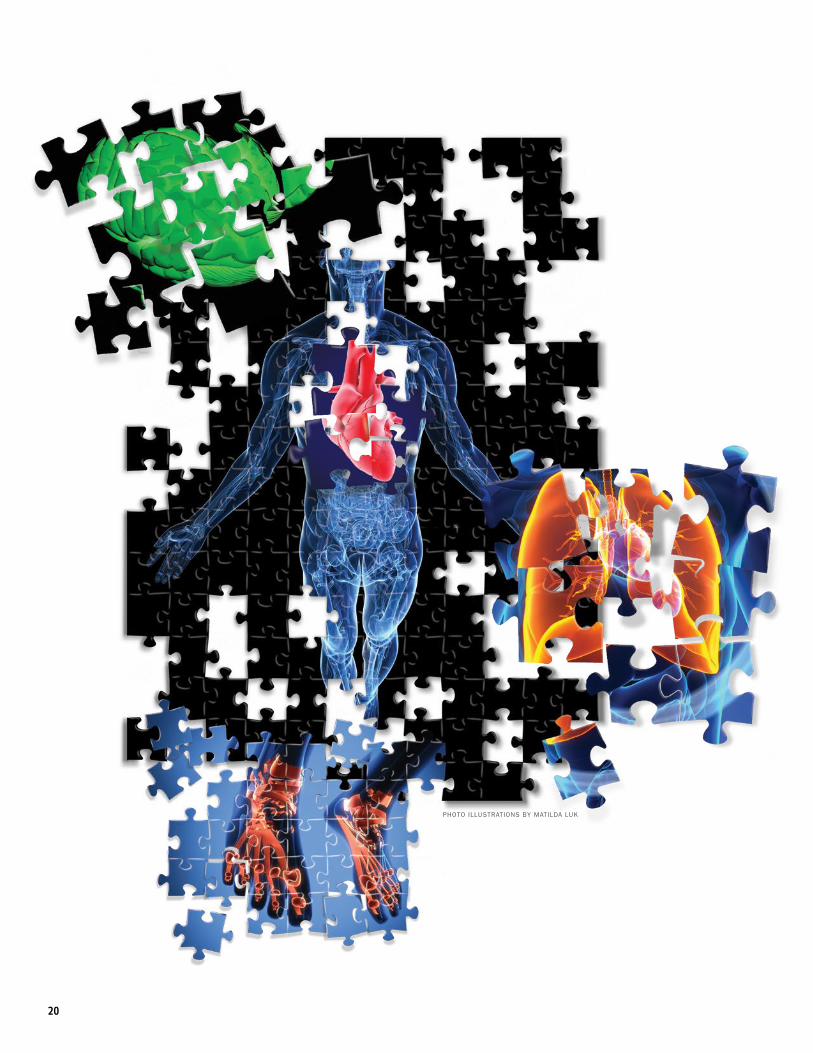
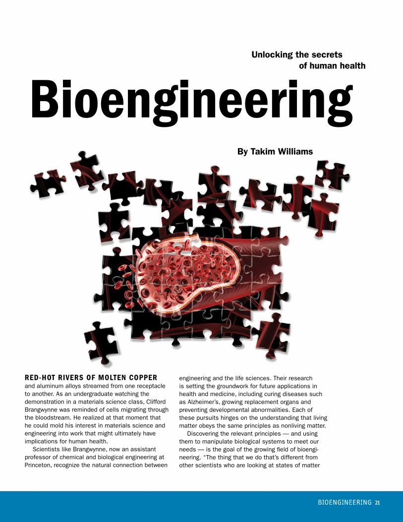
PHOTO ILLUSTRATIONS BY MATILDA LUK
20
BIOENGINEERING 21
By Takim Williams
RED-HOT RIVERS OF MOLTEN COPPER and aluminum alloys streamed from one receptacle to another. As an undergraduate watching the demonstration in a materials science class, Clifford Brangwynne was reminded of cells migrating through the bloodstream. He realized at that moment that he could mold his interest in materials science and engineering into work that might ultimately have implications for human health. Scientists like Brangwynne, now an assistant professor of chemical and biological engineering at Princeton, recognize the natural connection between
engineering and the life sciences. Their research is setting the groundwork for future applications in health and medicine, including curing diseases such as Alzheimer’s, growing replacement organs and preventing developmental abnormalities. Each of these pursuits hinges on the understanding that living matter obeys the same principles as nonliving matter. Discovering the relevant principles — and using them to manipulate biological systems to meet our needs — is the goal of the growing field of bioengi-neering. “The thing that we do that’s different from other scientists who are looking at states of matter
BioengineeringUnlocking the secrets of human health

“The living world involves flow almost by definition.”
–Howard Stone, the Donald R. Dixon ’69 and Elizabeth W.
Dixon Professor of Mechanical and Aerospace Engineering
22
and their properties is that we are doing it in the context of living cells,” Brangwynne said. “What is the state of matter inside of a cell and how does that enable biological function?” Brangwynne gestures toward a can of Gillette shaving foam next to a cylinder of silly putty on his desk and explains that the familiar grade school schema of three states of matter — solid, liquid, gas — is not entirely accurate. There are phases in between, and combinations with their own surprising properties. “A mound of foam is essentially a solid,” Brang- wynne said. “You can push on it and it deforms, and when you take your finger away it springs back into shape. You’ve taken something that is 95 percent gas — it’s mostly air — and 5 percent liquid, and you’ve combined those in such a way that you get a solid.” Correct biological function depends on transitions between these phases of matter. For example, your blood — typically a free-flowing liquid — clots to form a protective scab. However, these transitions can cause problems, for instance when an internal blood clot causes a stroke. Likewise, the liquid inside each cell — the cyto-plasm — regularly goes through local phase tran-sitions, some of which are disruptive. This issue is linked to neurodegenerative diseases, including Alzheimer’s disease and amyotrophic lateral sclero-sis (Lou Gehrig’s disease). In these cases, proteins aggregate and spontaneously transition from a liquid phase into a sticky, solid-like state, prohibiting normal function in the brain or nervous system. These phase
transitions are also thought to be involved in con-trolling cell size and growth, and thus diseases such as cancer. Brangwynne’s research focuses on gaining a better understanding of these living states of matter, and how they can be manipulated. Over the past few years, his group has published several studies exploring the molecules that control intracellular phase transitions and how the living matter within a cell affects gene regulation and cell size. In one particularly exciting study, graduate student Marina Feric discovered that liquid-phase droplets of RNA and protein are biophysically linked to cell size through the force of gravity. Brangwynne, who receives support from the National Science Foundation (NSF) and the National Institutes of Health (NIH), is opti-mistic that these fundamental studies will lead to medical applications. “We’re certainly hoping to use our findings to perturb these systems and keep cells in a healthier state,” he said.
IMAGE COURTESY OF CLIFFORD BRANGWYNNE LAB
Researchers in the Brangwynne lab use nucleoli from amphibian egg cells to study the role of gravity in determining the size of living cells.

Lung tissue extracted from a reptile embryo helps the Nelson lab study the effect of the fluid environment on lung development.
IMA
GE
CO
UR
TES
Y O
F CE
LES
TE N
ELS
ON
LAB
, E
XH
IBITE
D IN
AR
T OF
SC
IEN
CE
20
11
BIOENGINEERING 23
Swimming upstreamLike Brangwynne, Professor Howard Stone followed the flow of ideas from an area of engineering — fluid mechanics — to biology. “The living world involves flow almost by definition,” said Stone, who is the Donald R. Dixon ’69 and Elizabeth W. Dixon Profes-sor of Mechanical and Aerospace Engineering. “You circulate blood, you breathe streams of air in and out, you sweat to regulate temperature. If you study fluid mechanics, and if you’re somewhat open-minded, it’s easy to stumble across biological problems.” One of the biological problems that caught Stone’s attention is how bacteria move in a fluid. Moving through water is far more difficult for bacteria than it is for a human. For single-celled creatures a millionth of a meter long, the force of friction dominates their ability to swim in a given direction. Instead, bacteria are usually just carried along for the ride. The dominance of friction led to a discovery in Stone’s lab four years ago by visiting graduate student Yi Shen, who found that P. aeruginosa, a dangerous pathogen sometimes found in hospitals, can move against a current. The bacterium loses its flagellum — a long tail for swimming — when it adheres to a surface, which for many cells dictates the end of mobility. Yet P. aeruginosa can drag itself along a wall of, for example, a branched medical tube, by its small, tentacle-like pili, which are strong enough to resist the force of friction. After observing this phenomenon, Stone and his collaborators — Professor of Molecular Biology Zemer Gitai; Albert Siryaporn, an associate research scholar in molecular biology; and Minyoung Kevin Kim, a graduate student in chemistry — began researching this behavior in systems mimicking the human body. “Your blood vessels have branches. Your lungs are branched. We’ve made model branched systems and used a pump — like a heart — to drive fluid through it,” Stone said. “Bacteria inoculated into this flow sometimes end up in places you wouldn’t expect, and that, at this time, a simulation would never predict.” This basic research, which is supported by NSF, may allow us to better anticipate the movements of pathogens — in our bodies or our environments — in order to prevent infection and contamination.
Fluid environmentBacteria are single-celled organisms that can form colonies, but a more sophisticated arrangement occurs in our own bodies, where huge communities of cells organize into tissues and organs. Celeste Nelson, an associate professor of chemical and biological engineering, studies the fetal development of these organs, which depends on the fluid environ-ment in which they form. One of the organs that forms in a fluid environment is the lung. A fetus’s lungs are filled with fluid during
gestation. Nelson uses tissue cultures — parts of organs grown in laboratory dishes — and manipulates the speed and pressure of tiny streams of liquid that are directed onto the growing lung cells by small tubes. Nelson has discovered that the higher the pres-sure of this fluid in the fetal lung, the more quickly the lungs develop, whereas lower pressure leads to slower development. Several congenital disorders can derail lung development, and Nelson’s work — which is supported by NIH, NSF, the David and Lucile Packard Foundation, the Camille and Henry Dreyfus Foundation, the Burroughs Wellcome Fund, the Essig Enright Family Foundation, and Princeton’s Project X, which provides seed funding for unconventional research — may improve our ability to diagnose such problems early. The lungs, kidneys, mammary glands and other organs develop through a branched structure, which is an efficient space-filling strategy for functions that require maximum surface area. This exponential branching pattern is a highly reproducible self- assembly process, and in Nelson’s opinion, the forest of alveoli in the lungs is the most beautiful example. “The 23 generations of branches means several hundred million paths,” said Nelson. “Every one of those paths is needed for efficient diffusion of oxygen into the infant blood stream immediately after birth. “What’s amazing,” she said, “is that all of the branches in my lungs look exactly like the branches in your lungs.”
Nelson’s lab also studies a behavior in cancer cells called reversion, which — if it could be induced — would turn many cancers into benign, treatable illnesses. She collaborates with Derek Radisky, a researcher at the Mayo Clinic in Jacksonville, Florida. For Nelson, who started studying breast cancer while a postdoctoral fellow, the body’s organ systems have a mechanical elegance.
Timing is everythingStanislav Shvartsman is as fascinated by the chemical aspects of development as Nelson is by
24
the mechanical aspects. His research focuses on embryogenesis, the very early stages of fetal development. “When you want to bake a cake, it’s not enough to say that you need eggs and milk and flour,” said Shvartsman, a professor of chemical and biological engineering and the Lewis-Sigler Institute for Integra-tive Genomics. “Knowing the ingredients, and even knowing the sequence in which you add these ingre-dients — which is what we know from genetics — is not enough to bake a cake that tastes good.” When the recipe — the proper quantities of chemicals released by the cells of the embryo at the proper times — isn’t followed exactly, there are consequences for the developing organism. For example, a large class of developmental ab-normalities, known as RAS-opathies, is associated with asymmetry in the craniofacial complex, stunted height, congenital heart defects, developmental delays and other issues. Such defects are observed in one in every thousand births and are believed to be caused by mutations in genes of the Ras-MAPK pathway. Biologists know which genes are mutated, and even where to find these genes on our DNA. What they don’t know is why these particular mutations lead to a distinct set of clinical features. To find out, researchers turn to organisms that macroscopically look very different from us — such as bacteria and worms — but are very similar at the cellular level. Shvartsman’s research group, which is supported by NIH and NSF, uses the fruit fly to study embryogenesis. Initially, the handful of cells that make up an embryo are all identical. By the time of birth, that ho-mogenous handful will have given rise to brain, nerve, heart, blood and every other kind of cell required for a living, breathing organism. In order to differentiate into the right kind of cell at the right time, and to arrange into the correct three-dimensional shape, the embryonic cells have to communicate. They speak to each other through a language of chemical signals. The signals are actually protein molecules, Shvartsman said. A protein released by one cell at-taches to a receptor protein embedded in the surface of a neighboring cell. That surface protein reacts by changing shape, and in turn changing the internal environment of its cell. In this way cells “hear” each other. At any given time multiple cells are releasing various proteins, and the combination of signals floating through the embryonic environment tells a cell what to become, or when to divide to make more of itself. To crack the code, Shvartsman is looking at one signal at a time, beginning with a set of proteins that is well understood genetically thanks to the work
UNDERGRADUATE RESEARCH: BIOENGINEERING
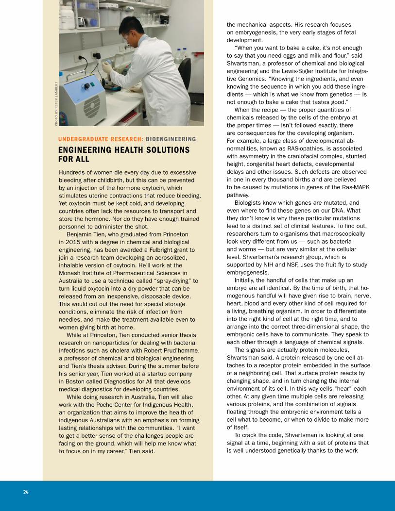
ENGINEERING HEALTH SOLUTIONS FOR ALLHundreds of women die every day due to excessive bleeding after childbirth, but this can be prevented by an injection of the hormone oxytocin, which stimulates uterine contractions that reduce bleeding. Yet oxytocin must be kept cold, and developing countries often lack the resources to transport and store the hormone. Nor do they have enough trained personnel to administer the shot. Benjamin Tien, who graduated from Princeton in 2015 with a degree in chemical and biological engineering, has been awarded a Fulbright grant to join a research team developing an aerosolized, inhalable version of oxytocin. He’ll work at the Monash Institute of Pharmaceutical Sciences in Australia to use a technique called “spray-drying” to turn liquid oxytocin into a dry powder that can be released from an inexpensive, disposable device. This would cut out the need for special storage conditions, eliminate the risk of infection from needles, and make the treatment available even to women giving birth at home. While at Princeton, Tien conducted senior thesis research on nanoparticles for dealing with bacterial infections such as cholera with Robert Prud’homme, a professor of chemical and biological engineering and Tien’s thesis adviser. During the summer before his senior year, Tien worked at a startup company in Boston called Diagnostics for All that develops medical diagnostics for developing countries. While doing research in Australia, Tien will also work with the Poche Center for Indigenous Health, an organization that aims to improve the health of indigenous Australians with an emphasis on forming lasting relationships with the communities. “I want to get a better sense of the challenges people are facing on the ground, which will help me know what to focus on in my career,” Tien said.
PH
OTO
BY
PE
TER
LA
MB
ER
T

BIOENGINEERING 25
of such Princeton biologists as Gertrud Schüpbach, the Henry Fairfield Osborn Professor of Biology. By controlling the amount of these proteins released in the fly embryo, Bomyi Lim, a former graduate student in Shvartsman’s lab and now a postdoctoral research associate at the Lewis-Sigler Institute for Integrative Genomics, has discovered the minimum dosage necessary for proper structural development. This is the first step in a long process, but it is a milestone, and Shvartsman is excited about continuing the process. “It’s very exciting to work in a field where there’s no risk of ever saying, ‘This is the end of the times-table. There is no more material to learn,’” he said.
Some assembly requiredBrangwynne views embryogenesis as the epitome of self-assembly, the process by which small, disorganized components interact based on simple rules to form complex structures without human intervention. A classic example is the snowflake, a delicate crystalline jewel formed in midair as water molecules freeze. Engineers have been trying to take advantage of self-assembly for some time — often in Brangwynne’s field of materials science — where time and money could be saved if certain synthetic materials would form on their own in a solution, rather than being pain- stakingly put together atom by atom. “The embryo of an organism like C. elegans essentially starts out as just a bag of molecules,” Brangwynne said. Once the egg is fertilized, it begins to organize, and the unstructured soup of molecules turns into a wriggling worm. “There’s absolutely nothing that human engineering can do that comes anywhere close to what I just described takes place in embryos all the time,” Brangwynne said. While Princeton scientists are importing methods and paradigms from engineering disciplines to bio- logy, they see a two-way street, recognizing that biology itself has methods to share. “We would like to learn how nature, through hundreds of millions of years of evolution, has
generated these systems that are just completely unbelievable in their level of sophistication,” Brangwynne said. “It’s as if we’ve been visited by an alien civilization that was millions of years more advanced than us. The first thing we would do would be to take a really close look at that spaceship. We’d try to figure out what it is made of, what are the prin-ciples that govern its flight and its control systems. That’s what we are doing with biological systems.”
The image shows thin slices of a part of fruit fly embryos where stem cells turn into mature eggs. Created by graduate students Yogesh Goyal and Bomyi Lim and postdoctoral researcher Miriam Osterfield in the laboratory of Stanislav Shvartsman, the image was selected for display in Princeton’s 2014 Art of Science competition.