biol315 special senses - dr. daniels websitejdaniels.huntingdon.edu/315/biol315 special...
TRANSCRIPT

1
The Special Senses
Chapter 14 in Open Stax
Chapter 17 in Martini
The Special Senses
• The five special senses are– Olfaction
– Gustation
Eq ilibri m– Equilibrium
– Hearing
– Vision
Olfaction Olfaction
• The sense of smell, or olfaction, is provided by a pair of olfactory organs
• These organs are located in the nasal cavity on either side of the nasal septum of the noseeither side of the nasal septum of the nose
• These organs consist of two layers; The olfactory epithelium and the lamina propria
The Ethmoid• The olfactory epithelium
contains the olfactory receptors, supporting cells, and basal cells
• The olfactory receptors are located in the epithelium that covers the inferior surfaces ofcovers the inferior surfaces of the cribriform plate, the medial surfaces of the superior nasal conchae, and the superior portion of the perpendicular plate
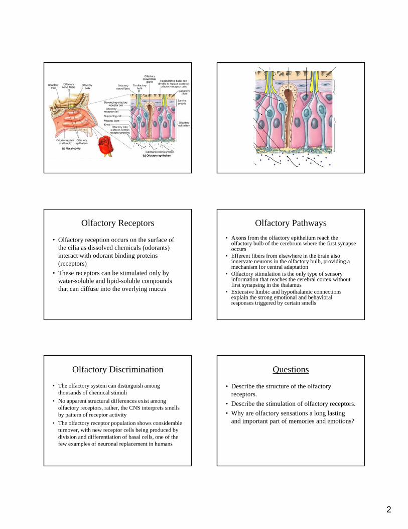
Olfactory Receptors
• The lamina propria contains blood vessels, nerves, and the olfactory, or Bowman’s glands
• Bowman’s glands form a thick, pigmented mucus
• The olfactory receptors are highly modified neuronsThe olfactory receptors are highly modified neurons
• The exposed tip of each receptor forms a knob that projects beyond the epithelial surface
• Cilia project from the knob and extend into the mucus
• The cilia lie parallel to the epithelial surface, exposing their surface to dissolved compounds
2
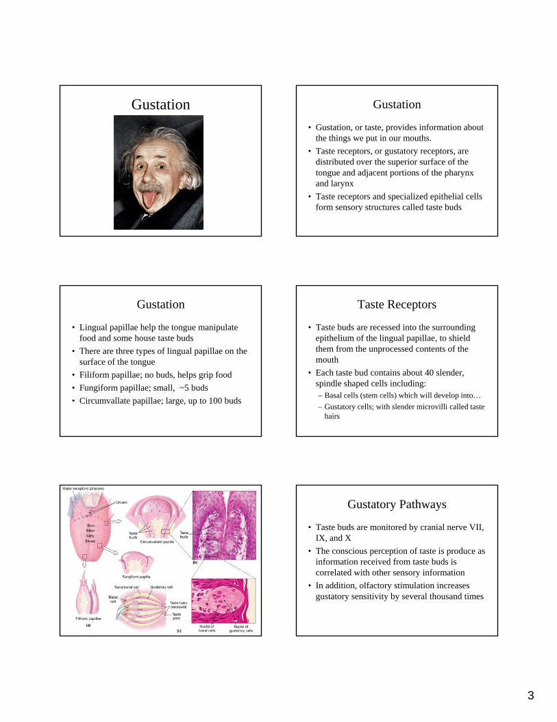
Olfactory Receptors
• Olfactory reception occurs on the surface of the cilia as dissolved chemicals (odorants) interact with odorant binding proteins (receptors)(receptors)
• These receptors can be stimulated only by water-soluble and lipid-soluble compounds that can diffuse into the overlying mucus
Olfactory Pathways
• Axons from the olfactory epithelium reach the olfactory bulb of the cerebrum where the first synapse occurs
• Efferent fibers from elsewhere in the brain also innervate neurons in the olfactory bulb, providing a y p gmechanism for central adaptation
• Olfactory stimulation is the only type of sensory information that reaches the cerebral cortex without first synapsing in the thalamus
• Extensive limbic and hypothalamic connections explain the strong emotional and behavioral responses triggered by certain smells
Olfactory Discrimination
• The olfactory system can distinguish among thousands of chemical stimuli
• No apparent structural differences exist among olfactory receptors, rather, the CNS interprets smells y p pby pattern of receptor activity
• The olfactory receptor population shows considerable turnover, with new receptor cells being produced by division and differentiation of basal cells, one of the few examples of neuronal replacement in humans
Questions
• Describe the structure of the olfactory receptors.
• Describe the stimulation of olfactory receptors.
h lf i l l i• Why are olfactory sensations a long lasting and important part of memories and emotions?
3
Gustation Gustation
• Gustation, or taste, provides information about the things we put in our mouths.
• Taste receptors, or gustatory receptors, are distributed over the superior surface of thedistributed over the superior surface of the tongue and adjacent portions of the pharynx and larynx
• Taste receptors and specialized epithelial cells form sensory structures called taste buds
Gustation
• Lingual papillae help the tongue manipulate food and some house taste buds
• There are three types of lingual papillae on the surface of the tonguesurface of the tongue
• Filiform papillae; no buds, helps grip food
• Fungiform papillae; small, ~5 buds
• Circumvallate papillae; large, up to 100 buds
Taste Receptors
• Taste buds are recessed into the surrounding epithelium of the lingual papillae, to shield them from the unprocessed contents of the mouthmouth
• Each taste bud contains about 40 slender, spindle shaped cells including:– Basal cells (stem cells) which will develop into…
– Gustatory cells; with slender microvilli called taste hairs
Gustatory Pathways
• Taste buds are monitored by cranial nerve VII, IX, and X
• The conscious perception of taste is produce as information received from taste buds isinformation received from taste buds is correlated with other sensory information
• In addition, olfactory stimulation increases gustatory sensitivity by several thousand times

4
Gustatory Discrimination
• The four primary taste sensations are sweet, salty, sour, and bitter
• At least two additional tastes have been discovered in humansdiscovered in humans – Umami; a pleasant, beef broth, chicken broth,
parmesan cheese taste, resulting from receptor sensitivity to AA’s, small peptides, and nucleotides
– Water; water receptors affect systems that deal with water balance
Mechanism of Gustatory Sensation• Dissolved chemicals contacting the taste hairs bind to
receptor proteins• Different tastes involve different receptor
mechanisms– Salt and sour are chemically gated ion channelsy g– Sweet, bitter, and umami, are G proteins (gustducins)
• The end result is neurotransmitter release which leads to an action potential on an afferent fiber
• Receptors adapt slowly, but central adaptation quickly reduces taste sensitivity
• Sensitivity to bitter and sour is much greater than to salty and sweet
Questions
• List and describe the three types of papillae on the human tongue
• Describe the structure of a taste bud
h h f i d d• What are the four primary and two secondary taste sensations?
Equilibrium and Hearing
Equilibrium and Hearing
• The special senses of equilibrium and hearing are provided by the inner ear
• Equilibrium sensations inform us of the position of the head in space by monitoring p p y ggravity, linear acceleration, and rotation
• Hearing enables us to detect and interpret sound waves
• In both cases, the receptors, called hair cells are mechanoreceptors
Anatomy of the Ear
• The ear is divided into three anatomical regions:– The external ear collects and directs sound toward
the middle earthe middle ear
– The middle ear collects and transmits sound waves to the inner ear
– The inner ear contains the sensory organs of equilibrium and hearing
5
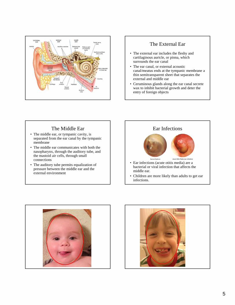
The External Ear
• The external ear includes the fleshy and cartilaginous auricle, or pinna, which surrounds the ear canal
• The ear canal, or external acoustic ,canal/meatus ends at the tympanic membrane a thin semitransparent sheet that separates the external and middle ear
• Ceruminous glands along the ear canal secrete wax to inhibit bacterial growth and deter the entry of foreign objects
The Middle Ear• The middle ear, or tympanic cavity, is
separated from the ear canal by the tympanic membrane
• The middle ear communicates with both the nasopharynx, through the auditory tube, andnasopharynx, through the auditory tube, and the mastoid air cells, through small connections
• The auditory tube permits equalization of pressure between the middle ear and the external environment
Ear Infections
• Ear infections (acute otitis media) are a bacterial or viral infection that affects the middle ear.
• Children are more likely than adults to get ear infections.

6
M2 M1 J
The Middle Ear
• The auditory ossicles are three tiny bones that transmit vibrations between the tympanic membrane and oval window of the inner ear
• The three auditory ossicles are the malleus• The three auditory ossicles are the malleus (hammer), the incus (anvil), and the stapes (stirrup)
The Inner Ear
• The sensory receptors of the inner ear lie within a collection of fluid filled tubes and chambers called the membranous labyrinth
• The labyrinth is filled with endolymph which has an electrolyte concentration similar to typical body y yp yfluids
• The bony labyrinth (temporal bone) surrounds and protects the membranous labyrinth
• Between the bony and membranous labyrinth is the fluid perilymph a liquid similar to CSF in its properties

7
The Inner Ear
• The bony labyrinth contains the vestibule, the three semicircular canals, and the cochlea
• The vestibule consists of a pair of membranous sacs the saccule and utriclesacs, the saccule and utricle
• the vestibule functions in static equilibrium, the semicircular canals in dynamic equilibrium and the cochlea provides the sense of hearing
Receptors of the Inner Ear
• The sensory receptors of the inner ear are called hair cells
• Each hair cell supports 80-100 long stereocilia, in the vestibule each hair cell also contains ain the vestibule each hair cell also contains a kinocilium, a single long cilium
• When external forces distort these processes it alters the rate at which the hair cell releases neurotransmitters
The Semicircular Ducts
• Sensory receptors in the semicircular ducts respond to rotational movement of the head (dynamic equillibrium)
• Each semicircular duct contains an ampulla, an expanded region containing the hair cells
• The hair cells form a raised structure called a crista and from this the stereo- and kinocilia of the hair cells are embedded in a gelatinous cupula
• When your head rotates, the movement of the endolymph deflects the cupula and stimulates the receptor processes
• Movement of the fluid in one direction causes excitation, movement in the opposite direction causes inhibition
• When movement stops the cupula rebounds to its normal position

8
The Utricle and Saccule
• The hair cells of the utricle and saccule are clustered in oval structures called maculae
• As in the ampullae the processes of the hair cells are embedded in a gelatinous massTh f f hi i d l k d• The surface of this mass contains densely packed calcium carbonate crystals called statoconia
• The complex as a whole is called an otolith• The weight of the statoconia on the macular surfaces
tells the body which way down is, likewise a shifting of the weight vector indicates linear acceleration
Questions
• List, in order, from outside to inside, the three auditory ossicles
• Describe the mechanisms of dynamic and static equilibriumstatic equilibrium
Hearing
• Receptors in the cochlear duct provide the sense of hearing
• As the auditory ossicles move in response to sound they initiate pressure fluctuations in the y pperilymph of the cochlea
• These waves in the perilymph stimulate hair cells along the cochlear spiral
• Nerve impulses from these hair cells are interpreted as sound

9
The Cochlea and Organ of Corti
• The cochlea is divided into three chambers– The scala vestibuli, or vestibular duct – The scala media, or cochlear duct– The scala tympani, or tympanic duct
• The scala vestibuli and scala tympani areThe scala vestibuli and scala tympani are interconnected at the tip of the cochlear spiral
• The organ of Corti, sits on the basilar membrane, which separates the cochlear duct from the tympanic duct
• The stereocilia of the hair cells in the organ of Corti extend upward toward the overlying tectorial membrane
The Cochlea and Organ of Corti
• As waves in the perilymph travel through the cochlear spiral they cause the basilar membrane to move up and down
• This movement stimulates the hair cells as• This movement stimulates the hair cells as their stereocilia deflect when they press against the tectorial membrane
The Hearing Process
• Outlined in Martini text on pages 585-586 good diagram on page 585
• Outlined in OpenStax on pages 569-570
Externalacousticmeatus
Malleus Incus
Movementof sound
waves
Stapes Oval window
Cochlear branchof cranial nerve VIII
Scala vestibuli(contains perilymph)
Vestibular membrane
Cochlear duct(contains endolymph)
Basilar membrane
Scala tympani(contains perilymph)
Tympanicmembrane
Roundwindow
(contains perilymph)
Sound wavesarrive attympanicmembrane.
Movement ofthe tympanicmembrane causesdisplacementof the auditoryossicles.
Movement ofthe stapes atthe oval windowestablishespressurewaves in theperilymphof the scalavestibuli.
The pressurewaves distortthe basilarmembrane ontheir way to theround windowof the scalatympani.
Vibration of the basilarmembranecauses vibrationof hair cellsagainst thetectorialmembrane.
Informationabout the regionand the intensityof stimulation isrelayed to theCNS over thecochlear branchof cranial nerveVIII.

10
The Hearing Process
• The location of the distortion of the basilar membrane varies with the frequency of the sound
• High frequency sounds have a short wavelength and vibrate the basilar membrane near the oval window
• Low frequency sounds have a longer wavelength andLow frequency sounds have a longer wavelength and vibrate the basilar membrane further from the oval window
• The intensity of the sound is directly proportion to the magnitude of movement by the stapes, this in turn dictates the degree of displacement by the basilar membrane, the louder the sound, the more the basilar membrane moves
Deafness
• Conductive, or conduction deafness results from conditions in the outer or middle ear that block the normal transfer of vibrations from the tympanic membrane to the oval window
• Nerve deafness results from problems with the cochlea or elsewhere along the auditory pathway
• Vibrations are transferred normally but either the receptors cannot respond or their responses do not reach their destinations
Questions
• What six basic steps are involved in the process of hearing?
• Describe the structure of the Cochlea and its function in hearingfunction in hearing
Vision
Accessory Structures of the Eye Accessory Structures of the Eye
• Eyelids function as windshield wipers for the eyes, keeping the surface lubricated and free of dust
• Eyelashes (palpebrae) are thick hairs that help prevent foreign objects from reaching the eyeT l l d difi d b l d h• Tarsal glands, are modified sebaceous glands that secrete a substance that helps keep the eyelids from sticking together
• The lacrimal caruncle contains glands that produce ‘sleep’ the gritty deposits that occur after a good night’s sleep

11
Accessory Structures of the Eye
• The conjunctiva is the epithelium covering the surface of the eye
• The palpebral conjunctiva covers the inner surface of the eyelidsy
• The ocular conjunctiva covers the anterior surface of the eye
• The ocular conjunctiva extends to the edge of the cornea, a transparent part of the outer layer of the eye
Lacrimal Apparatus
• The lacrimal apparatus produces, distributes and removes tears
• Tears form in the lacrimal glands, wash across the eye and collect in the lacrimal ylake
• The lacrimal punctae drain the lacrimal lake, through lacrimal canaliculi, to the lacrimal sac, and the nasolacrimal duct which empties into the nasal cavity
The eye
• The wall of the eye consists of three layers– Outer fibrous tunic consists of the sclera, and cornea
– Middle vascular tunic consists of the iris, ciliary body, and choroid
– Inner neural tunic consists of the Retina
• The eye is hollow and is divided into two cavities– The posterior cavity containing the vitreous body
– The anterior cavity (with two chambers) containing the aqueous humor
The Fibrous Tunic
• The fibrous tunic provides– Mechanical support and protection– Attachment for the extrinsic eye muscles– Assists focusing processg p
• The sclera, or ‘white of the eye, consists of dense connective tissue, and contains small blood vessels
• The transparent cornea is structurally continuous with the sclera and contains no blood vessels

12
The Vascular Tunic
• The vascular tunic, or uvea, contains numerous blood vessels, lymphatic vessels, and the intrinsic eye muscles
• Functions include:• Functions include:– Providing a route for blood and lymphatic vessels
– Regulating the amount of light entering the eye
– Secreting and reabsorbing the aqueous humor
– Controlling the shape of the lens
The Vascular Tunic
• The iris, contains blood vessels, pigment cells, and two layers of smooth muscle fibers
• When these muscles contract they change the diameter of the pupil, the central opening of the irisTh ill i l f i f• The pupillary constrictor muscles form a series of concentric circles around the pupil
• The pupillary dilator muscles extend radially away from the edge of the pupil
• Both muscle groups are controlled by the autonomic nervous system
The Ciliary body
• At its periphery the iris attaches to the ciliary body
• The bulk of the ciliary body consists of the ciliary muscle that projects into the interior of the eye
• The epithelium of this muscle forms folds calledThe epithelium of this muscle forms folds called ciliary processes
• The suspensory ligaments of the lens attach to the tips of these processes
• Essentially, the ciliary body helps hold the lens behind the iris and centered on the pupil
The Choroid
• The choroid is a vascular layer that separates the fibrous and neural layers
• The choroid lies between the sclera and the retina and contains an extensive capillaryretina and contains an extensive capillary network that delivers oxygen and nutrients to the retina

13
Questions
• Describe the two types of conjunctiva in the eye
• Describe the lacrimal apparatus
• Describe the muscles that change the diameter• Describe the muscles that change the diameter of the pupil
The Neural Tunic
• The neural tunic, or retina, is the innermost layer of the eye
• It consists of a thin outer pigmented part and a thicker inner layer the neural partthicker inner layer the neural part
• The pigmented part absorbs light as it passes through the retina preventing light from bouncing back through the neural part and stimulating it a second time
Organization of the Retina
• The retina contains several layers of cells• The layer closest to the pigmented part contains the
photoreceptors, cells that detect light• There are two types of photoreceptors; Rods and
ConesCones• Rods do not discriminate between colors of light,
they are highly sensitive and allow us to see in low light
• Cones provide color vision, there are three types of cones, cones give clear sharp vision, but require more light than rods
Organization of the Retina
• Rods are more numerous and densely distributed in a broad band around the periphery of the retina
• Cones are more densely distributed on the retina’s posterior surfacep
• Most cones are concentrated in the area where a visual image arrives after passing through the lens
• This area, the macula lutea, contains no rods• The highest density of cones occurs at the fovea
centralis, the site of sharpest vision

14
Organization of the Retina
• Rods and cones synapse with bipolar cells, which in turn synapse within the layer of neurons called ganglion cells
• A network of horizontal cells extends horizontally at the level of the synapses between the photoreceptors and the bipolar cells
• A comparable layer of amacrine cells occurs at the level of the synapse between the bipolar and ganglion cells
Organization of the Retina
• Horizontal and amacrine cells facilitate or inhibit communication between the photoreceptors and ganglion cells, thereby altering the sensitivity of the retinaaltering the sensitivity of the retina
• Important in adjusting the eye to dim or bright light
The Optic Disc
• The axons of the approximately 1 million ganglion cells exit the eye at the optic disc, just medial of the fovea
• The optic disc is the origin of the optic nerve• The optic disc is the origin of the optic nerve
• The central retinal artery and central retinal vein also exit the eye at the optic disc
• The optic disc has no photoreceptors and is referred to as the blind spot

15
Chambers of the Eye
• The anterior cavity of the eye is divided into the anterior chamber, which extends from the cornea to the iris, and the posterior chamber, between the iris and the ciliary body and lens
• Both chambers are filled with the aqueous humorq• The aqueous humor circulates in the anterior cavity
and provides nutrient and waste transport and contributes to the shape of the eye
• The pressure within the eye, or intraocular pressure, can be measured by bouncing a blast of air off the surface of the eye and measuring the resulting deflection pressure
Chambers of the Eye
• The vitreous humor, or vitreous body, lies within the posterior cavity of the eye.
• It is a gelatinous mass that helps stabilize the shape of the eye and gives physical support to p y g p y ppthe retina
• Its gelatinous consistency arises from collagen fibers and proteoglycans
• The vitreous humor is formed during development and is not replaced
The Lens• The lens lies posterior to the cornea and is held in
place by the suspensory ligaments• The primary function of the lens is to focus the visual
image on the photoreceptors, this is accomplished as the lens changes shape
• The lens consists of concentric layers of cells that are yprecisely organized
• The cells of the interior of the lens are called lens fibers, and have lost their nuclei and organelles.
• Lens fibers are slender and elongate and filled with transparent proteins called crystallins, which are responsible for both the clarity and focusing power of the lens
Anatomy of Rods and Cones
• The outer segment of a photoreceptor contains hundreds to thousands of flattened membranous discs
• The term rod or cone refers to the shape of the pouter segment
• A narrow stalk connects the outer segment to the inner segment, a region containing cellular organelles and forming the synapse where neurotransmitters are released
16
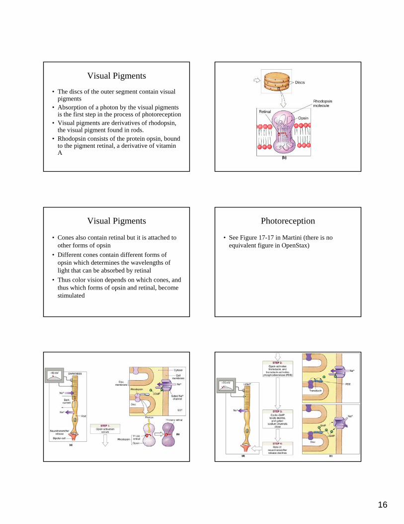
Visual Pigments
• The discs of the outer segment contain visual pigments
• Absorption of a photon by the visual pigments is the first step in the process of photoreceptionp p p p
• Visual pigments are derivatives of rhodopsin, the visual pigment found in rods.
• Rhodopsin consists of the protein opsin, bound to the pigment retinal, a derivative of vitamin A
Visual Pigments
• Cones also contain retinal but it is attached to other forms of opsin
• Different cones contain different forms of opsin which determines the wavelengths ofopsin which determines the wavelengths of light that can be absorbed by retinal
• Thus color vision depends on which cones, and thus which forms of opsin and retinal, become stimulated
Photoreception
• See Figure 17-17 in Martini (there is no equivalent figure in OpenStax)

17
Recovery From Stimulation
• Retinal does not spontaneously convert back to the 11-cis form.
• Instead the entire rhodopsin molecule must be broken down and reassembledBl hi i h b hi h h d i b k• Bleaching is the process by which rhodopsin breaks down into retinal and opsin
• Retinal is then converted back to the 11-cis form and reassembly proceeds using ATP
• This process takes time and contributes to lingering visual impressions or afterimages
Color Vision
• Color discrimination results from the integration of information from the three types of cones
• We have red, green, and blue cones each with , g ,a sensitivity to different wavelengths of light
• Normal individuals have 16% blue, 74% red, and 10% green cones
• If all three cones are stimulated we perceive the color as white
Light and Dark Adaptation
• The sensitivity of the visual system varies with the intensity of illumination
• The dark-adapted state occurs after about 30 minutes in the dark when almost all visual pigments will be sensitive to stimulation
• The light-adapted state occurs when sensitivity decreases as bleaching occurs
• From light to dark adaptation is about a 25K fold change in sensitivity
The Field of Vision
• Each eye receives a slightly different visual image, because the fovea are 5-7 cm apart, and the nose and eye socket block the view of the opposite sideopposite side
• Depth is perceived by comparing the relative positions of objects received by the left and right eyes
Questions
• Describe the anatomy of a photoreceptor
• Describe the process of photoreception and recovery from stimulation
ib h f l i i• Describe the nature of color vision