biological chemistry i: biochemical transformations iii · chemistry 5.07sc biological chemistry i...
TRANSCRIPT
1
Chemistry 5.07SC Biological Chemistry I
Fall Semester, 2013
Lectures 11 and 12. Biochemical Transformations III. Redox Cofactors: Oxidation and
Reduction and a quantitative description of spontaneity.
Energy released from the oxidation of standard building blocks (sugars, fatty acids, amino acids)
is harnessed in chemical bonds via NADH and used to make ATP. Oxidation and reduction in
biological systems is carried out using two organic cofactors nicotinamide adenine dinucleotide
(NAD) and flavin adenine dinucleotides (FAD, FMN or on occasion riboflavin) and inorganic
cofactors for electron transfer (ET). These cofactors play a central role in primary metabolism
specifically the respiratory chain and the mechanism by which a proton gradient is generated and
coupled for ATP production.
Outline:
I. Examine the chemistry of the two organic cofactors NAD and FAD involved in redox chem.
II. To introduce the inorganic cofactors involved in redox chemistry with focus on respiration.
III. To provide a quantitative description for spontaneity with redox reactions.
I. There are two major organic redox cofactors:
A. NAD+
B. FAD (FMN)
A. NAD+ /NADH (NADP+/NADPH)
1. Structure
The vitamin is nicotinic acid or nicotinamide (Niacin) and is a precursor to the actual cofactor.
Deficiency in the vitamin is associated with the disease pellagra.

2
Figure 1. Structure of NAD(P)+. NAD(P)+ is composed of an AMP phosphodiester and a nicotinamide ribose 5’-phosphate. Note the only difference between NAD+ and NADP+ is a phosphate attached to the hydroxyl at C2’ (green X). The business end of the molecule is the nicotinamide moiety. The oxidized form has a pyridinium ring and the reduced form has a 1,4-dihydropyridine ring.
Both of these forms have identical redox properties and chemical mechanisms. We will focus
ONLY on the redox reactivity of this cofactor. The NADH/NAD+ and NADP+/NADPH ratios
are of primary importance: the former in energy metabolism and the latter in reductive
biosynthesis. Metabolic enzymes can in general readily distinguish between these two
forms.
2. Scope of reactions
a. NADH is of primary importance in energy metabolism (catabolic processes). It supplies
reducing equivalents to reduce O2 to H2O in the respiratory chain and the energy released
generates a proton motif force used to make ATP. NADPH on the other hand is used
predominantly in biosynthesis (anabolic processes) and maintaining the redox status inside the
cell.
b. reactions in primary metabolism:
3
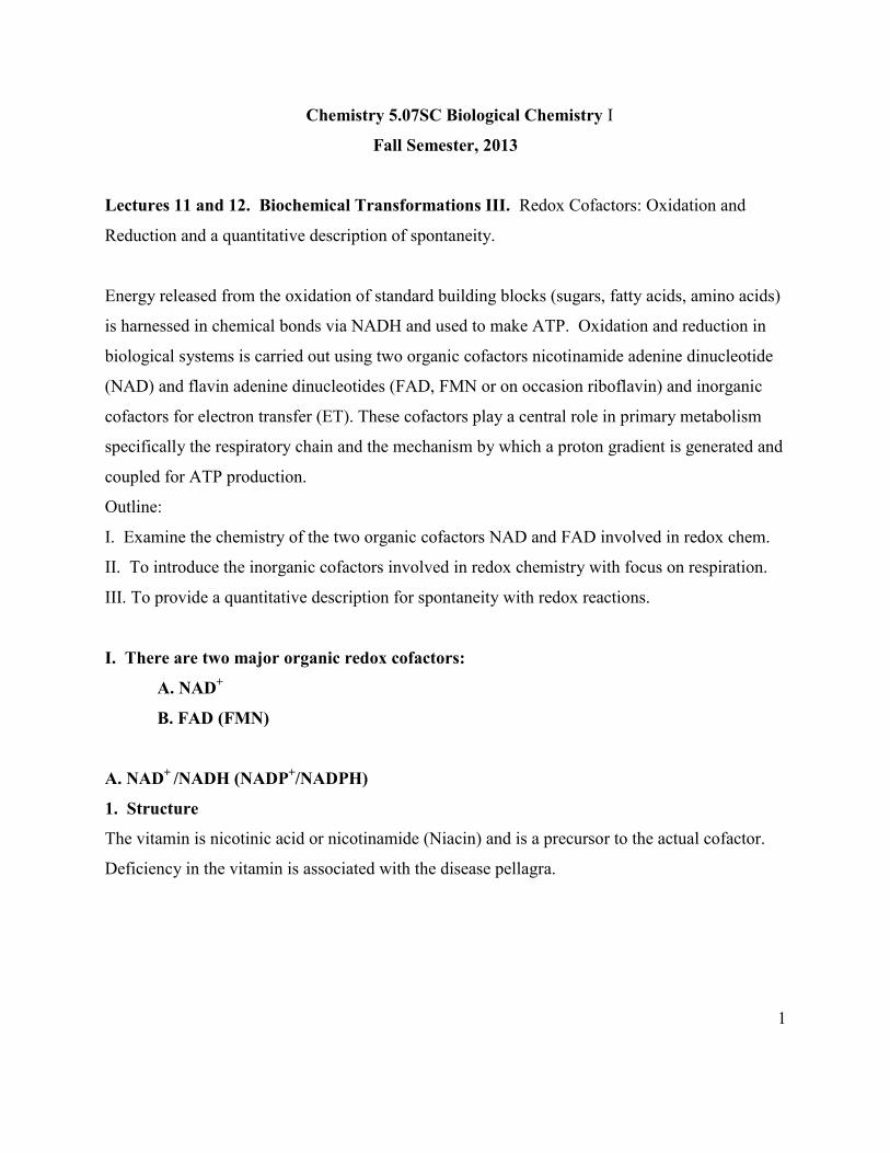
i. Reduction of a ketone to an alcohol with examples in the glycolysis pathway and
Krebs cycle – lactate dehydrogenase (LDH), malate dehydrogenase (MDH), alcohol
dehydrogenase (ADH). The reactions are reversible and pH dependent.
ii. Oxidation of an amine to an imine that hydrolyzes to a ketone and ammonia. An
example is glutamate dehydrogenase (GDH) that is a major enzyme involved in nitrogen
metabolism.
iii. Oxidation of an aldehyde to an acid. An example is glyceraldehyde 3-phosphate
(GAP) dehydrogenase (GAPDH) that you will see in the glycolysis pathway.
iv. Oxidation of saturated fatty acid thioesters to unsaturated fatty acid thioesters. Note
the oxidation requires and an activated hydrogen on the carbon (Cα) to the thioester.
This reaction plays a major role in fatty acid biosynthesis and degradation.
-CH2CH2-COSR to –CH=CH-COSR
v. Redox chemistry with the second organic redox cofactor FAD. Example: respiratory
chain.
3. Mechanism of the reactions shown above. The mechanism of this cofactor for ALL
reactions involves a hydride (H-) and proton (H+) transfer. An example of a typical reaction is
the reduction of an aldehyde to alcohol. This is one of the endings in the glycolysis pathway in
yeast, the mainstay of the beer and wine industry (fermentation, metabolism without O2). The
alcohol dehydrogenase (ADH) reaction is reversible and the proton makes the overall reaction
4
pH dependent. Thus by changing the concentration of one of the substrates [H+], the equilibrium
of the reaction can be shifted. NAD+/NADH are often used in coupled assays to measure
enzyme activity as NADH has absorption at 340 nm with a large extinction coefficient (ε = 6200
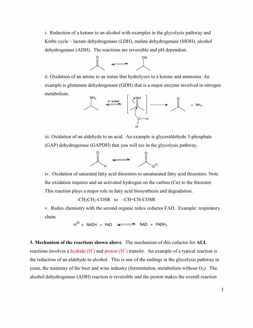
M-1cm-1), while NAD+ has no absorption at this wavelength. Note that in the ADH reaction that
in addition to the redox cofactor there is a Zn2+, which we previously encountered in the aldolase
reaction. It acts as a Lewis acid to polarize the carbonyl for nucleophilic attack. In this case the
Nu is the hydride (H-).
NOTE: H- does not attack the O of the carbonyl, it attacks the C of the carbonyl. Think
about Lecture 9 on carbonyl chemistry. In general, H- and H+ transfer occur in different
steps of the reaction as shown above.
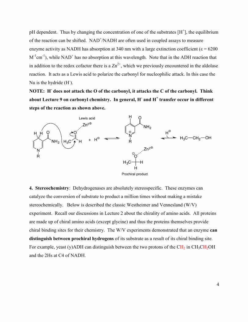
4. Stereochemistry: Dehydrogenases are absolutely stereospecific. These enzymes can
catalyze the conversion of substrate to product a million times without making a mistake
stereochemically. Below is described the classic Westheimer and Vennesland (W/V)
experiment. Recall our discussions in Lecture 2 about the chirality of amino acids. All proteins
are made up of chiral amino acids (except glycine) and thus the proteins themselves provide
chiral binding sites for their chemistry. The W/V experiments demonstrated that an enzyme can
distinguish between prochiral hydrogens of its substrate as a result of its chiral binding site.
For example, yeast (y)ADH can distinguish between the two protons of the CH2 in CH3CH2OH
and the 2Hs at C4 of NADH.

5
Digression on prochirality: We will NOT discuss the W/V experiment in Lecture. But as
chemists I hope you will all think about the beauty of this experiment and the wonder of protein
catalysts. The concept of prochirality is critical to understanding the TCA cycle since citrate has
prochiral arms (this is covered in later lectures in more depth). Understanding prochirality was
vital to determining the steps of the Kreb’s cycle.
Westheimer and Vennesland (W/V) Experiment
Both of the yeast (y) ADH enzyme substrates, ethanol and NADH at C-4 position, have prochiral
H’s.
W/V hypothesized that (y)ADH can distinguish between the two prochiral hydrogens. Their
method to demonstrate this involved using a stable isotope of H with an additional neutron
known as Deuterium (D). In their model, only one of the Ds would be transferred (DR is shown
below), leaving a chirally labeled NADH. DR has been stereospecifically transferred to C4.
To test their model, the [DR]-labeled NADH was incubated with acetylaldehyde and the fate of
DR was examined.
6
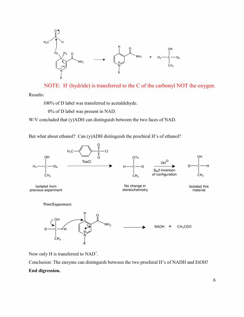
NOTE: H- (hydride) is transferred to the C of the carbonyl NOT the oxygen.
Results:
100% of D label was transferred to acetaldehyde.
0% of D label was present in NAD.
W/V concluded that (y)ADH can distinguish between the two faces of NAD.
But what about ethanol? Can (y)ADH distinguish the prochiral H’s of ethanol?
Now only H is transferred to NAD+.
Conclusion: The enzyme can distinguish between the two prochiral H’s of NADH and EtOH!
End digression.

7
5. Redox properties of NAD+
NAD+ + 2e- + H+ → NADH Eº’ = -0.32 v
NAD+ + 1e- → NAD• Eº’ = -0.79 v
NAD+ only undergoes two electron reductions inside the cell, as the one electron reduction
process is too far up hill (thermodynamically unfavorable).
a. The reduction potential of NAD+ is NOT modulated by the protein environment in
contrast with most redox active cofactors and is -0.32 v.
b. NAD+/NADH act as substrates with enzymes, that is, they bind and dissociate after each
turnover. This behavior contrasts with the FAD cofactors that are always tightly or covalently
bound to the enzyme.
c. NAD+ is O2 stable in contrast with most redox cofactors (FAD and the metal-based cofactors
that use iron and copper). Note O2 has two unpaired electrons that reside in its antibonding
orbitals. This property makes O2 unreactive with sugars, amino acids etc as their electrons are all
paired. However, both flavins and metals can carry out chemistry one electron at a time. Thus
they can react with O2 ; the chemistry is energetically favorable.
d. NAD+/NADH have a conserved binding domain called a Rossmann fold (see below and
Lecture 3) and is easily recognized by conservation of specific residues many of them glycines.
The cofactor is almost always bound in an extended conformation. Note the Rossmann fold
contains two “nucleotide” binding motifs (); one for nicotinamide ribose phosphate and
one for the AMP form of the NAD+ (see Figure 2).

8
A.
B.
Figure by O'Reilly Science Art for MIT OpenCourseWare.
B. PDB: 4P8R. Seattle Structural Genomics Center for Infectious Disease, Abendroth, J., Lorimer, D., Edwards, T.E. Structure of a glycosomal glyceraldehyde 3-phosphate dehydrogenase from Trypanosoma brucei. Figure 2. Rossmann fold has two nucleotide binding sites – repeats. A. 3D reconstruction of the Rossmann fold in glyceraldehyde-3-phosphate dehydrogenase. B. Plumbing diagram of the Rossmann fold. B. The second organic redox cofactor is FAD (FMN). See the Lexicon also. The chemistry
of this cofactor is complex and much less intuitive than NAD+. FAD and FMN are the common
forms used in cells. Riboflavin (RF) is used much less frequently.

9
Figure 3. Three forms of flavin (FMN, FAD, riboflavin).
1. This cofactor is the major mediator between 2 e- donors and 1 e- acceptors. In the
following figure you can see the available redox states. They all have different visible spectra
and can thus be identified by their unique features. The oxidized flavin (FAD or FMN) is planar
and bright yellow, while the reduced flavin (FADH2 or FMNH2) is butterfly shaped (non-planar
and is colorless). The two one e- reduced states are also colored with the color depending on
their protonation state.
10
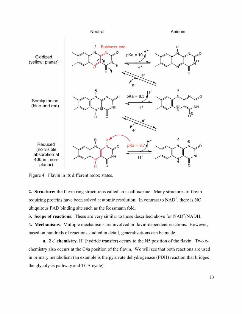
Figure 4. Flavin in its different redox states.
2. Structure: the flavin ring structure is called an isoalloxazine. Many structures of flavin
requiring proteins have been solved at atomic resolution. In contrast to NAD+, there is NO
ubiquitous FAD binding site such as the Rossmann fold.
3. Scope of reactions: These are very similar to those described above for NAD+/NADH.
4. Mechanisms: Multiple mechanisms are involved in flavin-dependent reactions. However,
based on hundreds of reactions studied in detail, generalizations can be made.
a. 2 e- chemistry. H- (hydride transfer) occurs to the N5 position of the flavin. Two e-
chemistry also occurs at the C4a position of the flavin. We will see that both reactions are used
in primary metabolism (an example is the pyruvate dehydrogenase (PDH) reaction that bridges
the glycolysis pathway and TCA cycle).
11
b. 1 e- chemistry. Chemistry also occurs 1 e- at a time. This chemistry is possible with
flavins because the semiquinone radical forms (the anionic red form and the neutral blue form)
are stable due to extensive electron delocalization over the isoalloxazine ring. One can draw
many resonance structures of the planar semiquinone radicals shown above.
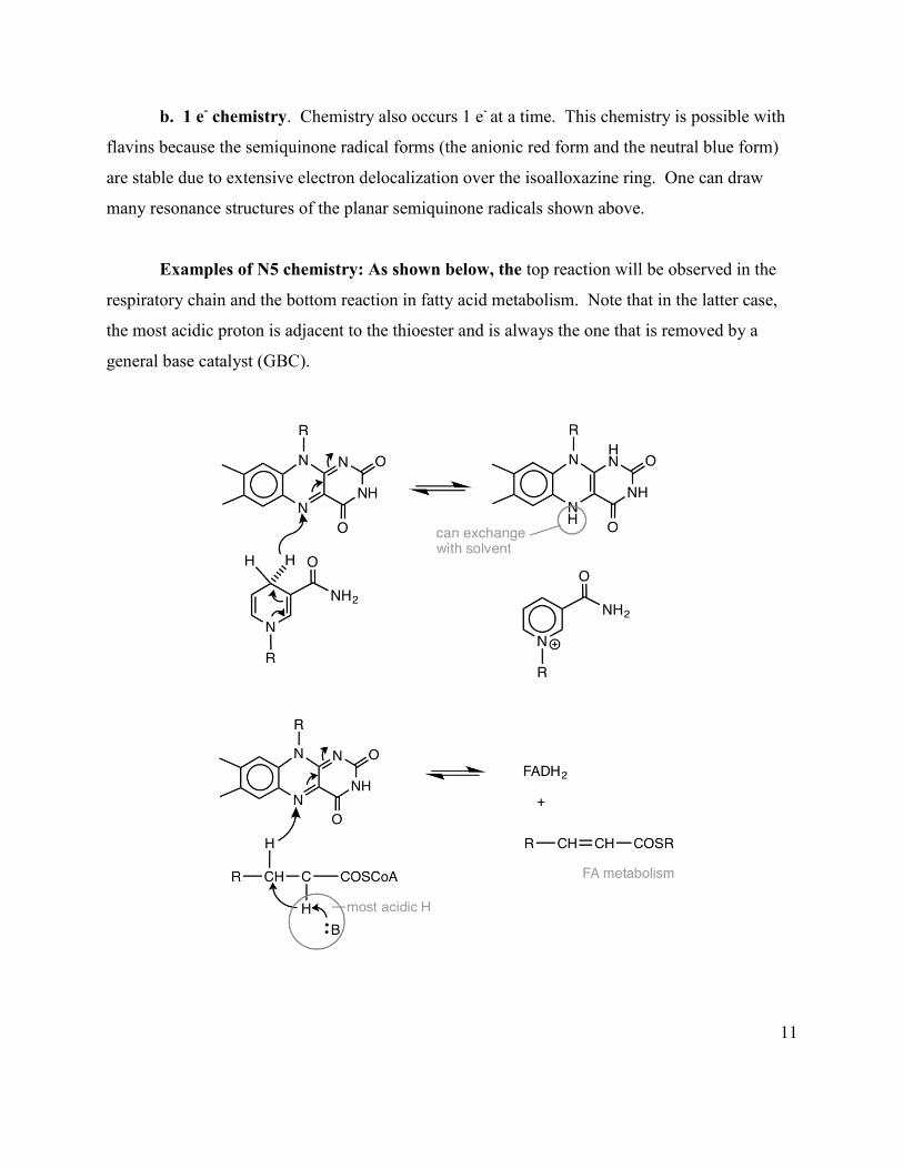
Examples of N5 chemistry: As shown below, the top reaction will be observed in the
respiratory chain and the bottom reaction in fatty acid metabolism. Note that in the latter case,
the most acidic proton is adjacent to the thioester and is always the one that is removed by a
general base catalyst (GBC).

12
Example of C4a chemistry from the pyruvate dehydrogenase (PDH) macromolecular
machine.
II. Inorganic redox-based cofactors. Metal clusters play an important role in catalysis and a
central role in respiration, photosynthesis, nitrogen fixation, and many enzymatic reactions.
Thirty to 35 % of all proteins use redox-inactive (structural role) or redox-active metallo-
cofactors (catalytic role). Recall the Zn2+ that we examined with the class II aldolase and ADH
dehydrogenase reaction. Zinc is d10 and is redox inert; the Zn2+ functions as a Lewis acid.
However, harnessing the energy of NADH oxidation via the respiratory chain, requires redox-
active cofactors that are predominantly iron and copper.
1. Structure: The following cofactors are widely used in biology: iron sulfur cluster (FeS)
clusters and hemes.

13
Iron Sulfur Clusters (the indicated charge [ ]n+ is based on the oxidation state of the Fe either
+2 or +3 and the sulfides (S-2)). These clusters undergo one electron reactions.
[Fe] 3+/2+ [2Fe2S]2+/1+ cluster [4Fe4S] 2+/1+cluster
Hemes Recall that we have previously seen hemes when discussing Hb and Mb. Hemes in Hb
and Mb function as reversible binders of O2. Hemes in the respiratory chain are involved in
electron transfer with other metallo-proteins and their iron changes redox state between Fe2+ and
Fe3+.
Figure 5. Heme or protoporphyrin IX in Hb and Mb. The equilateral ligands are “N” from the pyroles; the axial ligands vary depending on the protein. Example: An example of long range electron transfer is shown below based on a recent
structure and much biochemical analysis, Nature 2010 465, 441-5. Complex I
(NADH:ubiquinone oxidoreductase) is one the largest protein assemblies known and plays a

14
central role in energy production by the mitochondrial respiratory chain. Many mutations in
complex I subunits are associated with human neurodegenerative diseases.
C.
Courtesy of Macmillan Publishers, Ltd. © Nature. Source: Figure 3ab, 4 from Efremov, Rouslan G., Rozbeh Baradaran, and Leonid A. Sazanov. "The architecture of respiratory complex I." Nature 465, no. 7297 (2010): 441-445. Figure 6. A. and B. Side views, in the membrane plane of the entire complex I from Thermus
thermophilus. FeS clusters are shown as red and yellow spheres. C. NADH and FMN (magenta) donates two electrons by electron transfer (blue line for pathway), one at a time, to the chain of Fe-S clusters (red and yellow spheres) and finally passed from the N2 cluster to the quinone (dark blue). Electron transfer is coupled to conformational changes in the four helix bundle domain (green helices) and helix (red). These changes are transmitted to HL (magenta) that allows transport of three protons. The fourth proton is transported at the interface of the two domains. The hydrophilic domain surface is shown in grey, whereas the membrane domain surface is colored as in A. and B.
A. B.
15
This complex is a large L-shaped membrane bound enzyme. The reaction involves transfer of
electrons over 90 Å from NADH that binds to the hydrophilic domain, to ubiquinone in or near
the hydrophobic membrane bound domain (purple, Q).
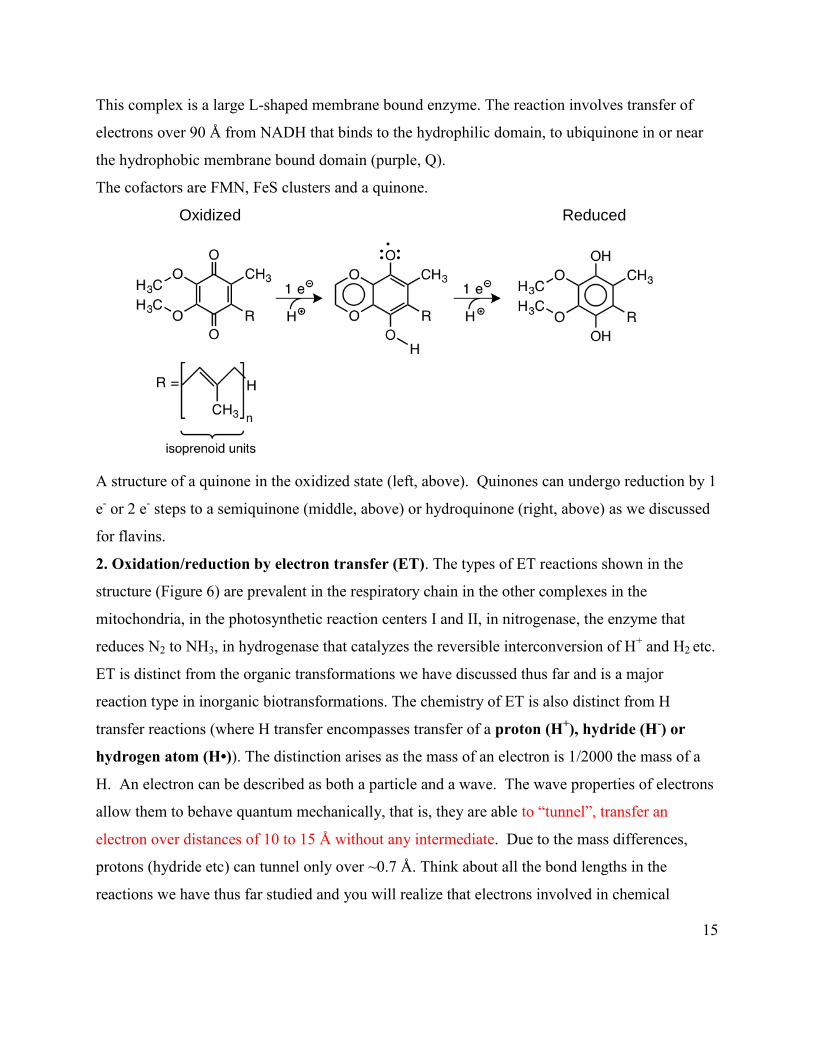
The cofactors are FMN, FeS clusters and a quinone.
Oxidized Reduced
A structure of a quinone in the oxidized state (left, above). Quinones can undergo reduction by 1
e- or 2 e- steps to a semiquinone (middle, above) or hydroquinone (right, above) as we discussed
for flavins.
2. Oxidation/reduction by electron transfer (ET). The types of ET reactions shown in the
structure (Figure 6) are prevalent in the respiratory chain in the other complexes in the
mitochondria, in the photosynthetic reaction centers I and II, in nitrogenase, the enzyme that
reduces N2 to NH3, in hydrogenase that catalyzes the reversible interconversion of H+ and H2 etc.
ET is distinct from the organic transformations we have discussed thus far and is a major
reaction type in inorganic biotransformations. The chemistry of ET is also distinct from H
transfer reactions (where H transfer encompasses transfer of a proton (H+), hydride (H-) or
hydrogen atom (H•)). The distinction arises as the mass of an electron is 1/2000 the mass of a
H. An electron can be described as both a particle and a wave. The wave properties of electrons
allow them to behave quantum mechanically, that is, they are able to “tunnel”, transfer an
electron over distances of 10 to 15 Å without any intermediate. Due to the mass differences,
protons (hydride etc) can tunnel only over ~0.7 Å. Think about all the bond lengths in the
reactions we have thus far studied and you will realize that electrons involved in chemical

16
transformations over 10 to 15 Å are unique. Another distinction between ET and H transfer are
the rate constants associated with these processes. Rate constants for ET (kET) are very fast, 108
s-1, while typical enzymatic reactions involving C-H bond cleavage occur with rate constants of 1
to 102 s-1. Based on Marcus/Levitch theory (equation below), the rate constant for ET under
optimized conditions falls off exponentially with distance. Typically it decreases by a factor of
10 for every 1.7 Å. The rate of ET is also dependent on the driving force for the reaction (ΔGº)
and the reorganization energy (λ) of the electron donor and acceptor and their environment. ΔGº
and λ are a direct consequence of protein structure. The first term H2AB is
the electronic overlap between the donor and the acceptor. Looking at hundreds of proteins with
metal cofactors one can make the generalization that Nature has figured out how to space redox
cofactors 10-15 Å apart so that ET occurs by TUNNELING---NO INTERMEDIATES. For
those interested in more details beyond the scope of this discussion see Nature Chem. Biol. 5,
543-550 (2009).
III. Quantitative Description of ET and Spontaneity and the relationship to free energy.
(revisit your notes on oxidation and reduction from Freshman Chemistry)
Definitions: reductant (reducing agent) gives up electrons; oxidant (oxidizing agent) accepts
electrons.
All redox reactions are divided into two half reactions: one describing the reduction step and the
other the oxidation step. Fe3+ + 1e- Fe2+ reduction
Cu1+ Cu2+ + 1e- oxidation
One can then ask the question can Cu1+ reduce Fe3+ ? Is the reaction spontaneous? Can we make
the reaction spontaneous? These questions are very similar to the ones we asked when
discussing ATP and the use of free energy changes to describe the spontaneity of a reaction. As
with free energy, we will note the mechanisms by which Nature can alter spontaneity.

17
Biochemists describe all transformations in terms of reduction potentials and use different
standard states from chemists.
In general if An + e- An-1 n is the oxidation state
Bn + e- Bn-1
then one can ask, is An + Bn-1 An-1 + Bn spontaneous?
Recall that ΔG = ΔGº + RTln [An-1][Bn]/[An][Bn-1 ] In this case ΔG is - welectrical where welectrical
= nFΔE. n is the number of moles of electrons; F is a faraday or the electrical charge of one
mole of e- (96,500JV-1 or calmol-1) and ΔE is the electron potential difference where the units of
E are volts (V), the number of joules of work (J) required to transfer 1 coulomb of charge, C.
This information gives rise to the Nernst Equation (instead of having group transfer of
phosphate as seen above with ATP, we now see group transfer with electrons).
ΔE = ΔEº - RTln [An-1][Bn]/[An][Bn-1 ]
since ΔG = -nFΔE, a positive ΔE defines a spontaneous reaction
As with ΔG, one needs a reference point or standard state. The biochemists’ standard state
differs from a chemist’s standard state by assigning the pH to 7. Reduction potentials, like free
energies, are defined with respect to an arbitrary standard state (hydrogen half reaction shown
below). The chemists standard state is the hydrogen half reaction:
2H+ +2e- H2(g)
in which the H+ is in equilibrium with H2 at a Pt electrode. This half reaction is arbitrarily
assigned to a standard reduction potential E° of 0 V at pH 0, 25ºC and 1 atm. For biochemists
E°’ is also 0 v and therefore at the biochemists standard state of pH 7,
E°’= - 0.42 v
See if you can determine the source of –0.42 v. One needs to think about how pH
can effect reduction potential. To do this one needs a H+ component of the half reaction.
For the general reaction: Mn + 1e- + H+ Mn-1
Eº =(-RT/nF) [Mn-1]/[Mn][H+] = (-RT/nF) [Mn-1]/[Mn ] + (RT/nF) ln[H+]
= (-RT/nF) [Mn-1]/[Mn ] – (RT/nF) pH
where RT/nF is 0.059v and includes the ln to log conversion.

18
For every pH unit (factor of 10) one observes a change in potential of - 59 mv. Therefore from pH 0 to 7, the standard reduction potential E°’ becomes - 0.42 v (0.059 x 7), the
standard state for the biochemist.
The more positive Eº’, the greater the tendency of the species to be reduced. Thus in the accompanying Table, O2 wants to be reduced (0.8v), while H2 wants to be oxidized.
This propensity for reduction is the basis for O2 as an end point of our respiratory chain.
Redox reactions in biology vary from -600 mv to + 815 mv (Table 1).
Table 1. Biologically-relevant redox reactions.
19
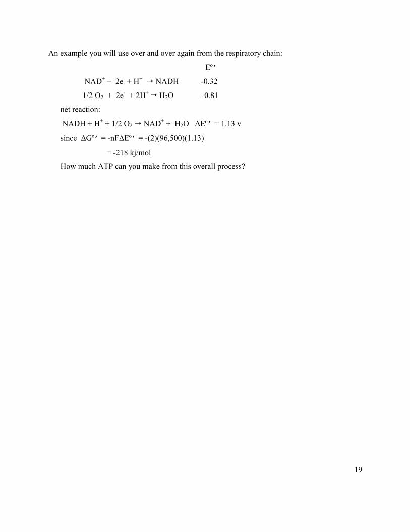
An example you will use over and over again from the respiratory chain:
Eº’
NAD+ + 2e- + H+ NADH -0.32
1/2 O2 + 2e- + 2H+ H2O + 0.81
net reaction:
NADH + H+ + 1/2 O2 NAD+ + H2O ΔEº’ = 1.13 v
since ΔGº’ = -nFΔEº’ = -(2)(96,500)(1.13)
= -218 kj/mol
How much ATP can you make from this overall process?

MIT OpenCourseWarehttps://ocw.mit.edu
5.07SC Biological Chemistry IFall 2013
For information about citing these materials or our Terms of Use, visit: https://ocw.mit.edu/terms.