biology 101 - lecture22 - musculoskeletal system · 10/30/2016 1 musculoskeletal system types of...
TRANSCRIPT

10/30/2016
1
Musculoskeletal SystemTypes of Skeletal Systems
1. Hydrostatic skeletons
2. Exoskeletons
3. Endoskeletons
2

10/30/2016
2
7
Hydrostatic Skeletons
Exoskeletons
• Surrounds the body as a rigid hard case.
• Composed of chitin in arthropods.
• Provides protection for internal organs and a site for muscle attachment.
• Must be periodically shed in order for the animal to grow.
• Not as strong as a bony skeleton.
• Respiratory system and gravity set limits on body size.
10

10/30/2016
3
Invertebrate Endoskeletons
• Rigid internal skeletons that form the body’s framework and offer surfaces for muscle attachment.
• Echinoderms have calcite skeletons.
• Made of calcium carbonate
18
10/30/2016
4
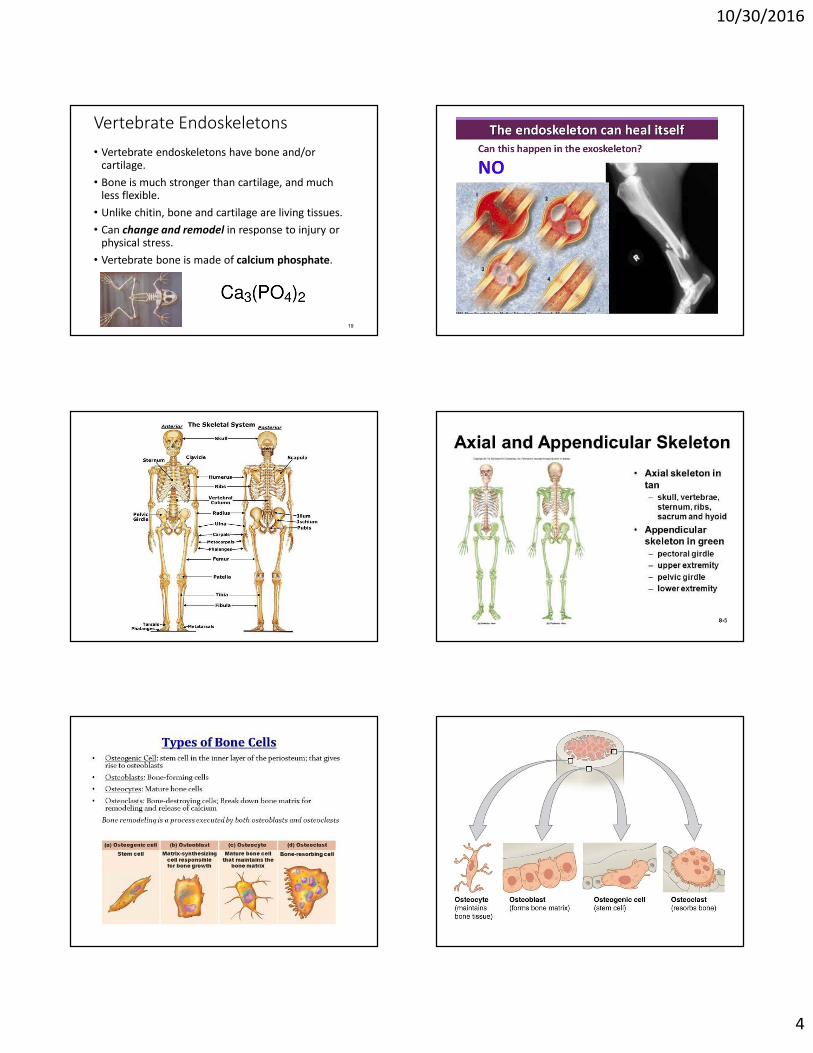
Vertebrate Endoskeletons
• Vertebrate endoskeletons have bone and/or cartilage.
• Bone is much stronger than cartilage, and much less flexible.
• Unlike chitin, bone and cartilage are living tissues.
• Can change and remodel in response to injury or physical stress.
• Vertebrate bone is made of calcium phosphate.
19

10/30/2016
5
Bone Structure
• In most mammals, bones retain internal blood vessels and are called vascular bones. • These typically have osteocytes and are also called
cellular bones or “living bones.”• Vascular bone has a special internal organization termed
the Haversian system.
• In birds and fishes, bones are avascular. • Lack osteocytes and are also called acellular bones.
26
Bone Structure
• Based on density and structure, bone falls into three categories:
1. Compact bone – outer dense layer.2. Medullary bone – lines the internal cavity.
• Contains bone marrow in vertebrates.• Bird bones are hollow.
3. Spongy bone – forms the epiphyses inside a thick shell of compact bone.
30
10/30/2016
6
31
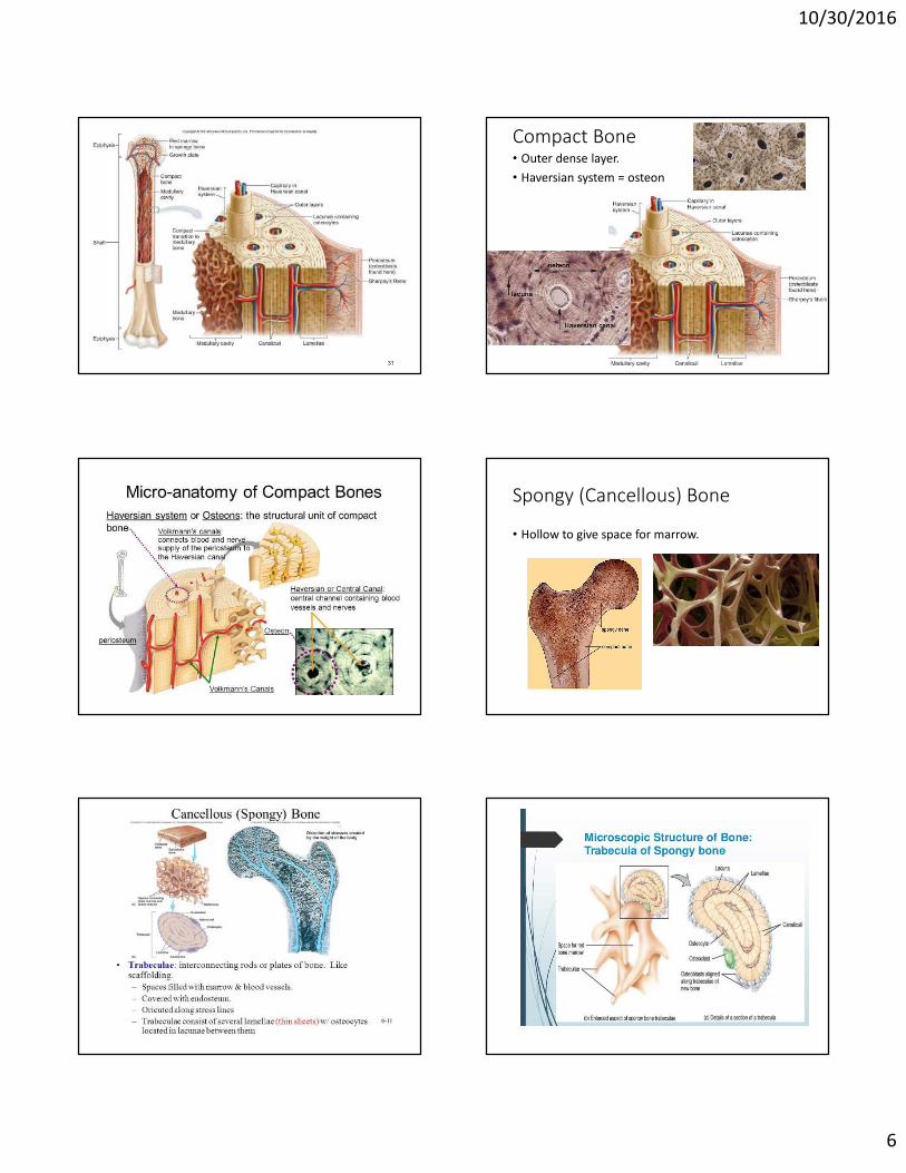
Compact Bone• Outer dense layer.
• Haversian system = osteon
Spongy (Cancellous) Bone
• Hollow to give space for marrow.

10/30/2016
7

10/30/2016
8
Osteoporosis
Joints (articulations)
• Joints: Locations where one bone meets another.
• 4 basic joint types:1. Ball-and-socket joints – permit movement in all
directions.2. Hinge joints – allow movement in only one plane3. Gliding joints – permit sliding of one surface over
another.4. Combination joints – movement characteristics of two
or more joint types.
48

10/30/2016
9
49
Copyright © The McGraw-Hill Companies, Inc. Permission required for reproduction or display.
Ball-and-Socket
Combination Joint
Hinge Joint Gliding Joint
a.
d.
b. c.
Muscles: What they do for you.
• Allow you to stand upright.
• Make it possible for you to move.
• Allow you to digest food.
• Help maintain a normal body temperature.
• Hold your skeleton together.
• Moves blood throughout the body.
Muscle tissue• Most abundant in animals.
• Consists of long cells called muscle fibers.
• Each contain many contractile proteins.
• Three kinds: smooth, skeletal, and cardiac.

10/30/2016
10
• Tendon:
attaches muscle
to bone.
• Ligament:
attaches bone
to bone.
Skeletal Muscle Movement
• Skeletal muscle fibers are attached to bones.• Directly to the periosteum by a tendon.
• One attachment of the muscle, the origin, remains stationary during contraction.
• The other end, the insertion, is attached to a bone that moves when muscle contracts.
• Muscles can be antagonistic.• One counters the action of the other.
58
59
Copyright © The McGraw-Hill Companies, Inc. Permission required for reproduction or display.
Extension
Flexion
Flexors
(hamstrings)
Tendon
Tendon
Extensors
(quadriceps)

10/30/2016
11
61
← Muscle fiber cells lengthwise view.

10/30/2016
12
Skeletal Muscle Contraction
• Thick filament • Composed of several myosin subunits packed together.• Myosin consists of two polypeptide chains wrapped
around each other.• Each chain ends with a globular head.
• Thin filament• Composed of two chains of actin proteins twisted
together in a helix.
69
←Thin filament
(actin)
← Thick
filament
(myosin)
71 72
10/30/2016
13
Skeletal Muscle Contraction
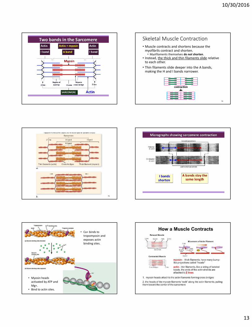
• Muscle contracts and shortens because the myofibrils contract and shorten.• Myofilaments themselves do not shorten.
• Instead, the thick and thin filaments slide relative to each other.
• Thin filaments slide deeper into the A bands, making the H and I bands narrower.
74
75
• Ca+ binds to
tropomyosin and
exposes actin
binding sites.
• Myosin heads
activated by ATP and
Mg+.
• Bind to actin sites.

10/30/2016
14
Skeletal Muscle Contraction
• In low Ca2+ levels, tropomyosin inhibits cross-bridge formation.
• In high Ca2+ levels, Ca2+ binds to troponin.• Tropomyosin is displaced, allowing the formation of
actin-myosin cross-bridges.
80
82
Neuromuscular Junction
• What stimulates the muscle to contract?
84
Skeletal Muscle Contraction
10/30/2016
15
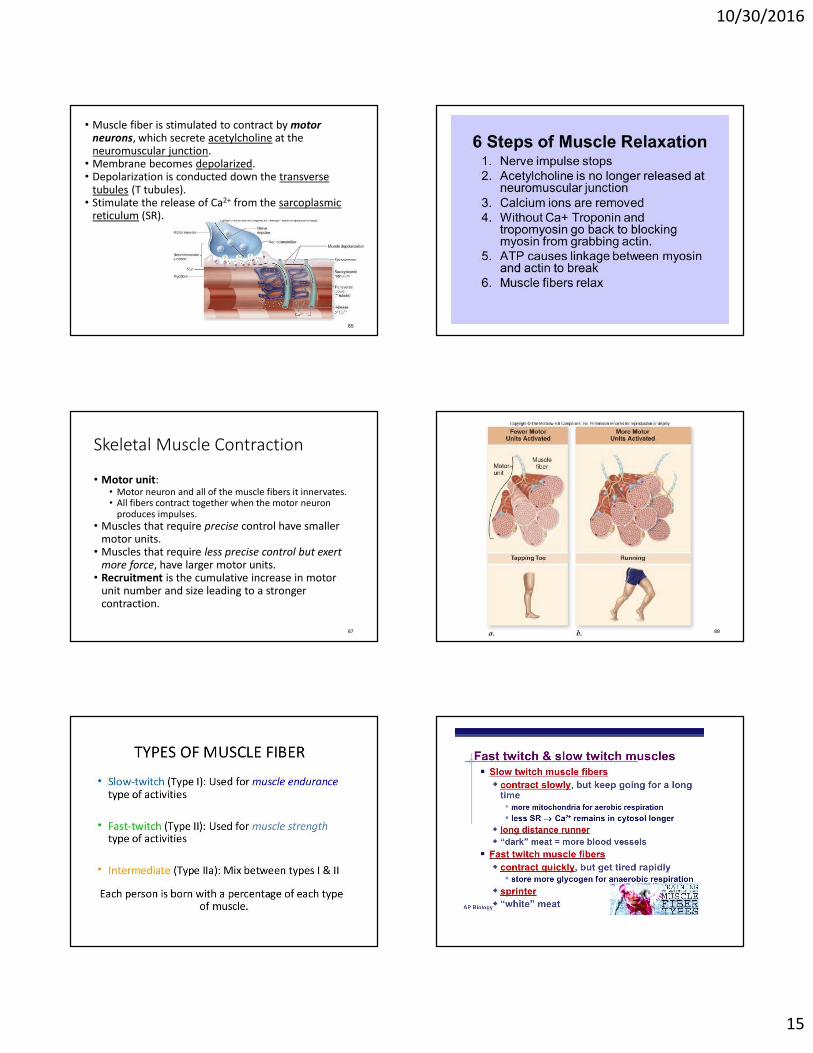
• Muscle fiber is stimulated to contract by motor
neurons, which secrete acetylcholine at the neuromuscular junction.
• Membrane becomes depolarized.• Depolarization is conducted down the transverse
tubules (T tubules).• Stimulate the release of Ca2+ from the sarcoplasmic
reticulum (SR).
85
Skeletal Muscle Contraction
• Motor unit: • Motor neuron and all of the muscle fibers it innervates. • All fibers contract together when the motor neuron
produces impulses.
• Muscles that require precise control have smaller motor units.
• Muscles that require less precise control but exert
more force, have larger motor units.• Recruitment is the cumulative increase in motor
unit number and size leading to a stronger contraction.
87 88
10/30/2016
16