biology 241: human anatomy and physiology 1 instructor: joel dahms
TRANSCRIPT

Biology 241: Human Anatomy and Physiology 1
Instructor: Joel Dahms

Introductions
Note cards Name Year you graduated HS and where Career goal List of classes you have taken that may help
prepare you for A&P and WHEN you took them (e.g. BIO 101, Fall’08)
List any other relevant experience you’ve had (job, internship, taking care of relatives, etc.).
Your preferred email address

Syllabus highlights - Day
Class meets:
MW 6:00PM - 7:40PM in AS 1520 (lecture)
MW 7:50PM - 9:30PM in AS 1615 (lab)
Holiday: 5/25 is Memorial Day, no class

Contact info
Email: [email protected]
email is the best way to contact me Office: IB 2324C Office hours: by appointment Office Phone: 527.3755 (Voice mail only)

Course website
The website has: Syllabus Lecture notes Answer keys to tests and quizzes (not yet though) Objectives (learning goals/study aids) for each unit Resources to help you study

Course website
Course Website:
https://frontpage.northseattle.edu/anp213jd/
User ID: anp213jd
Password: neuron

Required texts:
Human Anatomy and Physiology, Seventh Ed., Elaine N. Marieb & Katja Hoehn, Pearson Benjamin Cummings, 2007.
Human Anatomy and Physiology Laboratory Manual, Eighth Ed., Elaine N. Marieb& Susan J. Mitchell, Pearson Benjamin Cummings, 2008.
A Brief Atlas of the Human Body, Second Ed., Matt Hutchinson et al., Pearson Benjamin Cummings, 2007.

Required texts:

Required texts:

Optional texts:
Study Guide for Human Anatomy and Physiology, Seventh Ed., Elaine N. Marieb & Katja Hoehn, Pearson Benjamin Cummings, 2007.
The Anatomy Coloring Book, Third Ed.,Wynn Kapit and Lawrence M. Elson, Benjamin Cummings, 2001.
The Physiology Coloring Book, Wynn Kapit, Robert I. Macey, and Lawrence Meisami, Second Ed., Benjamin Cummings, 2000.
Fundamentals of Anatomy and Physiology, Seventh Ed., Frederic H. Martini, Benjamin Cummings, 2006.

Grading
Breakdown: Exams 400 points Lab Practicals 200 points Laboratory Exercises 200 points Quizzes & Assignments 100 points
Total 900 points
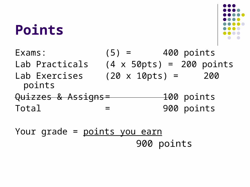
Points
Exams: (5) = 400 points Lab Practicals (4 x 50pts) = 200 pointsLab Exercises (20 x 10pts) = 200 pointsQuizzes & Assigns = 100 pointsTotal = 900 points
Your grade = points you earn
900 points

Grade percentages
4.0 - 3.5 A / A- 90 - 100%
3.4 - 2.9 B+/ B 80 - 89%
2.8 - 2.2 B-/ C+ 70 - 79%
2.1 - 1.5 C / C- 60 - 69
1.4 - 0.7 D+/ D 50 - 59%
0.0 E below 50%

Commitment
This is a very difficult class that requires learning what is essentially a new language
Because it is a prerequisite, the class is designed by the college as an overview: lots of breadth, little depth
Expect 20+ hours of reading and studying each week in addition to class sessions
The pace is a little frantic so missing class is not recommended. Spring Quarter especially!
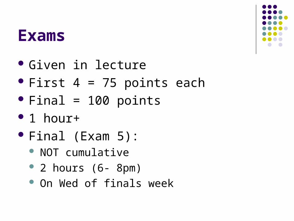
Exams
Given in lecture First 4 = 75 points each Final = 100 points 1 hour+ Final (Exam 5):
NOT cumulative 2 hours (6- 8pm) On Wed of finals week

Exams
Five exams:
Wed 4/15 Exam 1 (Chaps 1-3)
Wed 4/29 Exam 2 (Chaps 4, 5)
Wed 5/13 Exam 3 (Chaps 6, 7)
Wed 5/27 Exam 4 (Chaps 8 -10)
Wed 6/17 Exam 5 (Chaps 11 - 15)

Exams
A little more than half objective questions: multiple-choice, matching, true/false
The rest: fill-in-the-blank, short answer, short essay, and diagram labeling
You will need a Scantron form and a #2 pencil for each exam.
Not cumulative per se

Exams
Exams may not be rescheduled or made-up due to tardiness or absence. Students with extraordinary circumstances should discuss them with the instructor as soon as the situation occurs.
If you know ahead of time that you will miss an exam for a valid reason, I may be able to accommodate you but let me know as far ahead of time as possible.

Lab Practical Quizzes
Given in the lab Four practicals worth 50 points each Cover the material on the “Lab Practical Study
Guide” in the syllabus They will involve identifying slides, projected
pictures of slides, bones, muscle models, brain models, or diagrams.

Lab Practical Exams
Wed 5/6 Practical 1: Histology
Wed 5/20 Practical 2: Bones
Wed 6/3 Practical 3: Muscles
Mon 6/15 Practical 4: Nervous
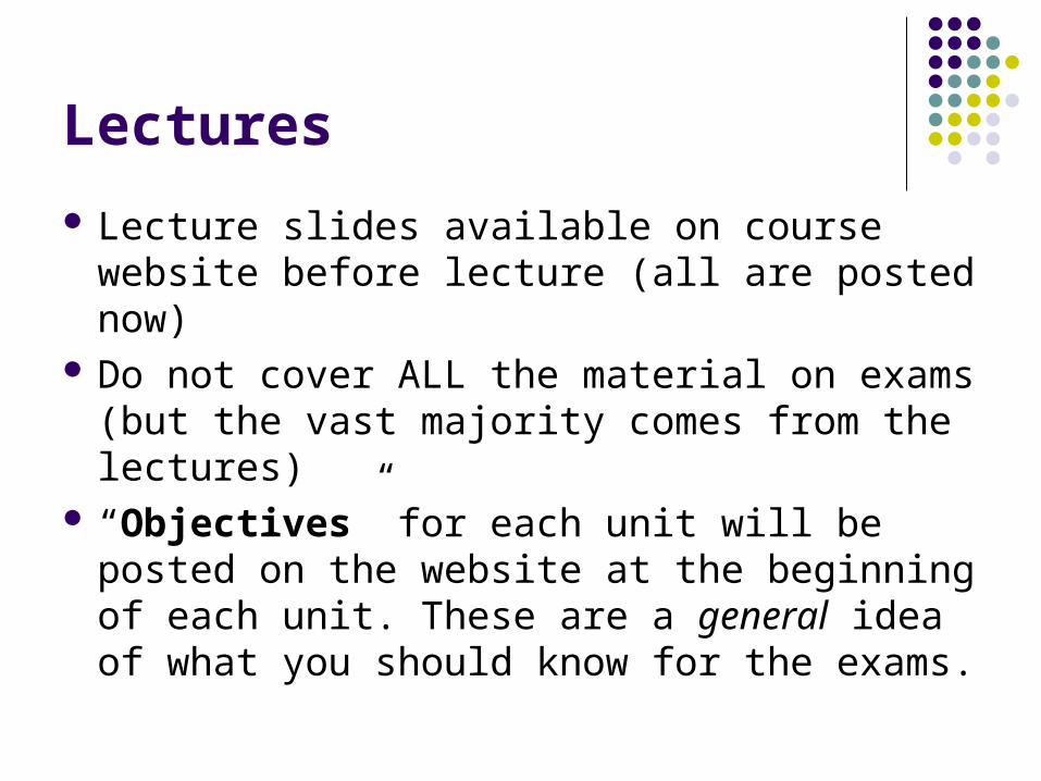
Lectures
Lecture slides available on course website before lecture (all are posted now)
Do not cover ALL the material on exams (but the vast majority comes from the lectures)
“Objectives” for each unit will be posted on the website at the beginning of each unit. These are a general idea of what you should know for the exams.

Objectives
List of learning goals that need to be achieved for you to do well in this class
Contain what the I and other teachers at NSCC have deemed to be the most important things for you to know to go on in a health-related career.
Available on the course website.

Attendance
Students should attend every class session. If you miss a class session, it is your responsibility to obtain the lecture notes, to make up laboratory experiments and to obtain handouts, assignments or other materials distributed in class. ESPECIALLY because we meet only twice a week.

Quizzes There will be three quizzes The first two quizzes will be in weeks 1 and 3
of class Quizzes may cover material presented in lab
or lecture Designed to prepare you for the types of
questions on the exam (multiple choice, T/F, fill-in-the-blank, short answer).
QUIZ 1 will be on Wed on material covered today in chapter 1(and lab)

Labs
Many laboratory exercises must be completed in the laboratory. Students who miss a laboratory exercise must schedule a make-up session with me or come in during open lab time to get credit for that exercise.
Lab exercises will be due the following week in lab on Wednesday.

Schedule of Lectures and Readings (Approximate)

Questions?

Introduction to the Human Body

Anatomy
“tome” means to cut in Greek Describes the structures of the body:
what they are made of where they are located associated structures

Physiology
Is the study of: functions of anatomical structures, both
individual and cooperative

KEY CONCEPT
All physiological functions are performed by specific anatomical structures
Principle of complementarity says that structure and function are complementary Function always reflects structure What a structure can do depends on its specific
form

Introduction
Key to learning anatomy is understanding function For example:
Left side of heart is larger than right. Why is that?
Structure (anatomy) and function (physiology) are intimately related

Gross Anatomy
Structures large enough that one can see with the unaided eye Surface Anatomy - study of superficial markings Regional Anatomy - The study of specific areas
of the body (e.g. head, trunk) Systemic Anatomy - Study of the 11* specific
organ systems

11 Organ systems
Integumentary*Nervous*Skeletal*EndocrineMuscular*Cardiovascular
Lymphatic
Urinary
Respiratory
Reproductive
Digestive

Microscopic Anatomy
Cf. Gross anatomy Involves studying anatomical structures that
cannot be seen with the unaided eye
1. Cytology - cells
2. Histology - tissue

Physiology = Function
Considers the operation of specific organ systems
Renal – kidney function Neurophysiology – workings of the nervous
system Cardiovascular – operation of the heart and
blood vessels Focuses on the functions of the body, often
at the cellular or molecular level

Anatomical Organization
We will start from the smallest and finish with the largest

Levels of Organization Chemical Level: - atoms (e.g. carbon)
combine to form molecules (e.g. glucose) Cellular level:
Smallest living units in organisms Cells contain organelles, each with a function
Tissue level - different groups of cells that perform a function
Organ Level - Different types of tissues that perform a common function
Organ system – consists of different organs that work closely together

1
2
4
5
6
3
Smooth muscle cellMolecules
Atoms
Smoothmuscletissue
Epithelialtissue
Heart
Bloodvessels
Smoothmuscletissue
Connectivetissue
Bloodvessel(organ)
Cardiovascularsystem
Cellular levelCells are made up ofmolecules.
Tissue levelTissues consist ofsimilar types of cells.
Organ levelOrgans are made upof different typesof tissues.
Organ system levelOrgan systems consist ofdifferent organs thatwork together closely.
Organismal levelThe human organismis made up of manyorgan systems.
Chemical levelAtoms combine toform molecules.
Levels of Structural Organization
Figure 1.1

Other Levels
Organismal Level - All systems working together (e.g. humans)
Ecological level - How organisms interact with each other and their environment

KEY CONCEPT
The body is divided into 11 organ systems All organ systems work together
Integration
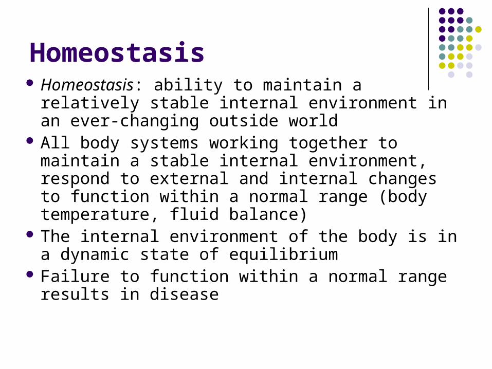
Homeostasis Homeostasis: ability to maintain a relatively stable
internal environment in an ever-changing outside world
All body systems working together to maintain a stable internal environment, respond to external and internal changes to function within a normal range (body temperature, fluid balance)
The internal environment of the body is in a dynamic state of equilibrium
Failure to function within a normal range results in disease

Homeostatic Control Mechanisms
Variables produce a change in the body The three interdependent components of
control mechanisms: Receptor – monitors the environments and
responds to changes (stimuli). Control center – determines the set point at which
the variable is maintained. Effector – provides the means to respond to
stimuli.

Homeostatic Control Mechanisms
Figure 1.4
Change detected by receptor
Stimulus: Produces changein variable
Input:Informationsent along afferentpathway to
Receptor (sensor) Effector
Controlcenter
Variable (in homeostasis)
Response ofeffector feedsback toinfluencemagnitude ofstimulus andreturns variableto homeostasis
Output:Information sentalong efferentpathway to
Imbalance
Imbalance
2
34
5
1

Regulation
Most regulatory systems in the body use extrinsic regulation: responses controlled by nervous and endocrine
systems, e.g. brain regulates body temp Usually occurs by negative feedback which
can be modeled as a thermostat:

Negative Feedback
Most common way that homeostasis is maintained in the body
In negative feedback systems the response of the effector negates or opposes the stimulus (shuts off the original stimulus)
Example: Regulation of room temperature

Figure 1.5
Signalwire turnsheater on
Signalwire turnsheater off
Response;temperaturerises
Response;temperaturedrops
Stimulus: rising roomtemperature
Stimulus: dropping roomtemperature
Balance
Effector(heater)
Effector(heater)
Setpoint
Control center(thermostat)
Heateroff
Setpoint
Receptor-sensor(thermometer inThermostat)
Control center(thermostat)
Heateron
Imbalance
Imbalance
Receptor-sensor(thermometer inThermostat)

Negative Feedback: Maintaining Normal Limits
Figure 1–3
Thermostat model

Figure 1–4
Negative Feedback

Positive Feedback
NOT a way to maintain homeostasis
Rare in nature because it is a “runaway train”
The response of the effector output reinforces or exaggerates the stimulus (e.g. blood clotting, ovulation, action potential)
Figure 1–5

Homeostatic Imbalance
Disturbance of homeostasis or the body’s normal equilibrium
Overwhelming the usual negative feedback mechanisms allows destructive positive feedback mechanisms to take over
This is often used as the definition of “disease”

Anatomical terms

Anatomical Position Hands at sides, palms forward

Orientation of terms
Note that Left/Right are reversed in anatomical figures
WHY?

Directional Terms
Superior and Inferior – toward and away from the head, respectively
Anterior and Posterior – toward the front and back of the body
Medial and Lateral – toward the midline, away from the midline
Proximal and Distal – closer to and farther from the origin of the body part (or from the torso)
Superficial and Deep – toward and away from the body surface
Cranial and Caudal – toward the head and toward the tail

Alternate Terms
Ventral (= Anterior) Dorsal (= Posterior)


Body Planes
Sometimes to gain a greater understanding of 3D images anatomists cut the image at different planes
Three planes exists in 3D space
-Two are parallel to the long axis of the body
-One is perpendicular to the long axis.

Body Planes
Figure 1.8
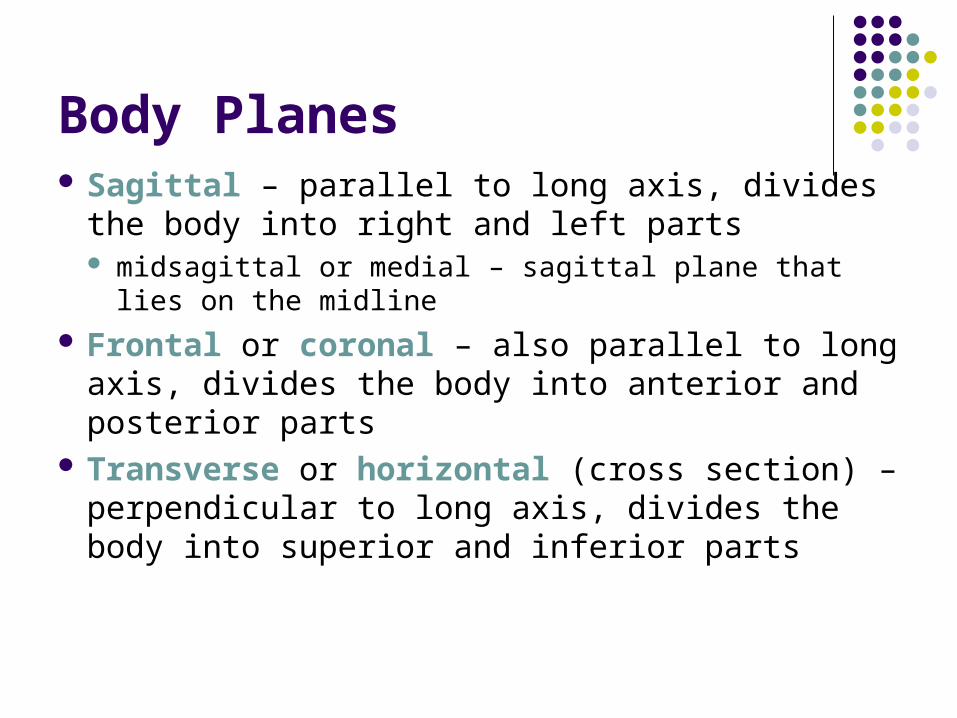
Body Planes Sagittal – parallel to long axis, divides the body
into right and left parts midsagittal or medial – sagittal plane that lies on the
midline Frontal or coronal – also parallel to long axis,
divides the body into anterior and posterior parts Transverse or horizontal (cross section) –
perpendicular to long axis, divides the body into superior and inferior parts

Anatomical Variability
Humans vary slightly in both external and internal anatomy
Over 90% of all anatomical structures match textbook descriptions, but: Nerves or blood vessels may be somewhat out of
place Small muscles may be missing
Extreme anatomical variations are seldom seen

Body Cavities
Dorsal cavity protects the nervous system, and is divided into two subdivisions Cranial cavity – within the skull; encases the brain Vertebral cavity – runs within the vertebral
column; encases the spinal cord Ventral cavity houses the internal organs
(viscera), and is divided into two subdivisions Thoracic Abdominopelvic

Body Cavities
Figure 1.9a
Cranial cavity(contains brain)
Dorsalbodycavity
Diaphragm
Abdominal cavity(contains digestiveviscera)
Pelvic cavity(contains bladder,reproductive organs,and rectum)
Vertebral cavity(contains spinal cord)
Key:
Dorsal body cavity
Ventral body cavity
Thoraciccavity(containsheartand lungs)
(a) Lateral view

Ventral Body Cavity Membranes
Called serous membranes or serosa Parietal serosa lines internal body walls Visceral serosa covers the internal organs Serous fluid separates the serosae

Heart Serosae
Figure 1.10b

Serous Membrane Relationship
Figure 1.10a

SUMMARY
Structure and function in anatomy and physiology
Levels of physical organization Homeostasis and feedback Systems integration and equilibrium Anatomical terms Locations and functions of major cavities Serous membranes in the ventral body cavity