biomechanical gait characteristics of …
TRANSCRIPT
1
BIOMECHANICAL GAIT CHARACTERISTICS OF UNSUCESSFUL FOOT CLEARANCE
DURING SWING IN INDIVIDUALS WITH CHRONIC STROKE
Jessica L. Burpee, SPT, Michael D Lewek, PT, PhD
DPT Capstone
Division of Physical Therapy
The University of North Carolina at Chapel Hill
2014
Advisory Committee
Michael D Lewek, PT, PhD Prudence Plummer, PhD

2
Abstract
Background and Objective: Individuals with hemiparesis secondary to chronic stroke
commonly display motor impairments that result in altered biomechanical characteristics of gait.
These abnormalities increase the risk of falls and injury during ambulation. Lower extremity
gait impairments have been previously described compared to the non-paretic limb or healthy
control subjects. Unfortunately, the biomechanical parameters underlying instances of
unsuccessful foot clearance (i.e., trips) during the swing phase of gait have yet to be examined.
The purpose of this study was to examine the paretic lower extremity spatiotemporal, kinematic,
and kinetic characteristics of trips during the gait cycle in individuals with chronic stroke.
Methods: Gait data for 26 subjects with chronic stroke were obtained during a 20 minute gait
training session on a dual-belt ‘instrumented’ treadmill. Force plate data were examined to
identify naturally occurring trips during the swing phase of the paretic limb. Instances of
successful foot swing immediately prior to each trip were also identified. Spatiotemporal
(double support time), kinematic (hip, knee, and ankle angles), and kinetic (propulsive impulse
and hip, knee, and ankle moments) measures of the paretic limb occurring during late stance, toe-
off, and swing were collected for each trip and step with successful foot clearance. These data
were compared across subjects and Pearson correlation coefficients were used to determine
relationships between variables.
Results: In the paretic limb, the ankle angle at toe off (p = 0.003), knee flexion velocity at toe off
(p < 0.001), and peak knee extension moment during terminal stance (p < 0.001) were
significantly different between trips and steps with successful foot clearance. Knee flexion
velocity moment correlated significantly (p < .05) with the other significant parameters. The
mean differences in significant parameters during trips were 1.1° more ankle plantarflexion at toe
off, knee flexion velocity reduced by 17.6°/sec, and peak knee extension moment increased by
0.01Nm/kg·m during terminal stance.
Conclusions: The qualitative differences in the parameters between instances of successful and
unsuccessful foot clearance are relatively small in individuals with hemiparesis secondary to
chronic stroke. Only minor changes in the movement of the paretic limb can result in a trip.
Additionally, the multi-joint biomechanical changes occurring in the paretic limb during
unsuccessful foot clearance result in a functionally longer limb. Thus, interventions targeting
multiple joints in the paretic limb may be needed to reduce the risk of trips in these individuals.

3
Introduction
Each year, nearly 800,000 people in the US experience a stroke.1 The multiple
consequences of stroke are a leading cause of impairment, disability, and death. Hemiparesis is a
common motor deficit occurring post-stoke that negatively impacts function and often persists
into the chronic stages of recovery following stroke.2 Individuals with chronic hemiparesis
secondary to stroke often exhibit disturbances in lower limb movement patterns that can be
attributed to the muscle weakness, abnormal motor control, and impaired coordination.3,4
These
impairments may adversely affect balance, transfers, and walking ability leading to falls and
further deficits in functional abilities.
Falls and fall-related injuries are common complications following stroke.5 The
detrimental consequences of falls include fractures, soft tissue injury, hospitalizations, decreased
mobility, and negative psychological impacts such as fear and anxiety.6 Additionally, falls after
stoke are associated with poor rehabilitation outcomes, loss of independence, and chronic
disability.7 Falls most often occur during mobility tasks such as walking and transfers in
individuals with chronic stroke.8 Common gait deviations that occur following stroke such as
decreased walking speed, reduced foot clearance, and stance time asymmetries are purported to
contribute to the risk for falling.6 Due to the increased risk for falls during ambulatory activities,
improvement of walking ability is a primary goal in stroke rehabilitation.
Quantifying hemiparetic gait dysfunction is a critical step towards designing effective
intervention strategies for gait rehabilitation. Hemiparetic gait impairments following stroke can
vary considerably amongst patients, and are largely influenced by the severity of the lower limb
neuromuscular deficits.9 Several previous investigations have identified and classified common
patterns of hemiparetic gait abnormalities.10,11
Decreased walking speed, reduced step length,

4
reduced stance time on the paretic limb, and increased time spent in double support are common
characteristics of hemiplegic gait.12
The causes of these spatiotemporal abnormalities have been
linked to alterations in lower limb mechanics.13
Specifically, paretic joint kinematic patterns
often include reductions in hip flexion, knee flexion, and ankle dorsiflexion during swing, as
well as decreased hip extension, knee flexion, and ankle plantar flexion at toe-off.11
Lower limb
kinetic alterations such as reductions in hip, knee, and ankle joint moments and powers during
late stance and pre-swing have also been identified.14,15
The overall result of these paretic limb biomechanical alterations is the presence of a
functionally longer limb during swing which challenges successful foot clearance.
Compensatory movements at the trunk and pelvis such as hip hiking and leg circumduction may
be utilized to overcome these abnormalities in distal joint motion.16
Altered late stance joint
mechanics and/or compensatory motions during swing may negatively impact paretic limb
advancement. Unsuccessful foot clearance (i.e., a trip) during the swing phase of gait can cause
falls and further injury to individuals following stroke. Previous studies have focused on
examining the biomechanical patterns that characterize hemiparetic gait10,11
as well as the
pathophysiology surrounding falls in post-stroke populations.5,6
However, the biomechanical
parameters underlying instances of unsuccessful foot clearance have yet to be examined. Our
aim is to examine the parameters leading to naturally occurring trips during ambulation. This
experimental design is notably different than previous studies that induced trips in subjects via
perturbations or unexpected obstacles.17,18
The purpose of this study is to determine the spatiotemporal, kinematic, and kinetic
characteristics of the paretic limb associated with unsuccessful foot clearance in subjects with
chronic hemiparesis secondary to stroke. We hypothesized that subjects will exhibit

5
biomechanical alterations during late stance, which will lead to a functionally longer paretic limb
in swing phase. The paretic leg posture we expected to see during trips, compared to instances
of successful foot clearance, included decreased pelvic obliquity throughout the swing phase,
decreased hip flexion at toe-off and during swing phase, decreased knee flexion during swing,
and increased ankle plantarflexion during swing. By exploring potential patterns demonstrated
during trips, we can design targeted interventions to improve walking ability and decrease falls
post-stroke.
Methods
Subjects
Forty subjects with chronic (>6 months) hemiparesis secondary to stroke were recruited
as part of a larger study examining gait kinematics and kinetics during treadmill ambulation.
Twenty six subjects who demonstrated unsuccessful foot clearance during training on the
treadmill were analyzed for this study. Additional inclusion criteria were (1) clinical symptoms
consistent with an ischemic or hemorrhagic unilateral cortical brain lesion, resulting in sensory
motor dysfunction more than 6 months prior to recruitment, and (2) the ability to walk a
minimum of 10 m overground without physical assistance. Stroke was determined via subject
self-report and confirmed by a physician while receiving medical clearance for study
participation. Participants were excluded from training if they exhibited additional neurologic
(e.g., Parkinson’s disease, vestibular disorders), musculoskeletal, or cardiovascular pathology
that would preclude gait training, or had a history of balance deficits or falls unrelated to the
stroke. Subjects who wore an ankle foot orthosis (AFO) during training were also excluded due
to the imposed alterations in lower limb joint mechanics from the AFO. All subjects signed an
informed consent form which was approved by the IRB at UNC Chapel Hill.

6
Data collection
Gait data for each subject were obtained from a 20 minute gait training session on a dual-
belt ‘instrumented’ treadmill (Bertec Corp., Columbus, OH). Treadmill speed for each subject
was chosen by the principal investigator as a speed that the subject could maintain for the
duration of the training session, with the goal of a heart rate of 70% of max or RPE of 14 on the
Borg scale. All participants wore an overhead safety harness while walking, which did not
restrict lower limb movement. Subjects did not use assistive devices during training and no
manual facilitation was provided to alter subjects’ gait patterns. A handrail was available for
subjects to hold onto, although they were discouraged from doing so.
While walking on the treadmill, limb movement data was collected in 3-dimensional
space. Lower extremity motion was recorded using an 8-camera motion capture system (Vicon
Corp.) at 120 Hz, as ground reaction forces (GRFs) were sampled concurrently at 960Hz from
both sides of the Bertec treadmill. Limb segments were tracked with retroreflective markers
taped to the subject’s pelvis, legs, and feet. Details describing marker placement setup have been
previously described.19
Following data collection, Visual3D software (C-Motion, Germantown,
MD) was used to identify the locations of the markers and model the three-dimensional motion
of the pelvis and lower limb segments. Marker trajectories and GRF data were low-pass filtered
at 6Hz and 20Hz, respectively. Hip, knee, and ankle joint angles were computed using Euler
angles and internal joint moments were computed using an inverse dynamics approach. Joint
powers were computed as the product of the joint moment and the angular velocity.
Data Management
All instances of unsuccessful foot clearance (trips) during the 20 minute gait training
session were initially identified. These trips were identified by the presence of a vertical GRF

7
which exceeded 10N on the paretic limb’s treadmill belt during its respective swing phase (see
Figure 1). Instances of successful foot swing during the stride immediately prior to each trip
were also identified and labeled. For subjects demonstrating a large number of trips during the
training session a maximum of 18 steps were used for analysis.
Because we were interested in the biomechanics influencing the swing phase dynamics of
the paretic limb, we focused our data collection on the late-stance, toe-off, and swing phases of
the gait cycle. The spatiotemporal measure of interest was double support time (non-paretic heel
strike to paretic toe-off in seconds). Pre-swing kinematic measures were peak hip extension
angle at late-stance, knee flexion velocity at toe-off, and ankle angle at toe-off. Swing-phase
kinematic measures included peak hip, knee, and ankle flexion angles and circumduction (frontal

8
plane excursion of the swing foot with respect to the stance foot). Kinetic measures collected
were propulsive impulse and the peak hip flexion, knee extension, and ankle plantarflexion
moments during late-stance. Spatiotemporal, kinetic, and kinematic variables were calculated
for each labeled step using custom written LabVIEW software (National Instruments, Austin,
TX). Outcome measures of successful and unsuccessful foot clearance were then averaged for
each subject.
Statistical analysis
Within-subject statistical analyses were performed for each outcome variable using
SPSS, ver 16 (SPSS, Chicago, IL). Paired-samples t-tests were used to compare outcome
measures between instances of successful and unsuccessful foot clearance. Adjustments for
multiple comparisons was performed by reducing the alpha level (.05) by the number of
comparisons made (e.g., 12 variables/0.05 = 0.004). A significance level of α=0.004 was used
for all between condition comparisons. Pearson correlations were then calculated to determine
the presence of relationships between the variables determined to be significantly different
between trip and non-trip steps. An α=0.05 was used to assess the correlations.
Results
Twenty-six subjects demonstrated naturally occurring trips during the gait training
sessions and were included in the gait analysis. Demographic data of the subjects are described
in table 1. Overground gait speed was 0.73 m/s (± 0.33; range 0.22-1.28), and treadmill gait
training speed was 0.68 m/s (± 0.27; range 0.20-1.3). An average of 10.0 (± 5.7) trips and
corresponding steps with successful foot clearance were analyzed for each of the 26 subjects.

9
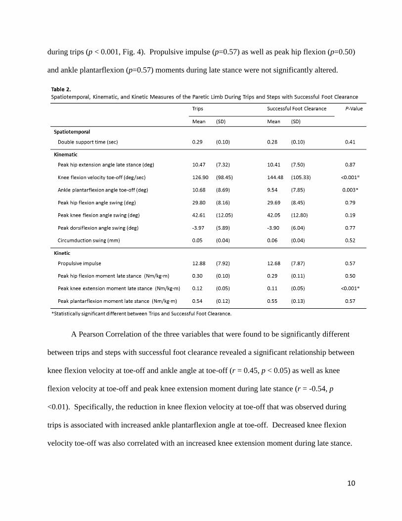
The means and standard deviations of the 12 variables measured during trips and steps
with successful foot clearance are presented in table 2. Two of the 8 spatiotemporal and
kinematic variables were significantly altered between trips and steps with successful foot
clearance. Ankle angle at toe-off had a mean difference of 1.1 degrees more plantarflexion
during trips (p = 0.003, Fig. 2). Knee flexion velocity at toe-off had a mean reduction of 17.6
degrees/second during trips (p < 0.001, Fig. 3). The spatiotemporal measure of double support
time was not significantly altered between trips and steps with successful foot clearance (p =
0.41). Similarly, 5 of the 7 kinematic variables were not found to be significantly different
during trips. These include peak hip extension at late stance (p=0.87), circumduction during
swing (p=0.52), and swing phase peak hip flexion, knee flexion, and ankle dorsiflexion (p=0.23,
p=0.19, p=0.77, respectively).
One kinetic measure was significantly different between trips and steps with successful
foot clearance. Peak knee extension moment at late stance had a mean increase of 0.01 Nm/kg·m
10
during trips (p < 0.001, Fig. 4). Propulsive impulse (p=0.57) as well as peak hip flexion (p=0.50)
and ankle plantarflexion (p=0.57) moments during late stance were not significantly altered.
A Pearson Correlation of the three variables that were found to be significantly different
between trips and steps with successful foot clearance revealed a significant relationship between
knee flexion velocity at toe-off and ankle angle at toe-off (r = 0.45, p < 0.05) as well as knee
flexion velocity at toe-off and peak knee extension moment during late stance (r = -0.54, p
<0.01). Specifically, the reduction in knee flexion velocity at toe-off that was observed during
trips is associated with increased ankle plantarflexion angle at toe-off. Decreased knee flexion
velocity toe-off was also correlated with an increased knee extension moment during late stance.

11

12
Discussion
The hypothesis that lower extremity kinematics and kinetics would be different during
instances of unsuccessful foot clearance was partially supported by the data. Comparisons
between steps with and without successful foot clearance revealed that peak knee extension
moment, knee flexion velocity, and ankle angle were significantly altered during trips.
Specifically, trips were characterized by an increase in the knee extension moment during late
stance and ankle plantarflexion angle at toe-off as well as a reduction in the knee flexion velocity
at toe-off. These kinematic and kinetic changes that occurred at the knee and ankle joints were
related to each other, and may have produced a functionally longer paretic limb during swing
phase which contributed to unsuccessful foot clearance. Contrary to our hypothesis, there were
no significant changes in biomechanical parameters at the hip or pelvis associated with steps that
resulted in a trip.
An overall lengthening of the paretic limb during swing has been demonstrated in many
kinematic and kinetic analyses of hemiparetic gait.10,11,13
Diminished flexion at the hip, knee,
and ankle are common post-stroke gait deviations occurring during pre-swing and swing phases.
These abnormalities have been attributed to a multitude of stroke-related impairments including
decreased strength, increased muscle tone, and irregularities in muscle activation timing.13,20
Paretic limb motor insufficiencies negatively impact the multi-joint flexion and effective limb
shortening that is needed in order to achieve foot clearance during swing phase. The kinematic
and kinetic changes that contribute to a longer paretic limb and the associated compensatory
movements have frequently been described in comparisons to the non-paretic limb or healthy
control subjects.10,11,21
However, the direct association between factors that would lengthen the
paretic limb and a resulting trip has not been previously reported in individuals post-stroke. Our
13
data suggest that diminished flexion at the knee and ankle joints contribute to the inability to
successfully shorten the limb and achieve sufficient foot clearance during swing.
We noted that the kinematic and kinetic differences between instances of successful and
unsuccessful paretic limb foot clearance are quantitatively small. This suggests that a trip can
result from only minor changes in the movement of the paretic limb at each joint. Nevertheless,
the biomechanical changes we identified leading to trips are similar in magnitude to the minimal
detectable change (MDC) values reported for gait parameters during treadmill walking in
individuals with post-stroke impairments.22
Although the alterations are small, their similarity in
size to these MDC values provides evidence that our findings represent real changes contributing
to trips.
Additionally, the magnitude of the changes we observed suggests that individuals with
hemiparesis are at a high risk of tripping with every step. In unimpaired subjects, the minimum
vertical distance between the toe and the walking surface (i.e., minimum foot clearance) occurs
approximately half-way through swing phase.23
It is suggested that this variable is directly
associated with the potential for trips during ambulation since it represents a critical time during
swing when the toe is at its closest distance to the ground, approximately 1.5 cm.23,24
Multiple
gait parameters such as segmental angular velocities, joint range of motion, and the coordination
of joint moments influence minimum toe clearance and successful forward limb progression
during swing.25
However, the trajectory of the swing limb and the conditions needed to achieve
minimum toe clearance are largely determined by kinematic and kinetic events occurring prior to
the initiation of swing.26,27
This is consistent with our finding that kinematic and kinetic
alterations occurring during late stance and toe-off lead to in the inability to successfully clear
the foot during swing and resultant trips.

14
Minimum toe clearance has not yet been examined in hemiparetic gait. However, an
increase in minimum toe clearance variability between strides has been identified in elderly
subjects and may contribute to an increased risk for trip-related falls due to the small margin for
error that this distance allows.28,29
While the underlying mechanisms of age-related increases in
toe clearance variability are not clear, they may be attributable to neuromuscular impairments,
such as reduced proprioceptive ability and altered motor control 29
, that are also common in
individuals with chronic stroke.30
Our study did not aim to quantify minimum toe clearance or
variability in toe clearance; however, we know that the minimum toe clearance was equal to zero
for each trip. If stroke-related impairments lead to an increase in toe clearance variability, this
may help explain why very small kinematic and kinetic alterations can lead to a trip.
An important finding in this investigation is that trips occurred due to biomechanical
changes at multiple joints in the paretic limb. This finding is in contrast with the idea that
unsuccessful foot clearance during paretic swing phase may be primarily due to single-joint
abnormalities, specifically ankle joint insufficiencies resulting in foot-drop or reduced
dorsiflexion.31-33
Ankle-foot orthoses (AFOs) are commonly prescribed for individuals with
hemiparesis secondary to stroke to assist with foot clearance and increase stability during
ambulatory activities.34
Improvements in gait speed, efficiency, and pattern have been
demonstrated with the use of AFOs.35
However, AFOs may only have negligible effects on
improving overall lower limb kinematics and kinetics to assist with successful foot clearance in
hemiparetic individuals.36,37
Our finding that trips result from alterations occurring at multiple
joints suggests that use of an intervention targeting single-joint gait abnormalities, such as an
AFO, by itself may not prevent all trips. All subjects included in this study were able to walk
on a treadmill without use of an AFO. Therefore, the influence of AFOs on trips cannot be

15
delineated from our investigation. However, given that the biomechanical changes which lead to
trips occurred at multiple joints, it is reasonable to suggest that interventions designed to address
multi-joint impairments may be more effective for reducing the risk for trips and resultant falls
during ambulation.
Biomechanical alterations at the ankle resulting in trips were observed in our study;
however, knee flexion and extension mechanics also significantly contributed to trips. Reduced
knee flexion velocity at toe-off and an increased knee extension moment during late stance are
causes of a stiff-knee gait pattern often demonstrated by individuals with neurological deficits
such as stroke and cerebral palsy.16
Previous studies have found that a stiff-knee gait pattern can
inhibit foot clearance and result in trips during walking.38
Stiff-knee gait in individuals with CP
and stroke is attributed to overactivity of the rectus femoris muscle during swing.39,40
Exaggerated activation of the quadriceps during late stance and early swing has been shown to
be elicited by hyperexcitable heteronymous reflex activity from hip flexor muscles following
stoke.41,42
That is, inappropriate knee extensor activity in stiff-knee gait during swing may be the
result of a reflexive response to hip extension during late stance.42
In contrast to those findings,
our study did not detect any alterations in hip kinematics associated with the observed reduction
in knee flexion velocity and increased knee extension moments that lead to trips, although we did
not assess hip extension velocity. Stiff-knee gait is often treated via surgical transfer of the
rectus femoris insertion to a site that reduces its contribution to the internal knee extension
moment. Post-operative improvements in walking ability in the CP population have been
associated with increased knee flexion velocity and reduced knee extension moments prior to
toe-off.26
Treatment of stiff-knee gait via botulinum toxin injection in the rectus femoris of

16
hemiparetic subjects has also been shown to effectively normalize knee angular position and
velocity during late stance and swing.43
We cannot directly attribute the changes in knee biomechanics we observed during trips
to altered muscle activity due to a lack of EMG data. However, the studies on individuals with
stiff-knee gait suggest that interventions targeting the muscles which influence knee angular
motion during swing can reduce some of the variables that lead to trips in individuals post-stoke.
Altering the swing phase dynamics alone may not be sufficient for reducing trips, however.
Increasing knee flexion torque during swing, for example, has not been shown to normalize gait
patterns following stroke. In fact, an orthotic which applied knee flexion torque to the paretic
limb during swing actually increased hip abduction, a compensatory movement used to facilitate
foot clearance.44
This supports our findings that successful foot clearance is not dependent upon
knee swing phase biomechanics, but instead is influenced primarily by kinematic and kinetic
conditions at late stance and toe-off.
Ankle plantarflexion at the end of stance can also critically influence swing phase
dynamics.23
We found that plantarflexion of the paretic ankle at toe-off is greater during trip
steps. During normal gait, the ankle plantarflexes at the end of stance to provide a forward
propulsive force, but then dorsiflexes during swing to allow toe clearance as the limb advances
forward.23,45
Excessive plantarflexion during late-stance and toe-off may result in insufficient
dorsiflexion at the time of critical toe clearance during swing.46
In individuals with chronic
stroke, excessive plantarflexion prior to toe-off may be due to contractures or spasticity in the
plantarflexor muscles or may be related to movements occurring further up the limb.25

17
Limitations
All subjects included in this study were able to independently ambulate for a total of
twenty minutes on a treadmill. In order to achieve twenty minutes of treadmill training with
minimal rest breaks, the treadmill speed was determined prior to testing and may not have been
representative of the subject’s self-selected or over-ground walking speed. Lower extremity
kinematics and kinetics are altered with walking speed 47
; however, comparisons were made
between trip and non-trip steps, so gait speed was not a factor in those differences.
Additionally, the subjects’ fall histories were not obtained during this study. Previous
investigations have noted variations in gait characteristics between elderly subjects with a history
of falls and elderly nonfallers.48,49
Fallers demonstrate increased stride frequency, decreased
stride length, reduced ankle plantarflexion and hip extension, and a greater minimum toe
clearance. These adaptations are likely demonstrated by fallers in an attempt to reduce gait
pattern variability and maximize stability.48
Without subject fall history we are unable to
speculate the potential this may have had on trip biomechanics.
In the future, interventions aimed at improving overall paretic limb flexion during late
stance/early swing by targeting multiple joints may be more effective at preventing trips than
those targeting flexion of a single joint. The pre-swing phase of the gait cycle during which we
observed significant alterations, suggests that gait rehabilitation efforts should focus on
increasing paretic limb muscle strength and joint coordination during this period to increase the
success of limb progression during swing. Implementing therapies that address the underlying
kinematic and kinetic abnormalities leading to trips may assist in preventing falls and falls-
related injuries in individuals with chronic stroke. Assessment of these trip-related biomechanics

18
in individuals with a history of falls may be useful for falls prediction or identification of falls
risk in the post-stoke population.
References
1. Go AS, Mozaffarian D, Roger VL, et al. Heart disease and stroke statistics--2013 update: A
report from the american heart association. Circulation. 2013;127(1):e6-e245.
2. Cauraugh JH, Kim SB. Chronic stroke motor recovery: Duration of active neuromuscular
stimulation. J Neurol Sci. 2003;215(1–2):13-19.
3. Bohannon R. Determinants of transfer capacity in patients with hemiparesis. Physiotherapy
Canada. 1988;40(4):236-239.
4. Bourbonnais D, Vanden Noven S, Pelletier R. Incoordination in patients with hemiparesis.
Can J Public Health. 1992;83 Suppl 2:S58-63.
5. Forster A, Young J. Incidence and consequences of falls due to stroke: A systematic inquiry.
BMJ. 1995;311(6997):83-86.
6. Weerdesteyn V, de Niet M, van Duijnhoven HJ, Geurts AC. Falls in individuals with stroke. J
Rehabil Res Dev. 2008;45(8).
7. Ramnemark A, Nilsson M, Borssen B, Gustafson Y. Stroke, a major and increasing risk factor
for femoral neck fracture. Stroke. 2000;31(7):1572-1577.
8. Schmid AA, Yaggi HK, Burrus N, et al. Circumstances and consequences of falls among
people with chronic stroke. J Rehabil Res Dev. 2014;50(9):1277-1286.
9. Perry J, Garrett M, Gronley JK, Mulroy SJ. Classification of walking handicap in the stroke
population. Stroke. 1995;26(6):982-989.
10. De Quervain I, Simon SR, Leurgans S, Pease W, McAllister D. Gait pattern in the early
recovery period after stroke. The Journal of Bone & Joint Surgery. 1996;78(10):1506-1514.
11. Mulroy S, Gronley J, Weiss W, Newsam C, Perry J. Use of cluster analysis for gait pattern
classification of patients in the early and late recovery phases following stroke. Gait Posture.
2003;18(1):114-125.
12. Von Schroeder HP, Coutts RD, Lyden PD, Billings E, Nickel VL. Gait parameters following
stroke: A practical assessment. Journal of rehabilitation research and development. 1995;32:25-
25.

19
13. Olney SJ, Richards C. Hemiparetic gait following stroke. part I: Characteristics. Gait
Posture. 1996;4(2):136-148.
14. Chen G, Patten C. Joint moment work during the stance-to-swing transition in hemiparetic
subjects. J Biomech. 2008;41(4):877-883.
15. Kerrigan DC, Karvosky ME, Riley PO. Spastic paretic stiff-legged gait: Joint kinetics. Am J
Phys Med Rehabil. 2001;80(4):244-249.
16. Perry J, Davids JR. Gait analysis: Normal and pathological function. Journal of Pediatric
Orthopaedics. 1992;12(6):815.
17. Smith GV, Forrester LW, Silver KH, Macko RF. Effects of treadmill training on translational
balance perturbation responses in chronic hemiparetic stroke patients. Journal of Stroke and
Cerebrovascular Diseases. 2000;9(5):238-245.
18. Owings TM, Pavol MJ, Grabiner MD. Mechanisms of failed recovery following postural
perturbations on a motorized treadmill mimic those associated with an actual forward trip. Clin
Biomech. 2001;16(9):813-819.
19. Cruz TH, Lewek MD, Dhaher YY. Biomechanical impairments and gait adaptations post-
stroke: Multi-factorial associations. J Biomech. 2009;42(11):1673-1677.
20. Moore S, Schurr K, Wales A, Moseley A. Observation and analysis of hemiplegic gait:
Swing phase. Australian Journal of Physiotherapy. 1993;39:271-271.
21. Olney SJ, Richards C. Hemiparetic gait following stroke. part I: Characteristics. Gait
Posture. 1996;4(2):136-148.
22. Kesar TM, Binder-Macleod SA, Hicks GE, Reisman DS. Minimal detectable change for gait
variables collected during treadmill walking in individuals post-stroke. Gait Posture.
2011;33(2):314-317.
23. Winter DA. Foot trajectory in human gait: A precise and multifactorial motor control task.
Phys Ther. 1992;72(1):45-53.
24. Begg R, Best R, Dell’Oro L, Taylor S. Minimum foot clearance during walking: Strategies
for the minimisation of trip-related falls. Gait Posture. 2007;25(2):191-198.
25. Mena D, Mansour J, Simon S. Analysis and synthesis of human swing leg motion during gait
and its clinical applications. J Biomech. 1981;14(12):823-832.
26. Goldberg SR, Ounpuu S, Arnold AS, Gage JR, Delp SL. Kinematic and kinetic factors that
correlate with improved knee flexion following treatment for stiff-knee gait. J Biomech.
2006;39(4):689-698.

20
27. Goldberg SR, Ounpuu S, Delp SL. The importance of swing-phase initial conditions in stiff-
knee gait. J Biomech. 2003;36(8):1111-1116.
28. Barrett R, Mills P, Begg R. A systematic review of the effect of ageing and falls history on
minimum foot clearance characteristics during level walking. Gait Posture. 2010;32(4):429-435.
29. Mills PM, Barrett RS, Morrison S. Toe clearance variability during walking in young and
elderly men. Gait Posture. 2008;28(1):101-107.
30. Cauraugh JH, Kim SB. Chronic stroke motor recovery: Duration of active neuromuscular
stimulation. J Neurol Sci. 2003;215(1):13-19.
31. Burridge JH, Taylor PN, Hagan SA, Wood DE, Swain ID. The effects of common peroneal
stimulation on the effort and speed of walking: A randomized controlled trial with chronic
hemiplegic patients. Clin Rehabil. 1997;11(3):201-210.
32. Bobath B. Adult hemiplegia: Evaluation and treatment. Butterworth-Heinemann London;
1990.
33. Lehmann JF, Condon SM, Price R, deLateur BJ. Gait abnormalities in hemiplegia: Their
correction by ankle-foot orthoses. Arch Phys Med Rehabil. 1987;68(11):763-771.
34. Shurr DG, Michael JW, Cook TM. Prosthetics and orthotics. Prentice Hall; 2002.
35. Leung J, Moseley A. Impact of ankle-foot orthoses on gait and leg muscle activity in adults
with hemiplegia: Systematic literature review. Physiotherapy. 2003;89(1):39-55.
36. Gok H, Kucukdeveci A, Altinkaynak H, Yavuzer G, Ergin S. Effects of ankle-foot orthoses
on hemiparetic gait. Clin Rehabil. 2003;17(2):137-139.
37. Cruz TH, Dhaher YY. Impact of ankle-foot-orthosis on frontal plane behaviors post-stroke.
Gait Posture. 2009;30(3):312-316.
38. Sutherland DH, Davids JR. Common gait abnormalities of the knee in cerebral palsy. Clin
Orthop. 1993;288:139-147.
39. Sutherland D, Santi M, Abel M. Treatment of stiff-knee gait in cerebral palsy: A comparison
by gait analysis of distal rectus femoris transfer versus proximal rectus release. Journal of
Pediatric Orthopaedics. 1990;10(4):433-441.
40. Kerrigan DC, Gronley J, Perry J. Stiff-legged gait in spastic paresis A study of quadriceps
and hamstrings muscle activity. American Journal of Physical Medicine & Rehabilitation.
1991;70(1):294-305.
41. Lewek MD, Schmit BD, Hornby TG, Dhaher YY. Hip joint position modulates volitional
knee extensor muscle activity after stroke. Muscle Nerve. 2006;34(6):767-774.

21
42. Lewek MD, Hornby TG, Dhaher YY, Schmit BD. Prolonged quadriceps activity following
imposed hip extension: A neurophysiological mechanism for stiff-knee gait? J Neurophysiol.
2007;98(6):3153-3162.
43. Hutin E, Pradon D, Barbier F, Gracies JM, Bussel B, Roche N. Lower limb coordination in
hemiparetic subjects: Impact of botulinum toxin injections into rectus femoris. Neurorehabil
Neural Repair. 2010;24(5):442-449.
44. Sulzer JS, Gordon KE, Dhaher YY, Peshkin MA, Patton JL. Preswing knee flexion
assistance is coupled with hip abduction in people with stiff-knee gait after stroke. Stroke.
2010;41(8):1709-1714.
45. Perry J, Davids JR. Gait analysis: Normal and pathological function. Journal of Pediatric
Orthopaedics. 1992;12(6):815.
46. Moosabhoy MA, Gard SA. Methodology for determining the sensitivity of swing leg toe
clearance and leg length to swing leg joint angles during gait. Gait Posture. 2006;24(4):493-501.
47. Bohannon RW. Gait performance of hemiparetic stroke patients: Selected variables. Arch
Phys Med Rehabil. 1987;68(11):777-781.
48. Barak Y, Wagenaar RC, Holt KG. Gait characteristics of elderly people with a history of
falls: A dynamic approach. Phys Ther. 2006;86(11):1501-1510.
49. Lai DT, Begg RK, Taylor S, Palaniswami M. Detection of tripping gait patterns in the elderly
using autoregressive features and support vector machines. J Biomech. 2008;41(8):1762-1772.