biomechanical study: resistance comparison of … · biomechanical study: resistance comparison of...
TRANSCRIPT
ISSN/$–see front matter © 2013 Sociedade Brasileira de Ortopedia e Traumatologia. Published by Elsevier Editora Ltda. All rights reserved.
Rev Bras Ortop. 2013;48(3):221-227
www.rbo.org.br/
*Corresponding author: Rua Dr. Cesareo Motta Jr, 112, Pavilhão Fernandinho Simonsen, Trauma Group Room. E-mail: [email protected]
article info
Article history:
Received on April 26, 2012
Accepted on September 11, 2012
Keywords:
Tibial fractures
Fibula
Ankle injuries
Bone plates
Osteosynthesis
Comparative study
Biomechanical study
Original Article
Biomechanical Study: Resistance Comparison of Posterior Antiglide Plate and Lateral Plate on Synthetic Bone Models Simulating Danis-Weber B Malleolar Fractures
Bruna Buscharino,1,* Rafael Gioso Moretti,1 José Octavio Soares Hungria,2 Ralph Walter Christian,3 Marcelo Mercadante,4 Fábio Raia,5 Hélio Pekelman6
1Resident Physician (3rd year), Department of Orthopedics and Traumatology of Santa Casa de São Paulo, São Paulo, Brazil.2PhD; Physician, First Assistant of the Trauma Group, Hospital Central da Santa Casa de São Paulo, São Paulo, Brazil.3PhD; Physician, First Assistant and Head of the Trauma Group, Hospital Central da Santa Casa de São Paulo, São Paulo, Brazil;Instructor-Professor, Department of Orthopedics and Traumatology, School of Medical Science, Santa Casa de São Paulo, São Paulo, Brazil.4PhD; Physician; Clinical Adjunct Head of the Trauma Group, Hospital Central da Santa Casa de São Paulo, São Paulo, Brazil; Assistant Professor, Department of Orthopedics and Traumatology, School of Medical Science, Santa Casa de São Paulo, São Paulo, Brazil.5Engineer; Doctoral degree; Professor, Second Assistant of the Department of Mechanical Engineering, Universidade Presbiteriana Mackenzie, São Paulo, Brazil.6Engineer; Master’s degree; Professor, First Assistant of the Department of Mechanical Engineering, Universidade Presbiteriana Mackenzie, São Paulo, Brazil.Work for this study was performed in the Department of Orthopedics and Traumatology of Santa Casa de São Paulo, Pavilhão Fernandinho Simonsen, São Paulo, Brazil.
doi: 10.1016/j.rbo.2012.09.005
a b s t r a c t
Objective: The purpose of this study was to compare different positions of plates in lateral
malleolar Danis-Weber B fractures on synthetic bone: a lateral plate and a posterior antiglide
plate. Methods: Short oblique fractures of distal fibula at the level of the syndesmosys were
simulated with a fibular osteotomy in sixteen synthetic fibula bones (Synbone®). Eight
fractures were fixed with lateral plating associated with an independent lag screw, and the
other eight were fixed with posterior antiglide plating with a lag screw through the plate.
A strain gage was installed at the center of each plate at the osteotomy site. Supination
and external rotation forces were applied to each of the two groups at the bend. Results:
The lateral position plate group suffered more deformity in response to supination forces
compared to the group with the posterior antiglide plate, but this result was not statistically
significant. In the tests with external rotation forces, the posterior antiglide plating group
had significantly higher resistance (p < 0.05). Conclusion: When subjected to external rotation
forces, osteosynthesis with posterior antiglide plate models simulating type B fractures of
the lateral malleolus of the ankle is more resistant than that of the neutralization plate.
© 2013 Sociedade Brasileira de Ortopedia e Traumatologia. Published by Elsevier Editora
Ltda. All rights reserved.
222 Rev Bras Ortop. 2013;48(3):221-227
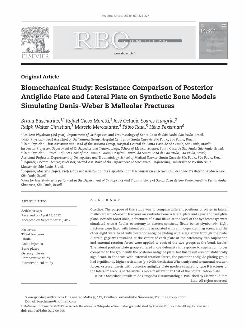
Fig. 1 - A right synthetic fibula was subjected to a simulation of a fracture using a 1-mm thick saw.
Introduction
The incidence of ankle fractures is increasing, and studies have
shown that fracture incidence among the elderly has doubled
over the last 40 years.1 Among athletes, both professional and
amateur, the incidence has also increased. Due to its position and
characteristics, the ankle is subject to numerous traumas, and
its fracture is the most frequent among the load-bearing joints.2
Risk factors for ankle fracture were elucidated by performing a
complementary analysis of multiple cases.3-5 Most of the cases
consisted of isolated malleolar fractures, representing two thirds
of total ankle fractures. One fourth of the patients presented
fractures in the two malleoli, and 7% of the patients presented
bimalleolar fractures with the third posterior fragment. Open
fractures are rare and represent only 2% of all ankle fractures.6
In various studies, the importance of anatomic reduction
and rigid fixation of these fractures to achieve complete
functional restitution has been highlighted.2,7-9 Danis-
Weber type-B fractures, adopted by the AO group, are quite
frequent.10,11 In the literature, several ways to stabilize this
type of fracture have been suggested: a single interfragmentary
lag screw, associated interfragmentary screws, tension bands,
intramedullary wire or nails, and intramedullary screw or
support plates and screws.10,12-14
Within this last option, there are several ways to install the
synthesis material. The most common method involves two
platings. A one-third tubular neutralization plate is placed laterally
and associated with an independent interfragmentary lag screw
outside the plate and a posterolateral antiglide plate can be
attached with an interfragmentary lag screw through the plate.15
Several researchers have conducted trials to compare
these two techniques with the use of plates and screws.
Some of them use case series and group them into the
techniques employed.12,16 Other researchers have conducted
biomechanical studies with cadaveric bones.9,14 There are
many advantages to posterior plating over lateral plating.
With lateral orientation, there is the possibility of intra-
articular positioning of the distal screws.17,18 There is also
greater incidence of surgical wound dehiscence using the
lateral technique.16 Greater resistance has been observed with
posterior plating17, and a greater number of patients report
satisfactory results regarding the positioning of the plate in
the posterior position.19
There are several arguments in favor of posteriorly placing the
plate with ankle fractures, and these arguments have motivated
us to attempt to prove these supposed advantages. Our study
provides a quantitative analysis concerning the difference in
mechanical efficiency obtained with the different dispositions
in the osteosynthesis of the lateral malleolus of the ankle.
Objective
This study aims to simluate Danis-Weber type-B fractures
and assess the mechanical resistance of osteosynthesis with
interfragmentary lag screw and lateral and posterior plates, which are subject to supination stress and external rotation.
Material and Methods
The present study used anatomical models of synthetic fibula bones that simulate the shape and bone characteristics of human fibulas (Synbone®).20 Sixteen fibulas of the same size and density were used and subjected to a simulated Danis-Weber type-B fracture. Oblique cuts from the anterior cortical bone to the posterior bone were made by using an oscillatory saw with a 1-mm thick blade at the height of the tibiotalar joint of each model (Fig. 1).
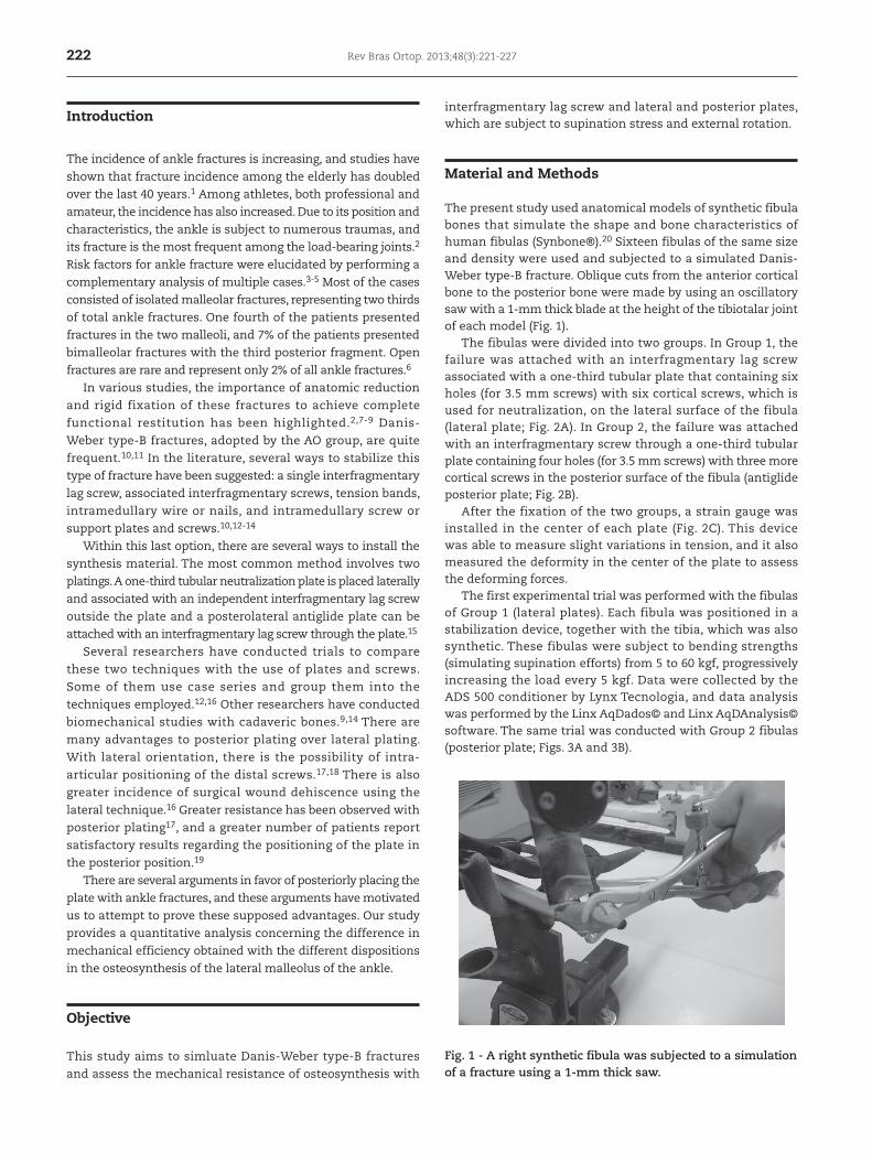
The fibulas were divided into two groups. In Group 1, the failure was attached with an interfragmentary lag screw associated with a one-third tubular plate that containing six holes (for 3.5 mm screws) with six cortical screws, which is used for neutralization, on the lateral surface of the fibula (lateral plate; Fig. 2A). In Group 2, the failure was attached with an interfragmentary screw through a one-third tubular plate containing four holes (for 3.5 mm screws) with three more cortical screws in the posterior surface of the fibula (antiglide posterior plate; Fig. 2B).
After the fixation of the two groups, a strain gauge was installed in the center of each plate (Fig. 2C). This device was able to measure slight variations in tension, and it also measured the deformity in the center of the plate to assess the deforming forces.
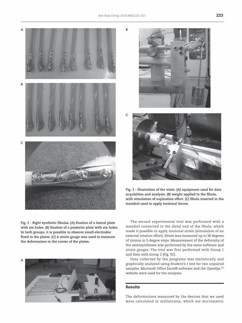
The first experimental trial was performed with the fibulas of Group 1 (lateral plates). Each fibula was positioned in a stabilization device, together with the tibia, which was also synthetic. These fibulas were subject to bending strengths (simulating supination efforts) from 5 to 60 kgf, progressively increasing the load every 5 kgf. Data were collected by the ADS 500 conditioner by Lynx Tecnologia, and data analysis was performed by the Linx AqDados© and Linx AqDAnalysis© software. The same trial was conducted with Group 2 fibulas (posterior plate; Figs. 3A and 3B).
Rev Bras Ortop. 2013;48(3):221-227 223
Fig. 2 - Right synthetic fibulas. (A) fixation of a lateral plate with six holes. (B) fixation of a posterior plate with six holes. In both groups, it is possible to observe small electrodes fixed to the plates. (C) A strain gauge was used to measure the deformation in the center of the plates.
A
A
B
C
The second experimental trial was performed with a mandrel connected to the distal end of the fibula, which made it possible to apply torsional strain (simulation of an external rotation effort). Strain was measured up to 30 degrees of torsion in 5-degree steps. Measurement of the deformity of the osteosyntheses was performed by the same software and strain gauges. The trial was first performed with Group 1 and then with Group 2 (Fig. 3C).
Data collected by the programs was statistically and graphically analyzed using Student’s t test for two unpaired samples. Microsoft Office Excel© software and the OpenEpi.21 website were used for the analyses.
Results
The deformations measured by the devices that we used were calculated in millistrains, which are micrometric
Fig. 3 - Illustration of the trials. (A) equipment used for data acquisition and analysis. (B) weight applied to the fibula, with simulation of supination effort. (C) fibula inserted in the mandrel used to apply torsional forces.
B
C
224 Rev Bras Ortop. 2013;48(3):221-227
Samples of Group 1L1 L2 L3 L4 L5 L6 L7
Applied force (kgf)
0 0.813333 1.3 0.956667 2.26 1.17 0.31 1.11
5 39.82 32.99333 31.96 56.68333 24.00333 48.10667 60.50333
10 79.96333 71.61667 67.64 110.2833 50.81 95.45667 119.9167
15 123.91 110.2467 101.64 177.42 78.77 140.2967 191.5367
20 172.25 162.8 140.11 254.6367 111.78 192.67 274.2533
25 218.2167 208.3167 189.4833 339.1033 141.8667 242.5633 343.0733
30 265.2767 275.82 247.8233 423.4933 175.5633 301.6667 401.0767
35 313.6733 325.4067 292.1033 508.6867 205.9133 354.53 446.0467
40 361.0567 382.18 345.1033 595.6067 242.1767 416.1867 498.4333
45 410.1833 435.92 390.4233 672.4967 276.8467 463.7233 542.0567
50 458.99 502.17 446.6667 788.63 354.4067 520.0433 599.9633
55 513.81 556.0233 470.3467 866.1567 392.7867 566.25 627.1333
60 566.2133 615.6767 500.4933 944.9733 432.4967 611.29 661.1167
Table 1 - Deformations measured (in millistrains) of the samples of group 1 in the first trial regarding the deforming strength (in kgf).
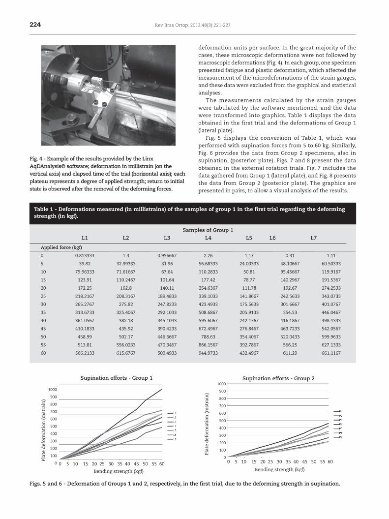
deformation units per surface. In the great majority of the cases, these microscopic deformations were not followed by macroscopic deformations (Fig. 4). In each group, one specimen presented fatigue and plastic deformation, which affected the measurement of the microdeformations of the strain gauges, and these data were excluded from the graphical and statistical analyses.
The measurements calculated by the strain gauges were tabulated by the software mentioned, and the data were transformed into graphics. Table 1 displays the data obtained in the first trial and the deformations of Group 1 (lateral plate).
Fig. 5 displays the conversion of Table 1, which was performed with supination forces from 5 to 60 kg. Similarly, Fig. 6 provides the data from Group 2 specimens, also in supination, (posterior plate). Figs. 7 and 8 present the data obtained in the external rotation trials. Fig. 7 includes the data gathered from Group 1 (lateral plate), and Fig. 8 presents the data from Group 2 (posterior plate). The graphics are presented in pairs, to allow a visual analysis of the results.
Fig. 4 - Example of the results provided by the Linx AqDAnalysis© software; deformation in millistrain (on the vertical axis) and elapsed time of the trial (horizontal axis); each plateau represents a degree of applied strength; return to initial state is observed after the removal of the deforming forces.
Figs. 5 and 6 - Deformation of Groups 1 and 2, respectively, in the first trial, due to the deforming strength in supination.
Supination efforts - Group 1
Plat
e d
efor
mat
ion
(mst
rain
)
1000
900
800
700
600
500
400
300
200
100
0
Bending strength (kgf)
0 5 10 15 20 25 30 35 40 45 50 55 60
Supination efforts - Group 2
Plat
e d
efor
mat
ion
(mst
rain
)
1000
900
800
700
600
500
400
300
200
100
0
Bending strength (kgf)
0 5 10 15 20 25 30 35 40 45 50 55 60
Rev Bras Ortop. 2013;48(3):221-227 225
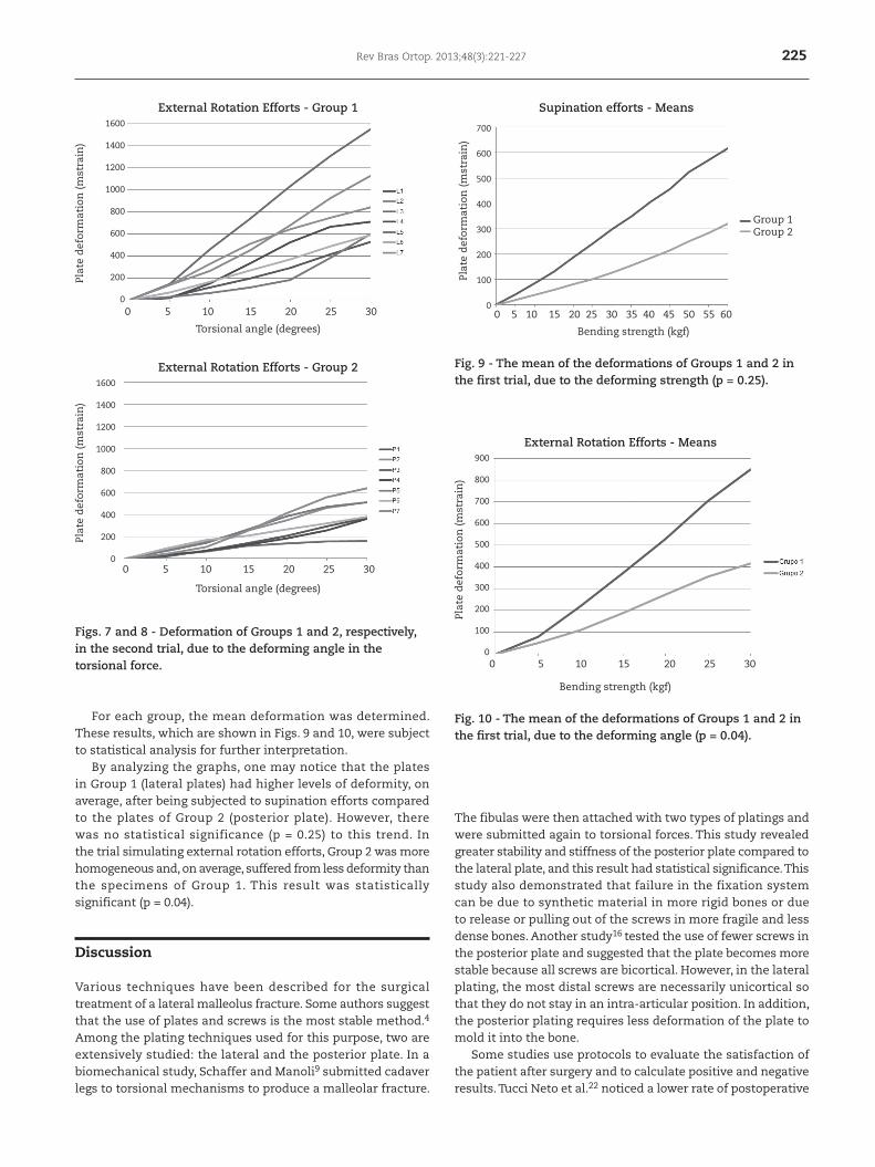
Figs. 7 and 8 - Deformation of Groups 1 and 2, respectively, in the second trial, due to the deforming angle in the torsional force.
The fibulas were then attached with two types of platings and were submitted again to torsional forces. This study revealed greater stability and stiffness of the posterior plate compared to the lateral plate, and this result had statistical significance. This study also demonstrated that failure in the fixation system can be due to synthetic material in more rigid bones or due to release or pulling out of the screws in more fragile and less dense bones. Another study16 tested the use of fewer screws in the posterior plate and suggested that the plate becomes more stable because all screws are bicortical. However, in the lateral plating, the most distal screws are necessarily unicortical so that they do not stay in an intra-articular position. In addition, the posterior plating requires less deformation of the plate to mold it into the bone.
Some studies use protocols to evaluate the satisfaction of the patient after surgery and to calculate positive and negative results. Tucci Neto et al.22 noticed a lower rate of postoperative
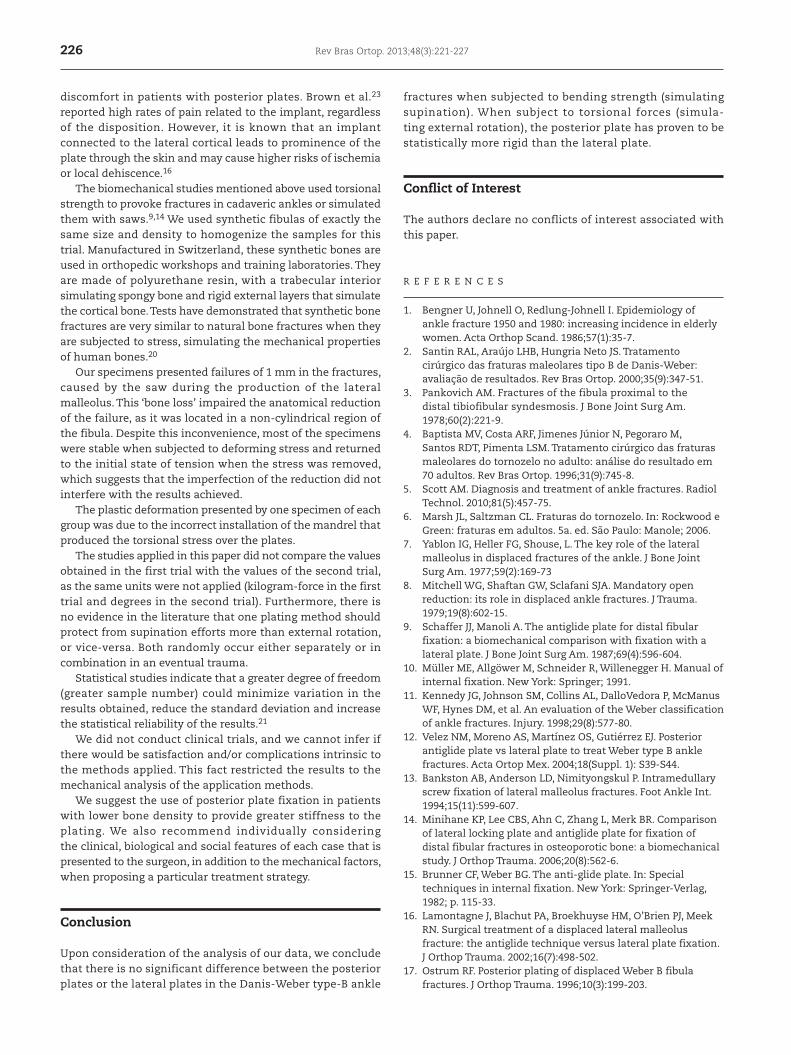
Fig. 9 - The mean of the deformations of Groups 1 and 2 in the first trial, due to the deforming strength (p = 0.25).
For each group, the mean deformation was determined. These results, which are shown in Figs. 9 and 10, were subject to statistical analysis for further interpretation.
By analyzing the graphs, one may notice that the plates in Group 1 (lateral plates) had higher levels of deformity, on average, after being subjected to supination efforts compared to the plates of Group 2 (posterior plate). However, there was no statistical significance (p = 0.25) to this trend. In the trial simulating external rotation efforts, Group 2 was more homogeneous and, on average, suffered from less deformity than the specimens of Group 1. This result was statistically significant (p = 0.04).
Discussion
Various techniques have been described for the surgical treatment of a lateral malleolus fracture. Some authors suggest that the use of plates and screws is the most stable method.4 Among the plating techniques used for this purpose, two are extensively studied: the lateral and the posterior plate. In a biomechanical study, Schaffer and Manoli9 submitted cadaver legs to torsional mechanisms to produce a malleolar fracture.
External Rotation Efforts - Group 1
Plat
e d
efor
mat
ion
(mst
rain
)
1600
1400
1200
1000
800
600
400
200
0
Torsional angle (degrees)
0 5 10 15 20 25 30
External Rotation Efforts - Group 2
Plat
e d
efor
mat
ion
(mst
rain
)
1600
1400
1200
1000
800
600
400
200
0
Torsional angle (degrees)
0 5 10 15 20 25 30
Supination efforts - Means
Plat
e d
efor
mat
ion
(mst
rain
)
700
600
500
400
300
200
100
0
Bending strength (kgf)
Group 1Group 2
0 5 10 15 20 25 30 35 40 45 50 55 60
Fig. 10 - The mean of the deformations of Groups 1 and 2 in the first trial, due to the deforming angle (p = 0.04).
Plat
e d
efor
mat
ion
(mst
rain
)900
800
700
600
500
400
300
200
100
0
Bending strength (kgf)
0 5 10 15 20 25 30
External Rotation Efforts - Means
226 Rev Bras Ortop. 2013;48(3):221-227
fractures when subjected to bending strength (simulating supination). When subject to torsional forces (simula- ting external rotation), the posterior plate has proven to be statistically more rigid than the lateral plate.
Conflict of Interest
The authors declare no conflicts of interest associated with this paper.
R E F E R E N C E S
1. Bengner U, Johnell O, Redlung-Johnell I. Epidemiology of ankle fracture 1950 and 1980: increasing incidence in elderly women. Acta Orthop Scand. 1986;57(1):35-7.
2. Santin RAL, Araújo LHB, Hungria Neto JS. Tratamento cirúrgico das fraturas maleolares tipo B de Danis-Weber: avaliação de resultados. Rev Bras Ortop. 2000;35(9):347-51.
3. Pankovich AM. Fractures of the fibula proximal to the distal tibiofibular syndesmosis. J Bone Joint Surg Am. 1978;60(2):221-9.
4. Baptista MV, Costa ARF, Jimenes Júnior N, Pegoraro M, Santos RDT, Pimenta LSM. Tratamento cirúrgico das fraturas maleolares do tornozelo no adulto: análise do resultado em 70 adultos. Rev Bras Ortop. 1996;31(9):745-8.
5. Scott AM. Diagnosis and treatment of ankle fractures. Radiol Technol. 2010;81(5):457-75.
6. Marsh JL, Saltzman CL. Fraturas do tornozelo. In: Rockwood e Green: fraturas em adultos. 5a. ed. São Paulo: Manole; 2006.
7. Yablon IG, Heller FG, Shouse, L. The key role of the lateral malleolus in displaced fractures of the ankle. J Bone Joint Surg Am. 1977;59(2):169-73
8. Mitchell WG, Shaftan GW, Sclafani SJA. Mandatory open reduction: its role in displaced ankle fractures. J Trauma. 1979;19(8):602-15.
9. Schaffer JJ, Manoli A. The antiglide plate for distal fibular fixation: a biomechanical comparison with fixation with a lateral plate. J Bone Joint Surg Am. 1987;69(4):596-604.
10. Müller ME, Allgöwer M, Schneider R, Willenegger H. Manual of internal fixation. New York: Springer; 1991.
11. Kennedy JG, Johnson SM, Collins AL, DalloVedora P, McManus WF, Hynes DM, et al. An evaluation of the Weber classification of ankle fractures. Injury. 1998;29(8):577-80.
12. Velez NM, Moreno AS, Martínez OS, Gutiérrez EJ. Posterior antiglide plate vs lateral plate to treat Weber type B ankle fractures. Acta Ortop Mex. 2004;18(Suppl. 1): S39-S44.
13. Bankston AB, Anderson LD, Nimityongskul P. Intramedullary screw fixation of lateral malleolus fractures. Foot Ankle Int. 1994;15(11):599-607.
14. Minihane KP, Lee CBS, Ahn C, Zhang L, Merk BR. Comparison of lateral locking plate and antiglide plate for fixation of distal fibular fractures in osteoporotic bone: a biomechanical study. J Orthop Trauma. 2006;20(8):562-6.
15. Brunner CF, Weber BG. The anti-glide plate. In: Special techniques in internal fixation. New York: Springer-Verlag, 1982; p. 115-33.
16. Lamontagne J, Blachut PA, Broekhuyse HM, O’Brien PJ, Meek RN. Surgical treatment of a displaced lateral malleolus fracture: the antiglide technique versus lateral plate fixation. J Orthop Trauma. 2002;16(7):498-502.
17. Ostrum RF. Posterior plating of displaced Weber B fibula fractures. J Orthop Trauma. 1996;10(3):199-203.
discomfort in patients with posterior plates. Brown et al.23 reported high rates of pain related to the implant, regardless of the disposition. However, it is known that an implant connected to the lateral cortical leads to prominence of the plate through the skin and may cause higher risks of ischemia or local dehiscence.16
The biomechanical studies mentioned above used torsional strength to provoke fractures in cadaveric ankles or simulated them with saws.9,14 We used synthetic fibulas of exactly the same size and density to homogenize the samples for this trial. Manufactured in Switzerland, these synthetic bones are used in orthopedic workshops and training laboratories. They are made of polyurethane resin, with a trabecular interior simulating spongy bone and rigid external layers that simulate the cortical bone. Tests have demonstrated that synthetic bone fractures are very similar to natural bone fractures when they are subjected to stress, simulating the mechanical properties of human bones.20
Our specimens presented failures of 1 mm in the fractures, caused by the saw during the production of the lateral malleolus. This ‘bone loss’ impaired the anatomical reduction of the failure, as it was located in a non-cylindrical region of the fibula. Despite this inconvenience, most of the specimens were stable when subjected to deforming stress and returned to the initial state of tension when the stress was removed, which suggests that the imperfection of the reduction did not interfere with the results achieved.
The plastic deformation presented by one specimen of each group was due to the incorrect installation of the mandrel that produced the torsional stress over the plates.
The studies applied in this paper did not compare the values obtained in the first trial with the values of the second trial, as the same units were not applied (kilogram-force in the first trial and degrees in the second trial). Furthermore, there is no evidence in the literature that one plating method should protect from supination efforts more than external rotation, or vice-versa. Both randomly occur either separately or in combination in an eventual trauma.
Statistical studies indicate that a greater degree of freedom (greater sample number) could minimize variation in the results obtained, reduce the standard deviation and increase the statistical reliability of the results.21
We did not conduct clinical trials, and we cannot infer if there would be satisfaction and/or complications intrinsic to the methods applied. This fact restricted the results to the mechanical analysis of the application methods.
We suggest the use of posterior plate fixation in patients with lower bone density to provide greater stiffness to the plating. We also recommend individually considering the clinical, biological and social features of each case that is presented to the surgeon, in addition to the mechanical factors, when proposing a particular treatment strategy.
Conclusion
Upon consideration of the analysis of our data, we conclude that there is no significant difference between the posterior plates or the lateral plates in the Danis-Weber type-B ankle

Rev Bras Ortop. 2013;48(3):221-227 227
18. Treadwell JR, Fallat LM. The antiglide plate for the Danis- Weber type-B fibular fracture: a review of 71 cases. J Foot Ankle Surg. 1993;32(6):573-9.
19. Winkler B, Weber BG, Simpson LA. The dorsal antiglide plate in treatment of Danis-Weber type B fractures of the distal fibula. Clin Orthop Relat Res. 1990;(259):204-9.
20. Synbone AG. Statement artificial vs. human bones, 2007. Disponível em www.synbone.ch. (acessado em 18 ago. 2011).
21. Box JF. Guinness, Gosset, Fisher, and small samples. Statistical Science;1987.
22. Tucci Neto C, Fernandes HGA, Neto PFT, Reis FB, Faloppa F. Tratamento de fraturas do tornozelo tipo Danis-Weber B com placa antideslizante póstero-lateral. Rev Bras Ortop. 2003;38(6):320-8.