bioorganic & medicinal chemistry - university of belgradeijuranic/bioorganicmedicinalchemistry...
TRANSCRIPT

Bioorganic & Medicinal Chemistry 23 (2015) 4649–4659
Contents lists available at ScienceDirect
Bioorganic & Medicinal Chemistry
journal homepage: www.elsevier .com/locate /bmc
5-Aryl-1H-pyrazole-3-carboxylic acids as selective inhibitorsof human carbonic anhydrases IX and XII
http://dx.doi.org/10.1016/j.bmc.2015.05.0520968-0896/� 2015 Elsevier Ltd. All rights reserved.
Abbreviations: CA, carbonic anhydrase; ZBG, zinc binding group; CAIs, carbonicanhydrase inhibitors; pKa, acid dissociation constant; Ki, inhibition constant; PDB,Protein Data Bank; MIFs, molecular interaction fields; HBD, hydrogen bond donor;HBA, hydrogen bond acceptor.⇑ Corresponding authors. Tel.: +39 055 4573005 (C.T.S.); tel.: +381 11 3336741
(B.J.D.).E-mail addresses: [email protected] (C.T. Supuran), [email protected].
rs (B.J. Drakulic).
Ilija N. Cvijetic a, Muhammet Tanç b, Ivan O. Juranic c, Tatjana Z. Verbic d, Claudiu T. Supuran b,⇑,Branko J. Drakulic c,⇑a Innovation Center of the Faculty of Chemistry, University of Belgrade, Studentski Trg 12-16, 11000 Belgrade, Serbiab NEUROFARBA Department, Sezione di Scienze Farmaceutiche e Nutraceutiche, Università degli Studi di Firenze, Via Ugo Schiff 6, 50019 Sesto Fiorentino, Florence, Italyc Department of Chemistry, Institute of Chemistry, Technology and Metallurgy, University of Belgrade, Njegoševa 12, 11000 Belgrade, Serbiad Faculty of Chemistry, University of Belgrade, Studentski Trg 12-16, 11000 Belgrade, Serbia
a r t i c l e i n f o a b s t r a c t
Article history:Received 15 April 2015Revised 26 May 2015Accepted 29 May 2015Available online 4 June 2015
Keywords:Carbonic anhydrasePhenyl-pyrazole-carboxylic acidsDockingMolecular interaction fieldsActive sites comparison
Inhibitory activity of a congeneric set of 23 phenyl-substituted 5-phenyl-pyrazole-3-carboxylic acidstoward human carbonic anhydrase (hCA, EC 4.2.1.1) isoforms I, II, IX and XII was evaluated by astopped-flow CO2 hydrase assay. These compounds exerted a clear, selective inhibition of hCA IX andXII over hCAI and II, with Ki in two to one digit micromolar concentrations (4–50 lM). Derivatives bearingbulkier substituents in para-position of the phenyl ring inhibited hCA XII at one-digit micromolar concen-trations, while derivatives having alkyl substituents in both ortho- and meta-positions inhibited hCA IXwith Kis ranging between 5 and 25 lM. Results of docking experiments offered a rational explanationon the selectivity of these compounds toward CA IX and XII, as well as on the substitution patterns lead-ing to best CA IX or CA XII inhibitors. By examining the active sites of these four isoforms with GRID gen-erated molecular-interaction fields, striking differences between hCA XII and the other three isoformswere observed. The field of hydrophobic probe (DRY) appeared significantly different in CA XII active site,comparing to other three isoforms studied. To the best of our knowledge such an observation was notreported in literature so far. Considering the selectivity of these carboxylates towards membrane-associ-ated over cytosolic CA isoforms, the title compounds could be useful for the development of isoform-specific non-sulfonamide CA inhibitors.
� 2015 Elsevier Ltd. All rights reserved.
1. Introduction
There are a few reports on biological activity of 5-aryl-pyrazole-3-carboxylic acid in the literature. Compounds were described aspartial agonists of the nicotinic acid receptors,1 as inhibitors ofreplication protein A protein–protein interactions,2 as scaffoldsthat lead to inhibitors of protein tyrosine phosphatase 1B,3 andas an inhibitors of a tissue-nonspecific alkaline phosphatase.4
Along with this, 5-heteroaryl-pyrazole-3-carboxylic acids, amongother compounds, were recently described as inhibitors of humancarbonic anhydrases (CAs, EC 4.2.1.1) I, II, IX and XII.5
The majority of reported small molecule CA inhibitors (CAIs)comprises a primary sulfonamide moiety as a zinc binding group(ZBG). The sulfonamide anion (–SO2NH�) coordinates the activesite Zn2+ ion and blocks catalysis. In Protein Data Bank more than300 unique sulfonamide-bearing small molecules co-crystallizedwith carbonic anhydrases (mainly human isoform II) can be found.On the other hand, significantly less non-sulfonamide CAIs, belong-ing to other classes of small organic molecules, such as for examplecarboxylic acids, were investigated so far. It was shown that somemolecules comprising –COOH moiety co-crystallized with differentCA isoforms, are bound within the CA active site, interacting withthe active site Zn2+ by water-mediated H-bonding, while otherwere found bound to the enzyme but did not interact with theactive site Zn2+ ion, or they were bound out of the active site.Best examples of the latter are, probably, ortho-hydroxy cinnamicacids—mechanism-based inhibitors originated in situ by hydrolysisof coumarins by Zn2+—hydroxyl ion couple in the CA active site.6–9
Isoform selectivity of this class of compounds depends on substi-tution pattern on coumarin phenyl ring. Recently reported

4650 I. N. Cvijetic et al. / Bioorg. Med. Chem. 23 (2015) 4649–4659
2-benzylsulfinylbenzoic acid10 was found bound to hCA II out ofthe active site. This compound constrained the proton-shuttle resi-due His64 in its ‘out’ conformation by a network of water mole-cules-mediated H-bonds. Inhibition study of hCA I and II, as wellas mycobaterial and fungal CA with a library of natural products,found adduct of cinnamic and maleic acid amide, 3-(3-chloro-4-hydroxyphenyl)-2-[(4-methoxy-4-oxobut-2-enoyl)amino]propanoicacid (CID 72785959, CAS 852690-88-1), as a low micromolar CAinhibitor.11 Structure of compound co-crystallized with hCA IIrevealed that carboxyl group of cinnamic acid moiety is notinvolved in interaction with catalytic Zn2+ ion. Inhibition ofmycobacterial CA Rv3588c by (+)-mispyric acid, natural productbearing two carboxyl groups, was also described,12 without addi-tional structural data. Smaller carboxylate-bearing molecules, suchas hydroxy and thio-substituted benzoic acids, were found boundto the CA active site, forming hydrogen bond to the Zn-boundwater molecule/hydroxide ion.13 In the same crystal structurestwo more molecules of substituted benzoic acids were found, occu-pying protein cavities out of the binding site. Inhibition of CAs withbiologically-relevant aliphatic carboxylates, along with benzoic and1,2,5,6-tetrafluoro-benzoic acids were also described, without addi-tional structural data.14 A congeneric set of di-methoxy-bro-mophenyl-substituted 2-benzyl-cyclopropyl carboxylic acids andderivatives, having the same topological distance between aryl andcarboxyl moieties as in a title compounds, were recently reportedas inhibitors of human CA isoforms I, II, IX and XII.15 Those com-pounds exerted strong preferences toward hCA I and II inhibition.Carboxylic acid derivatives briefly described in the previous linesinhibited CAs with Kis spanning range between the millimolar tomicromolar concentrations, with the exception of 2-benzyl-cyclo-propyl carboxylic acids, found as nanomolar inhibitors.
In this article we report the synthesis and inhibitory activity of acongeneric set of 23 phenyl-substituted 5-phenyl-pyrazole-3-car-boxylic acids (1–23) toward human CA isoforms I, II, IX and XII.
2. Results and discussion
2.1. Chemistry
The rationale of this work was to investigate different chemo-types incorporating the COOH moiety as a ZBG for obtainingCAIs. As heterocyclic carboxylic acids were rarely investigated asCAIs, we decided to synthesize a congeneric series of such deriva-tives incorporating the synthetically easily available pyrazole-3-carboxylic acid moiety.
Acetophenones II were prepared by Friedel–Crafts acylation ofthe corresponding aromatic substrates (I), or commercially avail-able substances were used. The aryl-diketo acids III were preparedfollowing previously reported procedures,16 by base-promotedcondensations of substituted acetophenones (II) and diethyl-ox-alate. Compounds 1–23 (IV) were synthesized from aryldiketoacids (III) and hydrazine-hydrate in glacial acetic acid (Scheme 1).
Identity and purity of compounds was confirmed by 1H and 13CNMR spectroscopy and LC-HR/ESI-MS.
2.2. Tautomeric preferences and protonation states
Aryl-pyrazole-carboxylic acid could appear in three tautomericforms (Scheme 2).
Scheme 1. Synthesis of 5-aryl-1H-pyrazole-3-carboxylic acids. (a) CH3COCl, AlCl3/CH2
CH3COOH, 8h, rt.
Tautomers A and B, with protonated pyrazole nitrogens, aremore probable than the tautomer C. In solid state, tautomeric formA was observed.17 To gather more data on tautomeric preferencesof examined compounds in solution, we recorded NOESY spectra of4-Me-derivative (1), in DMSO-d6. 4-Me-derivative (1) was chosenbecause of simple 1H NMR pattern of protons on phenyl ring.Spectra were recorded in DMSO due to good solubility of examinedcompounds and its polarity, which preclude possible aggregationof solute by intramolecular H-bonding, observed in solid state(see below). Clear NOESY signal between broad 1H peak at13.38 ppm and doublet at 7.71 ppm (Fig. 1) was observed.
Doublet at 7.71 ppm belongs to hydrogens in ortho-position ofthe phenyl ring, while broad signal at 13.38 ppm belongs to pyra-zole NH. Those data suggested that tautomer A is principal one inDMSO solution. Upon addition of one molar equivalent of piperi-dine, to deprotonate carboxyl group, broad signal at 13.38 ppmwas shifted to 9.75 ppm and NOESY signal disappeared (Fig. S1 inSupplementary Material). Our preliminary results suggested thattautomeric form A should not be predominant one when carboxylgroup of title compounds is in its anionic form, although it is wellknown that tautomeric preferences are different in different sol-vents. Tautomeric preferences of title compounds will be a subjectof separate studies. IR spectra of compounds were obtained by ATR(attenuated total reflectance) from solid samples. In spectra ofsome compounds we clearly observed broad IR bands in the region3100–3250 cm�1, where NH stretching bands were overlappedwith aromatic C–H bands. Such pattern in IR spectra suggestedaggregation via hydrogen bonding of some derivatives. Examplesare given in Supplementary Material, Figure S2. This is in accor-dance with crystal structure of unsubstituted derivative,17 wherein crystal packing intermolecular H-bonds between carboxyl groupand pyrazole –NH–N@ part is observed. We also estimated proto-nation states18 of compound 1 (Fig. S3 in Supplementary Material).pKa Value of carboxyl group is predicted to be within the range3.6–4.0; while pKa value of pyrazole nitrogen is predicted to bewithin the range 10.2–10.7, depending on tautomeric state(Fig. S4 in Supplementary Material). pKa1 Value (carboxyl group)of derivative 22 was experimentally determined using spectropho-tometric titration. Three wavelengths used for spectrophotometricpKa1 determination (270, 280, 290 nm) were selected as wave-lengths where the difference in absorptivity of species existing inthe solution is maximal. The UV/Vis spectra and the graph usedfor the pKa1 value determination are shown in Figure 2. For detailssee Section 4. The determined pKa1 of derivative 22 is 3.31 ± 0.07.
2.3. CA inhibition
Inhibitory activity of compounds toward hCA I, II, IX and XII wasevaluated by a stopped-flow CO2 hydrase assay. Results arereported as Ki values (Table 1).
All compounds tested were proved to be inactive towards CA Iand II, which were not inhibited up to concentrations of 50 lMinhibitor in the assay system. Depending on the substitution pat-tern on the phenyl ring, different subsets of compounds wereactive towards CA IX or towards CA XII. Along with Ki data reportedin Table 1, strong selective inhibition of 1–23 toward CA IX and CAXII over CA I and II can be noted (see also Supplementary Table 1).Dose-response curves for inhibitory activity of these compounds
Cl2, 6 h, 0 �C to rt; (b) 2 equiv Na/CH3OH, 15h, rt, then conc. HCl, (c) N2H4�H2O/

Scheme 2. Tautomers of aryl-pyrazole-carboxylic acids.
I. N. Cvijetic et al. / Bioorg. Med. Chem. 23 (2015) 4649–4659 4651
toward all CA isoforms appeared rather flat, still percentage ofenzyme activity of CA I and II were between 60% and 70% evenat 1 � 10�4 M concentrations of inhibitors. Providing selectivitytoward membrane-associated CA isoforms, considered as a goodanti-tumor targets,19 over cytosolic isoforms, the title compoundsmight be used as a good starting point for the design of isoform-se-lective inhibitors incorporating –COOH ZBGs. Compounds activetoward CA IX and CA XII inhibited those two isoforms in one- totwo-digit micromolar concentrations, comparable with thepotency of the majority of so-far reported CA inhibitors bearingthis ZBG.
2.4. Structure-activity study
In an attempt to explain selectivity of some of these compoundstoward CA IX and CA XII and to explain preferences of differentsubsets of compounds toward CA IX or CA XII, we performed dock-ing studies. For this purpose we chose Protein Data Bank (PDB)crystal structures derived from the wild-type CA isoforms and hav-ing a high resolution. CA I was modeled from PDB entry 1HCB, at1.60 Å resolution20; CA II from 3B4F, at 1.89 Å resolution21; CA IXfrom 3IAI, at 2.20 Å resolution22 and CA XII from 1JD0, at 1.50 Åresolution.23 Some of those enzymes were co-crystallized withsmall molecule inhibitors. For the docking experiments we
Figure 1. NOESY spectrum of 4-Me-derivative (1) in DMSO. Signa
removed all small molecules and waters, neutralized proteins withcounter-ions, than embedded them in water clusters and relaxedall structures by molecular dynamics minimization (see Section 4for details). In this point reasonable question arise: are minimiza-tion of the protein structure upon bound ligand removal can causecollapse of the active site? We believe that regarding CAs this pos-sibility is not an issue. CAs poses a wide and conical active site insignificant extent exposed to solvent. During catalytic cycle, thatis, CO2 hydration, small changes of protein conformation isobserved, and such changes mainly can be attributed to theHis64 (proton shuttle) conformation change. Consequently, differ-ent from some other enzyme classes, CAs in their crystal structuresare not ‘trapped’ in some local free-energy minimum for whichanother, close, local minimum can exist. Additionally, majority ofCA inhibitors are medium-sized molecules, which typically didnot significantly change conformation of active site, comparing touninhibited protein. By visual inspection of the 3D structures ofvery efficient inhibitors (typically nanomolar, from the sulfon-amide class) cocrystallized with the CAs it can be observed thatsuch molecules typically did not interact with its whole molecularsurface with the active site residues. Most probably this can beattributed to the wide CAs active site, as well as to very high affin-ity of ZBG to catalytic Zn2+ ion. On the other hand, from the mod-elling set-up view, it should be noted that MD minimization (as faras recent CHARMM force field variants and recent NAMD releasesare used) commonly very limited mobility of backbone appears,while position of side-chains are usually efficiently relaxed. In allso far reported crystal structures of CA co-crystallized with smallmolecules having a –COOH group within enzyme active site, thewater molecule bound to catalytic Zn2+ ion was present. Thus, forthe docking study we added this water molecule. Initial position
l between ortho-Ph-hydrogens and pyrazole NH is illustrated.

Figure 2. (a) UV/Vis Spectra of compound 22 in pH range 1.73–5.68 used for pKa1 determination (t = 25 ± 1 �C); wavelength range used for calculations is magnified; (b)Determination of Ka1 at 280 nm according to Eq. 1; slope = 4.0 ± 0.2 � 10�4, intercept = 0.764 ± 0.003, r2 = 0.973.
Table 1Structures of compounds 1–23 and inhibitory activities (given as Ki) toward four CAisoforms
Compound R- Ki* (lM)
hCA I hCA II hCA IX hCA XII
1 4-Me- >50 >50 >50 >502 4-Et- >50 >50 42 4.53 4-i-Pr- >50 >50 >50 >504 4-n-Bu- >50 >50 39 5.55 4-t-Bu- >50 >50 5 216 2,4-di-Me- >50 >50 45 >507 3,4-di-Me- >50 >50 48 >508 2,4,5-tri-Me- >50 >50 23.5 >509 2,3,5,6-tetra-Me- >50 >50 5 >5010 2,4,6-tri-Et- >50 >50 >50 >5011 2,4-di-i-Pr- >50 >50 16 >5012 2,4,6-tri-i-Pr- >50 >50 39 3913 b-Tetralinyl- >50 >50 >50 43.514 b-Naphtyl- >50 >50 >50 5.515 4-Ph- >50 >50 >50 416 4-Pyrrolidine- >50 >50 >50 33.517 4-F- >50 >50 >50 38.518 4-Cl- >50 >50 >50 5019 3-Br- >50 >50 23.5 >5020 4-OH- >50 >50 >50 4121 2-OMe- >50 >50 >50 5022 4-OMe- >50 >50 49.5 >5023 4-OMe-2,5-di-Me- >50 >50 22.5 >50AAZ** / 0.25 0.012 0.025 0.006
* Mean from three different assays, errors in the range of 5–10% of the reportedvalues (data not shown).
** Acetazolamide (5-acetamido-1,3,4-thiadiazole-2-sulfonamide, AAZ) was usedas a standard inhibitor.
4652 I. N. Cvijetic et al. / Bioorg. Med. Chem. 23 (2015) 4649–4659
of the water was estimated from PDB entry 4E3G,13 para-hydroxy-benzoyc acid co-crystallized with CA II, and further refined by theWaterFLAP protocol. All compounds were treated in their anionicforms, with deprotonated carboxyl group, in accordance with pre-dicted and experimentally determined pKa values. Both tautomersA and B, Scheme 2, were used for docking. Initial attempts of dock-ing compounds into the active site of each isoform appeared veryinconclusive, so we proceeded considering the whole enzyme asa target.
The docking study found all compounds bound out of the activesite of CA I (Fig. S4 in Supplementary Material), in the cleft lined bythe residues Pro13, Asn24, Thr100 and His103. The carboxylate
anion of these compounds makes polar interactions with Ser99and Thr100 hydroxyl groups, while the pyrazole nitrogens makepolar interactions with Asn245 side-chain and His103 backbonecarbonyl. Larger alkyl substituents were typically anchoredbetween side chains of Pro13 and Pro247. For CA II, the dockingstudy also found majority of the compounds out of the active site(Fig. S5 in Supplementary Material). The compounds are clusteredin a region lined with residues: Trp5, Lys170, Asn232 and Pro237.The carboxylate anion makes polar interactions with His4 NHbackbone and Lys170 side chain NH3
+, whereas the pyrazole NHwas hydrogen bounded with Phe231 backbone carbonyl. For CA Iand CA II similar docking solutions were found irrespective of theposition of the His64 (‘in’ or ‘out’).
Such results revealed higher affinity of these compoundstoward regions of CA I and CA II out of enzyme active site andcan offer possible explanation of inactivity of 1–23 toward thesecytosolic CA isoforms studied here.
Our docking study found all compounds bound close to theactive site Zn2+ ion of CA IX and CA XII (including those for whichKi > 50 lM were measured). All compounds were clustered withvery similar orientation of the pyrazole-COO� moiety in the activesite of each isoform, but orientation of this part of the moleculeappears different in CA IX and CA XII, respectively. In CA IX, the car-boxylate anion makes polar interactions with the hydroxyl groupof Thr199 and Thr200 and with the water coordinated to zinc.The pyrazole NH makes polar interactions with Gln67 side-chaincarbonyl. As a trend, compounds found as active toward CA IX(Table 1) have alkyl substituents in ortho- and meta-positions ofthe phenyl ring. Compounds having bulkier substituents in para-position appeared to be inactive. Visual inspection of dockingresults revealed that ortho- and meta-substituted compounds, aswell as those having medium-sized alkyl substituents in para-posi-tion assessed better shape complementarily with the cleft of activesite than the rest of the investigated compounds, which were inac-tive toward CA IX. This cleft is lined by backbone of residues Trp5,Asn62 and His64, and situated distal from the sequence 131–134.(Fig. 3 and Fig. S6 in Supplementary Material). These residues wereshown earlier to be involved in the binding of various classes ofCAIs or CA activators.24
This observation could explain the structural features that dif-ferentiate active from inactive compounds towards CA IX. Alongwith this, we observed that compounds 9 (2,3,5,6-tetra-methylderivative) and 5 (4-t-butyl derivative) with Ki < 10 lM, havemutually very similar 3D-dependent whole-molecular properties,particularly apolar surface area and volume (Table S2 inSupplementary Material).

Figure 3. Best-ranked solutions of compounds 9 (a) and 11 (b) docked into CAIX.
I. N. Cvijetic et al. / Bioorg. Med. Chem. 23 (2015) 4649–4659 4653
Docking solutions for CA XII found all compounds close to theactive site Zn2+ ion, but in orientations quite different comparedto CA IX. The carboxylate anion makes polar interactions withTyr7 and Thr199 hydroxyl groups, or Asn62 side-chain NH2. Thepyrazole NH and N make polar interactions with Asn62 side-chainor water-mediated interactions with the Thr199. Aromatic moietyof the compounds were directed toward the sequence 131–134(130’s segment). This part of the active site is specific for CA XIIand it was suggested that can be used for the design of isoformspecific inhibitors.23 Visual inspection of the docking resultsshowed that aryl moiety of all compounds found active towardsCA XII makes van-der Walls contacts with the 130’s segment, whilethe same moiety in inactive compounds cannot reach this segment(Fig. 4 and Fig. S7 in Supplementary Material). This rationalizes thestructure-activity relationship data of 1–23 observed towards CAXII and shown in Table 1.
It should be noted that we did not observe any regularitybetween calculated affinities of compounds (docking scores) and
Figure 4. Best-ranked solutions of compoun
the order of activity toward CA IX or CA XII. ChemGauss4 scoringfunction accounts for shape and electrostatic complementarities,as well as the binding site and the ligand desolvation penalty.Not overall scoring, nor shape complementarities show any trendwith the experimentally found Ki values. Considering the wideand conical shape of the CA active site, the congeneric set of exam-ined compounds—different in apolar moieties only, and the narrowrange of Ki values, such an observation is not surprising.
In literature, we found one systematic comparison of CAs I, II, IXand XII active sites.25 CA active site residues involved in ligandbinding were listed and similarity and differences commented. Inorder to elucidate more structural information that can explainselectivity of 1–23 toward membrane-associated isoforms, westudied active sites of CA I, II, IX and XII by GRID force field.26
Molecular interaction fields (MIFs) were calculated by hydrophobic(DRY), HBD (N1) and HBA (O) probes in the box encompassingactive site residues that interact with small molecules in so farreported crystal structures of CAs (for details see Section 4 and
ds 15 (a) and 14 (b) docked into CAXII.
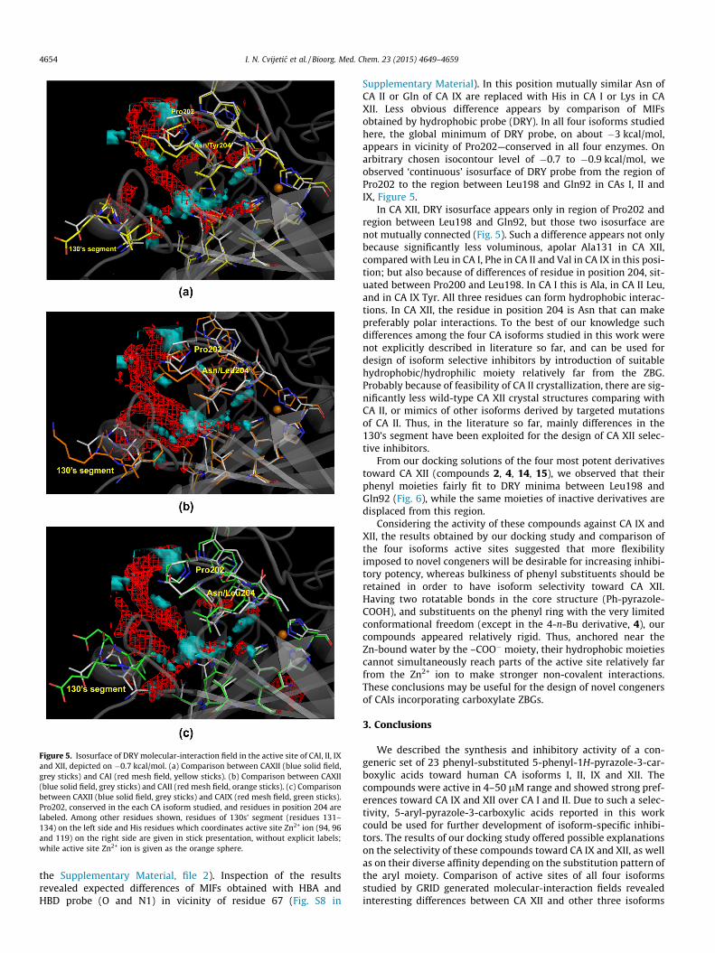
Figure 5. Isosurface of DRY molecular-interaction field in the active site of CAI, II, IXand XII, depicted on �0.7 kcal/mol. (a) Comparison between CAXII (blue solid field,grey sticks) and CAI (red mesh field, yellow sticks). (b) Comparison between CAXII(blue solid field, grey sticks) and CAII (red mesh field, orange sticks). (c) Comparisonbetween CAXII (blue solid field, grey sticks) and CAIX (red mesh field, green sticks).Pro202, conserved in the each CA isoform studied, and residues in position 204 arelabeled. Among other residues shown, residues of 130s’ segment (residues 131–134) on the left side and His residues which coordinates active site Zn2+ ion (94, 96and 119) on the right side are given in stick presentation, without explicit labels;while active site Zn2+ ion is given as the orange sphere.
4654 I. N. Cvijetic et al. / Bioorg. Med. Chem. 23 (2015) 4649–4659
the Supplementary Material, file 2). Inspection of the resultsrevealed expected differences of MIFs obtained with HBA andHBD probe (O and N1) in vicinity of residue 67 (Fig. S8 in
Supplementary Material). In this position mutually similar Asn ofCA II or Gln of CA IX are replaced with His in CA I or Lys in CAXII. Less obvious difference appears by comparison of MIFsobtained by hydrophobic probe (DRY). In all four isoforms studiedhere, the global minimum of DRY probe, on about �3 kcal/mol,appears in vicinity of Pro202—conserved in all four enzymes. Onarbitrary chosen isocontour level of �0.7 to �0.9 kcal/mol, weobserved ‘continuous’ isosurface of DRY probe from the region ofPro202 to the region between Leu198 and Gln92 in CAs I, II andIX, Figure 5.
In CA XII, DRY isosurface appears only in region of Pro202 andregion between Leu198 and Gln92, but those two isosurface arenot mutually connected (Fig. 5). Such a difference appears not onlybecause significantly less voluminous, apolar Ala131 in CA XII,compared with Leu in CA I, Phe in CA II and Val in CA IX in this posi-tion; but also because of differences of residue in position 204, sit-uated between Pro200 and Leu198. In CA I this is Ala, in CA II Leu,and in CA IX Tyr. All three residues can form hydrophobic interac-tions. In CA XII, the residue in position 204 is Asn that can makepreferably polar interactions. To the best of our knowledge suchdifferences among the four CA isoforms studied in this work werenot explicitly described in literature so far, and can be used fordesign of isoform selective inhibitors by introduction of suitablehydrophobic/hydrophilic moiety relatively far from the ZBG.Probably because of feasibility of CA II crystallization, there are sig-nificantly less wild-type CA XII crystal structures comparing withCA II, or mimics of other isoforms derived by targeted mutationsof CA II. Thus, in the literature so far, mainly differences in the130’s segment have been exploited for the design of CA XII selec-tive inhibitors.
From our docking solutions of the four most potent derivativestoward CA XII (compounds 2, 4, 14, 15), we observed that theirphenyl moieties fairly fit to DRY minima between Leu198 andGln92 (Fig. 6), while the same moieties of inactive derivatives aredisplaced from this region.
Considering the activity of these compounds against CA IX andXII, the results obtained by our docking study and comparison ofthe four isoforms active sites suggested that more flexibilityimposed to novel congeners will be desirable for increasing inhibi-tory potency, whereas bulkiness of phenyl substituents should beretained in order to have isoform selectivity toward CA XII.Having two rotatable bonds in the core structure (Ph-pyrazole-COOH), and substituents on the phenyl ring with the very limitedconformational freedom (except in the 4-n-Bu derivative, 4), ourcompounds appeared relatively rigid. Thus, anchored near theZn-bound water by the –COO� moiety, their hydrophobic moietiescannot simultaneously reach parts of the active site relatively farfrom the Zn2+ ion to make stronger non-covalent interactions.These conclusions may be useful for the design of novel congenersof CAIs incorporating carboxylate ZBGs.
3. Conclusions
We described the synthesis and inhibitory activity of a con-generic set of 23 phenyl-substituted 5-phenyl-1H-pyrazole-3-car-boxylic acids toward human CA isoforms I, II, IX and XII. Thecompounds were active in 4–50 lM range and showed strong pref-erences toward CA IX and XII over CA I and II. Due to such a selec-tivity, 5-aryl-pyrazole-3-carboxylic acids reported in this workcould be used for further development of isoform-specific inhibi-tors. The results of our docking study offered possible explanationson the selectivity of these compounds toward CA IX and XII, as wellas on their diverse affinity depending on the substitution pattern ofthe aryl moiety. Comparison of active sites of all four isoformsstudied by GRID generated molecular-interaction fields revealedinteresting differences between CA XII and other three isoforms

Figure 6. Best-ranked docking solutions for compounds 4 (yellow sticks), 14 (green sticks) and 15 (red sticks) overlapped with the isosurface of DRY probe (�0.7 kcal/mol) inthe CA XII active site. Pro202 and Asn204, given in stick presentation, are labeled. Among other residues shown, residues of 130s’ segment (residues 131–134) on the left sideand His residues which coordinates active site Zn2+ ion (94, 96 and 119) on the right side are given in stick presentation, without explicit labels; while active site Zn2+ ion isgiven as a red sphere.
I. N. Cvijetic et al. / Bioorg. Med. Chem. 23 (2015) 4649–4659 4655
in regions that have high affinity towards hydrophobic moieties. Tothe best of our knowledge such an observation was not reported sofar and can foster development of the selective CA XII inhibitors.
4. Experimental
4.1. Chemistry
All chemicals were purchased from Fluka, Aldrich, or Merck,having >98% purity, and were used as received. For the thin-layerchromatography, silica gel pre-coated plates with fluorescent indi-cator (254 nm) were used. Melting points were determined in opencapillary tubes on Stuart SMP-10 apparatus and are uncorrected.ESI-MS (Electrospray ionization mass spectrometry) analysis wasperformed in methanol on an Agilent Technologies 6210-1210TOF-LC-ESI-MS instrument in positive mode. IR (Infra-red) spectrawere recorded on a Thermo Nicolet 6700 FT-IC spectrophotometer,by ATR (Attenuated total reflectance). 1H, 13C NMR (Nuclear mag-netic resonance) and NOESY (Nuclear Overhauser effect spec-troscopy) spectra were recorded in DMSO-d6 on Bruker AVANCE500/125 MHz or on a Varian Gemini2000 200/50 MHz instruments.Chemical shifts are reported in parts per million (ppm) relative totetramethylsilane (TMS) and spin multiplicities are given as fol-lows: s (singlet), d (doublet), t (triplet), q (quartet), qn (quintet),sx (sextet), h (heptet) m (multiplet), br (broad). UV/Vis Spectrawere recorded on Thermo Scientific Evolution 60S spectropho-tometer (Thermo Fisher Scientific Inc, Waltham, Massachusetts,USA). pH Values were measured using CRISON pH-Burette 24 2Sequipped with CRISON 50 29 micro-combined pH electrode(Crison Instruments, S.A. Spain). The electrode was calibrated bystandard CRISON buffer solutions (pH 4.01, 7.00, and 9.21).
4.1.1. Typical experimental procedure for synthesis of 1–234.1.1.1. 5-(4-Methylphenyl)-1H-pyrazole-3-carboxylic acid(1). 0.4 g of 4-(4-methylphenyl)-2,4-dioxobutanoic acid was dis-solved in 20 ml of glacial acetic acid in 50 ml round-bottom flask.In a stirred solution, 280 lL (3 eq.) of hydrazine monohydrate
(N2H4�H2O) was added dropwise. The color of solution was chan-ged to pale yellow immediately after addition of first amount ofhydrazine. The progress of reaction was monitored by TLC. Aftercompletion, reaction mixture was poured in 50 ml H2O and leftto stir overnight. White precipitate was collected, dried on airand recrystallized from the mixture of hexane/EtOH (�9:1), giving0.34 g of product (87% of theoretical yield).
4.1.2. Characterization of compounds 1–234.1.2.1. 5-(4-Methylphenyl)-1H-pyrazole-3-carboxylic acid(1). White solid; mp = 253–254 �C (hexane/EtOH); C11H10N2O2,Mw = 202.21; ESI-MS: Calculated for C11H11N2O2 [M+H]+:203.08150, measured: 203.08063; IR (m, cm�1): 3264 (N–H), 1696(C@O), 1500, 1198; 1H NMR (500 MHz, DMSO-d6) d(ppm): 2.30(s, 3 H), 7.14 (s, 1 H), 7.22 (d, J = 7.83 Hz, 2 H), 7.71 (d,J = 8.31 Hz, 2 H), 13.38 (br). 13C NMR (125 MHz, DMSO-d6)d(ppm): 20.85, 104.91, 125.25, 128.13, 129.48, 137.58, 140.09,147.44, 162.04.
4.1.2.2. 5-(4-Ethylphenyl)-1H-pyrazole-3-carboxylic acid (2). Startingfrom 0.625 g of 4-(4-ethylphenyl)-2,4-dioxobutanoic acid,0.55 g of product was obtained (89% of theoretical yield). Whitesolid; mp = 229–230 �C (hexane/EtOH); C12H12N2O2,Mw = 216.24; ESI-MS: Calculated for C12H13N2O2 [M+H]+:217.09715, measured: 217.09634; IR (m, cm�1): 3265 (N–H),1692 (C@O), 1498, 1237; 1H NMR (200 MHz, DMSO-d6) d(ppm):1.17 (t, J = 7.30 Hz, 3 H), 2.60 (q, J = 7.30 Hz, 2 H), 7.14 (s, 1 H)7.26 (d, J = 7.86 Hz, 2 H), 7.74 (d, J = 8.42 Hz, 2 H), 12.93 (br);13C NMR (50 MHz, DMSO-d6) d(ppm): 15.74, 28.19, 105.19,125.60, 128.57, 140.48, 144.19, 147.67, 162.32.
4.1.2.3. 5-(4-Isopropylphenyl)-1H-pyrazole-3-carboxylic acid(3). Starting from 0.095 g of 4-(4-isopropylphenyl)-2,4-dioxobu-tanoic acid, 0.078 g of product was obtained (83% of theoreticalyield). White solid; mp = 241–243 �C (hexane/EtOH); C13H14N2O2,Mw = 230.26; ESI-MS: Calculated for C13H15N2O2 [M+H]+:231.11280, measured: 231.11197. IR (m, cm�1): 3229 (N–H), 1691

4656 I. N. Cvijetic et al. / Bioorg. Med. Chem. 23 (2015) 4649–4659
(C@O), 1499, 1194; 1H NMR (200 MHz, DMSO-d6) d(ppm): 1.22 (d,J = 6.74 Hz, 6 H) 2.91 (h, J = 6.74 Hz, 1 H) 7.14 (s, 1 H) 7.31 (d,J = 7.86 Hz, 2 H) 7.76 (d, J = 8.42 Hz, 2 H) 13.05 (br). 13C NMR(50 MHz, DMSO-d6) d(ppm): 23.99, 33.43, 105.11, 125.58, 127.05,128.78, 148.70, 162.27.
4.1.2.4. 5-(4-n-Butylphenyl)-1H-pyrazole-3-carboxylic acid(4). Starting from 0.52 g of 4-(4-n-butylphenyl)-2,4-dioxobu-tanoic acid, 0.435 g of product was obtained (85% of theoreticalyield). White solid; mp = 203–205 �C, dec. (hexane/EtOH);C14H16N2O2, Mw = 244.29; ESI-MS: Calculated for C14H17N2O2
[M+H]+: 245.12845, measured: 245.12744. IR (m, cm�1): 3320 (N–H), 1665 (C@O), 1455, 1265; 1H NMR (200 MHz, DMSO-d6)d(ppm): 0.85 (t, J = 7.30 Hz, 3 H), 1.26 (sx, J1,2 = 7.30 Hz, 2 H),1.52 (qn, J = 7.30 Hz, 2 H), 2.55 (t, J = 7.30 Hz, 2 H), 7.14 (s, 1 H),7.22 (d, J = 8.42 Hz, 2 H), 7.73 (d, J = 7.86 Hz, 2 H), 13.09 (br); 13CNMR (50 MHz, DMSO-d6) d(ppm): 14.03, 22.02, 33.31, 34.87,105.19, 125.54, 128.62, 129.09, 138.12, 142.77, 147.79, 162.38.
4.1.2.5. 5-(4-tert-Butylphenyl)-1H-pyrazole-3-carboxylic acid(5). Starting from 0.62 g of 4-(4-tert-butylphenyl)-2,4-dioxobu-tanoic acid, 0.48 g of product was obtained (79% of theoreticalyield). White solid; mp = 257–259 �C, dec. (hexane/EtOH);C14H16N2O2, Mw = 244.29; IR (m, cm�1): 3315 (N–H), 3145, 1723(C@O), 1584, 1210; ESI-MS: Calculated for C14H17N2O2 [M+H]+:245.12845, measured: 245.12888; 1H NMR (200 MHz, DMSO-d6)d(ppm): 2.50 (s, 9 H, overlapped with the solvent signal), 8.31 (s,1 H), 8.65 (d, J = 7.86 Hz, 2 H), 8.98 (d, J = 7.86 Hz, 2 H), 9.94 (br);13C NMR (50 MHz, DMSO-d6) d(ppm): 31.29, 34.56, 104.70,125.29, 125.82, 129.20, 141.27, 148.25, 150.65, 162.78.
4.1.2.6. 5-(2,4-di-Methylphenyl)-1H-pyrazole-3-carboxylic acid(6). Starting from 0.58 g of 4-(2,4-di-methylphenyl)-2,4-dioxobutanoic acid, 0.49 g of product was obtained (86% of theo-retical yield). White solid, mp = 248–249 �C (hexane/EtOH);C12H12N2O2, Mw = 216.24; ESI-MS: Calculated for C12H13N2O2
[M+H]+: 217.09715, measured: 217.09704; IR (m, cm�1): 3099(N–H, overlapped with C–H), 2975, 2917, 1695 (C@O), 1494,1423, 1279; 1H NMR (200 MHz, DMSO-d6) d(ppm): 3.51 (s, 3 H),3.58 (s, 3 H), 8.11 (s, 1 H), 8.24–8.36 (m, 3 H; overlapped ortho-Hdoublet with meta-H signals (doublet and splitted singlet)), 8.62(d, J = 7.86 Hz, 4 H), 14.29 (br). 13C NMR (50 MHz, DMSO-d6)d(ppm): 20.93, 108.01, 126.94, 127.78, 129.08, 131.79, 135.56,137.89, 140.56, 146.72, 162.69.
4.1.2.7. 5-(3,4-di-Methylphenyl)-1H-pyrazole-3-carboxylic acid(7). Starting from 0.38 g of 4-(3,4-di-methyl phenyl)-2,4-dioxobutanoic acid, 0.33 g of product was obtained (88% of theo-retical yield). White solid, mp = 276–277 �C (hexane/EtOH);C12H12N2O2, Mw = 216.24; ESI-MS: Calculated for C12H13N2O2
[M+H]+: 217.09715, measured: 217.09685; IR (m, cm�1): 3107(N–H, C–H), 2917, 1697 (C@O), 1508, 1469, 1270; 1H NMR(200 MHz, DMSO-d6) d(ppm): 2.24 (s, 3 H), 2.27 (s, 3 H), 7.15 (s,1 H), 7.20 (d, J = 7.86 Hz, 1 H), 7.57 (d, J = 7.86 Hz, 1 H), 7.65 (s, 1H), 13.38 (br); 13C NMR (50 MHz, DMSO-d6) d(ppm): 19.43,19.71, 105.08, 122.99, 126.67, 128.58, 130.22, 136.63, 137.01,140.60, 147.62, 162.45.
4.1.2.8. 5-(2,4,5-tri-Methylphenyl)-1H-pyrazole-3-carboxylicacid (8). Starting from 0.35 g of 4-(2,4,5-tri-methylphenyl)-2,4-dioxobutanoic acid, 0.28 g of product was obtained (81% of theo-retical yield). White solid, mp = 227–228 �C (hexane/EtOH);C13H14N2O2, Mw = 230.26; ESI-MS: Calculated for C13H15N2O2
[M+H]+: 231.11280, measured: 231.11234; IR (m, cm�1): 3163,3109 (N–H, overlapped with C–H), 1696 (C@O), 1514, 1277. 1HNMR (200 MHz, DMSO-d6) d(ppm): 2.22 (s, 6 H), 2.33 (s, 3 H),
3.91 (pyrazole CH2 of tautomer C (Scheme 2), �34% in respect tomain tautomer), 6.88 (s, 1 H), 7.07 (s, 1 H), 7.31 (s, 1 H), 13.33(br); 13C NMR (50 MHz, DMSO-d6) d(ppm): 19.20, 20.40, 51.64(pyrazole CH2 of tautomer C (Scheme 2)), 107.86, 127.80, 130.11,132.32, 132.73, 133.90, 136.56, 140.69, 146.57, 162.72.
4.1.2.9. 5-(2,3,5,6-tetra-Methylphenyl)-1H-pyrazole-3-car-boxylic acid (9). Starting from 0.37 g of 4-(2,3,5,6-tetra-methyl-phenyl)-2,4-dioxobutanoic acid, 0.33 g of product was obtained(91% of theoretical yield). White solid, mp = 246–248 �C, dec.(hexane/AcOEt); C14H16N2O2, Mw = 244.29; ESI-MS: Calculated forC14H17N2O2 [M+H]+: 245.12845, measured: 245.12844; IR (m,cm�1): 3244 (N–H), 3123, 2927, 1718 (C@O), 1471, 1410, 1242;1H NMR (500 MHz, DMSO-d6) d(ppm): 1.88 (s, 6 H), 2.20 (s, 6 H),6.57 (s, 1 H), 7.05 (s, 1 H), 13.06 (br). 13C NMR (125 MHz, DMSO-d6) d(ppm): 16.66, 19.69, 108.14, 130.47, 131.65, 133.19, 142.40,144.52, 163.03.
4.1.2.10. 5-(2,4,6-tri-Ethylphenyl)-1H-pyrazole-3-carboxylicacid (10). Starting from 0.41 g of 4-(2,4,6-tri-ethylphenyl)-2,4-dioxobutanoic acid, 0.33 g of product was obtained (82% of theo-retical yield). White solid, mp = 221–224 �C, dec. (hexane/AcOEt);C16H20N2O2, Mw = 272.34; ESI-MS: Calculated for C16H21N2O2
[M+H]+: 273.15975, measured: 273.15938; IR (m, cm�1): 3145(N–H, broad), 2967, 1712 (C@O), 1460, 1263; 1H NMR (200 MHz,DMSO-d6) d(ppm): 0.98 (t, J = 7.30 Hz, 6 H), 1.20 (t, J = 7.30 Hz, 4H), 2.30 (q, J1,2 = 7.30 Hz, 4 H), 2.60 (q, J = 7.30 Hz, 2 H) 6.65 (s, 1H) 7.00 (s, 2 H) 13.07 (br); 13C NMR (50 MHz, DMSO-d6) d(ppm):15.88, 16.01, 26.63, 28.37, 108.77, 125.56, 126.84, 143.48, 143.90,144.97, 163.23.
4.1.2.11. 5-(2,4-di-Isopropylphenyl)-1H-pyrazole-3-carboxylicacid (11). Starting from 0.35 g of 4-(2,4-di-isopropylphenyl)-2,4-dioxobutanoic acid, 0.28 g of product was obtained (79% of the-oretical yield). White solid, mp = 213–216 �C, dec. (hexane/AcOEt);C16H20N2O2, Mw = 272.34; ESI-MS: Calculated for C16H21N2O2
[M+H]+: 273.15975, measured: 273.15940; IR (m, cm�1): 3232(N–H), 2962, 1704 (C@O), 1465, 1273; 1H NMR (200 MHz,DMSO-d6) d(ppm): 1.16 (d, J = 6.74 Hz, 6 H), 1.22 (d, J = 6.74 Hz, 6H), 2.91 (h, J = 6.74 Hz, 1 H), 3.20 (h, J = 6.74 Hz, 1 H), 6.75 (s, 1H), 7.12 (d, splited J1,2 = 1.68 Hz, J1,3 = 7.86 Hz, 1 H), 7.25 (d, b,J = 7.86 Hz, 1 H), overlapped with 7.28 (s, 1H), 13.26 (br); 13CNMR (50 MHz, DMSO-d6) d(ppm): 29.28, 33.74, 108.21, 123.74,123.96, 127.55, 130.17, 146.99, 149.43, 162.70
4.1.2.12. 5-(2,4,6-tri-Isopropylphenyl)-1H-pyrazole-3-carboxylicacid (12). Starting from 0.32 g of 4-(2,4,6-tri-isopropylphenyl)-2,4-dioxobutanoic acid, 0.21 g of product was obtained (66% of the-oretical yield). ESI-MS: Pale yellow solid, mp = 217–219 �C, dec.(hexane/AcOEt); C19H26N2O2, Mw = 314.42; Calculated forC19H27N2O2 [M+H]+: 315.20670, measured: 315.20693; IR (m,cm�1): 3154 (N–H, C–H), 2961, 1700 (C@O), 1567, 1461; 1H NMR(200 MHz, DMSO-d6) d(ppm): 1.08 (d, J = 6.74 Hz, 12H), 1.24 (d,J = 6.74 Hz, 6 H), 2.49 (h, J = 6.74 Hz, 1H, overlapped with DMSO-d6 signal), 2.92 (h, J = 6.74 Hz, 1H), 3.82 (pyrazole CH2 of tautomerC (Scheme 2), �36% in respect to main tautomer), 6.65 (s, 1H), 7.10(s, 2H), 13.09 (br, 2H); 13C NMR (50 MHz, DMSO-d6) d(ppm): 23.98,30.20, 33.83, 51.53 (pyrazole CH2 of tautomer C (Scheme 2)),108.83, 120.40, 125.49, 148.16, 149.63, 163.13.
4.1.2.13. 5-(5,6,7,8-Tetrahydronaphthalen-2-yl)-1H-pyrazole-3-carboxylic acid.(13). Starting from 0.51 g of 4-(5,6,7,8-tetrahy-dronaphthalen-2-yl)-2,4-dioxobutanoic acid, 0.41 g of productwas obtained (82% of theoretical yield). White solid, mp = 234–236, dec. (hexane/EtOH); C14H14N2O2, Mw = 242.27; ESI-MS:Calculated for C14H15N2O2 [M+H]+: 243.11280, measured:

I. N. Cvijetic et al. / Bioorg. Med. Chem. 23 (2015) 4649–4659 4657
243.11257; IR (m, cm�1): 3104 (N–H, broad), 2925, 1697 (C@O),1430, 1270; 1H NMR (500 MHz, DMSO-d6) d(ppm): 1.72 (m, b, 4H) 2.70 (s, b, 2 H), 2.74 (s, b, 2H) 7.07–7.09 (m, overlapped pyrazoleCH singlet and meta-H doublet, 2 H) 7.50–7.52 (m, overlappedortho-Hs singlet and doublet, 2 H), 13.32 (br); 13C NMR(125 MHz, DMSO-d6) d(ppm): 22.71, 28.63, 28.87, 104.77, 122.54,125.75, 127.95, 129.42, 136.82, 137.11, 140.38, 147.35, 162.12.
4.1.2.14. 5-(Naphthalen-2-yl)-1H-pyrazole-3-carboxylic acid(14). Starting from 0.58 g of 4-(naphthalen-2-yl)-2,4-dioxobu-tanoic acid, 0.49 g of product was obtained (82% of theoreticalyield). White solid, mp = 274–276 �C, dec. (hexane/AcOEt);C14H10N2O2, Mw = 238.24; ESI-MS: Calculated for C14H11N2O2
[M+H]+: 239.08150, measured: 239.08065; IR (m, cm�1): 3206(N–H), 1683 (C@O), 1501, 1274; 1H NMR (200 MHz, DMSO-d6)d(ppm): 7.23 (s, 1 H), 7.50 (t, J1,2 = 3.93 Hz, 2 H), 7.89 (d, overlappedwith doublet at 7.95, 1 H), 7.95 (d, J = 8.42 Hz, 2 H), 8.04 (d,J = 8.42 Hz, 1 H), 8.39 (s, 1 H); 13C NMR (50 MHz, DMSO-d6)d(ppm): 104.77, 123.81, 124.00, 126.23, 126.71, 127.93, 128.31,128.55, 130.40, 132.77, 133.50, 141.89, 149.21, 163.07.
4.1.2.15. 5-((1,10-Biphenyl)-4-yl)-1H-pyrazole-3-carboxylic acid(15). Starting from 0.50 g of 4-((1,10-biphenyl)-2-yl)-2,4-dioxobutanoic acid, 0.42 g of product was obtained (85% of theo-retical yield). White solid, mp = 286–287 �C, dec. (hexane/AcOEt);C16H12N2O2, Mw = 264.28; ESI-MS: Calculated for C16H13N2O2
[M+H]+: 265.09715, measured: 265.09653; IR (m, cm�1): 3220(N–H), 1657 (C@O), 1451, 1275; 1H NMR (200 MHz, DMSO-d6)d(ppm): 7.28 (s, 1H), 7.36 (t, J = 7.30 Hz, 1 H), 7.46 (t, J = 7.86 Hz,2 H), 7.69 (d, 2 H, overlapped with doublet at 7.74), 7.74 (d,J = 7.86 Hz, 2 H), 7.95 (d, J = 7.86 Hz, 2 H), 13.21 (br); 13C NMR(50 MHz, DMSO-d6) d(ppm): 105.66, 126.13, 126.84, 127.38,127.89, 129.28, 130.40, 139.82, 140.00, 147.66, 162.14.
4.1.2.16. 5-(4-(Pyrrolidin-1-yl)phenyl)-1H-pyrazole-3-car-boxylic acid (16). Starting from 0.2 g of 4-(4-(pyrrolidin-1-yl)phenyl)-2,4-dioxobutanoic acid, 0.16 g of product was obtained(81% of theoretical yield). Pale red solid, mp = 276–279 �C, dec.(hexane); C14H15N3O2, Mw = 257.29; ESI-MS: Calculated forC14H16N3O2 [M+H]+: 258.12370, measured: 258.12359; IR (m,cm�1): 3207 (N–H), 2963, 1711(C@O), 1616, 1515, 1189; 1H NMR(200 MHz, DMSO-d6) d(ppm): 1.94 (br, t, 2 H), 3.24 (br, t, 2 H),6.54 (d, J = 8.42 Hz, 2 H), 6.91 (s, 1 H), 7.60 (d, J = 8.42 Hz, 2 H),8.91 (br); 13C NMR (50 MHz, DMSO-d6) d(ppm): 25.17, 47.48,103.04, 111.91, 118.28, 126.46, 142.10, 147.67, 147.94, 163.24.
4.1.2.17. 5-(4-Fluorophenyl)-1H-pyrazole-3-carboxylic acid(17). Starting from 0.58 g of 4-(4-fluorophenyl)-2,4-dioxobu-tanoic acid, 0.37 g of product was obtained (65% of theoreticalyield). White solid, mp = 227–228 �C (hexane/AcOEt); C10H7FN2O2,Mw = 206.17; ESI-MS: Calculated for C10H8FN2O2 [M+H]+:207.05643, measured: 207.05643; IR (m, cm�1): 3334 (N–H), 3091,1659 (C@O), 1453, 1240; 1H NMR (200 MHz, DMSO-d6) d(ppm):7.24 (s, 1 H), 7.30 (t, 3JHH = 8.42 Hz, 3JHF = 8.99 Hz, 2H), 7.92 (dd,3JHH = 8.42 Hz, 4JHF = 5.05 Hz, 2 H) 11.81–14.42 (s, br); 13C NMR(50 MHz, DMSO-d6) d(ppm): 105.57, 115.84, 116.28, 127.56,127.73, 128.20, 139.38, 147.74, 159.84, 161.92, 164.71.
4.1.2.18. 5-(4-Chlorophenyl)-1H-pyrazole-3-carboxylic acid(18). Starting from 0.27 g of 4-(4-chlorophenyl)-2,4-dioxobu-tanoic acid, 0.23 g of product was obtained (87% of theoreticalyield). White solid, mp = 256–257 �C (hexane/AcOEt); C10H7ClN2O2,Mw = 222.63; ESI-MS: Calculated for C10H8ClN2O2 [M+H]+:223.02688, measured: 223.02694; IR (m, cm�1): 3188 (N–H), 1679(C@O), 1488, 1271; 1H NMR (200 MHz, DMSO-d6) d(ppm): 8.47 (s,1 H) 8.71 (d, J = 8.42 Hz, 2 H) 9.10 (d, J = 8.99 Hz, 2 H); 13C NMR
(50 MHz, DMSO-d6) d(ppm): 105.86, 127.29, 129.18, 130.62,132.88, 161.76, two low-intensity signals from pyrazole C@N andC–NH not observed.
4.1.2.19. 5-(3-Bromophenyl)-1H-pyrazole-3-carboxylic acid(19). Starting from 0.57 g of 4-(3-bromophenyl)-2,4-dioxobu-tanoic acid, 0.45 g of product was obtained (80% of theoreticalyield). White solid, mp = 249–251 �C (hexane/AcOEt); C10H7BrN2O2,Mw = 267.08; ESI-MS: Calculated for C10H8BrN2O2 [M+H]+:266.97637, measured: 266.97587; IR (m, cm�1): 3246 (N–H), 1694(C@O), 1462, 1304; 1H NMR (200 MHz, DMSO-d6) d(ppm): 7.35 (s,1 H), 7.42 (t, J = 7.30 Hz, 1 H), 7.55 (d, J = 7.30 Hz, 1 H), 7.90 (d,J = 7.86 Hz, 1 H), 8.09 (s, 1 H), 13.43 (br); 13C NMR (50 MHz,DMSO-d6) d(ppm): 106.23, 122.61, 124.58, 125.58, 128.00, 129.20,131.04, 131.33, 161.70.
4.1.2.20. 5-(4-Hydroxyphenyl)-1H-pyrazole-3-carboxylic acid(20). Starting from 0.46 g of 4-(4-hydroxyphenyl)-2,4-dioxobu-tanoic acid, 0.39 g of product was obtained (87% of theoreticalyield). White solid, mp = 282–283 �C, dec. (hexane/AcOEt); C10H8N2O3,Mw = 204.18; ESI-MS: Calculated for C10H9N2O3 [M+H]+:205.06077, measured: 205.05983; IR (m, cm�1): 3290 (N–H), 1679(C@O), 1616, 1514, 1265; 1H NMR (200 MHz, DMSO-d6) d(ppm):6.85 (d, J = 8.42 Hz, 2 H), 7.04 (s, 1 H), 7.67 (d, J = 8.42 Hz, 2 H),9.80 (br) 12.90 (br); 13C NMR (50 MHz, DMSO-d6) d(ppm):104.31, 115.95, 122.01, 127.07, 141.07, 147.50, 157.90, 162.63.
4.1.2.21. 5-(2-Methoxyphenyl)-1H-pyrazole-3-carboxylic acid(21). Starting from 0.40 g of 4-(2-methoxyphenyl)-2,4-dioxobu-tanoic acid, 0.29 g of product was obtained (74% of theoreticalyield). White solid, mp = 234–235 �C (hexane/AcOEt); C11H10N2O3,Mw = 218.21; ESI-MS: Calculated for C11H11N2O3 [M+H]+:219.07642, measured: 219.07644; IR (m, cm�1): 3270, (N–H), 1708(C@O), 1470, 1179; 1H NMR (200 MHz, DMSO-d6) d(ppm): 3.91 (s,3 H), 7.05 (t, J = 7.30 Hz, 1 H) 7.13 (d, overlapped with singlet at7.17, 1 H), 7.17 (s, 1 H), 7.37 (t, J = 8.42, 1 H), 7.83 (dd,J1,2 = 7.86 Hz, J1,3 = 1.68 Hz, 1 H), 13.27 (br). 13C NMR (50 MHz,DMSO-d6) d(ppm): 55.76, 108.19, 112.18, 118.79, 120.97, 127.98,129.91, 141.22, 142.86, 156.19, 162.98.
4.1.2.22. 5-(4-Methoxyphenyl)-1H-pyrazole-3-carboxylic acid(22). Starting from 0.50 g of 4-(4-methoxyphenyl)-2,4-dioxobu-tanoic acid, 0.45 g of product was obtained (92% of theoreticalyield). White solid, mp = 226–228 �C, dec. (hexane/AcOEt);C11H10N2O3, Mw = 218.21; ESI-MS: Calculated for C11H11N2O3
[M+H]+: 219.07642, measured: 219.07540; IR (m, cm�1): 3309,3249 (N–H), 1679 (C@O), 1456, 1206; 1H NMR (200 MHz, DMSO-d6) d(ppm): 3.77 (s, 3 H) 6.99 (d, J = 8.99 Hz, 2 H), 7.09 (s, 1 H),7.76 (d, J = 8.42 Hz, 2 H), 13.21 (br); 13C NMR (50 MHz, DMSO-d6)d(ppm): 55.45, 104.75, 114.60, 123.70, 127.00, 140.62, 147.45,159.57, 162.43.
4.1.2.23. 5-(4-Methoxy-2,5-dimethylphenyl)-1H-pyrazole-3-car-boxylic acid (23). Starting from 0.62 g of 4-(4-methoxy-2,5-dimethylphenyl)-2,4-dioxobutanoic acid, 0.54 g of product wasobtained (89% of theoretical yield). Pale yellow solid, mp = 232–234 �C, dec. (hexane/AcOEt); C13H14N2O3, Mw = 246.26; ESI-MS:Calculated for C13H15N2O3 [M+H]+: 247.10772, measured:247.10769; IR (m, cm�1): 3304, 3164 (N–H), 1695 (C@O), 1510,1430, 1238; 1H NMR (200 MHz, DMSO-d6) d(ppm): 2.14 (s, 3 H),2.36 (s, 3 H), 3.81 (s, 3 H), 6.81 (s, 1 H), 6.87 (s, 1 H), 7.27 (s, 1H), 13.08 (br); 13C NMR (50 MHz, DMSO-d6) d(ppm): 15.77,20.96, 55.56, 107.68, 112.89, 122.74, 123.27, 131.17, 134.65,140.82, 146.27, 157.42, 162.83.
Representative 1H and 13C NMR spectra are provided inSupplementary Material, Figures S10–S31.
4658 I. N. Cvijetic et al. / Bioorg. Med. Chem. 23 (2015) 4649–4659
4.1.3. Determination of acidity constantThe acidity constant (pKa1) value of 4-MeO derivative (22) was
determined using spectrophotometric titration at 25 ± 1 �C. The50.0 ml of 1 � 10�4 M working solution was prepared in 10 mMphosphate buffer, pH = 1.73, I = 0.1 M (NaCl) and titrated with1 M KOH till pH 6 was reached (total volume change was less than1% at the end of titration). UV/Vis spectra were recorded in 220–420 nm range against the phosphate buffer as a blank. pKa1 Valuewas calculated according to equation:27
Ak ¼AkðA�Þ � Ak
½H3Oþ�� Ka1 þ AkðHAÞ ð1Þ
where AkðA�Þ is the absorbance of pure deprotonated compound(–COO�), AkðHAÞ is absorbance of molecular form (–COOH), and Ak
is the absorbance of solutions at different pH values where bothforms of compound 22 exist in the solution.
4.2. CA Inhibition
An SX.18MV-R Applied Photophysics stopped-flow instrumentwas used for assaying the CA-catalyzed CO2 hydration activity byusing the method of Khalifah.28 Inhibitor and enzyme were prein-cubated for 15 min prior to assay. IC50 values were obtained fromdose response curves working at seven different concentrationsof test compound (from 0.1 nM to 100 lM), by fitting the curvesusing PRISM (www.graphpad.com) and non-linear least squaresmethods. The obtained values represent the mean of at least threedifferent determinations, as described earlier by us.29,30 The inhibi-tion constants (Ki) were then derived by using the Cheng–Prusoffequation, as follows: Ki = IC50/(1 + [S]/Km), where [S] representsthe CO2 concentration at which the measurement was carriedout, and Km the concentration of substrate at which the enzymeactivity is at half maximal. All enzymes used were recombinant,produced in Escherichia coli as reported earlier.31–34 The concentra-tions of enzymes used in the assay were: hCA I, 12.3 nM; hCA II,7.7 nM; hCA IX, 9.9 nM and hCA XII, 12.3 nM.
4.3. Molecular modeling
Structures of compounds 1–23, in its anioic form, were pre-pared in SMILES notation (simplified molecular-input line-entrysystem). Both tautomers A and B (Scheme 2) were considered.Conformers of the each compound was generated in OMEGA2.4.3,35 using MMFF94s force field (build and search) and defaultsettings.36 With the exception of the 4-n-Bu derivative (4), confor-mation assemblies per molecule did not exceed 50 conformations.The use MMFF94s force filed without Coulomb interaction(NoEstat); and/or decrease of root-mean-square (rms) value ofatomic positions between conformers that should be included inconformational assembly, comparing to OMEGA default settings,did not produced more conformers per molecule. Carbonic anhy-drases were modeled from the following PDB entries: CA I—1HCB, CAII—3BF4, CAIX—3IAI and CAXII—1JD0. The one subunit(subunit A), of the CAIX and CAXII was used, and water and othersmall molecules were removed from all structures used for mod-elling. Protonation states of His residues was adopted from PDBentry 4G0C,37 obtained by neutron diffraction. His64 was treatedas a neutral, with protonated delta N. In the input PDB files, proto-nation states of histidine residues were specified by appropriateresidue names, following CHARMM convention (HSD or HSE).Hydrogens of other atoms are ascribed automatically during pro-teins preparation using CHARMM-GUI.38 Protonation states of ion-izable residues other than His was subsequently visually checkedand compared with protonation states of 4G0C structure. Each pro-tein was neutralized by addition of counter-ions and embedded inwater sphere surrounding protein by 10 Å layer from the edges.
CHARMM36 parameters were ascribed by CHARMM-GUI. Eachprotein was submitted to three cycles of minimization (minimiza-tion—heating—minimization. . .) during 20 ps each, without anyrestrains, in NAMD2.9.39 Conjugate gradient minimizer was used.In this way obtained protein structures were used for dockingstudies and for comparison of active sites. For docking studiesZn-bound water molecule was added. Docking of compounds 1–23 to all four proteins were performed by FRED 3.0.140,41 withdocking resolution set to high, without any constraint. The wholeprotein, considered as the receptor, was prepared in OpenEye‘MakeReceptor’ graphical user interface. Both His64 ‘in’ and ‘out’conformation was considered in each protein, in separate dockingruns. Ten top-ranked docking solutions were used for analysis. Forthe comparison of active sites proteins are aligned by their a-tracesusing DaliLight web interface.42,43 GRID26 molecular interactionfields (MIFs) with hydrophobic (DRY), HBD (N1), and HBA (O)probes, were calculated in FLAP2.1.44,45 Grid resolution was set to0.5 Å. MIFs were calculated in the box encompassing followingresidues (1JD0 residues numbering): Trp5, Thr60, Asn62, His64,Lys67, Asn69, Gln92, His94, His96, His119, Val121, Ala131,Ser132, Thr133, Ala134, Val143, Leu198, Thr199, Thr200, Pro202and the Zn2+ ion, for the each protein and having exactly the samesize. Box used for the GRID calculation, along with the importantresidues labeled, on the example of hCA XII, is depicted inSupplementary Material, Figure S9. The same box and the field ofDRY probe on isocontour level of �0.7 kcal/mol is enclosed as aPyMol state file (Supplementary Material, file 2). The 3D-depen-dent whole molecular properties (Surface area, Polar surface area,Apolar surface area, Volume and VirtualLogP46) of compounds 1–23 in its anionic forms were calculated in VegaZZ 3.0.547 fromstructures optimized by semiempirical molecular-orbital PM6method,48 as applied in MOPAC2012.49 For the calculation of 3D-dependent molecular properties, probe with radius of 0 Å wasused. VegaZZ were also used for handling of protein structuresand for set-up and analysis of MD calculations. MD and dockingcalculations were performed on Linux-based multi-node clustersequipped with the Intel Xeon E5345, or X5560 processors.50
Figures from FLAP or FRED outputs were prepared in PyMol1.7.051 or VIDA 4.2.1. 2D depictions provided in SupplementaryMaterial were made by OpenEye OEDepictTK.
Acknowledgements
Ministry of Education, Science, and Technological Developmentof Serbia (Grant No. 172035) and EU FP7 grants (Metoxia andDynano projects for CTS) supported this work. Authors gratefullyacknowledge OpenEye Scientific Software, Santa Fe, NM, for thefree, academic, licensing of software tools. Authors gratefullyacknowledge computational time provided on HPCG andPARADOX-III high-performance computing facilities. HPCG clusteris located at the Institute of Information and CommunicationTechnologies of the Bulgarian Academy of Science (IICT-BAS).PARADOX-III is located at the Scientific Computing Laboratory,Institute of Physics, Belgrade, and supported by national and EUgrants.
Supplementary data
Supplementary data associated with this article can be found, inthe online version, at http://dx.doi.org/10.1016/j.bmc.2015.05.052.
References
1. van Herk, T.; Brussee, J.; van den Nieuwendijk, A. M. C. H.; van der Klein, P. A.M.; Ijzerman, A. P.; Stannek, C.; Burmeister, A.; Lorenzen, A. J. Med. Chem. 2003,46, 3945.

I. N. Cvijetic et al. / Bioorg. Med. Chem. 23 (2015) 4649–4659 4659
2. Frank, A. O.; Feldkamp, M. D.; Kennedy, J. P.; Waterson, A. G.; Pelz, N. F.;Patrone, J. D.; Vangamudi, B.; Camper, D. V.; Rossanese, O. W.; Chazin, W. J.;Fesik, S. W. J. Med. Chem. 2013, 56, 9242.
3. Liu, G.; Xin, Z.; Pei, Z.; Hajduk, P. J.; Abad-Zapatero, C.; Hutchins, C. W.; Zhao, H.;Lubben, T. H.; Ballaron, S. J.; Haasch, D. L.; Kaszubska, W.; Rondinone, C. M.;Trevillyan, J. M.; Jirousek, M. R. J. Med. Chem. 2003, 46, 4232.
4. PubChem bio-assays: AID 1012 (uHTS identification of TNAP inhibitors in theabsence of phosphate acceptor performed in luminescent assay, BurnhamCenter for Chemical Genomics) and AID 1056 (SAR analysis of an In VitroTNAP Dose Response Luminescent Assay, Burnham Center for ChemicalGenomics).
5. Sechi, M.; Innocenti, A.; Pala, N.; Rogolino, D.; Carcelli, M.; Scozzafava, A.;Supuran, C. T. Bioorg. Med. Chem. Lett. 2012, 22, 5801.
6. Maresca, A.; Temperini, C.; Vu, H.; Pham, N. B.; Poulsen, S.-A.; Scozzafava, A.;Quinn, R. J.; Supuran, C. T. J. Am. Chem. Soc. 2009, 131, 3057.
7. Maresca, A.; Temperini, C.; Pochet, L.; Masereel, B.; Scozzafava, A.; Supuran, C.T. J. Med. Chem. 2010, 53, 335.
8. Maresca, A.; Supuran, C. T. Bioorg. Med. Chem. Lett. 2010, 20, 4511.9. Maresca, A.; Scozzafava, A.; Supuran, C. T. Bioorg. Med. Chem. Lett. 2010, 20,
7255.10. D’Ambrosio, K.; Carradori, S.; Monti, S. M.; Buonanno, M.; Secci, D.; Vullo, D.;
Supuran, C. T.; De Simone, G. Chem. Commun. 2015, 302.11. Davis, R. A.; Hofmann, A.; Osman, A.; Hall, R. A.; Mühlschlegel, F. A.; Vullo, D.;
Innocenti, A.; Supuran, C. T.; Poulsen, S.-A. J. Med. Chem. 2011, 54, 1682.12. von Gnielinski, N.; Nienaber, L.; Mason, L.; Ellis, S.; Triccas, J. A.; Davis, R. A.;
Hofmann, A. MedChemComm 2014, 5, 1563.13. Martin, D. P.; Cohen, S. M. Chem. Comm. 2012, 5259.14. Innocenti, A.; Vullo, D.; Scozzafava, A.; Casey, J. R.; Supuran, C. T. Bioorg. Med.
Chem. Lett. 2005, 15, 573.15. Boztas�, M.; Çetinkaya, Y.; Topal, M.; Gülçin, _I.; Menzek, A.; S�ahin, E.; Tanç, M.;
Supuran, C. T. J. Med. Chem. 2015, 58, 640.16. Sof’ina, O. A.; Igidov, N. M.; Koz’minykh, E. N.; Trapeznikova, N. N.; Kasatkina,
Yu. S.; Koz’minykh, V. O. Russ. J. Org. Chem. 2001, 37, 1017.17. Zhang, X.-Y.; Liu, W.; Tang, W.; Lai, Y.-B. Acta Crystallogr. E 2007, E63, o3764.18. ADMET Predictor, Version 7.1; Simulations Plus, 2014, http://www.
simulations-plus.com.19. Alterio, V.; Di Fiore, A.; D’Ambrosio, K.; Supuran, C. T.; De Simone, G. Chem. Rev.
2012, 112, 4421.20. Kumar, V.; Kannan, K. K. J. Mol. Biol. 1994, 241, 226.21. Güzel, Ö.; Temperini, C.; Innocenti, A.; Scozzafava, A.; Salman, A.; Supuran, C. T.
Bioorg. Med. Chem. Lett. 2008, 18, 152.22. Alterio, V.; Hilvo, M.; Di Fiore, A.; Supuran, C. T.; Pan, P.; Parkkila, S.; Scaloni, A.;
Pastorek, J.; Pastorekova, S.; Pedone, C.; Scozzafava, A.; Monti, S. M.; DeSimone, G. Proc. Natl. Acad. Sci. U.S.A. 2009, 106, 16233.
23. Whittington, D. A.; Waheed, A.; Ulmasov, B.; Shah, G. N.; Grubb, J. H.; Sly, W. S.;Christianson, D. W. Proc. Natl. Acad. Sci. U.S.A. 2001, 98, 9545.
24. Supuran, C. T. Nat. Rev. Drug Disc. 2008, 7, 168.
25. Güzel-Akdemir, Ö.; Akdemir, A.; Isik, S.; Vullo, D.; Supuran, C. T. Bioorg. Med.Chem. 2013, 21, 1386.
26. Goodford, P. J. J. Med. Chem. 1985, 28, 849. http://www.moldiscovery.com/software/grid/.
27. Albert, A.; Serjeant, E. P. The Determination of Ionization Constants, 2nd ed.;Chapman and Hall: London, 1971; p 44.
28. Khalifah, R. J. J. Biol. Chem. 1971, 246, 2561.29. Maresca, A.; Vullo, D.; Scozzafava, A.; Manole, G.; Supuran, C. T. J. Enzyme
Inhibit. Med. Chem. 2013, 28, 392.30. Maresca, A.; Scozzafava, A.; Vullo, D.; Supuran, C. T. J. Enzyme Inhibit. Med.
Chem. 2013, 28, 384.31. Scozzafava, A.; Menabuoni, L.; Mincione, F.; Mincione, G.; Supuran, C. T. Bioorg.
Med. Chem. Lett. 2001, 11, 575.32. Vomasta, D.; Innocenti, A.; König, B.; Supuran, C. T. Bioorg. Med. Chem. Lett.
2009, 19, 1283.33. Supuran, C. T.; Maresca, A.; Gregán, F.; Remko, M. J. Enzyme Inhibit. Med. Chem.
2013, 28, 289.34. Allouche, F.; Chabchoub, F.; Carta, F.; Supuran, C. T. J. Enzyme Inhibit. Med.
Chem. 2013, 28, 343.35. Hawkins, P. C. D.; Skillman, A. G.; Warren, G. L.; Ellingson, B. A.; Stahl, M. T. J.
Chem. Inf. Model. 2010, 50, 572. http://www.eyesopen.com/omega.36. Halgren, T. A. J. Comput. Chem. 1999, 20, 720.37. Fisher, S. Z.; Aggarwal, M.; Kovalevsky, A. Y.; Silverman, D. N.; McKenna, R. J.
Am. Chem. Soc. 2012, 134, 14726.38. Jo, S.; Kim, T.; Iyer, V. G.; Im, W. J. Comput. Chem. 1859, 2008, 29.39. Phillips, J. C.; Braun, R.; Wang, W.; Gumbart, J.; Tajkhorshid, E.; Villa, E.; Chipot,
C.; Skeel, R. D.; Kalé, L.; Schulten, K. S. J. Comput. Chem. 2005, 26, 1781. http://www.ks.uiuc.edu/Research/namd/.
40. McGann, M. R.; Almond, H. R.; Nicholls, A.; Grant, J. A.; Brown, F. K. Biopolymers2003, 68, 76.
41. McGann, M. J. Chem. Inf. Model. 2011, 51, 578. http://www.eyesopen.com/oedocking.
42. Goujon, M.; McWilliam, H.; Li, W.; Valentin, F.; Squizzato, S.; Paern, J.; Lopez, R.Nucleic Acids Res. 2010, 38, W695.
43. Holm, L.; Park, J. Bioinformatics 2000, 16, 566.44. Baroni, M.; Cruciani, G.; Sciabola, S.; Perruccio, F.; Mason, J. S. J. Chem. Inf.
Model. 2007, 47, 279. http://www.moldiscovery.com/software/flap/.45. Cross, S.; Baroni, M.; Goracci, L.; Cruciani, G. J. Chem. Inf. Model. 2012, 52, 2587.46. Gaillard, P.; Carrupt, P. A.; Testa, B.; Boudon, A. J. Comput. Aided Mol. Des. 1994,
8, 83.47. Pedretti, A.; Villa, L.; Vistoli, G. J. Comput. Aided Mol. Des. 2004, 18, 167. http://
ddl.unimi.it.48. Stewart, J. J. P. J. Mol. Model. 2007, 13, 1173.49. Stewart, J. J. P. J. Comput. Aided Mol. Des. 1990, 4, 1. MOPAC2012, Stewart
Computational Chemistry, Colorado Springs, CO, USA. http://OpenMOPAC.net.50. Atanassov, E.; Gurov, T.; Karaivanova, A. Autom. Inf. 2011, 2, 7.51. The PyMol Molecular Graphics System, Version 1.7.1.3, Schrodinger LLC.