biopsy diagnosis in colitis: keeping yourself and the ... · pdf filebiopsy diagnosis in...
TRANSCRIPT

Biopsy diagnosis in colitis: Keeping yourself and
the patient out of trouble
Robert H. Riddell, MD, FRCPC, FRCPath Mt Sinai Hospital
Prof of Lab Medicine & Pathobiology University of Toronto November 14 2015

Disclosures
Ø None
Reporting biopsies in IBD Ø First colonoscopy (pressure to make a 1o diagnosis)
Ø Does the patient have IBD or any form of colitis that will explain their symptoms?
Ø Can it be narrowed down/ a definitive diagnosis made? Ø Does the patient have dysplasia or carcinoma? Ø Variations and Traps
Ø Follow up colonoscopy (no pressure for a 1o diagnosis) Ø Why is the patient still symptomatic/not responding? Ø Is there active disease/the wrong disease Ø In an asymptomatic patient
Ø Any features of increased risk of relapse? Ø Is there dysplasia or carcinoma

Diagnosing IBD on biopsies - needs evidence of
Prior mucosal damage (crypt changes) Ø Architecture, atrophy, Paneth cell metaplasia,
duplicated muscularis mucosae Chronicity - XS of chronic inflammation, ideally with
deep plasma cells (Note - N around ICV) Together = some form of IBD until proven otherwise
Does not distinguish between…..
Ø Ulcerative colitis and Crohn's disease Ø Microscopic colitis – LC, CC, Ø Some medications/drugs Ø Chronic infections - amebiasis, TB, Syphilis, LGV
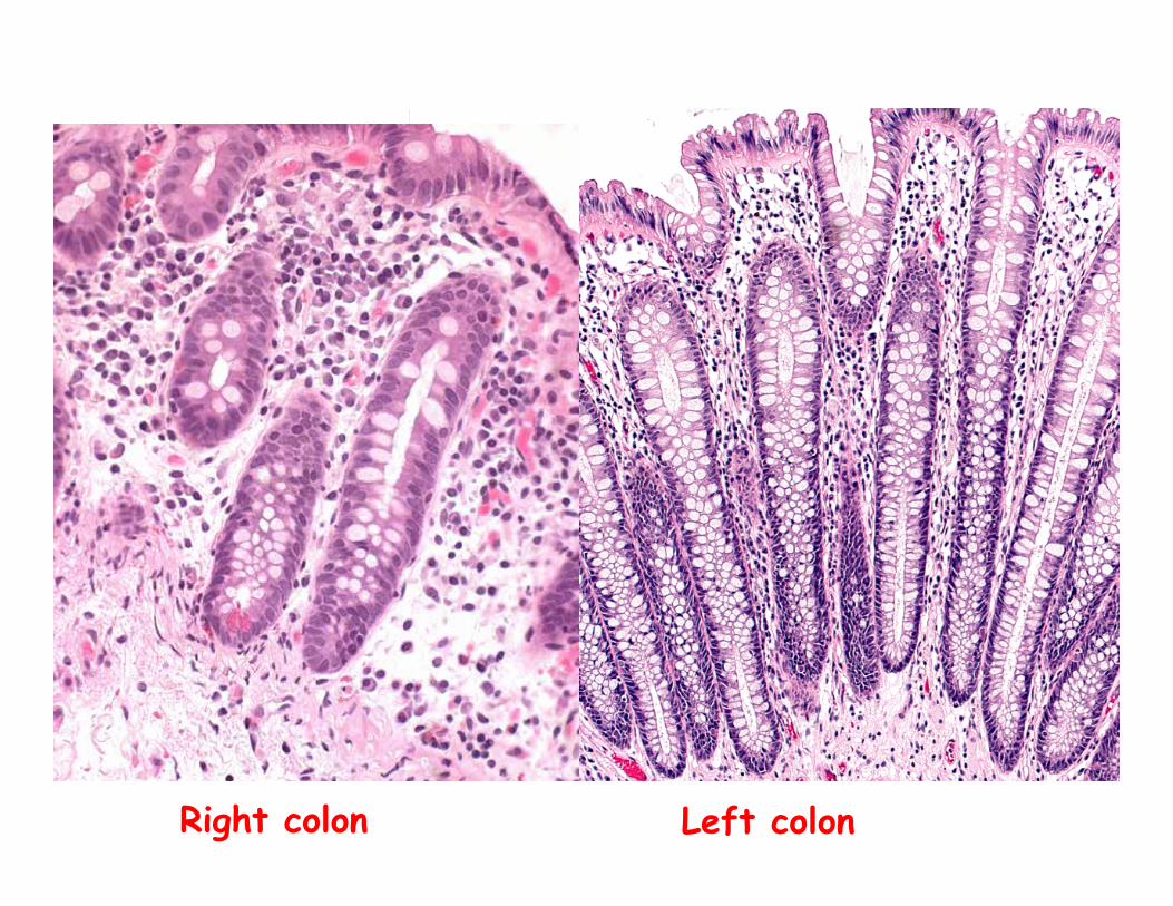
Right colon Left colon

Architectural distortion (+ chronic inflammation)
= Archetypal IBD

Distal Paneth cell metaplasia

Thickened / duplicated muscularis mucosae – indicates prior ulceration

Noteduplicatedmuscularismucosae–butthemucosahasreturnedtonormal
AnapparentlynormalbiopsydoesnotexcludeUC
SO“ThereisnoevidenceofUCinthesebiopsies”
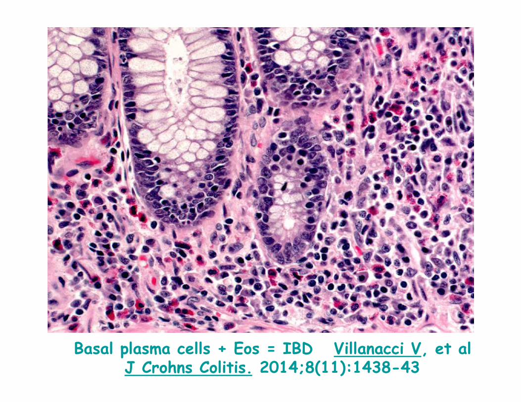
Basal plasma cells + Eos = IBD Villanacci V, et al J Crohns Colitis. 2014;8(11):1438-43
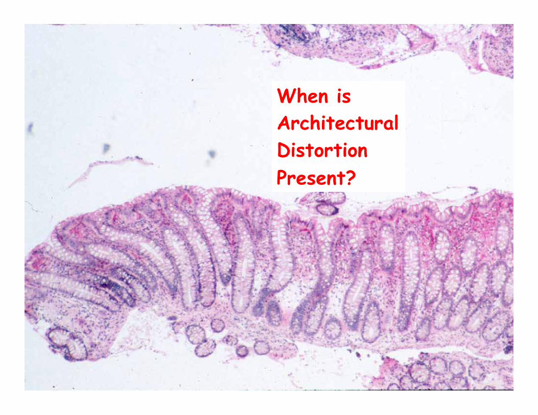
When is Architectural Distortion Present?
Ulcerative Colitis is No Fun
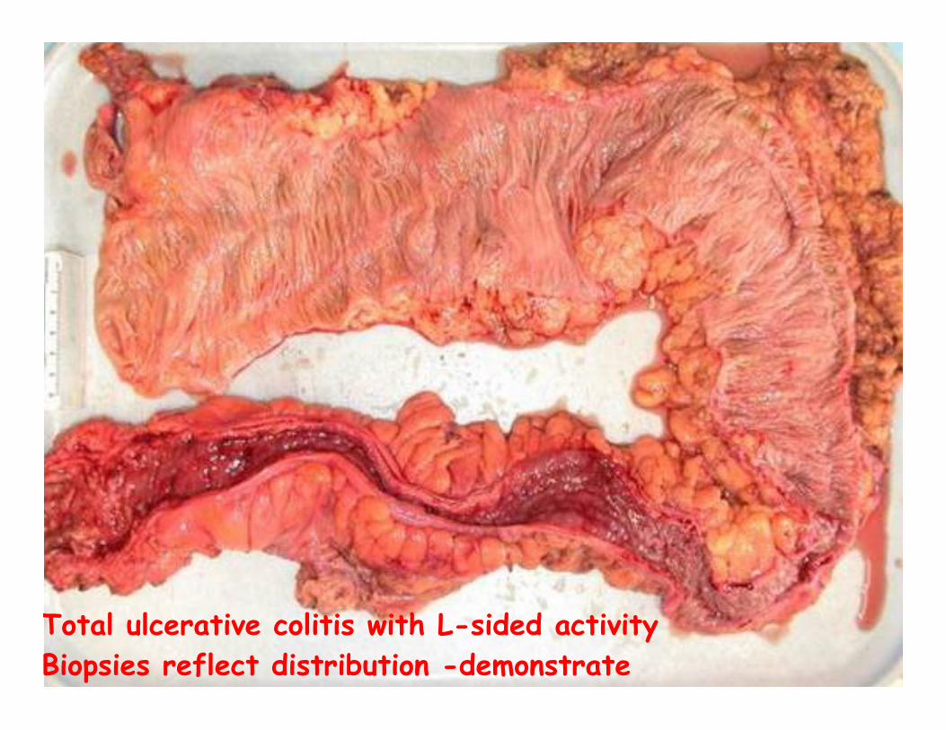
Total ulcerative colitis with L-sided activity Biopsies reflect distribution -demonstrate

M55 2/52 bloody diarrhea
Rectal Bx Is this IBD?


How to report Ø 1) Description Arch distortion + Marked chronic inflammation
c. basal plasma cells (=IBD) Ø 2) Interpretation
Rectal biopsy The features are those of active
inflammatory bowel disease. The location and diffuseness of the
inflammation favor ulcerative colitis – ideally after reviewing endo report
NOTE what is NOT in the report ““Chronic active colitis”” ““Clinical correlation is recommended”” ““This could be IBD, drugs or infection””

How extensive? In one biopsy – who knows? Needs set of biopsies to determine
Extent Distribution of disease
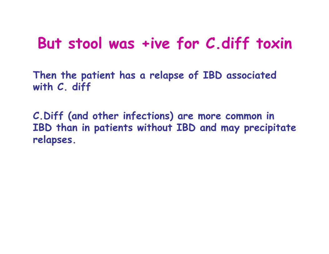
But stool was +ive for C.diff toxin
Then the patient has a relapse of IBD associated with C. diff C.Diff (and other infections) are more common in IBD than in patients without IBD and may precipitate relapses.
Ulcerative colitis - unusual patterns Ø Cecal and peri-appendiceal patches Ø Return of rectal mucosal architecture to normal
Ø Post therapy Ø Gradually over time
Ø Giant cells & granulomas - mucin (ruptured crypts) Ø Rectal sparing of inflammation
Diverticular colitis, After therapeutic enemas Immunosuppressives, PSC
Ø Patchy inflammation - ? Following Rx & “designer” drugs Ø Follicular proctitis (Diversion, UC, CD, Chlamydia/LGV) Ø Aphthoid ulcers esp fulminant disease - both ends
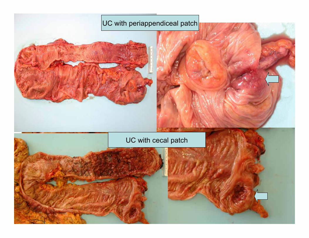
UC with cecal patch
UC with periappendiceal patch
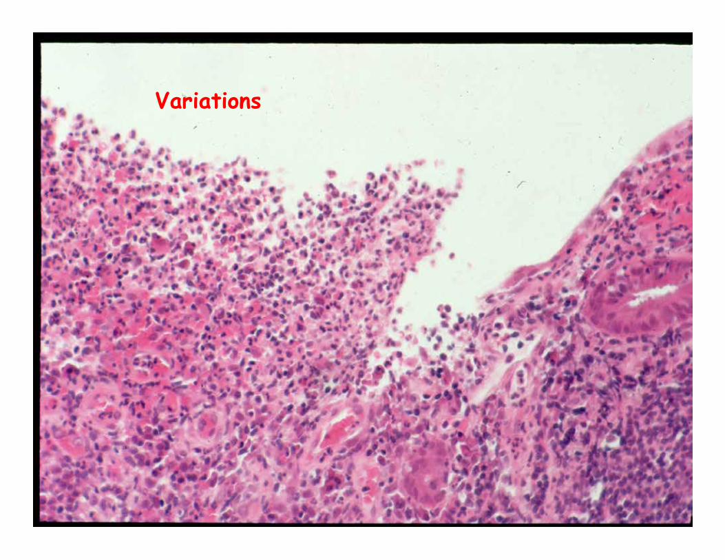
Variations
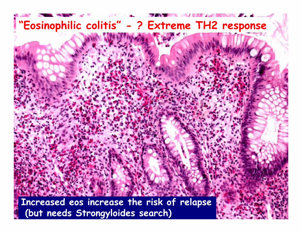
“Eosinophilic colitis” - ? Extreme TH2 response
Increased eos increase the risk of relapse (but needs Strongyloides search)
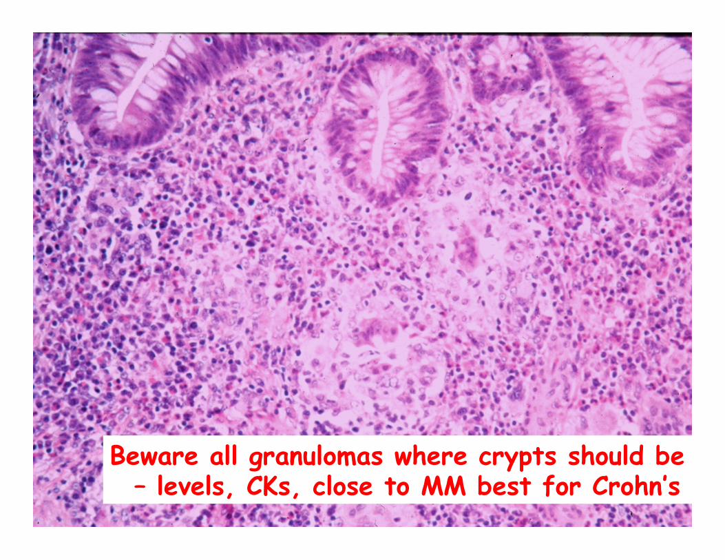
Beware all granulomas where crypts should be – levels, CKs, close to MM best for Crohn’s

Histological Crohn’s disease
Ø Resections – for complications only – but a good chance to examine the focality of the disease
Ø Biopsy diagnosis is an extension of the changes seen in resections, namely: Ø Marked focal inflammation++ within & between biopsies Ø Erosions/ulcers on a background of minimally inflamed mucosa Ø In a patient in an appropriate clinical setting
NOT fulminant colitis
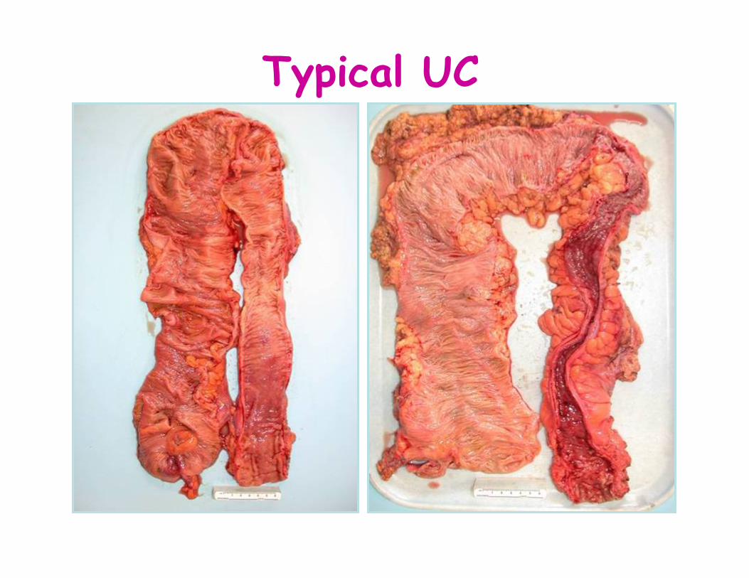
Typical UC
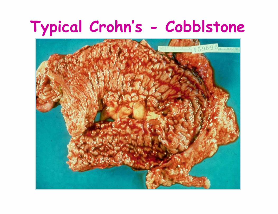
Typical Crohn’s - Cobblstone
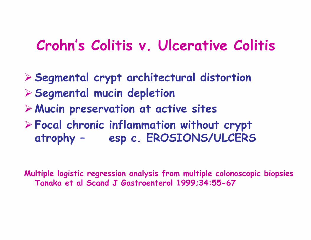
Crohn’s Colitis v. Ulcerative Colitis
Ø Segmental crypt architectural distortion Ø Segmental mucin depletion Ø Mucin preservation at active sites Ø Focal chronic inflammation without crypt
atrophy – esp c. EROSIONS/ULCERS Multiple logistic regression analysis from multiple colonoscopic biopsies
Tanaka et al Scand J Gastroenterol 1999;34:55-67
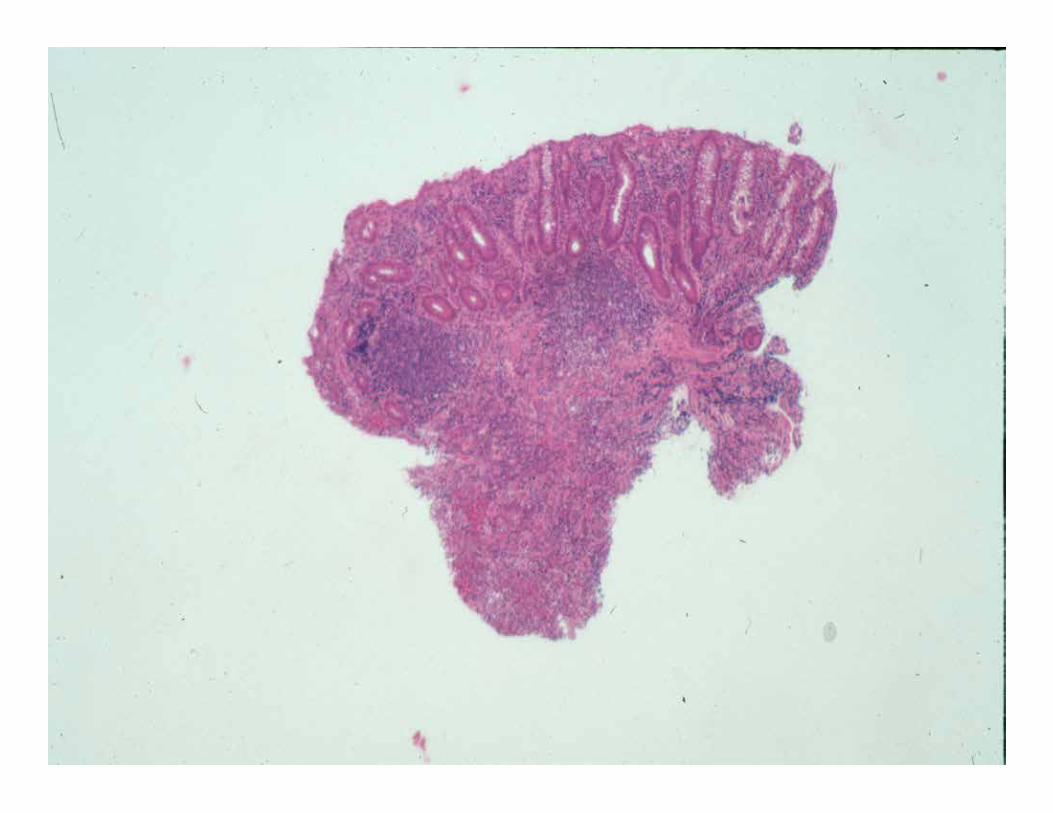
Does the patient have IBD?
? Ulcerative colitis ? Crohn’s disease ? Diversion colitis ? Drug effect ? Other
With 4 different biopsies it is impossible to
understand the distribution of the disease
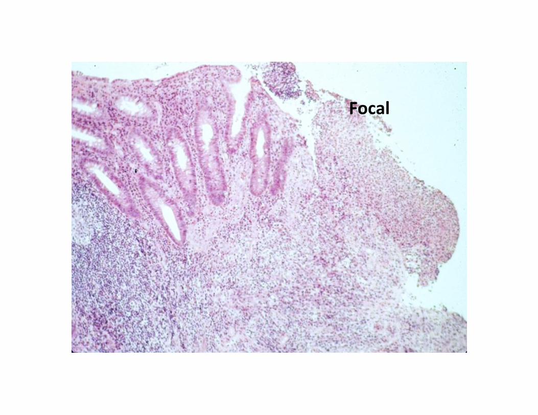
Focal

Focal
Terminal ileal biopsy – CD or medications (NSAIDs/ASA)
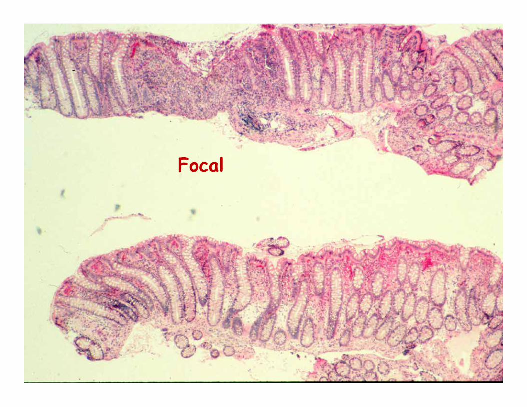
Focal
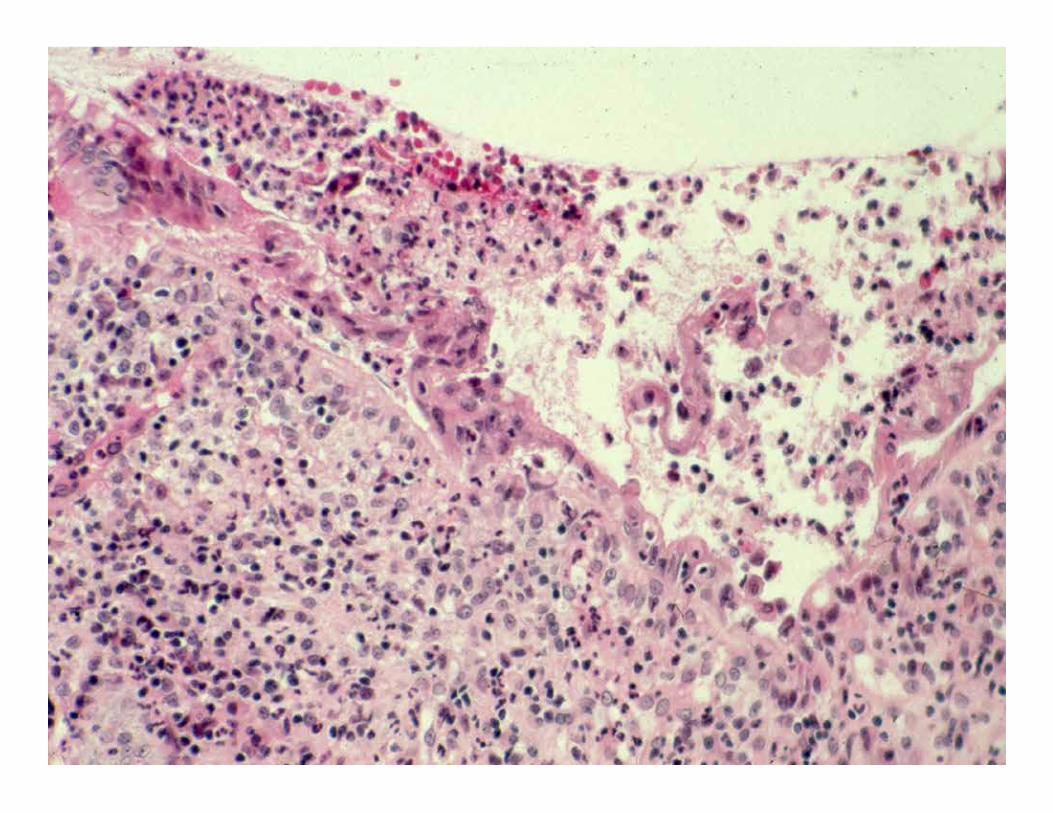

Focal
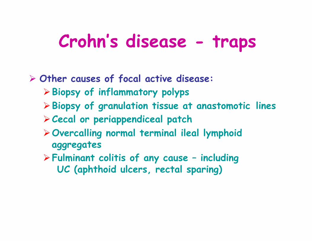
Crohn’s disease - traps
Ø Other causes of focal active disease: Ø Biopsy of inflammatory polyps Ø Biopsy of granulation tissue at anastomotic lines Ø Cecal or periappendiceal patch Ø Overcalling normal terminal ileal lymphoid
aggregates Ø Fulminant colitis of any cause – including
UC (aphthoid ulcers, rectal sparing)

Getting the diagnosis from patterns of inflammation
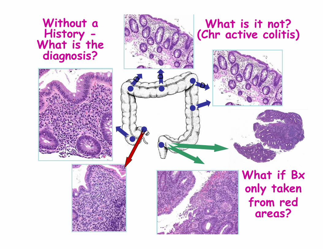
D
D
Without a History -
What is the diagnosis?
What if Bx only taken from red areas?
What is it not? (Chr active colitis)
D
D
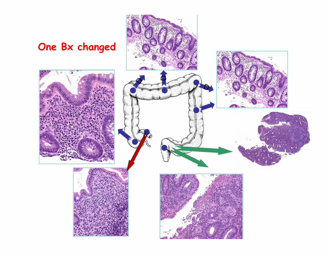
One Bx changed

D
D
Ulcerative proctitis with Cecal patch
One Bx changed What’’s the trap?

Pan UC
Does any other disease produce this combination of changes?

Diverticular Colitis or CD provided……there are diverticula
How about this variation??

PredomR-sidedUCThinkPSC

Crohn’’s disease
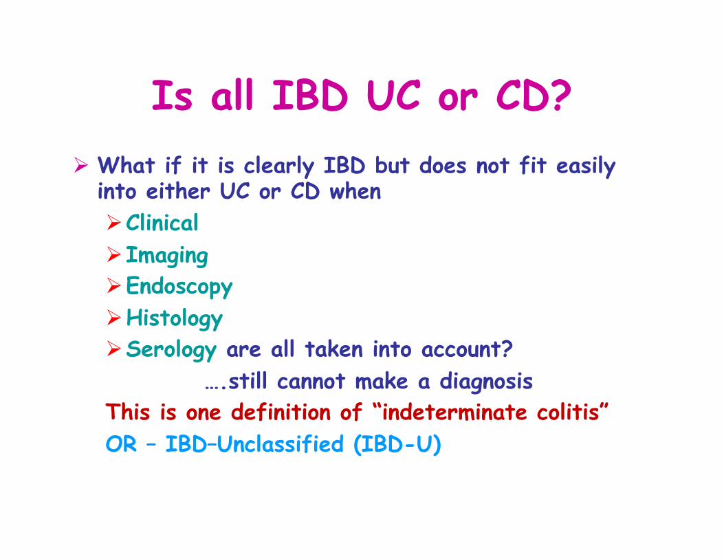
Is all IBD UC or CD? Ø What if it is clearly IBD but does not fit easily
into either UC or CD when Ø Clinical Ø Imaging Ø Endoscopy Ø Histology Ø Serology are all taken into account? ….still cannot make a diagnosis
This is one definition of “indeterminate colitis” OR – IBD–Unclassified (IBD-U)

IBD-U @ first diagnosis Ø IBD - Usually pancolitis (varying degrees of arch
distortion + inflammation with basal plasma cells BUT Can’’t ““pull the trigger”” on either UC or CD e.g. Some degree of rectal sparing but Sigmoid inflamed Focality but not enough for CD (no erosions on a background of normal mucosa) No granulomas i.e. Treat as large bowel IBD
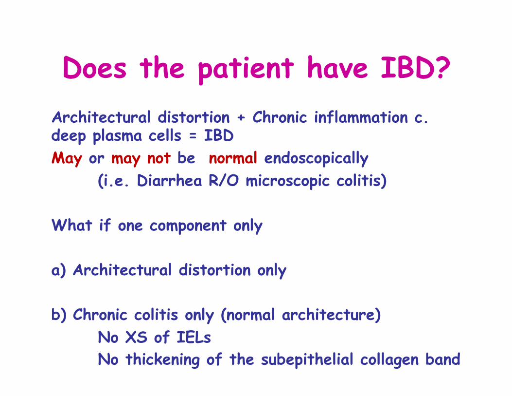
Does the patient have IBD? Architectural distortion + Chronic inflammation c. deep plasma cells = IBD May or may not be normal endoscopically
(i.e. Diarrhea R/O microscopic colitis) What if one component only a) Architectural distortion only b) Chronic colitis only (normal architecture)
No XS of IELs No thickening of the subepithelial collagen band

Architectural distortion only - Mild architectural distortion is present without
inflammation, and is indicative of prior damage, although the cause of this is unclear.
- It may represent a prior episode of infectious colitis but the possibility of quiescent IBD or medication-associated injury (e.g. NSAIDs) cannot be excluded.
- If the patient’s symptoms exacerbate, consideration should be given to repeating the colonoscopy with biopsies to better elucidate the nature of the underlying disease or cause.
- If symptomatic at the time of scoping, these changes cannot explain the symptoms. IS THERE A PATTERN? Diffuse, increase distally? UC Ensure not obvious e.g. Post solid organ transplant

Arch normal, diffuse basal plasmacytosis The diffuse chronic inflammation with basal
plasma cells in most biopsies is indicative of a mild but definite chronic colitis.
This may represent mild IBD (although the lack of architectural changes are against this); a variant of microscopic colitis or medication associated injury, especially NSAIDs. There is no excess of intraepithelial lymphocytes or a thickened sub-epithelial collagen band to suggest either lymphocytic or collagenous colitis. However the changes could represent the healing phase of either of those conditions.
If the patient’s symptoms exacerbate, consideration should be given to repeating the colonoscopy with biopsies to better elucidate the nature of the underlying disease.

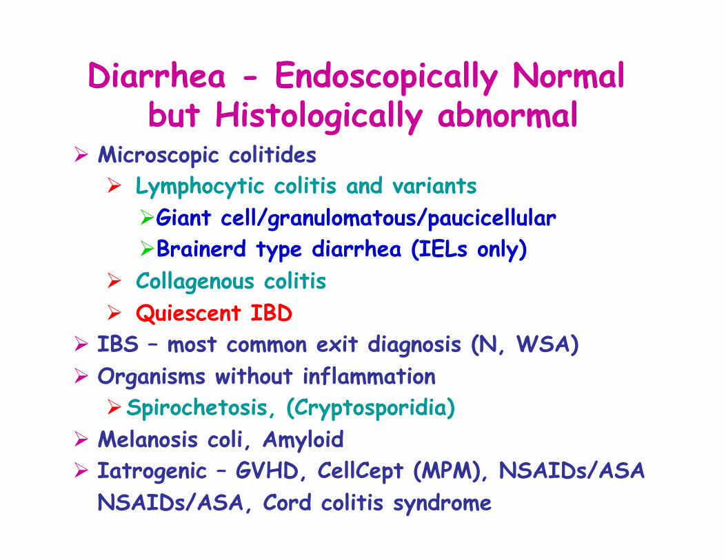
Diarrhea - Endoscopically Normal but Histologically abnormal
Ø Microscopic colitides Ø Lymphocytic colitis and variants
Ø Giant cell/granulomatous/paucicellular Ø Brainerd type diarrhea (IELs only)
Ø Collagenous colitis Ø Quiescent IBD
Ø IBS – most common exit diagnosis (N, WSA) Ø Organisms without inflammation
Ø Spirochetosis, (Cryptosporidia) Ø Melanosis coli, Amyloid Ø Iatrogenic – GVHD, CellCept (MPM), NSAIDs/ASA NSAIDs/ASA, Cord colitis syndrome

If she hasn't yet, she will soon ! . . .
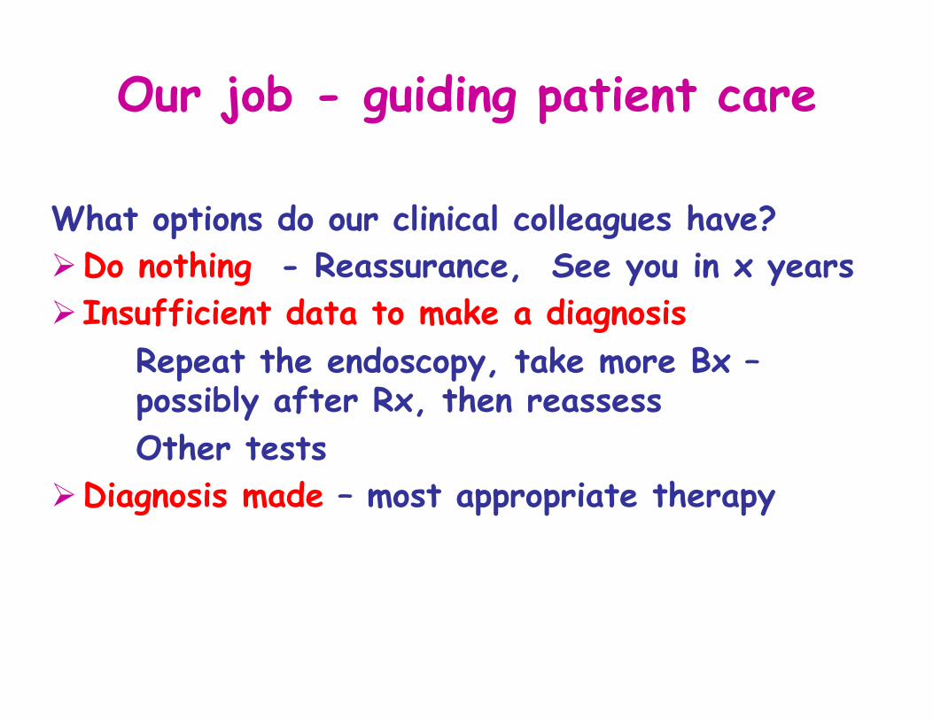
Our job - guiding patient care
What options do our clinical colleagues have? Ø Do nothing - Reassurance, See you in x years Ø Insufficient data to make a diagnosis Repeat the endoscopy, take more Bx –
possibly after Rx, then reassess Other tests
Ø Diagnosis made – most appropriate therapy
Interpretation & History Description /micro if required Initial colonoscopy: Biopsies from XX: The features are those of active
inflammatory bowel disease. The diffuseness and pattern of the disease favors this being active ulcerative colitis
Follow-up colonoscopy – Previous diagnosis of UC Biopsies from XX: Features of active ulcerative
colitis. There is no evidence of dysplasia.

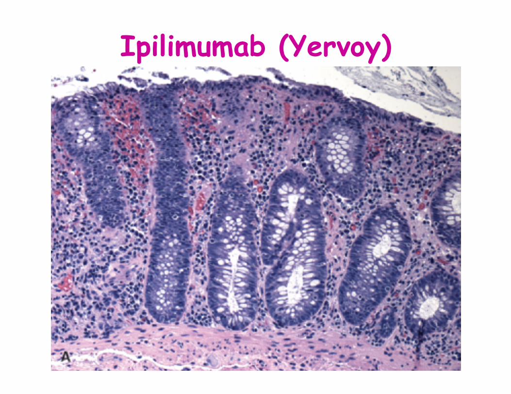
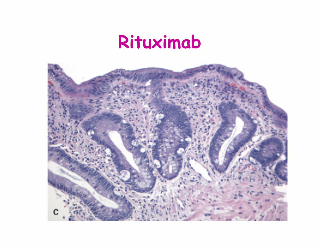
Drugs associated with an IBD-like picture
CTLA-4 antagonists – MM – Colitis –IBD-like Yervoy (ipilimumab), Opdivo (nivolumab)
CD20 – Retuximab – Crohn’’s-like – usually no plasma cells, apoptosis
Zydelig (idelalisib) P13Kdd - Phosphatidylinositol-3-kinasedd - activated in B-cell lymphomas
(Entero)colitis MMF Cocaine, oral contraceptives, K= resins (kayexylate)
can all cause ischemia
Ipilimumab (Yervoy)
Rituximab
Note – all new patients with IBD
Comment The features are highly suggestive of IBD,
especially if the patient is not taking immunosuppressives or receiving monoclonal antibodies such as ipilimumab (Yervoy), nivolumab (Opdivo), retuximab (Rituxan) or Idelalisib (Zydelig). These can cause a colitis that can mimic IBD

Finally – the great imitator
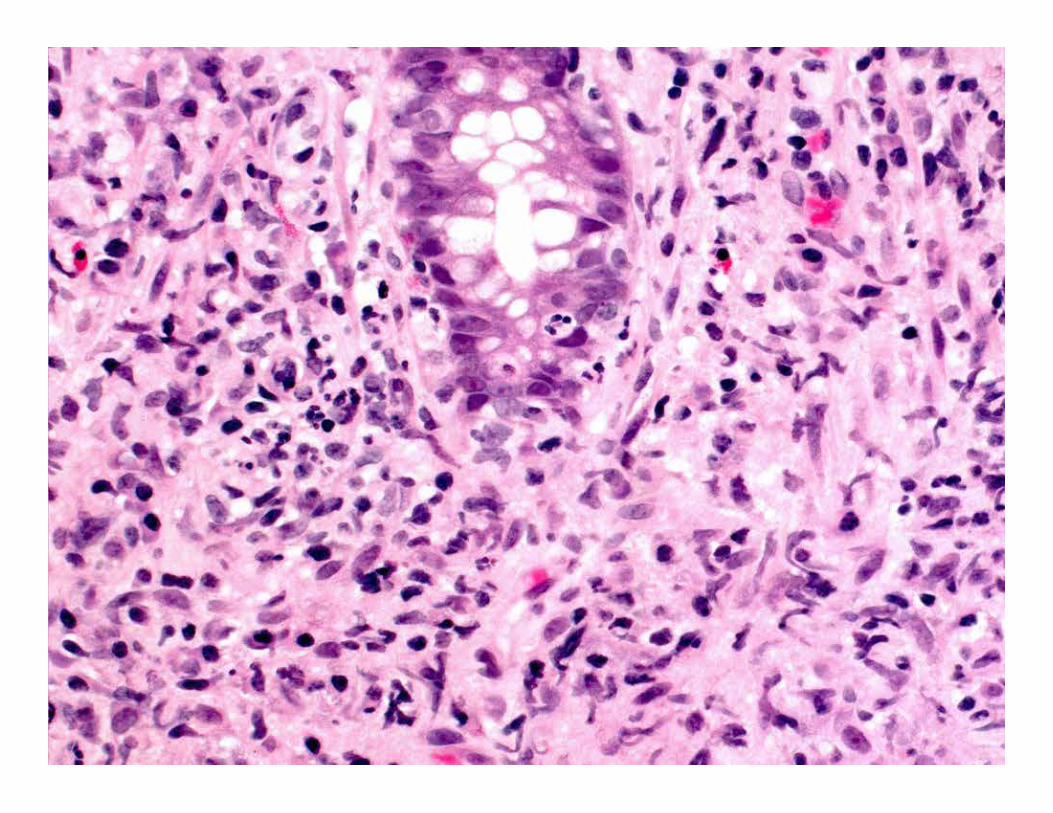
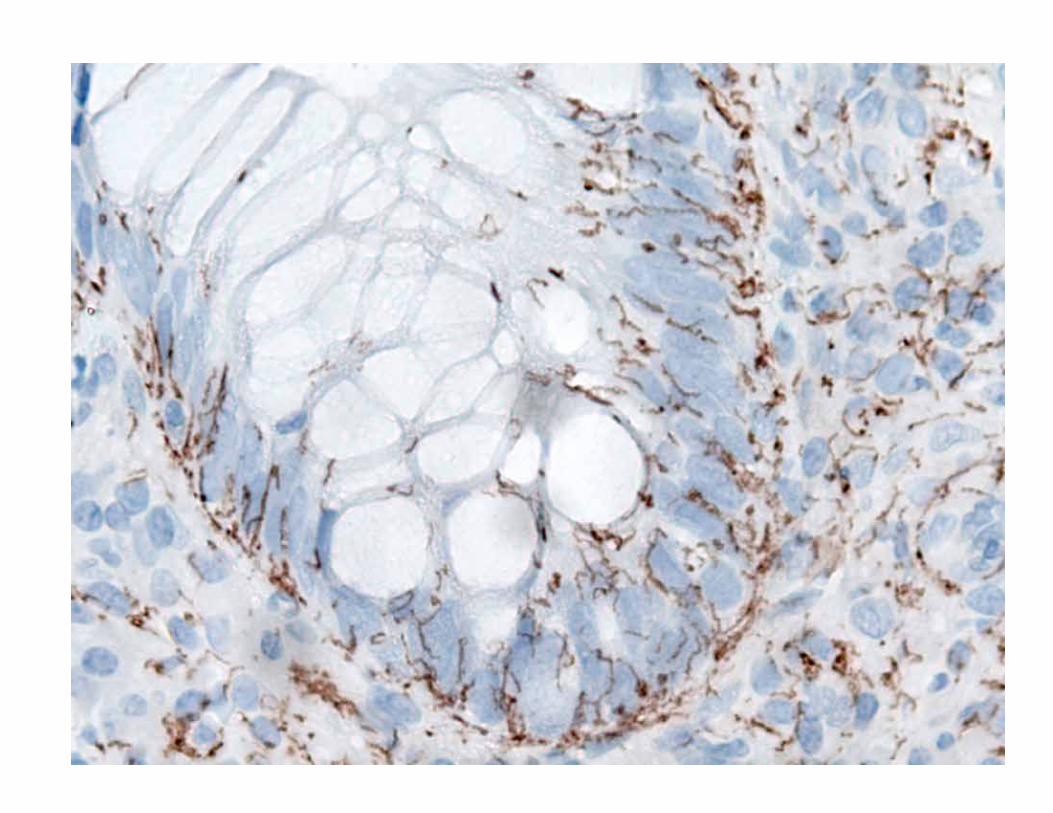
Rectal lues Ø Proctitis only
Ø Diff diagnosis of ulcerative proctitis Ø No ““massive plasmacytosis”” –
lymphoplasmacytic Ø High endothelial venules not overtly obvious
Ø Think of luetic infection /LGC whenever there is a proctitis that does not readily ““fit””, esp if male
Keeping everyone out of trouble
Chronic active colitis Consistent with Non-specific colitis Clinical correlation is required

”Chronic active colitis” …… is a description, NOT a diagnosis It needs an interpretation If WE do not interpret it who does?
…using what criteria? Who saw the slides? If the patient has IBD it is a lifelong diagnosis
Is it even IBD? Always IBD?

How was this reported? Chronic active colitis What would we expect the history to be? What was it? F23 proctosigmoiditis
– outside diagnosis – “chronic active colitis” (Rx as IBD)
What was the result of the new diagnosis? Advised to stop taking Chinese herbal enemas – these
contain vincristine/vinblastine (from the Madagascan periwinkle in the enema)
Beware of (the epidemic of) ““Consistent with”” How should that be interpreted?
How do we want it interpreted? .. Might be … what else is it c/w ?
Usually interpreted as ““diagnostic of””
(now ““histologically proven””) = permission to treat ………. with steroids, biologicals, surgery
It is not a medico-legal escape cause -
= in all reasonable medical probability When does it matter? When it directs therapy
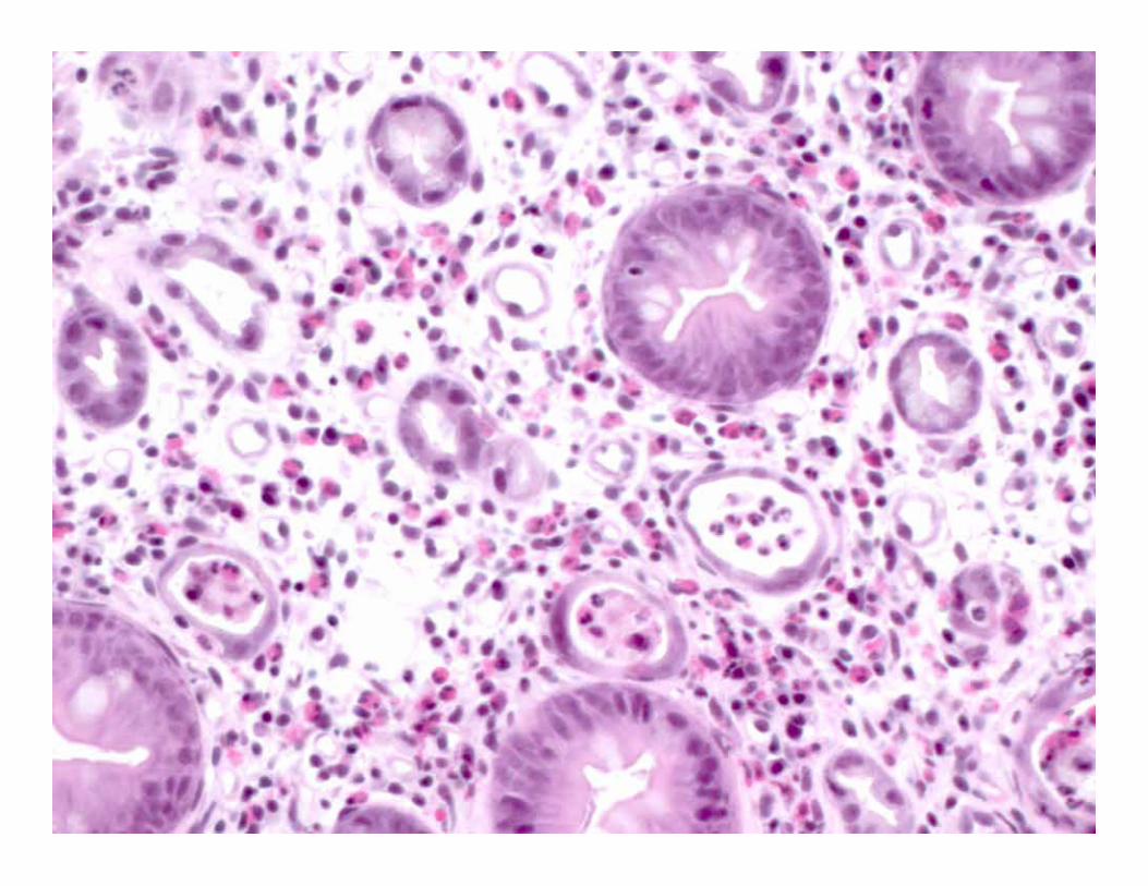
“Mild non-specific colitis”, Ø in the absence of an endoscopic abnormality =
microscopic colitis – AND it is now “histologically proven” =
- permission to treat – was that your intent?
Ø All inflammation is non-specific unless you can see the cause (“bugs”, drugs)
They need an interpretation – what DO they mean?
Are they really pathological? Are they causing the patients symptoms?
How do YOU want the recipient to interpret it? (It is YOUR job – not theirs)

““Clinical correlation is required””
Does this really mean ““Clinico-pathology correlation is required”” Who does the ““pathology”” part of the discussion? How? Report, E-mail, Ph call

Do’’s and Don’’t’’s Ø Do not use descriptions of ““colitis”” without
interpretations Ø - You are the one that has seen the slides
Ø Do not say ““C/W IBD, Infection or Drugs”” – need to say which (they know it is inflamed)
Ø Do not say C/W Crohn’’s in Ø Pouch biopsies Ø Diverted bowel Ø Upper GI biopsies in patients with ““hot”” colitis
