biosynthesis of bioactive microbial metabolites and its ... · to the structural studies and...
TRANSCRIPT
VOL.49 NO.8 THE JOURNAL OF ANTIBIOTICS
REVIEW ARTICLE
Biosynthesis of Bioactive Microbial Metabolites and Its Applicationto the Structural Studies and Production of Hybrid Compounds
Akira Nakagawaand Satoshi Omura* *
Department of Biosciences, Teikyo University,Toyosatodai, Utsunomiya 320, Japan
1 Research Center for Biological Function, The Kitasato Institute,Minato-ku, Tokyo 108, Japan
(Received for publication April 3, 1996)
I. Biosynthesis of Bioactive Compoundsfrom MicroorganismsA. HitachimycinB. OkilactomycinC. SetomimycinD. FuraquinocinE. VineomycinF. AsukamycinG. ReductiomycinH. Purpactin
I. LactacystinJ. Pyripyropene
II. Structure Elucidation on Polyketide Antibiotics by Biosynthetic MeansA. ElasninB. HerbimycinC. IrumamycinD. PhthoramycinE. AurantininF. Xanthoquinodin
III. Biosynthesis and Stereochemistry of Macrolide Antibiotics and Production of New Hybrid Macrolidesand Polyketide
A. Biosynthesis of the Aglycone Moiety in 16-Membered Macrolides, Leucomycins and TylosinB. The Intact Incorporation of a Chain-elongation Intermediate into MacrolidesC. Biosynthesis of 16-MemberedMacrolides after the Formation of the Lactone RingD. Stereochemistry of Tylosin and Its Related Compounds by Microbial ConversionE. Production of Hybrid Macrolides by Microbial TransformationF. Production of a New Hybrid Antibiotic, Mederrohdin by Genetic Manipulation
Microorganisms produce a variety of secondary me-tabolites with interesting structural features and biologi-cal activities as we can not imagine. Someof them haveoccupied a weighty position as medicines and agriculturaldrugs and for animal health. Recent progress of isolationand purification techniques and apparatus such as NMRspectroscopy in addition to establishment of newscreening methods for bioactive microbial metaboliteshave resulted in discovery of novel biologically activecompounds1*.Biosynthetic studies on secondary metabo-lites using compounds enriched with 1 3C and other stableisotopic atoms followed by NMRspectroscopic analysesprovided us not only biosynthetic information of thecarbon skeleton but also enzymatic and stereochemical
717
aspects for a C-C bond formation and its reconstruction.Especially, the labeling patterns of 13C enriched com-
pounds to polyketide skeletons which are typified by poly-cyclic aromatic and macrocyclic compounds, branched-chain fatty acid lactone, and their complexes inform usthe regularity in C-C bond formation2 ~4). Furthermore,it suggests that the biosynthetic findings deduced fromthe feeding experiments of 13C atom and its NMR
spectrometry would be useful means for the structureelucidation of novel compounds5). Particularly, the ap-proach of the enzymatic function in each condensationstep in the chain-elongation process and moleculargenetics to polyketide biosynthesis will occupy adominant position in production of new compounds.
718 THE JOURNAL OF ANTIBIOTICS AUG. 1996
Present article is reviewed in centering around ourhitherto biosynthetic studies on bioactive compounds,including the biosynthesis and stereochemistry ofclinically useful macrolide antibiotics and the produc-tion of new hybrid macrolides by microbial conversionand of a novel polyketide by gene manupilation.
I. Biosynthesis of Bioactive Compoundsfrom Microorganisms
During past 25 years, Omura and his coworkers6'7)
have found manynovel compoundswith a variety ofbiological activities from microorganisms by introducingand constructing effective screening systems. Some of
new compoundspossess unique structural skeleton de-riving from pathways via polyketide, shikimate, follow-ed by condensation with segments from amino acid andor mevalonate pathways. In this item, biosynthetic path-ways and its mechanismsinvolving a unique formation,cleavage, rearrangement ofC-C bond and their enzymaticreactions, in addition to biosynthetic origin for the
carbon skeleton via metabolic pathway consisting oflower organic acids, isoprenoid, shikimate, amino acidsand their complex are described in exemplifying someof bioactive compounds, hitachimycin, okilactomycin,setomimycin, furaquinocin, vineomycin, asukamycin, re-
ductiomycin, purpactin A, lactacystin, and pyripyropene.A. Hitachimycin
Hitachimycin8'9) is an antiprotozoal antibiotic isolatedfrom the culture broth of actinomycete strain No. KM4927. A novel 19-membered ring lactam skeleton forhitachimycin is substantially distinguishable with ansa-mycin antibiotics10) in the point that the antibioticcontains no aromatic or quinoid nucleus in the ansa-
chain moiety. The labeling pattern by [I-13C]acetateof hitachimycin molecule indicated that the antibioticconsists of eight malonates, one methylmalonate and onephenylalanine11}, as illustrated in Fig. 1. A high level ofenrichment at C-19 in the feeding experiment of
Fig. 1. The biosynthetic building unit of hitachimycin.
D,L-[l-13C]phenylalanine suggests the conversion of
phenylalanine to /?-phenylalanine by aminomutase in ahitachimycin-producing strain and then its incorporationinto a polyketide chain presumably as a starter unit. Theoccurrence of the intramolecular rearrangement of an
amino group from a- to ^-position was evidenced fromthe relative intensity with natural occurrence of the signalof an amide nitrogen atom in the 15NNMRspectrumof hitachimycin enriched with DL[1 5N]-a-phenylalanine.
Although tyrosine aminomutase has been reported in thebiosynthesis of a peptide antibiotic, edeine12), the occur-rence of phenylalanine aminomutase in a hitachimycin-producing strain may be the first finding in microbialsecondary metabolites.
B. OkilactomycinOkilactomycin13'14) is an antitumor antibiotic pro-
duced by Streptomyces griseoflavus subsp. zamamiensissubsp. nov. The antibiotic possesses a unique 13-
membered ring with the intra-ether bridge forming atetrahydro-<5-pyrone ring with the exo-methylene at the^-position. The feeding experiments of [l-13C]acetate,[l-13C]propionate, and l-[methyl-13C]methionine re-
vealed that the carbon skeleton of okilactomycin is built
up from four acetates, four propionates, one methionine,and three carbons at C-12, C-13, and C-14, as shown inFig. 2. In general, it seems to be a commonobservationthat occurence of C-methyl group on a polyketide chainin actinomycetes is derived biosynthetically from methylofpropionate, rather than from methionine. In this point,occurrence of the methyl group at C-l l deriving frommethionine seems to be a rare case in a biosynthetic
pattern of metabolites from actinomycetes. The bio-synthetic origin of the remaining three carbons was
deduced from the feeding experiment of 75%enrichedD-[U-13C6]glucose. The observation of the 13C-13C
coupling pattern among C-13, C-14 and C-15 impliesthat the unit arises from an intact glycerol unit derivedvia glucose15). This finding suggests that the C3 unit intetrocarcin A aglycone which contains a structurally
Fig. 2. The biosynthetic building unit of okilactomycin.

VOL. 49 NO. 8 THE JOURNAL OF ANTIBIOTICS 719
Fig. 3. The biosytithetic pathway of setomimycin.
similar unit as okilactomycin is also derived from glycerolas a precursor16).
C. SetomimycinSetomimycin17) is an antibacterial and antitumor
antibiotic which was isolated from the cultured brothof Streptomyces pseudovenezuelae. The structure of theantibiotic was determined to -be a unique substituted9,9'-bianthryl skeleton by means of various 13C NMRtechniques, including ^C^H} selective decoupling,^C^H} selective population transfer, and ^C^H}NOE experiments. Setomimycin appears to be con-structed by the oxidative coupling of substituetd
hydroanthracene monomerwhich may be derived frompolyacetate in its carbon skeleton. Appearance of 16enriched signals in the 13C NMR spectrum ofsetomimycin labeled with sodium[l-13C]acetate clearlyindicated that the antibiotic is biosynthesized from twononaketides in which each contains nine carbon atomsaccompanied by the loss of each one of the carboxylcarbons via path A or B, as shown in Fig. 3. The site ofdecarboxylation in the nonaketide was. evidenced fromthe appearance of two enriched singlet signals assignableto the methyl groups (C-12 and C-12') of acetate unit inthe NMRspectrum of [1 ,2-13C2]acetate-labeled setomi-mycin. Appearance of triplet signals due to 13C-13C
couplings for all carbons except for the above mentionedmethyl groups implies that setomimycin is derived fromtwo nonaketide metabolites via decarboxylation at theterminals, as demonstrated in path A18).
D. FuraquinocinFuraquinocins19'20) are a novel antibiotic complex
with a cytotoxic activity against HeLa S3 cells, producedby Streptomyces sp. KO-3988. The antibiotic was pre-dicted to consist of two biosynthetic units, a naphtho-
Fig. 4. The biosynthetic pathway of furaquinocin A.
quinone chromophoreand an isoprenoid-like side chainfrom the structural feature. The feeding of [1,2-13C2]-acetate exhibited the enrichment of twenty carbons in
which eleven carbons derived from C-2 of acetate, asshown in Fig. 4. Each carbon in the naphthoquinonering enriched with 13C showed two kinds of couplingpattern with equal signal intensities. This means that thenaphthoquinone ring may be formed by two differentmanners originated in acetate arrangement, which arib
also indicated by the 2D-INADEQUATEspectrum Of[l ,2-13C2]acetate labeled furaquinocin. The antibiotic ismost likely produced through a synmetric intermediate,1 ,3,6,8-tetrahydroxynaphthalene. On the other hand, thelabeling pattern of [1,2-13C2]acetate for the C10 unitconsisting of the dihydrofuran ring and the side chain
indicates that the unit derives from mevalonate pathwayfrom the appearance of singlet signal for C-10 and C-14and eight additional satellite peaks for the other carbonsof the side chain. The biosynthetic origin of the remaining
720 THE JOURNAL OF ANTIBIOTICS AUG. 1996
two methyl carbons (MeO-7 and Me-8) comes from amethyl ofL-methionine, not from a methyl ofpropionate.The concurrent attachment of an isoprenoid side chainand a C1 unit from methionine onto polyketide backboneis also interesting. Although a few isoprenoid antibi-otics produced by actinomycetes have been reported,furaquinocins may be the first example involving a
carbon-carbon bond between an inside position of theisoprenoid chain (C-3) and the polyketide nucleus(C-3a)21).
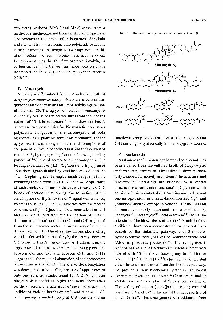
E. Vineomycin
Vineomycins22), isolated from the cultured broth ofStreptomycesmatensis subsp. vineus are a benzanthra-quinone antibiotic with an antitumor activity against sol-id Sarcoma 180. The aglycone moieties of vineomycinsA1 and B2 consist of ten acetate units from the labelingpattern of 13C labeled acetate23>24), as shown in Fig. 5.There are two possibilities for biosynthetic process onpolyacetate elongation of the chromophore of bothaglycones. As a plausible formation mechanism for theaglycones, it was thought that the chromophore ofcomponent Ax would be formed first and then convertedto that of B2 by ring opening from the following labelingpattern of 13C labeled acetate to the chromophore. Thefeeding experiment of [l,2-13C2]acetate to B2 appeared16 carbon signals flanked by satellite signals due to the13C-13Csplitting and the singlet signals assignable to theremaining three carbons, C-l , C-3', and C-4'. Appearanceof each singlet signal means cleavages at least two C-Cbonds of acetate units during the formation of thechromophore of B2. Since the C-4' signal was enriched,whereas those at C-l and C-3' were not from the feedingexperiment of [I-13C]acetate, it was concluded that C-land C-3' are derived from the C-2 carbon of acetate.This means that both carbons at C-l and C-4' originatedfrom the sameacetate molecule via pathwayof a simpledecaacetate for B2. Therefore, the chromophore of B2would be derived from that of Ax by the cleavage betweenC-l2b and C-l in A1 via pathway A. Furthermore, theappearance of at least two 13C-13C coupling pairs, i.e.,between C-5 and C-6 and between C-ll and C-lla
suggests that the mode of elongation of the decaacetateis the same as that of B2. The site of decarboxylationwas determined to be at C-2, because of appearance ofonly one enriched singlet signal for C-2. Vineomycin
biosynthesis is confident to give the useful informationfor the structural characteristics of novel isotetracenoneantibiotics such as kerriamycins25) and urdamycins26)
which possess a methyl group at C-3 position and an
Fig. 5. The biosynthetic pathway ofvineomycins At and B2.
functional group of oxygen atom at C-l, C-7, C-8 andC- 1 2 deriving biosynthetically from an oxygen of acetate.
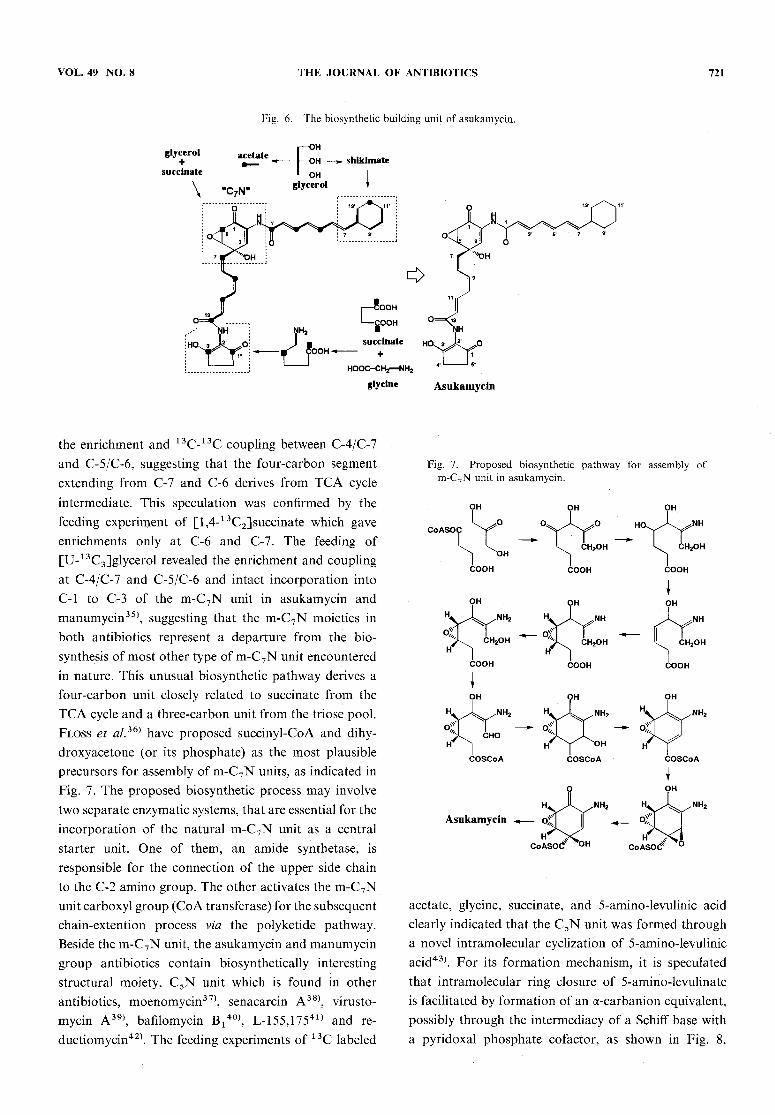
F. Asukamycin
Asukamycin27'28), a new antibacterial compound, wasbeen isolated from the cultured broth of Streptomycesnodosus subsp. asukaensis. The antibiotic shows particu-larly anticoccidial activity in chickens. The structural andbiosynthetic interestings are intensed to a centralstructural element a multifunctional m-C7N unit whichconsists of a six-membered ring carrying one carbon andone nitrogen atom in a meta disposition and C5N unit(2-amino-3-hydroxycyclopent-2-enone). The m-C7N unitis most commonly quininoid as exemplified byrifamycin29), pactamycin30), geldanamycin3 1), and ansa-mitocin32). The biosynthesis of the m-C7N unit in theseantibiotics have been demonstrated to proceed by abranch of the shikimate pathway, with 3-amino-5-hydroxybenzoic acid (AHBA) or 3-aminobenzoic acid(ABA) as proximate precursors33). The feeding experi-
ment of AHBAand ABAwhich are potential precursorslabeled with 13C in the carboxyl group in addition tofeeding of [1-13C] and [l,2-13C2]acetate, indicated thateither the unit is not derived from the shikimate pathway.To provide a new biochemical pathway, additionalexperiments were conducted with 1 3C precursors such asacetate, succinate and glycerol34), as shown in Fig. 6.The feeding of sodium [I-13C]acetate clearly enrichedpositions C-6 and C-7 in the m-C7Nunit, suggestive ofa "tail-to-tail". This arrangement was evidenced from
VOL. 49 NO. 8 THE JOURNAL OF ANTIBIOTICS 721
Fig. 6. The biosynthetic building unit of asukamycin.
the enrichment and 13C-13C coupling between C-4/C-7and C-5/C-6, suggesting that the four-carbon segmentextending from C-7 and C-6 derives from TCA cycleintermediate. This speculation was confirmed by thefeeding experiment of [l,4-13C2]succinate which gave
enrichments only at C-6 and C-7. The feeding of
[U-13C3]glycerol revealed the enrichment and couplingat C-4/C-7 and C-5/C-6 and intact incorporation intoC-l to C-3 of the m-C7N unit in asukamycin and
manumycin35), suggesting that the m-C7Nmoieties inboth antibiotics represent a departure from the bio-synthesis of most other type of m-C7Nunit encounteredin nature. This unusual biosynthetic pathway derives afour-carbon unit closely related to succinate from theTCAcycle and a three-carbon unit from the triose pool.Floss et al.36) have proposed succinyl-CoA and dihy-droxyacetone (or its phosphate) as the most plausibleprecursors for assembly of m-C7Nunits, as indicated inFig. 7. The proposed biosynthetic process may involvetwo separate enzymatic systems, that are essential for theincorporation of the natural m-C7Nunit as a centralstarter unit. One of them, an amide synthetase, isresponsible for the connection of the upper side chainto the C-2 amino group. The other activates the m-C7Nunit carboxyl group (CoAtransferase) for the subsequentchain-extention process via the polyketide pathway.Beside the m-C7Nunit, the asukamycin and manumycingroup antibiotics contain biosynthetically interesting
structural moiety, C5Nunit which is found in otherantibiotics, moenomycin37), senacarcin A38), virusto-mycin A39), bafilomycin B/^, L-155,17541) and re-
ductiomycin42). The feeding experiments of 13C labeled
rig. /. .proposed Diosyntnetic patnway tor assembly otm-C7Nunit in asukamycin.
acetate, glycine, succinate, and 5-amino-levulinic acid
clearly indicated that the C5N unit was formed througha novel intramolecular cyclization of 5-amino-levulinicacid43). For its formation mechanism, it is speculatedthat intramolecular ring closure of 5-amino-levulinate
is facilitated by formation of an a-carbanion equivalent,possibly through the intermediacy of a Schiff base witha pyridoxal phosphate co factor, as shown in Fig. 8.

722 THE JOURNAL OF ANTIBIOTICS AUG. 1996
The evidence for the carbon source for the remainingfunctional units, two triene systems and cyclohexane ringcontaining C-7' in asukamycin, was obtained from theincorporation of 1 3C labeled acetate and [U-1 3C3]glycer-ol. The formation of the cyclohexane ring and C-7' wasconfirmed by the shikimate-type labeling pattern,
namely, two doubly coupled spin systems (C-T/CSr/C-9'
Fig. 8. Formation mechanism of the 2-amino-3-hydroxy-cyclopent-2-enone moiety in asukamycin.
and C-l r/C-127C-13') and a single enriched, noncoupledsignal for C-10\ from [U-13C3]glycerol.
G. ReductiomycinReductiomycin42'44), an antibiotic produced by
Streptomyces xanthochromogenus, possesses antitumorand antibacterial activities. Reductiomycin consists oftwo structural units, C5N unit which is found in
asukamycin molecule and an acetoxydihydrofuran unitbearing an acrylic acid side chain, as shown in Fig. 9.The biosynthetic origin of the C5N unit was unequivo-cally proven by feeding of [4,5-1 3C2] aminolevulinic acid,as described in asukamycin biosynthesis45). Regarding
to the carbon source of the dihydrofuranylacrylic acidmoiety, it was thought that the entire-nine carbon
assembly, including the acetoxyl group, could be derivedby a ring cleavage of phenylalanine or tyrosine, followedby a Baeyer-Villiger oxidation. This assumption on theorigin of the remaining seven carbon atoms of thedihydrofuran moiety was elucidated from the labelingpattern of reductiomycin derived from [U-13C]glycer-ol45), in addition to the incorporation of [1,6-14C]shikimate and L-[U-14C]phenylalanine. The 2D-
INADEQUATE spectrum clearly indicates the twopossible arrangements of an intact three-carbon unitinvoling C-3' and O4', i.e., C-y/C-4'/C-5f and C-3'/C-47C-3" for seven carbons labeled with 13C in thedihydrofuran moiety, as shown in Fig. 9. The resultsobtained from the feeding experiment of [U- 1 3C]glycerolgive rise to two species of reductiomycin, one showingthe labeling pattern (a) and the other pattern (b).
Coupling pattern (b) means ring cleavage between C-4and C-5 of shikimate or a metabolite thereof. On the
Fig. 9. The biosynthetic pathway of reductiomycin.
VOL. 49 NO. 8 THE JOURNAL OF ANTIBIOTICS 723
other hand, coupling pattern (a) reflects bond cleavagebetween C-3 and C-4. A high level of the incorporationof 4-hydroxy [7-13C]benzoate to C-5' of reductiomycinindicated that 4-hydroxybenzoate or a closely relatedproduct, e.g., the corresponding aldehyde must be thesubstrate for the ring cleavage reaction leading to theformation of the dihydrofuran moiety. The hypotheticalpathway and the formation mechanism of the dihydro-furanylacrylic moiety have been proposed by analyzingthe incorporation pattern of 4-hydroxybenzoic acid andits aldehyde labeled with 13C and deuterium46).
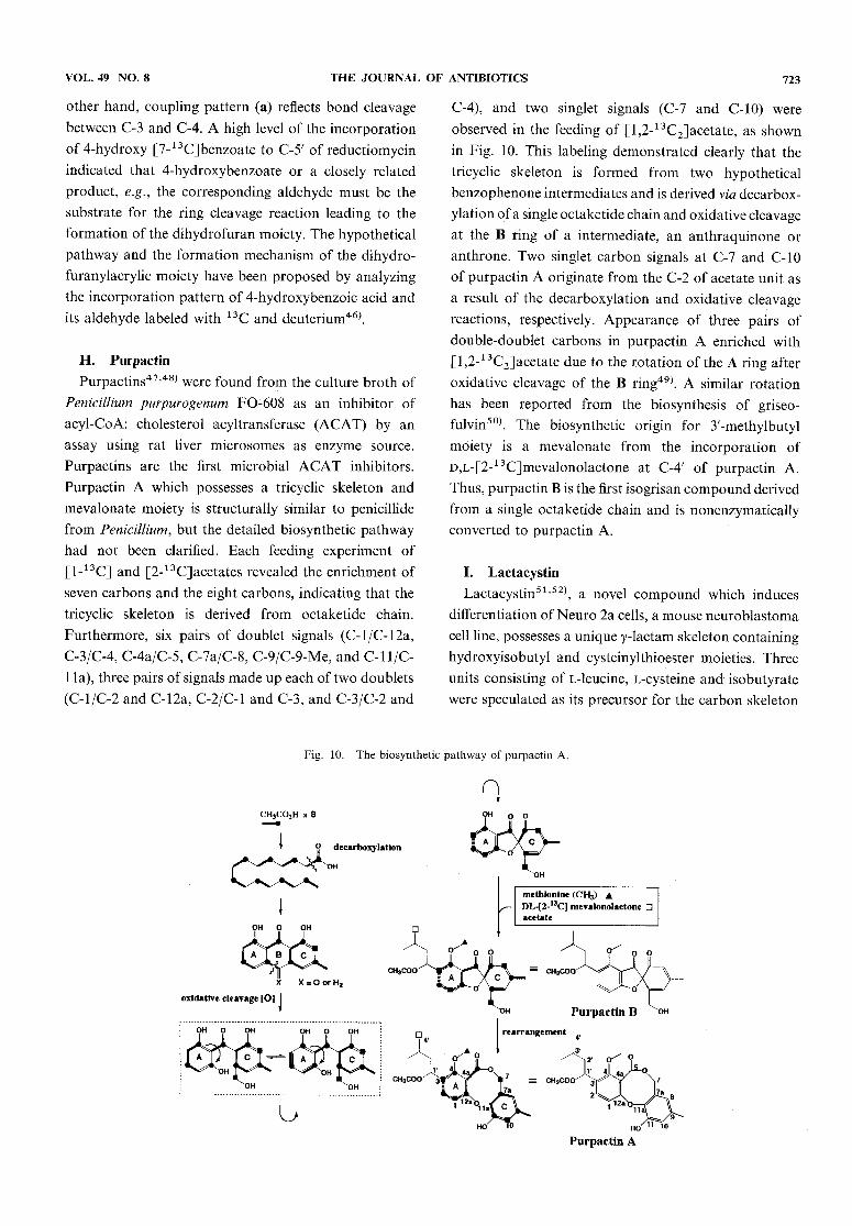
H. Purpactin
Purpactins47'48) were found from the culture broth ofPenicillium purpurogenum FO-608 as an inhibitor ofacyl-CoA: cholesterol acyltransferase (ACAT) by anassay using rat liver microsomes as enzyme source.Purpactins are the first microbial ACAT inhibitors.Purpactin A which possesses a tricyclic skeleton andmevalonate moiety is structurally similar to penicillidefrom Penicillium, but the detailed biosynthetic pathwayhad not been clarified. Each feeding experiment of[1-13C] and [2-13C]acetates revealed the enrichment of
seven carbons and the eight carbons, indicating that thetricyclic skeleton is derived from octaketide chain.Furthermore, six pairs of doublet signals (C-l/C-12a,
C-3/C-4, C-4a/C-5, C-7a/C-8, C-9/C-9-Me, and C-l l/C-
l la), three pairs of signals made up each of two doublets(C-l/C-2 and C-12a, C-2/C-1 and C-3, and C-3/C-2 and
C-4), and two singlet signals (C-7 and C-10) wereobserved in the feeding of [l,2-13C2]acetate, as shown
in Fig. 10. This labeling demonstrated clearly that thetricyclic skeleton is formed from two hypothetical
benzophenone intermediates and is derived via decarbox-ylation ofa single octaketide chain and oxidative cleavageat the B ring of a intermediate, an anthraquinone oranthrone. Two singlet carbon signals at C-7 and C-10ofpurpactin A originate from the C-2 of acetate unit asa result of the decarboxylation and oxidative cleavagereactions, respectively. Appearance of three pairs of
double-doublet carbons in purpactin A enriched with[l,2-13C2]acetate due to the rotation of the A ring afteroxidative cleavage of the B ring49). A similar rotationhas been reported from the biosynthesis of griseo-fulvin50). The biosynthetic origin for 3'-methylbutylmoiety is a mevalonate from the incorporation ofD,L-[2-13C]mevalonolactone at C-4' of purpactin A.
Thus, purpactin B is the first isogrisan compoundderivedfrom a single octaketide chain and is nonenzymatically
converted to purpactin A.
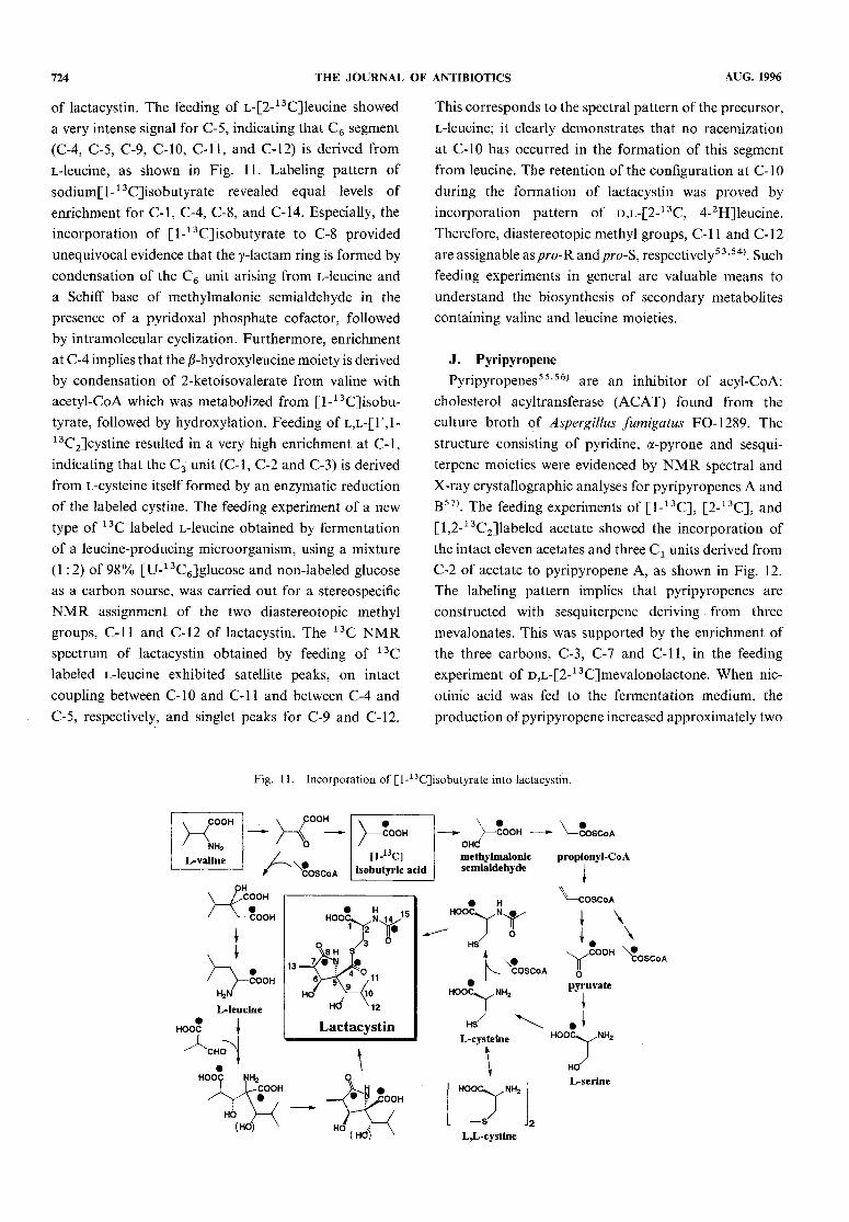
I. Lactacystin
Lactacystin51'52), a novel compound which inducesdifferentiation of Neuro2a cells, a mouseneuroblastomacell line, possesses a unique y-lactam skeleton containinghydroxyisobutyl and cysteinylthioester moieties. Threeunits consisting of L-leucine, L-cysteine and isobutyratewere speculated as its precursor for the carbon skeleton
Fig. 10. The biosynthetic pathway of purpactin A.
724 THE JOURNAL OF ANTIBIOTICS AUG. 1996
of lactacystin. The feeding of L-[2-13C]leucine showeda very intense signal for C-5, indicating that C6 segment(C-4, C-5, C-9, C-10, C-ll, and C-12) is derived fromL-leucine, as shown in Fig. ll. Labeling pattern ofsodium[l-13C]isobutyrate revealed equal levels of
enrichment for C-l, C-4, C-8, and C-14. Especially, theincorporation of [l-13C]isobutyrate to C-8 provided
unequivocal evidence that the y-lactam ring is formed bycondensation of the C6 unit arising from L-leucine anda Schiff base of methylmalonic semialdehyde in thepresence of a pyridoxal phosphate co factor, followedby intramolecular cyclization. Furthermore, enrichment
at C-4 implies that the /Miydroxyleucine moiety is derivedby condensation of 2-ketoisovalerate from valine withacetyl-CoA which was metabolized from [l-13C]isobu-tyrate, followed by hydroxylation. Feeding of l,l-[1',1-13C2]cystine resulted in a very high enrichment at C-l,indicating that the C3 unit (C-l, C-2 and C-3) is derivedfrom L-cysteine itself formed by an enzymatic reductionof the labeled cystine. The feeding experiment of a newtype of 13C labeled L-leucine obtained by fermentationof a leucine-producing microorganism, using a mixture(1 : 2) of 98% [U-13C6]glucose and non-labeled glucoseas a carbon sourse, was carried out for a stereospecificNMR assignment of the two diastereotopic methylgroups, C-ll and C-12 of lactacystin. The 13C NMRspectrum of lactacystin obtained by feeding of 13Clabeled L-leucine exhibited satellite peaks, on intact
coupling between C-10 and C-ll and between C-4 andC-5, respectively, and singlet peaks for C-9 and C-12.
This corresponds to the spectral pattern of the precursor,L-leucine; it clearly demonstrates that no racemizationat C-10 has occurred in the formation of this segmentfrom leucine. The retention of the configuration at C-10during the formation of lactacystin was proved byincorporation pattern of d,l-[2-13C, 4-2H]leucine.
Therefore, diastereotopic methyl groups, C-l l and C-12are assignable as/?roR and/?ro-S, respectively53'54). Suchfeeding experiments in general are valuable means tounderstand the biosynthesis of secondary metabolitescontaining valine and leucine moieties.
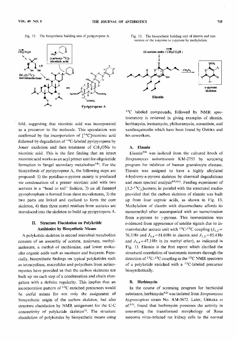
J. PyripyropenePyripyropenes55'56) are an inhibitor of acyl-CoA:
cholesterol acyltransferase (ACAT) found from theculture broth of Aspergillus fumigatus FO-1289. Thestructure consisting of pyridine, a-pyrone and sesqui-
terpene moieties were evidenced by NMRspectral andX-ray crystallographic analyses for pyripyropenes A andB57). The feeding experiments of [1-13C], [2-13C], and[l,2-13C2]labeled acetate showed the incorporation of
the intact eleven acetates and three Cx units derived fromC-2 of acetate to pyripyropene A, as shown in Fig. 12.The labeling pattern implies that pyripyropenes areconstructed with sesquiterpene deriving from threemevalonates. This was supported by the enrichment ofthe three carbons, C-3, C-7 and C-ll, in the feedingexperiment of D,L-[2-13C]mevalonolactone. Whennic-otinic acid was fed to the fermentation medium, the
production of pyripyropene increased approximately two
Fig. ll. Incorporation of [l-13C]isobutyrate into lactacystin.
VOL. 49 NO. 8 THE JOURNAL OF ANTIBIOTICS 725
Fig. 12. The biosynthetic building unit of pyripyropene A.
fold, suggesting that nicotinic acid was incorporated
as a precursor to the molecule. This speculation wasconfirmed by the incorporation of [14C]nicotinic acid
followed by degradation of 14C-labeled pyripyropene byJones' oxidation and then treatment of CH3ONa to
nicotinic acid. This is the first finding that an intactnicotinic acid works as an acyl primer unit for oligoketideformation in fungal secondary metabolites58). For thebiosynthesis of pyripyropene A, the following steps areproposed: 1) the pyridino-a-pyrone moiety is producedvia condensation of a primer nicotinic acid with twoacetates in a "head to tail" fashion, 2) an all farnesyl
pyrophosphate is formed from three mevalonates, 3) thetwo parts are linked and cyclized to form the coreskeleton, 4) then three acetyl residues from acetates areintroduced into the skeleton to build up pyripyropene A.
II. Structure Elucidation on PolyketideAntibiotics by Biosynthetic Means
A polyketide skeleton in second microbial metabolitesconsists of an assembly of acetate, malonate, methyl-malonate, a methyl of methionine, and lower molec-ular organic acids such as succinate and butyrate. Espe-cially, biosynthetic findings on typical polyketides suchas tetracyclines, macrolides and polyethers from actino-mycetes have provided us that the carbon skeletons arebuilt up via each step of a condensation and chain elon-gation with a definite regularity. This implies that anincorporation pattern of 13Cenriched precursors wouldbe useful means for not only the assignment ofbiosynthetic origin of the carbon skeleton, but also
structure elucidation by NMRassignment for the C-Cconnectivity of polyketide skeleton5*. The structure
elucidation of polyketides by biosynthetic means using
Fig. 13. The biosynthetic building unit of elasnin and con-version of the a-pyrone to y-pyrone by methylation.
13C labeled compounds, followed by NMR spec-trometory is reviewed in giving examples of elasnin,herbimycin, irumamycin, phthoramycin, aurantinin, and
xanthoquinodin which have been found by Omura andhis coworkers.
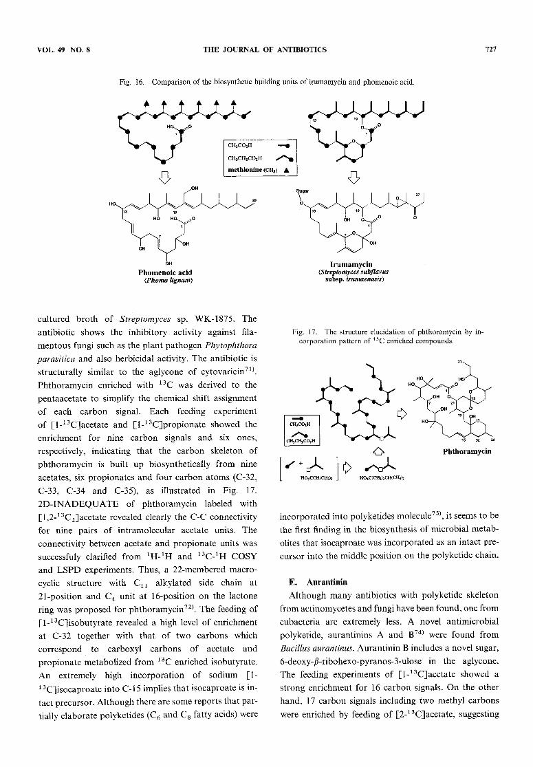
A. ElasninElasnin59) was isolated from the cultured broth of
Streptomyces noboritoensis KM-2753 by screening
program for inhibitor of human granulocyte elastase.Elasnin was assigned to have a highly alkylated
4-hydroxy-a-pyrone skeleton by chemical degradationsand mass spectral analysis60'61*. Feeding experiment of[l,2-13C2]acetate, in parallel with the structural studiesprovided that the carbon skeleton of elasnin was builtup from four caproic acids, as shown in Fig. 13.
Methylation of elasnin with diazomethane affords itsmonomethylether accompanied with an isomerizationfrom a-pyrone to y-pyrone. This isomerization was
evidenced from appearance of satelite signals due to in-tramolecular acetate unit with 13C-13C coupling (Jli2 =76.3Hz and 73>4=61.0Hz in elasnin and Jx 2=85.4Hz
and J3 4=47.3Hz in its methyl ether), as indicated inFig. 13. Elasnin is the first report which clarified the
structural correlation of tautomeric isomers through thedetection of 13C-13C coupling in the 13C NMRspectrumof a polyketide enriched with a 13C-labeled precursorbiosynthetically.
B. Herbimycin
In the course of screening program for herbicidal
substance, herbimycin62) was isolated from Streptomyceshygroscopicus strain No. AM-3672. Later, Uehara etal.63\ found that herbimycin possesses the activity inconverting the transformed morphology of Roussarcoma virus-infected rat kidney cells to the normal

726 THE JOURNAL OF ANTIBIOTICS AUG. 1996
morphology. In those days, the application of 100 MHzXHNMRspectroscopy to the structural elucidation ofherbimycin led to the proposal of partial structuresconsisting of a benzoquinone nucleus A as thechromophore and three fragments B~D, but not the
whole structure. The connectivity among three fragmentsin the ansa-chain was deduced from a high level ofincorporation for four carbon signals at C-l, C-7, C-9and C-l3 by the feeding experiments of sodium[l-13C]propionate64), as shown in Fig. 14. The labeling
pattern of [l-13C]propionate demonstrates that herbi-
mycin might be derived from m-C7Nunit via shikimatepathway for a benzoquinone moiety and from a glycolate(or glycerate) for C-l l/C-12, two acetates for C-3/C-4and C-5/C-6, four propionates and four O-methyls
from methionine in the ansa-chain moiety.
C. Irumamycin
A new macrolide antibiotic, irumamycin65) which isproduced by Streptomyces subflavus inhibits strongly thegrowth of the phytopathogenic fungi. The structure
elucidation ofirumamycin was successfully assigned from
the feeding experiments using 13C doubly labeled acetateand propionate, in addition to chemical degradation66).Feeding experiments of [1-13C]acetate and [l-13C]pro-pionate showed a high level of enrichment for five carbonsignals and for eight signals, respectively, indicating thatthe aglycone consists of five acetate and eight propionateunits, as shown in Fig. 15. Each 13C NMRspectrum of
Fig. 14. The structure of herbimycin A and the proposedbiosynthetic building unit.
irumamycin enriched with [1,2-13C2]acetate and [1,2-1 3C2]propionate exhibited additional satellites based onintermolecular and intramolecular 1 3C-13C coupling foreach of the carbon signals which appeared as doublet.The 13C-13Cdecoupling experiments of these satellitepeaks deduced a 20-membered macrocyclic structurecontaining a 6-membered ring hemiketal. Appearance ofa long range 13C-13C coupling (3/cc= ll.3Hz) betweenC-24 and C-26 of ethyl ketone supported the exsistenceof a,/?-epoxy-a',/?'-ethyl ketone moiety in irumamycin.The proposed structure was also evidenced from thestructural feature of an ozonolysis products ofdiacetylirumamycin. In general, the carbon skeleton ofpolyketides produced by actinomycetes originates frommalonate (and/or acetate) as the biosynthetic origin forC2 unit and methylmalonate (and/or propionate) for C3unit in polyketide chain. On the other hand, fungal
polyketides add extra carbons from the methyl group ofmethionine. This is especially evident when the labelingpatterns of fungal metabolite such as phomenoicacid67)which is produced by Phoma lingam are compared withthose of irumamycin, as illustrated in Fig. 16.
During the biosynthetic studies of irumamycin, its
aglycones, irumanolides I and II were found from amutant of an irumamycin-producing strain obtained byNTG treatment68). Both aglycones were microbiallyconverted to irumamycin under the presence of a
polyketide biosynthetic inhibitor, cerulenin69) during thefermentation of the parent strain. This implies that bothaglycones are converted to irumamycinvia an epoxida-tion and glycosidation.
D. PhthoramycinIn the course of screening program for cellulose
biosynthesis inhibitor, phthoramycin70) was found in the
Fig. 15. The structure elucidation of irumamycin by in-corporation pattern of [1,2-13C2]acetate and [1,2-13C2]-propionate.
VOL. 49 NO. 8 THE JOURNAL OF ANTIBIOTICS 727
Fig. 16. Comparison of the biosynthetic building units of irumamycin and phomenoic acid.
cultured broth of Streptomyces sp. WK-1875. Theantibiotic shows the inhibitory activity against fila-
mentous fungi such as the plant pathogen Phytophthoraparasitica and also herbicidal activity. The antibiotic isstructurally similar to the aglycone of cytovaricin71).
Phthoramycin enriched with 13C was derived to thepentaacetate to simplify the chemical shift assignmentof each carbon signal. Each feeding experimentof [l-13C]acetate and [l-13C]propionate showed theenrichment for nine carbon signals and six ones,respectively, indicating that the carbon skeleton ofphthoramycin is built up biosynthetically from nineacetates, six propionates and four carbon atoms (C-32,C-33, C-34 and C-35), as illustrated in Fig. 17.
2D-INADEQUATE of phthoramycin labeled with
[l,2-13C2]acetate revealed clearly the C-C connectivityfor nine pairs of intramolecular acetate units. The
connectivity between acetate and propionate units wassuccessfuly clarified from ^^H and ^C^H COSYand LSPDexperiments. Thus, a 22-membered macro-cyclic structure with C1X alkylated side chain at21-position and C4 unit at 16-position on the lactonering was proposed for phthoramycin72). The feeding of[l-13C]isobutyrate revealed a high level of enrichmentat C-32 together with that of two carbons whichcorrespond to carboxyl carbons of acetate and
propionate metabolized from 13C enriched isobutyrate.An extremely high incorporation of sodium [1-
13C]isocaproate into C-1 5 implies that isocaproate is in-tact precursor. Although there are somereports that par-tially elaborate polyketides (C6 and C8 fatty acids) were
Fig. 17. The structure elucidation of phthoramycin by in-corporation pattern of 13C enriched compounds.
incorporated into polyketides molecule73), it seems to bethe first finding in the biosynthesis of microbial metab-olites that isocaproate was incorporated as an intact pre-cursor into the middle position on the polyketide chain.
E. Aurantinin
Although many antibiotics with polyketide skeletonfrom actinomycetes and fungi have been found, one fromeubacteria are extremely less. A novel antimicrobialpolyketide, aurantinins A and B74) were found from
Bacillus aurantinus. Aurantinin B includes a novel sugar,6-deoxy-j?-ribohexo-pyranos-3-ulose in the aglycone.The feeding experiments of [1-13C]acetate showed astrong enrichment for 16 carbon signals. On the other
hand, 17 carbon signals including two methyl carbonswere enriched by feeding of [2-13C]acetate, suggesting
728 THE JOURNAL OF ANTIBIOTICS AUG. 1996
that the polyketide chain of aurantinin consists of 16malonate units. The 13C spectrum of component Benriched with [l,2-13C2]acetate exhibited additional
satellite peaks for all carbon signals except for methylcarbons arising from methionine as a biosyntheticprecursor and carbons from sugar moiety. Theobservation of intra- and intermolecular 13C-13Ccoupling patterns of acetate units in the 2D-IN-
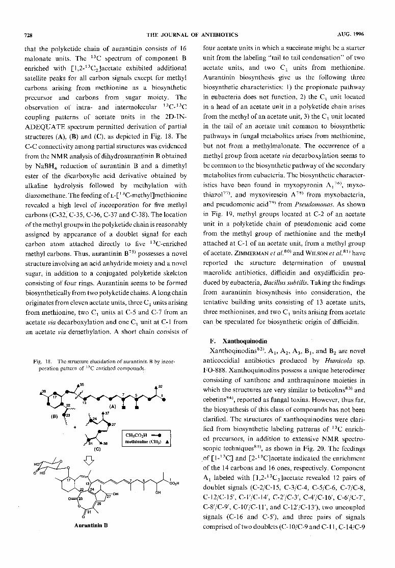
ADEQUATEspectrum permitted derivation of partialstructures (A), (B) and (C), as depicted in Fig. 18. TheC-Cconnectivity amongpartial structures was evidencedfrom the NMRanalysis of dihydroaurantinin B obtainedby NaBH4reduction of aurantinin B and a dimethylester of the dicarboxylic acid derivative obtained byalkaline hydrolysis followed by methylation with
diazomethane. The feeding of L-[ 1 3C-methyl]methioninerevealed a high level of incorporation for five methylcarbons (C-32, C-35, C-36, C-37 and C-38). The locationof the methyl groups in the polyketide chain is reasonablyassigned by appearance of a doublet signal for eachcarbon atom attached directly to five 13C-enriched
methyl carbons. Thus, aurantinin B75) possesses a novelstructure involving an acid anhydride moiety and a novelsugar, in addition to a conjugated polyketide skeletonconsisting of four rings. Aurantinin seems to be formedbiosynthetically from two polyketide chains. A long chainoriginates from eleven acetate units, three C1 units arisingfrom methionine, two Cx units at C-5 and C-7 from anacetate via decarboxylation and one Cx unit at C-l froman acetate via demethylation. A short chain consists of
Fig. 18. The structure elucidation ofaurantinin B by incor-poration pattern of 13C enriched compounds.
four acetate units in which a succinate might be a starterunit from the labeling "tail to tail condensation" of twoacetate units, and two C1 units from methionine.Aurantinin biosynthesis -give us the following threebiosynthetic characteristics: 1) the propionate pathway
in eubacteria does not function, 2) the Cx unit locatedin a head of an acetate unit in a polyketide chain arisesfrom the methyl of an acetate unit, 3) the Cx unit locatedin the tail of an acetate unit commonto biosynthetic
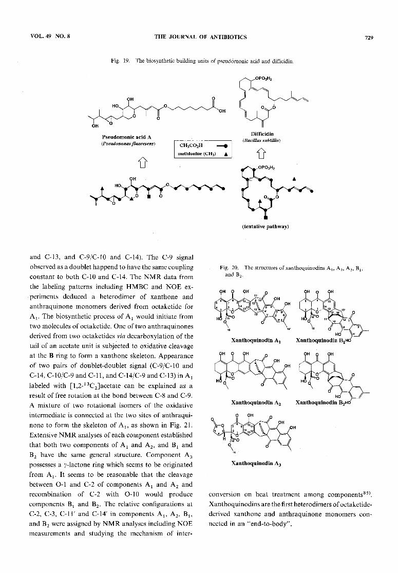
pathways in fungal metabolites arises from methionine,but not from a methylmalonate. The occurrence of amethyl group from acetate via decarboxylation seems tobe commonto the biosynthetic pathway of the secondarymetabolites from eubacteria. The biosynthetic character-istics have been found in myxopyronin Ax16\ myxo-thiazol77), and myxovirescin A78) from myxobacteria,and pseudomonic acid79) from Pseudomonas. As shownin Fig. 19, methyl groups located at C-2 of an acetateunit in a polyketide chain of pseudomonicacid comefrom the methyl group of methionine and the methylattached at C-l of an acetate unit, from a methyl groupof acetate. Zimmermanet al.m and Wilson et al.81) havereported the structure determination of unusualmacrolide antibiotics, difficidin and oxydifficidin pro-
duced by eubacteria, Bacillus subtilis. Taking the findingsfrom aurantinin biosynthesis into consideration, thetentative building units consisting of 13 acetate units,
three methionines, and two Cx units arising from acetatecan be speculated for biosynthetic origin of difficidin.
F. Xanthoquinodin
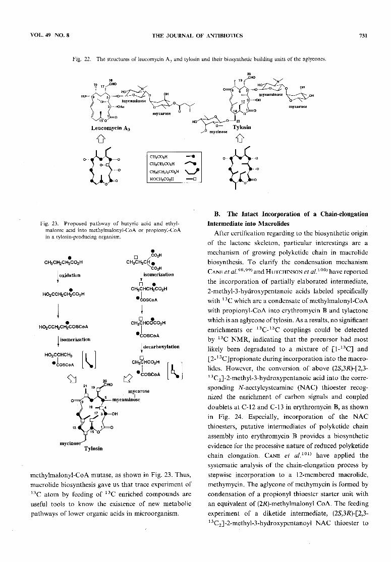
Xanthoqinodins82), Al5 A2, A3, Bl5 and B2 are novelanticoccidial antibiotics produced by Humicola sp.FO-888. Xanthoquinodins possess a unique heterodimerconsisting of xanthone and anthraquinone moieties inwhich the structures are very similar to beticolins83) andcebetins84), reported as fungal toxins. However, thus far,the biosynthesis of this class of compounds has not beenclarified. The structures of xanthoquinodins were clari-fied from biosynthetic labeling patterns of 13C enrich-ed precursors, in addition to extensive NMRspectro-scopic techniques85), as shown in Fig. 20. The feedingsof [1-13C] and [2-13C]acetate indicated the enrichmentof the 14 carbons and 16 ones, respectively. ComponentA1 labeled with [1,2-13C2]acetate revealed 12 pairs ofdoublet signals (C-2/C-15, C-3/C-4, C-5/C-6, C-7/C-8,
C-12/C-15', C-r/C-14', C-2'/C-3', C-47C-16', C-6'/C-7',
C-8'/C-9', C-lO'/C-l l', and C-127C-13'), two uncoupledsignals (C-l6 and C-5'), and three pairs of signals
comprised of two doublets (C-10/C-9 and C-l 1, C-14/C-9
VOL. 49 NO. 8 THE JOURNAL OF ANTIBIOTICS 729
Fig. 19. The biosynthetic building units of pseudomonic acid and difficidin.
and C-13, and C-9/C-10 and C-14). The C-9 signal
observed as a doublet happend to have the same couplingconstant to both C-10 and C-14. The NMRdata fromthe labeling patterns including HMBCand NOEex-periments deduced a heterodimer of xanthone and
anthraquinone monomersderived from octaketide forAx. The biosynthetic process of Ax would initiate fromtwo molecules of octaketide. Oneof two anthraquinonesderived from two octaketides via decarboxylation of thetail of an acetate unit is subjected to oxidative cleavageat the B ring to form a xanthone skeleton. Appearanceof two pairs of doublet-doublet signal (C-9/C-10 andC-14, C-lO/C-9 and C-ll, and C-14/C-9 and C-13) in A,labeled with [l,2-13C2]acetate can be explained as a
result of free rotation at the bond between C-8 and C-9.A mixture of two rotational isomers of the oxidativeintermediate is connected at the two sites of anthraqui-none to form the skeleton of A1? as shown in Fig. 21.Extensive NMRanalyses of each component establishedthat both two components of Ax and A2, and Bx andB2 have the same general structure. Component A3
possesses a y-lactone ring which seems to be originatedfrom Ax. It seems to be reasonable that the cleavagebetween O-l and C-2 of components A1 and A2 andrecombination of C-2 with O-10 would produce
components Bx and B2. The relative configurations atC-2, C-3, C-1T and C-14' in components A1? A2, Bl5and B2 were assigned by NMRanalyses including NOEmeasurements and studying the mechanism of inter-
Fig. 20. The structures ofxanthoquinodins Als A2, A3, Bl3andB9.
conversion on heat treatment among components85).
Xanthoquinodins are the first heterodimers of octaketide-derived xanthone and anthraquinone monomerscon-nected in an "end-to-body".

730 THE JOURNAL OF ANTIBIOTICS AUG. 1996
Fig. 21. The biosynthetic pathway of xanthoquinodin A1
III. Biosynthesis and Stereochemistry of MacrolideAntibiotics and Production of NewHybrid
Macrolides and PolyketideMacrolide antibiotics are classified as 12-, 14-, or
1 6-membered ring macrolides which contain amino sugarand/or neutral sugar moieties, according to the size ofthe macrocyclic lactone ring of the aglycone86~ 88). Thesemacrolides have occupied a large position in antibiotics,because of their excellent antibacterial activities, particu-larly against Gram-positive bacteria and Mycoplasma.Among macrolide antibiotics, 14-membered macro-lides, erythromycin and oleandomycin, 16-membered
ones, leucomycin (josamycin), spiramycin, midecamycin,and tylosin, and their derivatives have been used inmedical and veterinary fields. To date, macrolides
antibiotics have been a focus of the world's attentionin organic chemistry and biochemistry owing to thestructural and biological characteristics. Biosyntheticorigin of the carbon skeleton of the aglycone, the
stereochemical biosynthetic pathway and the regulation
are described in this chapter. This review also deals withthe production of new hybrid macrolides by a microbialtransformation and a polyketide by gene manipuration.
A. Biosynthesis of the Aglycone Moiety in 16-
Membered Macrolides, Leucomycins and TylosinIn the biosynthetic origin of the carbon skeleton of
the lactone ring, magnamycin A has been studied in-tensively using 14C-precursor by groups of Woodwardet al.89\ Achenbach et al.90\ and Srinivasan and
Srinivasan91). Improvement of 13C NMRspectroscopyand the propagation of 13C enriched compounds haveresulted in a great contribution for studying thebiosynthesis of macrolides92~95). The biosynthetic
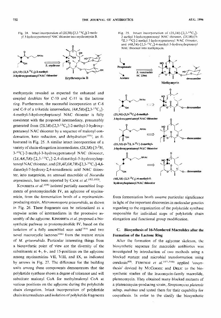
origin for the aglycone moiety of leucomycin A3 wasestablished from the feeding experiments of [1-13C]
and [2-13C]acetate, [l-13C]propionate, and [l-13C]bu-
tyrate, as shown in Fig. 22. Indeed, a high level ofincorporation by the feeding of [1-13C]butyrate and[4-13C]2-ethylmalonate is related to the finding that an
aglycone, platenolide containing a methyl group in placeof the formyl group was isolated from a mutant of aplatenomycin-producing strain. It is interesting findings
that three carbons at positions 7, 8 and 19 correspondingto a propionate unit, were weakly enriched by the feedingof [2-13C]acetate but not by [l-13C]acetate. This meansthat the propionate pathway through the tricarboxylicacid cycle via succinyl-CoA and methylmalonyl-CoA isactive in a leucomycin-producing microorganism.Evidence for the biosynthetic origin of the C2 unit at
C-3 and C-4 was obtained finally from the enrichmentat C-4 by the feeding of [2-13C]glycerol96), indicating
that glycolate metabolized from glycerol may be a directprecursor for the C2 unit.Similarly, it has been clarified that the aglycon moietyof tylosin derives from two acetate units, five propionateunits, and one butyrate unit, as shown in Fig. 23. Aninteresting thing is a high level of incorporation by thefeeding of [1-13C]butyrate not only at C-5 but also at
carbons, 3, 7, ll, 13, and 15 which should be derivedfrom the carboxyl carbon of propionate. This labelingpattern indicates obviously that butyrate are metabolizedinto propionyl-CoA or methylmalonyl-CoA via succinateinvolving an oxidation and isomerization97). Further, thefeeding experiment of [l,3,r-13C3]2-ethylmalonic acid
showed an additional enrichment at carbons, 3, 4, ll,12, 13, 14, 15, and 16 accompanied with a doublet signalarising from 1 3C-13C coupling, in addition to enrichmentat carbons 5 and 19, indicating the metabolic pathwayof [l,3,r-13C3]2-ethylmalonate to methylmalonate via
VOL. 49 NO. 8 THE JOURNAL OF ANTIBIOTICS 731
Fig. 22. The structures of leucomycin A3 and tylosin and their biosynthetic building units of the aglycones.
Fig. 23. Proposed pathway of butyric acid and ethyl-malonic acid into methylmalonyl-CoA or propionyl-CoAin a tylosin-producing organism.
methylmalonyl-CoA mutase, as shown in Fig. 23. Thus,macrolide biosynthesis gave us that trace experiment of13C atom by feeding of 13C enriched compounds areuseful tools to knowthe existence of newmetabolicpathwaysof lower organic acids in microorganism.
B. The Intact Incorporation of a Chain-elongation
Intermediate into MacrolidesAfter certification regarding to the biosynthetic originof the lactone skeleton, particular interestings are amechanism of growing polyketide chain in macrolidebiosynthesis. To clarify the condensation mechanismCane et aL98'99) and Hutchinson et al. 100) have reported
the incorporation of partially elaborated intermediate,2-methyl-3-hydroxypentanoic acids labeled specifically
with 13C which are a condensate of methylmalonyl-CoAwith propionyl-CoA into erythromycin B and tylactonewhich is an aglycone oftylosin. As a results, no significantenrichments or 13C-13C couplings could be detected
by 13C NMR,indicating that the precursor had mostlikely been degradated to a mixture of [1-13C] and[2-1 3C]propionate during incorporation into the macro-lides. However, the conversion of above (2,S,3jR)-[2,3-1 3C2]-2-methyl-3-hydroxypentanoic acid into the corre-sponding 7V-acetylcysteamine (NAC) thioester recog-nized the enrichment of carbon signals and coupleddoublets at C-12 and C-13 in erythromycin B, as shownin Fig. 24. Especially, incorporation of the NACthioesters, putative intermediates of polyketide chain
assembly into erythromycin B provides a biosyntheticevidence for the processive nature of reduced polyketidechain elongation. Cane et a/.101) have applied thesystematic analysis of the chain-elongation process by
stepwise incorporation to a 12-membered macrolide,methymycin. The aglycone of methymycin is formed bycondensation of a propionyl thioester starter unit withan equivalent of (2jR)-methylmalonyl CoA. The feedingexperiment of a diketide intermediate, (2S,3JR)-[2,3-13C2]-2-methyl-3-hydroxypentanoyl NAC thioester to
732 THE JOURNAL OF ANTIBIOTICS AUG. 1996
Fig. 24. Intact incorporation of (2S,3K)-[2,3-13C2]-2-meth-yl-3-hydroxypentanoyl NACthioester into erythromycin B.
methymycin revealed as expected the enhanced andcoupled doublets for C-10 and C-ll in the lactonering. Furthermore, the successful incorporation at C-8
and C-9 of a triketide intermediate, (4R,5K)-[2,3-13C2]-4-methyl-5-hydroxyheptanoyl NAC thioester is fully
consistent with the proposed intermediacy, presumablygenerated from (2S,3K)-[2,3-1 3C2]-2-methyl-3-hydroxy-
pentanoyl NACthioester by a sequence of malonyl con-densation, keto reduction, and dehydration101), as il-lustrated in Fig. 25. A similar intact incorporation of avariety of chain-elongation intermediates, (2S,3R)-[3-2H,3-13C]-2-methyl-3-hydroxypentanoyl NAC thioester,(2£,4^,5K)-[2,3-13C2]-2,4-dimethyl-5-hydroxyhep-
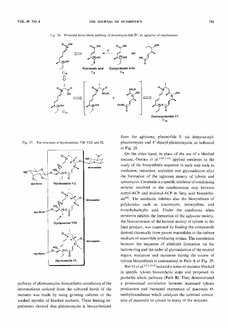
tenoyl NACthioester, and (2£,4£,6#,7#)-[2,3-1 3C2]-4,6-dimethyl-7-hydroxy-2,4-nonadienoic acid NACthioes-ter, into nargenicin, an unusual macrolide of Nocardiaargentinesis, has been reported by Cane et al.102'103\Kinoshita et al.104) isolated partially assembled frag-ments of protomycinolide IV, an aglycone of mycina-micin, from the fermentation broth of a mycinamicin-producing strain, Micromonospora griseorubida, as shownin Fig. 26. These fragments can be rationalized as astepwise series of intermediates in the processive as-sembly of the aglycone. Kinoshita et al. proposed a bio-
synthetic pathway to protomycinolide IV, based on theisolation of a fully assembled seco acid105) and two
novel macrocyclic lactones106) from the mutant strainof M. griseorubida. Particular interesting things froma biosynthetic point of view are the diversity of thesubstituents at 4-, 6-, and 15-positions on the aglycone
among mycinamicins VII, VIII, and IX, as indicatedby arrows in Fig. 27. The difference for the buildingunits among these components demonstrates that thepolyketide synthase shows a degree of tolerance and willsubstitute malonyl CoA for methylmalonyl CoA at
various positions on the aglycone during the polyketidechain elongation. Intact incorporation of polyketidechain intermediates and isolation ofpolyketide fragments
Fig. 25. Intact incorporation of (2S,3K)-[2,3-13C2]-2-methyl-3-hydroxypentanoyl NAC thioester, (25,3l?)-[3-
2H,3-13C]-2-methyl-3-hydroxypentanoyl NAC thioester,and (4#,5#)-[2,3-13C2]-4-methyl-5-hydroxyheptanoyl
NACthioester into methymycin.
from fermentation broth assume particular significancein light of the important discoveries in molecular geneticsregarding to the organization of the polyketide synthaseresponsible for individual steps of polyketide chainelongation and functional group modification.
C. Biosynthesis of 16-Membered Macrolides after theFormation of the Lactone Ring
After the formation of the aglycone skeleton, thebiosynthetic sequence for macrolide antibiotics was
investigated by introduction of two methods using ablocked mutant and microbial transformation usingcerulenin69). Furumai et al.101'108) applied 'cosyn-
thesis' deviced by McCormic and Delic to the bio-synthetic studies of the leucomycin-family macrolide,platenomycin. They obtained manyblocked mutants ofa platenomycin-producing strain, Streptomyces platensissubsp. malvinus and tested them for their capability forcosynthesis. In order to the clarify the biosynthetic
VOL. 49 NO. 8 THE JOURNAL OF ANTIBIOTICS 733
Fig. 26. Proposed biosynthetic pathway of protomycinolide IV, an aglycone of mycinamicin.
Fig. 27. The structures ofmycinamicins, VII, VIII, and IX.
pathway of platenomycin, biosynthetic correlation of theintermediates isolated from the cultured broth of themutants was made by using growing cultures or the
washed mycelia of blocked mutants. These feeding ex-periments showed that platenomycin is biosynthesized
from the aglycone, platenolide I, via demycarosyl-
platenomycin and 4//-deacyl-platenomycin, as indicatedin Fig. 28.
On the other hand, in place of the use of a blockedmutant, Omura et al.109>110) applied cerulenin to thestudy of the biosynthetic sequence in each step such asoxidation, reduction, acylation and glycosidation afterthe formation of the aglycone moiety of tylosin andspiramycin. Cerulenin is a specific inhibitor of condensingenzyme involved in the condensation step betweenacetyl-ACP and malonyl-ACP in fatty acid biosynthe-sis69). The antibiotic inhibits also the biosynthesis of
polyketides such as leucomycin, tetracycline, and6-methylsalicylic acid. Under the conditions when
cerulenin inhibits the formation of the aglycone moiety,the bioconversion of the lactone moiety of tylosin to thefinal product, was examined by feeding the compoundsderived chemically from parent macrolides to the culturemediumof macrolide producing strains. The correlationbetween the sequence of aldehyde formation on thelactone ring and the order of glycosidation of the neutralsugars, mycarose and mycinose during the course oftylosin biosynthesis is summarized in Path A of Fig. 29.Baltz et al.1 1 1'11^ isolated a series of mutants blockedin specific tylosin biosynthetic steps and proposed its
probable whole pathway (Path B). They demonstrateda pronounced correlation between increased tylosin
production and increased expression of macrocin O-methyltransferase which catalyzes the terminal conver-sion of macrocin to tylosin in manyof the mutants.

734 THE JOURNAL OF ANTIBIOTICS AUG- 1996
Fig. 28. The biosynthetic pathway of platenomycin A3
D. Stereochemistry of Tylosin and Its Related
Compoundsby Microbial ConversionThe majority of 14- and 16-membered macrolides
possesses various functional groups with the structuraland stereochemical regularity at the definite position onthe lactone ring. W. D. Celmer113'114) has proposed apredictive model of configurational uniformity amongmacrolides, based on the configurational assignment ofoleandomycin and biogenetic consideration. After that,this model was applied to 16-memberedmacrolides suchas leucomycin A3, magnamycin B and tylosin, and theconfiguration at C-14 and C-15 in tylosin has been
proposed to be R and S, respectively, as illustrated inFig. 30. Comparative NMRand CDanalyses of tylosinand the derivatives with the aglycone derived from leuco-mycin A3 whose absolute configuration has been determin-ed by X-ray crystallographic analysis115) revealed the3R, 4S, 5S, 6R and 8#-configurations for tylosin. How-ever, the configurations at C-14 and C-15 of tylosin havenot been specified. For configurational assignment of all
asymmetric centers of tylosin, a biosynthetic intermediate,protylonolide assigned by X-ray crystallographic anal-ysis1 1651 17) was transformed microbially to tylosin underthe inhibition of the production of tylosin aglycone inthe presence of cerulenin. Retention with the configura-tion of the protylonolide during the microbial transfor-mation to tylosin was evidenced from comparative CDanalysis between protylonolide and 5-0-mycaminosyl-
protylonolide which was derived from the transformedtylosin. As a result, the configuration for the aglycone
moiety oftylosin was assigned to be 3R,AS, 5S, 6R, 8R,14S and 15R1 17\ Therefore, 1^-configuration of tylosinproposed by Celmer was revised to be 14S. After that,the stereochemistry of 16-memberedmacrolides suchas rosaramicin1 18), acumycin119), and mycinamicin120)which possess an asymmetric center at C-14 was con-firmed to possess 14iS-configuration by X-ray crystallo-graphic analysis. Furthermore, it has been clarified
that an antibiotic, M-4365 G121) also possesses thesame configuration with the aglycone of tylosin from themicrobial transformation of protylonolide to M-4365Gx with retention of the configuration122).
E. Production of Hybrid Macrolides by MicrobialTransformation
The isolation of aglycone from a mutant may servenot only for the biosynthetic studies described above,but also as a starting material for the construction ofnew macrolide antibiotics. Maezawaet al.123) has ap-plied to the production of new macrolide by a microbialglycosidation of narbonolide which is an aglycone of a14-membered macrolide, narbomycin, with mycaminosefinding in only 16-membered macrolides. 5-0-Mycami-nosyl-narbonolide was obtained successfully by the
feeding experiments of narbonolide to the fermentationbroth of a platenomycin-producing strain. Attempt ofthe glycosidation of platenolide with desosamine in-volving in only 14-membered macrolides affords also anew hybrid macrolide, 5-(9-desosaminyl platenolide124),as shown in Fig. 31. Both new hybrid macrolides possessantibacterial activity against Gram-positive bacteria.Omura et al.125) attempted the microbial productionof new hybrid macrolides using a biosynthetic inhibitorof polyketide, cerulenin. By means of so called "hybridbiosynthesis", the biologically inactive tylosin aglyconewas converted into novel antibacterial macrolide,chimeramycin by a spiramycin-producing strain Sm.
ambofaciens KA-448 in the presence of cerulenin. Novelhybrid macrolides, chimeramycins A and B, in whichspiramycin constituted sugars, mycaminosyl-mycarose

VOL. 49 NO. 8 THE JOURNAL OF ANTIBIOTICS
Fig. 29. The biosynthetic pathways of tylosin and macrocin.
and forosamine were bonded glycosidically at C-5, andC-9 of protylonolide, respectively, were successfullyproduced by the method, as shown in Fig. 32.Chimeramycins exhibited in vitro antimicrobial activityas that of tylosin and spiramycin I. Demycarosyl-chimeramycins obtained by an acidic hydrolysis of
chimeramycins were stronger activity than that of parentantibiotics. A hybrid biosynthesis using cerulenin wouldbe a useful method for the production of new bioactivecompounds for secondary metabolites which consist ofpolyketide skeleton containing sugars, amino acids, andfragments arised via a shikimate pathway.
F. Production of a New Hybrid Antibiotic, Mederrho-din by Genetic ManipulationRecent advances in understanding the biosynthesis ofbacterial polyketide have led to the concept of polyketidesynthase which can utilize different starter and chain-extender units and can process the poly-/?-ketone inter-mediates in a certain manner126~128). Huchinsonet al.129~133) have vigorously investigated enzymatic
synthesis of a bacterial aromatic polyketide, tetra-
cenomycin biosynthesis in Streptomyces from acetyl andmalonyl coenzyme A and demonstrated the similarityof biochemical connection between the biosynthesis ofbacterial fatty acids and polyketides by isolation and
735

736 THE JOURNAL OF ANTIBIOTICS AUG. 1996
Fig. 30. Configurational model of macrolide antibiotics.
(a) Celmer model (1966)(b) Correct configurational model of tylosin
(1980)
Fig. 31. Production of new hybrid macrolides, 5-0-my-caminosyl-narbonolide and 5-O-desosaminyl-platenolide.
Fig. 32. Production of a new hybrid macrolide, chimera-mycin B.
purification of acyl carrier protein acyltransferase frombacteria.
Interest in the biosynthesis of natural products hasrisen recently, because of advances in the geneticunderstanding ofpolyketide metabolism and the prospectof manufacturing new polyketide-derived drugs withrecombinant microorganisms. Hopwood et al.13*\planned the development of this method by genetic
manipulation and reported the first case of the productionof new hybrid antibiotics by genetically engineeredstrains, as shown in Fig. 33. Whole genes for actinorhodin
biosynthesis of Streptomyces coelicolor A3 had been
already isolated and the position of genes correspondingto each biosynthetic step on the cloned DNAfragmenthad been also determined135). Then, it was tried to in-troduce the whole or a part of fragments of actinorhodinbiosynthetic genes into other Streptomyces strains whichproduced actinorhodin-related antibiotics. Since most ofStreptomyces strains possessed the restriction system, itwas difficult to introduce recombinant plasmids,
however, transformants were obtained from two kindsof strains, kalafungin producer Streptomyces tanashiensis

VOL. 49 NO. 8 THE JOURNAL OF ANTIBIOTICS 737
Fig. 33. Production ofa new hybrid antibiotics, mederrhodinA by genetic manipulation.
and medermycin producer Streptomyces sp. AM7161,but not those of kalafungin producer. Newantibiotics,mederrhodins A and B were produced by thetransformants carrying PIJ 2301, 2315 or 2316 whichcontained a part of actinorhodin biosynthtic genes. Onthe other hand, the transformant introduced PIJ 2303
including whole genes for actinorhdin biosynthesis didnot produce mederrhodins, however, produced acti-norhodin and medermycin. The biosynthesis and
productivity of mederrhodins by transformants wererelatively high and quite stable. All recombinant plasmids(PIJ 2301, 2315 and 2316) contained the transcriptionalunit of act V and the inserted DNAfragment would bementioned as follow: 1) medermycin was synthesized bybiosynthetic genes of Streptomyces sp. AM7161. 2) thehydroxyl residue was substituted at C-6 position of thenaphthoquinone skeleton by hydroxylase which is a
gene product of an inserted DNAfragment (act V) ofPIJ 2315. Antimicrobial activity of mederrhodin A wassimilar to that of medermycin. In a future, knowledgesfrom the breeding of the mederrhodin's producer willgive manyinformations to molecular breeding of otherantibiotic-producing organisms.
References
1) Omura, S.: {Ed). The search for bioactive compoundsfrom microorganisms. Springer-Verlag. 1989
2) O'Hagan, D.: Biosynthesis offatty acid and polyketidemetabolites. Nat. Prod. Rep. 10: 593-624, 1993
3) O'Hagan, D.: Biosynthesis of polyketide metabolites.Nat. Prod. Rep. 9: 447-479, 1992
4) Simpson, T. J.: The biosynthesis of polyketides. Nat.Prod. Reo. 8: 573-602, 1991
5) Omura, S. & A. Nakagawa: Structure elucidation andproduction of new antibiotics using biosynthetic means.
Studies in Natural Products Chemistry. Volume 5.Structure Elucidation {Ed. Rahman, A-u.). Elsevie,
Amsterdam. Oxford. New York. Tokyo, pp. 589-620,1989
6) Omura, S.: Philosophy of new drug discovery.Microbiological Rev. 50: 259-279, 1986
7) Omura, S.: Trends in the search for bioactive microbialmetabolites. J. Indust. Microbiol. 10: 135- 156, 1992
8) Umezawa, I.; Y. Takeshima, K. Komiyama, Y. Koh,
H. Yamamoto& M. Kawaguchi: A new antitumorantibiotic, stubomycin. J. Antibiotics 34: 259 - 265, 19819) Komiyama, K.; K. Edanami, H. Yamamoto & I.
Umezawa: Antitumor activity of a new antitumorantibiotic, stubomycin. J. Antibiotics 35: 703 - 706, 1982
10) Lancini, G. & M. Grandi: Biosynthesis ofansamycins.Antibiotics IV Biosynthesis {Ed. Corcoran, J. W.)
Springer-Verlag, Berlin. Heiderberg. New York. pp.12-40, 1981
ll) Omura, S.; A. Nakagawa, K, Shibata & H. Sano: Thestructure of hitachimycin. A novel macrocyclic lactam
involving /?-phenylalanine. Tetrahedron Lett. 1982:4713-4716, 1982
12) Borowska Z. K. & R. J. Parry: Biosynthesis ofaminoacid. Investigation of the mechanism of jS-tyrosine
formation. J. Am. Chem. Soc. 102: 836-837, 198013) Imai, H.; K. Suzuki, M. Morioka, Y. Numasaki, S.
Kadota, K. Nagai, T. Sato, M. Iwatani & T. Saito:Okilactomycin, a novel antibiotic produced by aStreptomyces species. I. Taxonomy, fermentation,isolation and characterization. J. Antibiotics 40:1475-1482, 1987
14) Imai, H.; H. Kaniwa, T. Tokunaga, S. Fugita, T.
Furuya, H. Matsumoto & M. Shimizu: Okilactomycin,a novel antibiotic produced by a Streptomyces species.
II. Structure determination. J. Antibiotics 40: 1483-1489, 1987
15) Imai, H.; A. Nakagawa & S. Omura: Biosynthesis ofthe antibiotic okilactomycin. J. Antibiotics 42: 1321 -
1323, 1989
16) Tamaoki, T. & F. Omit: Biosynthesis of tetrocarcin.Incorporation of 14C- and 13C-labeled compounds into
tetrocarcin. J. Antibiotics 36: 595-598, 198317) Omura, S.; H. Tanaka, Y. Iwai, K. Nishigaki, J.
Awaya, Y. Takahashi & R. Masuma: A new antibiotic,setomimycin, produced ,by a strain of Streptomyces. J.Antibiotics 26: 1091 - 1098, 1978
18) Kakinuma, K.; N. Imamura, N. Ikekawa, H. Tanaka,S. Minami & S. Omura: Structure and biosynthesis ofsetomimycin. A novel 9,9'-bianthryl antibiotic. J. Am.
Chem. Soc. 102: 7493-7498,,1980
19) Komiyama, K.; S. Funayama, Y. Anraku, M. Ishibashi,Y. Tanaka & S. Omura: Novel antibiotics, fur-aquinocins A and B. Taxonomy, fermentation, isolation
and physico-chemical and biological characteristic. J.Antibiotics 42: 247-252, 1989
20) Funayama, S.; M. Ishibashi, Y. Anraku, K. Komiyama& S. Omura: Structures of novel antibiotics, fur-
aquinocins A and B. Tetrahedron Lett. 1989: 7427-7430, 1989
21) Funayama, S.; M. Ishibashi, K. Komiyama& S. Omura:Biosynthesis of furaquinocins A and B. J. Org. Chem.

738 THE JOURNAL OF ANTIBIOTICS
55: 1132-1133, 1990
22) Omura, S.; H. Tanaka, R. Oiwa, J. Awaya, R. Masuma& Y. Tanaka: New antitumor antibiotics OS-4742 A1?A2, Bl9 and B2 produced by a strain of Streptomyces.
J. Antibiotics 30: 908-916, 1977
23) Imamura, N.; K. Kakinuma, N. Ikekawa, H. Tanaka& S. Omura: Structure of vineomycin B2. J. Antibiotics
34: 1517-1518, 1981
24) Imamura, N ; K. Kakinuma, N. Ikekawa, H. Tanaka& S. Omura: Biosynthesis of vineomycins Ax and B2.
J. Antibiotics 35: 602-608, 1982
25) Hayakawa, Y.; K. Furihata, H. Seto & N. Otake:The structures of new isotetracenone antibiotics,kerriamycins A, B and C. Tetrahedron Lett. 1985:3475-3478, 1985
26) Rohr, J. & A. Zeek: Metabolic products ofmicroorgan-isms. Urdamycin, new angucycline antibiotics fromStreptomycesfradiae. II. Structural studies of urdamycinB to F. J. Antibiotics 40: 459-467, 1987
27) Omura, S.; C. Kitao, H. Tanaka, R. Oiwa, Y.
Takahashi, A. Nakagawa, M. SIiimada & Y. Iwai: Anew antibiotic, asukamycin, produced by Streptomyces.
J. Antibiotics 29: 876-881, 197628) Kakinuma, K.; N. Ikekawa, A. Nakagawa & S.
Omura: The structure of asukamycin, a possible shuntmetabolite from 3-dehydroquinic acid in the shikimate
pathway. J. Am. Chem. Soc. 101: 3402-3404, 1979
29) White, R. J.; E. Martinelli, G. G. Gallo, G. C.
Lancini & P. Beynon: Rifamycin biosynthesis studiedwith 13C enriched precursors and carbon magnetic
resonance. Nature (London) 243: 273-277, 197330) Rinehart, Jr. K. L.; M. Potgieter, D. L. Delaware
& H. Seto: Direct evidence from multiple 13C labelingand homonuclear decoupling for the labeling pattern byglucose of the m-aminobenzoyl (C7N) unit of pactamy-cin. J. Am. Chem. Soc. 103: 2099-2101, 1981
31) Harber, A.; R. D. Johnson & K. L. RiNEHART,Jr.:Biosynthetic origin of the C2 units of geldanamycin and
distribution of label from D-[6-13C]glucose. J. Am.Chem. Soc. 99: 3541-3544, 1977
32) Higashide, E.; M. Asai, K. Ootsu, S. Tanida, Y. Kozai,T. Hasegawa, T. Kishi, Y. Sugino & Y. Yoneda:Ansamitocin, a group of novel maytansinoid antibiotics
with antitumor properties from Nocardia. Nature(London). 270: 721 -722, 1977
33) Becker, A. M.; R. W. Richards & R. F. C. Brown(1983): An expedient synthesis of 3-amino-5-hydroxy-benzoic acid and its Af-alkyl analogues. Tetrahedron39: 4189-4192, 1977
34) Beal, J. M.; R. E. Herrold, H. G. Floss, R. Thiericke,A. Zeeck, A. Nakagawa & S. Omura: Studies on the
biosynthesis of the m-C7N unit in the antibioticsmanumycin and asukamycin. J. Am. Chem. Soc. 110:4435^4437, 1988
35) Zeeck, A.; K. Schroder, K. Frobel, R. Grote & R.Thiericke: The structure of manumycin. I. Characteriza-
tion, structure elucidation and biological activity. J.Antibiotics 40: 1530- 1540, 1987
36) Thiericke, R.; A. Zeeck, A. Nakagawa, S. Omura, R.E. Herrold, T. S. Wu, M. Beal & H. G. Floss:Biosynthesis of the manumycin group antibiotics. J. Am.
Chem. Soc. 112: 3979-3987, 1990
37) WelzeL, P.; F. J. Witteler, D. Muller & W. Reimer:
AUG. 1996
Structure of the antibiotic moenomycin A. Angew.Chem. 93: 121-123, 1981
38) Nakano, H.; M. Yoshida, K. Shirahata, S. Ishii, Y.Arai, M. Morimoto & F. Tomita: Senacarcin A, a new
antitumor antibiotic produced by Streptomyces endussubsp. aureus. J. Antibiotics 35: 760-762, 1982
39) Omura, S.; N. Imamura, K. Hinotozawa, K. Otoguro,G. Lukacs, R. Faghih, R. Tolmann, R. H. Arison& J. K. Smith: The structure of virustomycin A. J.Antibiotics 36: 1783- 1786, 1983
40) Werner, G.; H. Hagenmair, H. Drautz, A. Baumgart-ner & H. Zahner: Metabolic products of microorgan-isms. 224. Bafilomycins, a new group of macrolideantibiotics. Production, isolation, chemical structure andbiological activity. J. Antibiotics 37: 110- 117, 1984
41) Goetz, M. A.; P. A. McCormic, R. L. Monaghan, D.A. Ostlind, O. D. Hensens, J. M. Liesch & G.
Albers-Schnberg: L-155,175: a new antiparasiticmacrolide. Fermentation, isolation and structure. J.Antibiotics 38: 161-168, 1985
42) Konda, Y.; K. Onda, K. Hinotozawa & S. Omura:Structure of antitumor alkaloid AM-6201. J. Antibiotics
34: 1222-1223, 1981
43) Nakagawa, A.; T.-S. Wu, P. J. Keller, J. P. Lee, S.Omura & H. G. Floss: Biosynthesis of asukamycin.Formation of the 2-amino-3-hydroxycyclopent-2-enonemoiety. J. Chem. Soc. Chem. Commun. 1985: 519-521,
1985
44) Shimizu, K. & G. Tamura: Reductiomycin, a newantibiotic. I. Taxonomy, fermentation, isolation, char-acterization and biological activities. J. Antibiotics 34:
649-653, 1981
45) Beal, J. M.; J. P. Lee, A. Nakagawa, S. Omura & H.G. Floss: Biosynthesis of the antibiotic reductiomycin.J. Am. Chem. Soc. 108: 331-332, 1986
46) Cho, H.; J. M. Beale, C. Graff, U. Mocek, A.
Nakagawa, S. Omura & H. G. Floss: Studies on thebiosynthesis of the antibiotic reductiomycin in Strepto-
myces xanthochromogenus. J. Am. Chem. Soc. 115:12296- 12304, 1993
47) Tomoda, H.; H. Nishida, R. Masuma, J. Cao, S. Okuda& S. Omura: Purpactins, new inhibitors of acyl-CoA:
cholesterol acyltransferase produced by Penicilliumpurpurogenum. I. Production, isolation and physico-chemical and biological properties. J. Antibiotics 44:136-143, 1991
48) Nishida, H.; H. Tomoda, J. Cao, S. Okuda& S. Omura:Purpactins, new inhibitors of acyl-CoA: cholesterol
acyltransferase produced by Penicillium purpurogenum.II. Structure elucidation of purpactins A, B and C. J.Antibiotics 44: 144- 151, 1991
49) Nishida, H.; H. Tomoda, S. Okuda & S. Omura:Biosynthesis of purpactin A. J. Org. Chem. 57:1271-1274, 1992
50) Sato, Y. & T. Oda: Griseofulvin biosynthesis.Tetrahedron Lett. 1976: 3971 -3974, 1976
51) Omura, S.; T. Fujimoto, K. Otoguro, K. Matsuzaki,R. Moriguci, H. Tanaka & Y. Sasaki: Lactacystin,a novel microbial metabolite, induces neuritogenesis ofneuroblastoma cells. J. Antibiotics 44: 1 13- 116, 1991
52) Omura, S.; K. Matsuzaki, T. Fujimoto, K. Kosuge,
T. Furuyua, S. Fujita & A. Nakagawa: Structure oflactacystin, a new microbial metabolite which induces

VOL.49 NO.8 THE JOURNAL OF ANTIBIOTICS
differentiation of neuroblastoma cells. J. Antibiotics 44:117-118, 1991
Nakagawa, A.; S. Takahashi, K. Uchida, K.Matsuzaki, S. Omura, A. Nakamura, N. Kurihara,T. Nakamatsu, Y. Miyake, K. Take & M; Kainosho:
Biosynthesis of lactacystin. Origin of the carbons andstereospecific NMRassignment of the two diastereotopicmethyl group. Tetrahedron Lett. 35: 5009-5012, 1994Takahashi, S.; K. Uchida, A. Nakagawa, Y. Miyake,M. Kainosho, K. Matsuzaki & S. Omura: Biosynthesisof lactacystin. J. Antibiotics 48: 1015- 1020, 1995Omura, S.; H. Tomoda, Y. K. Kim & H. Nishida:Pyripyropenes, highly protein inhibitors of acyl-CoA :
cholesterol acyltransferase produced by Aspergillusfumigatus. J. Antibiotics 46: 1168- 1169, 1993Tomoda, H.; Y. K. Kim, H. Nishida, R. Masuma & S.
Omura: Pyripyropenes, novel inhibitors of acyl-CoA:cholesterol acyltransferase produced by Aspergillus
fumigatus. J. Antibiotics 47: 148- 153, 1994Tomoda, H.; H. Nishida, Y. K. Kim, R. Obata, T.Sunazuka, S. Omura, J. Border, M. Guadiana, P. G.Dormer & A. B. Smith III: Relative and absolute
stereochemistry of pyripyropene A, a potent, bioavail-able inhibitor of acyl-CoA: cholesterol acyltransferase(ACAT). J. Am. Chem. Soc. 116: 12097-12098, 1994
Tomoda, H.; N. Tabata, Y. Nakata, H. Nishida, T.Kaneko, R. Obata & S. Omura: Biosynthesis of
pyripyropene A. J. Org. Chem. 61: 882-886, 1996Ohno, H.; T. Saeki, A. Awaya, A. Nakagawa & S.Omura: Isolation and characterization of elasnin, a new
humangranulocyte elastase inhibitor produced by astrain of Streptomyces. J. Antibiotics 31: 1116-1123,
1978
Omura, S.; A. Nakagawa & H. Ohno: Structure ofelasnin, a novel elastase inhibitor. J. Am. Chem. Soc.
101: 4386-4388, 1979
Nakagawa, A.; H. Ohno, K. Miyano & S. Omura:Structure of elasnin, a novel elastase inhibitor containingan a-pyrone ring. J. Org. Chem. 45: 3268-3274, 1980Omura, S.; Y. Iwai, Y. Takahashi, N. Sadakane, A.Nakagawa, H. Oiwa, Y. Hasegawa & T. Ikai:Herbimycin, a new antibiotic produced by a strain of
Streptomyces. J. Antibiotics 32: 255-261, 1979
Uehara, Y.; M. Hori, T. Takeuchi & H. Umezawa:Screening of agents which convert 'transformed
morphology' of rous sarcoma virus-infected rat kidneycells to 'normal morphology': identification of an active
agent as herbimycin and its inhibition of intracellularsrc kinase. Jpn. J. Cancer Res. 76: 672-675, 1985Omura, S.; A. Nakagawa & N. Sadakane: Structureof herbimycin, a new ansamycin antibiotic. Tetrahedron
Lett. 1979: 4323-4326, 1979
Omura, S.; Y. Tanaka, A. Nakagawa, Y. Iwai, M.Inoue & H. Tanaka: Irumamycin, a new antibioticactive against phytopathogenic fungi. J. Antibiotics 35:
256-257, 1982
Omura, S.; A. Nakagawa & Y. Tanaka: Structure ofa new antifungal antibiotic, irumamycin. J. Org. Chem.
47: 5413-5415, 1982
Devys, M.; J.-P. Ferezou, R. S. Topgi, M. Barbier,J.-F. Bpisqiet & A. Kollman: Structure and biosynthesisof phomenoic acid, an antifungal compound isolatedfrom Phoma lingam tode. J. Chem. Soc. Perkin Trans.
739
1: 2133-2137, 1984
Sadakane, N.; Y. Tanaka & S. Omura: New20-membered lactones, irumanolides I and II, producedby a mutant ofStreptomyces. J. Antibiotics 36: 931 - 933,
1983
Omura, S.: The antibiotic cerulenin, a novel tool forbiochemistry as an inhibitor of fatty acid synthesis.
Bacteriol. Rev. 40: 681 -697, 1976Omura, S.; Y. Tanaka, K. Hisatome, S. Miura, Y.Takahashi, A. Nakagawa, H. Imai & H. B Woodruff:Phthoramycin, a new antibiotic active against a plant
pathogen, Phytophthora sp. J. Antibiotics 41: 1910-1912, 1988
Kihara, T.; H. Kusakabe, G. Nakamura, T. Sakurai& K. Isono: Cytovaricin, a novel antibiotic. J. Antibiotics
34: 1073-1074, 1981
Nakagawa, A.; S. Miura, H. Imai, N. Imamura & S.Omura: Structure and biosynthesis of a new antifungalantibiotic, phthoramycin. J. Antibiotics 42: 1324- 1327,
1989
Townsend, C. A.; S. B. Christensen & K. Trautwein:Hexanoate as a starter unit in polyketide biosynthesis.
J. Am. Chem. Soc. 106: 3868-3869, 1984
Konda, Y.; A. Nakagawa, Y. Harigaya, M. Onda,R. Masuma& S. Omura: Aurantinin B, a new
antimicrobial antibiotic from bacterial origin. J.Antibiotics 41: 268-270, 1988
Nakagawa, A.; Y. Konda, A. Hatano, Y. Harigaya,M. Onda & S. Omura: Structure and biosynthesis of
novel antibiotics, aurantinins A and B produced byBacillusaurantinus. J. Org. Chem. 53: 2660-2661, 1988Kohl, W.; H. Irschik, H. Reichenbach & G. Hofle:Antibiotika aus Gleitenden Bakterien, XXII Die
biosynthese des antibiotikums myxopyronin A ausMyxococcusfulvus Stamm Mx f50. Liebigs Ann. Chem.
1088-1093, 1984
Trowitzsch, W.; G. Reifenstahl, V. Wray & K.Gerth: Myxothiazol, an antibiotic from Myxococcus
fulvus (myxobacterales). II. Structure elucidation. J.Antibiotics 33: 1480- 1490, 1980
Trowitzsch, W.; K. Gerth, V. Wray & G. Hofle: Onthe biosynthesis of the antibiotic myxovirescin Ax by
Myxococcus virescens. J. Chem. Soc, Chem. Commun.
1174-1175, 1983
Feline, T. C; R. B. Jones, G. Mellows & L. Phillips:Pseudmonic acid. Part.2. Biosynthesis of pseudmonicacid A. J. Chem. Soc. Perkin I. 309-318, 1977Zimmerman, S. B.; C. D. Schwartz, R. L. Monagham,B. A. Pelak, B. Weissberger, Gilfilan, S. Mochales,S. Hernandez, S. A. Currie, E. Tejera & E. O. Stapley:
Difficidin and oxydifficidin: novel broad spectrum
antibacterial antibiotics produced by Bacillus subtilis. I.Production, taxonomy and antibacterial activity. J.
Antibiotics 40: 1677- 1681, 1987Wilson, K. E.; J. E. Flor, R. E. Schwartz, H. Joshua,J. P. Smith, A. Pelak & O. D. Hensens: Difficidin andoxydifficidin: novel broad spectrum antibacterial anti-biotics produced by Bacillus subtilis. II. Isolation and
physico-chemical characterization. J. Antibiotics 40:1682-1691, 1987
Tabata, N.; Y. Suzumura, H. Tomoda, R. Masuma,K. Haneda, M. Kishi, Y. Iwai & S. Omura:
Xanthoquinodins, new anticoccidial agents produced by
740 THE JOURNAL OF ANTIBIOTICS
Penicillium sp. Production, isolation and physico-chemical and biological properties. J. Antibiotics 46:749-755, 1993
Milat, M.-L.; T. Prange, P.-H. Ducrot, J.-C. Tabet,J. Einhorn, J.-P. Blein & J.-Y. Lakkeknabd: Structures
of the beticolins, the yellow toxins produced byCercospora beticola. J. Am. Chem. Soc. 1 14: 1478 - 1479,
1992
Jalal, M. A. F.; M. B. Hossain, D. J. Robeson & D.Helm: Cercospora beticola phtotoxin: Cebetins that arephotoactive, Mg2 + -binding, chlorinated anthraquinone-
xanthone conjugates. J. Am. Chem. Soc. 114: 5967-
5971, 1992
Tabata, N.; H. Tomoda, K. Matsuzaki & S. Omura:Structure and biosynthesis of xanthoquinodins, anti-coccidial antibiotics. J. Am. Chem. Soc. 115: 8558-8564, 1993
Nakagawa, A. & S. Omura: Structure and stereo-chemistry of macrolides. In Macrolide Antibiotics,
Chemistry, Biology, and Practice. (Omura, S., Ed.), pp.37-84, Academic Press, 1984Omura, S. & Y. Tanaka: Biochemistry, reguration, andgenetics of macrolide production. In Macrolide Anti-biotics, Chemistry, Biology, and Practice. (Omura, S.,å Ed.), pp. 199-229, Academic Press, 1984
Omura, S. & H. Tanak: Production and antimicrobialactivity of macrolide. In Macrolide Antibiotics, Chem-istry, Biology, and Practice. (Omura, S., Ed.), pp. 3 -36,Academic Press, 1984
Woodward, R. B: Struktur und Biogenese der
Makrolide. Eine neue Klasse von Naturstoffen. Angew.Chem. 69: 50-58, 1957
Achenbach, H. & H. Grosebacj: Zur Biogenese dermacrolide, XI. Weitere Untersuchungen zur Biogenesedesmagnamycins. Z. Naturforsh 19b (7). 561 - 568, 1964Srinivasan, D. & P. R. Srinivasan: Studies on thebiosynthesis of magnamycin. Biochem. 6: 3111 -3118,
1967
Omura, S. & A. Nakagawa: Biosynthesis of 16-Membered Macrolide Antibiotics, In Antibiotics IV,
Biosynthesis (Corcoran, J. W., Ed.) pp. 175-192,
Springer-Verlag, Berlin. Heiderberg. New York, 1981Omura, S.; A. Nakagawa, A. Neszmelyi, S. D. Gero,A.-M. Sepulchre, F. Piriou & G. Lukacs: Carbon-13
nuclear magnetic resonance spectral analysis of 16-membered macrolide antibiotics. J. Am. Chem. Soc. 97:4001 -4009, 1974
Omura, S.; A. Nakagawa, H. Takeshima, K. Atsumi,J. Miyazawa, F. Piriou & G. Lukacs: Biosynthetic
studies using 13C enriched precursors on the 16-
membered macrolide antibiotic leucomycin A3. J. Am.Chem. Soc. 97: 6600-6602, 1975
Omura, S.; H. Takeshima, A. Nakagawa, J. Miyaza-wa, F. Piriou & G. Lukacs: Studies on the biosynthesisof 16-memberedmacrolide antibiotics using carbon-13Cnuclear magnetic resonance spectroscopy. Biochem. 16:
2860-2866, 1977
Omura, S.; K. Tsuzuki, A. Nakagawa & G. Lukacs:Biosynthetic origin of carbons 3 and 4 of leucomycinaglycone. J. Antibiotics 36: 611 -613, 1983Omura, S.; A. Nakagawa, H. Takeshima, J. Miyazawa& C. Kitao: A 13C nuclear magnetic resonance study
of the biosynthesis of the 16- membered macrolide
AUG. 1996
antibiotic tylosin. Tetrahedron Lett. 1975: 4503 -4506,1975
98) Cane, D. E. & C.-C. J. Yang: Macrolide biosynthesis.4. Intact incorporation of a chain-elongation inter-mediate into erythromycin. J. Am. Chem. Soc. 109:1255-1257, 1987
99) Cane, D. E: Polyketide biosynthesis: Molecularrecongnition or genetic programing? Science 263:338-340, 1994
100) Yue, S.; J. S. Duncan, Y. Yamamoto & C. R.
Hutchinson: Macrolide biosynthesis. Tylactone forma-tion involevs the processive addition of three carbon
units. J. Am. Chem. Soc. 109: 1253-1255, 1987101) Cane, D. E.; R. H. Lambalot, P. C. Prabhakaran &
W. R. Ott: Macrolide biosynthesis. 7. Incorporation ofpolyketide chain elongation intermediates into methy-
mycin. J. Am. Chem. Soc. 115: 522-526, 1993
102) Cane, D. E. & W. R Ott: Macrolide biosynthesis. 5.Intact incorporation of a chain-elongation intermediate
into nargenicin. J. Am. Chem. Soc. 110: 4840-4841,
1988
103) Cane, D. E.; W. Tan & W. R. Ott: Nargenicinbiosynthesis. Incorporation of polyketide chain elon-gation intermediates and support for a proposed
intramolecular Diels-Alder cyclization. J. Am. Chem.Soc. 115: 527-535, 1993
104) Kinoshita, K.; Takenaka, S. & M. Hayashi:
Mycinamicin biosynthesis: isolation and structureelucidation of mycinoic acids, proposed intermediates
for formation of mycinamicins. X-Ray molecularstructure of /?-bromophenacyl 5-hydroxy-4-methylhept-
2-enoate. J. Chem. Soc, Perkin. Trans. I: 2547-2553,1991
105) Kinoshita, K.; S. Takenaka, H. Suzuki, T. Yamamoto,T. Morohoshi & M. Hayashi: Mycinamicin biosynth-
esis: Isolation and structural elucidation of novel
macrolactones and a seco acid produced by a mutant ofMicromonospora griseorubida. J. Chem. Soc. Chem.Commun. 957-959, 1992
106) Kinoshita, K.; S. Takenaka, H. Suzuki, T. Yamamoto,T. Morohoshi & M. Hayashi: Mycinamicins, newmacrolide antibiotics. XIII. Isolation and structures ofnovel fermentation products from Micromonospora
griseorubida. J. Antibiotics 45: 1 -9, 1992107) Furumai, T.; Y. Seki, K. Takeda, A. Kinumaki & M.
Suzuki: An approach to the biosynthesis of macrolideantibiotic platenomycin. J. Antibiotics 26: 708-710,1973
108) Furumai, T.; K. Takeda & M. Suzuki: Studies onbiosynthesis of basic 1 6-membered macrolide antibiotic,
platenomycins. IV. Biosynthesis of platenomycins. J.Antibiotics 28: 789-797, 1975
109) Omura, S. & H. Takeshima: Inhibition of the
biosynthesis of leucomycin, a macrolide antibiotic, bycerulenin. J. Biochem. 75: 193- 195, 1974
1 10) Omura, S.; N. Sadakane & H. Matsubara: Bioconver-sion and biosynthesis of 16-membered macrolide, and
its structure- activity relationship. Chem. Pharm. Bull.(Jap.) 30: 223-229, 1982
111) Baltz, R. H.; E. T. Seno,J. Stonesifer&G. M. Wild:Biosynthesis of the macrolide antibiotic tylosin. Apreferred pathway from tylactone to tylosin. J.Antibiotics 36: 131-141, 1983

VOL.49 NO.8 THE JOURNAL OF ANTIBIOTICS
112) Seno, E. T. & R. H. Baltz: S-Adenosyl-L-methionine:Macrocin Omethyltransferase activities in a series ofStrepyomyces fradiae mutants that produce differentlevels of the macrolide antibiotic tylosin. Antimicrob.
Agents Chemother. 27: 758-763, 1982113) W. D. Celmer: Macrolide stereochemistry. III. A
configurational model for macrolide antibiotics. J. Am.Chem. Soc. 87: 1801-1802, 1965
114) Celmer, W. D.: Macrolide stereochemistry. IV. On thetotal absolute configuration of carbomycin (magnamy-
cin). J. Am. Chem. Soc. 88: 5028-5030, 1966
115) Omura, S.; A. Nakagawa, M. Machida & H. Imai:Evidence for configurational identity between leucomy-cin andtylosin. Tetrahedron Lett. 12: 1045 - 1048, 1977
116) Omura, S.; C. Kitao & H. Matsubara: Isolation andcharacterization of a new 16-membered lactone,
protylonolide from a mutant of tylosin producing strain,Streptomycesfradiae KA-427. Chem. Pharm. Bull. (Jap.)
28: 1963-1965, 1980117) Omura, S.; H. Matsubara, A. Nakagawa, A.
Furusaki & T. Matsumoto: X-Ray crystallography ofprotylonolide and absolute configuration of tylosin. J.Antibiotics 33: 915-917, 1980
118) Reimann, H. & R. S. Jaret: Structure of rosamicin, anew macrolide from Micromonospora. J. Chem. Soc,
Chem. Commun. 1270-1271, 1981119) Claudy, J.; J. F. Moore, L. Weiler & D. C. Wiley:
The structure of acumycin. A 16-membered ringmacrolide antibiotic. Tetrahedron 37: 91 -97, 1981
120) Hayashi, M.; M. Ohno, S. Katsumata, S. Saito, K.
Harada, M. Takeda & M. Suzuki: Mycinamicins, newmacrolide antibiotics. IV. Structure of mycinamicin III.
J. Antibiotics 34: 276-281, 1981
121) Kinumaki, A.; K. Harada, T. Suzuki, M. Suzuki &
T. Okuda: Macrolide antibiotics M-4365 produced byMicromonospora II. Chemical structures. J. Antibiotics30: 450-454, 1977
122) Omura, S.; H. Ikeda, H. Matsubara & N. Sadakane:Hybrid biosynthesis and absolute configuration ofmacrolide antibiotic M-4365 Gx. J. Antibiotics 33:1570-1572, 1980
123) Maezawa, I.; A. Kinumaki & M. Suzuki: Biologicalglycosidation of macrolide aglycones. I. Isolation and
characterization of 5-0-mycaminosyl narbonolide and9-dihydro-5-mycaminosyl narbonolide. J. Antibiotics 29:
1203-1208, 1976
124) Maezawa, I.; A. Kinumaki & M. Suzuki: Biologicalglycosidation of macrolide aglycones. II. Isolation and
741
characterization of desosaminyl-platenolide I. J. Anti-biotics 31: 309-318, 1978
Omura, S.; N. Sadakane, Y. Tanaka & H. Matsubara:Chimeramycins: new macrolide antibiotics produced byhybrid biosynthesis. J. Antibiotics 36: 927-930, 1983Donadio, S. & L. Katz: Organization of the enzymatic
domains in the multifunctional polyketide synthaseinvoled in erythromycin in Saccharopolyspora erythraea.
Gene 111: 51-60, 1992
Donadio, S.; M. J. Staver, J. B. Mcalpine, S. J.
Swanson & L. Katz: Modular organization of genesrequired for complex polyketide biosynthesis. Science252: 675-679, 1991
Bevitt, D. J.; J. Cortes, S. F. Haydock & P. F.Leadlay: 6-Deoxy-erythronolide-B synthase 2 from
Saccharopolyspora erythraea. Cloning of the structuralgene, sequence analysis and inferred domainstructure
of the multifunctional enzyme. Eur. J. Biochem. 204:39-49, 1992
Shen, B. & C. R. Hutchinson: Enzymatic synthesisof a bacterial polyketide from acetyl and malonyl
coenzyme A. Science 262: 1535- 1540, 1993Meurer, G. & C. R. Hutchinson: Daunorubicin type
II polyketide synthase enzymes DpsA and DpsBdetermine neither the choice of starter unit nor thecyclization pattern of aromatic polyketides. J. Am.Chem. Soc. 117: 5899-5900, 1995
Summers, R. S.; A. Ali, B. Shen, W. A. Wessel & C.R. Hutchinson: Malonyl-coenzyme A: A possible link
between fatty acid polyketide biosynthesis. Biochem. 34:9389-9402, 1995
Mcdain, R.; C. R. Hutchinson & C. Khosla:Engineered biosynthesis of novel polyketides-Analysis
ofTcmn function in tetracenomycin biosynthesis. J. Am.Chem. Soc. 117: 6805-6810, 1995
Shen, B.; R. G. Summers, E. Wendtpienkowski & C.R. Hutchinson: The Streptomyces glancescens tern KL
polyketide synthase and tcm N polyketide cyclase genesgovern the size and shape of aromatic polyketides. J.
Am. Chem. Soc. 117: 6811-6821, 1995Hopwood, D. A.; F. Malpartida, H. M. Kiesel, H.Ikeda, J. Duncan, I. Fujii, B. A. M. Rudd, H. G. Floss
& S. Omura: Production of hybrid antibiotics by geneticengineering. Nature 314: 642-644, 1985
Malpartida, F. & D. A. Hopwood: Molecular cloningof the whole biosynthetic pathway of a Streptomyces
antibiotic and its expression in a heterologous host.Nature 309: 462-464, 1984