biosynthesis of oxygen and nitrogen-containing ... · biosynthesis of oxygen and...
TRANSCRIPT
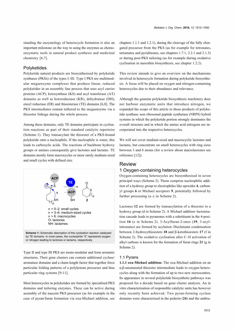
1512
Biosynthesis of oxygen and nitrogen-containing heterocyclesin polyketidesFranziska Hemmerling1,2 and Frank Hahn*1,2
Review Open Access
Address:1Institut für Organische Chemie and Zentrum für BiomolekulareWirkstoffe, Gottfried Wilhelm Leibniz Universität Hannover,Schneiderberg 38, 30167 Hannover, Germany and 2Fakultät fürBiologie, Chemie und Geowissenschaften, Universität Bayreuth,Universitätsstraße 30, 95440 Bayreuth, Germany
Email:Frank Hahn* - [email protected]
* Corresponding author
Keywords:biosynthesis; chemoenzymatic synthesis; enzymology; heterocycles;polyketides
Beilstein J. Org. Chem. 2016, 12, 1512–1550.doi:10.3762/bjoc.12.148
Received: 01 April 2016Accepted: 22 June 2016Published: 20 July 2016
This article is part of the Thematic Series "Natural products in synthesisand biosynthesis II".
Guest Editor: J. S. Dickschat
© 2016 Hemmerling and Hahn; licensee Beilstein-Institut.License and terms: see end of document.
AbstractThis review highlights the biosynthesis of heterocycles in polyketide natural products with a focus on oxygen and nitrogen-contain-
ing heterocycles with ring sizes between 3 and 6 atoms. Heterocycles are abundant structural elements of natural products from all
classes and they often contribute significantly to their biological activity. Progress in recent years has led to a much better under-
standing of their biosynthesis. In this context, plenty of novel enzymology has been discovered, suggesting that these pathways are
an attractive target for future studies.
1512
IntroductionHeterocyclesHeterocycles are important structural elements, which are
present in natural products from all classes and also in many
biologically active synthetic compounds. They often contribute
significantly to their structural and physical properties as well
as to their biological activity [1-3]. Heterocycles can for exam-
ple be involved in cation complexation as known for ionophoric
polyethers or introduce conformational rigidity into a molecule,
which is crucial for target binding [4].
Oxygen heterocycles are mainly found in carbohydrates,
polyketides, peptides and terpenoids. Nitrogen heterocycles are
part of peptides and alkaloids. Both can of course also occur in
the respective hybrid natural products. Sulphur-containing
heterocycles are present in few polyketides and more wide-
spread in peptidic natural products of both, non-ribosomal and
post-ribosomal modified origin [5].
The biosynthetic mechanisms for heterocycle formation are nu-
merous, and range from simple addition or condensation reac-
tions to oxidative ring closures. The large number of mechanis-
tically different cyclisation modes triggers the interest on the re-
sponsible enzymes. Due to the relevance of heterocycles, under-
Beilstein J. Org. Chem. 2016, 12, 1512–1550.
1513
standing the enzymology of heterocycle formation is also an
important milestone on the way to using the enzymes as chemo-
enzymatic tools in natural product synthesis and medicinal
chemistry [6,7].
PolyketidesPolyketide natural products are biosynthesised by polyketide
synthases (PKSs) of the types I–III. Type I PKS are multimod-
ular megaenzyme complexes that produce linear, reduced
polyketides in an assembly line process that uses acyl carrier
proteins (ACP), ketosynthase (KS) and acyl transferase (AT)
domains as well as ketoreductase (KR), dehydratase (DH),
enoyl reductase (ER) and thioesterase (TE) domains [6,8]. The
PKS intermediates remain tethered to the megaenzyme via a
thioester linkage during the whole process.
Among these domains, only TE domains participate in cyclisa-
tion reactions as part of their standard catalytic repertoire
(Scheme 1). They transacylate the thioester of a PKS-bound
polyketide onto a nucleophile. If the nucleophile is water, this
leads to carboxylic acids. The reactions of backbone hydroxy
groups or amines consequently give lactones and lactams. TE
domains mostly form macrocycles or more rarely medium-sized
and small cycles with defined size.
Scheme 1: Schematic description of the cyclisation reaction catalysedby TE domains. In most cases, the nucleophile “X” represents oxygenor nitrogen leading to lactones or lactams, respectively.
Type II and type III PKS are mono-modular and form aromatic
structures. Their gene clusters can contain additional cyclase/
aromatase domains and a chain-length factor that together force
particular folding patterns of a polyketone precursor and thus
particular ring systems [9-11].
Most heterocycles in polyketides are formed by specialised PKS
domains and tailoring enzymes. These can be active during
assembly of the nascent PKS precursor (as for example in the
case of pyran/furan formation via oxa-Michael addition, see
chapters 1.1.1 and 1.2.1), during the cleavage of the fully elon-
gated precursor from the PKS (as for example for tetronates,
tetramates and pyridinones, see chapters 1.7.1, 2.2.1 and 2.1.3)
or during post-PKS tailoring (as for example during oxidative
cyclisation in aureothin biosynthesis, see chapter 1.2.2).
This review intends to give an overview on the mechanisms
involved in heterocycle formation during polyketide biosynthe-
sis. A focus will be placed on oxygen and nitrogen-containing
heterocycles due to their abundance and relevance.
Although the genuine polyketide biosynthesis machinery does
not harbour enzymatic units that introduce nitrogen, we
expanded the scope of this article to those products of polyke-
tide synthase–non ribosomal peptide synthetase (NRPS) hybrid
systems in which the polyketide portion strongly dominates the
overall structure and in which the amino acid nitrogens are in-
corporated into the respective heterocycles.
We will not cover medium-sized and macrocyclic lactones and
lactams, but concentrate on small heterocycles with ring sizes
between 3 and 6 atoms (for a review about macrolactones see
reference [12]).
Review1 Oxygen-containing heterocyclesOxygen-containing heterocycles are biosynthesised in seven
principal ways (Scheme 2). Those comprise nucleophilic addi-
tion of a hydroxy group to electrophiles like epoxides 4, carbon-
yl groups 6 or Michael acceptors 9, potentially followed by
further processing (a–c in Scheme 2).
Lactones 12 are formed by transacylation of a thioester to a
hydroxy group (d in Scheme 2). A Michael addition–lactonisa-
tion cascade leads to pyranones with a substituent in the 4-posi-
tion 16 (e in Scheme 2). 3-Acylfuran-2-ones (19, 3-acyl-
tetronates) are formed by acylation–Dieckmann condensation
between 2-hydroxythioesters 18 and β-ketothioesters 17 (f in
Scheme 2). The oxidative cyclisation after C–H activation of
alkyl carbons is known for the formation of furan rings 21 (g in
Scheme 2).
1.1 Pyrans1.1.1 oxa-Michael addition: The oxa-Michael addition on an
α,β-unsaturated thioester intermediate leads to oxygen hetero-
cycles along with the formation of up to two new stereocentres.
Its appearance in several polyketide biosynthetic pathways was
proposed for a decade based on gene cluster analysis. An in
vitro characterisation of responsible catalytic units has however
only recently been achieved. Two pyran-forming cyclase
domains were characterised in the pederin (24) and the ambru-

Beilstein J. Org. Chem. 2016, 12, 1512–1550.
1514
Scheme 2: Mechanisms for the formation of oxygen heterocycles. The degree of substitution can differ from that shown in the scheme. In b, othermodes of processing are possible in the second step. Partially redrawn from [13].
ticin (28) biosynthetic pathways (Scheme 3 and Scheme 4)
[14,15].
Scheme 3: Pyran-ring formation in pederin (24) biosynthesis. Incuba-tion of recombinant PedPS7 with substrate surrogate 22 gave conver-sion into cyclic stereoisomers anti-23 and syn-23 [14].
PedPS7 is a monofunctional pyran synthase (PS) domain that
was predicted to catalyse ring formation from an α,β-unsatu-
Scheme 4: The domain AmbDH3 from ambruticin biosynthesis cataly-ses the dehydration of 25 and subsequent cyclisation to tetrahydro-pyran 27 with high stereoselectivity [15].
rated intermediate in the biosynthesis of the PKS–NRPS hybrid
product pederin (24) [16,17]. The recombinant, isolated domain
transformed both enantiomers of the structurally simplified
tetraketidic precursor surrogate 22 into cyclised products anti-
23 and syn-23 (Scheme 3a) [14]. The in vitro reaction with
PedPS7 proceeds with moderate stereoselectivity irrespective of
the configuration of the substrate at C7.
Beilstein J. Org. Chem. 2016, 12, 1512–1550.
1515
Scheme 5: SalBIII catalyses dehydration of 29 and subsequent cyclisation to tetrahydropyran 30 [18].
PS domains are common in trans-AT PKS clusters and partici-
pate in the biosynthesis of such important compounds as bryo-
statin and sorangicin. They are related to DH domains on the
amino acid sequence level, but show a significant mutation of a
DH-characteristic aspartic acid to a histidine or an asparagine
residue in their active site. This exchange avoids the dehydra-
tion reaction and might facilitate the activation of the hydroxy
group for nucleophilic attack on the Michael system by proton
abstraction. PS domains also form a distinct phylogenetic clade
compared to DH domains. Within a module, PS domains are
usually located adjacent to DH domains and act on their tran-
siently formed dehydration product [14].
The arrangement is somewhat different in the case of AmbDH3
from ambruticin biosynthesis (Scheme 4) [15]. This bifunc-
tional domain catalyses both steps, dehydration of a 3-hydroxy-
thioester intermediate 25 and subsequent cyclisation to a tetra-
hydropyran ring 27. AmbDH3 is currently the only known case
of a pyran-forming domain in a cis-AT PKS.
Hahn et al. showed that AmbDH3 catalyses dehydration of only
the 2-D,3-D-configured precursor 25 to the E-configured olefin
intermediate 26 and subsequent cyclisation to 27 (Scheme 4).
The C6 epimers of compounds 25 and 26 were also accepted,
but with much lower conversion. In both cases, the configura-
tion at C2 in the cyclic product was exclusively D, highlighting
the high stereoselectivity of the domain-catalysed reaction. The
ambruticins contain a second hydropyran ring that is estab-
lished by epoxide opening (see chapter 1.1.3).
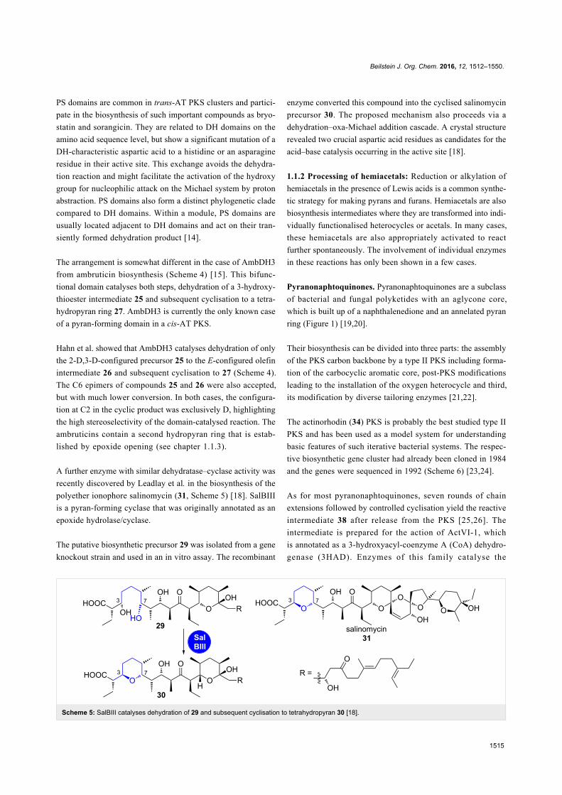
A further enzyme with similar dehydratase–cyclase activity was
recently discovered by Leadlay et al. in the biosynthesis of the
polyether ionophore salinomycin (31, Scheme 5) [18]. SalBIII
is a pyran-forming cyclase that was originally annotated as an
epoxide hydrolase/cyclase.
The putative biosynthetic precursor 29 was isolated from a gene
knockout strain and used in an in vitro assay. The recombinant
enzyme converted this compound into the cyclised salinomycin
precursor 30. The proposed mechanism also proceeds via a
dehydration–oxa-Michael addition cascade. A crystal structure
revealed two crucial aspartic acid residues as candidates for the
acid–base catalysis occurring in the active site [18].
1.1.2 Processing of hemiacetals: Reduction or alkylation of
hemiacetals in the presence of Lewis acids is a common synthe-
tic strategy for making pyrans and furans. Hemiacetals are also
biosynthesis intermediates where they are transformed into indi-
vidually functionalised heterocycles or acetals. In many cases,
these hemiacetals are also appropriately activated to react
further spontaneously. The involvement of individual enzymes
in these reactions has only been shown in a few cases.
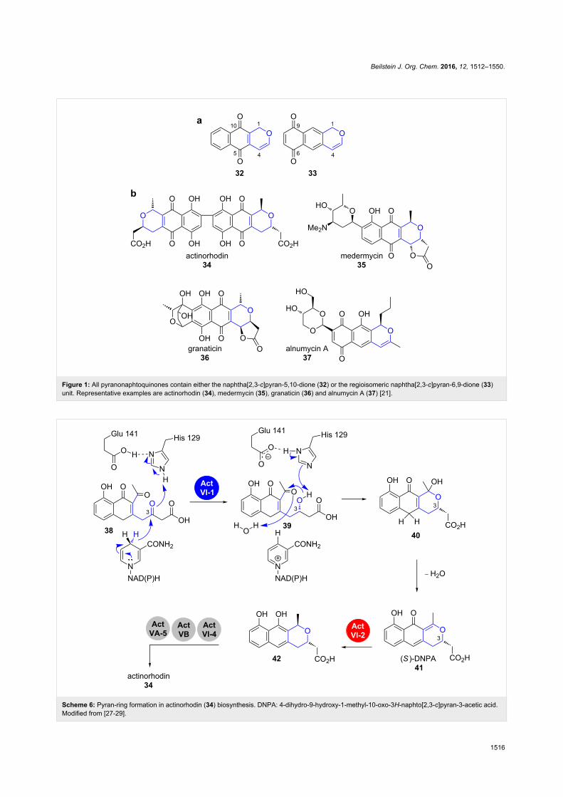
Pyranonaphtoquinones. Pyranonaphtoquinones are a subclass
of bacterial and fungal polyketides with an aglycone core,
which is built up of a naphthalenedione and an annelated pyran
ring (Figure 1) [19,20].
Their biosynthesis can be divided into three parts: the assembly
of the PKS carbon backbone by a type II PKS including forma-
tion of the carbocyclic aromatic core, post-PKS modifications
leading to the installation of the oxygen heterocycle and third,
its modification by diverse tailoring enzymes [21,22].
The actinorhodin (34) PKS is probably the best studied type II
PKS and has been used as a model system for understanding
basic features of such iterative bacterial systems. The respec-
tive biosynthetic gene cluster had already been cloned in 1984
and the genes were sequenced in 1992 (Scheme 6) [23,24].
As for most pyranonaphtoquinones, seven rounds of chain
extensions followed by controlled cyclisation yield the reactive
intermediate 38 after release from the PKS [25,26]. The
intermediate is prepared for the action of ActVI-1, which
is annotated as a 3-hydroxyacyl-coenzyme A (CoA) dehydro-
genase (3HAD). Enzymes of this family catalyse the
Beilstein J. Org. Chem. 2016, 12, 1512–1550.
1516
Figure 1: All pyranonaphtoquinones contain either the naphtha[2,3-c]pyran-5,10-dione (32) or the regioisomeric naphtha[2,3-c]pyran-6,9-dione (33)unit. Representative examples are actinorhodin (34), medermycin (35), granaticin (36) and alnumycin A (37) [21].
Scheme 6: Pyran-ring formation in actinorhodin (34) biosynthesis. DNPA: 4-dihydro-9-hydroxy-1-methyl-10-oxo-3H-naphto[2,3-c]pyran-3-acetic acid.Modified from [27-29].
Beilstein J. Org. Chem. 2016, 12, 1512–1550.
1517
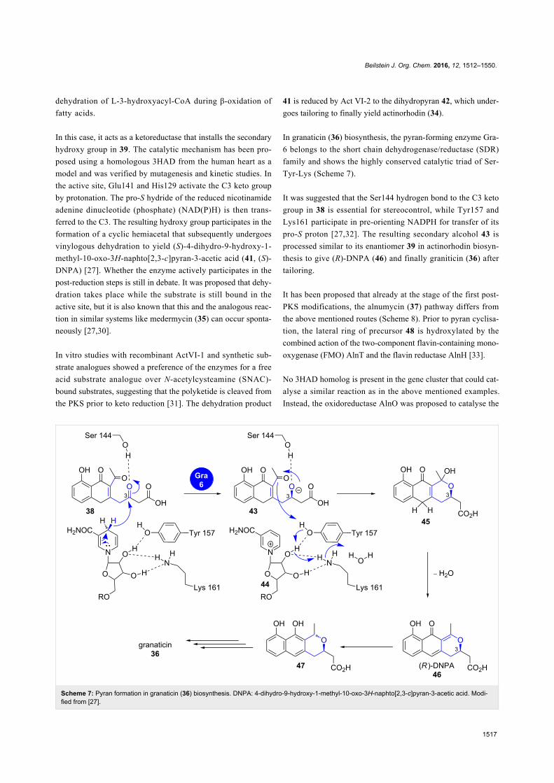
Scheme 7: Pyran formation in granaticin (36) biosynthesis. DNPA: 4-dihydro-9-hydroxy-1-methyl-10-oxo-3H-naphto[2,3-c]pyran-3-acetic acid. Modi-fied from [27].
dehydration of L-3-hydroxyacyl-CoA during β-oxidation of
fatty acids.
In this case, it acts as a ketoreductase that installs the secondary
hydroxy group in 39. The catalytic mechanism has been pro-
posed using a homologous 3HAD from the human heart as a
model and was verified by mutagenesis and kinetic studies. In
the active site, Glu141 and His129 activate the C3 keto group
by protonation. The pro-S hydride of the reduced nicotinamide
adenine dinucleotide (phosphate) (NAD(P)H) is then trans-
ferred to the C3. The resulting hydroxy group participates in the
formation of a cyclic hemiacetal that subsequently undergoes
vinylogous dehydration to yield (S)-4-dihydro-9-hydroxy-1-
methyl-10-oxo-3H-naphto[2,3-c]pyran-3-acetic acid (41, (S)-
DNPA) [27]. Whether the enzyme actively participates in the
post-reduction steps is still in debate. It was proposed that dehy-
dration takes place while the substrate is still bound in the
active site, but it is also known that this and the analogous reac-
tion in similar systems like medermycin (35) can occur sponta-
neously [27,30].
In vitro studies with recombinant ActVI-1 and synthetic sub-
strate analogues showed a preference of the enzymes for a free
acid substrate analogue over N-acetylcysteamine (SNAC)-
bound substrates, suggesting that the polyketide is cleaved from
the PKS prior to keto reduction [31]. The dehydration product
41 is reduced by Act VI-2 to the dihydropyran 42, which under-
goes tailoring to finally yield actinorhodin (34).
In granaticin (36) biosynthesis, the pyran-forming enzyme Gra-
6 belongs to the short chain dehydrogenase/reductase (SDR)
family and shows the highly conserved catalytic triad of Ser-
Tyr-Lys (Scheme 7).
It was suggested that the Ser144 hydrogen bond to the C3 keto
group in 38 is essential for stereocontrol, while Tyr157 and
Lys161 participate in pre-orienting NADPH for transfer of its
pro-S proton [27,32]. The resulting secondary alcohol 43 is
processed similar to its enantiomer 39 in actinorhodin biosyn-
thesis to give (R)-DNPA (46) and finally graniticin (36) after
tailoring.
It has been proposed that already at the stage of the first post-
PKS modifications, the alnumycin (37) pathway differs from
the above mentioned routes (Scheme 8). Prior to pyran cyclisa-
tion, the lateral ring of precursor 48 is hydroxylated by the
combined action of the two-component flavin-containing mono-
oxygenase (FMO) AlnT and the flavin reductase AlnH [33].
No 3HAD homolog is present in the gene cluster that could cat-
alyse a similar reaction as in the above mentioned examples.
Instead, the oxidoreductase AlnO was proposed to catalyse the

Beilstein J. Org. Chem. 2016, 12, 1512–1550.
1518
Scheme 8: Pyran formation in alnumycin (37) biosynthesis. Adapted from [21].
stereoselective reduction of the ketone at C15 in 49. The pyran
51 would then be obtained by spontaneous or enzyme-sup-
ported hemiacetalisation followed by dehydration [34]. The
tricyclic core unit is oxidised further and heavily decorated by
tailoring enzymes, also involving an unusual rearrangement
leading to the dioxane unit, whose carbon atoms originally
derive from a sugar building block [34-36].
1.1.3 Epoxide opening: The nucleophilic opening of epoxides
is probably the most abundant type of reaction leading to furans
and pyrans. It, for example, plays an important role in the bio-
synthesis of ionophoric terrestrial and marine polyethers (see
chapter 1.3). In this chapter, we will focus on two examples in
which one pyran ring is formed. Both characteristically deviate
from the typical polyether-specific interplay between one epoxi-
dase and one or a few epoxide hydrolases that collaboratively
set up multiple oxygen heterocycles.
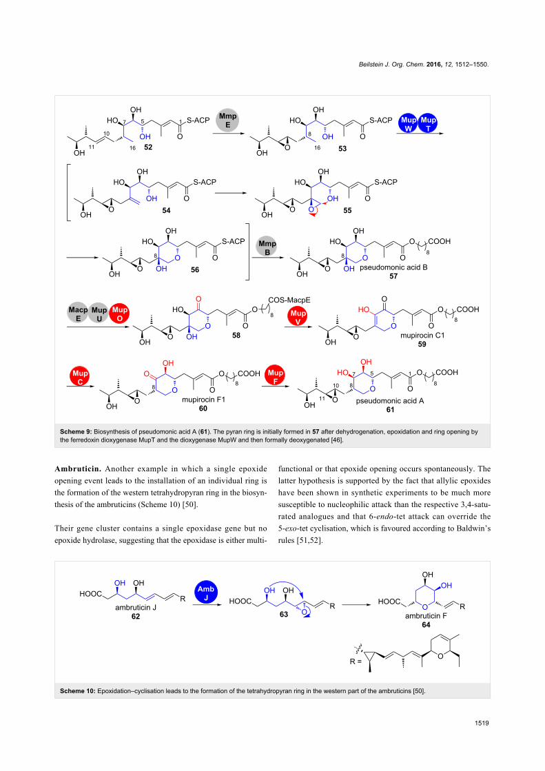
Pseudomonic acid A. Mupirocin is a clinically important anti-
biotic against Gram-positive bacteria, which consists of a mix-
ture of pseudomonic acids from Pseudomonas fluorescens
NCIMB 10586 with pseudomonic acid A (61) being the main
compound (Scheme 9) [37-44]. It belongs to the group of trans-
AT-PKS products and the gene cluster harbours genes that code
for a β-hydroxymethylglutaryl-CoA synthase (HCS) cassette
(mupG, mupH, mupJ, mupK and macpC) and an iteratively
acting type I fatty acid synthase (FAS) (mmpB).
During the biosynthesis of the pseudomonic acids, the initially
formed PKS product 52 undergoes a complex tailoring path-
way (Scheme 9) [45]. A remarkable feature is the tightly regu-
lated steps that lead to the formation and decoration of the
pyran-ring-containing region between C5 and C11 in 61 [46-
49]. This has been studied by a series of fermentation and gene
deletion–intermediate isolation experiments.
The process starts by oxidoreductase domain MmpE-catalysed
epoxidation of the double bond between C10 and C11. Olefin
53 is thus a branching point from which two series of analo-
gous C10–C11 epoxides (53–61) and C10–C11 (not shown)
olefins arise (Scheme 9). The fact that the wild-type titers of the
respective olefins are much lower than the analogous epoxides
53–61 suggests that epoxidation has a strong influence on the
performance of the downstream enzymes.
The dioxygenase MupW together with its associated ferredoxin
dioxygenase MupT then catalyse dehydrogenation and epoxida-
tion on C8 and C16 of 53. Whether the pyran-ring closure is
also mediated by an enzymatic activity or if this reaction is a
spontaneous process could not be clarified yet and may be
subject for in vitro studies with the purified enzymes.
The net-deoxygenation on C8 of pseudomonic acid B (57) is
obtained by a multistep process (Scheme 9). After elongation by
the iterative type I fatty acid synthase MmpB, redox transfor-
mations and a dehydration on the MacpE-bound substrate 58
finally lead to pseudomonic acid A (61) with a 3,4-dihydroxy-
2,5-disubstituted pyran ring. The reason for the elaborate oxida-
tion–reduction on the C6 and C7 hydroxy groups during this
biosynthetic endgame remains enigmatic [46].
Beilstein J. Org. Chem. 2016, 12, 1512–1550.
1519
Scheme 9: Biosynthesis of pseudomonic acid A (61). The pyran ring is initially formed in 57 after dehydrogenation, epoxidation and ring opening bythe ferredoxin dioxygenase MupT and the dioxygenase MupW and then formally deoxygenated [46].
Scheme 10: Epoxidation–cyclisation leads to the formation of the tetrahydropyran ring in the western part of the ambruticins [50].
Ambruticin. Another example in which a single epoxide
opening event leads to the installation of an individual ring is
the formation of the western tetrahydropyran ring in the biosyn-
thesis of the ambruticins (Scheme 10) [50].
Their gene cluster contains a single epoxidase gene but no
epoxide hydrolase, suggesting that the epoxidase is either multi-
functional or that epoxide opening occurs spontaneously. The
latter hypothesis is supported by the fact that allylic epoxides
have been shown in synthetic experiments to be much more
susceptible to nucleophilic attack than the respective 3,4-satu-
rated analogues and that 6-endo-tet attack can override the
5-exo-tet cyclisation, which is favoured according to Baldwin’s
rules [51,52].
Beilstein J. Org. Chem. 2016, 12, 1512–1550.
1520
Scheme 11: a) Nonactin (70) is formed from heterodimers of (−)(+)-dimeric nonactic acid and (+)(−)-dimeric nonactic acid. b) The product of the nonSgene catalyses the cyclisation of (6R,8R,E)-6,8-dihydroxy-2-methylnon-2-enoic acid thioester (71a and 71 b) to (+)-nonactic acid thioester (69b/72)[53,56,58].
1.2 Furans1.2.1 oxa-Michael addition: Similar to the PS domains de-
scribed in chapter 1.1.1, furan rings can also be biosynthesised
via oxa-Michael additions.
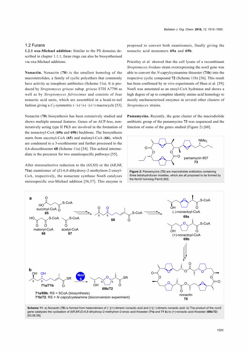
Nonactin. Nonactin (70) is the smallest homolog of the
macrotetrolides, a family of cyclic polyethers that commonly
have activity as ionophore antibiotics (Scheme 11a). It is pro-
duced by Streptomyces griseus subsp. griseus ETH A7796 as
well as by Streptomyces fulvissimus and consists of four
nonactic acid units, which are assembled in a head-to-tail
fashion giving a C2-symmetric (−)-(+)-(−)-(+) macrocycle [53].
Nonactin (70) biosynthesis has been extensively studied and
shows multiple unusual features. Genes of an ACP-less, non-
iteratively acting type II PKS are involved in the formation of
the nonactoyl-CoA (69a and 69b) backbone. The biosynthesis
starts from succinyl-CoA (65) and malonyl-CoA (66), which
are condensed to a 3-oxothioester and further processed to the
4,6-dioxothioester 68 (Scheme 11a) [54]. This achiral interme-
diate is the precursor for two enantiospecific pathways [55].
After stereoselective reduction to the (6S,8S) or the (6R,8R,
71a) enantiomer of (E)-6,8-dihydroxy-2-methylnon-2-enoyl-
CoA, respectively, the nonactate synthase NonS catalyses
stereospecific oxa-Michael addition [56,57]. This enzyme is
proposed to convert both enantiomers, finally giving the
nonactic acid monomers 69a and 69b.
Priestley et al. showed that the cell lysate of a recombinant
Streptomyces lividans strain overexpressing the nonS gene was
able to convert the N-caprylcysteamine thioester (71b) into the
respective cyclic compound 72 (Scheme 11b) [56]. This result
has been confirmed by in vivo experiments of Shen et al. [59].
NonS was annotated as an enoyl-CoA hydratase and shows a
high degree of up to complete identity amino acid homology to
mostly uncharacterised enzymes in several other clusters of
Streptomyces strains.
Pamamycins. Recently, the gene cluster of the macrodiolide
antibiotic group of the pamamycins 73 was sequenced and the
function of some of the genes studied (Figure 2) [60].
Figure 2: Pamamycins (73) are macrodiolide antibiotics containingthree tetrahydrofuran moieties, which are all proposed to be formed bythe NonS homolog PamS [60].
Beilstein J. Org. Chem. 2016, 12, 1512–1550.
1521
Scheme 12: A PS domain homolog in oocydin A (76) biosynthesis is proposed to catalyse furan formation via an oxa-Michael addition [61].
Scheme 13: Mechanism of oxidation–furan cyclisation by AurH, which converts (+)-deoxyaureothin (77) into (+)-aureothin (79) [13].
This cluster contains a NonS homolog, PamS, that was pro-
posed to catalyse all three oxa-Michael additions that lead to
tetrahydrofuran formation during biosynthesis. As the enzyme
must act on biosynthetic intermediates of strongly varying size,
this attributes a remarkably broad substrate tolerance to PamS.
No detailed characterisation of PamS has been carried out yet.
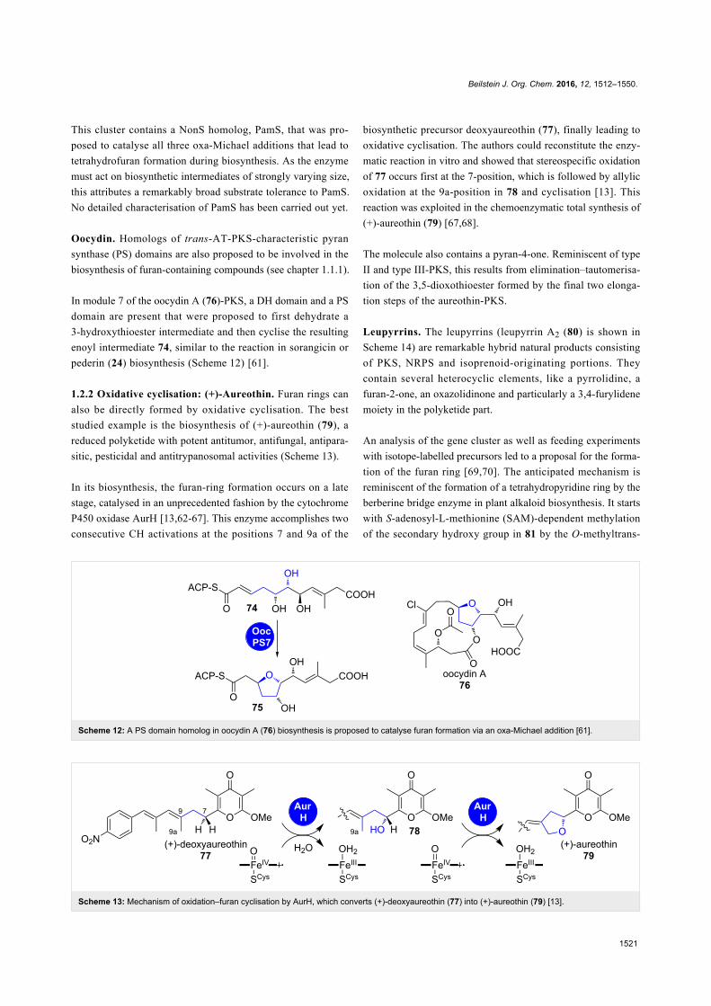
Oocydin. Homologs of trans-AT-PKS-characteristic pyran
synthase (PS) domains are also proposed to be involved in the
biosynthesis of furan-containing compounds (see chapter 1.1.1).
In module 7 of the oocydin A (76)-PKS, a DH domain and a PS
domain are present that were proposed to first dehydrate a
3-hydroxythioester intermediate and then cyclise the resulting
enoyl intermediate 74, similar to the reaction in sorangicin or
pederin (24) biosynthesis (Scheme 12) [61].
1.2.2 Oxidative cyclisation: (+)-Aureothin. Furan rings can
also be directly formed by oxidative cyclisation. The best
studied example is the biosynthesis of (+)-aureothin (79), a
reduced polyketide with potent antitumor, antifungal, antipara-
sitic, pesticidal and antitrypanosomal activities (Scheme 13).
In its biosynthesis, the furan-ring formation occurs on a late
stage, catalysed in an unprecedented fashion by the cytochrome
P450 oxidase AurH [13,62-67]. This enzyme accomplishes two
consecutive CH activations at the positions 7 and 9a of the
biosynthetic precursor deoxyaureothin (77), finally leading to
oxidative cyclisation. The authors could reconstitute the enzy-
matic reaction in vitro and showed that stereospecific oxidation
of 77 occurs first at the 7-position, which is followed by allylic
oxidation at the 9a-position in 78 and cyclisation [13]. This
reaction was exploited in the chemoenzymatic total synthesis of
(+)-aureothin (79) [67,68].
The molecule also contains a pyran-4-one. Reminiscent of type
II and type III-PKS, this results from elimination–tautomerisa-
tion of the 3,5-dioxothioester formed by the final two elonga-
tion steps of the aureothin-PKS.
Leupyrrins. The leupyrrins (leupyrrin A2 (80) is shown in
Scheme 14) are remarkable hybrid natural products consisting
of PKS, NRPS and isoprenoid-originating portions. They
contain several heterocyclic elements, like a pyrrolidine, a
furan-2-one, an oxazolidinone and particularly a 3,4-furylidene
moiety in the polyketide part.
An analysis of the gene cluster as well as feeding experiments
with isotope-labelled precursors led to a proposal for the forma-
tion of the furan ring [69,70]. The anticipated mechanism is
reminiscent of the formation of a tetrahydropyridine ring by the
berberine bridge enzyme in plant alkaloid biosynthesis. It starts
with S-adenosyl-L-methionine (SAM)-dependent methylation
of the secondary hydroxy group in 81 by the O-methyltrans-

Beilstein J. Org. Chem. 2016, 12, 1512–1550.
1522
Scheme 14: Leupyrrin A2 (80) and the proposed biosynthesis of its furylidene moiety [69,70].
Scheme 15: Asperfuranone (93) biosynthesis, adapted from [75].
ferase Leu14 (Scheme 14a) [71-74]. Oxidation of the methoxy
group in 82 by the cluster-encoded dehydrogenase Leu8 is fol-
lowed by a Prins-type cyclisation. No enzyme candidate for the
cyclisation reaction to 84 could be identified in the cluster.
1.2.3 Processing of hemiacetals: Asperfuranone. Asperfura-
none (93) consists of a polyketide side chain, attached to the C3
of an oxidised isobenzofuran (Scheme 15). The respective
biosynthetic cluster contains seven genes and has been identi-

Beilstein J. Org. Chem. 2016, 12, 1512–1550.
1523
fied by Wang and co-workers through a genome mining ap-
proach in Aspergillus nidulans [76]. Later on, the same group
annotated a highly homologous gene cluster in Aspergillus
terreus and elucidated the timing and mechanism of asperfura-
none biosynthesis by step-wise heterologous expression of the
individual genes in A. nidulans [77]. Thus, genes involved in
asperfuranone biosynthesis have been renamed from “afo” to
“ateafo”.
This bipartite azaphilone structure corresponds to its assembly
by the highly reducing (HR)-PKS AteafoG, followed by a non-
reducing (NR)-PKS AteafoE. The product of the HR-PKS
AteafoG, tetraketide 85, is transferred to the starter unit:ACP
transacylase (SAT) domain of the NR-PKS AteafoE. After the
elongation by four further ketide units, reductive PKS release
and Knoevenagel condensation yield the benzaldehyde interme-
diate 88. Oxidative dearomatisation of 88 catalysed by the sali-
cylate monooxygenase AteafoD gives 89, which is hydroxylat-
ed at C8 by the oxygenase AteafoF. The positioning of this
newly formed hydroxy group forces the formation of a five-
membered ring hemiacetal in 91. Spontaneous dehydration
installs the furan moiety and after keto reduction by an endoge-
nous reductase, asperfuranone (93) is obtained.
Aflatoxins. Aflatoxins 94–99 are highly toxic carcinogens pro-
duced in several Aspergillus species (Figure 3). The respective
pathway gene clusters have been identified and homologies be-
tween Aspergillus species were compared for example by the
groups of Bennett and Ehrlich [78,79]. Structurally, aflatoxins
belong to the group of furanocoumarins and consist of a penta-
cyclic system in which a benzobisfuran is annelated with a
δ-lactone and a cyclopentanone or oxidation products of the
latter.
Aflatoxin biosynthesis has been studied since the late 1960s and
has attracted attention, because the polyketide undergoes a
series of oxidative rearrangements, which drastically alter the
molecular scaffold. Due to the complexity of these processes,
we will focus on the steps directly associated with heterocycle
formation [82-84].
Aflatoxin B1 (94) is considered as the most toxic aflatoxin. It is
derived in multiple enzymatic conversions from norsolorinic
acid anthrone 100, which is produced by the norsolorinic acid
synthase (NorS) (Scheme 16) [83,85]. NorS is a complex of a
NR-PKS PksA and a pair of yeast-like fatty acid synthases
HexA/HexB, which provide an unusual hexanoyl-CoA starter
unit [86]. Norsolorinic acid (100) undergoes three oxidative re-
arrangements towards aflatoxin B1 (94): The first rearrange-
ment sets up the benzobisfuran motif in 106, the second
rearranges the anthraquinone in 106 to the xanthone in 107 and
Figure 3: The four major aflatoxins produced by Aspergilli are thetypes B1, B2, G1 and G2 (94–97). In the digestive tract of animals,aflatoxins B1 and B2 (94 and 95) are oxidized to M1 and M2 (98 and99), respectively [80,81].
the third is an oxidative ring contraction towards the cyclopen-
tanone in 94 (Scheme 16).
After several enzymatic post-PKS modifications, the oxoaver-
antin (OAVN) cyclase transforms 5’-oxoaverantin (101) into
averufin (102) by intramolecular acetal formation [87]. To date,
it is not clear, how exactly the OAVN cyclase participates in
this process [88]. Interestingly, the OAVN cyclase operates
cofactor-free, although it contains a NAD(P)+-binding Rossman
fold. Furthermore, this enzyme is also capable of catalysing the
later conversion of versiconal (105) to versicolorin B (106)
[88].
Averufin (102) is the starting point for the first oxidative rear-
rangement. Feeding experiments with isotope-labelled averufins
(102) showed that their C5’ and C6’-carbons (pink) are excised
on the way to aflatoxin B1 (94) and that the oxidation state of
C1’ (green) changes from that of an alcohol to an aldehyde,
implying that the rearrangement must be oxidative
[82,89,91,92,107].
The biosynthetic mechanims of the conversion of averufin (102)
into 1’-hydroxyversicolorone (103) has been the subject of
intensive studies. Gene disruption experiments in the aflatoxi-

Beilstein J. Org. Chem. 2016, 12, 1512–1550.
1524
Scheme 16: Overview on aflatoxin B1 (94) biosynthesis. HOMST = 11-hydroxy-O-methylsterigmatocystin [78,79,82-106].
genic strain A. parasiticus NRRL 2999 revealed that this step is
in fact catalysed by the cytochrome P450 enzyme AVR mono-
oxygenase via an undeciphered mechanism (encoded by the
gene cypX, see Scheme 16) [93]. The same study also revealed
the participation of the FMO MoxY in a Baeyer–Villiger oxida-
tion, which yields versiconal acetate (104) [93,94]. This is then
hydrolysed by a cytosolic esterase (putatively also coded in the
aflatoxin gene cluster as estA) to versiconal (105) [95]. The
bisfuran moiety of versicolorin B (106), which is crucial for the
mutagenic DNA binding, is then set up stereospecifically by the
versiconal cyclase, which accepts both enantiomers (2’R and
2’S) of versiconal (105) [96,108]. Heterologous expression and
characterisation by Townsend and co-workers revealed that the
versicolorin B synthase (VBS) does not require any cofactors,
in spite of its flavin adenine dinucleotide (FAD) binding site
[98,99].
The reaction mechanisms and biosynthetic enzymes involved in
the rearrangement of versicolorin B (106) to demethylsterigma-
tocystin (107) have also been discussed controversely. Up to
four genes (aflM, aflN, aflX and aflY) have been implied in
biosynthetic studies to code for enzymes that are participating
in this complex conversion [100]. Henry and Townsend sug-
gested an oxidation–reduction–oxidation sequence mediated by
putative NADPH-dependent oxidoreductase AflM and
cytochrome P450 enzyme AflN [101]. Gene disruption experi-
ments by Cary et al. have shown that the NADH-dependent
oxidoreductase AflX also takes part in the conversion [102].
Beilstein J. Org. Chem. 2016, 12, 1512–1550.
1525
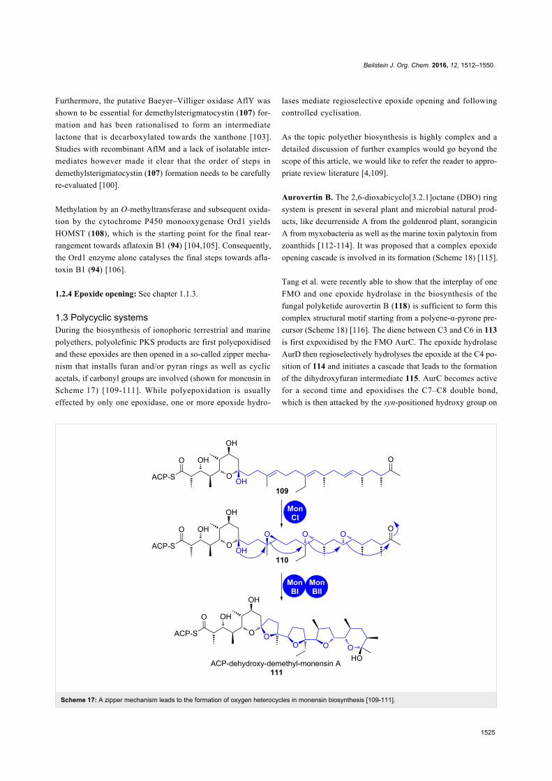
Scheme 17: A zipper mechanism leads to the formation of oxygen heterocycles in monensin biosynthesis [109-111].
Furthermore, the putative Baeyer–Villiger oxidase AflY was
shown to be essential for demethylsterigmatocystin (107) for-
mation and has been rationalised to form an intermediate
lactone that is decarboxylated towards the xanthone [103].
Studies with recombinant AflM and a lack of isolatable inter-
mediates however made it clear that the order of steps in
demethylsterigmatocystin (107) formation needs to be carefully
re-evaluated [100].
Methylation by an O-methyltransferase and subsequent oxida-
tion by the cytochrome P450 monooxygenase Ord1 yields
HOMST (108), which is the starting point for the final rear-
rangement towards aflatoxin B1 (94) [104,105]. Consequently,
the Ord1 enzyme alone catalyses the final steps towards afla-
toxin B1 (94) [106].
1.2.4 Epoxide opening: See chapter 1.1.3.
1.3 Polycyclic systemsDuring the biosynthesis of ionophoric terrestrial and marine
polyethers, polyolefinic PKS products are first polyepoxidised
and these epoxides are then opened in a so-called zipper mecha-
nism that installs furan and/or pyran rings as well as cyclic
acetals, if carbonyl groups are involved (shown for monensin in
Scheme 17) [109-111]. While polyepoxidation is usually
effected by only one epoxidase, one or more epoxide hydro-
lases mediate regioselective epoxide opening and following
controlled cyclisation.
As the topic polyether biosynthesis is highly complex and a
detailed discussion of further examples would go beyond the
scope of this article, we would like to refer the reader to appro-
priate review literature [4,109].
Aurovertin B. The 2,6-dioxabicyclo[3.2.1]octane (DBO) ring
system is present in several plant and microbial natural prod-
ucts, like decurrenside A from the goldenrod plant, sorangicin
A from myxobacteria as well as the marine toxin palytoxin from
zoanthids [112-114]. It was proposed that a complex epoxide
opening cascade is involved in its formation (Scheme 18) [115].
Tang et al. were recently able to show that the interplay of one
FMO and one epoxide hydrolase in the biosynthesis of the
fungal polyketide aurovertin B (118) is sufficient to form this
complex structural motif starting from a polyene-α-pyrone pre-
cursor (Scheme 18) [116]. The diene between C3 and C6 in 113
is first expoxidised by the FMO AurC. The epoxide hydrolase
AurD then regioselectively hydrolyses the epoxide at the C4 po-
sition of 114 and initiates a cascade that leads to the formation
of the dihydroxyfuran intermediate 115. AurC becomes active
for a second time and epoxidises the C7–C8 double bond,
which is then attacked by the syn-positioned hydroxy group on
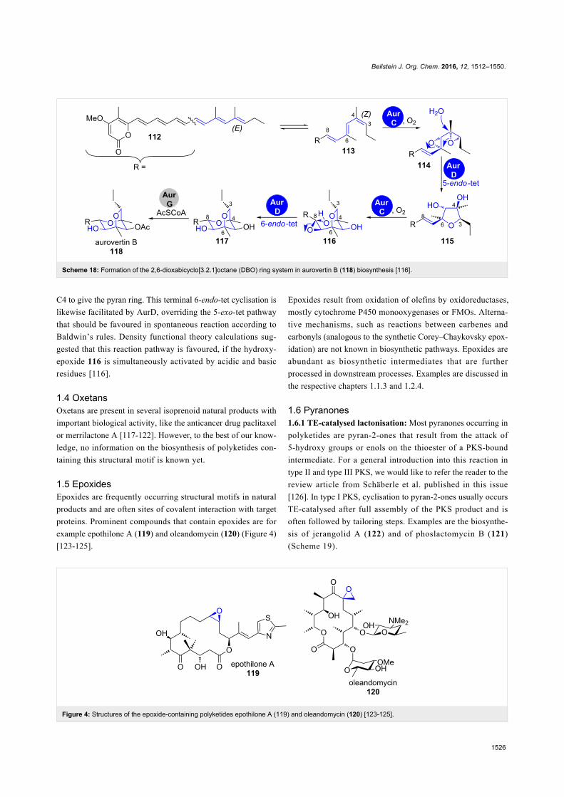
Beilstein J. Org. Chem. 2016, 12, 1512–1550.
1526
Scheme 18: Formation of the 2,6-dioxabicyclo[3.2.1]octane (DBO) ring system in aurovertin B (118) biosynthesis [116].
Figure 4: Structures of the epoxide-containing polyketides epothilone A (119) and oleandomycin (120) [123-125].
C4 to give the pyran ring. This terminal 6-endo-tet cyclisation is
likewise facilitated by AurD, overriding the 5-exo-tet pathway
that should be favoured in spontaneous reaction according to
Baldwin’s rules. Density functional theory calculations sug-
gested that this reaction pathway is favoured, if the hydroxy-
epoxide 116 is simultaneously activated by acidic and basic
residues [116].
1.4 OxetansOxetans are present in several isoprenoid natural products with
important biological activity, like the anticancer drug paclitaxel
or merrilactone A [117-122]. However, to the best of our know-
ledge, no information on the biosynthesis of polyketides con-
taining this structural motif is known yet.
1.5 EpoxidesEpoxides are frequently occurring structural motifs in natural
products and are often sites of covalent interaction with target
proteins. Prominent compounds that contain epoxides are for
example epothilone A (119) and oleandomycin (120) (Figure 4)
[123-125].
Epoxides result from oxidation of olefins by oxidoreductases,
mostly cytochrome P450 monooxygenases or FMOs. Alterna-
tive mechanisms, such as reactions between carbenes and
carbonyls (analogous to the synthetic Corey–Chaykovsky epox-
idation) are not known in biosynthetic pathways. Epoxides are
abundant as biosynthetic intermediates that are further
processed in downstream processes. Examples are discussed in
the respective chapters 1.1.3 and 1.2.4.
1.6 Pyranones1.6.1 TE-catalysed lactonisation: Most pyranones occurring in
polyketides are pyran-2-ones that result from the attack of
5-hydroxy groups or enols on the thioester of a PKS-bound
intermediate. For a general introduction into this reaction in
type II and type III PKS, we would like to refer the reader to the
review article from Schäberle et al. published in this issue
[126]. In type I PKS, cyclisation to pyran-2-ones usually occurs
TE-catalysed after full assembly of the PKS product and is
often followed by tailoring steps. Examples are the biosynthe-
sis of jerangolid A (122) and of phoslactomycin B (121)
(Scheme 19).

Beilstein J. Org. Chem. 2016, 12, 1512–1550.
1527
Scheme 19: Structures of phoslactomycin B (121) (a) and jerangolid A (122) (b). The heterocycle-forming steps in their biosynthesis are shown onthe bottom [50,127].
In phoslactomycin B (121) biosynthesis, the 4-hydroxytetra-
hydro-2H-pyran-2-one 124 is formally dehydrated by consecu-
tive malonylation–elimination to finally give a 5,6-dihydro-2H-
pyran-2-one 127 [127]. The tailoring enzyme PlmT2 was
proposed to catalyse the decarboxylative elimination of
malonoyl halfester 126. It is not clear, whether the initial
malonylation was catalysed by an AT domain or another en-
zyme in the cluster. Similar chemistry occurs during the biosyn-
thesis of related compounds like fostriecin and leptomycin
[128,129].
In jerangolid A biosynthesis, the dihydro-2H-pyran-2,4(3H)-
dione 128 is transformed into a 4-methoxy-5,6-dihydro-2H-
pyran-2-one 129 by action of the O-methyltransferase JerF [50].
1.6.2 Michael addition–lactonisation: A novel mechanism for
the integration of pyran-2-ones into polyketide backbones has
recently been discovered.
Rhizoxin. In 2013, Hertweck and co-workers provided detailed
insight into the unprecedented enzyme catalysis involved in the
formation of 4-substituted δ-lactones and the structurally
closely related glutarimides, respectively (Scheme 20) [130].
The assembly of both moieties includes a β-branching event of
the polyketide carbon backbone that is mechanistically differ-
ent from that occurring during isoprenoid biosynthesis. The
designated branching modules of lactone and glutarimide-pro-
ducing PKS show similar designs: a branching domain (B or
X), which is flanked by a KS and an ACP domain (Scheme 20b
and c).
In vitro reconstitution experiments with the branching module
of the macrolide rhizoxin (130) (rhiPKS) and synthetic SNAC-
thioesters revealed that the chain branch originates from a syn-
selective Michael addition of an ACP-bound malonate unit 133
to a KS-bound α,β-unsaturated thioester 132 (Scheme 20b)
[130]. This results in an intermediate 134 in which the ACP and
the KS domain are covalently linked by the branched polyke-
tide. Subsequent nucleophilic attack of the δ-hydroxy group on
the thioester then yields the ACP-bound δ-lactone 135 and the
polyketide chain can be passed downstream on the assembly
line.
When testing the substrate scope of the rhizoxin (130)
branching module, C3-substituted as well as amino and carbox-
amide nucleophiles in lieu of a hydroxy group in 132 were
accepted, yielding δ-lactam and glutarimide moieties, respec-
tively [131,132]. When the B-domain of the rhiPKS was
exchanged with an X-domain of glutarimide-producing PKS
from the 9-methylstreptimidone PKS of S. himastatinicus, both,
glutarimides and lactones were obtained from respective sub-
strate conversions. Thus, the domains can be seen as function-
ally equivalent [133]. Supported by kinetic analyses and muta-
tional studies it was shown that B-domains, neither have an in-
fluence on the substrate selectivity nor on the turnover and
furthermore do not catalytically take part in the branching or
heterocyclisation event. Solely their double-hotdog fold is struc-
turally essential for the branching module. Consequently, the
B-domain has even been mimicked with a dehydratase domain
that bears the same folding motif. It is thus most interesting that
the branching KS domain alone mediates the entire catalytic se-
quence and represents a unique family of ligase-cyclase.

Beilstein J. Org. Chem. 2016, 12, 1512–1550.
1528
Scheme 20: a) Structures of rhizoxin (130) and cycloheximide (131). Model for the formation of δ-lactones (b) or glutarimides (c), respectively.Adapted from [133].
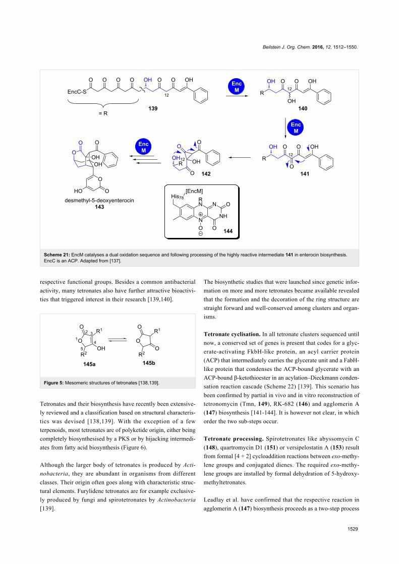
1.6.3 Favorskii rearrangement: Enterocin. Another mecha-
nism applies for the δ-lactone embedded in the tricyclic, caged
core of the bacteriostatic agent enterocin that is produced in
Streptomyces species [134,135]. The respective biosynthetic
pathway has been fully reconstituted in an in vitro one-pot reac-
tion [136]. The flavoprotein EncM transforms the C12 methy-
lene group of the octaketidic PKS type II product 139 in a two-
step oxidation sequence using the unprecedented, enzyme-
bound flavin-N5-oxide 144 (Scheme 21) [137].
The resulting ketone 141 undergoes a Favorskii rearrangement,
finally leading to the formation of the δ-lactone moiety. EncM
has also been rationalised to participate in the stereoselectivity
of the subsequent aldol condensations and the final lactonisa-
tion yielding the pyran-2-one attached to the caged ring system
[137].
1.7 Furanones1.7.1 Acylation–Dieckmann condensation: Tetronates.
Tetronates (4-hydroxyfuran-2(5H)-ones, 145a/145b) are an
abundant type of heterocycles with a broad spectrum of biologi-
cal activities (Figure 5) [138,139]. In polyketides, they mostly
appear in form of 3-acyltetronates and it was proposed that this
structural motif is able to mimic corresponding anions of acidic
functional groups like phosphates, sulphates or carboxylates. In
fact, tetronates often act by inhibiting enzymes that process the
Beilstein J. Org. Chem. 2016, 12, 1512–1550.
1529
Scheme 21: EncM catalyses a dual oxidation sequence and following processing of the highly reactive intermediate 141 in enterocin biosynthesis.EncC is an ACP. Adapted from [137].
respective functional groups. Besides a common antibacterial
activity, many tetronates also have further attractive bioactivi-
ties that triggered interest in their research [139,140].
Figure 5: Mesomeric structures of tetronates [138,139].
Tetronates and their biosynthesis have recently been extensive-
ly reviewed and a classification based on structural characteris-
tics was devised [138,139]. With the exception of a few
terpenoids, most tetronates are of polyketide origin, either being
completely biosynthesised by a PKS or by hijacking intermedi-
ates from fatty acid biosynthesis (Figure 6).
Although the larger body of tetronates is produced by Acti-
nobacteria, they are abundant in organisms from different
classes. Their origin often goes along with characteristic struc-
tural elements. Furylidene tetronates are for example exclusive-
ly produced by fungi and spirotetronates by Actinobacteria
[139].
The biosynthetic studies that were launched since genetic infor-
mation on more and more tetronates became available revealed
that the formation and the decoration of the ring structure are
straight forward and well-conserved among clusters and organ-
isms.
Tetronate cyclisation. In all tetronate clusters sequenced until
now, a conserved set of genes is present that codes for a glyc-
erate-activating FkbH-like protein, an acyl carrier protein
(ACP) that intermediately carries the glycerate unit and a FabH-
like protein that condenses the ACP-bound glycerate with an
ACP-bound β-ketothioester in an acylation–Dieckmann conden-
sation reaction cascade (Scheme 22) [139]. This scenario has
been confirmed by partial in vivo and in vitro reconstruction of
tetronomycin (Tmn, 149), RK-682 (146) and agglomerin A
(147) biosynthesis [141-144]. It is however not clear, in which
order the two sub-steps occur.
Tetronate processing. Spirotetronates like abyssomycin C
(148), quartromycin D1 (151) or versipelostatin A (153) result
from formal [4 + 2] cycloaddition reactions between exo-methy-
lene groups and conjugated dienes. The required exo-methy-
lene groups are installed by formal dehydration of 5-hydroxy-
methyltetronates.
Leadlay et al. have confirmed that the respective reaction in
agglomerin A (147) biosynthesis proceeds as a two-step process

Beilstein J. Org. Chem. 2016, 12, 1512–1550.
1530
Figure 6: Structures of tetronates for which gene clusters have been sequenced. The tetronate moiety is shown in blue. All structural elements thatderive from tailoring processes on the tetronate are shown in red. Kijanimicin is not shown [138,139].
[143]. An initial acyl transferase Agg4-catalysed acetylation of
the primary hydroxy group in 158 is followed by dehydratase
Agg5-catalysed acetic acid elimination, leading to olefin 147
(Scheme 22). This mechanism was confirmed by gene knockout
and complementation experiments as well as by in vitro recon-
stitution using purified enzymes. Agg4 and Agg5 showed sub-
strate tolerance and also accepted RK-682 as a substrate,
thereby generating a novel agglomerin derivative. Similar genes
are coded in all known clusters of spirotetronates. An analo-
gous acetylation–elimination process was experimentally con-
firmed for quartromicin D1 (151) biosynthesis (Scheme 22)
[145].
VstJ has been identified as a probable candidate for the enzyme-
catalysed [4 + 2] cycloaddition in versipelostatin A (153) bio-
synthesis by heterologous expression, gene knockout experi-
ments and in vitro reaction with the purified enzyme
(Scheme 23) [146].

Beilstein J. Org. Chem. 2016, 12, 1512–1550.
1531
Scheme 22: Conserved steps for formation and processing in several 3-acyl-tetronate biosynthetic pathways were confirmed by in vitro studies.Tmn7a, RkC, RkF and Agg3 are ACPs. Fragments, which are established by tetronate processing are shown in red [139,141-143].
Interestingly, homologs of vstJ are also present in the biosyn-
thetic gene clusters of the spirotetronate-containing polyketides
abissomycin C (148), tetrocarcin (150), quartromicin D1 (151),
chlorothricin (152), lobophorin and kijanimicin. All these genes
are remarkably small in size (vstJ for example codes for only
142 amino acids) and have no significant sequence similarity to
other characterised proteins [146,147].
The homologous qmnH from quartromicin D1 (151) biosynthe-
sis contains two tandem-vstJ sequences in agreement with the

Beilstein J. Org. Chem. 2016, 12, 1512–1550.
1532
Scheme 23: In versipelostatin A (153) biosynthesis, VstJ is a candidate enzyme for catalysing the [4 + 2] cycloaddition. VST: versipelostatin A [146].
fact that four [4 + 2] cycloaddition events need to take place to
assemble the four monomers into the highly symmetrical
natural product [147].
Thiotetronates. Recently, Leadlay et al. presented their find-
ings on the biosynthesis of thiotetronate antibiotics (Scheme 24)
[148]. These small heterocyclic compounds are produced by a
range of actinomycetes and a deeper understanding of their bio-
synthesis was for a long time hampered by the inability to iden-
tify their biosynthetic genes.
Those were finally discovered by a comparative genomics ap-
proach in which the clusters of thiolactomycin (165), thiotetro-
mycin (166), 834-B1 (167) and Tü 3010 (168) were sequenced
and genetically manipulated (Scheme 24a). Gene knockout ex-
periments and heterologous expression of the whole clusters as
well as versions devoid of key genes revealed an unprece-
dented mechanism for heterocycle formation (shown for thio-
lactomycin (165) in Scheme 24b).
For thiolactomycin (165), an iteratively acting PKS module
produces a tetraketide 169 that contains all backbone carbon
atoms of the natural product and which is regioselectively epox-
idised at the C4 and C5 carbons by the cytochrome P450 mono-
oxygenase TlmD1 to give 170. The peptidyl carrier protein
(PCP) of the downstream NRPS module is loaded with an
L-cysteine, which serves as a sulphur donor. From 177, sulphur
is transferred by the NifS-like cysteine desulphurase TlmS to
the tRNA-specific and adenosine triphosphate (ATP)-depend-
ent 2-thiouridylase TlmJ, which is thereby converted into its
disulphide form 171.
Disulphide attack on the C5 position of 170, activation of the
resulting secondary hydroxy group as the adenosine monophos-
phate (AMP) ester 172 and nucleophilic attack of the sulphur on
the C4 position leads to thiirane 173 formation. The cyclase
domain of the NRPS module would be responsible for double-
bond shift and ring opening of the thiirane 173 with concomi-
tant nucleophilic attack of the thiolate on the thioester, leading
to thiolactone 165 formation along with the cleavage from the
multienzyme.
As all key genes are also present in the clusters of the other
thiotetronates 166–168, it was postulated that this mechanism is
general for the formation of this type of heterocycle.
1.7.2 Oxidative cyclisation: Aurones. Aurones are yellow
coloured pigments of ornamental flowers that belong to the
flavonoids. They are structurally closely related to chalcones,
from which they differ by a central, annelated furan-3-one
moiety instead of an acrylate unit (Scheme 25) [149]. Their bio-
synthesis proceeds from chalcones by an oxidation–conjugate
addition cascade catalysed by plant phenol oxidases (PPOs)
[150,151].
The PPO aureusidin synthase plays a central role in aurone bio-
synthesis in Antirrhinum majus [152,153]. It catalyses the oxi-
dation of phenols 180 and o-catechols 181 to o-quinones 182
and concomitant conjugate addition of a phenolic hydroxy
group, leading to the formation of the central furan-3-one unit
[154]. This enzyme is flavin-dependent and acts under con-
sumption of hydrogen peroxide.
It has been shown that the AS is substrate tolerant and accepts
different hydroxylation patterns as well as glycosylations on the
chalcone A and B rings [154]. However, the oxidative half-
reaction only occurs with chalcones and not with other aryl
substrates like L-tyrosine, 3,4-dihydroxy-L-phenylalanine
(L-DOPA), 4-coumaric acid or caffeic acid.
Grisanes. Many fungal spirobenzofuranones contain the
grisane (191) moiety as the central structural motif

Beilstein J. Org. Chem. 2016, 12, 1512–1550.
1533
Scheme 24: a) Structures of some thiotetronate antibiotics. b) Biosynthesis of thiolactomycin (165) as proposed by Leadlay and co-workers. The con-figuration of the stereocentres in the PKS intermediates is postulated based on the assumption that all reactions on the way to the structurally fullyelucidated product 165 are occurring stereospecifically. Cyc: cyclase domain, A: adenylation domain, PCP: peptidyl carrier protein [148].
(Scheme 26a). The spiro linkage between the B and C rings is
installed by oxidative phenol coupling starting from type
II-PKS-derived anthraquinone precursors [155].
(+)-Geodin (189) was the first chlorinated compound isolated
from fungi [156]. During its biosynthesis in Aspergillus terreus,
the furan-3-one ring is closed by action of the multicopper blue
protein dihydrogeodin oxidase on dihydrogeodin (186)
(Scheme 26a) [157,158]. The direct precursor of dihydrogeodin
(186) in this pathway, sulochrin (185), is also a substrate for a
close homologue of dihydrogeodin oxidase (DHO). Sulochrin
oxidase (SO) converts sulochrin (185) into (+)-bisdechloroge-
odin (188), which then spontaneously hydrates to asterric acid
(192), the end product of this pathway in Penecillium frequen-
tans [158].
In 2010, the gene cluster of griseofulvin (193) was sequenced
and analysed [159]. This cluster does not contain a multicopper
blue protein, but instead the cytochrome P450 oxygenase GsfF.
This enzyme has no other obvious role in biosynthesis and was
proposed to catalyse the stereospecific oxidative radical-cou-
pling reaction of griseophenone C (187, Scheme 26b).

Beilstein J. Org. Chem. 2016, 12, 1512–1550.
1534
Scheme 25: Aureusidine synthase (AS) catalyses phenolic oxidation and conjugate addition of chalcones leading to aureusidine (184). THC:2’,4,4’,6’- tetrahydroxychalcone; PHC: 2’,3,4,4’,6’- pentahydroxychalcone [154].
Scheme 26: a) Oxidative cyclisation is a key step in the biosynthesis of spirobenzofuranes 189, 192 and 193. b) Mechanism of the proposedcytochrome P450-catalysed stereospecific radical coupling in the biosynthesis of griseofulvin (193). CP: chloroperoxidase; SO: sulochrin oxidase;DHO: dihydrogeodin oxidase [157-159].
Beilstein J. Org. Chem. 2016, 12, 1512–1550.
1535
Scheme 27: A bicyclisation mechanism forms a β-lactone and a pyrrolidinone and removes the precursor from the assembly line in salinosporamideA (199) biosynthesis [160,161].
Scheme 28: Spontaneous cyclisation leads to off-loading of ebelactone A (201) from the PKS machinery [163].
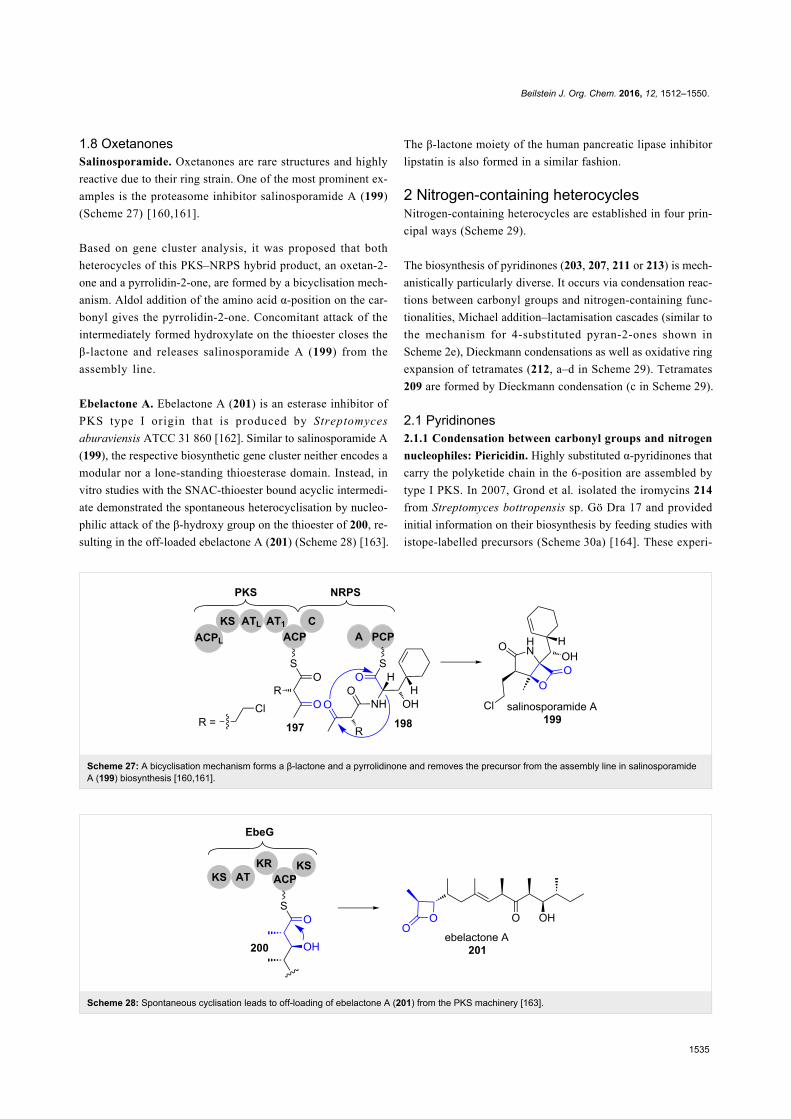
1.8 OxetanonesSalinosporamide. Oxetanones are rare structures and highly
reactive due to their ring strain. One of the most prominent ex-
amples is the proteasome inhibitor salinosporamide A (199)
(Scheme 27) [160,161].
Based on gene cluster analysis, it was proposed that both
heterocycles of this PKS–NRPS hybrid product, an oxetan-2-
one and a pyrrolidin-2-one, are formed by a bicyclisation mech-
anism. Aldol addition of the amino acid α-position on the car-
bonyl gives the pyrrolidin-2-one. Concomitant attack of the
intermediately formed hydroxylate on the thioester closes the
β-lactone and releases salinosporamide A (199) from the
assembly line.
Ebelactone A. Ebelactone A (201) is an esterase inhibitor of
PKS type I origin that is produced by Streptomyces
aburaviensis ATCC 31 860 [162]. Similar to salinosporamide A
(199), the respective biosynthetic gene cluster neither encodes a
modular nor a lone-standing thioesterase domain. Instead, in
vitro studies with the SNAC-thioester bound acyclic intermedi-
ate demonstrated the spontaneous heterocyclisation by nucleo-
philic attack of the β-hydroxy group on the thioester of 200, re-
sulting in the off-loaded ebelactone A (201) (Scheme 28) [163].
The β-lactone moiety of the human pancreatic lipase inhibitor
lipstatin is also formed in a similar fashion.
2 Nitrogen-containing heterocyclesNitrogen-containing heterocycles are established in four prin-
cipal ways (Scheme 29).
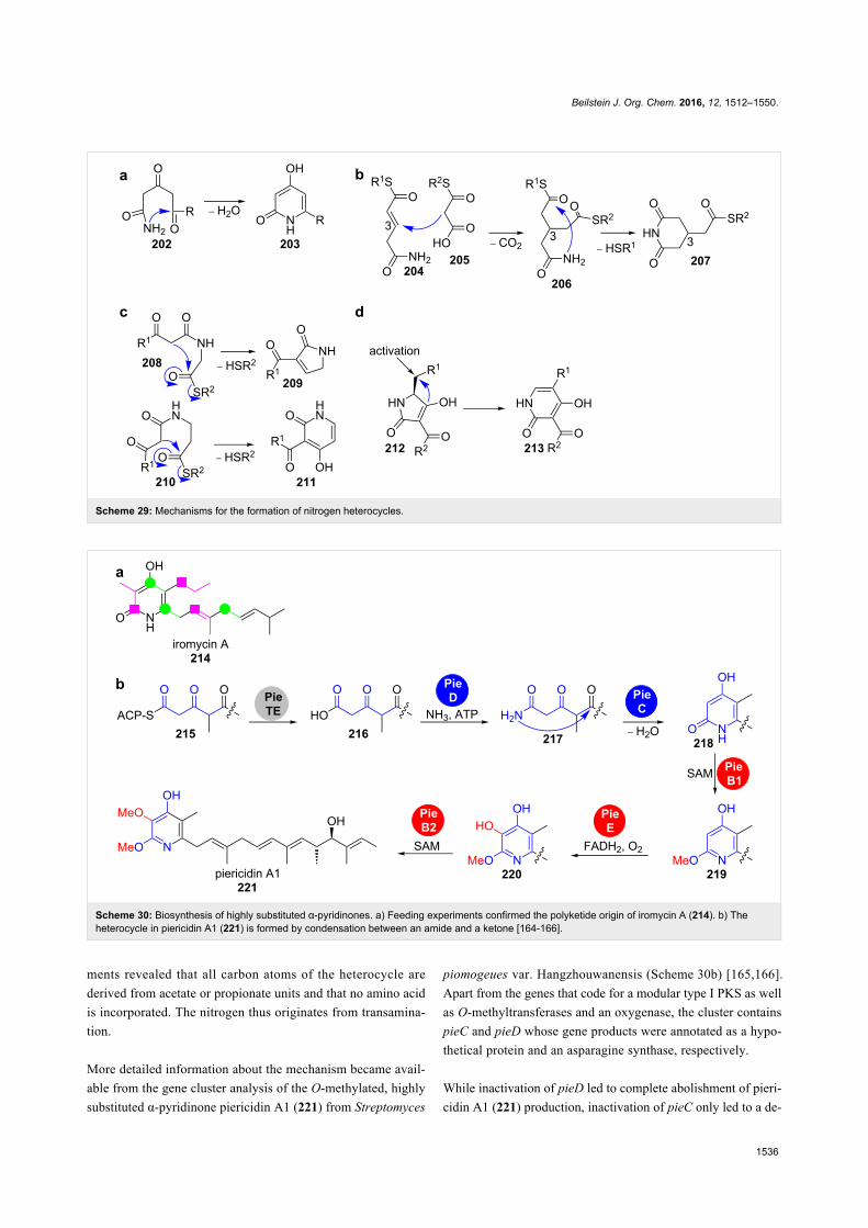
The biosynthesis of pyridinones (203, 207, 211 or 213) is mech-
anistically particularly diverse. It occurs via condensation reac-
tions between carbonyl groups and nitrogen-containing func-
tionalities, Michael addition–lactamisation cascades (similar to
the mechanism for 4-substituted pyran-2-ones shown in
Scheme 2e), Dieckmann condensations as well as oxidative ring
expansion of tetramates (212, a–d in Scheme 29). Tetramates
209 are formed by Dieckmann condensation (c in Scheme 29).
2.1 Pyridinones2.1.1 Condensation between carbonyl groups and nitrogen
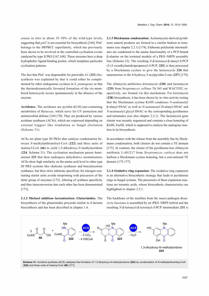
nucleophiles: Piericidin. Highly substituted α-pyridinones that
carry the polyketide chain in the 6-position are assembled by
type I PKS. In 2007, Grond et al. isolated the iromycins 214
from Streptomyces bottropensis sp. Gö Dra 17 and provided
initial information on their biosynthesis by feeding studies with
istope-labelled precursors (Scheme 30a) [164]. These experi-
Beilstein J. Org. Chem. 2016, 12, 1512–1550.
1536
Scheme 29: Mechanisms for the formation of nitrogen heterocycles.
Scheme 30: Biosynthesis of highly substituted α-pyridinones. a) Feeding experiments confirmed the polyketide origin of iromycin A (214). b) Theheterocycle in piericidin A1 (221) is formed by condensation between an amide and a ketone [164-166].
ments revealed that all carbon atoms of the heterocycle are
derived from acetate or propionate units and that no amino acid
is incorporated. The nitrogen thus originates from transamina-
tion.
More detailed information about the mechanism became avail-
able from the gene cluster analysis of the O-methylated, highly
substituted α-pyridinone piericidin A1 (221) from Streptomyces
piomogeues var. Hangzhouwanensis (Scheme 30b) [165,166].
Apart from the genes that code for a modular type I PKS as well
as O-methyltransferases and an oxygenase, the cluster contains
pieC and pieD whose gene products were annotated as a hypo-
thetical protein and an asparagine synthase, respectively.
While inactivation of pieD led to complete abolishment of pieri-
cidin A1 (221) production, inactivation of pieC only led to a de-
Beilstein J. Org. Chem. 2016, 12, 1512–1550.
1537
Scheme 31: Acridone synthase (ACS) catalyses the formation of 1,3-dihydroxy-N-methylacridone (224) by condensation of N-methylanthraniloyl-CoA(222) and three units of malonyl-CoA (66) [171].
crease in titre to about 35–50% of the wild-type levels,
suggesting that pieC is not essential for biosynthesis [164]. PieC
belongs to the SRPBCC superfamily, which has previously
been shown to be involved in the controlled cyclisation events
catalysed by type II PKS [167,168]. These enzymes have a deep
hydrophobic ligand-binding pocket, which templates particular
cyclisation patterns.
The fact that PieC was dispensable for piericidin A1 (221) bio-
synthesis was explained by that it could either be comple-
mented by other endogenous cyclases in S. piomogeues or that
the thermodynamically favoured formation of the six-mem-
bered heterocycle occurs spontaneously in the absence of the
enzyme.
Acridones. The acridones are pyridin-4(1H)-one-containing
metabolites of Rutaceae, which serve for UV protection and
antimicrobial defense [169,170]. They are produced by various
acridone synthases (ACSs), which are expressed depending on
external triggers like irradiation or fungal elicitation
(Scheme 31).
ACSs are plant type III PKSs that catalyse condensation be-
tween N-methylanthraniloyl-CoA (222) and three units of
malonyl-CoA (66) to yield 1,3-dihydroxy-N-methylacridone
(224, Scheme 31). The cyclisation mechanism passes hemi-
aminal 223 that then undergoes dehydrative aromatisation.
ACSs show high similarity on the amino acid level to other type
III PKS systems like chalcone synthases and benzalacetone
synthases, but their strict substrate specificity for nitrogen-con-
taining starter units avoids mispriming with precursors of the
latter group of enzymes [172]. Altering of synthase specificity
and thus interconversion into each other has been demonstrated
[173].
2.1.2 Michael addition–lactamisation: Glutarimides. The
biosynthesis of the glutarimides proceeds similar to δ-lactone
biosynthesis and has been described in chapter 1.6.
2.1.3 Dieckmann condensation: Actinomycete-derived pyridi-
none natural products are formed in a similar fashion as tetra-
mates (see chapter 2.2.1) [174]. Elaborate polyketide intermedi-
ates are condensed to the amine functionality of a PCP-bound
β-alanine on the terminal module of a PKS–NRPS assembly
line (Scheme 32). The resulting N-β-ketoacyl-β-alanyl-S-PCP
(3-(3-oxoalkylamido)propanoyl-S-PCP, 225) is then processed
by a Dieckmann cyclase to give the heterocycle 226 that
tautomerises to the 4-hydroxy-3-acylpyridin-2-one (227) [174].
The elfamycin antibiotics kirromycin (228) and factumycin
(229) from Streptomyces collinus Tü 365 and WAC5292, re-
spectively, are formed via this mechanism. For kirromycin
(228) biosynthesis, it has been shown by in vitro activity testing
that the Dieckmann cyclase KirHI condenses N-acetoacetyl
β-alanyl-SNAC as well as N-acetoacetyl D-alanyl-SNAC and
N-acetoacetyl glycyl-SNAC to the corresponding pyridinones
and tetramates (see also chapter 2.2.1). The factumycin gene
cluster was recently sequenced and contains a close homolog of
KirHI, FacHI, which is supposed to catalyse the analogous reac-
tion in its biosynthesis.
In accordance with the release from the assembly line by Dieck-
mann condensation, both clusters do not contain a TE domain
[175]. In contrast, the cluster of the pyridinone-less elfamycin
antibiotic L-681217 from Streptomyces cattleya does not
harbour a Dieckmann cyclase homolog, but a conventional TE
domain [175-177].
2.1.4 Oxidative ring expansion: The oxidative ring expansion
is an alternative biosynthetic strategy that leads to pyridinone
rings in fungal systems. The precursors of these expansion reac-
tions are tetramic acids, whose biosynthetic characteristics are
highlighted in chapter 2.2.1.
The backbone of the tenellins from the insect pathogen Beau-
veria bassiana is assembled by an iPKS–NRPS hybrid and the
resulting N-β-ketoacyl-β-tyrosinyl-S-PCP intermediate 231 is

Beilstein J. Org. Chem. 2016, 12, 1512–1550.
1538
Scheme 32: A Dieckmann condensation leads to the formation of a 3-acyl-4-hydroxypyridin-2-one 227 and removes the biosynthetic precursor fromthe PKS–NRPS hybrid assembly line during kirromycin (228) biosynthesis [174].
cyclised by an R* domain to yield the tetramic acid pretenellin
A (232, Scheme 33a). Two cytochrome P450 monooxygenases
then catalyse the consecutive ring expansion to the pyridinone
and N-hydroxylation. TenA was annotated as the ring expand-
ase responsible for pyridinone formation.
The mechanism of this unusual ring-expansion reaction remains
unclear in detail. The authors however presented preliminary in-
dications that point towards a radical mechanism without
isolable intermediates (Scheme 33b) [178]. This was supported
by the presence of the shunt product prototenellin D (240) in the
wild-type strain and in several knockout transformants. Conver-
sion experiments with cell-free extracts showed that 240 is not a
competent substrate of the tailoring enzymes in the cluster. It
was suggested that other oxidising enzymes with appropriate
substrate specificity must be encoded in the Beauveria bassiana
genome and responsible for prototenellin (240) formation. A
similar situation must be given for compounds 238, 239 and
241–243, whose clusters contain ring expandase candidates
with high identity to TenA and for which similarly hydroxylat-
ed metabolites were isolated (Scheme 33c). Other authors sug-
gested mechanisms that pass a quinonemethide intermediate
[179].
The N-hydroxylation reaction occurring from pretenellin B
(233) to tenellin (234) is catalysed by the second cytochrome
P450 monooxygenase TenB. This type of reaction is usually
rather catalysed by FAD-dependent monooxygenases and
nonheme iron-containing monooxygenases [181-185].
The cytochrome P450 monooxygenase ApdE (48% amino acid
identity to TenA) was shown to catalyse a similar ring-expan-
sion reaction in aspyridone A (238) biosynthesis (Scheme 33c).
This enzyme, however, shows a more diverse oxidation chem-
istry leading not only to the pyridinone, but also to a β-hydroxy-
tetramic acid as well as a dephenylated product.
Oxazoles. Natural products featuring oxazole moieties are pre-
dominantly derived from the nucleophilic attack of a serine side
chain hydroxy group on a carbonyl carbon of the peptide back-
bone. This has been shown for the oxazoles in thiazole/oxazole-
modified microcins (TOMMs) which are a group of riboso-
mally synthesised and posttranslationally modified peptides as
well as for NRPS-derived natural products [186]. In the case of
NRPS, the assembly is accomplished by a modified condensa-
tion domain (designated as heterocyclisation domain) and the
resulting oxazoline is often subsequently aromatised to the
oxazole by a flavin-dependent oxygenase domain [187]. How-
ever, some PKS–NRPS derived oxazoles originate from a dif-
ferent biosynthetic route.
Oxazolomycin (244) is a polyene spiro-linked γ-lactam/β-lacton
antibiotic that was originally isolated from Streptomyces albus
(Scheme 34a) [188,189].
Isotope-labelling studies have shown that instead of serine,
three molecules of glycine are incorporated into its carbon
backbone. The analysis of the respective biosynthetic gene
cluster revealed the absence of canonical heterocyclisation or
Beilstein J. Org. Chem. 2016, 12, 1512–1550.
1539
Scheme 33: a) Biosynthesis of the pyridinone tenellin (234). b) A radical mechanism was proposed for the ring-expansion reaction catalysed byTenA. c) Other fungal pyridinone-containing hybrid iPKS–NRPS natural products [178,180].
oxidation domains [190]. Instead, the loading module OzmO
contains a formylation domain that transfers the formyl group
of formyl-tetrahydrofolate onto glycin-S-PCP (Scheme 34b)
[191]. The resulting formyl-glycin-S-PCP 246 serves as the pre-
cursor for cyclisation.
Recently, Leadlay and co-workers proposed a mechanism for
oxazole formation in the biosynthesis of the C2-symmetrical
macrodiolide conglobatin (245) that was isolated from Strepto-
myces conglobatus ATCC 31005 [192,193]. In the biosynthetic
gene cluster, a putative cyclodehydratase CongE is coded that is
homologous to OzmP from the oxazolomycin (244) gene
cluster. Molecular modelling studies suggested, that CongE
belongs to the family of N-type ATP (pyro)phosphohydrolases
and contains the conserved ATP-binding motif SGGKDS. In
analogy to a mechanism previously reported by Dunbar and
Beilstein J. Org. Chem. 2016, 12, 1512–1550.
1540
Scheme 34: a) Oxazole-containing PKS–NRPS-derived natural products oxazolomycin (244) and conglobatin (245). b) Formylglycyl-S-PCP precur-sor for oxazole formation. c) and d) Proposed mechanisms for oxazole formation as suggested by Leadlay and co-workers [192].
co-workers, Leadlay and co-workers hypothesised that CongE
promotes oxazole formation by activation of one of the carbon-
yl amide oxygens by either phosphorylation or adenyl transfer
followed by nucleophilic attack and elimination (Scheme 34c
and d) [192,194].
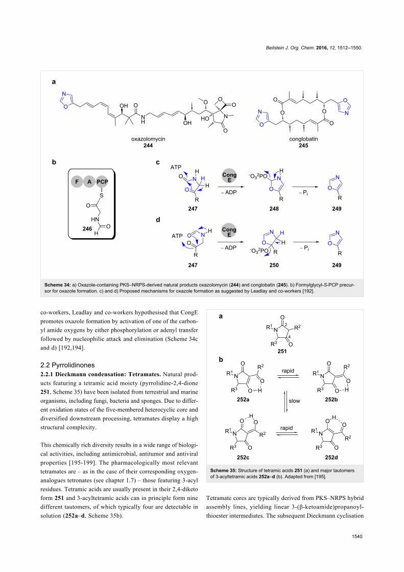
2.2 Pyrrolidinones2.2.1 Dieckmann condensation: Tetramates. Natural prod-
ucts featuring a tetramic acid moiety (pyrrolidine-2,4-dione
251, Scheme 35) have been isolated from terrestrial and marine
organisms, including fungi, bacteria and sponges. Due to differ-
ent oxidation states of the five-membered heterocyclic core and
diversified downstream processing, tetramates display a high
structural complexity.
This chemically rich diversity results in a wide range of biologi-
cal activities, including antimicrobial, antitumor and antiviral
properties [195-199]. The pharmacologically most relevant
tetramates are – as in the case of their corresponding oxygen-
analogues tetronates (see chapter 1.7) – those featuring 3-acyl
residues. Tetramic acids are usually present in their 2,4-diketo
form 251 and 3-acyltetramic acids can in principle form nine
different tautomers, of which typically four are detectable in
solution (252a–d, Scheme 35b).
Scheme 35: Structure of tetramic acids 251 (a) and major tautomersof 3-acyltetramic acids 252a–d (b). Adapted from [195].
Tetramate cores are typically derived from PKS–NRPS hybrid
assembly lines, yielding linear 3-(β-ketoamide)propanoyl-
thioester intermediates. The subsequent Dieckmann cyclisation

Beilstein J. Org. Chem. 2016, 12, 1512–1550.
1541
Scheme 36: Equisetin biosynthesis. R*: terminal reductive domain. Adapted from [202].
releases the tetramate from the megasynthetase. There are basi-
cally four different types of enzymatic units responsible for this
process, which are described to date: module-embedded R*-
and TE-domains, as well as lone-standing PyrD3/PyrD4-
homologs and Dieckmann cyclases.
In fungal iPKS–NRPS systems, a terminal reductive domain
(R*) directly catalyses the tetramate cyclisation without inter-
mediacy of a free aldehyde intermediate [179]. Studies of
Schmidt et al. provided the first evidence for this biosynthetic
route, utilising the R* domain of the equisetin (255) pathway
from Fusarium strains (Scheme 36) [200,201].
The reaction required no cofactor, despite a conserved N-termi-
nal NAD(P)H binding motif that is characteristic for the SDR
superfamily. In addition, phylogenetic analyses revealed that
R*-domains represent a distinct branch in the SDR superfamily
tree. Subsequent studies showed that the equisetin synthetase
genes had been misidentified as the fusaridione A synthetase
genes and the cluster was reassigned correctly [202]. Sequence
alignments in the same study also identified corresponding
R*-domains in the biosynthetic pathways of the spiro-tetra-
mates.
Pseurotins are Aspergillal natural products from the group of
the 3-spirotetramates, which display a wide array of biological
activities (Scheme 37a).
Their spiro centre is installed by epoxidation of the C3–C4
double bond of the tetramate ring in 264 and subsequent
epoxide opening by the 3’-enol oxygen of the side chain. Inter-
estingly, it was shown that the bifunctional epoxidase/C-MT
PsoF also catalyses a gate-keeping methylation in trans on the
stage of the nascent tetraketide (Scheme 37, highlighted in
boxes). This modification is crucial for the acyl-chain transfer
from PKS (261) to NRPS (263) as well as the epoxidation reac-
tion that yields the final spiro structure in 265 and pseurotin A
(256). Multiple methylation and oxidation steps give rise to a
high chemical diversity in the pseurotin compound family
[203,204]. This gate-keeping methylation was also proposed for
other fungal tetramates (Scheme 37b).
For the polycyclic tetramate macrolactams (PTMs), a module-
embedded TE domain that belongs to the α/β-hydrolase family
adopts the function of the fungal R*-domain [205,206]. The
tetramic acid is incorporated into a macrolactam ring, which is
fused to a set of two or three carbacycles of varying size, cycli-
zation pattern and oxidation level (Figure 7). This rich struc-
tural diversity of PTMs, which are produced in phylogeneti-
cally diverse bacteria results in a broad spectrum of biological
activities, including compounds with antifungal, antibiotic, and
antitumoural properties [207].
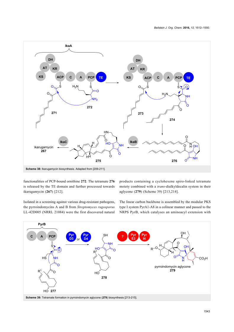
Ikarugamycin (267) is a PTM produced by various Strepto-
myces species that shows a broad spectrum of biological activi-
ty including antimicrobial and cytotoxic properties [208]. Its
biosynthesis has been reconstituted in E. coli and has shown to
be remarkably streamlined, utilising only the three enzymes
IkaABC to build up its highly complex structure (Scheme 38)
[209,210].
IkaA is a mixed iPKS–NRPS, in which the iPKS provides two
ACP-bound hexaketides 271 and 273. The condensation domain
of the NRPS attaches these two polyketide chains to the amine
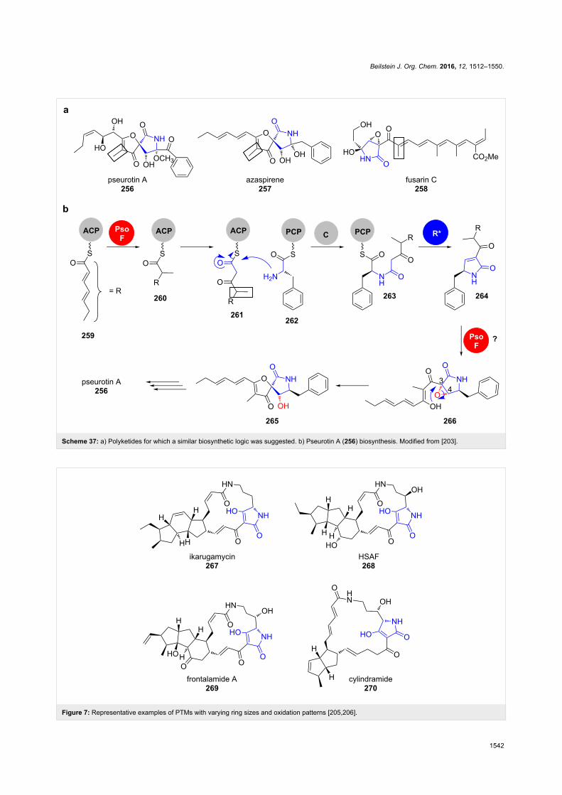
Beilstein J. Org. Chem. 2016, 12, 1512–1550.
1542
Scheme 37: a) Polyketides for which a similar biosynthetic logic was suggested. b) Pseurotin A (256) biosynthesis. Modified from [203].
Figure 7: Representative examples of PTMs with varying ring sizes and oxidation patterns [205,206].
Beilstein J. Org. Chem. 2016, 12, 1512–1550.
1543
Scheme 38: Ikarugamycin biosynthesis. Adapted from [209-211].
Scheme 39: Tetramate formation in pyrroindomycin aglycone (279) biosynthesis [213-215].
functionalities of PCP-bound ornithine 272. The tetramate 276
is released by the TE domain and further processed towards
ikarugamycin (267) [212].
Isolated in a screening against various drug-resistant pathogens,
the pyrroindomycins A and B from Streptomyces rugosporus
LL-42D005 (NRRL 21084) were the first discovered natural
products containing a cyclohexene spiro-linked tetramate
moiety combined with a trans-dialkyldecalin system in their
aglycone (279) (Scheme 39) [213,214].
The linear carbon backbone is assembled by the modular PKS
type I system PyrA1-A8 in a colinear manner and passed to the
NRPS PyrB, which catalyses an aminoacyl extension with

Beilstein J. Org. Chem. 2016, 12, 1512–1550.
1544
Scheme 40: Dieckmann cyclases catalyse tetramate or 2-pyridone formation in the biosynthesis of, for example, tirandamycin B (281), streptolyldigin(282), α-lipomycin (283) and kirromycin (228), respectively. DCy: Dieckmann cyclase. Adapted from [174].
L-cysteine [215]. This linear precursor 277 is then cleaved off
the megaenzyme by a Dieckmann condensation yielding the
tetramate moiety in 278. Gene deletion experiments and in vitro
assays revealed that this reaction is catalysed individually by
the two phylogenetically distinct enzymes PyrD3 and PyrD4.
Their respective biosynthetic genes are closely clustered and lo-
cated centrally in the PKS gene cluster. Homologs of PyrD3
and PyrD4 were found in biosynthetic gene clusters of the struc-
turally related spirotetronates. The introduction of chlD4 from
the spirotetronate chlorothricin (152) biosynthesis even partially
restored pyrroindomycin production in a ΔpyrD3-D4 deletion
mutant, highlighting the similarities in the tetronate/tetramate
formation chemistry in these natural products.
Surprisingly, in some actinomycete-derived tetramic acid-con-
taining natural products, R* or TE domains as well as PyrD3/
PyrD4-homologs are absent in the respective gene clusters. On
the contrary, recent in vitro studies revealed conserved dedi-
cated Dieckmann cyclases as catalysts in the biosynthetic path-
ways towards tirandamycin B (281), streptoylydigin (282) and
α-lipomycin (283), respectively (Scheme 40) [174].
In all these pathways, homologous genes are found directly
upstream of the NRPS genes and in their deletion mutants,
tetramate formation was abolished. In in vitro reactions, simpli-
fied SNAC thioester substrate surrogates were converted into
tetramates and mutational studies revealed a conserved catalyt-
ic triad consisting of Cys88-Asp115-His253. The respective
Dieckmann cyclases are phylogenetically distinct from fungal
R* domains, bacterial TE domains and PyrD3/PyrD4. Interest-
ingly, this paradigm also applies for bacterially derived 2-pyri-
done scaffolds such as kirromycin (228) (see chapter 2.1.3).
ConclusionDue to their attractive biological activity and abundance,
oxygen and nitrogen heterocycles-containing polyketides are
highly relevant. Recent years have seen a steady progress in the
understanding of their biosynthesis and plenty of novel enzy-
mology has been uncovered in this context. It is now clear that
heterocycle formation occurs by an exceptionally broad range
of mechanisms. Nevertheless, there is still plenty of room for
future studies on the biosynthesis of other types of heterocycles
as well as on the catalytic mechanisms and the structures of

Beilstein J. Org. Chem. 2016, 12, 1512–1550.
1545
cyclising enzymes. In principle, all these enzymes also repre-
sent future candidates for the development of novel types of
biocatalysts for chemoenzymatic synthesis.
AcknowledgementsWe thank the Deutsche Forschungsgemeinschaft for funding by
the Emmy Noether program as well as the European Union for
a Career Integration Grant.
References1. Taylor, R. D.; MacCoss, M.; Lawson, A. D. G. J. Med. Chem. 2014,
57, 5845–5859. doi:10.1021/jm40176252. Pozharskii, A. F.; Soldatenkov, A. T.; Katritzky, A. R. Heterocycles in
Life and Society: An Introduction to Heterocyclic Chemistry,Biochemistry and Applications, 2nd ed.; Wiley-VCH: Weinheim, 2011.doi:10.1002/9781119998372
3. Dua, R.; Shrivastava, S.; Sonwane, S. K.; Shrivastava, S. K.Adv. Biol. Res. 2011, 5, 120–144.
4. Liu, T.; Cane, D. E.; Deng, Z. The Enzymology of PolyetherBiosynthesis. In Complex Enzymes in Microbial Natural ProductBiosynthesis, Part B: Polyketides, Aminocoumarins andCarbohydrates; Hopwood, D. A., Ed.; Methods in Enzymology, Vol.459; Academic Press, 2009; pp 187–214.
5. Fischbach, M. A.; Walsh, C. T. Chem. Rev. 2006, 106, 3468–3496.doi:10.1021/cr0503097
6. Friedrich, S.; Hahn, F. Tetrahedron 2015, 71, 1473–1508.doi:10.1016/j.tet.2014.12.026
7. Lechner, H.; Pressnitz, D.; Kroutil, W. Biotechnol. Adv. 2015, 33,457–480. doi:10.1016/j.biotechadv.2015.01.012
8. Hertweck, C. Angew. Chem., Int. Ed. 2009, 48, 4688–4716.doi:10.1002/anie.200806121
9. Tang, Y.; Tsai, S.-C.; Khosla, C. J. Am. Chem. Soc. 2003, 125,12708–12709. doi:10.1021/ja0378759
10. Weissman, K. J.; Leadlay, P. F. Nat. Rev. Microbiol. 2005, 3,925–936. doi:10.1038/nrmicro1287
11. Staunton, J.; Weissman, K. J. Nat. Prod. Rep. 2001, 18, 380–416.doi:10.1039/a909079g
12. Parenty, A.; Moreau, X.; Campagne, J.-M. Chem. Rev. 2006, 106,911–939. doi:10.1021/cr0301402
13. Richter, M. E. A.; Traitcheva, N.; Knüpfer, U.; Hertweck, C.Angew. Chem., Int. Ed. 2008, 47, 8872–8875.doi:10.1002/anie.200803714
14. Pöplau, P.; Frank, S.; Morinaka, B. I.; Piel, J. Angew. Chem., Int. Ed.2013, 52, 13215–13218. doi:10.1002/anie.201307406
15. Berkhan, G.; Hahn, F. Angew. Chem., Int. Ed. 2014, 53,14240–14244. doi:10.1002/anie.201407979
16. Sudek, S.; Lopanik, N. B.; Waggoner, L. E.; Hildebrand, M.;Anderson, C.; Liu, H.; Patel, A.; Sherman, D. H.; Haygood, M. G.J. Nat. Prod. 2007, 70, 67–74. doi:10.1021/np060361d
17. Piel, J. Proc. Natl. Acad. Sci. U. S. A. 2002, 99, 14002–14007.doi:10.1073/pnas.222481399
18. Luhavaya, H.; Dias, M. V. B.; Williams, S. R.; Hong, H.;de Oliveira, L. G.; Leadlay, P. F. Angew. Chem., Int. Ed. 2015, 54,13622–13625. doi:10.1002/anie.201507090
19. Bieber, B.; Nüske, J.; Ritzau, M.; Gräfe, U. J. Antibiot. 1998, 51,381–382. doi:10.7164/antibiotics.51.381
20. Naruse, N.; Goto, M.; Watanabe, Y.; Terasawa, T.; Dobashi, K.J. Antibiot. 1998, 51, 545–552. doi:10.7164/antibiotics.51.545
21. Metsä-Ketelä, M.; Oja, T.; Taguchi, T.; Okamoto, S.; Ichinose, K.Curr. Opin. Chem. Biol. 2013, 17, 562–570.doi:10.1016/j.cbpa.2013.06.032
22. Das, A.; Khosla, C. Acc. Chem. Res. 2009, 42, 631–639.doi:10.1021/ar8002249
23. Malpartida, F.; Hopwood, D. A. Nature 1984, 309, 462–464.doi:10.1038/309462a0
24. Fernández-Moreno, M. A.; Martínez, E.; Boto, L.; Hopwood, D. A.;Malpartida, F. J. Biol. Chem. 1992, 267, 19278–19290.
25. Keatinge-Clay, A. T.; Maltby, D. A.; Medzihradszky, K. F.; Khosla, C.;Stroud, R. M. Nat. Struct. Mol. Biol. 2004, 11, 888–893.doi:10.1038/nsmb808
26. McDaniel, R.; Ebert-Khosla, S.; Hopwood, D. A.; Khosla, C.J. Am. Chem. Soc. 1993, 115, 11671–11675.doi:10.1021/ja00078a002
27. Taguchi, T.; Kunieda, K.; Takeda-Shitaka, M.; Takaya, D.;Kawano, N.; Kimberley, M. R.; Booker-Milburn, K. I.;Stephenson, G. R.; Umeyama, H.; Ebizuka, Y.; Ichinose, K.Bioorg. Med. Chem. 2004, 12, 5917–5927.doi:10.1016/j.bmc.2004.08.026
28. Caballero, J. L.; Martinez, E.; Malpartida, F.; Hopwood, D. A.MGG, Mol. Gen. Genet. 1991, 230, 401–412.doi:10.1007/BF00280297
29. Cole, S. P.; Rudd, B. A.; Hopwood, D. A.; Chang, C.-J.; Floss, H. G.J. Antibiot. 1987, 40, 340–347. doi:10.7164/antibiotics.40.340
30. He, Q.; Li, L.; Yang, T.; Li, R.; Li, A. PLoS One 2015, 10, e0132431.doi:10.1371/journal.pone.0132431
31. Itoh, T.; Taguchi, T.; Kimberley, M. R.; Booker-Milburn, K. I.;Stephenson, G. R.; Ebizuka, Y.; Ichinose, K. Biochemistry 2007, 46,8181–8188. doi:10.1021/bi700190p
32. Taguchi, T.; Ebizuka, Y.; Hopwood, D. A.; Ichinose, K.J. Am. Chem. Soc. 2001, 123, 11376–11380. doi:10.1021/ja015981+
33. Grocholski, T.; Oja, T.; Humphrey, L.; Mäntsälä, P.; Niemi, J.;Metsä-Ketelä, M. J. Bacteriol. 2012, 194, 2829–2836.doi:10.1128/JB.00228-12
34. Oja, T.; Palmu, K.; Lehmussola, H.; Leppäranta, O.; Hännikäinen, K.;Niemi, J.; Mäntsälä, P.; Metsä-Ketelä, M. Chem. Biol. 2008, 15,1046–1057. doi:10.1016/j.chembiol.2008.07.022
35. Oja, T.; Klika, K. D.; Appassamy, L.; Sinkkonen, J.; Mäntsälä, P.;Niemi, J.; Metsä-Ketelä, M. Proc. Natl. Acad. Sci. U. S. A. 2012, 109,6024–6029. doi:10.1073/pnas.1201530109
36. Oja, T.; Niiranen, L.; Sandalova, T.; Klika, K. D.; Niemi, J.;Mäntsälä, P.; Schneider, G.; Metsä-Ketelä, M.Proc. Natl. Acad. Sci. U. S. A. 2013, 110, 1291–1296.doi:10.1073/pnas.1207407110
37. Fuller, A. T.; Mellows, G.; Woolford, M.; Banks, G. T.; Barrow, K. D.;Chain, E. B. Nature 1971, 234, 416–417. doi:10.1038/234416a0
38. Chain, E. B.; Mellows, G. J. Chem. Soc., Chem. Commun. 1974,847–848. doi:10.1039/c39740000847
39. Chain, E. B.; Mellows, G. J. Chem. Soc., Perkin Trans. 1 1977,294–309. doi:10.1039/p19770000294
40. Clayton, J. P.; O’Hanlon, P. J.; Rogers, N. H. Tetrahedron Lett. 1980,21, 881–884. doi:10.1016/S0040-4039(00)71533-4
41. Shiozawa, H.; Shimada, A.; Takahashi, S. J. Antibiot. 1997, 50,449–452. doi:10.7164/antibiotics.50.449
42. Hughes, J.; Mellows, G. Biochem. J. 1978, 176, 305–318.doi:10.1042/bj1760305
43. Hughes, J.; Mellows, G. J. Antibiot. 1978, 31, 330–335.doi:10.7164/antibiotics.31.330
Beilstein J. Org. Chem. 2016, 12, 1512–1550.
1546
44. Hughes, J.; Mellows, G. Biochem. J. 1980, 191, 209–219.doi:10.1042/bj1910209
45. El-Sayed, A. K.; Hothersall, J.; Cooper, S. M.; Stephens, E.;Simpson, T. J.; Thomas, C. M. Chem. Biol. 2003, 10, 419–430.doi:10.1016/S1074-5521(03)00091-7
46. Gao, S.-S.; Hothersall, J.; Wu, J.; Murphy, A. C.; Song, Z.;Stephens, E. R.; Thomas, C. M.; Crump, M. P.; Cox, R. J.;Simpson, T. J.; Willis, C. L. J. Am. Chem. Soc. 2014, 136, 5501–5507.doi:10.1021/ja501731p
47. Cooper, S. M.; Laosripaiboon, W.; Rahman, A. S.; Hothersall, J.;El-Sayed, A. K.; Winfield, C.; Crosby, J.; Cox, R. J.; Simpson, T. J.;Thomas, C. M. Chem. Biol. 2005, 12, 825–833.doi:10.1016/j.chembiol.2005.05.015
48. Hothersall, J.; Wu, J.; Rahman, A. S.; Shields, J. A.; Haddock, J.;Johnson, N.; Cooper, S. M.; Stephens, E. R.; Cox, R. J.; Crosby, J.;Willis, C. L.; Simpson, T. J.; Thomas, C. M. J. Biol. Chem. 2007, 282,15451–15461. doi:10.1074/jbc.M701490200
49. Cooper, S. M.; Cox, R. J.; Crosby, J.; Crump, M. P.; Hothersall, J.;Laosripaiboon, W.; Simpson, T. J.; Thomas, C. M. Chem. Commun.2005, 1179–1181. doi:10.1039/b414781b
50. Julien, B.; Tian, Z.-Q.; Reid, R.; Reeves, C. D. Chem. Biol. 2006, 13,1277–1286. doi:10.1016/j.chembiol.2006.10.004
51. Nicolaou, K. C.; Prasad, C. V. C.; Somers, P. K.; Hwang, C. K.J. Am. Chem. Soc. 1989, 111, 5335–5340. doi:10.1021/ja00196a044
52. Nicolaou, K. C.; Prasad, C. V. C.; Somers, P. K.; Hwang, C. K.J. Am. Chem. Soc. 1989, 111, 5330–5334. doi:10.1021/ja00196a043
53. Shen, B.; Kwon, H.-J. Chem. Rec. 2002, 2, 389–396.doi:10.1002/tcr.10042
54. Nelson, M. E.; Priestley, N. D. J. Am. Chem. Soc. 2002, 124,2894–2902. doi:10.1021/ja016965f
55. Ashworth, D. M.; Robinson, J. A.; Turner, D. L.J. Chem. Soc., Perkin Trans. 1 1988, 1719–1727.doi:10.1039/p19880001719
56. Woo, A. J.; Strohl, W. R.; Priestley, N. D.Antimicrob. Agents Chemother. 1999, 43, 1662–1668.
57. Earle, M. J.; Priestley, N. D. Bioorg. Med. Chem. Lett. 1997, 7,2187–2192. doi:10.1016/S0960-894X(97)00393-4
58. Rong, J.; Nelson, M. E.; Kusche, B.; Priestley, N. D. J. Nat. Prod.2010, 73, 2009–2012. doi:10.1021/np100421v
59. Smith, W. C.; Xiang, L.; Shen, B. Antimicrob. Agents Chemother.2000, 44, 1809–1817. doi:10.1128/AAC.44.7.1809-1817.2000
60. Rebets, Y.; Brötz, E.; Manderscheid, N.; Tokovenko, B.;Myronovskyi, M.; Metz, P.; Petzke, L.; Luzhetskyy, A.Angew. Chem., Int. Ed. 2015, 54, 2280–2284.doi:10.1002/anie.201408901
61. Matilla, M. A.; Stöckmann, H.; Leeper, F. J.; Salmond, G. P. C.J. Biol. Chem. 2012, 287, 39125–39138.doi:10.1074/jbc.M112.401026
62. Müller, M.; Kusebauch, B.; Liang, G.; Beaudry, C. M.; Trauner, D.;Hertweck, C. Angew. Chem., Int. Ed. 2006, 45, 7835–7838.doi:10.1002/anie.200602840
63. He, J.; Müller, M.; Hertweck, C. J. Am. Chem. Soc. 2004, 126,16742–16743. doi:10.1021/ja046104h
64. He, J.; Hertweck, C. Chem. Biol. 2003, 10, 1225–1232.doi:10.1016/j.chembiol.2003.11.009
65. He, J.; Hertweck, C. J. Am. Chem. Soc. 2004, 126, 3694–3695.doi:10.1021/ja039328t
66. Werneburg, M.; Busch, B.; He, J.; Richter, M. E. A.; Xiang, L.;Moore, B. S.; Roth, M.; Dahse, H.-M.; Hertweck, C.J. Am. Chem. Soc. 2010, 132, 10407–10413. doi:10.1021/ja102751h
67. Werneburg, M.; Hertweck, C. ChemBioChem 2008, 9, 2064–2066.doi:10.1002/cbic.200800301
68. Henrot, M.; Richter, M. E. A.; Maddaluno, J.; Hertweck, C.;De Paolis, M. Angew. Chem., Int. Ed. 2012, 51, 9587–9591.doi:10.1002/anie.201204259
69. Bode, H. B.; Wenzel, S. C.; Irschik, H.; Höfle, G.; Müller, R.Angew. Chem., Int. Ed. 2004, 43, 4163–4167.doi:10.1002/anie.200454240
70. Kopp, M.; Irschik, H.; Gemperlein, K.; Buntin, K.; Meiser, P.;Weissman, K. J.; Bode, H. B.; Müller, R. Mol. BioSyst. 2011, 7,1549–1563. doi:10.1039/c0mb00240b
71. Winkler, A.; Łyskowski, A.; Riedl, S.; Puhl, M.; Kutchan, T. M.;Macheroux, P.; Gruber, K. Nat. Chem. Biol. 2008, 4, 739–741.doi:10.1038/nchembio.123
72. Winkler, A.; Motz, K.; Riedl, S.; Puhl, M.; Macheroux, P.; Gruber, K.J. Biol. Chem. 2009, 284, 19993–20001.doi:10.1074/jbc.M109.015727
73. Winkler, A.; Hartner, F.; Kutchan, T. M.; Glieder, A.; Macheroux, P.J. Biol. Chem. 2006, 281, 21276–21285. doi:10.1074/jbc.M603267200
74. Gaweska, H. M.; Roberts, K. M.; Fitzpatrick, P. F. Biochemistry 2012,51, 7342–7347. doi:10.1021/bi300887m
75. Guo, C.-J.; Wang, C. C. C. Front. Microbiol. 2014, 5, No. 717.doi:10.3389/fmicb.2014.00717
76. Chiang, Y.-M.; Szewczyk, E.; Davidson, A. D.; Keller, N.;Oakley, B. R.; Wang, C. C. C. J. Am. Chem. Soc. 2009, 131,2965–2970. doi:10.1021/ja8088185
77. Chiang, Y.-M.; Oakley, C. E.; Ahuja, M.; Entwistle, R.; Schultz, A.;Chang, S.-L.; Sung, C. T.; Wang, C. C. C.; Oakley, B. R.J. Am. Chem. Soc. 2013, 135, 7720–7731. doi:10.1021/ja401945a
78. Yu, J.; Chang, P.-K.; Ehrlich, K. C.; Cary, J. W.; Bhatnagar, D.;Cleveland, T. E.; Payne, G. A.; Linz, J. E.; Woloshuk, C. P.;Bennett, J. W. Appl. Environ. Microbiol. 2004, 70, 1253–1262.doi:10.1128/AEM.70.3.1253-1262.2004
79. Ehrlich, K. C.; Yu, J.; Cotty, P. J. J. Appl. Microbiol. 2005, 99,518–527. doi:10.1111/j.1365-2672.2005.02637.x
80. Asao, T.; Büchi, G.; Abdel-Kader, M. M.; Chang, S. B.; Wick, E. L.;Wogan, G. N. J. Am. Chem. Soc. 1965, 87, 882–886.doi:10.1021/ja01082a031
81. Holzapfel, C. W.; Steyn, P. S.; Purchase, I. F. H. Tetrahedron Lett.1966, 7, 2799–2803. doi:10.1016/S0040-4039(01)99863-6
82. Cox, R. Nat. Prod. Rep. 2014, 31, 1405–1424.doi:10.1039/C4NP00059E
83. Minto, R. E.; Townsend, C. A. Chem. Rev. 1997, 97, 2537–2556.doi:10.1021/cr960032y
84. Townsend, C. A. Nat. Prod. Rep. 2014, 31, 1260–1265.doi:10.1039/C4NP00092G
85. Crawford, J. M.; Thomas, P. M.; Scheerer, J. R.; Vagstad, A. L.;Kelleher, N. L.; Townsend, C. A. Science 2008, 320, 243–246.doi:10.1126/science.1154711
86. Watanabe, C. M. H.; Townsend, C. A. Chem. Biol. 2002, 9, 981–988.doi:10.1016/S1074-5521(02)00213-2
87. Sakuno, E.; Yabe, K.; Nakajima, H. Appl. Environ. Microbiol. 2003, 69,6418–6426. doi:10.1128/AEM.69.11.6418-6426.2003
88. Sakuno, E.; Wen, Y.; Hatabayashi, H.; Arai, H.; Aoki, C.; Yabe, K.;Nakajima, H. Appl. Environ. Microbiol. 2005, 71, 2999–3006.doi:10.1128/AEM.71.6.2999-3006.2005
89. Simpson, T. J.; de Jesus, A. E.; Steyn, P. S.; Vleggaar, R.J. Chem. Soc., Chem. Commun. 1982, 631–632.doi:10.1039/C39820000631

Beilstein J. Org. Chem. 2016, 12, 1512–1550.
1547
90. Townsend, C. A.; Christensen, S. B.; Davis, S. G. J. Am. Chem. Soc.1982, 104, 6154–6155. doi:10.1021/ja00386a070
91. Townsend, C. A.; Christensen, S. B. J. Am. Chem. Soc. 1985, 107,270–271. doi:10.1021/ja00287a059
92. Townsend, C. A.; Christensen, S. B.; Davis, S. G.J. Chem. Soc., Perkin Trans. 1 1988, 839–861.doi:10.1039/p19880000839
93. Wen, Y.; Hatabayashi, H.; Arai, H.; Kitamoto, H. K.; Yabe, K.Appl. Environ. Microbiol. 2005, 71, 3192–3198.doi:10.1128/AEM.71.6.3192-3198.2005
94. McGuire, S. M.; Townsend, C. A. Bioorg. Med. Chem. Lett. 1993, 3,653–656. doi:10.1016/S0960-894X(01)81247-6
95. Chang, P.-K.; Yabe, K.; Yu, J. Appl. Environ. Microbiol. 2004, 70,3593–3599. doi:10.1128/AEM.70.6.3593-3599.2004
96. Lin, B.-K.; Anderson, J. A. Arch. Biochem. Biophys. 1992, 293, 67–70.doi:10.1016/0003-9861(92)90366-5
97. Yabe, K.; Matsuyama, Y.; Ando, Y.; Nakajima, H.; Hamasaki, T.Appl. Environ. Microbiol. 1993, 59, 2486–2492.
98. McGuire, S. M.; Silva, J. C.; Casillas, E. G.; Townsend, C. A.Biochemistry 1996, 35, 11470–11486. doi:10.1021/bi960924s
99. Silva, J. C.; Minto, R. E.; Barry, C. E., III; Holland, K. A.;Townsend, C. A. J. Biol. Chem. 1996, 271, 13600–13608.doi:10.1074/jbc.271.23.13600
100.Conradt, D.; Schätzle, M. A.; Haas, J.; Townsend, C. A.; Müller, M.J. Am. Chem. Soc. 2015, 137, 10867–10869.doi:10.1021/jacs.5b06770
101.Henry, K. M.; Townsend, C. A. J. Am. Chem. Soc. 2005, 127,3724–3733. doi:10.1021/ja0455188
102.Cary, J. W.; Ehrlich, K. C.; Bland, J. M.; Montalbano, B. G.Appl. Environ. Microbiol. 2006, 72, 1096–1101.doi:10.1128/AEM.72.2.1096-1101.2006
103.Ehrlich, K. C.; Montalbano, B.; Boué, S. M.; Bhatnagar, D.Appl. Environ. Microbiol. 2005, 71, 8963–8965.doi:10.1128/AEM.71.12.8963-8965.2005
104.Yabe, K.; Matsushima, K.; Koyama, T.; Hamasaki, T.Appl. Environ. Microbiol. 1998, 64, 166–171.
105.Prieto, R.; Woloshuk, C. P. Appl. Environ. Microbiol. 1997, 63,1661–1666.
106.Udwary, D. W.; Casillas, L. K.; Townsend, C. A. J. Am. Chem. Soc.2002, 124, 5294–5303. doi:10.1021/ja012185v
107.Townsend, C. A.; Christensen, S. B.; Davis, S. G. J. Am. Chem. Soc.1982, 104, 6152–6153. doi:10.1021/ja00386a069
108.Yabe, K.; Hamasaki, T. Appl. Environ. Microbiol. 1993, 59,2493–2500.
109.Liu, T.; Cane, D. E.; Deng, Z. Methods Enzymol. 2009, 459, 187–214.doi:10.1016/S0076-6879(09)04609-6
110.Bhatt, A.; Stark, C. B. W.; Harvey, B. M.; Gallimore, A. R.;Demydchuk, Y. A.; Spencer, J. B.; Staunton, J.; Leadlay, P. F.Angew. Chem., Int. Ed. 2005, 44, 7075–7078.doi:10.1002/anie.200501757
111.Gallimore, A. R.; Stark, C. B. W.; Bhatt, A.; Harvey, B. M.;Demydchuk, Y.; Bolanos-Garcia, V.; Fowler, D. J.; Staunton, J.;Leadlay, P. F.; Spencer, J. B. Chem. Biol. 2006, 13, 453–460.doi:10.1016/j.chembiol.2006.01.013
112.Shiraiwa, K.; Yuan, S.; Fujiyama, A.; Matsuo, Y.; Tanaka, T.;Jiang, Z.-H.; Kouno, I. J. Nat. Prod. 2012, 75, 88–92.doi:10.1021/np2007582
113.Smith, A. B., III; Dong, S.; Brenneman, J. B.; Fox, R. J.J. Am. Chem. Soc. 2009, 131, 12109–12111. doi:10.1021/ja906115a
114.Moore, R. E.; Scheuer, P. J. Science 1971, 172, 495–498.doi:10.1126/science.172.3982.495
115.Steyn, P. S.; Vleggaar, R. J. Chem. Soc., Chem. Commun. 1985,1796–1798. doi:10.1039/c39850001796
116.Mao, X.-M.; Zhan, Z.-J.; Grayson, M. N.; Tang, M.-C.; Xu, W.;Li, Y.-Q.; Yin, W.-B.; Lin, H.-C.; Chooi, Y.-H.; Houk, K. N.; Tang, Y.J. Am. Chem. Soc. 2015, 137, 11904–11907.doi:10.1021/jacs.5b07816
117.Huang, J.-m.; Yokoyama, R.; Yang, C.-s.; Fukuyama, Y.Tetrahedron Lett. 2000, 41, 6111–6114.doi:10.1016/S0040-4039(00)01023-6
118.Inoue, M.; Lee, N.; Kasuya, S.; Sato, T.; Hirama, M.; Moriyama, M.;Fukuyama, Y. J. Org. Chem. 2007, 72, 3065–3075.doi:10.1021/jo0700474
119.Holton, R. A.; Somoza, C.; Kim, H. B.; Liang, F.; Biediger, R. J.;Boatman, P. D.; Shindo, M.; Smith, C. C.; Kim, S. J. Am. Chem. Soc.1994, 116, 1597–1598. doi:10.1021/ja00083a066
120.Holton, R. A.; Kim, H. B.; Somoza, C.; Liang, F.; Biediger, R. J.;Boatman, P. D.; Shindo, M.; Smith, C. C.; Kim, S. J. Am. Chem. Soc.1994, 116, 1599–1600. doi:10.1021/ja00083a067
121.Wender, P. A.; Badham, N. F.; Conway, S. P.; Floreancig, P. E.;Glass, T. E.; Gränicher, C.; Houze, J. B.; Jänichen, J.; Lee, D.;Marquess, D. G.; McGrane, P. L.; Meng, W.; Mucciaro, T. P.;Mühlebach, M.; Natchus, M. G.; Paulsen, H.; Rawlins, D. B.;Satkofsky, J.; Shuker, A. J.; Sutton, J. C.; Taylor, R. E.; Tomooka, K.J. Am. Chem. Soc. 1997, 119, 2755–2756. doi:10.1021/ja9635387
122.Wender, P. A.; Badham, N. F.; Conway, S. P.; Floreancig, P. E.;Glass, T. E.; Houze, J. B.; Krauss, N. E.; Lee, D.; Marquess, D. G.;McGrane, P. L.; Meng, W.; Natchus, M. G.; Shuker, A. J.;Sutton, J. C.; Taylor, R. E. J. Am. Chem. Soc. 1997, 119, 2757–2758.doi:10.1021/ja963539z
123.Montemiglio, L. C.; Parisi, G.; Scaglione, A.; Sciara, G.; Savino, C.;Vallone, B. Biochim. Biophys. Acta, Gen. Subj. 2016, 1860, 465–475.doi:10.1016/j.bbagen.2015.10.009
124.Nagano, S.; Li, H.; Shimizu, H.; Nishida, C.; Ogura, H.;Ortiz de Montellano, P. R.; Poulos, T. L. J. Biol. Chem. 2003, 278,44886–44893. doi:10.1074/jbc.M308115200
125.Ogura, H.; Nishida, C. R.; Hoch, U. R.; Perera, R.; Dawson, J. H.;Ortiz de Montellano, P. R. Biochemistry 2004, 43, 14712–14721.doi:10.1021/bi048980d
126.Schäberle, T. F. Beilstein J. Org. Chem. 2016, 12, 571–588.doi:10.3762/bjoc.12.56
127.Palaniappan, N.; Alhamadsheh, M. M.; Reynolds, K. A.J. Am. Chem. Soc. 2008, 130, 12236–12237. doi:10.1021/ja8044162
128.Hu, Z.; Reid, R.; Gramajo, H. J. Antibiot. 2005, 58, 625–633.doi:10.1038/ja.2005.86
129.Liu, X.-j.; Kong, R.-x.; Niu, M.-s.; Qiu, R.; Tang, L. J. Nat. Prod. 2013,76, 524–529. doi:10.1021/np300667r
130.Bretschneider, T.; Heim, J. B.; Heine, D.; Winkler, R.; Busch, B.;Kusebauch, B.; Stehle, T.; Zocher, G.; Hertweck, C. Nature 2013,502, 124–128. doi:10.1038/nature12588
131.Heine, D.; Bretschneider, T.; Sundaram, S.; Hertweck, C.Angew. Chem., Int. Ed. 2014, 53, 11645–11649.doi:10.1002/anie.201407282
132.Heine, D.; Sundaram, S.; Bretschneider, T.; Hertweck, C.Chem. Commun. 2015, 51, 9872–9875. doi:10.1039/C5CC03085D
133.Sundaram, S.; Heine, D.; Hertweck, C. Nat. Chem. Biol. 2015, 11,949–951. doi:10.1038/nchembio.1932

Beilstein J. Org. Chem. 2016, 12, 1512–1550.
1548
134.Piel, J.; Hertweck, C.; Shipley, P. R.; Hunt, D. M.; Newman, M. S.;Moore, B. S. Chem. Biol. 2000, 7, 943–955.doi:10.1016/S1074-5521(00)00044-2
135.Teufel, R.; Miyanaga, A.; Michaudel, Q.; Stull, F.; Louie, G.;Noel, J. P.; Baran, P. S.; Palfey, B.; Moore, B. S. Nature 2013, 503,552–556. doi:10.1038/nature12643
136.Cheng, Q.; Xiang, L.; Izumikawa, M.; Meluzzi, D.; Moore, B. S.Nat. Chem. Biol. 2007, 3, 557–558. doi:10.1038/nchembio.2007.22
137.Teufel, R.; Stull, F.; Meehan, M. J.; Michaudel, Q.; Dorrestein, P. C.;Palfey, B.; Moore, B. S. J. Am. Chem. Soc. 2015, 137, 8078–8085.doi:10.1021/jacs.5b03983
138.Tao, W.; Zhu, M.; Deng, Z.; Sun, Y. Sci. China: Chem. 2013, 56,1364–1371. doi:10.1007/s11426-013-4921-x
139.Vieweg, L.; Reichau, S.; Schobert, R.; Leadlay, P. F.; Süssmuth, R. D.Nat. Prod. Rep. 2014, 31, 1554–1584. doi:10.1039/C4NP00015C
140.Lacoske, M. H.; Theodorakis, E. A. J. Nat. Prod. 2015, 78, 562–575.doi:10.1021/np500757w
141.Demydchuk, Y.; Sun, Y.; Hong, H.; Staunton, J.; Spencer, J. B.;Leadlay, P. F. ChemBioChem 2008, 9, 1136–1145.doi:10.1002/cbic.200700715
142.Sun, Y.; Hahn, F.; Demydchuk, Y.; Chettle, J.; Tosin, M.; Osada, H.;Leadlay, P. F. Nat. Chem. Biol. 2010, 6, 99–101.doi:10.1038/nchembio.285
143.Kanchanabanca, C.; Tao, W.; Hong, H.; Liu, Y.; Hahn, F.;Samborskyy, M.; Deng, Z.; Sun, Y.; Leadlay, P. F.Angew. Chem., Int. Ed. 2013, 52, 5785–5788.doi:10.1002/anie.201301680
144.Sun, Y.; Hong, H.; Gillies, F.; Spencer, J. B.; Leadlay, P. F.ChemBioChem 2008, 9, 150–156. doi:10.1002/cbic.200700492
145.Wu, L.-F.; He, H.-Y.; Pan, H.-X.; Han, L.; Wang, R.; Tang, G.-L.Org. Lett. 2014, 16, 1578–1581. doi:10.1021/ol500111n
146.Hashimoto, T.; Hashimoto, J.; Teruya, K.; Hirano, T.; Shin-ya, K.;Ikeda, H.; Liu, H.-w.; Nishiyama, M.; Kuzuyama, T. J. Am. Chem. Soc.2015, 137, 572–575. doi:10.1021/ja510711x
147.Tian, Z.; Sun, P.; Yan, Y.; Wu, Z.; Zheng, Q.; Zhou, S.; Zhang, H.;Yu, F.; Jia, X.; Chen, D.; Mándi, A.; Kurtán, T.; Liu, W.Nat. Chem. Biol. 2015, 11, 259–265. doi:10.1038/nchembio.1769
148.Tao, W.; Yurkovich, M. E.; Wen, S.; Lebe, K. E.; Samborskyy, M.;Liu, Y.; Yang, A.; Liu, Y.; Ju, Y.; Deng, Z.; Tosin, M.; Sun, Y.;Leadlay, P. F. Chem. Sci. 2016, 7, 376–385.doi:10.1039/C5SC03059E
149.Harborne, J. B., Ed. The Flavonoids: Advances in Research Since1986; Chapman & Hall: London, U.K., 1993.
150.Vaughn, K. C.; Lax, A. R.; Duke, S. O. Physiol. Plant. 1988, 72,659–665. doi:10.1111/j.1399-3054.1988.tb09180.x
151.Mayer, A. M. Phytochemistry 1986, 26, 11–20.doi:10.1016/S0031-9422(00)81472-7
152.Sato, T.; Nakayama, T.; Kikuchi, S.; Fukui, Y.;Yonekura-Sakakibara, K.; Ueda, T.; Nishino, T.; Tanaka, Y.;Kusumi, T. Plant Sci. 2001, 160, 229–236.doi:10.1016/S0168-9452(00)00385-X
153.Nakayama, T.; Yonekura-Sakakibara, K.; Sato, T.; Kikuchi, S.;Fukui, Y.; Fukuchi-Mizutani, M.; Ueda, T.; Nakao, M.; Tanaka, Y.;Kusumi, T.; Nishino, T. Science 2000, 290, 1163–1166.doi:10.1126/science.290.5494.1163
154.Nakayama, T.; Sato, T.; Fukui, Y.; Yonekura-Sakakibara, K.;Hayashi, H.; Tanaka, Y.; Kusumi, T.; Nishino, T. FEBS Lett. 2001,499, 107–111. doi:10.1016/S0014-5793(01)02529-7
155.Lin, Z.; Zachariah, M. M.; Marett, L.; Hughen, R. W.; Teichert, R. W.;Concepcion, G. P.; Haygood, M. G.; Olivera, B. M.; Light, A. R.;Schmidt, E. W. J. Nat. Prod. 2014, 77, 1224–1230.doi:10.1021/np500155d
156.Raistrick, H.; Smith, G. Biochem. J. 1936, 30, 1315–1322.doi:10.1042/bj0301315
157.Fujii, I.; Iijima, H.; Tsukita, S.; Ebizuka, Y.; Sankawa, U. J. Biochem.1987, 101, 11–18.
158.Huang, K.; Yoshida, Y.; Mikawa, K.; Fujii, I.; Ebizuka, Y.; Sankawa, U.Biol. Pharm. Bull. 1996, 19, 42–46. doi:10.1248/bpb.19.42
159.Chooi, Y.-H.; Cacho, R.; Tang, Y. Chem. Biol. 2010, 17, 483–494.doi:10.1016/j.chembiol.2010.03.015
160.Udwary, D. W.; Zeigler, L.; Asolkar, R. N.; Singan, V.; Lapidus, A.;Fenical, W.; Jensen, P. R.; Moore, B. S. Proc. Natl. Acad. Sci. U. S. A.2007, 104, 10376–10381. doi:10.1073/pnas.0700962104
161.Gulder, T. A. M.; Moore, B. S. Angew. Chem., Int. Ed. 2010, 49,9346–9367. doi:10.1002/anie.201000728
162.Umezawa, H.; Aoyagi, T.; Uotani, K.; Hamada, M.; Takeuchi, T.;Takahashi, S. J. Antibiot. 1980, 33, 1594–1596.doi:10.7164/antibiotics.33.1594
163.Wyatt, M. A.; Ahilan, Y.; Argyropoulos, P.; Boddy, C. N.;Magarvey, N. A.; Harrison, P. H. M. J. Antibiot. 2013, 66, 421–430.doi:10.1038/ja.2013.48
164.Surup, F.; Wagner, O.; von Frieling, J.; Schleicher, M.; Oess, S.;Müller, P.; Grond, S. J. Org. Chem. 2007, 72, 5085–5090.doi:10.1021/jo0703303
165.Liu, Q.; Yao, F.; Chooi, Y. H.; Kang, Q.; Xu, W.; Li, Y.; Shao, Y.;Shi, Y.; Deng, Z.; Tang, Y.; You, D. Chem. Biol. 2012, 19, 243–253.doi:10.1016/j.chembiol.2011.12.018
166.Chen, Y.; Zhang, W.; Zhu, Y.; Zhang, Q.; Tian, X.; Zhang, S.;Zhang, C. Org. Lett. 2014, 16, 736–739. doi:10.1021/ol4034176
167.Zhang, W.; Tang, Y. In Vitro Analysis of Type II Polyketide Synthase.In Complex Enzymes in Microbial Natural Product Biosynthesis, PartB: Polyketides, Aminocoumarins and Carbohydrates; Hopwood, D. A.,Ed.; Methods in Enzymology, Vol. 459; Academic Press, 2009;pp 367–393.
168.Kendrew, S. G.; Katayama, K.; Deutsch, E.; Madduri, K.;Hutchinson, C. R. Biochemistry 1999, 38, 4794–4799.doi:10.1021/bi9827924
169.Maier, W.; Baumert, A.; Schumann, B.; Furukawa, H.; Gröger, D.Phytochemistry 1993, 32, 691–698.doi:10.1016/S0031-9422(00)95155-0
170.Junghanns, K. T.; Kneusel, R. E.; Baumert, A.; Maier, W.; Gröger, D.;Matern, U. Plant Mol. Biol. 1995, 27, 681–692.doi:10.1007/BF00020222
171.Lukačin, R.; Springob, K.; Urbanke, C.; Ernwein, C.; Schröder, G.;Schröder, J.; Matern, U. FEBS Lett. 1999, 448, 135–140.doi:10.1016/S0014-5793(99)00355-5
172.Lukačin, R.; Schreiner, S.; Silber, K.; Matern, U. Phytochemistry 2005,66, 277–284. doi:10.1016/j.phytochem.2004.11.023
173.Lukačin, R.; Schreiner, S.; Matern, U. FEBS Lett. 2001, 508, 413–417.doi:10.1016/S0014-5793(01)03061-7
174.Gui, C.; Li, Q.; Mo, X.; Qin, X.; Ma, J.; Ju, J. Org. Lett. 2015, 17,628–631. doi:10.1021/ol5036497
175.Thaker, M. N.; García, M.; Koteva, K.; Waglechner, N.; Sorensen, D.;Medina, R.; Wright, G. D. MedChemComm 2012, 3, 1020–1026.doi:10.1039/c2md20038d
176.Brötz, E.; Kulik, A.; Vikineswary, S.; Lim, C.-T.; Tan, G. Y. A.;Zinecker, H.; Imhoff, J. F.; Paululat, T.; Fiedler, H.-P. J. Antibiot. 2011,64, 257–266. doi:10.1038/ja.2010.170
Beilstein J. Org. Chem. 2016, 12, 1512–1550.
1549
177.Kempf, A. J.; Wilson, K. E.; Hensens, O. D.; Monaghan, R. L.;Zimmerman, S. B.; Dulaney, E. L. J. Antibiot. 1986, 39, 1361–1367.doi:10.7164/antibiotics.39.1361
178.Halo, L. M.; Heneghan, M. N.; Yakasai, A. A.; Song, Z.; Williams, K.;Bailey, A. M.; Cox, R. J.; Lazarus, C. M.; Simpson, T. J.J. Am. Chem. Soc. 2008, 130, 17988–17996. doi:10.1021/ja807052c
179.Boettger, D.; Hertweck, C. ChemBioChem 2013, 14, 28–42.doi:10.1002/cbic.201200624
180.Schmidt, K.; Riese, U.; Li, Z.; Hamburger, M. J. Nat. Prod. 2003, 66,378–383. doi:10.1021/np020430y
181.Rydberg, P.; Ryde, U.; Olsen, L. J. Chem. Theory Comput. 2008, 4,1369–1377. doi:10.1021/ct800101v
182.Choi, Y. S.; Zhang, H.; Brunzelle, J. S.; Nair, S. K.; Zhao, H.Proc. Natl. Acad. Sci. U. S. A. 2008, 105, 6858–6863.doi:10.1073/pnas.0712073105
183.Krithika, R.; Marathe, U.; Saxena, P.; Ansari, M. Z.; Mohanty, D.;Gokhale, R. S. Proc. Natl. Acad. Sci. U. S. A. 2006, 103, 2069–2074.doi:10.1073/pnas.0507924103
184.Yamada, O.; Nan, S. N.; Akao, T.; Tominaga, M.; Watanabe, H.;Satoh, T.; Enei, H.; Akita, O. J. Biosci. Bioeng. 2003, 95, 82–88.doi:10.1016/S1389-1723(03)80153-6
185.Lee, J.; Simurdiak, M.; Zhao, H. J. Biol. Chem. 2005, 280,36719–36727. doi:10.1074/jbc.M505334200
186.Metelev, M. V.; Ghilarov, D. A. Mol. Biol. 2014, 48, 29–45.doi:10.1134/S0026893314010105
187.Roy, R. S.; Gehring, A. M.; Milne, J. C.; Belshaw, P. J.; Walsh, C. T.Nat. Prod. Rep. 1999, 16, 249–263. doi:10.1039/a806930a
188.Mori, T.; Takahashi, K.; Kashiwabara, M.; Uemura, D.; Katayama, C.;Iwadare, S.; Shizuri, Y.; Mitomo, R.; Nakano, F.; Matsuzaki, A.Tetrahedron Lett. 1985, 26, 1073–1076.doi:10.1016/S0040-4039(00)98515-0
189.Gräfe, U.; Kluge, H.; Thiericke, R. Liebigs Ann. Chem. 1992, 429–432.doi:10.1002/jlac.199219920178
190.Zhao, C.; Ju, J.; Christenson, S. D.; Smith, W. C.; Song, D.; Zhou, X.;Shen, B.; Deng, Z. J. Bacteriol. 2006, 188, 4142–4147.doi:10.1128/JB.00173-06
191.Zhao, C.; Coughlin, J. M.; Ju, J.; Zhu, D.; Wendt-Pienkowski, E.;Zhou, X.; Wang, Z.; Shen, B.; Deng, Z. J. Biol. Chem. 2010, 285,20097–20108. doi:10.1074/jbc.M109.090092
192.Zhou, Y.; Murphy, A. C.; Samborskyy, M.; Prediger, P.; Dias, L. C.;Leadlay, P. F. Chem. Biol. 2015, 22, 745–754.doi:10.1016/j.chembiol.2015.05.010
193.Westley, J. W.; Liu, C.-M.; Evans, R. H.; Blount, J. F. J. Antibiot. 1979,32, 874–877. doi:10.7164/antibiotics.32.874
194.Dunbar, K. L.; Chekan, J. R.; Cox, C. L.; Burkhart, B. J.; Nair, S. K.;Mitchell, D. A. Nat. Chem. Biol. 2014, 10, 823–829.doi:10.1038/nchembio.1608
195.Schobert, R.; Schlenk, A. Bioorg. Med. Chem. 2008, 16, 4203–4221.doi:10.1016/j.bmc.2008.02.069
196.Mo, X.; Li, Q.; Ju, J. RSC Adv. 2014, 4, 50566–50593.doi:10.1039/C4RA09047K
197.Ghisalberti, E. L. Bioactive tetramic acid metabolites. In BiactiveNatural Products (Part I); Atta-ur-Rahman, Ed.; Studies in NaturalProducts Chemistry, Vol. 28; Elsevier, 2003; pp 109–163.doi:10.1016/s1572-5995(03)80140-0
198.Gossauer, A. In Monopyrrolic Natural Compounds Including TetramicAcid Derivatives; Herz, W.; Falk, H.; Kirby, G. W., Eds.; Progress inthe Chemistry of Organic Natural Products, Vol. 86; Springer Verlag:Wien, 2003. doi:10.1007/978-3-7091-6029-9
199.Royles, B. J. L. Chem. Rev. 1995, 95, 1981–2001.doi:10.1021/cr00038a009
200.Sims, J. W.; Schmidt, E. W. J. Am. Chem. Soc. 2008, 130,11149–11155. doi:10.1021/ja803078z
201.Singh, S. B.; Zink, D. L.; Goetz, M. A.; Dombrowski, A. W.;Polishook, J. D.; Hazuda, D. J. Tetrahedron Lett. 1998, 39,2243–2246. doi:10.1016/S0040-4039(98)00269-X
202.Kakule, T. B.; Sardar, D.; Lin, Z.; Schmidt, E. W. ACS Chem. Biol.2013, 8, 1549–1557. doi:10.1021/cb400159f
203.Zou, Y.; Xu, W.; Tsunematsu, Y.; Tang, M.; Watanabe, K.; Tang, Y.Org. Lett. 2014, 16, 6390–6393. doi:10.1021/ol503179v
204.Tsunematsu, Y.; Fukutomi, M.; Saruwatari, T.; Noguchi, H.; Hotta, K.;Tang, Y.; Watanabe, K. Angew. Chem., Int. Ed. 2014, 53, 8475–8479.doi:10.1002/anie.201404804
205.Du, L.; Lou, L. Nat. Prod. Rep. 2010, 27, 255–278.doi:10.1039/B912037H
206.Lou, L.; Qian, G.; Xie, Y.; Hang, J.; Chen, H.; Zaleta-Rivera, K.; Li, Y.;Shen, Y.; Dussault, P. H.; Liu, F.; Du, L. J. Am. Chem. Soc. 2011,133, 643–645. doi:10.1021/ja105732c
207.Blodgett, J. A. V.; Oh, D.-C.; Cao, S.; Currie, C. R.; Kolter, R.;Clardy, J. Proc. Natl. Acad. Sci. U. S. A. 2010, 107, 11692–11697.doi:10.1073/pnas.1001513107
208.Jomon, K.; Kuroda, Y.; Ajisaka, M.; Sakai, H. J. Antibiot. 1972, 25,271–280. doi:10.7164/antibiotics.25.271
209.Zhang, G.; Zhang, W.; Zhang, Q.; Shi, T.; Ma, L.; Zhu, Y.; Li, S.;Zhang, H.; Zhao, Y.-L.; Shi, R.; Zhang, C. Angew. Chem., Int. Ed.2014, 53, 4840–4844. doi:10.1002/anie.201402078
210.Antosch, J.; Schaefers, F.; Gulder, T. A. M. Angew. Chem., Int. Ed.2014, 53, 3011–3014. doi:10.1002/anie.201310641
211.Greunke, C.; Antosch, J.; Gulder, T. A. M. Chem. Commun. 2015, 51,5334–5336. doi:10.1039/C5CC00843C
212.Li, Y.; Chen, H.; Ding, Y.; Xie, Y.; Wang, H.; Cerny, R. L.; Shen, Y.;Du, L. Angew. Chem., Int. Ed. 2014, 53, 7524–7530.doi:10.1002/anie.201403500
213.Ding, W.; Williams, D. R.; Northcote, P.; Siegel, M. M.; Tsao, R.;Ashcroft, J.; Morton, G. O.; Alluri, M.; Abbanat, D.; Maiese, W. M.;Ellestad, G. A. J. Antibiot. 1994, 47, 1250–1257.doi:10.7164/antibiotics.47.1250
214.Singh, M. P.; Petersen, P. J.; Jacobus, N. V.;Mroczenski-Wildey, M. J.; Maiese, W. M.; Greenstein, M.;Steinberg, D. A. J. Antibiot. 1994, 47, 1258–1265.doi:10.7164/antibiotics.47.1258
215.Wu, Q.; Wu, Z.; Qu, X.; Liu, W. J. Am. Chem. Soc. 2012, 134,17342–17345. doi:10.1021/ja304829g
Beilstein J. Org. Chem. 2016, 12, 1512–1550.
1550
License and TermsThis is an Open Access article under the terms of the
Creative Commons Attribution License
(http://creativecommons.org/licenses/by/2.0), which
permits unrestricted use, distribution, and reproduction in
any medium, provided the original work is properly cited.
The license is subject to the Beilstein Journal of Organic
Chemistry terms and conditions:
(http://www.beilstein-journals.org/bjoc)
The definitive version of this article is the electronic one
which can be found at:
doi:10.3762/bjoc.12.148