biosynthesis of the tunicamycin antibiotics proceeds via...
TRANSCRIPT

Biosynthesis of the tunicamycin antibioticsproceeds via unique exo-glycal intermediatesFilip J. Wyszynski1†, Seung Seo Lee1†, Tomoaki Yabe1, Hua Wang1, Juan Pablo Gomez-Escribano2,
Mervyn J. Bibb2, Soo Jae Lee3, Gideon J. Davies4 and Benjamin G. Davis1*
The tunicamycins are archetypal nucleoside antibiotics targeting bacterial peptidoglycan biosynthesis and eukaryoticprotein N-glycosylation. Understanding the biosynthesis of their unusual carbon framework may lead to variants withimproved selectivity. Here, we demonstrate in vitro recapitulation of key sugar-manipulating enzymes from this pathway.TunA is found to exhibit unusual regioselectivity in the reduction of a key a,b-unsaturated ketone. The product ofthis reaction is shown to be the preferred substrate for TunF—an epimerase that converts the glucose derivative to agalactose. In Streptomyces strains in which another gene (tunB) is deleted, the biosynthesis is shown to stall at thisexo-glycal product. These investigations confirm the combined TunA/F activity and delineate the ordering of events in themetabolic pathway. This is the first time these surprising exo-glycal intermediates have been seen in biology. They suggestthat construction of the aminodialdose core of tunicamycin exploits their enol ether motif in a mode of C–C bond formationnot previously observed in nature, to create an 11-carbon chain.
The tunicamycins are fatty acyl nucleoside antibiotics withpotent inhibitory activity towards bacterial cell wall biosyn-thesis and eukaryotic protein N-glycosylation1–3. They are
produced by Streptomyces lysosuperificus and chartreusis strains4,5,and their structures comprise a unique 11-carbon core (tunicamine)decorated with uracil, N-acetylglucosamine (GlcNAc) and variablefatty acyl moieties (Fig. 1a)6,7. Several other natural products sharethe same carbohydrate core: streptovirudins8, corynetoxins9,MM1929010, mycospocidin11 and antibiotic 2401012. The biosyn-thetic pathways to this core are all unknown.
Tunicamycins were the first compounds found to specificallyinhibit the formation of peptidoglycan precursor lipid I (targetingenzyme MraY) during bacterial cell wall biosynthesis1,13, a modeof action orthogonal to existing antibiotics1,14,15. These antibioticsalso inhibit eukaryotic N-linked glycoprotein synthesis at the firstcommitted step2. Although this results in cytotoxicity to mamma-lian cells and currently precludes clinical use, it renders tunicamycina crucial tool in glycobiology16.
Synthetic studies towards tunicamycins have been published17–20,including two total syntheses21,22, but biosynthetic investigationshave been limited23. Recent identification of the biosyntheticgene cluster and the proposal of a possible metabolic pathway,however, have opened the door to more detailed investigations24–26.A deeper understanding of the enzymatic processes involved mayallow this unique natural product scaffold to be readily altered.New analogues could be more selective inhibitors of MraY, as wellas rationally designed inhibitors of other enzymes.
The biogenesis of the undecose tunicamine core has beenpredicted23–26 to involve some form of unusual monosaccharidetail-to-tail C–C coupling (Fig. 1b). Although associated geneshave been suggested24, the actual activity of the gene products andprecise ordering of the transformations remain unknown. Here,we show how two enzymes act together at the first committedstages. TunA is shown to have a previously unreported activity in
biology as a 5,6-dehydratase, and kinetic analyses and structureprovide insights into this novel, unique selectivity. TunF is foundto act as a sugar-4-epimerase with unusual substrate tolerance,which helps to delineate pathway ordering. A revised pathway isproposed, involving conjugation of an exo-glycal (the first invokedin any natural pathway) with a suitable uridine derivative.
ResultsBioinformatic mining of the tun gene cluster. Within thebiosynthetic pathway deduced from the S. chartreusis tun genecluster24, putative gene products TunA and TunF were suggestedto participate in the early construction of the tunicaminesubunit (Fig. 1b). Sequence homology comparison and alignment(Supplementary Figs S1 and S2) revealed that TunA was similarto NAD-dependent hexose epimerases and dehydratases from avariety of sources. This suggested TunA to be a member ofthe short-chain dehydrogenase/reductase (SDR) family. The threeclosest homologues (identities 27–32%) with Protein Data Bank(PDB)27 entries are well-characterized 4,6-dehydratases thatoperate on a substrate very different to that in the tunicamycinpathway, deoxythymidine-diphosphate glucose (dTDP-Glc):DesIV from Streptomyces venezuelae, RmlB from Salmonellaenterica and RmlB from Streptococcus suis28,29. Multiple sequencealignments suggest mechanistically important amino-acid residues(Supplementary Fig. S1). Importantly, the putative TunA sequencehas key conserved motifs for nucleotide binding (TGxxGxxG nearN-terminus) and a signature TYK catalytic triad (Thr119, Tyr143,Lys147). In other enzymes, TYK, together with cofactor NADþ,performs the initial oxidation of the 4-OH of hexose substrates29.
The TunF sequence showed similarity (28–29%) with two of the bestcharacterized NAD-dependent hexose epimerases: WbpPfrom Pseudomonas aeruginosa and WbgU from Plesiomonasshigelloides30,31. This led to early predictions as a UDP-GlcNAc 4-epi-merase and suggested it as the first committed pathway enzyme.
1Chemistry Research Laboratory, University of Oxford, 12 Mansfield Road, Oxford OX1 3TA, UK, 2Department of Molecular Microbiology, John InnesCentre, Norwich Research Park, Norwich NR4 7UH, UK, 3College of Pharmacy, Chungbuk National University, Gaeshin-dong, Heungduk-gu, Cheongju,Chungbuk, Korea, 4Department of Chemistry, The University of York, Heslington, York YO10 5DD, UK; †These authors contributed equally to this work.
*e-mail: [email protected].
ARTICLESPUBLISHED ONLINE: 20 MAY 2012 | DOI: 10.1038/NCHEM.1351
NATURE CHEMISTRY | VOL 4 | JULY 2012 | www.nature.com/naturechemistry 539
© 2012 Macmillan Publishers Limited. All rights reserved.
Alignments of TunF with human UDP-glucose-4-epimerase (hGalE)and WbpP and WbgU (Supplementary Fig. S2) revealed that residues3–35 resemble the NADþ-binding pocket of hGalE32 and contain theconserved TGxxGxxG signature sequence (6–13)33. An Sx24Yx3K cata-lytic triad, characteristic of SDR enzymes, is present at positions 115–144 (ref. 33). Ser289 is predicted to act as ‘gatekeeper’ residue, allowingN-acetylated sugar nucleotides into the active site. This is in accord withother UDP-GlcNAc 4-epimerases and hGalE, which accept acetylatedsubstrates and contain unhindered residues at this position. Conversely,it contrasts with E. coli GalE (eGalE), which only epimerizes UDP-galactose and has a bulky tyrosine gatekeeper residue34. This supportsthe hypothesis that TunF acts upon an N-acetylated sugar.
TunF is a UDP-GlcNAc 4-epimerase. To probe TunF function anddetermine the substrate(s), a codon-optimized tunF gene was
synthesized and cloned into expression vector pET16b. This wasexpressed and the protein purified from E. coli as an N-terminal-His10-fusion (Supplementary Fig. S3). The 37.8 kDa protein wasincubated with UDP-GlcNAc and conversion was monitored by1H NMR (Supplementary Fig. S4). This revealed the formation ofa product with chemical shifts indistinguishable from UDP-GalNAc standard: a decrease in the integration of UDP-GlcNAc-derived peaks mirrored an increase in UDP-GalNAc. In controlexperiments with heat-denatured TunF, no product was formed.The time course of the reaction (Supplementary Fig. S4) showedthe formation of �30% equilibrium position (Keq¼ 0.43),consistent with related epimerases and reflecting a greater stabilityof equatorial over axial 4-OH (ref. 35). A high-performance liquidchromatography (HPLC) assay (Supplementary Fig. S5) allowedthe determination of kinetic parameters (KM¼ 3.6(+0.2) mM andkcat/KM¼ 34.0(+2.8) mM21 min21) for TunF towards UDP-GlcNAc.
TunA is a UDP-GlcNAc 5,6-dehydratase. The predicted activityof TunA was based on sequence similarity with DesIV and RmlB.Both catalyse oxidation of 4-OH in dTDP-glucose and subsequentE1cB elimination to form a,b-unsaturated-ketone dTDP-4-keto-glucose-5,6-ene28,29. In these enzymes, this intermediate undergoes1,4-conjugate addition of hydride to yield dTDP-6-deoxy-4-keto-glucose (Supplementary Fig. S6).
Codon-optimized tunA gene was synthesized and cloned intopET16b. This was expressed and the protein purified from E. colicells as an N-terminal-His10-fusion (Supplementary Fig. S7).The 37.9 kDa protein product was incubated with putative sub-strates. Product was only formed from UDP-GlcNAc, rather thanthe proposed substrate UDP-GalNAc (Supplementary Fig. S8).Surprisingly, the product of TunF was not processed by TunA,and our initial proposals24 for the order of events during tunicaminebiosynthesis were incorrect.
After TunA was incubated with UDP-GlcNAc and NaBD4, massspectrometry revealed a product compound with m/z¼ 589. The18-unit reduction in m/z from substrate (UDP-GlcNAc) to productcorresponded simply to a dehydrated form. This result and NMRresults suggested that no deuterium incorporation/trapping hadoccurred (Fig. 2). This gave the first suggestion that TunA yieldsneither the intermediate UDP-4-keto-6-deoxy-GlcNAc-5,6-ene northe typical dehydrated product UDP-6-deoxy-4-keto-GlcNAc, butinstead its allylic alcohol isomer, UDP-6-deoxy-5, 6-ene-GlcNAc.
To distinguish possible dehydration pathways (Fig. 2), the reactionmixture was analysed in detail by 1H NMR. UDP-GlcNAc was incu-bated with TunA in buffered D2O. New resonances from product weredistinctly visible: dH¼ 5.53 ppm was assigned to an H-1′′ anomericproton with multiplicity and coupling constants very similar tothose of UDP-GlcNAc. Resonances at dH¼ 4.85 ppm were indicativeof a 1,1-disubstituted-alkenyl moiety, potentially corresponding to H-6′′a,6′′b of a terminal alkene and arguing for the formation of a UDP-6-deoxy-GlcNAc-5,6-ene product. This was further supported by theabsence of C-6′′ methyl group resonance, which would be present ifthe typical product of dehydratases, UDP-4-keto-6-deoxy-GlcNAc,had been formed. The double-triplet (dt) multiplicity for the reson-ance at dH¼ 4.05 ppm was assigned to H-4′′; critically, this displayedlong-range allylic coupling to protons on C-6′′ and the 3J4′ ′ ,3′ ′ (9.4 Hz)coupling constant closely matched that of the corresponding H-4′′
peak of UDP-GlcNAc; this strongly suggested unchanged stereo-chemistry at C-4′′.
This unusual product of the TunA-catalysed reaction was pro-duced and purified on scale and fully characterized by 1H, 13C,31P, correlation spectroscopy (COSY), heteronuclear multiple-quantum correlation (HMQC) and total correlation spectroscopy(TOCSY) NMR experiments and high-resolution mass spec-troscopy, and was unambiguously assigned as UDP-6-deoxy-GlcNAc-5,6-ene (Supplementary Figs S9 and S10). To the best of
O
HO
NH O
OH
O
AcHNHO
HO
OH
O
NH
O
O
N
HO OH
OHO
7 Tun13:1
8 Tun14:1A
9 Tun15:1B
10 Tun16:1A
11 Tun17:1B
Tun15:0
Tunicamycins
x
x =
9 Tun14:1B
10 Tun15:1A
11 Tun16:1B
12 Tun17:1A
y
y =
9
O
NH
O
O
N
HO OH
OHO
AcHN
HOHO
OH
OUDP
O
HO
AcHN
O
OH
O
NH
O
O
N
HO OH
OH
UDP
TunN,G
TunA,F,B,(M?)
Uridine 5'-triphosphateUDP-GlcNAc Uridine
O
NH
O
O
N
HO OH
PPPO
UDP-N-acetyl-tunicamine
Tunicamycins
a
b
1'1
2'3'4'
5'
6'
7'
8'9'
10'11'
1''2''3''
4'' 5''
6''
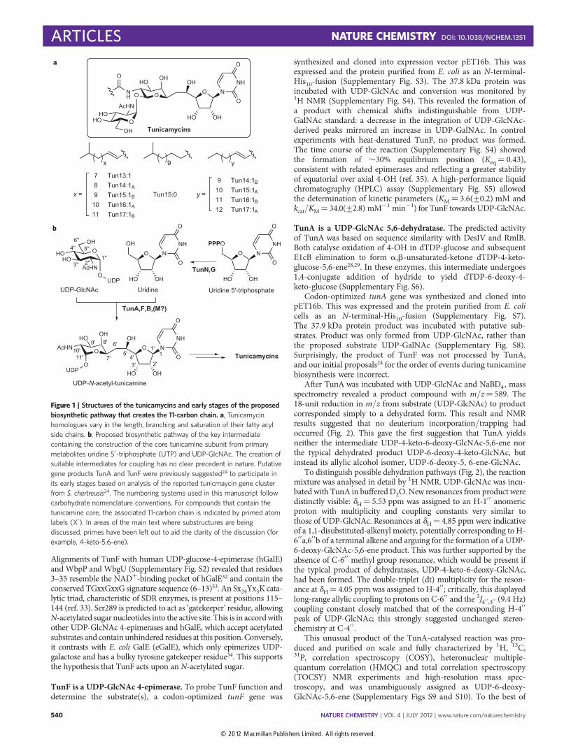
Figure 1 | Structures of the tunicamycins and early stages of the proposed
biosynthetic pathway that creates the 11-carbon chain. a, Tunicamycin
homologues vary in the length, branching and saturation of their fatty acyl
side chains. b, Proposed biosynthetic pathway of the key intermediate
containing the construction of the core tunicamine subunit from primary
metabolites uridine 5′-triphosphate (UTP) and UDP-GlcNAc. The creation of
suitable intermediates for coupling has no clear precedent in nature. Putative
gene products TunA and TunF were previously suggested24 to participate in
its early stages based on analysis of the reported tunicmaycin gene cluster
from S. chartreusis24. The numbering systems used in this manuscript follow
carbohydrate nomenclature conventions. For compounds that contain the
tunicamine core, the associated 11-carbon chain is indicated by primed atom
labels (X′). In areas of the main text where substructures are being
discussed, primes have been left out to aid the clarity of the discussion (for
example, 4-keto-5,6-ene).
ARTICLES NATURE CHEMISTRY DOI: 10.1038/NCHEM.1351
NATURE CHEMISTRY | VOL 4 | JULY 2012 | www.nature.com/naturechemistry540
© 2012 Macmillan Publishers Limited. All rights reserved.
our knowledge, TunA is the first enzyme to exhibit NDP-sugar-5,6-dehydratase activity, converting 4′′, 5′′ and 6′′ positions of UDP-GlcNAc into an allylic alcohol moiety. This is in stark contrast toall other reported NDP-sugar-4,6-dehydratases that generateNDP-4-keto-6-methyl sugars. HPLC and NMR analyses indicatedthat TunA catalysed the establishment from UDP-GlcNAc of anequilibrium (Keq¼ 0.16) with kcat¼ 0.41(+0.01) s21 and KM¼5.5(+0.3) mM (Supplementary Fig. S11).
Insight into the TunA mechanism was gained from other obser-vations. In particular, when the reaction was conducted in bufferedD2O, the integration of the H-5′′ resonance for starting substrateUDP-GlcNAc was lower in the 1H NMR than for other protonresonances. This reduction suggested partial incorporation ofdeuterium at C-5′′; this site-specific H-5′′ ↔ 2H-5′′ exchange wasconfirmed by 2H NMR. An additional minor doublet resonancewas also observed (J¼ 9.1 Hz) at dH¼ 3.52 ppm, which overlappedwith the H-4′′ triplet peak of UDP-GlcNAc. This supported theexistence of two closely related compounds differing only by thepresence of deuterium at C-5′′. This partial deuteration implied‘wash out’ by deuterated solvent36 and suggests a rapid, reversiblestep consistent with an E1cB reaction mechanism, supporting theinvolvement of a UDP-4-keto-GlcNAc intermediate that is readilydeprotonated at C-5′′.
Control of reductive regioselectivity in Tun A. Given the nearidentical conservation of key residues in TunA (a 5,6-dehydrataseusing 1,2-reduction) and, for example, DesIV (a 4,6-dehydrataseusing 1,4-reduction)29 suggested by primary sequence alignment(vide supra), we sought to determine the mechanism behind thiscontrol of regioselectivity (1,2 versus 1,4). TunA was crystallizedin the presence of both substrate UDP-GlcNAc and cofactorNADþ at a pH remote from that of optimal activity to minimizeturnover—the resulting X-ray crystal structure was determined to
a resolution of 1.9 Å (Supplementary Table S8) as a ternarycomplex (Fig. 3). The overall structure was superimposed with theternary complex structures of other dehydratases from the SDRfamily—dTDP-Glc-4,6-dehydratases DesIV and both RmlBs—with a root-mean-square deviation of 1.3–1.8 Å. The overallstructure of TunA can be divided into two domains. TheN-terminal domain contains the NADþ-binding site and showsthe typical Rossmann fold (seven parallel b-strands surroundedby seven a-helices). The smaller C-terminal domain bindsUDP-GlcNAc and consists of mixed strands of four b-sheets andfour a-helices, features also characteristic of the SDR family. Thechemistry catalysed by the enzyme occurs at the interface ofthese domains.
Overall, the interactions of NADþ and UDP-GlcNAc in theirbinding sites in TunA are very similar to those at equivalentpositions in DesIV and both RmlBs (Fig. 3b and SupplementaryFigs S12 and S14). Consistent with the bioinformatics analysis(vide supra), mutation of each residue in the TYK motif (to therespective single-point alanine mutants Thr119Ala, Tyr143Ala,Lys147Ala) reduced the enzymatic activity to an undetectablelevel (Supplementary Table S9). Closer inspection of the sitewhere the GlcNAc moiety and the nicotinamide ring of NADþ
are in close contact, however, reveals some interesting subtleties,which may play important roles in altering the catalytic activity ofTunA and hence explain the unique observed reactivity. C-4 ofthe nicotinamide ring lies 3.4 Å from C-4′′ of the GlcNAc ring, aposition normally ideal for subsequent hydride transfer37. Therelative positions of the nicotinamide and hexosamine/hexosering planes are particularly notable (Fig. 4 and SupplementaryFig. S13). The ribose of NADþ bearing the nicotinamide group isplaced closer to the GlcNAc moiety in the TunA complex than inDesIV/RmlB structures. Moreover, the plane of the nicotinamidering in TunA is rotated �208 with respect to the UDP-GlcNAc
O
OUDP
AcHN
HOH
O
HOH
Tyr143
O–
H
NAD+
HO Thr119 O
OUDP
AcHN
HO
HOH
Tyr143
O
NADH
HO Thr119O
H
Glu121 CO2
O
OUDP
AcHN
HO
Tyr143
O
NADH
HO Thr119O
H
Glu121 CO2H
O
OUDP
AcHN
HOH
OH
UDP-GlcNAc
UDP-4-keto-GlcNAc
UDP-4-keto-6-deoxy-GlcNAc-5,6-eneUDP-6-deoxy-GlcNAc-5,6-ene
S Cys120
HS Cys120
O
OUDP
AcHN
HO
OH
O
via E1cBmechanism
O
OUDP
AcHN
HO O
O
OUDP
AcHN
HOOH
H
UDP-6-deoxy-GalNAc-5,6-ene
1,2-axial-addition
1,2-equatorial-addition
1,4-addition
–
–
–
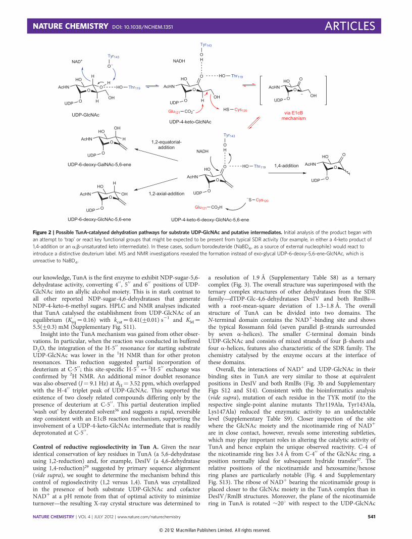
Figure 2 | Possible TunA-catalysed dehydration pathways for substrate UDP-GlcNAc and putative intermediates. Initial analysis of the product began with
an attempt to ‘trap’ or react key functional groups that might be expected to be present from typical SDR activity (for example, in either a 4-keto product of
1,4-addition or an a,b-unsaturated keto intermediate). In these cases, sodium borodeuteride (NaBD4, as a source of external nucleophile) would react to
introduce a distinctive deuterium label. MS and NMR investigations revealed the formation instead of exo-glycal UDP-6-deoxy-5,6-ene-GlcNAc, which is
unreactive to NaBD4.
NATURE CHEMISTRY DOI: 10.1038/NCHEM.1351 ARTICLES
NATURE CHEMISTRY | VOL 4 | JULY 2012 | www.nature.com/naturechemistry 541
© 2012 Macmillan Publishers Limited. All rights reserved.
ring plane; in the other dehydratases it is approximately parallel tothe hexosamines (Fig. 4). The amide carbonyl of the nicotinamidewithin TunA makes a hydrogen bond to the Val174 backboneamide, thus stabilizing this ‘rotated’ position (SupplementaryFig. S14); in DesIV/RmlBs the equivalent residue is Asn.This rotation observed only in TunA places C-4 of NADþ/NADH nicotinamide directly above C-4′′ of GlcNAc, whereas inDesIV/RmlBs this lies much closer to C-6′′ of the substrate sugar.More importantly, the C-4 si-face of nicotinamide faces away
from C-6′′ of GlcNAc in TunA, but maintains a favourable positionand angle for hydride delivery at C-4′′. This subtle rotation isessentially the sole element controlling the absolute observedswitch in 1,2 versus 1,4 reductive regioselectivity (Fig. 2 andSupplementary Fig. S15).
The positions of amino-acid residues probably involved indehydration across C-5′′ and C-6′′ are also clearly visible. Themost notable Cys120, 2.7 Å from OH-6′, we suggest as a generalacid for OH-6′′ protonation (Fig. 2). In other dehydratases, Asp atcorresponding positions plays this role28,29. The adjacent Glu121—conserved across all dehydratases discussed here—is suggested tofunction as a general base in the abstraction of the H-5′′ proton,lying 3.6 Å from C-5′′ and with good alignment for axial H-5′′. InTunA, Glu121 also forms a 3.1 Å hydrogen bond with OH-6′′, aninteraction absent from the other dehydratases. Consistent withthese suggested critical roles in catalysis, the corresponding singlepoint alanine mutants—Cys120Ala and Glu121Ala—had no detect-able enzymatic activities (Supplementary Table S9).
It is notable that the Lys residue hydrogen-bonded to the OH-2′′
of dTDP-glucose in the DesIV and RmlBs is replaced by smallerAla183 in TunA, thus allowing the larger C-2′′-acetamido substitu-ent of UDP-GlcNAc. Consistent with this role, the mutationAla183 � Lys, blocking this acetamide binding site, removed anydetectable activity (Supplementary Table S9). In the binding ofdTDP-glucose, the DesIV/RmlBs structures show a His residueforming a hydrogen bond to the only hydroxyl (OH-3′) of the sub-strate 2′-deoxyribose ring. In TunA, Glu267 replaces this residue,forming hydrogen bonds to both OH-2′,3′ of the ribose moiety inUDP-GlcNAc. Interestingly, alteration of Glu267 � Ala gave asingle-point mutant that retained some activity, albeit with eightfoldreduction in kcat and fivefold increase in KM (SupplementaryTable S9). Notably, the structure determined here indicates thatadditional hydrogen bonding to OH-2′ provided by the Glu201side chain may mitigate the associated loss of binding energy tothe ribose diol unit during catalysis.
TunF acts on more than one substrate. The apparent processing ofUDP-GlcNAc as substrate by both TunA and TunF created aconundrum regarding the starting point and ordering of the tunpathway (Fig. 5a). Despite the demonstrated UDP-GlcNAc-4-epimerase activity (vide supra), TunF could, in principle act uponother substrates (Supplementary Fig. S16).
To investigate this possibility, UDP-GlcNAc was incubated witha mixture of TunF and TunA. Four 1H NMR resonances withcharacteristic multiplicities in the dH¼ 5.4–5.7 ppm H-1′′ regionindicated four distinct species (Fig. 5b). Three corresponded toUDP-GlcNAc, UDP-GalNAc and UDP-6-deoxy-GlcNAc-5,6-ene;the fourth, formed only in the presence of both TunF and TunA,was deduced to be UDP-6-deoxy-GalNAc-5,6-ene (Fig. 5).
To test this hypothesis, a sample of UDP-6-deoxy-GlcNAc-5,6-ene was isolated following incubation of UDP-GlcNAc with5,6-dehydratase TunA. This alternative substrate was incubatedwith TunF (Supplementary Fig. S17). As for UDP-GlcNAc assubstrate, TunF also catalysed the establishment of an equilibrium(Keq¼ 0.43) from UDP-6-deoxy-GlcNAc-5,6-ene. Notably, thekinetic parameters revealed that UDP-6-deoxy-GlcNAc-5,6-ene isthe preferred substrate for TunF (kcat/KM¼ 245(+25) versus34.0(+ 2.8) mM21 min21).
In vivo detection of an exo-glycal intermediate. To assess therelevance of these in vitro experiments to tunicamycinbiosynthesis in vivo, we created an in-frame deletion mutant oftunB in which the entire protein-coding sequence was removed.The construction of an in-frame deletion should prevent anypolar effects on the expression of downstream genes, whiledeletion of tunB was predicted to lead to the accumulation of
Gly17
Gly20
Gly14
NAD
Lys147Tyr143
Thr119Glu121
Cys120UDP-GlcNAc
b
a
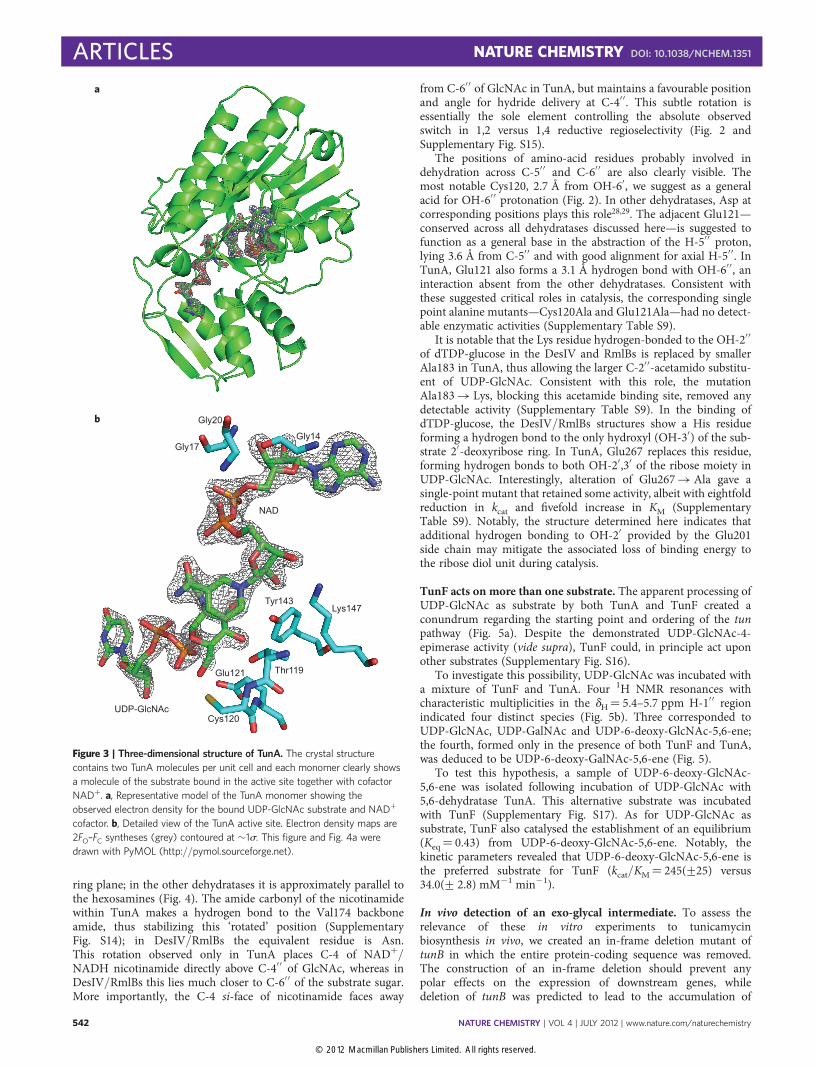
Figure 3 | Three-dimensional structure of TunA. The crystal structure
contains two TunA molecules per unit cell and each monomer clearly shows
a molecule of the substrate bound in the active site together with cofactor
NADþ. a, Representative model of the TunA monomer showing the
observed electron density for the bound UDP-GlcNAc substrate and NADþ
cofactor. b, Detailed view of the TunA active site. Electron density maps are
2FO–FC syntheses (grey) contoured at �1s. This figure and Fig. 4a were
drawn with PyMOL (http://pymol.sourceforge.net).
ARTICLES NATURE CHEMISTRY DOI: 10.1038/NCHEM.1351
NATURE CHEMISTRY | VOL 4 | JULY 2012 | www.nature.com/naturechemistry542
© 2012 Macmillan Publishers Limited. All rights reserved.
sufficient exo-glycal for direct observation. This mutational analysiswas performed on the cloned tunicamycin biosynthetic gene (tun)cluster present in pIJ12003a24 in the heterologous expression hostStreptomyces coelicolor M1152. TunB is predicted to catalyse theformation of the C–C bond between C5 of the uridyl moiety andC6 of the central sugar in tunicamycin (see below). If the exo-glycal is the source of this central sugar moiety, then it would beexpected to accumulate in a tunB mutant. The mutant construct,created by polymerase chain reaction (PCR)-targeting38, wastransferred into S. coelicolor M115224 by conjugation via tri-parental mating39. Successful integration of the mutant constructinto the chromosomal wBT1 attachment site was determined bysingle-colony PCR of kanamycin-resistant exconjugants, yieldingS. coelicolor M1481.
No tunicamycins were detected from the M1481 culture(Supplementary Fig. S24). However, the nucleotide-rich fraction(Supplementary Fig. S22) contained not only compounds associatedwith typical metabolism (UDP-HexNAcs (m/z 606 [M–H]2),UDP-hexoses (m/z 565 [M–H]2)), but also compound
O
AcHNHO
HO
OH
OUDP
O
AcHNHO
HO
OUDP
UDP-6-deoxy-5,6-ene-GlcNAc
i. UDP-GlcNAc
ii. UDP-GlcNAc + TunF
iii. UDP-GlcNAc + TunA
UDP-6-deoxy-GlcNAc-5,6-ene
UDP-6-deoxy-GalNAc-5,6-ene
iv. UDP-GlcNAc + TunF + TunA
UDP-GaINAc
5.60 5.55 5.50 ppm
5.60 5.55 5.50 ppm
5.60 5.55 5.50 ppm
5.60 5.55 5.50 ppm
UDP-GlcNAc
TunA
O
AcHNHO
HO
OUDP
UDP-6-deoxy-5,6-ene-GalNAc
TunF
O
AcHNHO
OH
OUDP
UDP-GlcNAc UDP-GalNAc
TunA
Tun
a
F
HO
b
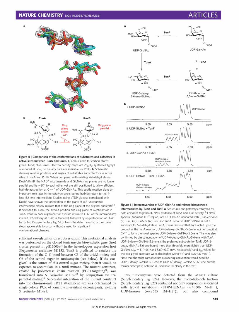
Figure 5 | Interconversion of UDP-GlcNAc and related biosynthetic
intermediates by TunA and TunF. a, Structures and pathways catalysed by
both enzymes together. b, NMR evidence of TunA and TunF activity. 1H NMR
spectra (anomeric H-1′ ′ region) of UDP-GlcNAc incubated with (i) no enzyme,
(ii) TunF, (iii) TunA or (iv) TunF and TunA. Because UDP-GalNAc is not a
substrate for 5,6-dehydratase TunA, it was deduced that TunF acted upon the
product of the TunA reaction, UDP-6-deoxy-GlcNAc-5,6-ene, epimerizing it at
C-4′ ′ to form the novel species UDP-6-deoxy-GalNAc-5,6-ene. This was also
confirmed by direct incubation of UDP-6-deoxy-GlcNAc-5,6-ene with TunF.
UDP-6-deoxy-GlcNAc-5,6-ene is the preferred substrate for TunF; UDP-6-
deoxy-GlcNAc-5,6-ene bound more than threefold more tightly than UDP-
GlcNAc (KM¼ 1.1(+0.1) and 3.6(+0.2) mM, respectively) and kcat values for
the exo-glycal substrate were also higher (269(+4) and 122(+3) min21).
Note that the strict carbohydrate numbering convention would describe
UDP-6-deoxy-GlcNAc-5,6-ene as UDP-6′ ′-deoxy-GlcNAc-5′ ′,6′ ′-ene but the
former descriptive notation is used here for clarity in the text.
H
HOH
AcHNH
O
OUDP
OH
N+
H2N
OR
H
HOH
HOH
O
OTDP
OH
N+
H2NO
R
3.3 Å3.9 Å
Ring plane angle:
22º 3.7 Å3.7 Å
Ring plane parallel
TunAb
a
RmlB
Figure 4 | Comparison of the conformations of substrates and cofactors in
active sites between TunA and RmlB. a, Colour code for carbon atoms:
green, TunA; blue, RmlB. Electron density maps are 2FO–FC syntheses (grey)
contoured at �1s; no density data are available for RmlB. b, Schematic
showing relative positions and angles of substrates and cofactors in active
sites of TunA and RmlB. When compared with existing 4,6-dehydratases
DesIV/RmlB, the NADþ nicotinamide and GlcNAc ring planes are no longer
parallel and lie �208 to each other, yet are still positioned to allow efficient
hydride-abstraction at C24′ ′ of UDP-GlcNAc. This subtle rotation plays an
important role later in the catalytic cycle, during hydride return to the 4-
keto-5,6-ene intermediate. Studies using dTDP-glucose complexed with
DesIV have shown that orientation of the plane of a,b-unsaturated
intermediate closely mirrors that of the ring plane of the original substrate29.
If extended to TunA, the altered position and ring plane of nicotinamide in
TunA result in poor alignment for hydride return to C-6′ ′ of the intermediate;
instead, 1,2-delivery at C-4′ ′ is favoured, followed by re-protonation of O-4′ ′
by Tyr143 (Supplementary Fig. S15). From the determined structure these
steps appear able to occur without a need for significant
conformational changes.
NATURE CHEMISTRY DOI: 10.1038/NCHEM.1351 ARTICLES
NATURE CHEMISTRY | VOL 4 | JULY 2012 | www.nature.com/naturechemistry 543
© 2012 Macmillan Publishers Limited. All rights reserved.
corresponding to the predicted exo-glycal (m/z 588 [M–H]2)(Supplementary Fig. S22). Such a disrupted pathway would lead tothe accumulation of both exo-glycal epimers. The presence of UDP-6-deoxy-GlcNAc-5,6-ene was confirmed by the addition of an auth-entic sample (Supplementary Fig. S22). No exo-glycal was detectedfrom S. coelicolor M1040 (M1152 harbouring pIJ12003a containingintact tun cluster24) and the non-producing strain S. coelicolorM1037 (M1152 containing only vector24) (Supplementary Fig. S23).
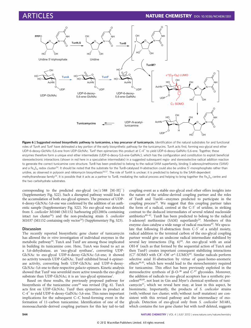
DiscussionThe recently reported biosynthetic gene cluster of tunicamycinhas allowed the in vitro investigation of individual enzymes in themetabolic pathway24. TunA and TunF are among those implicatedin building its tunicamine core. Here, TunA was found to act asa 5,6-dehydratase, an ‘exo-glycal synthase’, converting UDP-GlcNAc to exo-glycal UDP-6-deoxy-GlcNAc-5,6-ene; it showedno activity towards UDP-GalNAc. TunF exhibited broad 4-epimer-ase activity, converting both UDP-GlcNAc and UDP-6-deoxy-GlcNAc-5,6-ene to their respective galacto-epimers. Kinetic analysisshowed that TunF was sevenfold more active towards the exo-glycalsubstrate than UDP-GlcNAc; it is an ‘exo-glycal epimerase’.
Based on these results, the previously proposed pathway forbiosynthesis of the tunicamine core24 was revised (Fig. 6). TunAacts first on UDP-GlcNAc. TunF then epimerizes its product atC-4′′ to yield UDP-6-deoxy-GalNAc-5,6-ene. This raises importantimplications for the subsequent C–C bond-forming event in theformation of 11-carbon tunicamine. Identification of one of themonosaccharide-derived coupling partners for this key tail-to-tail
coupling event as a stable exo-glycal enol ether offers insights intothe nature of the uridine-derived coupling partner and the rolesof TunB and TunM—enzymes predicted to participate in thecoupling process24. We suggest that this coupling partner takesthe form of a radical, centred at the C-5′ of uridine, in strikingcontrast to the deduced intermediates of several related nucleosideantibiotics40–42. TunB has been predicted to belong to the radicalS-adenosyl methionine (SAM) superfamily24. Members of thisenzyme class catalyse a wide range of radical reactions43. We specu-late that following H-abstraction from C-5′ of a uridyl moiety,radical addition to the terminal carbon of the exo-glycal couplingpartner would give an undecose radical intermediate stabilized byseveral key interactions (Fig. 6)44. An exo-glycal with an axialOH-8′ (such as that formed by the sequential action of TunA andthen TunF) creates important consequent stabilizing interactions(C7′-SOMO with C8′–O8′-s*-LUMO)44. Similar radicals performselective axial H-abstraction by virtue of quasi-homo-anomericeffects44,45, which here would lead to the stereochemistry requiredfor tunicamine. This effect has been previously exploited in thestereoselective synthesis of b-O-46 and C-47 glycosides. Moreover,the addition of radicals to exo-glycal acceptors has a synthetic pre-cedent48,49, not least in Gin and Myer’s chemical synthesis of tuni-camycin21, which we reveal here may, at least in this aspect, bebiomimetic. Importantly, the products of S. coelicolor strains(with/without tun cluster; with/without tunB mutation) are con-sistent with this revised pathway and the intermediacy of exo-glycals. Detection of exo-glycal only from S. coelicolor M1481,which contains the tun gene cluster but with tunB deleted, suggests
Uridine 5'-triphosphate
O
NH
O
O
N
HO OH
PPPO
TunN,GO
NH
O
O
N
HO OH
OH
Uridine
O
AcHNHO
HO
OH
OUDP
UDP-GlcNAc
O
AcHNHO
HO
OUDP
UDP-6-deoxy-5,6-ene-GlcNAc
TunAO
AcHNHO
HO
OUDP
UDP-6-deoxy-5,6-ene-GalNAc
TunF
TunM?
O
HO
AcHN
O
OH
O
NH
O
O
N
HO OH
OH
UDP
UDP-N-acetyl-tunicamine
Tunicamycins
TunBO
NH
O
O
N
HO OH
OH
O
HO
AcHN
O
OH
O
NH
O
O
N
HO OH
OH
UDP
H
σ*(C8'-O8') LUMO
C7' SOMO
Figure 6 | Suggested revised biosynthetic pathway to tunicamine, a key precursor of tunicamycin. Identification of the natural substrates for and functional
roles of TunA and TunF have delineated a key portion of the early biosynthetic pathway for the tunicamycins. TunA acts first, forming exo-glycal enol ether
UDP-6-deoxy-GlcNAc-5,6-ene from UDP-GlcNAc. TunF then epimerizes this product at C-4′ ′ to yield UDP-6-deoxy-GalNAc-5,6-ene. Together, these
enzymes therefore form a unique enol ether intermediate (UDP-6-deoxy-5,6-ene-GalNAc), which has the configuration and constitution to exploit beneficial
stereoelectronic interactions (shown in red here in a speculative intermediate) in a suggested subsequent regio- and stereoselective radical addition reaction
to generate the correct tunicamine core structure. TunB has been predicted to belong to the radical SAM superfamily, binding S-adenosylmethionine (SAM)
and a Fe4S4 redox cluster24. It should be noted that the substrate for the TunB-catalysed H-abstraction could also be uridine 5′-monophosphate rather than
uridine, as observed in polyoxin and nikkomycin biosynthesis56,57. The role of TunM is unclear; it is predicted to belong to the SAM-dependent
methyltransferase family24. It is possible that it acts as a partner to TunB, mediating the radical process and helping to bring together the Fe4S4 centre and
the two carbohydrate substrates.
ARTICLES NATURE CHEMISTRY DOI: 10.1038/NCHEM.1351
NATURE CHEMISTRY | VOL 4 | JULY 2012 | www.nature.com/naturechemistry544
© 2012 Macmillan Publishers Limited. All rights reserved.
that exo-glycals are formed only transiently and at low levels inuninterrupted biosynthesis and not at all in cells that do notproduce tunicamycin. The loss of tunicamycin production causedby tunB deletion also confirms its essential role.
The structure of TunA is similar to those of others from the SDRfamily but differs critically in active site arrangement. Indeed, wesuggest that this unique 5,6-dehydratase shares the same firststeps of other SDRs (Supplementary Fig. S6), deviating only at thea,b-unsaturated-keto intermediate. 1,2-Hydride delivery, observedin TunA for the first time, forms a 5,6-ene product and contrastswith the 1,4-regioselectivity observed thus far in the SDRs(Fig. 2). The major source of this altered regioselectivity is attributedto subtle changes in the relative positions of the NADþ cofactor andUDP-GlcNAc substrate within the TunA active site (Fig. 3).
TunA contains an active-site Cys that is absent from other SDRenzymes. Within the 4,6-dehydratases DesIV/RmlB, a conservedAsp/Glu diad is involved in dehydration across C-5′′/C-6′′. At equiv-alent positions in TunA, Glu121 is retained while Cys120 lies in placeof Asp, and mutation of either abolishes activity. Although expected toact as a general acid like aspartic acid, it is possible that the higher pKaof cysteine may prevent unwanted protonation (which might be cata-lysed by an Asp were this to be found at position 120) of the uniqueenol ether product formed by TunA, thus protecting it from unwantedhydrolysis in the enzyme active site. Cys120 may also provide morefavourable hydrophobic interactions with the C¼C double bondthat is uniquely formed here.
Together, the TunA and TunF enzymes have revealed not onlynew modes of biosynthesis used in the assembly of the unique11-carbon chain that makes up the core of tunicamycin, theyhave also given insight into surprising biological intermediates(exo-glycals) and the mechanistic subtlety behind modes of biocata-lytic selectivity (1,2 versus 1,4 reduction) that are used for theirformation. Remarkably, this discovery of a new pathway, whichwe suggest exploits the enol ether moiety of these exo-glycals,until now had no biological precedent but does have chemicalsynthetic precedent21. TunA and TunF may act together to delivera substrate for radical coupling that is chemically logical in ensuringboth regioselecivity and stereoselectivity (Fig. 5). It therefore pre-sents, to our knowledge, a rare example of the post hoc validationof a chemical synthetic strategy21 as being biomimetic.
MethodsChemicals, enzymes and bacterial strains. Unless otherwise stated, chemicalreagents were purchased from Sigma-Aldrich, media components from Sigma-Aldrich or Melford Laboratories and enzymes from Sigma-Aldrich or Invitrogen.Media and general methods for the growth and propagation of E. coli were asdescribed in ref. 50.
Cloning and expression of tunA and tunF. The gene sequences of tunA and tunFfrom S. chartreusis NRRL 3882 (Genbank accession HQ172897) were codonoptimized for E. coli and synthesized by GenScript USA. The genes, both supplied invector pET16b (Novagen), were introduced into E. coli BL21(DE3) bytransformation and expressed using standard procedures (Supplementary Methods).Protein purification after cell harvest was performed using immobilized metalaffinity chromatography according to standard methods (Supplementary Methods).
HPLC activity assay. UDP-GlcNAc 4,6-dehydratase activity was typically assayedwith a reaction mixture volume of 100 ml, incubated at 30 8C for 20 min, andcontaining 5 mM UDP-GlcNAc (or UDP-GalNAc) with or without 2 mM NADþ in50 mM Tris, pH 7.5, and with an appropriate amount of the enzyme extract. For thetrapping experiments, 25 mM sodium borodeuteride was added to the mixture andincubated at 22 8C for an additional 10 min. Control reactions lacking particularcomponents were carried out to determine their necessity for enzymatic conversion.The reaction mixtures were analysed by HPLC (Supplementary Methods).
HPLC time course assay. UDP-GlcNAc (5 mM) was prepared in 0.05 M Tris buffer,pH 7.5, with or without 5 mM MgCl2. TunA was added to a final concentration of0.12 mg ml21. The reaction was mixed by vortexing and incubated at 30 8C. Smallaliquots were withdrawn at a range of time intervals, filtered through viva spin(Sartorius, 10K NMWCO) and analysed by HPLC (Supplementary Methods).Substrate and product peaks eluted at 18 and 19 min, respectively. These wereintegrated and a time course of TunA activity plotted (Supplementary Fig. S11).
HPLC kinetic assay. Appropriate concentrations of UDP-GlcNAc were prepared in0.05 M Tris buffer, pH 7.5, containing 5 mM MgCl2. TunA was added to a finalconcentration of 0.12 mg ml21. The reaction was mixed by vortexing and incubated at30 8C. Each reaction was stopped after 5 min by flash freezing in liquid nitrogen. Eachsample was then boiled for 1 min, filtered through viva spin (Sartorius, 10K NMWCO)and analysed by HPLC (Supplementary Methods). Substrate and product peaks elutedat 26 and 29 min, respectively. From the integration of these peaks a Michaelis–Mentencurve of TunA activity was constructed (Supplementary Fig. S11).
NMR activity assay. Enzymatic reactions were performed in 25 mM Tris, pH 8.0,with 25–100 mg of freshly purified TunA and 77 mM UDP-GlcNAc in a totalreaction volume of 300 ml. After incubation for 1 h at 37 8C, the mixture was flashfrozen and lyophilized. The resulting residue was dissolved in D2O (600 ml) andanalysed by NMR. As control experiments, the same procedures were applied tosamples lacking either TunA enzyme or UDP-GlcNAc. For isolation and fullcharacterization of the UDP-6-deoxy-GlcNAc-5,6-ene product see SupplementaryMethods and Supplementary Figs S8, S9 and S10.
Functional and kinetic assay of TunF. The activity of TunF was measured in asimilar way to that of TunA. See Supplementary Methods for details.
TunA crystallization and structure solution. The cubic-shaped crystals of TunAprotein, typically 115 × 115 mm and belonging to space group P1 (a¼ 45.5,b¼ 51.1, c¼ 67.8(Å), a¼ 98.13, b¼ 106.65, g¼ 94.36 (8), two molecules perasymmetric unit), were obtained after six days at 20 8C using a hanging drop methodwith 20 mg ml21 protein, 20 mM Tris-HCL (pH 7.8) and 2 mM UDP-GlcNAc inthe protein solution, and 20% polyethylene glycol 3350, 100 mM 8 v/v tacsimate(pH 6) in the reservoir solution. A 1.8 Å data set was collected from a single crystalusing a Rigaku Saturn 944þ charge-coupled device detector and FREþ SuperBrightX-ray generator equipped with a copper anode at 100 K. The X-ray data wereprocessed with HKL2000 and scaled with SCALEPACK51. The structure was solvedby molecular replacement using an alanine model constructed from the structure ofdTDP-glucose 4,6-dehydratase (PDB ID 1R66)29. Successive initial model buildingwas carried out using the Phenix program (v1.6)52 and subsequent modelbuilding/rebuilding were performed in COOT53,54. Model refinement wasperformed with Refmac in the CCP4 program suite, by setting aside 5% of theobserved reflection data for cross-validation. The structure was refined to aresolution of 1.9 Å. The final model has an R factor of 18.5% and an Rfree of 23.3%.Relevant X-ray data collection and structure solution statistics are presented inSupplementary Table S8. Data are deposited with the code PDB ID code 3VPS.
Construction of the tunB deletion mutant. S. coelicolor M1481 (M1152 carryingthe tunB-deleted construct pIJ12541 integrated at the chromosomal wBT1attachment (att) site) was made by first replacing tunB in pIJ12003a24 with anapramycin resistance gene using PCR-targeting38 with the pIJ773 apramycin cassetteamplified with primers tunB20 (CTTCCAAGAGGAGGGGGCCGACTGATGACCGGCTACACCATTCCGGGGATCCGTCGACC) and tunB19 (GGCCTCCCTGGACAAGGCCTACCTCACCTCCGCGCCTTCTGTAGGCTGGAGCTGCTTC). Correct targeting was confirmed by PCR using primers tunBtstF(TCGCGACTTCACCTACATCA) and tunBtstR (TGAGGTCGTACAGGCGTATG). The apramycin resistance cassette was then removed by site-specificrecombination using FLP-recombinase38 generating pIJ12541. This construct wasintroduced in S. coelicolor M1152 by conjugation38 to yield S. coelicolor M1481.Correct integration of pIJ12541 at the chromosomal wBT1 att site was confirmed byPCR using primer pairs BT1V1(GGTGCGAATAA GGGACAGTG) plus BT1C1(CACGAGCGGAAACGTACC), and BT1C2 (GTACCAGTTGGCCGTCACC)plus BT1V2 (ACGTCCACGAACTCACCTG).
Construction of S. coelicolor M1037. pRT80255 was introduced into S. coelicolorM1152 by conjugation, generating S. coelicolor M1037. pRT802 integrates at thechromosomal wBT1 att site.
Construction of S. coelicolor M1040. pIJ12003a, a pRT802 derivate containing theminimal tunicamycin gene cluster24, was introduced into S. coelicolor M1152 byconjugation, generating strain S. coelicolor M1040.
Received 28 June 2011; accepted 3 April 2012;published online 20 May 2012
References1. Brandish, P. E. et al. Modes of action of tunicamycin, liposidomycin B, and
mureidomycin A: inhibition of phospho-N-acetylmuramyl-pentapeptidetranslocase from Escherichia coli. Antimicrob. Agents Chemother. 40,1640–1644 (1996).
2. Heifetz, A., Keenan, R. W. & Elbein, A. D. Mechanism of action of tunicamycinon the UDP-GlcNAc:dolichyl-phosphate GlcNAc-1-phosphate transferase.Biochemistry 18, 2186–2192 (1979).
3. Tamura, G. Tunicamycin (Japan Scientific Societies Press, 1982).4. Hamill, R. L., Hoehn, M. H. & Boeck, L. D. Process for preparing tunicamycin.
US patent 4,336,333 (1980).
NATURE CHEMISTRY DOI: 10.1038/NCHEM.1351 ARTICLES
NATURE CHEMISTRY | VOL 4 | JULY 2012 | www.nature.com/naturechemistry 545
© 2012 Macmillan Publishers Limited. All rights reserved.
5. Takatsuki, A., Arima, K. & Tamura, G. Tunicamycin, a new antibiotic:1. Isolation and characterization of tunicamycin. J. Antibiot. 24, 215–223 (1971).
6. Takatsuki, A. et al. Structural elucidation of tunicamycin: 2. Structure oftunicamycin. Agric. Biol. Chem. 41, 2307–2309 (1977).
7. Tsvetanova, B. C. & Price, N. P. J. Liquid chromatography-electrospraymass spectrometry of tunicamycin-type antibiotics. Anal. Biochem. 289,147–156 (2001).
8. Thrum, H. et al. Streptovirudins, new antibiotics with antibacterial and antiviralactivity: 1. Culture taxonomy, fermentation and production of streptovirudincomplex. J. Antibiot. 28, 514–521 (1975).
9. Vogel, P. et al. Isolation of a group of glycolipid toxins from seedheads of annualryegrass (Lolium rigidum Gaud.) infected by Corynebacterium rathayi. Austr. J.Exp. Biol. Med. Sci. 59, 455–467 (1981).
10. Kenig, M. & Reading, C. Holomycin and an antibiotic (MM 19290) relatedto tunicamycin, metabolites of Streptomyces clavuligerus. J. Antibiot. 32,549–554 (1979).
11. Nakamura, S., Arai, M., Karasawa, K. & Yonehara, H. On an antibiotic,mycospocidin. J. Antibiot. 10, 248–253 (1957).
12. Mizuno, M., Shimojima, Y., Sugawara, T. & Takeda, I. Antibiotic 24010.J. Antibiot. 24, 896–899 (1971).
13. Tamura, G., Sasaki, T., Matsuhashi, M., Takatsuki, A. & Yamasaki, M.Tunicamycin inhibits formation of lipid intermediate in cell-free peptidoglycansynthesis of bacteria. Agric. Biol. Chem. 40, 447–449 (1976).
14. Walsh, C. Where will new antibiotics come from? Nature Rev. Microbiol. 1,65–70 (2003).
15. Xu, L., Appell, M., Kennedy, S., Momany Frank, A. & Price Neil, P. J.Conformational analysis of chirally deuterated tunicamycin as an active siteprobe of UDP-N-acetylhexosamine:polyprenol-P N-acetylhexosamine-1-Ptranslocases. Biochemistry 43, 13248–13255 (2004).
16. Elbein, A. D. The tunicamycins — useful tools for studies on glycoproteins.Trends Biochem. Sci. 6, 219–221 (1981).
17. Danishefsky, S. J., Deninno, S. L., Chen, S., Boisvert, L. & Barbachyn, M. Fullysynthetic stereoselective routes to the differentially protected subunits of thetunicamycins. J. Am. Chem. Soc. 111, 5810–5818 (1989).
18. Ichikawa, S. & Matsuda, A. Synthesis of tunicaminyluracil derivatives.Nucleosides Nucleotides Nucleic Acids 23, 239–253 (2004).
19. Karpiesiuk, W. & Banaszek, A. Stereoselective syntheses of the O,N-protectedsubunits of the tunicamycins. Carbohydr. Res. 299, 245–252 (1997).
20. Ramza, J. & Zamojski, A. New convenient synthesis of tunicamine. Tetrahedron48, 6123–6134 (1992).
21. Myers, A. G., Gin, D. Y. & Rogers, D. H. Synthetic studies of the tunicamycinantibiotics. Preparation of (þ)-tunicaminyluracil, (þ)-tunicamycin-V, and5′-epi-Tunicamycin-V. J. Am. Chem. Soc. 116, 4697–4718 (1994).
22. Suami, T., Sasai, H., Matsuno, K. & Suzuki, N. Synthetic approaches towardantibiotic tunicamycins. Part VIII. Total synthesis of tunicamycin. Carbohydr.Res. 143, 85–96 (1985).
23. Tsvetanova, B. C., Kiemle, D. J. & Price, N. P. J. Biosynthesis of tunicamycin andmetabolic origin of the 11-carbon dialdose sugar, tunicamine. J. Biol. Chem. 277,35289–35296 (2002).
24. Wyszynski, F. J., Hesketh, A. R., Bibb, M. J. & Davis, B. G. Dissectingtunicamycin biosynthesis by genome mining: cloning and heterologousexpression of a minimal gene cluster. Chem. Sci. 1, 581–589 (2010).
25. Chen, W. et al. Characterization of the tunicamycin gene cluster unveilingunique steps involved in its biosynthesis. Prot. Cell 1, 1093–1105 (2010).
26. Karki, S., Kwon, S-Y. & Kwon, H-J. Cloning of tunicamycin biosynthetic genecluster from Streptomyces chartreusis NRRL 3882. J. Kor. Soc. Appl. Biol. Chem.54, 136–140 (2011).
27. Berman, H., Henrick, K. & Nakamura, H. Announcing the worldwide ProteinData Bank. Nature Struct. Biol. 10, 980–980 (2003).
28. Allard, S. T. M. et al. Toward a structural understanding of the dehydratasemechanism. Structure 10, 81–92 (2002).
29. Allard, S. T. M., Cleland, W. W. & Holden, H. M. High resolution X-ray structureof dTDP-glucose 4,6-dehydratase from Streptomyces venezuelae. J. Biol. Chem.279, 2211–2220 (2004).
30. Demendi, M., Ishiyama, N., Lam, J. S., Berghuis, A. M. & Creuzenet, C. Towardsa better understanding of the substrate specificity of the UDP-N-acetylglucosamine C4 epimerase WbpP. Biochem. J. 389, 173–180 (2005).
31. Kowal, P. & Wang, P. G. New UDP-GlcNAc C4 epimerase involved in thebiosynthesis of 2-acetamino-2-deoxy-L-altruronic acid in the O-antigenrepeating units of Plesiomonas shigelloides O17. Biochemistry 41,15410–15414 (2002).
32. Jornvall, H. et al. Short-chain dehydrogenases reductases (SDR). Biochemistry34, 6003–6013 (1995).
33. Filling, C. et al. Critical residues for structure and catalysis in short-chaindehydrogenases/reductases. J. Biol. Chem. 277, 25677–25684 (2002).
34. Schulz, J. M. et al. Determinants of function and substrate specificity in humanUDP-galactose 4′-epimerase. J. Biol. Chem. 279, 32796–32803 (2004).
35. Creuzenet, C., Belanger, M., Wakarchuk, W. W. & Lam, J. S. Expression,purifcation, and biochemical characterization of WbpP, a new UDP-GlcNAc C4
epimerase from Pseudomonas aeruginosa serotype O6. J. Biol. Chem. 275,19060–19067 (2000).
36. Gross, J. W., Hegeman, A. D., Vestling, M. M. & Frey, P. A. Characterization ofenzymatic processes by rapid mix-quench mass spectrometry: the case of dTDP-glucose 4,6-dehydratase. Biochemistry 39, 13633–13640 (2000).
37. Allard, S. T. M. et al. The crystal structure of dTDP-D-glucose 4,6-dehydratase(RmlB) from Salmonella enterica serovar Typhimurium, the second enzyme inthe dTDP-L-rhamnose pathway. J. Mol. Biol. 307, 283–295 (2001).
38. Gust, B. et al. Lambda red-mediated genetic manipulation of antibiotic-producing Streptomyces. Adv. Appl. Microbiol. 54, 107–128 (2004).
39. Kieser, T., Bibb, M. J., Buttner, M. J., Chater, K. F. & Hopwood, D. A. PracticalStreptomyces Genetics (The John Innes Foundation, 2000).
40. Kaysser, L. et al. Identification and manipulation of the caprazamycin genecluster lead to new simplified liponucleoside antibiotics and give insights intothe biosynthetic pathway. J. Biol. Chem. 284, 14987–14996 (2009).
41. Kaysser, L., Siebenberg, S., Kammerer, B. & Gust, B. Analysis of theliposidomycin gene cluster leads to the identification of new caprazamycinderivatives. ChemBioChem 11, 191–196 (2010).
42. Rackham, E. J., Gruschow, S., Ragab, A. E., Dickens, S. & Goss, R. J. M.Pacidamycin biosynthesis: identification and heterologous expression of the firsturidyl peptide antibiotic gene cluster. ChemBioChem 11, 1700–1709 (2010).
43. Marsh, E. N. G., Patterson, D. P. & Li, L. Adenosyl radical: reagent and catalyst inenzyme reactions. ChemBioChem 11, 604–621 (2010).
44. Giese, B. The stereoselectivity of intermolecular free radical reactions. Angew.Chem. Int. Ed. Engl. 28, 969–980 (1989).
45. Giese, B. & Dupuis, J. Anomeric effect of radicals. Tetrahedron Lett. 25,1349–1352 (1984).
46. Kahne, D., Yang, D., Lim, J. J., Miller, R. & Paguaga, E. The use of alkoxy-substituted anomeric radicals for the construction of beta-glycosides. J. Am.Chem. Soc. 110, 8716–8717 (1988).
47. Crich, D. & Lim, L. B. L. Synthesis of 2-deoxy-b-C-pyranosides bydiastereoselective hydrogen-atom transfer. Tetrahedron Lett. 31, 1897–1900 (1990).
48. Cipolla, L., Liguori, L., Nicotra, F., Torri, G. & Vismara, E. Glycomimetics via anew glycoexoenitols-malonyl radical C–C bond formation. Chem. Commun.1253–1254 (1996).
49. Vauzeilles, B. & Sinay, P. Selective radical synthesis of b-C-disaccharides.Tetrahedron Lett. 42, 7269–7272 (2001).
50. Sambrook, J. & Russell, D. Molecular Cloning: A Laboratory Manual (ColdSpring Harbor Laboratory Press, 2000).
51. Otwinowski, Z. & Minor, W. Processing of X-ray diffraction data collected inoscillation mode. Methods Enzymol. 276, 307–326 (1997).
52. Adams, P. D. et al. PHENIX: a comprehensive Python-based system formacromolecular structure solution. Acta Crystallogr. D 66, 213–221 (2010).
53. Emsley, P. & Cowtan, K. COOT: model-building tools for molecular graphics.Acta Crystallogr. D 60, 2126–2132 (2004).
54. Emsley, P., Lohkamp, B., Scott, W. G. & Cowtan, K. Features and development ofCOOT. Acta Crystallogr. D 66, 486–501 (2010).
55. Gregory, M. A., Till, R. & Smith, M. C. M. Integration site for Streptomycesphage BT1 and development of site-specific integrating vectors. J. Bacteriol. 185,5320–5323 (2003).
56. Chen, W. et al. Characterization of the polyoxin biosynthetic gene cluster fromStreptomyces cacaoi and engineered production of polyoxin H. J. Biol. Chem.284, 10627–10638 (2009).
57. Ginj, C., Ruegger, H., Amrhein, N. & Macheroux, P. 3′-Enolpyruvyl-UMP, anovel and unexpected metabolite in nikkomycin biosynthesis. ChemBioChem 6,1974–1976 (2005).
AcknowledgementsThe authors gratefully acknowledge B. Odell (Oxford University) for help with the NMRexperiments. This work was supported by the EPSRC (DTA studentship for F.J.W.), the Billand Melinda Gates Foundation (S.S.L.) and BBSRC (J.P.G-E. and M.J.B). G.J.D and B.G.D.are Royal Society Wolfson Research Merit Award recipients. This manuscript is dedicatedto the memory of Professor David Gin.
Author contributionsF.J.W. cloned tunF, purified the protein and performed functional and kinetic analyses ofTunF. S.S.L. and T.Y. cloned tunA and its mutants, purified the resulting proteins andperformed functional analyses. S.S.L. performed the kinetic analyses. TunA was crystallizedby T.Y. The 3D structure was determined by S.J.L., and S.S.L., B.G.D and G.J.D. carried outits analysis. H.W. and J.P.G-E. created the tunB mutant strain and extracts. M.J.B. designedthe deletion strategy. H.W., M.J.B. and B.G.D. analysed the extracts. The manuscript waswritten by F.J.W., S.S.L., M.J.B., G.J.D. and B.G.D.
Additional informationThe authors declare competing financial interests: details accompany the full-text HTMLversion of the paper at www.nature.com/naturechemistry. Supplementary informationaccompanies this paper at www.nature.com/naturechemistry. Reprints and permissioninformation is available online at http://www.nature.com/reprints. Correspondence andrequests for materials should be addressed to B.G.D.
ARTICLES NATURE CHEMISTRY DOI: 10.1038/NCHEM.1351
NATURE CHEMISTRY | VOL 4 | JULY 2012 | www.nature.com/naturechemistry546
© 2012 Macmillan Publishers Limited. All rights reserved.