biotechnologies for restoration of … · fluorescence specifically emitted by the prosthetic group...
TRANSCRIPT
UNIVERSITÀ DEGLI STUDI DI MILANO
FACULTY OF AGRICULTURAL AND FOOD SCIENCE
THE DEPARTMENT OF FOOD ENVIRONMENTAL AND NUTRITIONAL SCIENCES
PHILOSOPHY DOCTORATE SCHOOL IN
MOLECULAR SCIENCE AND AGRICULTURAL, FOOD AND ENVIRONMENTAL BIOTECHNOLOGY
PHILOSOPHY DOCTORATE COURSE IN CHEMISTRY, BIOCHEMISTRY AND ECOLOGY OF PESTICIDES
XXVII CYCLE
PHILOSOPHY DOCTORATE THESIS
BIOTECHNOLOGIES FOR RESTORATION OF CULTURAL HERITAGE
EMANUELA LOMBARDI NO. MATR. R09789
SUPERVISOR: PROFESSOR DANIELE GIUSEPPE DAFFONCHIO COORDINATOR: PROFESSOR DANIELE GIUSEPPE DAFFONCHIO
ACADEMIC YEAR 2013/2014


Summary
Abstract ......p.5 Riassunto ......p.9 Aim of the Thesis ......p.13 Chapter 1 Biotechnologies for cultural heritage
…..p.15
Chapter 2 Physiology, biochemistry and biotechnological applications of Desulfovibrio genus
......p.35
Chapter 3 Optimization of the production process and conservation of D.vulgaris biomass
......p.59
Chapter 4 Set up of new molecular methods for D.vulgaris quantification
......p.69
Chapter 5 Successful combination of chemical and biological treatments for the cleaning of stone artworks
......p.91
Chapter 6 Evaluation of effectiveness of biocleaning in sulfates removal from mural paintings of Queen Teodolinda Chapel in Monza Cathedral
......p.109
Chapter 7 Analysis of the microbial community present on the reverse side of a deteriorated canvas
......p.119
Chapter 8 Identification of microorganisms deteriorating a paper print of the Monastery of “S. Maria al Carrobiolo” in Monza
......p.138
General conclusions and perspectives ......p.147
Acknowledgements ......p.151
Activities performed during the Ph.D. ......p.152

Abstract
5
Abstract In recent years, operators in the restoration sector are adding to their historical-artistic competences also scientific knowledge in order to find solutions more and more effective and respectful toward the cultural heritage, the operator and the environment. Among the different scientific branches, biotechnologies allow for an innovative and precise approach to the complexity of the problems that the restorer has to face in his own daily work. Biotechnology research in the field of cultural heritage develops in two directions: on the one hand focuses on the development of accurate diagnostic techniques, useful for the correct identification and characterization of alterations and biodeteriogens; on the other hand focuses on the development of innovative restoration methods, based on the employment of new products. The employment of biotechnologies in restoration of cultural heritage is the main topic of the present PhD doctoral thesis, which deals with both of the aforementioned sides. In Chapter 1 a review on the employment of biotechnologies in the field of cultural heritage is presented, considering both diagnostic techniques for characterization of biodeteriogens, and the use of microorganisms and enzymes for restoration. The first part of the thesis focuses on a microbial product, based on sulfate-reducing bacteria (SRB) belonging to D. vulgaris species, applied for the removal of sulfate crusts from artwork surfaces. In studies carried out in the last decades, this product has turned out to be very promising and favorable, compared to traditional restoration techniques, thanks to its capability of combining effectiveness to selectivity and safety for the restorer and the environment. Such a technology, original, innovative and sustainable, has been successfully experimented on important artworks. In Chapter 2 a review on the current knowledge of Desulfovibrio genus is presented, in particular concerning its physiology, biochemistry and biotechnological applications. Nevertheless, the D. vulgaris-based product presented four limitations: i) low production yields and inability of long-term conservation of D. vulgaris biomass; ii) lack of an appropriate method for monitoring of abundance and activity of the biomass; iii) time-consuming application technique; iv) application limited to stone surfaces. The overcoming of the above mentioned limitations has been the aim of the first part of the present work. A research work, structured in different phases, has been conducted for the optimization of the production process and the development of a method for the long-term conservation of the bacterial biomass (Chapter 3). Initially the laboratory protocol has been set up on small volumes at liter scale, in order to define the growth curve of the bacterium and evaluate its metabolic response to different substrates and growth conditions. subsequently the fermentative process has been transferred from the flask to 5 lt fermentor, optimizing the control of pH and H2S concentration. H2S is the main metabolic product in the fermentative process of SRB, its accumulation is toxic for bacteria, leading to unfavorable growth conditions. These improvements allowed a significant increase in biomass production, from a concentration of 1*108 cell/ml in 120h of fermentation in flask, to the concentration of 3*109 cell/ml in 72h of fermentation in bioreactor. For the long-term conservation of D. vulgaris biomass, freeze-drying has been carried out, testing the effectiveness of different cryoprotective agents. Among them, the best in terms of cell viability post-rehydration resulted to be lactose, which ensured the stability of the product for a minimum of 6 months. Chapter 4 deals with the development of new molecular approaches for monitoring of D. vulgaris biomass concentration and viability since traditionally employed methods, such as Most Probable Number (MPN) and microscope counting, resulted unsuitable. The
Abstract
6
research focused on the set up of a method applicable not only to liquid cultures, but also to cells embedded in the delivery system used for the applications of D. vulgaris cells on the surfaces during the biorestoration treatment. Among all the tested methods, the most effective and suitable resulted to be the spectrophotometric measurement of the fluorescence specifically emitted by the prosthetic group of bisulfite reductase, a key enzyme in dissimilatory sulfate reduction. The results showed that fluorescence emission is proportional to viable cells present in liquid culture as well as when embedded in the delivery system. Real-time PCR quantification of the SRB-specific dsr gene allowed to significantly quantify D. vulgaris cells in liquid culture, but when applied on cells embedded in the delivery system the detection limit (107 cell/ml) was too high to make this method efficient. The development of new methodologies for the application of D.vulgaris-based product, aimed at the reduction of time and number of required applications for the removal of sulfations, has been conducted on the funeral monument realized in memory of ‘Neera’, the poetess Anna Zuccari, located in the Cimitero Monumentale in Milan (Chapter 5). Besides biological treatment, two other methods have been tested: chemical treatment, based on the non-ionic detergent Tween 20 and a combined treatment, consisting in a chemical pre-treatment followed by the biological treatment. The combined method resulted to be effective in the removal of the black crust, without altering the underlying stone, obtaining a 70% reduction in cleaning time. Moreover, the combined method preserved all the advantages of the biocleaning approach: selectivity toward the alteration and respectfulness toward the original material. For the purpose of extending this biocleaning approach to substrates other than stone artworks, such as mural paintings, an experimentation has been carried out on two scenes belonging to the pictorial cycle decorating ‘Queen Teodolinda Chapel’ in Monza Cathedral (Chapter 6). The applicability test on surfaces characterised by fragility, such as pigmented surfaces, is of primary importance for the further development of this technology. The obtained results can be regarded very promising, in terms of sulfations removal and respectfulness towards such a delicate surface. However, this study has to be considered merely preliminary and incomplete, and further research must be conducted in order to verify the compatibility of the treatment with different kinds of materials, such as pigments. The last part of this thesis focuses on diagnostic methodologies for the identification and characterisation of biodeteriogens from two artworks. In the past, microorganisms responsible for deterioration of cultural assets were identified through conventional methods based on the cultivation of potential biodeteriogen microorganisms and their identification and phenotypic characterisation. Here molecular biology technologies independent from bacteria cultivation were employed, which complete and expand the information provided by the cultivation-dependent approach. These methodologies have been employed for the analysis of an acrylic monochrome painting on canvas realised by the artist E. Castellani (Chapter 7) and of a paper print realized in the 17th century, conserved in the Monastery of “S. Maria al Carrobiolo” in Monza (Chapter 8). The acrylic monochrome painting on canvas presented alterations characterised by yellow-earth/red point areas of different extensions, spread on the whole posterior surface. Molecular analyses on the total microbial community have been carried out through Denaturing Gradient Gel Electrophoresis (DGGE) method. Among the identified bacteria, the most abundant were Bacillus subtilis, Bacillus licheniformis and Deinococcus gobiensis; whereas among fungi, Leptosphaerulina and Penicillium genera. Culture-dependent techniques confirmed the dominance of bacteria belonging to Bacillus genus, whilst no fungal species has been isolated. However, presence of fungi was
Abstract
7
confirmed by microscope analysis, which allowed the visualization of fungal hyphae and spores in all the samples. Considering morphology and dimensions, these structures visualized by microscope were ascribable to mycelia and spores of fungi belonging to Penicillium genus, confirmed also by comparison with literature images. Afterwards the characterization of microbial community, the activity of four different biocides on the potential biodeteriogens was evaluated. Biotin N, Biotin R, New Des 50 and Amuchina (in single and mixed) were tested toward the single microbial isolates and the whole microbial community. According to the antibiogram test, the combination of Biotin R 4% in ethyl acetate + Biotin N 4% in white spirit resulted to be the most effective in terms of inhibiting activity, both on single strains and on the whole bacterial community. The paper print realized in the 17th century, conserved in the Monastery of “S. Maria al Carrobiolo” in Monza, presented whitenings and small dark spots, on the obverse and on the reverse side, respectively. In this case, a microbial investigation was executed through culture-dependent techniques for the isolation of bacteria and fungi, in order to characterize the possible deteriogens and determine their phylogenetic affiliation. The results demonstrated a negligible presence of bacteria. The most frequently cultured strains belonged to the Staphylococcus genus, which is associated to human skin, and to the Sphingomonas genus, which is an environmental bacterium, which have never been associated to biodeteriogen activity. As concerns fungi, the results showed a dominant presence of Neurospora pannonica, both on the obverse and on the reverse side of the print. In summary, the research emphasized the importance of biotechnologies in the field of cultural heritage. The optimization of D. vulgaris-based product, described in the first part of the work, has been successful, therefore this result underlines the importance of research for the improvements of biotechnological methodologies employed in restoration. The overall results suggest that further research is required for additional enhancement of this sulfates removal methodology and for the development of novel approaches, more and more effective and convenient, to be used in the field of cultural heritage.

8

Riassunto
9
Riassunto
Negli ultimi anni, gli operatori del settore del restauro sempre più stanno affiancando alle proprie competenze storico-artistiche e tecniche, quelle sviluppate in contesti scientifici, al fine di trovare soluzioni sempre più efficaci e rispettose del bene, dell’operatore e dell’ambiente. Tra le diverse branche scientifiche, quelle legate alle biotecnologie permettono un approccio innovativo e puntuale alla complessità dei problemi che un restauratore deve affrontare nel proprio quotidiano lavoro. La ricerca biotecnologica nell’ambito dei beni culturali si sviluppa in due direzioni: da un lato è focalizzata sullo sviluppo di accurate tecniche diagnostiche, utili per la corretta identificazione e caratterizzazione delle alterazioni e degli agenti biodeteriogeni; dall’altro sullo sviluppo di innovativi metodi di restauro, basati sull’utilizzo di nuovi prodotti. L’impiego delle biotecnologie nel restauro dei beni culturali è il tema principale di questa tesi di dottorato, che affronta entrambi gli aspetti. Nel Capitolo 1 è presentata una review sull’impiego delle biotecnologie nel campo dei beni culturali, considerando sia le tecniche diagnostiche per la caratterizzazione dei biodeteriogeni, sia l’uso di microorganismi ed enzimi per il restauro. La prima parte della tesi è incentrata su un formulato microbico a base di batteri solfato-riduttori della specie D. vulgaris, per la rimozione di solfatazioni. In studi condotti negli ultimi decenni, il prodotto si è rivelato molto promettente e vantaggioso rispetto alle tecniche di restauro tradizionali, per la sua capacità di coniugare l’efficacia alla selettività e sicurezza per l’operatore e per l’ambiente. Tale tecnologia, originale, innovativa ed ecosostenibile, è stata sperimentata con successo su importanti opere d’arte. Nel Capitolo 2 è presentata una review riguardante il genere Desulfovibrio, in particolare gli aspetti legati alla fisiologia, biochimica e applicazioni biotecnologiche. Tuttavia, il prodotto presentava quattro grossi limiti: i) ridotta capacità produttiva e impossibilità di conservare il prodotto per lunghi periodi; ii) assenza di un metodo adeguato per il monitoraggio dell’abbondanza e attività della biomassa; iii) lunghi tempi di messa in opera; iv) casi applicativi limitati alle superfici litoidi. Il superamento dei limiti sopracitati è stato l’obiettivo della prima parte del presente lavoro. Un lavoro di ricerca, articolato in diverse tappe, è stato svolto per ottimizzare il processo produttivo e sviluppare un metodo per la conservazione nel lungo periodo della biomassa batterica prodotta (Capitolo 4). Inizialmente il protocollo sviluppato in laboratorio è stato riprodotto su piccoli volumi (beute da 1 lt), al fine di definire la curva di crescita del batterio e verificare la sua risposta metabolica a diversi substrati e condizioni di crescita, successivamente il processo fermentativo è stato trasferito dalla beuta al fermentatore da 5 lt, riuscendo così a tenere sotto controllo il pH e la concentrazione di H2S, prodotto metabolico principale del processo fermentativo dei batteri solfato-riduttori. Un eccessivo accumulo di H2S è tossico per i batteri, determinando sfavorevoli condizioni di crescita. Le modifiche apportate hanno consentito di ottenere un significativo aumento della produzione di biomassa, passando dalla concentrazione di 1*108 cell/ml in 120h di fermentazione in beuta, alla concentrazione di 3*109 cell/ml in 72h di fermentazione in bioreattore. Per la conservazione nel lungo periodo della biomassa batterica, si è proceduto con la liofilizzazione, testando l’efficacia di diversi crioprotettori. Tra essi, il migliore in termini di vitalità cellulare post-reidratazione è risultato il lattosio, garantendo la stabilità del prodotto per almeno 6 mesi. Il Capitolo 3 riguarda lo sviluppo di nuovi approcci molecolari per il monitoraggio della concentrazione e vitalità della biomassa di D.vulgaris. Infatti, i metodi tradizionalmente
Riassunto
10
impiegati, quali Most Probable Number (MPN) and conta al microscopio, risultavano inadeguati. La ricerca si è focalizzata sull’individuazione di un metodo che potesse essere applicato non solo alle colture liquide, ma anche alle cellule incorporate nel sistema veicolante, usato per le applicazioni sulle superfici da trattare. Tra tutti i metodi testati, il più efficace e adatto si è dimostrato la misura spettrofotometrica della fluorescenza, emessa specificamente dal gruppo prostetico della bisolfito reduttasi, enzima chiave nella solfato riduzione dissimilativa. I test condotti hanno dimostrato che l’emissione di fluorescenza è proporzionale alle cellule vive presenti in coltura liquida o incorporate nel sistema veicolante. La quantificazione del gene dsr, specifico dei batteri solfato-riduttori, mediante Real-time PCR, ha permesso di quantificare in maniera significativa le cellule di D. vulgaris in coltura liquida. Nel caso di cellule incorporate nel sistema veicolante, il limite di detection (107 cell/ml) era troppo alto, per cui il metodo si è mostrato poco efficiente. Lo sviluppo di nuove metodologie di applicazione del formulato microbico basato su D. vulgaris, finalizzate a ridurre i tempi e il numero delle applicazioni necessarie alla rimozione delle alterazioni solfatiche, è stato condotto sul monumento funebre della scrittrice Anna Zuccari, in arte Neera, situato nel Cimitero Monumentale di Milano (Capitolo 5). Oltre al trattamento biologico, sono state testate altre metodologie: trattamento chimico, basato sull’utilizzo del detergente non ionico Tween 20 e trattamento combinato, ossia un pretrattamento chimico seguito da un trattamento biologico. Il metodo combinato risultava essere efficace nella rimozione della crosta nera, senza alterare la pietra sottostante, ottenendo una riduzione del tempo e numero di applicazioni del 70%. Inoltre, il metodo combinato manteneva tutti i vantaggi del trattamento biologico: selettività nei confronti dell’alterazione e rispettosità nei confronti del materiale originario. Per estendere l’applicabilità del formulato ad altri tipi di substrati, quali ad esempio i dipinti murali, è stato condotto un test su due scene componenti il ciclo pittorico che decora la Cappella della Regina Teodolinda nel Duomo di Monza (Capitolo 6). La verifica dell’applicabilità su superfici caratterizzate da fragilità e delicatezza, quali le superfici pigmentate, risulta di fondamentale importanza per l’ulteriore sviluppo della tecnologia. I risultati ottenuti in termini di rimozione delle solfatazioni e rispettosità per una superficie così delicata sono promettenti, tuttavia questo studio è da considerarsi preliminare ed incompleto, ulteriore ricerca è necessaria per verificare la compatibilità con diversi tipi di materiali, quali pigmenti. La seconda parte di questa tesi è focalizzata sulle tecniche diagnostiche per l’identificazione e caratterizzazione degli agenti biodeteriogeni da due opere d’arte. In passato i microorganismi responsabili del deterioramento del materiale in oggetto venivano identificati mediante metodi convenzionali, basati sulla coltivazione dei potenziali biodeteriogeni e loro identificazione e caratterizzazione fenotipica. In questo lavoro sono state impiegate tecniche di biologia molecolare indipendenti dalla coltivazione, in grado di complementare ed espandere le informazioni fornite dai convenzionali metodi di identificazione. Tali tecniche sono state impiegate per l’analisi di un acrilico monocromo su tela realizzato dal pittore E. Castellani (Capitolo 7) e di una stampa del ‘600, proveniente dal Convento di S. Maria al Carrobiolo di Monza (Capitolo 8). L’acrilico monocromo su tela presentava vistose alterazioni puntiformi di colore giallo ocra-arancio su tutta la superficie del retro della tela. Le analisi molecolari sull’intera comunità batterica sono state condotte mediante l’utilizzo della tecnica Denaturing Gradient Gel Electrophoresis (DGGE). Tra i batteri identificati, i più abbondanti risultano quelli ascrivibili alle specie Bacillus subtilis, Bacillus licheniformis e
Riassunto
11
Deinococcus gobiensis; mentre tra i funghi, spicca la presenza dei generi Leptosphaerulina e Penicillium. Le analisi coltura-dipendenti hanno confermato la dominanza di batteri appartenenti al genere Bacillus, mentre non è stato possibile isolare alcuna specie fungina. La presenza di funghi è stata comunque confermata mediante analisi al microscopio ottico, che ha consentito la visualizzazione di ife e spore fungine in tutti i campioni. Per morfologia e dimensioni, le strutture visualizzate al microscopio sono riconducibili a miceli e spore di funghi ascrivibili al genere Penicillium, come confermato anche dal confronto con immagini di letteratura. Successivamente alla caratterizzazione della comunità microbica, sono stati condotti studi di valutazione dell’attività biocida di quattro prodotti: Biotin N, Biotin R, New Des 50 e Amuchina (in singolo e come mix) sui singoli ceppi batterici e sull’intera comunità microbica. Secondo gli antibiogrammi condotti con i differenti biocidi, Biotin R 4% in etil acetato, Biotin N 4% in white spirit e soprattutto la loro combinazione sono risultati essere i prodotti più efficaci in termini di attività inibente, sia sui singoli ceppi che sull’intera comunità batterica. La stampa del ‘600, proveniente dal Convento di S. Maria al Carrobiolo di Monza, presentava sbiancamenti e piccole macchie scure rispettivamente sul fronte e sul retro. In questo caso, si è proceduto con un’indagine microbica mediante metodi coltura-dipendenti per l’isolamento di batteri e funghi al fine di individuare i possibili biodeteriogeni e determinarne l’affiliazione filogenetica. Dai risultati ottenuti si evince una trascurabile presenza di batteri. Inoltre i batteri individuati con più frequenza appartengono ai generi Staphylococcus, microrganismo associato alla pelle dell’uomo, e Sphingomonas, batterio ambientale, i quali non sono mai stati associati ad un’attività biodeteriogena. Per quanto riguarda i funghi, è dominante la presenza del fungo Neurospora pannonica, sia sul fronte sia sul retro della stampa. In conclusione, questo lavoro di ricerca ha enfatizzato l’importanza delle biotecnologie nel settore dei beni culturali. L’ottimizzazione del prodotto a base di D. vulgaris, descritta nella prima parte del lavoro, è andata a buon fine, questo risultato sottolinea l’importanza della ricerca per migliorare le metodologie biotecnologiche impiegate nel restauro. I risultati complessivi suggeriscono che ulteriore ricerca è necessaria per l’ulteriore miglioramento della metodologia di rimozione dei solfati e per lo sviluppo di approcci innovativi, sempre più efficaci e convenienti, da utilizzare nel settore dei beni culturali.

12
Aim of the Thesis
13
Aim of the Thesis
The continuous biotechnological innovation, applied to the field of cultural heritage, has opened new scenarios both for the development of innovative products and techniques for restoration, and for the employment of diagnostic molecular methodologies. Research in the field of biological cleaning of artworks started at the beginning of 1990s. Today it is recognized as being a valid alternative to traditional chemical treatments such as organic solvents or other aggressive conservation methods like mechanical treatments. Among the developed products based on microorganisms as cleaning agents, one of the most successful and object of several studies is a microbial formulate based on Desulfovibrio vulgaris, used for the removal of sulfates altering the surface of artworks exposed to a polluted atmosphere. Such technology has been successfully experimented in applications on important monuments and artworks, among them: 2 statues realized by the artist J. Eberle, belonging to the statuary decor of Buon Consiglio Castle in Trento (Polo et al., 2010); the sculpture “Allegoria della Morte” by G. Lazzerini (Gioventù et al., 2013); a tile of Milan Cathedral (Cappitelli et al., 2007) and an area of Pietà Rondanini base (Cappitelli et al., 2005). The increasing focus on this technology is mainly due to its ability to conjugate efficiency, selectivity and safety for the restorer and the environment. In spite of its promising properties, the product in question presented some limitations. The principal aim of the present PhD doctoral thesis is indeed to deal with these limitations, for the optimization of this cleaning technology. The first chapter will discuss about biotechnologies in the field of cultural heritage, in particular on the use of microorganisms and enzymes for restoration. The current fields of employment of microorganisms in restoration are various: from removal of organic and inorganic matter, to stone consolidation. Another treated issue is the use of molecular-based techniques for the detection of microorganisms colonizing the artworks. In the second chapter, a deeper review on the current knowledge of Desulfovibrio genus is presented, in particular concerning its response to oxygen exposure, energy metabolism and biotechnological applications. In the third chapter one of the problems linked to the use of D. vulgaris for biorestoration is presented: the difficulty of monitoring the biomass. The traditional techniques employed for the estimation of the cellular concentration are based on Most Probable Number (MPN) and microscope counting, but they proved to be inappropriate: the first because D. vulgaris cells tend to aggregate, therefore serial decimal dilutions required in MPN are not reliable, giving incoherent results; the latter because it didn’t allow to distinguish between live and dead cells, overestimating the cellular concentration and also because the count is subjective. The need for an analytical, unequivocal method was evident, therefore the development of new molecular approaches for this purpose was studied. Another problem, described in the fourth chapter, is the reduced production capacity and the long-term conservation of D. vulgaris biomass. The production of a large amount of bacterial biomass resulted hard, therefore the scale-up of the laboratory procedure has been carried out, reaching the optimization of the process and reducing the total productive cost. As concerns the long-term conservation of bacterial biomass, a freeze-dried procedure has been defined, testing the effectiveness of different cryoprotectants. In the fifth chapter, it is presented another drawback: the biorestoration practice employing D. vulgaris is limited to small surface areas, mainly because the technique is time-consuming in the presence of thick and compact crusts. Therefore, the development
Aim of the Thesis
14
of a new methodology of application is described, directed at the reduction of the time and number of applications required for the complete removal of black crusts. A new strategy, consisting in the combination of biocleaning with a chemical pre-treatment, was tested on a one-century-old artistic marble statue realized by the artist Lina Arpesani in memory of ‘Neera’, the poetess Anna Zuccari. The issue treated in the sixth chapter is the extension of this biocleaning approach to other tipologies of surface. In fact it has been experimented exclusively on lithoid materials, but an important goal for the expansion of the methodology is the test on mural paintings, often altered by sulfations, by their nature characterized by extreme fragility. The applicability on pigmented surfaces was tested on the mural paintings narrating the ‘Stories of Queen Teodolinda’, situated in Queen Teodolinda Chapel of Monza Cathedral. This study has to be considered merely preliminary, the matter requires in-depth examinations. Another aim of the present PhD doctoral thesis is to deal with the employment of diagnostic molecular methodologies for the identification and characterization of agents responsible for biodeterioration of cultural heritage. In the seventh chapter these methodologies were used for the analysis of the microbial community present on the reverse side of a deteriorated canvas, realized by the artist Enrico Castellani in the second half of ‘900. The demonstration of the possibility of employment of such methodologies for different constituent materials of artworks is illustrated in the eighth chapter, in which they are used for identification of microorganisms altering a paper print dating at ‘600, coming from the Monastery of “S. Maria al Carrobiolo” in Monza. Finally, the Conclusions chapter summarizes general conclusions of this Ph.D. thesis and suggests future perspectives. References Cappitelli, F., Zanardini, E., Toniolo, L., Abbruscato, P., Ranalli, G., Sorlini, C. (2005) Bioconservation of the marble base of the Pietà Rondanini by Michelangelo Buonarroti. Geophysical Research Abstracts 7, 06675. Cappitelli, F., Toniolo, L., Sansonetti, A., Gulotta, D., Ranalli, G., Zanardini, E., Sorlini, C. (2007) Advantages of using microbial technology over traditional chemical technology in removal of black crusts from stone surfaces of historical monuments. Appl. Environ. Microbiol. 73, 5671–5. Gioventù, E., Lorenzi, P. (2013) Bio-removal of black crust from marble surface: comparison with traditional methodologies and application on a sculpture from the Florence’s English Cemetery. Procedia Chemistry, 8:123-129. Polo, A., Cappitelli, F., Brusetti, L., Principi, P., Villa, F., Giacomucci, L., Ranalli, G., Sorlini, C. (2010) Feasibility of removing surface deposits on stone using biological and chemical remediation methods. Microb. Ecol. 60, 1–14.
Chapter I
15
Biotechnologies for cultural heritage Biotechnology has a wide range of applications fields. At the beginning of ‘90s important biotechnological applications to artwork preservation emerged (Ramirez et al., 2005). It was the beginning of a series of promising studies, going hand in hand with advances in biotechnology and opening new horizons in art preservation. In the past, the contribution of applied microbiology and biotechnology for the preservation and restoration of artworks involved only the identification of the living organisms accountable for the deterioration of materials by classic phenotypic identification methods (Fernandes, 2006). Recently, a change is in action, given the amount of published works, focusing in the introduction of molecular-based techniques for the detection of microorganisms on the surface of artworks and in the utilization of microorganisms for artwork cleaning and restoration. Artworks undergo a continuous process of deterioration caused by several factors: aging, attack of biological, physical, chemical agents. These factors become more aggressive in the presence of environmental pollution (Sorlini et al., 2010). As concerns biological deterioration, it is caused by microorganisms, which can be considered one of the most important causes of this kind of deterioration and this role has been object of several studies. Bacteria, fungi, lichens, algae, cyanobacteria are able to colonize artworks made of different materials: wood, canvas, paper, parchment, leather, silk, cotton, papyrus, tapestries, stone, ceramics, etc. Over the year, the development of advanced biotechnology has led to the application of the latter for colonization studies and for the diagnosis of stone pathologies. Another kind of deterioration is caused by natural physical and chemical agents specific to the environment and climatic area, accelerated by airborne pollutants. From partial combustion of fossil fuels derive organic pollutants such as aromatic, aliphatic and polycyclic hydrocarbons; other pollutants released in the atmosphere are carbonaceous particles, metals, dusts, fine particulates and acidic gases (sulfur dioxide SO2 and nitrogen oxides NOx). All these compounds represent a serious threat especially to artwork in the open air. Another factor responsible of deterioration is the result of incorrect or inappropriate restoration interventions, realized especially in the past decades, which leaves undesirable and harmful materials on the surface of the artwork. Especially in these two last kinds of deterioration, that is physical-chemical and derived by incorrect restoration, microorganisms and enzymes constitute a valid help in recovery. Indeed, from the end of ‘80s, studies on the use of microorganism as biorestoration agents started, on the basis of their metabolic properties. Microorganisms and enzymes can play a very important biocleaning role when traditional techniques fail or give not satisfactory results, or when operators and artworks are put at risk. Therefore they are able to remove layers of undesirable materials, which is the result of the interaction between pollutants and the surface or residual matter left after incorrect restoration interventions. They can be applied for the treatment of organic and inorganic deterioration.
1. Microorganisms for removal of organic matter The surface of artworks, in particular stonework, frescoes and mortar often presents organic patinas, result of the polluted atmosphere and aging process. Airborne carbonaceous particles derived from oil and coal combustion, in particular polycyclic

Chapter I
16
aromatic hydrocarbons, have been identified on the surface of artworks (Saiz-Jimenez, 1997). Therefore these surfaces passively entrap airborne particulate matter and organic compounds, creating a patina which undergoes deterioration, with the consequent unaesthetic alteration of the material. A solution to this problem is the use of bacteria, on the basis of their ability to remove organic compounds. In fact, several bacteria are able to remove aliphatic and aromatic hydrocarbons. An important example is the use of bacteria for the removal of organic matter applied in the past to stone surfaces during restoration interventions. In a study dating at 2005, (Ranalli et al., 2005) aerobic heterotrophic viable bacterial cells were applied to a fresco of the 14th century, “Conversione di S. Efisio e battaglia” (Conversion of S. Efisio and battle) by Spinello Aretino in the Monumental Cemetery of Pisa. This fresco, 20 years ago, was detached from the wall using the “tear-off” technique, realized by pasting a gauze cloth on the surface of the fresco using animal glue and then removing from the wall the cloth with the attached fresco, when the glue is dry. In order to dissolve the glue and detach the fresco from the gauze, proteolytic enzymes (collagenases and proteases) were tested, resulting not effective. Furthermore, neither physical nor chemical techniques gave better results. This failure was ascribed to the presence of formalin, added in the past as biocide, responsible for the formation of insoluble compounds. Therefore, a suspension of Pseudomonas stutzeri (A29 strain) was applied to 20 square meters of the fresco. The bacterial suspension was embedded in hydrophilic cotton strips which were laid over it. After 8 to 12 hours this suspension removed the glue, allowing the separation of the gauze from the fresco. Antonioli and colleagues (Antonioli et al., 2005) tested three different carbon sources for the cultivation of A29 strain of Pseudomonas stutzeri (glucose, animal glue and aged animal glue), observing that cells grown on glucose could not degrade the glue present on the Spinello Aretino fresco, instead cells grown on animal glue and aged animal glue were able to do it. Proteomic analysis showed that this degradation can be attributed to the expression of caseolytic and collagenase activity of these cells, in fact the most abundant components of animal glues are collagen and casein. Another fresco treated using this approach is “Stories of the Holy Fathers” painted by Buonamico Buffalmacco in the 14th century and situated at Camposanto Monumentale in Pisa (Lustrato et al., 2012). As in the previous case, during World War II the fresco was quickly removed from the original walls because of a bombardment. It was detached using the “tear-off” technique with gauze and a layer of warm animal glue, and it was then stored by rolling it up. In this case, for the first time, this method was applied for full-scale biocleaning of ancient mediaeval frescos. Before and after the biocleaning, chemical and microbiological analyses were performed, showing that bioremoval with the A29 strain of P. stutzeri successfully removed animal glue and casein proteins from the fresco. The GC-MS (Gas chromatography-mass spectrometry) and PY/GC-MS (Pyrolysis/Gas chromatography-mass spectrometry) analytical procedures proved that animal glue and casein had been almost completely removed (85% and 80%, respectively). Three different times of application were tested: 2, 3 and 6 h, all of them resulted to be effective, as after them the animal glue and casein were no longer detectable by visual inspection. Moreover, as the optimal result was already obtained in the first 2 h after application, this cleaning procedure can be considered very quick. After the biocleaning treatment, the fresco was subjected to short- and medium-term microbial monitoring to assess microbial colonization, activity, and continued presence of any viable P. stutzeri cells. The absence of viable cells in the fresco after biotreatment, and thus of any potential negative effects due to their metabolism, was confirmed.

Chapter I
17
The biocleaning method, compared to traditional chemical and mechanical methods, offers a lot of advantages: it is non invasive, extremely selective, environmental friendly and it doesn’t require any specialized equipment. In fact, it is non-destructive and removes only extraneous substances or altered compounds from the painting, uses only safe microorganisms (neither pathogenic nor spore-forming bacteria), both for the operators and for the environment. Biotechnological techniques are powerful, low-cost, environmental friendly solutions, which are of low risk to human health. Another field of viable bacterial cells application can be seen in the removal of organic synthetic polymers used in the conservation and as original constituents of artworks. In fact, from 20th century, artistic objects made of plastics were realized, moreover synthetic polymers began to be employed for treatments of artifacts as adhesives, consolidants and protective coatings (Cappitelli and Sorlini, 2008). However synthetic polymeric materials suffer different forms of deterioration, including chemical, physical and biological. In 2014 Troiano and colleagues (Troiano et al., 2014) suggest a methodology for the selection of bacteria able to remove synthetic polymers. They tested the ability of five bacteria to attack a four-year old Paraloid B72, the most commonly used polymer in conservation treatments, through optical and scanning electron microscopy observations, weight loss measurements, Fourier transform infrared spectroscopy and differential scanning calorimetric analysis. However, none of the bacteria were able to attack Paraloid B72, maybe because this polymer was not sufficiently aged. In fact, the behaviour of freshly dried Paraloid B72 may be completely different from that of the same resin after aging. Nevertheless, the developed methodology can be applied to select other bacteria with this ability, offering a reference for future research on bioremoval of synthetic resins. A valid strategy for the identification of bacteria characterized by the ability of remove these materials consists in their isolation directly from polymeric items (Chen et al., 2007; Arutchelvan et al., 2005; Saleem et al., 2008). For example, (McNamara et al., 2004) from a bronze statue treated with Incralac (an ethyl methacrylate and butyl acrylate copolymer) a yeast was isolated. This microorganism was found to be responsible of the deterioration of the coating, for this reason it could be used as biocleaning agents.
2. Microorganisms for removal of inorganic matter Artworks deterioration caused by inorganic agents is mainly due to air pollutants or residual matters left after inappropriate restoration interventions. In the case of stone, a frequent phenomenon is sulfatation. Sulfur dioxide (SO2) is a pollutant converted to sulfite ions (SO3
2-) in the presence of moisture (reaction 1) and then oxidized to sulfate ions (SO4
2-) by oxidant agents as oxygen, ozone and hydrogen peroxide (reaction 2, 3, 4) (Sorlini et al., 2010).
SO2 SO32- (1)
SO32-+ 1/2 O2 SO4
2- (2) SO3
2- + 1/3 O3 SO42- (3)
SO32- + H2O2 SO4
2- (4)
In these conditions sulfates enter the stone pores and, after water evaporation, they climb to the surface and precipitate as bihydrate calcium sulfate or gypsum (CaSO4·2H2O). Gypsum undergoes dissolution and recrystallization processes on the areas exposed to the rain, resulting in the erosion of the altered stone; instead if the surface is not exposed to
Chapter I
18
rain, gypsum, which is quite pouros, includes minerals and smog particles, leading to the formation of black crusts. Black crust is composed of gypsum crystals, calcite, carbonaceous particles, silicon particles, aromatic, polycyclic and aliphatic compounds, sometimes enclosing calcium oxalates, fragments of mycelium, bacterial cells and potassium-hydrated phosphates. From the combustion of heavy oils derive carbonaceous particles, which catalize the reaction between acidic air pollutants (SO2 and NOx) and the stone carbonates, leading to large porous formations. Traditional chemical and mechanical treatments are not selective towards the alteration, attacking not only the black crust but also the sound stone. An alternative cleaning technology, employing sulphate reducing bacteria (SRB) was first proposed by Atlas et al. (1988). They used Desulfovibrio desulfuricans for the removal of sulfates, by immersion of the sample in a broth culture for 24 hours. The sample was a stone cornice of the Museum of Natural History of Chicago. Another sample treated in the same way was a statue from the Cave Hill Cemetery in Louisville, KY. No method was performed to prove the removal of sulphate, but the discoloration of the black crust, turning to a lighter colour, took place (Gauri et al., 1992). The authors suggested that Desulfovibrio desulfuricans enzymes dissociated the gypsum and reduced the sulphate ions, while the calcium ions reacted with carbon dioxide, allowing the precipitation of calcite (reaction 5).
6CaSO4 + 4H2O + 6CO2 6CaCO3 + 4H2S + 2S + 11O2 (5)
In 1997, Ranalli and colleagues (Ranalli et al., 1997) tested in batch different strains of Desulfovibrio in pure and mixed cultures to verify their sulfate-reducing potentiality. D. desulfuricans ATCC strain 13541 and D. vulgaris ATCC strain 29579 were selected and their biomasses were applied under anaerobic conditions on stone sample surfaces, using sepiolite as delivery system. The following stone surfaces were used as samples: samples artificially enriched with sulfates (marble and Lecce stone), real fragments of a marble column section from Pisa and of a marble statue representing a horse hoof. Sulfate removal was measured by ion-exchange chromatography and resulted to be more effective on real samples than on artificially enriched samples. The best result was obtained with a 36 h treatment of D.vulgaris, in fact 81% sulfate removal was achieved. In these experiments the cells colonized the delivery system (i.e. carrier) sepiolite, and then were applied on the surface, allowing in this way a close contact with the surface to be treated. However, this method still required a long time (10 to 14 days) for the colonization of the sepiolite, and moreover hydrogen sulfide could react with the iron in the medium, forming iron sulfide precipitates, resulting in the formation of black spots on the surface of the samples. An improved methodology for the removal of black crusts using Desulfovibrio vulgaris subsp. vulgaris ATCC 29579 was described in 2006 by Cappitelli et al. (2006). In order to allow an easy application of bacteria, to keep a good contact between the cells and the surface to be treated, and to easily remove the cells after the treatment, three delivery systems were tested and compared: the mineral matrix sepiolite and the two organic gels Hydrobiogel-97 and Carbogel. In the case of organic gels, bacterial cells are entrapped in about 10 minutes during gel formation, whereas in the case of the inorganic matrix, they must colonize the matrix by growing and adhering on the surface of the particles, in a process lasting at least 2 days. Carbogel was selected as the best delivery system for the high number of viable bacteria that were retained at the end of the experiment. In fact,

Chapter I
19
after 20 h the cell number in Carbogel changed from 108 to 106 cells g-1, while with the two other carriers the decrease of cell numbers was higher. To overcome the problem of precipitation of the generated sulfide, resulting in the formation of black spots on the treated stone surface, two strategies were adopted. The first was the use of a DSMZ 63 medium modified by eliminating any iron source. The second strategy to avoid sulfide precipitation on the stone was the filtration of the culture on cellulose filters which allowed the bacteria to pass through but trapped the residual iron sulfide precipitates. After filtration, a final cell centrifugation and washing step were used to eliminate by-products, which could cause corrosion and undesirable stains on the stone surface due to bacterial metabolites in the exhausted medium. Then the cell pellet was resuspended in anoxic phosphate buffer at pH 7.0 (KH2PO4, 0.408 g liter-1; K2HPO4, 0.522 g liter-1) added with 0.599 g liter-1 sodium lactate, to reach a cell density of around 108 cells ml-1. Carbogel was added to the bacterial suspension. The obtained bacterial product was applied to a Candoglia marble fragment from the Milan Cathedral, Italy, altered by a 2- to 3-mm thick black crust (35% sulfates). The stone sample was divided into three pieces: one was treated with D. vulgaris subsp. vulgaris ATCC 29579 cells mixed into Carbogel, another was treated with Carbogel without microorganisms, and the last one was left untreated. Each application was 15 h long and the removal of the cells after the treatment resulted to be easy. Two methods were adopted to evaluate the efficiency of the biotreatment: ion-exchange chromatography (IC) and color measurements. The results showed that the treatment was very successful. The strain removed 98% of the sulfates of the crust in a 45-h treatment. However, the feasibility of the treatment should be evaluated on a case-by-case basis. The number of applications and the overall treatment time must be evaluated considering different factors such as the chemical nature and thickness and uniformity of the black crust. In another study (Cappitelli et al., 2007), the biocleaning technology using viable microorganisms was compared to the traditional chemical technologies. In situ experiments were carried out on a lunette of Candoglia marble from the Milan Cathedral, which was completely covered by black crust. The chemical treatment consisted in ammonium carbonate-EDTA mixture, instead the biological one involved the sulfate-reducing bacterium Desulfovibrio vulgaris subsp. vulgaris ATCC 29579. The differently treated samples were analysed using optical microscopy, scanning electron microscopy-energy dispersive spectroscopy (SEM-EDS) and FTIR analysis. The results showed that the biological procedure resulted in more homogeneous removal of the surface deposits and preserved the noble patina under the black crust. Whereas both of the treatments converted gypsum to calcite, allowing consolidation, the chemical treatment also formed undesirable sodium sulfate. The biological treatment resulted to be superior because it removed the black crust homogeneously, without altering the noble patina under the black crust, proving its selectivity. After this test on marble, in 2010 D. vulgaris was applied for the first time on limestone, which is more porous than marble (Polo et al., 2010). Two sculptures were treated: Demetra and Cronos statues situated in the courtyard of the Buonconsiglio Castle in Trento. Chemical analyses were performed before and after the biocleaning and they showed that bioremediation with D. vulgaris successfully removed gypsum black crust from the treated sculptures. After three applications, the black crusts were no longer detectable by visual inspection, and the FTIR analyses proved that the gypsum was almost completely removed. Moreover, as previously demonstrated for marble (Cappitelli et al., 2007), both optical evidence and FTIR analysis showed that the noble patina was preserved.

Chapter I
20
Three different methodologies for the removal of black crust were compared in a study of 2011 (Gioventù et al., 2011): laser, chemical and biological treatment. These cleaning procedures were applied on three different lithotypes of the external walls of the Florence Cathedral: green serpentine, red marlstone and Carrara white marble. The chemical treatment consisted in the mixture of ammonium carbonate and the non-ionic detergent Tween 20. The biological treatment consisted in a D.vulgaris biomass entrapped in Carbogel, as described in Cappitelli et al. (2006). The effects of the different procedures on the surfaces were evaluated by scanning electron microscopy coupled with energy dispersive X-ray (SEM/EDS) spectroscopy, Fourier transform infrared (FTIR) spectroscopy and color measurements. Color measurements were performed also one year later. It was found that chemical cleaning led to non-homogeneous crust removal and sometimes it caused the detachment of fragments. On the contrary, biological cleaning led to a uniform removal of the black crust. Laser treatment left a thin yellow layer visible by the naked-eye, and particularly on the white marble, it left a residual layer of gypsum. Instead microbial and chemical cleaning managed to remove all the gypsum residuals from the surfaces. Overall, the microbial cleaning process can be considered the most satisfactory treatment. Further biocleaning interventions have been carried out in situ in Italy for the removal of the black crusts : the base of the Pietà Rondanini by Michelangelo Buonarroti in Milan (Cappitelli et al., 2005), Castello Sforzesco in Milan, the sculpture “Allegoria della Morte” by G. Lazzerini in Florence (Gioventù et al., 2013), some areas of the façade of S. Maria delle Grazie in Milan, tuff stone external wall of 12th century Matera Cathedral (Alfano et al., 2011).
Another phenomenon regarding artworks deterioration caused by inorganic agents is calcareous stone nitration. Nitrogen dioxide (NO2) is an atmospheric pollutant which is oxidized to N2O5 and then to nitric acid:
N2O5 + H2O 2HNO3
Nitric acid interacts with calcium carbonate to produce calcium nitrate:
CaCO3 + HNO3 Ca(NO3)2 + H2O + CO2
Calcium nitrate is highly soluble, so it undergoes leaching by rain, exposing the sound stone to consumption. The formation of nitrate salt efflorescence on the surface of wall paintings is amongst the most important causes and mechanisms of deterioration in artworks located in indoor environments. The increasing volume of the crystals formed exerts a pressure on the wall. This produces traction forces that can exceed the strength of the material, thus generating micro-cracks in the wall painting. An alternative cleaning technology employing nitrate-reducing bacteria was first proposed by Ranalli et al. (1996). They used a strain of Pseudomonas stutzeri (GB94), delivered in sepiolite. Real samples of Vicenza stone altered by nitrates and artificially aged samples were treated. The application lasted 30 hours, with the resulting 88% of nitrates removal. In another application on the external walls of the Cathedral of Matera (May et al., 2008), Carbogel was used as delivery system. A good yield was obtained also in this case, nevertheless in both cases a contribution of the carrier was observed, in particular in the first application the nitrate removal in the control was 20%. In a study of 2011 (Alfano et al., 2011) the middle and long-term monitoring of in situ biocleaning was realised. In fact, six years before, the tuff stone external wall of 12th century Matera Cathedral, altered by nitrates and sulfates, had been treated using nitrate

Chapter I
21
and sulphate reducing bacteria. The bioremoval treatment was based on the direct application onto the altered stone surfaces of a Pseudomonas pseudoalcaligenes KF707 strain and Desulfovibrio vulgaris ATCC 29579 cells. The two strains were entrapped in a Carbogel and applied individually and together to the vertical wall. The biological procedure resulted in an efficient, homogeneous removal. In fact, after 24h the strains had removed 55% of the nitrate and 85% of the sulfate deposits, respectively. The aim of this work was the setting up of an advanced protocol to maximise biocleaning to vertical walls using a multilayer biosystem, evaluation of sulfate and nitrate removal using physical-chemical analyses and determination of middle and long-term effects of bioremoval. Physical and chemical analysis (IC analyses and colour measurement) proved the effectiveness of the bioremoval. For the first time, a biocleaning biosystem allowing the simultaneous removal of nitrates and sulphates from stone tuff was employed. The effectiveness of this advanced system was confirmed by long-term data monitoring (6 years). In 2013 (Bosch-Roig et al., 2013) this system based on the use of P.stutzeri was applied for the first time on a wall painting altered by nitrate salt efflorescence. The research was carried out for the cleaning of wall paintings placed in the lunettes of the central vault of the Santos Juanes church in Valencia, Spain. First of all, different strains of P.stutzeri were tested and P. stutzeri DSMZ 5190 was selected as it showed to have greater efficiency in the reduction of nitrate into molecular nitrogen. Different application supports were tested: cotton, sepiolite, carbogel, agar and agarose. Each support was tested with and without Japanese paper, showing that, in the treatment of wall paintings, it provides a better protection and allows a proper removal of the application “pack”. Among the tested supports, agar was chosen as the most efficient in the removal on vertical surfaces, and as the most proper because it reduces the risks to the fresco by minimizing the volume of water filtration as well as the time of contact. Using these experimental conditions, a reduction of 92% in nitrate efflorescence was proved by Ion chromatography. Monitoring of the biocleaning treatment was realized by means of ATP assay, immediately after treatment to be sure that no P.stutzeri, water or agar residues have been left on the fresco. In fact they could promote the growth of other microorganisms, causing biodegradation of the wall painting. The same monitoring was performed one month after the treatment, in both of the two cases no growth of microorganisms was reported. Nevertheless, the biological removal of nitrates has aroused less interest than the bioremoval of sulfates, perhaps because nitrates don’t cause marked discoloration of the artifact, as sulfates do.
3. Enzymes Another kind of biological agent for artwork conservation is represented by enzymes. They can be applied on different kinds of materials: paper, painted surfaces, furniture, but few applications of enzymes on stone have been made and only on frescoes. Enzymes are employed especially for the treatment of high-value objects. Specificity for the target molecules and quick activity are their strong points, however they present also some disadvantages: high sensitivity to experimental conditions such as temperature, pH, inhibitors (Bellucci and Cremonesi, 1994). In 2003, Cremonesi proposed a new methodology for the application of enzymes, by immobilization of enzymes in gels (Cremonesi, 2003). Immobilized enzymes allow to reduce the amount of water released to the artwork and an easier removal after the treatment.
Chapter I
22
The enzyme Alcalase has been used (Beutel et al., 2002) for the removal of casein from a medieval fresco. The success of this technique was proved by reverse high-pressure liquid chromatography. The enzyme laccase produced by the fungus Trametes was used for the removal of red-brown stains caused by Serratia Marcescens on the marble sculpture Slide Mantra (Konkol et al., 2009). Moreover, proteases, lipases and amylases are generally used enzymes against organic deterioration. Lipases are also able to remove organic synthetic polymers used in the conservation of artworks (Bellucci et al., 1999). For instance, the acrylic resin Paraloid B72 present on the surfaces of a 15th century tempera painting on panels and a 19th century oil painting on canvas, was removed using lipases. In 2013 (Palla et al., 2013) proteases isolated from marine invertebrate organisms were applied to remove protein layers form works of art surface. Proteolytic zymography assay evidenced that these enzymes are active in a broad temperature range, between 4 degrees and 37 degrees C. The enzymatic cleaning by these proteases, tested on wooden furniture of the second half of the eighteenth century showed positive results, without needing to heat the enzyme solution or the surface on which they were applied. These novel proteases resulted more appropriate than previously employed proteases, which usually are active at temperature >= 37 degrees C. Another work published in 2013 (Pereira et al., 2013) established a multiscale and multitechnique nondestructive approach for monitoring surface properties and evaluating the effectiveness of enzymatic removal of varnishes from paintings/polychrome artefacts. This methodology consists in the use of microscopy [stereomicroscopy (SM), optical microscopy (OM), atomic force microscopy (AFM), and scanning electron microscopy (SEM)] and colorimetric (CIE L*a*b* system) techniques, used for characterization of the reconstruction surfaces at different scales (macro-scale by SM and OM; micro-scale by SEM and nano-scale by AFM) and also to monitor the cleaning treatment.
4. Microorganisms for stone consolidation One of the effect of deterioration of stone materials is the increase of porosity, leading to the loss of mechanical properties (Sorlini et al., 2010). In the past, synthetic polymers were employed for stone consolidation, but this kind of treatment was not long-lasting and presented several drawbacks: rupture of the synthetic film caused by water evaporation, change in colour, waterproofing of stone and other aesthetic damages. Another treatment consisted in the limewater technique, based on the use of Ca(OH)2, which reacting with CO2, produces calcite. Also this technique is not free from problems: the formed carbonate crystals are very small, creating a superficial and friable layer. An alternative methodology consists in the employment of biocalcificating agents, that is microorganisms able to precipitate salt crystals. Biocalcification reaction is shown in the following reaction:
Ca2+ + CO32- CaCO3 ↓
This phenomenon was observed in the presence of different genera of aerobic heterotrophic bacteria, such as Bacillus, Myxococcus, Escherichia coli and Pseudomonas. The first articles about the effect of calcifying bacteria on deteriorated stone were written by Gauri and Atlas (Atlas et al., 1988; Gauri et al., 1988; Gauri et al., 1992). They used Desulfovibrio desulfuricans for the removal of black crusts from stone and observed also the phenomenon of biocalcification. Since then, different teams of researchers proposed microbially induced calcium carbonate precipitation as an eco-friendly method to protect and restore degraded ornamental stones (Le Metayer-Levrel et al., 1999; Stocks-Fischer

Chapter I
23
et al., 1999; Ramachandran et al., 2001; Ramakrishnan et al., 2001; De Muynck et al., 2008a,b; Orial et al., 1992; Castanier et al., 1999). Nevertheless, the biological mechanism of crystal formation is not completely clear. Microbially induced calcium carbonate precipitation (MICCP) is a process where an organism creates a local microenvironment, allowing precipitation of carbonates (Hamilton, 2003). Bacteria isolated from different natural habitats have been reported for their ability to precipitate calcium carbonate (Krumbein, 1979; Rodriguez-Navarro et al., 2003). Calcium carbonate precipitation is a chemical process influenced by four main factors: the calcium concentration, amount of dissolved inorganic carbon (DIC), availability of nucleation sites and pH (Hammes and Verstraete, 2002). In 2002 Hammes and Verstraete suggested that microorganisms influence precipitation by altering any of the precipitation parameters described above, either separately or in various combinations with one another. MICCP has gained increasing interest in the last 20 years and found to be the primary focus of research in bio geo civil engineering because of its numerous applications (Dhami et al., 2014). There are mainly four groups of microorganisms involved in the process, which are: (i) photosynthetic organisms such as cyanobacteria and algae, (ii) sulfate reducing bacteria responsible for dissimilatory reduction of sulfates, (iii) organisms utilizing organic acids, and (iv) organisms that are involved in nitrogen cycle either by ammonification of amino acids/nitrate reduction or hydrolysis of urea (Stocks-Fischer et al., 1999; Hammes and Verstraete, 2002; Jargeat et al., 2003). As concerns the second pathway, in the presence of organic matter and absence of oxygen, sulfate reducing bacteria can reduce sulfate to H2S and release HCO3
- (Equation 7) (Ehrlich, 1998; Castanier et al., 1999; Wright, 1999). This reaction takes place when the abiotic dissolution of gypsum (CaSO4·2H2O) (Equation 6) provides an environment that is rich in both sulfate and calcium ions. The resulting H2S release leads to an increase in pH and favors the precipitation of calcium carbonate (Equation 8) (Castanier et al., 1999).
CaSO4·2H2O→Ca2+ + SO42- + 2H2O (6)
2 (CH2O)+SO42- →HS-+HCO3- +CO2+H2O (7)
Ca2+ + HCO3- + OH- →CaCO3 + 2H2O (8) Tiano et al. (1999) studied the effect of microbial calcite crystals on Lecce bioclastic limestone by Micrococcus spp. and Bacillus subtilis and their results showed a significant reduction in water absorption. The authors also highlighted some drawbacks, such as the formation of new products due to chemical reactions between stone minerals and some by-products originating from the metabolism of bacteria, and the formation of stains caused by the growth of air-borne fungi. To avoid such problems, the authors used some natural and synthetic polypeptides to control the calcite crystal growth in the pores. Use of organic matrix macromolecules (OMM) isolated from Mytilus californianus shells was proposed to induce the precipitation of calcium carbonate within the pores of the stone, because of their crystal-nucleating activity (Tiano et al., 1992; Tiano, 1995). Nonviable cells were employed, to overcome problems linked to the use of living cells, such as production of exopolymers and microbial contamination. Tests were carried out on Lecce stone and Gioia marble (Tiano et al., 1999; Tiano et al., 2003), monitoring them by capillarity absorption, superficial hardness, colour variation and drilling resistance. The results were good, nevertheless this procedure was too expensive and time-consuming. Hence, in place of OMM, Tiano et al. (2006) proposed to use acid functionalized proteins such as polyaspartic acid. Calcium and carbonate ions were supplied for calcite crystal growth, by addition of ammonium carbonate and calcium chloride solution or a solution
Chapter I
24
of saturated bicarbonate. This process was less expensive and easier, but the consolidating effect was observed to be very low compared to OMM (Tiano et al., 2006). Myxococcus xanthus was tested by Rodriguez-Navarro et al. (2003), resulting in 26% reduction of stone porosity, moreover the newly formed crystals showed greater resistance to physical stress than the natural crystals of sound stone. This property can be attributed to the incorporation of bacterial metabolites into the crystals. Urzì et al. in 1999 showed that most of the bacteria isolated from stone can mediate carbonate precipitation. On the basis of this observation, experiments were carried out by immersing samples of stone in a culture without calcite-forming bacteria. The culture medium activated the growth of calcite-forming bacteria naturally present in the microbial community colonizing the stone surface. Currently, a French company is specialized in the production of biological mortars and cements, using a strain of Bacillus cereus mixed to stone powder. Precipitation of calcite crystals by fresh water bacteria on limestone significantly reduced the pore sizes of the stone (Zamarreno et al., 2009). Calcite crystals were deposited around and inside open pore spaces, filling 43–49% of the open pore spaces, which was 20% higher than the application of the medium alone. De Muynck et al. (2012) reported that B. sphaericus was very efficient for consolidation of limestone specimens at range of temperatures (10, 20, 28, 37◦C). This isolate led to 64% lower weight loss upon sonication and 46% decreased sorptivity in treated limestone specimens compared to the control specimens. De Muynck et al. (2011) recently applied bacterial calcite in two types of stones: microporous and macroporous. They reported that application of bacterial carbonates is more successful in macroporous stone. Although this technology presents several advantages, there are also a few limitations. In comparison to chemical treatments, biobased treatments are found to be more complex because the microbial activity depends on many environmental factors such as temperature, pH, concentrations of donors and acceptors of electrons, concentrations and diffusion rates of nutrients and metabolites. Moreover, design of experiments for biodeposition treatments require a huge data of the biological processes (growth, biosynthesis, specific enzymatic activities), chemical reactions accompanied with formation of insoluble compounds, physic-chemical processes as precipitation, crystallization, and adhesion. Because of this complexity, its usage at large-scale has not been so encouraging. As the amounts of carbonate precipitates formed are dependent on amount of calcium added, increased concentration of calcium leads to accumulation of salts and favours efflorescence and damage to crystallization. The survival of bacteria within the stone material is another limiting factor, influencing the extent of calcification. Another hindrance for the large scale production is the cost of laboratory grade nutrient media, so there is great need to look for alternative economical and cheap medium ingredients.
5. Molecular-based techniques for the detection of microorganisms As biological agents have an important role in deterioration of artworks, the need for advanced methodologies for their detection has acquired a crucial role. Traditional phenotypic identification methods generally used to study microbial populations present on the surface of artworks are time-consuming and not always effective in the detection of specific characteristics of the microbial ecotypes. Only less than 10% of the microorganisms, particularly bacteria, present in a given environment can be cultured on standard media (Fernandes, 2006). On the other hand,
Chapter I
25
phenotypic identification methods are well established, based in mature technology and easy to implement. In recent years, molecular-based techniques, or genotypic identification methods, have been successfully used to examine the biological diversity in deteriorated artifacts. These methods do not need cultivation of the organism, or even its extraction from the environmental sample, and require smaller samples than those needed for phenotypic identification methods. The use of genotypic identification methods expanded the array of identifiable microorganisms present in samples collected from artifacts. Phenotypic and genotypic identification methods can be advantageously combined to provide a picture of the microbial diversity responsible for the deterioration of artworks. In 2000 Daffonchio et al. investigated the use of molecular techniques for the study of microbial diversity on stoneworks. The employed molecular tools were: PCR (polymerase chain reaction), simple and effective cloning and electrophoretic systems (SSCP, DGGE, TGGE) for nucleic acids separation, automated DNA sequencing, databases for the comparison of ribosomal gene sequences and a rapid identification of microorganisms. Microbial biodiversity of Carrara marble and other stone materials was studied, Geodermatophilus-like organisms were identified and in particular a strain phenotipically similar to Geodermatophilus, but genotipically divergent. 16s rDNA partial sequencing confirmed that the strain may represent a new species and genus of actinomycetes. Two new methods were set up: the first for detection of genes involved in aromatic hydrocarbon degradation directly from the stone samples, the second for the detection of Bacillus strains, using a PCR-based method, as B.cereus are often found on stone surfaces. In 2006 McNamara and colleagues (McNamara et al., 2006) studied the biodeterioration of Maya archaeological sites in southern Mexico, made of limestone, by means of molecular methodologies. High temperature and humidity had resulted in substantial microbial growth on stone surfaces at many of the sites. Despite the porous nature of limestone and the common occurrence of endolithic microorganisms in many habitats, little was known about the microbial flora living inside the stone. They found a large endolithic bacterial community in limestone from the interior of the Maya archaeological site Ek’ Balam. DNA extraction of the total community from stone samples and construction of a clone library were carried out. Analysis of 16S rDNA clones demonstrated disparate communities (endolithic: 80% Actinobacteria, Acidobacteria, and Low GC Firmicutes; epilithic: 50% Proteobacteria). The epilithic community was dominated by Proteobacteria with substantial numbers of Actinobacteria and the presence of photosynthetic microorganisms, whereas the endolithic community was dominated by Actinobacteria and contained large numbers of Acidobacteria and Low GC Firmicutes. They demonstrated the presence of an endolithic bacterial community in limestone from the Maya site Ek’ Balam that is distinctly different from the community on the stone surface. In previous studies of microorganisms on Maya stone, based on culture methods and microscopy, Cyanobacteria and a few groups of readily culturable heterotrophic bacteria (e.g., Pseudomonas sp. and Bacillus sp.) were identified as the most common organisms. The dominance of Pseudomonas and Bacillus is not surprising, given that these genera are readily culturable. McNamara and colleagues in their work found that a large percentage of the clones were closely related to the Proteobacteria and Low GC Firmicutes, perhaps indicating that the dominance of Pseudomonas and Bacillus in culturing studies is, in some respects, an accurate description of the bacterial community on Maya stone. An important observation (Saiz-Jimenez, 1995) is that the presence of an organism on deteriorated materials does not necessarily imply that it is responsible for the damage.
Chapter I
26
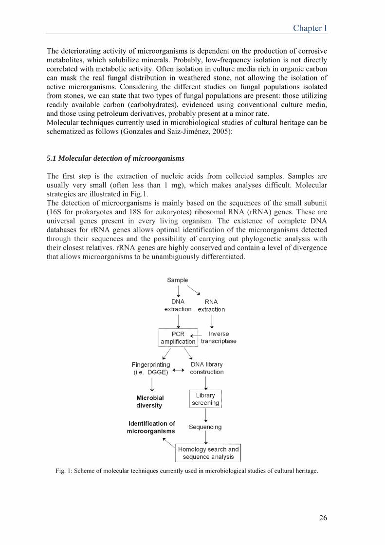
The deteriorating activity of microorganisms is dependent on the production of corrosive metabolites, which solubilize minerals. Probably, low-frequency isolation is not directly correlated with metabolic activity. Often isolation in culture media rich in organic carbon can mask the real fungal distribution in weathered stone, not allowing the isolation of active microrganisms. Considering the different studies on fungal populations isolated from stones, we can state that two types of fungal populations are present: those utilizing readily available carbon (carbohydrates), evidenced using conventional culture media, and those using petroleum derivatives, probably present at a minor rate. Molecular techniques currently used in microbiological studies of cultural heritage can be schematized as follows (Gonzales and Saiz-Jiménez, 2005): 5.1 Molecular detection of microorganisms The first step is the extraction of nucleic acids from collected samples. Samples are usually very small (often less than 1 mg), which makes analyses difficult. Molecular strategies are illustrated in Fig.1. The detection of microorganisms is mainly based on the sequences of the small subunit (16S for prokaryotes and 18S for eukaryotes) ribosomal RNA (rRNA) genes. These are universal genes present in every living organism. The existence of complete DNA databases for rRNA genes allows optimal identification of the microorganisms detected through their sequences and the possibility of carrying out phylogenetic analysis with their closest relatives. rRNA genes are highly conserved and contain a level of divergence that allows microorganisms to be unambiguously differentiated.
Fig. 1: Scheme of molecular techniques currently used in microbiological studies of cultural heritage.
Chapter I
27
5.2 Amplification of target genes As samples collected from artworks are highly limited in size, some kinds of analyses are impossible to carry out. The solution to this problem is the amplification of target genes. In the basic molecular protocol, specific target genes (rRNA genes) are PCR-amplified in order to obtain a large number of copies of these DNA fragments. The PCR technique requires two gene-specific primers and is carried out through 25–35 thermal cycles consisting of a denaturation step, annealing of the primers, and extension of the newly synthesized DNA fragment. Currently available primers are able to target every class of microorganism within a microbial community, such as Bacteria, Archaea, or Eukarya. The use of these primer pairs allows the detection and distinction of the three domains of life. Moreover, the sequences of microbial group-specific primers, e.g. for sulfate-reducing bacteria, nitrate-reducing bacteria, and methanotrophs, are available in the literature, and these primers can be applied in monitoring cultural assets. 5.3 Community fingerprinting PCR amplification products can be processed through: construction of a rRNA gene library or microbial community fingerprint. In the latter, the amplified rRNA genes from different microorganisms result in different electrophoretic patterns of migration. As a consequence, the microbial community of a sample can be characterized by its electrophoretic profile, which produces a so-called microbial community fingerprint. This allows the microbial diversity in each analyzed sample to be easily visualized and compared to the fingerprint from other samples or sites. Currently, there are several methods to obtain microbial community fingerprints from natural samples. For studies of artworks, the most used technique is denaturing gradient gel electrophoresis (DGGE). Other techniques, such as analysis of terminal restriction fragment length polymorphisms (t-RFLP), are less frequently used. DGGE analysis requires previous amplification of a specific portion of the 16S (or 18S for eukaryotes) rRNA genes. These DNA fragments are then separated in a chemical denaturing gradient (formed by urea and formamide) and then amplified with a set of primers. In order to stabilize the migration of the DNA fragments during DGGE, primers have a 40-bp GC-rich tail. 5.4 Identification of microorganisms Microorganisms constituting a given microbial community in a sample are usually identified through cloning and sequencing of the amplified PCR products obtained from the samples. The products are cloned into adequate vectors, screened, and then sequenced. A homology search of the sequence against DNA databases provides information on the taxonomic and phylogenetic lineage of the microorganism corresponding to that sequence. The most commonly used homology search algorithm is Blast, which is available online at the US National Center for Biotechnology Information [http://www.ncbi.nlm.nih.gov/BLAST/]. By this method, key information on the composition of microbial communities thriving on cultural assets are obtained. Moreover, in situ identification procedures, such as fluorescent in situ hybridization (FISH), can be used to detect specific microbial groups.
Chapter I
28
6. Future perspectives The present article highlights the importance of further research on this matter. As concerns the use of microorganisms and enzymes as biorestoration agents, studies aimed at the development of easily-applied, ready-to-use and safe biocleaning products, are essential, as is the cost-to-benefit evaluation. At that time biocleaning technologies will be confirmed to be not only environmentally sustainable, but also economically viable. As regards molecular-based techniques for the detection of microorganisms, their development has been very quick over the last few years, nevertheless they present also some limitations. For example, linking function to phylogeny is a crucial point in molecular microbial analyses, and novel approaches have been proposed; however, they have not yet been applied to microorganisms involved in the biodeterioration of cultural heritage. A common practice is to infer phylogeny from function, nevertheless this is appropriate only in the comparison of highly related microorganisms, it may be risky in microorganisms with increasing divergence. The development of biotechnologies for art conservation requires a strong and continuous dialogue between the art world and the world of science. Only through the combination of these fields more and more innovative methods to preserve our historical and cultural heritage can be developed, opening new horizons for art preservation. References Alfano, G., Lustrato, G., Belli, C., Zanardini, E., Cappitelli, F. Mello, E., Sorlini, C., Ranalli G. (2011) The bioremoval of nitrate and sulfate alterations on artistic stonework:The case-study of Matera Cathedral after six years from the treatment. International Biodeterioration & Biodegradation 65, 1004-1011. Antonioli, P., Zapparoli G., Abbruscato, P., Sorlini, C., Ranalli G. and Righetti, P.G. (2005) Art-loving bugs: the resurrection of Spinello Aretino from Pisa’s cemetery. Proteomics 15, 2453–2459. Arutchelvan, V., Kanakasabai, V., Nagarajan, S., Muralikrishnan, V. (2005) Isolation and identification of novel high strength phenol degrading bacterial strains from phenol-formaldehyde resin manufacturing industrial wastewater. J Hazard Mater 127(1):238-43. Atlas, R.M., Chowdhury, A.N. and Gauri, L.K. (1988) Microbial calcification of gypsum-rock and sulphated marble. Stud. Conserv. 33:149-153. Bellucci, R., Cremonesi, P., and Pignaroli, G. (1999) A preliminary note on the use of enzymes in conservation: the removal of aged acrylic resin coatings with lipase. Stud. Conserv. 44:278-281. Bellucci, R. and Cremonesi, P. (1994) The use of enzymes in the conservation and restoration of paintings. Kermes Arte Tec. Restauro 21:45-64. Beutel, S., Klein, K., Knobbe, G., Konigfeld, P., Petersen, K., Ulber, R., and Schelper, T. (2002) Controlled enzymatic removal of damaging casein layers on medieval wall paintings. Biotechnol. Bioeng. 80:13-21.
Chapter I
29
Bosch Roig, P., Regidor Ros, J.L., Montes Estellés, R. (2013) Biocleaning of nitrate alterations on wall paintings by Pseudomonas stutzeri. International Biodeterioration & Biodegradation 84, 266-274. Cappitelli, F., et al. (2005) Bioconservation of the marble base of the Pietà Rondanini by Michelangelo Buonarroti. Geophysical Research Abstracts 7, 06675. Cappitelli, F. and Sorlini, C. (2008) Microorganisms attack synthetic polymers in items representing our cultural heritage. Appl. Environ. Microbiol. 74:564-569.
Cappitelli, F., Zanardini, E., Ranalli, G., Mello, E., Daffonchio, D., & Sorlini, C. (2006). Improved Methodology for Bioremoval of Black Crusts on Historical Stone Artworks by Use of Sulfate-Reducing Bacteria, Applied and Environmental Microbiology, 72(5), 3733–3737.
Cappitelli, F., Toniolo, L., Sansonetti, A., Gulotta, D., Ranalli, G., Zanardini, E., Sorlini, C. (2007) Advantages of using microbial technology over traditional chemical technology in removal of black crusts from stone surfaces of historical monuments. Appl. Environ. Microbiol. 73, 5671–5. Castanier, S., Le Metayer-Levrel, G., Perthuisot, J.P. (1999) Ca-carbonates precipitation and limestone genesis-the microbiogeologist point of view. Sedimentary Geology 126, 9-23. Chen, J., Zhang, Y., Du, G.C., Hua, Z.Z., Zhu, Y. (2007) Biodegradation of polyvinyl alcohol by a mixed microbial culture. Enzyme Microb Technol.40(7):1686-91. Cremonesi, P. (2003) An approach to cleaning, p. 59-63. In P. Cremonesi (ed.), Materiali Tradizionali ed Innovativi Nella Pulitura dei Dipinti e delle Opere Policrome Mobili. Atti del Convegno, Piazzolla sul Brenta (Padova), 25-26 Ottobre 2002. Il Prato, Padova, Italy. Daffonchio, D., Borin, S., Zanardini, E., Abbruscato, P., Urzì, C. and Sorlini, C. (2000) Molecular tools applied to the study of deteriorated artworks, p. 21-38. In O. Ciferri, P. Tiano, and G. Mastromei (ed.), Of Microbes and Art: the Role of Microbial Communities on the Degradation and Protection of Cultural Heritage. Kluwer Academic/Plenum Publishers, New York, NY. De Muynck, W., Cox, K., De Belie, N., and Verstraete, W. (2008a). Bacterial carbonate precipitation as an alternative surface treatment for concrete. Constr. Build. Mater. 22, 875–885. De Muynck, W., Debrouwer, D., De Belie, N., and Verstraete,W. (2008b). Bacterial carbonate precipitation improves the durability of cementitious materials. Cem. Concr. Res. 38, 1005–1014. De Muynck, W., Leuridan, S., Van Loo, D., Verbeken, K., Cnudde, V., De Belie, N., et al.. (2011). Influence of pore structure on the effectiveness of a biogenic carbonate surface treatment for limestone conservation. Appl. Environ. Microbiol. 77, 6808–6820. Dhami, N.K., Reddy, M.S., and Mukherjee, A. (2014) Application of calcifying bacteria for remediation of stones and cultural heritages. Frontiers in Microbiology 5, art 304.
Chapter I
30
Ehrlich, H. L. (1998). Geomicrobiology: its significance for geology. Earth Sci. Rev. 45, 45–60. Fernandes, P. (2006) Applied microbiology and biotechnology in the conservation of stone cultural heritage materials. Appl Microbiol Biotechnol 73:291–296. Gauri, K.L., Parks, L., Jaynes, J. and Atlas, R. (1992) Removal of sulphated crust from marble using sulphate-reducing bacteria, p-160-165. In R. G. M. Webster (ed.), Stone Cleaning and the Nature, Soiling and Decay Mechanisms of Stone. Donhead, London, United Kingdom. Gauri, K.L. and Chowdhury A.N. (1988) Experimental studies on conversion of gypsum to calcite by microbes, p. 545-550. In J. Ciabach (ed.), Proceedings of 6th International Congress on Deterioration and Conservation of Stone, 12-14 Sept. Nicholas Copernicus University, Torùn, Poland.
Gioventù, E., Lorenzi, P. F., Villa, F., Sorlini, C., Rizzi, M., Cagnini, A., Griffo, A., Cappitelli, F. (2011). Comparing the bioremoval of black crusts on colored artistic lithotypes of the Cathedral of Florence with chemical and laser treatment. International Biodeterioration & Biodegradation, 65(6), 832–839.
Gioventù, E. et al. (2013) Bio-removal of black crust from marble surface: comparison with traditional methodologies and application on a sculpture from the Florence’s English Cemetery. Procedia Chemistry, 8:123-129. Gonzales, J.M. and Saiz-Jimenez, C. (2005) Application of molecular nucleic acid-based techniques for the study of microbial communities in monuments and artworks. Int. Microbiol. 8:189-194. Hamilton, W. A. (2003) Microbially influenced corrosion as a model system for the study of metal microbe interactions: a unifying electron transfer hypothesis. Biofoulin 19, 65-76. Hammes, F. and Verstraete, W. (2002) Key roles of pH and calcium metabolism in microbial carbonate precipitation. Re/Views in Environmental Science & Bio/Technology 1: 3–7. Jargeat, P., Rekangalt, D., Verner, M. C., Gay, G., Debaud, J. C., Marmeisse, R., et al.. (2003). Characterisation and expression analysis of a nitrate transporter and nitrite reductase genes, two members of a gene cluster for nitrate assimilation from the symbiotic basidiomycete Hebeloma cylindrosporum. Curr. Genet. 43, 199–205. Konkol, N., McNamara, C., Sembrat, J., Rabinowitz, M. and Mitchell, R. (2009) Enzymatic decolorization of bacterial pigments from culturally significant marble. J. Cult. Herit. 10: 362-366. Krumbein, W. E. (1979). Photolithotrophic and chemoorganotrophic activity of bacteria and algae as related to beach rock formation and degradation (Gulf of Aqaba, Sinai), Geomicrobiol. J. 1, 139–203.
Chapter I
31
Le Metayer-Levrel, G., Castanier, S., Orial, G., Loubiere J.F. and Perthuisot J.P. (1999) Application of bacterial carbonatogenesis to the protection and regeneration of limestones in buildings and historical patrimony. Sediment. Geol. 126:25-34. Lustrato, G., Alfano, G., Andreotti, A., Colombini, M.P, Ranalli, G. (2012) Fast biocleaning of mediaeval frescoes using viable bacterial cells. International Biodeterioration & Biodegradation 69, 51-61. May, E., Webster, A.M., Inkpen, R., Zamarreno, D., Kuever, J., Rudolph, C., Warsheid, T., Sorlini, C., Cappitelli, F., Zanardini, E., Ranalli, G., Krage, L., Vgenopoulos, A., Katsinis, D., Mello, E. and Malagodi, M. (2008) The Biobrush project for bioremediation of heritage stone, p. 76-93. In E. May and J. Mitchell (ed.), Heritage Microbiology and Science: Microbes, Monuments and Maritime Materials. Royal Society of Chemistry, Cambridge, United Kingdom. McNamara, C.J., Perry, T.D., Bearce, K.A., Hernandez-Duque, G., and Mitchell, R. (2006) Epilithic and endolithic bacterial communities in limestone from a Maya archaeological site. Microb. Ecol. 51:51-64. McNamara, C.J., Breuker, M., Helms, M., Perry, T.D., Mitchell, R. (2004) Biodeterioration of incralac used for the protection of bronze monuments. J Cult Herit.5:361-364. Orial, G., Castanier, S., Le Metayer, G., and Loubiere, J. F. (1992). “Biomineralization: a new process to protect calcareous stone; applied to historic monuments,” in Biodeterioration of Cultural Property 2: Proceedings of the 2nd International Conference on Biodeterioration of Cultural Property (Yokohama). Palla, F., Cammarata, M., Trapani, M., Ghersi, G., Salamone, M., & Sebastianelli, M. (2013). Novel proteases from marine organisms with potential interest in restoration procedure. Science and Technology for the Conservation of Cultural Heritage, 279. Pereira, C., Busani, T., Branco, L. C., Joosten, I., and Sandu, I. C. A. (2013). Nondestructive Characterization and Enzyme Cleaning of Painted Surfaces: Assessment from the Macro to Nano Level. Microscopy and Microanalysis, 19(06), 1632-1644. Polo, A., Cappitelli, F., Brusetti, L., Principi, P., Villa, F., Giacomucci, L., Ranalli, G., Sorlini, C. (2010) Feasibility of removing surface deposits on stone using biological and chemical remediation methods. Microb. Ecol. 60, 1–14. Ramachandran, S. K., Ramakrishnan, V., and Bang, S. S. (2001) Remediation of Concrete using Microorganisms. ACIMater. J. 98, 3–9. Ramakrishnan, S. K., Panchalan, R. K., and Bang, S. S. (2001) “Improvement of concrete durability by bacterial mineral precipitation,” in Proceedings of 11th International Conference on Fracture (Turin). Ramirez, J.L., Santana, M.A., Galindo-Castro, I. and Gonzalez A. (2005) The role of biotechnology in art preservation. TRENDS in Biotechnology 23(12):584-588.
Chapter I
32
Ranalli, G., Alfano G., Belli, C. Lustrato, G., Colombini, M.P., Bonaduce, I., Zanardini, E., Abbruscato, P., Cappitelli F. and Sorlini C. (2005) Biotechnology applied to cultural heritage: biorestoration of frescoes using viable bacterial cells and enzymes. J. Appl. Microbiol. 98, 73–83.
Ranalli, G., Chiavarini, M., Guidetti, V., Marsala, F., Matteini, M., Zanardini, E., Sorlini, C. (1997). The use of microorganisms for the removal of sulfates on artistic stoneworks. Int. Biodet. Biodeg. 40, 255-261.
Ranalli, G., Chiavarini, M., Guidetti, V., Marsala, F., Matteini, M., Zanardini, E., Sorlini, C. (1996). The use of microorganisms for the removal of nitrates and organic substances on artistic stoneworks. In: Riederer, J., (Ed.), Proceedings of the eighth International Congress on Deterioration and Conservation of Stone, Berlin, pp. 1415-1420. Rodriguez-Navarro, C., Rodriguez-Gallego, M., Ben Chekroun, K. and Gonzalez-Muñoz M.T. (2003) Conservation of ornamental stone by Myxococcus xanthus-Induced Carbonate Biomineralization. Appl. Environ. Microbiol 69(4):2182-2193. Saiz-Jimenez, C. (1997) Biodeterioration vs Biodegradation: the Role of Microorganisms in the Removal of Pollutants Deposited on Historic Buildings International Biodeterioration & Biodegradation 40, (2-4): 225-232. Saleem, M., Brim, H., Hussain, S., Arshad, M., Leigh, M.B. (2008) Perspectives on microbial cell surface display in bioremediation. Biotechnol Adv 26(2):151. Sorlini, C., Ranalli, G. and Zanardini, E. (2010) Stone Bioconservation. In Cultural Heritage Microbiology: Fundamental Studies in Conservation Science Ed. By R. Mitchell and C.J. McNamara ASM Press, Washington, DC. Stocks-Fischer, S., Galinat, J.K., Bang, S.S. (1999) Microbiological precipitation of CaCO3. Soil Biology and Biochemistry 31 1563-1571. Tiano, P. (1995) Stone Reinforcement by Calcite Crystal Precipitation Induced by Organic Matrix Macromolecules. Studies in Conservation, 40(3): 171-176. Tiano, P., Biagiotti, L., Mastromei, G. (1999) Bacterial bio-mediated calcite precipitation for monumental stones conservation: methods of evaluation. Journal of Microbiological Methods 36, 139–145. Tiano, P., Cantisani, E., Sutherland , I., Paget, J.M. (2006) Biomediated reinforcement of weathered calcareous stones. Journal of Cultural Heritage 7, 49-55. Tiano, P., Bracci, S. and Rescic S. (2003) Biomediated calcite precipitation for the reinforcement of monumental stones, p. 486-497. In R. J. Koestler, V. H. Koestler, A. E. Charola, and F. E. Nieto-Fernandez (ed.), Art, Biology and Conservation; Biodeterioration of Works of Art. Metropolitan Museum of Art, New York, NY.

Chapter I
33
Tiano, P., Addadi, L., and Weiner, S. (1992). “Stone reinforcement by induction of calcite crystals using organic matrix macromolecules: feasibility study,” in 7th International Congress on Deterioration and Conservation of Stone (Lisbon), 1317–1326. Troiano , F., Vicini, S., Gioventù, E., Lorenzi, P.F., Improta, C.M., Cappitelli, F. (2014) A methodology to select bacteria able to remove synthetic polymers. Polymer Degradation and Stability 107, 321-327. Urzì, C., Garcia Valles, M., Vendrell, M. and Pernice A. (1999) Biomineralization processes on rock and monument surfaces observed in field and laboratory conditions. Geomicrobiol. J. 16:39-54. Wright, D. T. (1999). The role of sulphate-reducing bacteria and cyanobacteria in dolomite formation in distal ephemeral lakes of the Coorong region, South Australia. Sediment. Geol. 126, 147–157. Zamarreno, D. V., Inkpen, R., and May, E. (2009). Carbonate crystals precipitated by freshwater bacteria and their use as a limestone consolidant. Appl. Environ. Microbiol. 75, 5981–5990.

34
Chapter II
35
Physiology, biochemistry and biotechnological applications of Desulfovibrio genus
1. Introduction
Desulfovibrio organisms belong to a heterogeneous group of sulfate-reducing, motile, anaerobic bacteria with more than 50 proposed species. This genus was described for the very first time in 1895, when the Dutch microbiologist Martinus Beijerinck, reported on a “Spirillum desulfuricans”, a bacterium producing hydrogen sulfide in sediments and anoxic water bottoms by sulfate reduction (Cypionka, 2000). Nevertheless, because of the difficulty in cultivation of anaerobes in pure culture, the knowledge of the sulfate-reducing bacteria remained stationary for about the following 50 years. However, much research has focused on Desulfovibrio as a model to explore the sulfate-reducing bacteria (SRB), due to the great importance that this bacteria have in the biogeochemical cycling of sulfur, carbon and nitrogen (Muyzer et al., 2008). Before the estabilshment of suitable growth techniques for anaerobes, the sulfate reducers were assumed to be a small, specialized group of bacteria able to oxidize incompletely few substrates (Ishimoto et al., 1954). This view was changed dramatically in the last 40 years thanks to the work of Widdel and Pfennig (Widdel F. and Pfennig N., 1977; Widdel F. and Pfennig N., 1981), who described new species of SRB able to completely oxidize a variety of organic substrates. Another important contribution to the understanding of SRB was given by a study conducted by Bak F. and Cypionka H. (Bak F. and Cypionka H., 1987) that described a novel type of energy metabolism involving the disproportionation of inorganic sulphur compounds. Initially discovered in a newly isolated sulphate-reducing bacterium, Desulfovibrio sulfodismutans, this process was confirmed in other sulphate-reducing bacteria. In recent years, new species were found to be able to grow on more diverse and less degradable substrates such as hydrocarbons or aromatic compounds (Rabus et al., 2006) and it was demonstrated the occurrence of sulphate reduction also in oxic environments (Muyzer et al, 2008). Another interesting aspect of the high versatility of their energy metabolism is the ability to precipitate toxic metal ions like copper (II), nickel (II) and cadmium (II) as metal sulfides in acidic aquatic environments (e.g., mine effluents). Additionally, SRB can deliver electrons directly to oxidized toxic metal ions, including uranium (VI), technetium (VII), and chromium (VI), converting them into less soluble, reduced forms. Thanks to all this evidences, today, the sulfate-reducing bacteria are considered of greats importance not only for their ecological roles, but also for their economic impact due to their involvement in biocorrosion of ferrous metals, occurring in anaerobic environments such as in offshore oil production or waterlogged clay soils (Hamilton, 1985) and in bioremediation processes of sulfate and heavy metals in different natural and anthropic environments (Quillet et al., 2012; Lenz et al., 2008). For all this reason the study of this class of bacteria continues to be object of great attention among the scientists, with particular focus on the impact of this bacteria on the environment and the economy. For example, recently, in order to put new light on their physiology and ecology, two strains beloging to the genus Desulfovibrio (D. desulfuricans G20 and D. vulgaris subsp. vulgaris str. Hildenborough) have already been genomically sequenced (Heidelberg et al., 2004) and one is currently in progress (D. magneticus). In this Review we provide an overview of recent advances in physiology, with a particular focus on response to oxygen exposure, biochemistry and the biotechnological applications of bacteria belonging to the genus Desulfovibrio.
Chapter II
36
2. Responses to oxygen exposure The view that sulphate reducers are strictly anaerobic, which can still be found in recent publications, started to change with the demonstration of the occurrence of sulphate reduction in oxic environments (Muyzer et al., 2008). As oxygen represents anyway an environmental stress for SRB, they have developed adaptation strategies to protect themselves against oxygen. These strategies can be distinguished into two groups: behavioral strategies and molecular strategies. The former includes aggregation, migration to anoxic zones and aerotaxis, while the latter allows SRB to remove oxygen in order to protect themselves from harmful effects (Dolla et al., 2006). As concerns Desulfovibrio, three kinds of behavioral reactions have been observed: aggregation, migration to anoxic zones, and band formation at the oxic-anoxic boundary. The latter includes both positive and negative responses to oxygen (Cypionka, 2000). 2.1 Behavioral strategies against oxygen
2.1.1 Aggregation
In conditions of oxygen exposure, SRB tend to form aggregates. Aggregates may be formed with bacteria only or with other particles like iron sulfides. Clumping appears to be a defensive emergency reaction, indeed aggregated bacteria better survive oxygen exposure than do free-living bacteria, because of the anoxicity of the inner volume of the aggregate, preserved by the respiratory activity (Cypionka, 2000). The proposed mechanism of aggregate formation is similar to the one observed in the purple sulfur bacterium Amoebobacter purpureus (Overmann et al., 1992). This bacterium forms dense cell aggregates after depletion of sulfide. After the addition of sulfide or various thiol compounds, disintegration of cell aggregates takes place. Also in the sulfate-reducing bacteria, the aggregates are formed under conditions in which oxygen causes sulfide depletion and sometimes sulfur droplets are visible in these cell aggregates. However, in SRB such disintegration as that observed with A. purpureus has not yet been observed (Cypionka, 2000). 2.1.2 Migration to anoxic zones
Another behavioral response of sulfate-reducing bacteria to oxygen is migration to anoxic zones. In microbial mats, migration of sulfur-oxidizing and sulfate-reducing bacteria, caused by the changing oxic conditions during day and night, is well known. A study of 1997 showed that MPN counts of sulfate-reducing bacteria in the upper 3 mm of a cyanobacterial mat of Solar Lake (Sinai, Egypt) were 20-fold lower during the day under oxic conditions than at night in the absence of oxygen (Krekeler et al., 1997). 2.1.3 Aerotaxis
Desulfovibrio’s behavior can be considered not simply aerophobic, but involve both positive and negative responses to oxygen. In fact Desulfovibrio species don’t swim into the anoxic zone as expected for obligate anaerobes. Instead they accumulated inside the outer edge of the oxic zone at concentrations of ≤20% air saturation (Eschemann et al., 1999). At high concentrations, oxygen functions as a repellent, in fact SRB move away from the oxygen bubble. This kind of response to pure oxygen is showed also by many
Chapter II
37
aerobic bacteria. The mechanisms of negative aerotaxis are not yet well understood neither in sulfate-reducing nor in aerobic bacteria. Nevertheless, different receptors are involved in positive and in negative aerotaxis in E. coli and D. vulgaris Hildenborough presents oxygen-sensing mechanisms similar to those of aerobic bacteria. A c-type heme-containing methyl-accepting protein, named DcrA, was found to function as the sensor of the oxygen concentration or redox potential of the environment, proved by the decrease in oxygen tolerance when the gene encoding for DcrA is deleted (Voordouw et al., 1998). Accumulation in bands depends on the availability of an electron donor, indeed the cells accumulate only when they are in the presence of an electron donor, which provides a chance to reduce oxygen and thus to remove it and to gain energy from respiration. Cells in the oxic zones reduce oxygen, so it is consumed, instead cells that have moved to the anoxic part would not change diffusion and oxygen concentrations in their environment. Inside the band oxygen is reduced and finally consumed completely (Cypionka, 2000). Another interesting observation is that part of the cells in the bands came from the outer anoxic area, these cells enter the oxic zone and are trapped there. This observation was confirmed by the motility pattern showed by D. desulfuricans CSN, when it passed from the anoxic to the oxic zone. Within the bands, the cells moved mainly in circles, whereas, in anoxic environments, they showed straight movements. The circular movement would not allow them to escape from the oxic zone. This behavior can be considered as a strategy developed by cells for the removal of oxygen from the environment to establish anoxic conditions for growth (Eschemann et al., 1999). The mechanism by which the cells are forced to move in circles remains still unknown. D. vulgaris doesn’t move in circles in the presence of oxygen, but moves to a higher swimming speed (Johnson et al., 1997). Dilling and Cypionka in 1991 described a SRB able to respire using oxygen and can even couple aerobic respiration to ATP formation: Desulfovibrio oxyclinae. It was isolated from the upper layer of a hypersaline microbial mat and showed oxygen-dependent growth, as indicated by higher growth yields after exposure to oxygen. Thus for the first time it was shown that some sulfate reducers can use molecular oxygen as a terminal electron acceptor (Muyzer et al., 2008). 2.2 Molecular strategies against oxygen 2.2.1 Oxygen Toxicity and Protective Mechanisms in Sulfate-Reducing Bacteria Several studies have shown that pure cultures of SRB survive hours or even days of exposure to air (Hardy et al., 1981; Sass et al., 1996), moreover from microbial mats that are saturated with air or pure oxygen during the day several sulfate reducers were isolated. This indicates that oxygen by itself is not toxic; instead, compounds that are formed by the reaction of reduced compounds with oxygen are toxic (Cypionka, 2000). Therefore, oxygen is generally more toxic to metabolizing than to resting cells. Compounds with thiol groups were found to drastically increase the toxicity, as hydrogen peroxide and superoxide radicals are formed by chemical reactions. In SRB catalase and superoxide dismutase have been detected. These enzymes catalyze detoxification of aggressive oxygen species, producing oxygen (Hewitt et al., 1975). Recently an alternative mechanism in the detoxification of superoxide without production of oxygen was described: a superoxide rubredoxin oxidoreductase, which produces H2O2 without concomitant production of O2 (Jenney et al., 1999). NADP dehydrogenase, alcohol dehydrogenase, lactate dehydrogenase and hydrogenase are substrate-oxidizing enzymes of SRB which were found to be oxygen sensitive.
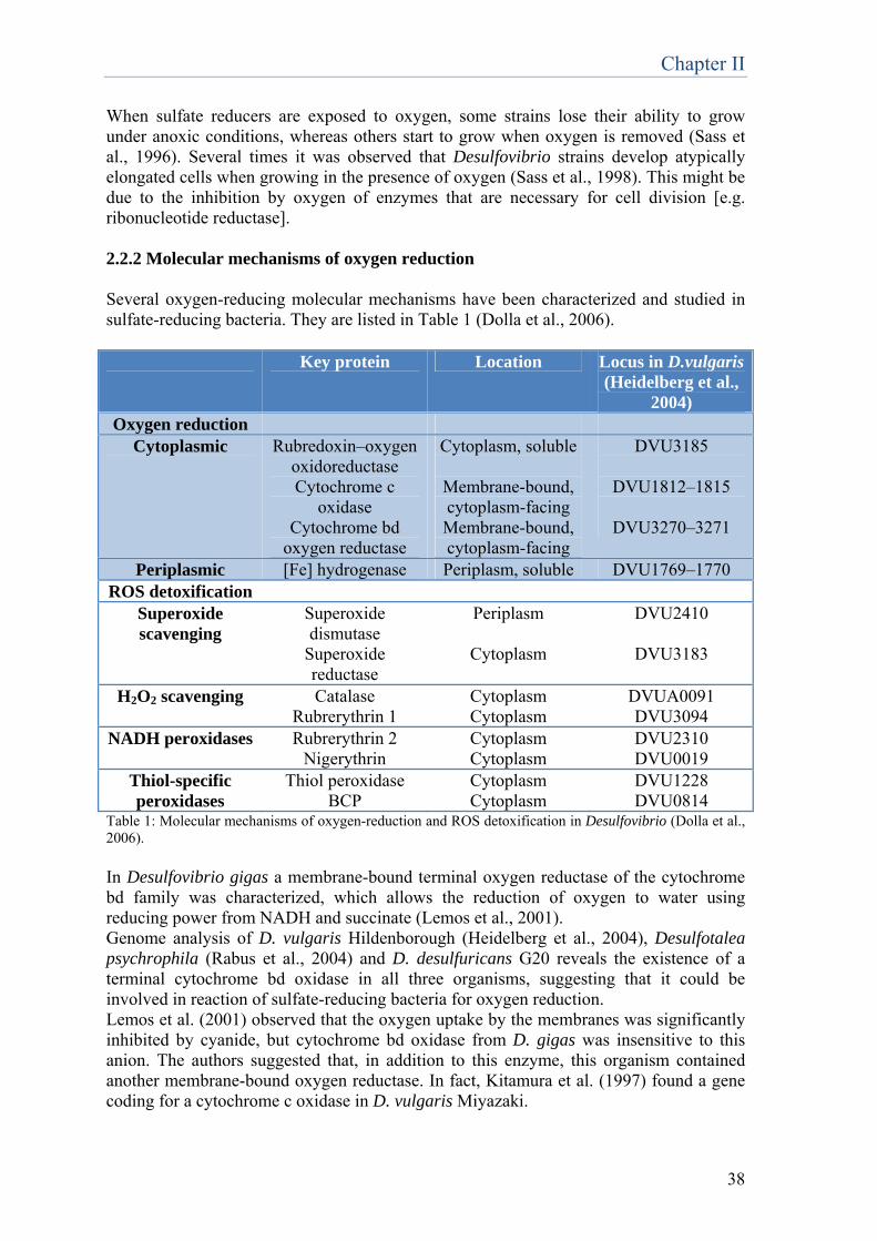
Chapter II
38
When sulfate reducers are exposed to oxygen, some strains lose their ability to grow under anoxic conditions, whereas others start to grow when oxygen is removed (Sass et al., 1996). Several times it was observed that Desulfovibrio strains develop atypically elongated cells when growing in the presence of oxygen (Sass et al., 1998). This might be due to the inhibition by oxygen of enzymes that are necessary for cell division [e.g. ribonucleotide reductase]. 2.2.2 Molecular mechanisms of oxygen reduction Several oxygen-reducing molecular mechanisms have been characterized and studied in sulfate-reducing bacteria. They are listed in Table 1 (Dolla et al., 2006). Key protein Location Locus in D.vulgaris
(Heidelberg et al., 2004)
Oxygen reduction Cytoplasmic Rubredoxin–oxygen
oxidoreductase Cytochrome c
oxidase Cytochrome bd
oxygen reductase
Cytoplasm, soluble
Membrane-bound, cytoplasm-facing Membrane-bound, cytoplasm-facing
DVU3185
DVU1812–1815
DVU3270–3271
Periplasmic [Fe] hydrogenase Periplasm, soluble DVU1769–1770 ROS detoxification
Superoxide scavenging
Superoxide dismutase
Superoxide reductase
Periplasm
Cytoplasm
DVU2410
DVU3183
H2O2 scavenging Catalase Rubrerythrin 1
Cytoplasm Cytoplasm
DVUA0091 DVU3094
NADH peroxidases Rubrerythrin 2 Nigerythrin
Cytoplasm Cytoplasm
DVU2310 DVU0019
Thiol-specific peroxidases
Thiol peroxidase BCP
Cytoplasm Cytoplasm
DVU1228 DVU0814
Table 1: Molecular mechanisms of oxygen-reduction and ROS detoxification in Desulfovibrio (Dolla et al., 2006). In Desulfovibrio gigas a membrane-bound terminal oxygen reductase of the cytochrome bd family was characterized, which allows the reduction of oxygen to water using reducing power from NADH and succinate (Lemos et al., 2001). Genome analysis of D. vulgaris Hildenborough (Heidelberg et al., 2004), Desulfotalea psychrophila (Rabus et al., 2004) and D. desulfuricans G20 reveals the existence of a terminal cytochrome bd oxidase in all three organisms, suggesting that it could be involved in reaction of sulfate-reducing bacteria for oxygen reduction. Lemos et al. (2001) observed that the oxygen uptake by the membranes was significantly inhibited by cyanide, but cytochrome bd oxidase from D. gigas was insensitive to this anion. The authors suggested that, in addition to this enzyme, this organism contained another membrane-bound oxygen reductase. In fact, Kitamura et al. (1997) found a gene coding for a cytochrome c oxidase in D. vulgaris Miyazaki.
Chapter II
39
SRB are able to reduce oxygen using not only electron acceptors from the medium, but also storage compounds. An example is Desulfovibrio salexigens, which accumulate massive amounts of polyglucose during its anaerobic growth. It has been shown that the endogenous oxygen reduction rate of D. salexigens Mast1 was proportional to the polyglucose content and was entirely due to a NADH oxidase activity (van Niel and Gottschal, 1998). It was proposed a NADH-linked electron transfer chain enabling the transfer of reducing power, derived from polyglucose, to oxygen. This electron transfer chain involves three proteins: a NADH–rubredoxinoxidoreductase (Rbo), a rubredoxin and a rubredoxin oxygen oxidoreductase (Roo). Rubredoxins are small cytoplasmic electron-carrying proteins that contain a single iron atom coordinated to four cysteinyl sulfur atoms. This protein acts as an electron shuttle between the NADH-rubredoxin oxygen oxidoreductase and the rubredoxin-oxygen oxidoreductase (Chen at al., 1993). The NADH-rubredoxin oxygen oxidoreductase is a dimeric protein able to reduce rubredoxin from NADH. This enzyme is able to reduce incompletely oxygen to hydrogen peroxide. The rubredoxin gives electron to its other redox partner, the rubredoxin-oxygen oxidoreductase (Roo). Roo contains a di-iron center, where the reduction of oxygen to water has been proposed to occur and a flavodoxin-like domain, which has been proposed to be the intermediate bridge in the electron transfer between the rubredoxin and the Roo di-iron center (Frazao et al., 2000). A cluster of genes encoding desulfoferrodoxin, rubredoxin and rubredoxin-oxygen oxidoreductase is present in the D. vulgaris Hildenborough genome (Heidelberg et al., 2004) and in D. desulfuricans G20 genome (www.jgi.doe.gov). Therefore, this cytoplasmic oxygen reduction chain appears to be a conserved mechanism among Desulfovibrio species and might be an efficient system to eliminate oxygen, and thus protect the bacteria in oxic conditions. More recently, Fournier et al. (2004) have reported the involvement of [Fe] hydrogenase and cytochrome c in the response of D. vulgaris Hildenborough to oxidative stress. When the cells were exposed to oxygen, the hydrogenase activity in the periplasm was higher than the one in cells kept under anaerobic conditions. In this bacterium, three hydrogenases have been characterized as periplasmic-facing enzymes, one periplasmic soluble [Fe] hydrogenase and two membrane-bound [NiFe] hydrogenases (Heidelberg et al., 2004; Voordouw and Brenner, 1985). However, only [Fe] hydrogenase activity was reported to be affected by the presence of oxygen. It has been shown that the increase in [Fe] hydrogenase activity is directly correlated to the increase in the amount of protein and the corresponding RNA (Fournier et al., 2004; Fournier et al., 2006). The hydAB genes, encoding [Fe] hydrogenase, are up-regulated in the presence of oxygen. In the same way, an increase in the cytochrome c content of the periplasmic space was also observed following the oxygen exposure (Fournier et al., 2004). [Fe] hydrogenase and cytochromes are thus involved in the response to an oxidative stress induced by either the presence of oxygen or a strong redox potential. 2.2.3 Reactive oxygen species (ROS) detoxification When sulfate-reducing bacteria are in contact with oxygen, highly toxic reactive oxygen species (ROS) are generated, as for aerobic organisms. This factor needs to be taken into consideration when survival of SRB in oxic conditions is studied. These ROS are: superoxide (O2•-), hydrogen peroxide (H2O2) and hydroxyl radical (HO•). ROS are responsible for the O2 toxicity due to their high reactivity which generates different types of damages inside cells (Dolla et al., 2006). They induce oxidations of proteins and
Chapter II
40
aminoacids, leading to a cleavage of the polypeptide chain and to the formation of cross-linked protein aggregates. An important effect of superoxide is its ability to impair Fe-S clusters, causing its disintegration and a loss of enzyme activity. As a result of iron-sulfur cluster damage, iron is released into the cytosol, where it catalyzes the oxidation of DNA in conjunction with hydrogen peroxide. Hydrogen peroxide oxidizes enzyme thiols and thus is likely to inactivate enzymes. It also reacts with free Fe2+ to product hydroxyl radical, a powerful oxidant that reacts with most of the biomolecules. Because iron can localize along the phosphodiester backbone of nucleic acids, DNA is a target of hydroxyl radical. A wide variety of DNA lesions are formed like base and sugar damages, tandem lesions and DNA-protein cross-links. In SRB these ROS can be either generated by enzymatic oxygen reduction, involving the reactions described above, or by auto-oxidation of some reduced proteins as cytochromes or flavoproteins (Cypionka, 2000). In addition, the chemical auto-oxidation of the hydrogen sulfide, which is normally produced by these bacteria, could also result in the formation of ROS, which therefore increases the SRB sensitivity to oxygen (Cypionka et al., 1985). However, in addition to the effects induced by ROS, oxidative stress is also a consequence of the direct inactivation by molecular oxygen of several key enzymes, like hydrogenase (Cammacket al., 1994) or lactate dehydrogenase (Stams and Hansen, 1982) and of the modification of physicochemical parameters of the environment like redox potential and pH. Genomic studies on D.vulgaris H revealed that this microorganism possesses a baseline protection given by the constitutive expression of sodB and kat, that is of genes having a known function in ROS protection also in other organisms. Instead, the expression of ROS protection genes in the PerR regulon was inducible, depending on the levels of O2
(Zhou et al., 2010). At weak oxidative stress conditions (0.1 % O2 and 1 mM of H2O2) the expression of PerR is higher, by contrast at high levels of oxidative stress conditions (between 21 and 100 % O2), its expression is lower. In this last condition, the upregulation of genes encoding for protein repair and degradation has been observed. This observation suggests the presence of a mechanisms of shift from ROS elimination in the response strategy, in order to prevent against further oxidative damage in case of severe conditions (Zhou et al., 2011). Another interesting mechanism implicated in the protection against ROS, investigated in recent studies (Figueiredo et al, 2012), is the role of bacterioferritin in the defence against oxygen. Intracellular free iron is under aerobic conditions a catalyst for the formation of harmful reactive oxygen species. For this reason, the relation between intracellular iron storage and oxidative stress response in the sulfate reducing bacterium D. vulgaris Hildenborough was analysed (Figueiredo et al, 2012). The viability of both wild-type and bacterioferritin mutant, in the presence of high concentration of oxygen, was compared. The mutant strain exhibited lower viability and a higher content of intracellular ROS. Furthermore, the bacterioferritin gene is under the control of the PerR regulon. All these data confirmed the importance of iron storage by bacterioferritin to contribute to the oxygen tolerance of D.vulgaris. 2.2.3.1 Superoxide anion detoxification In Desulfovibrio species, two superoxide scavengers, superoxide dismutase (SOD) and superoxide reductase (SOR), have been characterized (Table 1). Superoxide dismutase, which is common also in aerobic microrganisms, eliminates superoxide ions by dismutation to hydrogen peroxide and molecular oxygen (Dolla et al., 2006). The
Chapter II
41
presence of a superoxide dismutase in anaerobic bacteria has been demonstrated for the first time by Hatchikian and Henry (1977) in a Desulfovibrio strain. More recently, an iron-containing superoxide dismutase has been purified and characterized from different species of Desulfovibrio. Moreover, the genes encoding a superoxide dismutase have been cloned from D. vulgaris Hildenborough (Lumppio et al., 2001) and D. vulgaris Miyazaki (Nakanishi et al., 2003). Superoxide dismutase has been found to be constitutively expressed during anaerobic growth in D. gigas (Dos Santos et al., 2000). Moreover, no significant differences in superoxide dismutase activity have been observed between cultures of D. gigas exposed to various oxygen concentrations (up to 120 M) and the cells maintained under anoxic conditions. In addition, a study of Fareleira et al. (2003) showed that exposure time to oxygen does not have significant effect on the activity level. Moreover, the amount of SOD does not change when D. vulgaris Hildenborough is exposed to oxygen (Fournier et al., 2006). These data suggest that superoxide dismutase genes are constitutively expressed in Desulfovibrio species. Fournier et al. (2003) have shown that the deletion of the sod gene in D. vulgaris Hildenborough induces an increase in the sensitivity of the strain to externally produced superoxide, which is not induced by exposure to air only. If not scavenged, superoxide induces damages like oxidation and destabilization of iron-sulfur clusters of some enzymes which could lead to their inactivation (Flint et al., 1993). In particular, Desulfovibrio periplasm contains several iron-sulfur-containing enzymes, like hydrogenases or formate dehydrogenases, involved in the energy metabolism, sensitive to superoxide (Heidelberg et al., 2004). When cells are exposed to oxygen, also for a short time, the periplasmic [Fe] hydrogenase activity is higher in the wild-type strain of D. vulgaris Hildenborough than in the sod mutant (Fournier et al., 2004). This indicates the role of periplasmic super-oxide dismutase in the protection of sensitive enzymes against superoxide-induced damages. Before the end of 1990s, the only known mechanism for scavenging superoxide ions was dismutation catalyzed by SOD. However, Lombard et al. (2000) discovered a superoxide reductase activity (SOR), carried out by desulfoferrodoxin from Desulfoarculus baarsii. This protein has been also purified and characterized from Desulfovibrio desulfuricans ATCC27774 and D. vulgaris Hildenborough (Moura et al., 1990; Tavares et al., 1994). Another enzyme, named neelaredoxin, which shows superoxide reductase activity, has been characterized from D. gigas (Silva et al., 2001). These two iron proteins, having SOR activity, are also found in other anaerobic prokaryotes such as Archaeoglobus fulgidus, a sulfate-reducing archaebacterium and Treponema pallidum (Abreu et al., 2000; Jovanovic et al., 2000; Hazlett et al., 2002). It should be noted that the Archaeglobus fulgidus genome contains both genes coding for desulfoferrodoxin and neelaredoxin (Klenk et al., 1998). Rubredoxins, small iron-containing proteins, have been shown to be efficient electron donors for the superoxide reductase activity enzymes (desulfoferrodoxin and neelaredoxin) in D. vulgaris Hildenborough (Coulter and Kurtz, 2001), Archaeoglobus fulgidus (Auchere et al., 2004) and Treponema pallidum (Rodrigues et al., 2005). Moreover, in D. vulgaris Hildenborough and D. desulfuricans G20, the desulfoferrodoxin (SOR), rubredoxin and rubredoxin-oxygen oxidoreductase are in the same genes cluster. This genes organization suggests that SOR and ROO may collaborate for the reduction and the detoxification of oxygen entering the cytoplasm through the use of rubredoxin as a common intermediary electron donor. Involvement of SORs as an oxygen defense protein has been shown by Voordouw and Voordouw (1998), who deleted the gene encoding desulfoferrodoxin in D. vulgaris Hildenborough (sor mutant). This deletion does not affect the growth under anaerobic conditions but increases the sensitivity of the strain to oxygen exposure. In the same way, the sor mutant is more sensitive to exposure to superoxide than the sod mutant which is more sensitive than the

Chapter II
42
wild-type strain (Lumppio et al., 2001; Fournier et al., 2003). Comparison of the sensitivity of sor and sod mutants to various oxidative stresses indicates that under aerated conditions, SOR is the key oxygen defense factor. This observation can be related with the fact that, in the cytoplasm, ROS can induce damages on both proteins and DNA, which are much more deleterious than those occurring in the periplasm. SOD is involved in the removal of periplasmic superoxide to protect sensitive enzymes. SOD (superoxide dismutase), is localized in the periplasm. For this reason, its involvement in the protection of periplasmic ironsulfur proteins, such as [Fe] hydrogenases, was proposed. [Fe] hydrogenases have an important role in the production of energy according to the hydrogen cycling model proposed by Odom and Peck, in fact they convert H2 into protons and electrons. According to this hypothesis, SOD would protect [Fe] hydrogenase against superoxide-induced damage. In order to confirm this hypothesis, the periplasmic [Fe] hydrogenase activity was followed in both wild-type and sod deletant strains. When cells were kept in anaerobic conditions, the hydrogenase activity was observed in both mutant and wild-type strains. On the other hand, when cells were exposed for a short time to pure oxygen, the activity was higher in the wild-type than in the strain lacking the SOD, suggesting that SOD protects the hydrogenase from damage induced by ROS. SOR and SOD can be regarded as complementary components of an efficient defense system able to scavenge superoxide in the two compartments of the cell. 2.2.3.2 Hydrogen peroxide detoxification As for superoxide detoxification, two enzymatic reactions have been described in Desulfovibrio for hydrogen peroxide elimination (Table 1). The first reaction is catalyzed by the well-known enzyme catalase (Kat), widely distributed also in aerobic organisms (Dolla et al, 2006). Catalase activity does not seem to be present in all known Desulfovibrio species. While D. vulgaris, D. gigas and Desulfomicrobium norvegicum are catalase positive, D. salexigens and D. desulfuricans (strain Essex 6) are catalase negative (Dos Santos et al., 2000). In D. gigas, Fareleira et al. (2003) have shown that catalase activity increases as cells are exposed to increasing oxygen concentration. The second reaction, responsible of hydrogen peroxide elimination, is catalyzed by NAD(P)H-dependent peroxidases. Rubrerythrin (Rbr) and nigerythrin, which exhibit this NADH peroxidase activity in vitro, have been isolated from D. vulgaris Hildenborough (Coulter et al., 1999). The deletion of the gene encoding rubrerythrin in D. vulgaris Hildenborough does not alter the sensitivity of the strain to any oxidative stress (Fournier et al., 2003). However, the lack of a strong oxygen stress phenotype for the mutant strain can be explained by the presence in the genome of genes encoding a nigerythrin and a second homolog to rubrerythrin (Table 1), which may compensate for the deletion (Heidelberg et al., 2004). Another detoxification system is represented by two proteins having thiol-peroxidase activity. Their expression increases when D. vulgaris Hildenborough is exposed to oxygen for one hour (Fournier et al., 2006). D.vulgaris H. possesses another set of genes encoding enzymes for peroxide reduction, such as AphC, Rbr2, Rbr and Rdl, controlled by the transcriptional regulator peroxide-responsive repressor (PerR) (Zhou et al., 2011).
Chapter II
43
3. Energy metabolism of SRB
3.1 Pathways of sulfate reduction
There are two biological pathways of sulfate reduction. In the assimilatory pathway, which is widespread in the three domains of life, sulfate is reduced to sulfide in small amounts and this is transformed into cysteine, from which other biological sulfur-containing molecules are derived (Grein et al., 2013). In the dissimilatory pathway, which is restricted to five bacterial and two archaeal lineages, sulfate is the terminal electron acceptor of the respiratory pathway producing large quantities of sulfide (Barton et al., 2009). A key difference between the assimilatory and dissimilatory sulfite reductases is that the former reduce sulfite directly to sulfide, whereas the latter forms, in vitro, a mixture of products including also trithionate and thiosulfate, in relative proportions that depend on reaction conditions (Akagi, 1995). The two pathways (Fig. 1) start with activation of sulfate by reaction with ATP to form adenosine-5′-phosphosulfate (APS), a step catalyzed by the trimeric sulfate adenylyl transferase (Sat), also known as ATP sulfurylase (Taguchi et al., 2004). The formation of APS is endergonic and is driven by hydrolysis of the pyrophosphate formed by a pyrophosphatase (soluble or membrane-bound). So, the activation of sulfate to APS is considered to consume two ATP equivalents. In the prokaryotic assimilatory pathway APS is converted to 3′-phosphoadenosine-5′-phosphosulfate (PAPS) by the adenylyl sulfate kinase (CysC), PAPS is reduced to sulfite by a thioredoxin-dependent PAPS reductase (CysH), and finally sulfite is reduced to sulfide by an assimilatory sulfite reductase that is either multimeric and NADPH-dependent (CysIJ) or a monomeric ferredoxin-dependent enzyme (Crane et al., 1996). In the dissimilatory pathway APS is reduced to sulfite by the APS reductase (AprBA), a heterodimeric iron-sulfur flavoenzyme. Sulfite is reduced by the dissimilatory sulfite reductase DsrAB, a siroheme containing protein, with the involvement of the small protein DsrC (Moura et al., 1988). DsrC was initially believed to be a third subunit of DsrAB, on the contrary the majority of DsrC is not associated with DsrAB and is thus free to interact with other proteins (Venceslau et al., 2013). Another small protein DsrD, which is often encoded downstream of dsrAB, might also be involved in sulfite reduction, possibly in a regulatory role, but its exact function is still unknown. Interestingly, the dsrD gene is strongly downregulated in the presence of high sulfide concentrations (Caffrey et al., 2010).
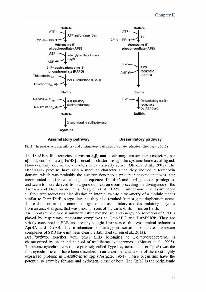
Chapter II
44
Fig.1: The prokaryotic assimilatory and dissimilatory pathways of sulfate reduction (Grein et al., 2013)
The DsrAB sulfite reductase forms an α2β2 unit, containing two siroheme cofactors, per αβ unit, coupled to a [4Fe\4S] iron-sulfur cluster through the cysteine heme axial ligand. However, only one of the cofactors is catalytically active (Oliveira et al., 2008). The DsrA/DsrB proteins have also a modular character since they include a ferredoxin domain, which was probably the electron donor to a precursor enzyme that was later incorporated into the reductase gene sequence. The dsrA and dsrB genes are paralogous, and seem to have derived from a gene duplication event preceding the divergence of the Archaea and Bacteria domains (Wagner et al., 1998). Furthermore, the assimilatory sulfite/nitrite reductases also display an internal two-fold symmetry of a module that is similar to DsrA/DsrB, suggesting that they also resulted from a gene duplication event. These data confirm the common origin of the assimilatory and dissimilatory enzymes from an ancestral gene that was present in one of the earliest life forms on Earth. An important role in dissimilatory sulfur metabolism and energy conservation of SRB is played by respiratory membrane complexes as QmoABC and DsrMKJOP. They are strictly conserved in SRB and are physiological partners of the two terminal reductases AprBA and DsrAB. The mechanisms of energy conservation of these membrane complexes of SRB have not been clearly established (Grein et al., 2013). Desulfovibrio, together with other SRB belonging to Deltaproteobacteria, is characterized by an abundant pool of multiheme cytochromes c (Matias et al., 2005). Tetraheme cytochrome c3 (more precisely called Type I cytochrome c3 or TpIc3) was the first cytochrome c to have been described in an anaerobe, and is one of the most highly expressed proteins in Desulfovibrio spp (Postgate, 1954). These organisms have the potential to grow by formate and hydrogen, either or both. The TpIc3 is the periplasmic
Chapter II
45
electron acceptor of hydrogenases and formate dehydrogenases. The presence of the cycA gene coding for the TpIc3 in the genomes of SRB is correlated with the presence of periplasmic hydrogenases and formate dehydrogenases that lack a membrane subunit for direct quinone reduction (Pereira et al., 2011), in contrast to most bacteria. In several cases these enzymes have a dedicated cytochrome c3 subunit. These soluble uptake hydrogenases and formate dehydrogenases are usually present in several copies in the deltaproteobacterial SRB (Pereira et al., 2011). The TpIc3 performs a proton-coupled two-electron transfer. As it receives electrons from H2 or formate oxidation, it can deliver this reducing power to several membrane complexes (Matias et al., 2005). Using soluble dehydrogenases and TpIc3, rather than direct quinone reduction, the deltaproteobacterial SRB acquire a higher metabolic flexibility, as electrons can be shuttled through several alternative pathways. Thus, in contrast to the other groups of SRB, the Deltaproteobacteria SRB may derive additional electrons from intracellular cycling of redox intermediates such as hydrogen and formate (Odom and Peck, 1981). A high content of multiheme cytochromes c seems to be characteristic of soil and sediment Proteobacteria, such as Geobacter, Shewanella, Anaeromyxobacter and Desulfovibrio, which are subjected to variable redox conditions. Thomas et al. (2008) have argued that having a high number of multiheme cytochromes c is a property of metabolically versatile anaerobes that have to adapt to environments with fluctuating redox conditions. The versatile nature of SRB is reflected in the fact that they can even grow in the absence of sulfate, in syntrophy with other organisms that consume H2 and/or formate, such as methanogens. In fact, SRB were found to be still abundant in methanogenic zones of marine sediments. A revised model for dissimilatory sulfate reduction was proposed in 2011 by Bradley and colleagues, on the basis of data obtained from the sedimentary sulfur isotope record. According to them, sulfite is reduced in the DsrAB-C complex in sequential two- electron transfers (Fig 2). Sulfite is reduced first to S2+, then to S0. The zero-valent sulfur may be directly reduced to sulfide as a side reaction or bind to DsrC, forming a disulfide bond between cysteines, releasing sulfide as product. DsrC acts as a shuttle carrying oxidizing capacity to the membrane. This scheme suggests a mechanism by which oxidizing capacity can be transported from cytoplasmic DsrAB to the membrane. DsrAB transfers only 4 electron to sulfite. The fifth and sixth electron involved in sulfite reduction are given by the two conserved cysteine residues.
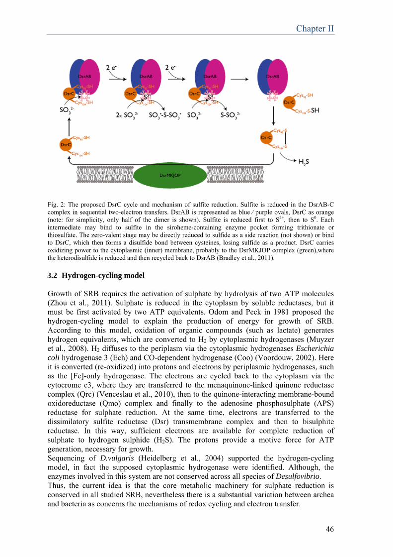
Chapter II
46
Fig. 2: The proposed DsrC cycle and mechanism of sulfite reduction. Sulfite is reduced in the DsrAB-C complex in sequential two-electron transfers. DsrAB is represented as blue ⁄ purple ovals, DsrC as orange (note: for simplicity, only half of the dimer is shown). Sulfite is reduced first to S2+, then to S0. Each intermediate may bind to sulfite in the siroheme-containing enzyme pocket forming trithionate or thiosulfate. The zero-valent stage may be directly reduced to sulfide as a side reaction (not shown) or bind to DsrC, which then forms a disulfide bond between cysteines, losing sulfide as a product. DsrC carries oxidizing power to the cytoplasmic (inner) membrane, probably to the DsrMKJOP complex (green),where the heterodisulfide is reduced and then recycled back to DsrAB (Bradley et al., 2011). 3.2 Hydrogen-cycling model
Growth of SRB requires the activation of sulphate by hydrolysis of two ATP molecules (Zhou et al., 2011). Sulphate is reduced in the cytoplasm by soluble reductases, but it must be first activated by two ATP equivalents. Odom and Peck in 1981 proposed the hydrogen-cycling model to explain the production of energy for growth of SRB. According to this model, oxidation of organic compounds (such as lactate) generates hydrogen equivalents, which are converted to H2 by cytoplasmic hydrogenases (Muyzer et al., 2008). H2 diffuses to the periplasm via the cytoplasmic hydrogenases Escherichia coli hydrogenase 3 (Ech) and CO-dependent hydrogenase (Coo) (Voordouw, 2002). Here it is converted (re-oxidized) into protons and electrons by periplasmic hydrogenases, such as the [Fe]-only hydrogenase. The electrons are cycled back to the cytoplasm via the cytocrome c3, where they are transferred to the menaquinone-linked quinone reductase complex (Qrc) (Venceslau et al., 2010), then to the quinone-interacting membrane-bound oxidoreductase (Qmo) complex and finally to the adenosine phosphosulphate (APS) reductase for sulphate reduction. At the same time, electrons are transferred to the dissimilatory sulfite reductase (Dsr) transmembrane complex and then to bisulphite reductase. In this way, sufficient electrons are available for complete reduction of sulphate to hydrogen sulphide (H2S). The protons provide a motive force for ATP generation, necessary for growth. Sequencing of D.vulgaris (Heidelberg et al., 2004) supported the hydrogen-cycling model, in fact the supposed cytoplasmic hydrogenase were identified. Although, the enzymes involved in this system are not conserved across all species of Desulfovibrio. Thus, the current idea is that the core metabolic machinery for sulphate reduction is conserved in all studied SRB, nevertheless there is a substantial variation between archea and bacteria as concerns the mechanisms of redox cycling and electron transfer.
Chapter II
47
3.3 Thermodynamic considerations
Oxygen allows much more ATP conservation than sulphate: first, because of the positive redox potential, much higher free energy is available (Cypionka, 1995). For example, oxidation of H2 with sulfate and oxygen at pH 7 yields free energies of -39 and -237 kJ mol-1, respectively. Second, as a gas, oxygen can be reduced without a requirement for energy-dependent transport (Cypionka, 1994). Third, oxygen can be reduced without ATP-consuming activation as is required for sulfate. In a corresponding manner, sulfate reducers growing with nitrate have higher growth yields than with sulfate (Cypionka, 1986). In a study of Cypionka (2000) on the electron transport-driven proton translocation, various electron acceptors were compared for their capacity to induce proton translocation when added in small pulses to cells incubated in the presence of electron donors. By this method, he observed that oxygen gave the fastest and highest proton release [(7.8±1.8 H+)/O2)]. 3.4 Aerobic growth and syntrophy
In spite of their ability of respiration with oxygen, aerobic growth of sulfate-reducing bacteria in pure culture is poor or absent. The situation changes completely if other aerobic organisms are present, creating a syntrophic growth of aerobic and sulfate-reducing bacteria (Zhou et al., 2011). Syntrophy literally means ‘feeding together’ and refers to any interaction in which two species cooperate, the one completing a metabolic reaction from which the other species can’t gain energy. A model of syntrophic interaction was developed involving D. vulgaris H. and Methanococcus maripaludis S2, a hydrogenotrophic methanogen (Stolyar et al., 2007). In media without an electron acceptor, D. vulgaris H. cooperated by transferring H2, a waste product of lactate fermentation, to M. maripaludis and in return benefited from a chemical environment (a low H2 concentration) in which lactate fermentation was thermodynamically favourable. In this way, an end product that inhibits energy generation from fermentation in one species is consumed by a second species, allowing both species to gain energy. These syntrophic interactions often occur in anaerobic environments lacking appropriate electron acceptors, such as lake sediments and anaerobic digestors and involve methanogenic archaea that consume intermediates produced by firmicutes such as Desulfotomaculum, or by deltaproteobacteria such as Desulfovibrio (Stams et al., 2009). The two strains associating in the syntrophic interaction originate from very different environments, thus they adapt to syntrophy or to living with each other gradually. Two factors may limit the growth of the microorganisms in this nascent syntrophic association. First, the two partners suffer from low levels of energy being available for growth when they are relying on syntrophy for survival, in comparison to the growth conditions in pure culture (Stams et al., 2009). Second, the ability of these organisms to access their energy source depends on the distribution and continued cooperation of their partner species. This situation could lead to unstable growth, especially if one species is inhibited (Shou et al., 2007). A recent experiment with 24 independently evolving co-cultures confirmed these predictions (Hillesland and Stahl, 2010). Initially, growth was unstable. However, by 300 generations, growth of the co-cultures stabilized, and it was 80% faster than the growth of the ancestors. Both species acquired mutations contributing to the improved productivity. These results demonstrate that improved stability and productivity are typical adaptations to the initial stress of living in a community.
Chapter II
48
5. Biotechnological applications
SRB can be employed for a wide range of biotechnological application: removal of heavy metals from groundwater and waste water, removal of sulphur compounds from waste water and off gases, acid mine drainage decontamination, hydrogen production, biorestoration of monuments and artworks. SRB, together with sulphur-oxidizing microorganisms, can be successfully exploited in the sustainable clean-up of industrial waste streams. Sulphate reduction can be applied beneficially to biotechnology, such as the removal of heavy metals from groundwater and waste water (Muyzer et al., 2008). This application takes advantage of differences in the chemical properties of metal sulphates and sulphides. Metal sulphates (cadmium, cobalt, copper, iron, nickel and zinc) are highly soluble, but the corresponding metal sulphides have low solubility. Thus, by sulphate reduction, metals can be precipitated, recovered and reused. In 2012, Quillet and colleagues published a study on the diversity and activity of SRB in a 3.5-m sediment core taken from a heavy metal-contaminated site in the UK. The abundance of SRB was quantified by qPCR of the dissimilatory sulfite reductase gene β- subunit (dsrB). Gene libraries for dsrA (dissimilatory sulfite reductase α-subunit) were constructed both from the surface sediment (top 20 cm), which is strongly contaminated by heavy metals and from a deeper zone (250 cm), less contaminated and sulfate-depleted. They found high transcriptional activity of dsrA belonging to Desulfovibrio species in the surface sediment. These results may suggest that members of the Desulfovibrionaceae are more active than other SRB groups in heavy metal-contaminated surface sediments. Another study (Lovley et al., 1994) has shown that washed cell suspensions of Desulfovibrio vulgaris are able to rapidly reduce Cr (VI) to Cr (III) with H2 as the electron donor. The c3 cytochrome from this organism worked as a Cr (VI) reductase. D. vulgaris may have advantages over previously described reducing bacteria for the bioremediation of Cr (VI)-contaminated waters, because differently from them its Cr (VI)-reducing capability is not inhibited by heavy metals and sulfate present in the waste. Another biotechnological application for SRB is the removal and reuse of sulphur compounds from waste water and off gases (Muyzer et al., 2008). Under oxygen-limitation, sulphide-oxidizing bacteria mainly produce elemental sulphur instead of sulphate. The combination of this reaction with an anaerobic step performed by SRB allows to treat water and gas that contain oxidized sulphur compounds. The oxidized sulphur compounds are reduced to sulphide, which is then partially oxidized to elemental sulphur. In the first step, sulphur dioxide is removed from the flue gas with an alkaline solution to form sulphite. The presence of oxygen in the flue gas results in the oxidation of part of the sulphite to sulphate. In an anaerobic bioreactor, sulphite and sulphate are reduced by SRB to sulphide. Then, in a micro-aerobic reactor, sulphide is partially oxidized to elemental sulphur by autotrophic sulphide-oxidizing bacteria. Biological treatment with SRB can be considered as the most promising alternative for acid mine drainage (AMD) decontamination (Martins et al., 2009). These wastewaters contain high concentrations of sulphate and heavy metals. The use of SRB for these applications has generally one important limitation: lack of bacterial resistance to metals. Heavy metals are generally toxic for microorganisms, including SRB, due to substitution of essential ions on cellular sites, and blockage of functional groups of important molecules such as enzymes. For this reason, the search for SRB highly resistant to metals is extremely important for the development of a bioremediation technology. A SRB

Chapter II
49
consortium resistant to high concentrations of heavy metals (Fe, Cu and Zn) was obtained. The phylogenetic analysis of the dsr gene sequence revealed that this consortium contains species of SRB affiliated to Desulfovibrio desulfuricans and Desulfobulbus rhabdoformis. The results show that the presence of usually lethal concentrations of Fe (400mg/L), Zn (150mg/L) and Cu (80mg/L) is not toxic for the sulphate-reducing bacteria present in this sample. As a consequence, a very good efficiency in terms of sulphate reduction and metals removal was obtained. It was observed that copper was the first element to be removed, followed by zinc and then iron. This result can be explained by the solubilities of CuS, ZnS and FeS, which are respectively 5.83×10⁻¹⁸ mg/L, 2.31×10⁻⁷ mg/L and 3.43×10⁻⁵ mg/L. Therefore, copper needs the least amount of sulphide to precipitate, while iron needs the highest. Moreover, SRB could reduce structural Fe (III) in clay minerals (Liu et al., 2012). Smectite illitization is one of the most important reactions in clay mineralogy and geochemistry. Extensive research has been carried out to understand the mechanism of smectite illitization. D. vulgaris could promote smectite illitization through two different reactions: an indirect mechanism in which sulfide produced by sulfate reduction can chemically reduce Fe (III) and a direct enzymatic reduction of structural Fe (III) in clay minerals, mediated by cytochromes. Illite formation has been proven by SEM and TEM analysis. Another important application of SRB is represented by hydrogen production (Martins et al., 2013). Hydrogen is often recognized as an ideal energy carrier for the future, due to its high energy content per unit (2.75-fold greater than hydrocarbon fuels), and clean combustion that does not generate greenhouse gases. However, the technologies involved in hydrogen production still rely on very energy-intensive processes that are not sustainable. Hence, there is an urgent demand for efficient approaches to produce H2 in a renewable, sustainable and environmentally friendly way. SRB cultures represent a potential valid alternative for hydrogen production. In particular, D. vulgaris can be a suitable microorganism to be used in second stage systems, for hydrogen production from dark-fermentation effluents containing formate. In fact, this organism is characterized by very high level of hydrogenases, the enzymes responsible for production/consumption of H2, with multiple copies usually being present in the genome. The proposed mechanism consist in a two-stage system, where a second process allows further production of H2 by a photo-fermentation or a microbial electrolysis process, or in alternative, the production of methane by an anaerobic digestion process. This last option mimics the natural events in anoxic environments, where primary fermenters produce H2, formate and short-chain fatty acids that are then used by terminal oxidizing organisms such as methanogens, acetogens and sulfate reducers. One of the most fashionable applications of SRB, on which I focused my attention during my PhD, is their use for biorestoration of artworks and monuments. From the beginning of 1990s, studies on biological cleaning in the field of cultural heritage started. Today the use of microorganisms is recognized as a valid alternative to traditional chemical treatments such as organic solvents or other aggressive conservation methods like mechanical treatments such as laser. It has been shown, in laboratory scale trials, that Pseudomonas stutzeri, Pseudomonas aeruginosa, Desulfovibrio vulgaris and Desulfovibrio desulfuricans are able to remove nitrates, sulfates and organic matter on marble, brick and calcareous stones (Gauri et al., 1989; Heselmeyer et al., 1991; Ranalli et al., 1997; Ranalli et al., 1996; Cappitelli et al., 2006). Microorganisms can be regarded as the new bioagents for the recovery and conservation of artwork and monuments (Bosch-Roig et al., 2014).
Chapter II
50
In the last decades, an alternative cleaning technology employing sulfate reducing bacteria (SRB) for the removal of sulfate-based crusts has developed. Sulfation of the surfaces of monuments and artworks is a very common problem, especially in polluted environment. Traditionally, solfatic black crusts are removed by means of chemical methods (for example ammonium carbonate packs) or mechanical methods (scalpel, sandblastings). Although these methods are effective, they present several problems linked to their non selectivity and impact with artwork and environment. The use of SRB represents a valid alternative, in virtue of the sulfate-reducing ability of these bacteria. In this kind of treatment, known as biorestoration, SRB come in contact with the black crust, attack the sulfate present in its structure and use it as electron acceptor for their metabolism. In this way, thanks to their high selectivity towards sulfate, only the sulphate is removed, not altering the underlying surface. The first study on SRB as biorestoration agents dates at 1997 (Ranalli et al.). Different strains of Desulfovibrio in pure and mixed cultures were tested to verify their sulfate-reducing potentiality, defining D. vulgaris as the most effective in sulphate removal. Since then, advancements in this study have been achieved. The optimal delivery system for the applications of the cells was defined and the procedure of cell cultivation was improved in order to eliminate by-products present in the medium, which could cause corrosion and undesirable stains on the stone surface (Cappitelli et al, 2006). The biocleaning technology using viable microorganisms was compared to the traditional chemical technologies (ammonium carbonate-EDTA mixture) (Cappitelli et al, 2007), showing that showed that the biological procedure resulted in more homogeneous removal of the surface deposits and preserved the noble patina under the black crust. This result obtained on marble was confirmed by the test carried out on limestone (Polo et al., 2010). In 2011 (Gioventù et al.) laser, chemical and biological treatment for the removal of black crusts were compared. These cleaning procedures were applied on three different lithotypes: green serpentine, red marlstone and Carrara white marble, showing that the microbial cleaning process can be considered the most satisfactory treatment.
6. Concluding remarks Biotechnological applications of SRB are very promising: the field of application of SRB, and in particular of Desulfovibrio species, is really wide. The results till now obtained in the employment and exploitation of these microorganisms are very cheering and challenging, nevertheless further research has to be conducted, aimed both at the optimization of these methodologies, and at the elucidation of mechanisms implicated in oxygen tolerance and metabolism of Desulfovibrio genus. The current knowledge is the results of decades of studies, however studies focusing on the biotechnological application are quite recent, therefore a great effort has to be done in this direction, in order to explore new fields of application and further develop the existing technologies. References Abreu, I.A., Saraiva, L.M., Carita, J., Huber, H., Stetter, K.O., Cabelli, D., Teixeira, M. (2000) Oxygen detoxification in the strict anaerobic archaeon Archaeoglobus fulgidus: superoxide scavenging by neelaredoxin. Mol. Microbiol. 38, 322–334. Akagi, J.M. (1995) Respiratory sulfate reduction, in: L.L. Barton (Ed.), Sulfate-reducing Bacteria, Plenum Press, New York, pp. 89–111.
Chapter II
51
Auchere, F., Sikkink, R., Cordas, C., Raleiras, P., Tavares, P., Moura, I., Moura, J.J. (2004) Overexpression and purification of Treponema pallidum rubredoxin; kinetic evidence for a superoxide-mediated electron transfer with the superoxide reductase neelaredoxin. J. Biol. Inorg. Chem. 9, 839–849. Bak, F. and Cypionka, H. (1987) A novel type of energy metabolism involving fermentation of inorganic sulphur compounds. Nature 326, 891-892. Barton, Larry L. and Fauque, Guy D. (2009) Biochemistry, Physiology and Biotechnology of Sulfate-Reducing Bacteria. Advances in Applied Microbiology 68, 41–98. Beijerinck, W.M. (1895) Ueber Spirillum desulfuricans als Ursache von Sulfatreduction. Centralbl. Bakt. II Abt. 1:1–9, 49–59, 104–14. Bradley, A.S., Leavitt, W.D., Johnston, D.T. (2011) Revisiting the dissimilatory sulfate reduction pathway. Geobiology 9, 446-457. Bosch-Roig, P., and Ranalli, G. (2014) The safety of biocleaning technologies for cultural heritage. Frontiers in Microbiology, 5(April), 155. Caffrey, S.M., Voordouw, G. (2010) Effect of sulfide on growth physiology and gene expression of Desulfovibrio vulgaris Hildenborough, Antonie Van Leeuwenhoek 97 11-20.
Cammack, R., Fernandez,V.M., Hatchikian, E.C. (1994) Nickel–iron hydrogenase. Methods Enzymol. 243, 43–68.
Cappitelli, F., Zanardini, E., Ranalli, G., Mello, E., Daffonchio, D., and Sorlini, C. (2006) Improved Methodology for Bioremoval of Black Crusts on Historical Stone Artworks by Use of Sulfate-Reducing Bacteria, Applied and Environmental Microbiology, 72(5), 3733–3737.
Cappitelli, F., Toniolo, L., Sansonetti, A., Gulotta, D., Ranalli, G., Zanardini, E., Sorlini, C. (2007) Advantages of using microbial technology over traditional chemical technology in removal of black crusts from stone surfaces of historical monuments. Appl. Environ. Microbiol. 73, 5671–5. Chen, L., Liu, M.Y., LeGall, J., Fareleira, P., Santos, H., Xavier, A.V. (1993) Rubredoxin oxidase, a new flavo-hemo-protein, is the site of oxygen reduction to water by the “strict anaerobe” Desulfovibrio gigas. Biochem. Biophys. Res. Comm. 193, 100- 105. Coulter, E.D., Shenvi, N.V., Kurtz Jr., D.M., (1999) NADH peroxidase activity of rubrerythrin. Biochem. Biophys. Res. Com. 255, 317–323. Coulter, E.D., Kurtz Jr., D.M. (2001) A role for rubredoxin in oxidative stress protection in Desulfovibrio vulgaris: catalytic electron transfer to rubrerythrin and two-iron superoxide reductase. Arch. Biochem. Biophys. 394, 76–86.
Chapter II
52
Crane, B.R., Getzoff, E.D. (1996) The relationship between structure and function for the sulfite reductases, Curr. Opin. Struct. Biol. 6 744–756. Cypionka H. (1994) Sulfate transport. Methods Enzymol. 243:3–14 Cypionka, H. (1995) Solute transport and cell energetics. See Ref. 5a, pp. 151–84. Cypionka, H., Pfennig, N. (1986) Growth yields of Desulfotomaculum orientis with hydrogen in chemostat culture. Arch. Microbiol. 143:366–69. Cypionka, H. (2000) Oxygen respiration by Desulfovibrio species. Annu. Rev. Microbiol, 54, 827–48. Dilling W., Cypionka H. (1990) Aerobic respiration in sulfate-reducing bacteria. FEMS Microbiol. Lett. 71:123–28.
Dolla, A., Fournier, M., and Dermoun, Z. (2006) Oxygen defense in sulfate- reducing bacteria. Journal of Biotechnology, 126(1), 87–100.
Dos Santos, W.G., Pacheco, I., Liu, M.Y., Teixeira, M., Xavier, A.V., LeGall, J. (2000) Purification and characterization of an iron superoxide dismutase and a catalase from the sulfate-reducing bacterium Desulfovibrio gigas. J. Bacteriol. 182, 796–804. Eschemann, A., Kuhl, M., Cypionka H. (1999) Aerotaxis in Desulfovibrio. Environ. Microbiol. 1:489–95 Fareleira, P., Santos, B.S., Antonio, C., Moradas-Ferreira, P., LeGall, J., Xavier, A.V., Santos, H. (2003) Response of a strict anaerobe to oxygen: survival strategies in Desulfovibrio gigas. Microbiology 149, 1513–1522. Figueiredo, M. C. O., Lobo, S. a L., Carita, J. N., Nobre, L. S. and Saraiva, L. M. (2012) Bacterioferritin protects the anaerobe Desulfovibrio vulgaris Hildenborough against oxygen. Anaerobe 18, 454–8. Flint, D.H., Tuminello, J.F., Emptage, M.H. (1993) The inactivation of Fe–S cluster containing hydrolyases by superoxide. J. Biol. Chem. 268, 22369–22376. Fournier, M., Dermoun, Z., Durand, M.-C. and Dolla, A. (2004) A new function of the Desulfovibrio vulgaris Hildenborough [Fe] hydrogenase in the protection against oxidative stress. J. Biol. Chem. 279, 1787–93.
Fournier, M., Aubert, C., Dermoun, Z., Durand, M.-C., Moinier, D., Dolla, A. (2006) Response of the anaerobe Desulfovibrio vulgaris Hildenborough to oxidative conditions: proteome and transcript analysis. Biochimie 88, 85–94.
Fournier, M., Zhang, Y., Wildschut, J.D., Dolla, A., Voordouw, J.K., Schriemer, D.C., Voordouw, G. (2003) Function of oxygen resistance proteins in the anaerobic, sulfate-reducing bacterium Desulfovibrio vulgaris Hildenborough. J. Bacteriol. 185, 71–79.

Chapter II
53
Frazao, C., Silva, G., Gomes, C.M., Matias, P., Coelho, R., Sieker, L., Macedo, S., Liu, M.Y., Oliveira, S., Teixeira, M., Xavier, A.V., Rodrigues-Pousada, C., Carrondo, M.A., Le Gall, J. (2000) Structure of a dioxygen reduction enzyme from Desulfovibrio gigas. Nat. Struct. Biol. 7, 1041–1045.
Gioventù, E., Lorenzi, P. F., Villa, F., Sorlini, C., Rizzi, M., Cagnini, A., Griffo, A., Cappitelli, F. (2011) Comparing the bioremoval of black crusts on colored artistic lithotypes of the Cathedral of Florence with chemical and laser treatment. International Biodeterioration & Biodegradation, 65(6), 832–839.
Grein, F., Ramos, A. R., Venceslau, S. S. and Pereira, I. a C. (2013) Unifying concepts in anaerobic respiration: insights from dissimilatory sulfur metabolism. Biochim. Biophys. Acta 1827, 145–60. Hamilton, W. A. (1985). Sulphate-reducing bacteria and anaerobic corrosion. Annual Review of Microbiology 39, 195-217. Hardy, J.A., Hamilton, W.A. (1981) The oxygen tolerance of sulphate-reducing bacteria isolated from North Sea waters. Curr. Microbiol. 6:259–62. Hatchikian, E.C., Henry, Y.A. (1977) An iron-containing superoxide dismutase from the strict anaerobe Desulfovibrio desulfuricans (Norway 4). Biochimie 59, 153–161. Hazlett, K.R., Cox, D.L., Sikkink, R.A., Auchere, F., Rusnak, F., Radolf, J.D. (2002) Contribution of neelaredoxin to oxygen tolerance by Treponema pallidum. Methods Enzymol. 353, 140–156. Heidelberg, J. F. et al. (2004) The genome sequence of the anaerobic, sulfate-reducing bacterium Desulfovibrio vulgaris Hildenborough. Nat. Biotechnol. 22, 554–9. Hewitt, J., Morris, J.G. (1975) Superoxide dismutase in some obligately anaerobic bacteria. FEBS Lett. 50:315–18. Hillesland, K. L. & Stahl, D. A. (2010) Rapid evolution of stability and productivity at the origin of a microbial mutualism. Proc. Natl Acad. Sci. USA 107, 2124–2129. Ishimoto, M., Koyama, J., Nagai, Y. (1954) Biochemical studies on sulfate-reducing bacteria. IV. The cytochrome system of sulfate-reducing bacteria. J. Biochem. 41:763-770. Jenney, F.E., Verhagen, M.F.J.M., Cui, X., Adams, M.W.W. (1999) Anaerobic microbes: detoxification without superoxide dismutase. Science 86:306–09. Johnson, M.S., Zhulin, I.G., Gapuzan, M.E.R., Taylor, B.L. (1997) Oxygen-dependent growth of the obligate anaerobe Desulfovibrio vulgaris Hildenborough. J. Bacteriol. 179, 5598–601. Jovanovic, T., Ascenso, C., Hazlett, K.R., Sikkink, R., Krebs, C., Litwiller, R., Benson, L.M., Moura, I., Moura, J.J., Radolf, J.D., Huynh, B.H., Naylor, S., Rusnak, F. (2000)
Chapter II
54
Neelaredoxin, an iron-binding protein from the syphilis spirochete, Treponema pallidum, is a superoxide reductase. J. Biol. Chem. 275, 28439–28448. Kitamura, M., Mizugai, K., Taniguchi, M., Akutsu, H., Kumagai, I., Nakaya, T. (1997) A gene encoding a cytochrome c oxidase-like protein is located closely to the cytochrome c553 gene in the anaerobic bacterium, Desulfovibrio vulgaris (Miyazaki F). Microbiol. Immunol. 39, 75–80. Klenk, H.P., et al. (1998) The complete genome sequence of the hyperthermophilic, sulphate-reducong archaeon Archeoglobus fulgidus. Nature 390, 364–370. Krekeler, D., Teske, A., Cypionka, H. (1997) Strategies of sulfate-reducing bacteria to escape oxygen stress in a cyanobacterial mat. FEMS Microbiol. Ecol. 25:89–96 Lemos, R.S., Gomes, C.M., Santana, M., LeGall, J., Xavier, A.V., Teixeira, M., (2001) The “strict anaerobe” Desulfovibrio gigas contains a membrane-bound oxygen-reducing respiratory chain. FEBS Lett. 496, 40–43. Lenz, M., Van Hullebusch, E.D., Hommes, G. (2008) Selenate removal in methanogenic and sulfate-reducing upflow anaerobic sludge bed reactors. Wat. Res. 42, 2184-2194.
Liu, D., Dong, H., Bishop, M. E., Zhang, J., Wang, H., Xie, S., Wang, S., Huang, L., Eberl, D. D. (2012) Microbial reduction of structural iron in interstratified illite-smectite minerals by a sulfate-reducing bacterium. Geobiology, 10(2), 150–62.
Lombard, M., Fontecave, M., Touati, D., Niviere, V. (2000) Reaction of the desulfoferrodoxin from Desulfoarculus barsii with superoxide anion. J. Biol. Chem. 275, 115–121.
Lovley, D. R., & Phillips, E. J. P. (1994) Reduction of chromate by Desulfovibrio vulgaris and its c3 Cytochrome. Appl. Environ. Microbiol. 60(2), 726-728.
Lumppio, H.L., Shenvi, N.V., Summers, A.O.,Voordouw, G.,Kurtz, D.M. (2001) Rubrerythrin and rubredoxin oxidoreductase in Desulfovibrio vulgaris: a novel oxidative stress protection sys- tem. J. Bacteriol. 183, 101–108.
Martins, M., & Pereira, I. a. C. (2013) Sulfate-reducing bacteria as new microorganisms for biological hydrogen production. International Journal of Hydrogen Energy, 38(28), 12294–12301.
Martins, M., Faleiro, M. L., Barros, R. J., Veríssimo, a R., Barreiros, M. A., Costa, M.C. (2009) Characterization and activity studies of highly heavy metal resistant sulphate-reducing bacteria to be used in acid mine drainage decontamination. J. Hazard. Mater. 166, 706–13.
Matias, P.M., Pereira, I.A.C., Soares, C.M., Carrondo, M.A. (2005) Sulphate respiration from hydrogen in Desulfovibrio bacteria: a structural biology overview, Prog. Biophys. Mol. Biol. 89 292–329.
Chapter II
55
Moura, I., LeGall, J., Lino, A.R., Peck, H.D., Fauque, G., Xavier, A.V., Dervartanian, D.V., Moura, J.J.G., Huynh, B.H. (1988) Characterization of two dissimilatory sulfite reductases (desulforubidin and desulfoviridin) from the sulfate-reducing bacteria- mossbauer and EPR studies, J. Am. Chem. Soc. 110 1075–1082.
Moura, I., Tavares, P.,Moura, J.J.G., Ravi, N., Huynh, B.H., Liu, M.Y., LeGall, J. (1990) Purification and characterization of desulfoferrodoxin. J. Biol. Chem. 265, 21596–21602.
Muyzer, G. & Stams, A. J. M. (2008) The ecology and biotechnology of sulphate-reducing bacteria. Nat. Rev. Microbiol. 6, 441–54.
Nakanishi, T., Inoue, H., Kitamura, M. (2003) Cloning and expression of the superoxide dismutase gene from the obligate anaerobic bacterium Desulfovibrio vulgaris (Miyazaki F). J. Biochem. (Tokyo) 133, 387–393. Odom, J. M. & Peck, H. D. Jr. (1981) Hydrogen cycling as a general mechanism for energy coupling in the sulfate-reducing bacteria, Desulfovibrio sp. FEMS Microbiol. Lett. 12, 47–50. Oliveira, T.F., Vonrhein, C., Matias, P.M., Venceslau, S.S., Pereira, I.A.C., Archer, M. (2008) The crystal structure of Desulfovibrio vulgaris dissimilatory sulfite reductase bound to DsrC provides novel insights into the mechanism of sulfate respiration, J. Biol. Chem. 283 34141–34149. Overmann, J., Pfennig, N. (1992) Buoyancy regulation and aggregate formation in Amoebobacter purpureus from Mahoney Lake. FEMS Microbiol. Ecol. 101:67– 79 Pereira, I.A.C., Ramos, A.R., Grein, F., Marques, M.C., da Silva, S.M., Venceslau, S.S. (2011) A comparative genomic analysis of energy metabolism in sulfate reducing bacteria and archaea, Front. Microbiol. 2, 69. Polo, A., Cappitelli, F., Brusetti, L., Principi, P., Villa, F., Giacomucci, L., Ranalli, G., Sorlini, C. (2010) Feasibility of removing surface deposits on stone using biological and chemical remediation methods. Microb. Ecol. 60, 1–14.
Postgate, J.R. (1954) Presence of cytochrome in an obligate anaerobe, Biochem. J. 56 xi.
Quillet, L., Besaury, L., Popova, M., Paissé, S., Deloffre, J., and Ouddane, B. (2012) Abundance, diversity and activity of sulfate-reducing prokaryotes in heavy metal-contaminated sediment from a salt marsh in the Medway Estuary (UK). Marine Biotechnology (New York, N.Y.), 14(3), 363–81.
Rabus, R., Hansen, T., Widdel, F. (2006) Dissimilatory sulfate- and sulfur-reducing prokaryotes. In: Dworkin, M., Falkow, S., Rosenberg, E., Schleifer, K.H. and Stackebrandt, E.(eds).The Prokaryotes: Ecophysiology and Biochemistry. Springer New York: New York, NY, USA.
Ranalli, G., Chiavarini, M., Guidetti, V., Marsala, F., Matteini, M., Zanardini, E., Sorlini, C. (1997) The use of microorganisms for the removal of sulfates on artistic stoneworks. Int. Biodet. Biodeg. 40, 255-261.
Chapter II
56
Ranalli, G., Chiavarini, M., Guidetti, V., Marsala, F., Matteini, M., Zanardini, E., Sorlini, C. (1996) The use of microorganisms for the removal of nitrates and organic substances on artistic stoneworks. In: Riederer, J., (Ed.), Proceedings of the eighth International Congress on Deterioration and Conservation of Stone, Berlin, pp. 1415-1420.
Rodrigues, J.V., Abreu, I.A., Saraiva, L.M., Teixeira, M. (2005) Rubredoxin acts as an electron donor for neelaredoxin in Archaeoglobus fulgidus. Biochem. Biophys. Res. Commun. 329, 1300–1305.
Sass, H., Cypionka, H., Babenzien, H-D. (1996) Sulfate-reducing bacteria from the oxic sediment layers of the oligotrophic Lake Stechlin. Arch. Hydrobiol. Spec. Issues Adv. Limnol. 48:241–48.
Sass, H., Berchtold, M., Branke, J., Konig, H., Cypionka, H., Babenzien, H-D. (1998) Psychrotolerant sulfate-reducing bacteria from an oxic freshwater sediment, description of Desulfovibrio cuneatus sp. nov. and Desulfovibrio litoralis sp. nov. Syst. Appl. Microbiol. 21:212-19.
Shou, W., Ram, S. & Vilar, J. M. G. (2007) Synthetic cooperation in engineered yeast populations. Proc. Natl Acad. Sci. USA 104, 1877–1882.
Silva, G., Oliveira, S., LeGall, J., Xavier, A.V., Teixeira, M., Roudigues-Pousada, C. (2001) Molecular characterization of Desulfovibrio gigas neelaredoxin, a protein involved in oxygen detoxification in anaerobes. J. Bacteriol. 183, 4413–4420.
Stams, A.J.M., Hansen,T.A. (1982) Oxygen-labile l(+) lactate dehydrogenase activity in Desulfovibrio desulfuricans. FEMSMicrobiol. Lett. 13, 389–394. Stams, A. J. M. & Plugge, C. M. (2009) Electron transfer in syntrophic communities of anaerobic bacteria and archaea. Nature Rev. Microbiol. 7, 568–577. Stolyar, S. et al. (2007) Metabolic modeling of a mutualistic microbial community. Mol. Syst. Biol. 3, 92. Taguchi, Y., Sugishima, M., Fukuyama, K. (2004) Crystal structure of a novel zinc-binding ATP sulfurylase from Thermus thermophilus HB8, Biochemistry 43 4111-4118. Tavares, P., Ravi, N., Moura, J.J., LeGall, J., Huang, Y.H., Crouse, B.R., Johnson, M.K., Huynh, B.H., Moura, I. (1994) Spectroscopic properties of desulfoferrodoxin from Desulfovibrio desulfuricans (ATCC 27774). J. Biol. Chem. 269, 10504–10510. Thomas, S.H., Wagner, R.D., Arakaki, A.K., Skolnick, J., Kirby, J.R., Shimkets, L.J., Sanford, R.A., Loffler, F.E. (2008) The mosaic genome of Anaeromyxobacter dehalogenans strain 2CP-C suggests an aerobic common ancestor to the delta-proteobacteria, PLoS One 3 e2103. Van Niel, E.W.J., Gottschal, J.C. (1998). Oxygen consumption by Desulfovibrio strains with and without polyglucose. Appl. Environ. Microbiol. 64, 1034–1039.
Chapter II
57
Venceslau, S. S., Lino, R. R. et al. (2010) The Qrc membrane complex, related to the alternative complex III, is a menaquinone reductase involved in sulfate respiration. J. Biol. Chem. 285, 22774–22783. Venceslau, S. S. et al. (2013) Redox states of Desulfovibrio vulgaris DsrC, a key protein in dissimilatory sulfite reduction. Biochem. Biophys. Res. Commun. 441, 732–6. Voordouw, G., Brenner, S. (1985) Nucleotide sequence of the gene encoding the hydrogenase from Desulfovibrio vulgaris (Hildenborough). Eur. J. Biochem. 148, 515–520. Voordouw, G. (2002) Carbon monoxide cycling by Desulfovibrio vulgaris Hildenborough. J. Bacteriol. 184, 5903–5911.
Voordouw, J.,Voordouw, G. (1998) Deletion of the rbo gene increases the oxygen sensitivity of the sulfate-reducing bacterium Desulfovibrio vulgaris Hildenborough. Appl. Env. Microbiol. 64, 2882–2887.
Wagner, M., Roger, A.J., Flax, J.L., Brusseau, G.A., Stahl, D.A. (1998) Phylogeny of dissimilatory sulfite reductases supports an early origin of sulfate respiration, J. Bacteriol. 180 2975-2982. Widdel, F., Pfennig, N. (1977) A new anaerobic, sporing, acetate-oxidizing, sulfate- reducing bacterium, Desulfotomaculum. Annu. Rev. Microbiol. 2000.54:827-848.
Widdel, F., Pfennig, N. (1981) Studies on dissimilatory sulfate-reducing bacteria that decompose fatty acids. I. Isolation of new sulfate-reducing bacteria enriched with acetate from saline environments: description of Desulfobacter postgatei gen. nov., sp. nov. Arch. Microbiol. 129:395-400.
Zhou, A. et al. (2010) Hydrogen peroxide-induced oxidative stress responses in Desulfovibrio vulgaris Hildenborough. Environ. Microbiol. 12, 2645–2657.
Zhou, J., et al. (2011) How sulphate-reducing microorganisms cope with stress: lessons from systems biology. Nature Reviews. Microbiology, 9(6), 452–66.

58
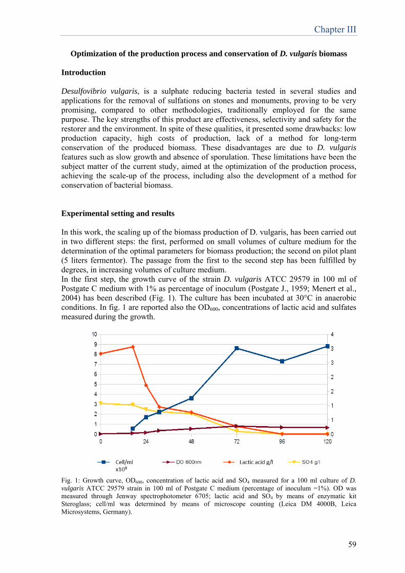
Chapter III
59
Optimization of the production process and conservation of D. vulgaris biomass Introduction Desulfovibrio vulgaris, is a sulphate reducing bacteria tested in several studies and applications for the removal of sulfations on stones and monuments, proving to be very promising, compared to other methodologies, traditionally employed for the same purpose. The key strengths of this product are effectiveness, selectivity and safety for the restorer and the environment. In spite of these qualities, it presented some drawbacks: low production capacity, high costs of production, lack of a method for long-term conservation of the produced biomass. These disadvantages are due to D. vulgaris features such as slow growth and absence of sporulation. These limitations have been the subject matter of the current study, aimed at the optimization of the production process, achieving the scale-up of the process, including also the development of a method for conservation of bacterial biomass. Experimental setting and results In this work, the scaling up of the biomass production of D. vulgaris, has been carried out in two different steps: the first, performed on small volumes of culture medium for the determination of the optimal parameters for biomass production; the second on pilot plant (5 liters fermentor). The passage from the first to the second step has been fulfilled by degrees, in increasing volumes of culture medium. In the first step, the growth curve of the strain D. vulgaris ATCC 29579 in 100 ml of Postgate C medium with 1% as percentage of inoculum (Postgate J., 1959; Menert et al., 2004) has been described (Fig. 1). The culture has been incubated at 30°C in anaerobic conditions. In fig. 1 are reported also the OD600, concentrations of lactic acid and sulfates measured during the growth.
Fig. 1: Growth curve, OD600, concentration of lactic acid and SO4 measured for a 100 ml culture of D. vulgaris ATCC 29579 strain in 100 ml of Postgate C medium (percentage of inoculum =1%). OD was measured through Jenway spectrophotometer 6705; lactic acid and SO4 by means of enzymatic kit Steroglass; cell/ml was determined by means of microscope counting (Leica DM 4000B, Leica Microsystems, Germany).
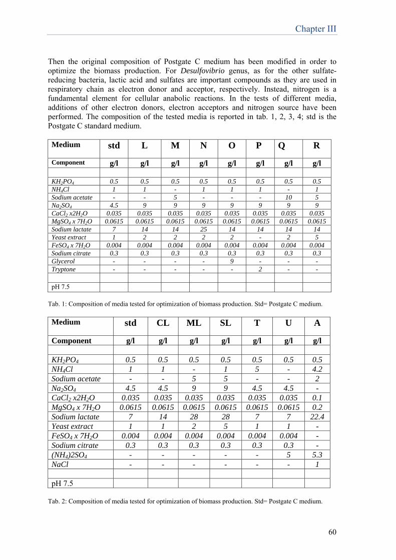
Chapter III
60
Then the original composition of Postgate C medium has been modified in order to optimize the biomass production. For Desulfovibrio genus, as for the other sulfate-reducing bacteria, lactic acid and sulfates are important compounds as they are used in respiratory chain as electron donor and acceptor, respectively. Instead, nitrogen is a fundamental element for cellular anabolic reactions. In the tests of different media, additions of other electron donors, electron acceptors and nitrogen source have been performed. The composition of the tested media is reported in tab. 1, 2, 3, 4; std is the Postgate C standard medium. Medium
std L M N O P Q R
Component g/l g/l g/l g/l g/l g/l g/l g/l KH2PO4 0.5 0.5 0.5 0.5 0.5 0.5 0.5 0.5 NH4Cl 1 1 - 1 1 1 - 1 Sodium acetate - - 5 - - - 10 5 Na2SO4 4.5 9 9 9 9 9 9 9 CaCl2 x2H2O 0.035 0.035 0.035 0.035 0.035 0.035 0.035 0.035 MgSO4 x 7H2O 0.0615 0.0615 0.0615 0.0615 0.0615 0.0615 0.0615 0.0615 Sodium lactate 7 14 14 25 14 14 14 14 Yeast extract 1 2 2 2 2 - 2 5 FeSO4 x 7H2O 0.004 0.004 0.004 0.004 0.004 0.004 0.004 0.004 Sodium citrate 0.3 0.3 0.3 0.3 0.3 0.3 0.3 0.3 Glycerol - - - - 9 - - - Tryptone - - - - - 2 - -
pH 7.5
Tab. 1: Composition of media tested for optimization of biomass production. Std= Postgate C medium. Medium
std CL ML SL T U A
Component g/l g/l g/l g/l g/l g/l g/l KH2PO4 0.5 0.5 0.5 0.5 0.5 0.5 0.5 NH4Cl 1 1 - 1 5 - 4.2 Sodium acetate - - 5 5 - - 2 Na2SO4 4.5 4.5 9 9 4.5 4.5 - CaCl2 x2H2O 0.035 0.035 0.035 0.035 0.035 0.035 0.1 MgSO4 x 7H2O 0.0615 0.0615 0.0615 0.0615 0.0615 0.0615 0.2 Sodium lactate 7 14 28 28 7 7 22.4 Yeast extract 1 1 2 5 1 1 - FeSO4 x 7H2O 0.004 0.004 0.004 0.004 0.004 0.004 - Sodium citrate 0.3 0.3 0.3 0.3 0.3 0.3 - (NH4)2SO4 - - - - - 5 5.3 NaCl - - - - - - 1
pH 7.5
Tab. 2: Composition of media tested for optimization of biomass production. Std= Postgate C medium.
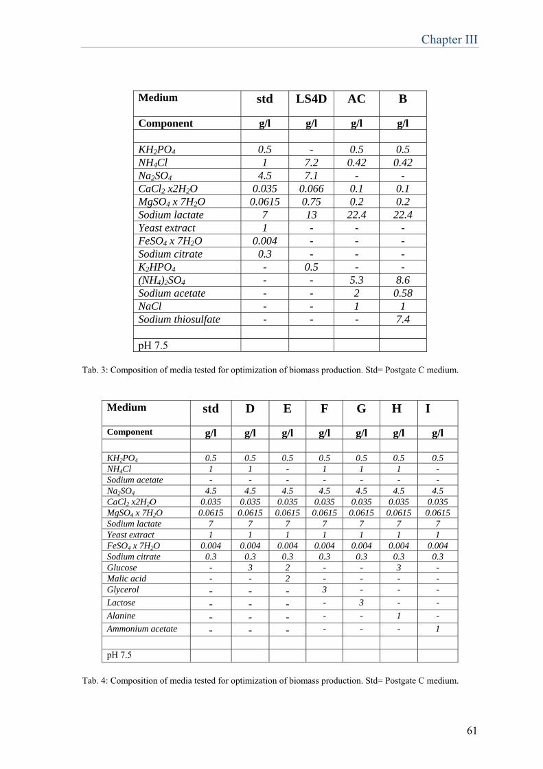
Chapter III
61
Medium
std LS4D AC B
Component g/l g/l g/l g/l KH2PO4 0.5 - 0.5 0.5 NH4Cl 1 7.2 0.42 0.42 Na2SO4 4.5 7.1 - - CaCl2 x2H2O 0.035 0.066 0.1 0.1 MgSO4 x 7H2O 0.0615 0.75 0.2 0.2 Sodium lactate 7 13 22.4 22.4 Yeast extract 1 - - - FeSO4 x 7H2O 0.004 - - - Sodium citrate 0.3 - - - K2HPO4 - 0.5 - - (NH4)2SO4 - - 5.3 8.6 Sodium acetate - - 2 0.58 NaCl - - 1 1 Sodium thiosulfate - - - 7.4 pH 7.5
Tab. 3: Composition of media tested for optimization of biomass production. Std= Postgate C medium.
Medium
std D E F G H I
Component g/l g/l g/l g/l g/l g/l g/l KH2PO4 0.5 0.5 0.5 0.5 0.5 0.5 0.5 NH4Cl 1 1 - 1 1 1 - Sodium acetate - - - - - - - Na2SO4 4.5 4.5 4.5 4.5 4.5 4.5 4.5 CaCl2 x2H2O 0.035 0.035 0.035 0.035 0.035 0.035 0.035 MgSO4 x 7H2O 0.0615 0.0615 0.0615 0.0615 0.0615 0.0615 0.0615Sodium lactate 7 7 7 7 7 7 7 Yeast extract 1 1 1 1 1 1 1 FeSO4 x 7H2O 0.004 0.004 0.004 0.004 0.004 0.004 0.004 Sodium citrate 0.3 0.3 0.3 0.3 0.3 0.3 0.3 Glucose - 3 2 - - 3 - Malic acid - - 2 - - - - Glycerol - - - 3 - - - Lactose - - - - 3 - - Alanine - - - - - 1 - Ammonium acetate - - - - - - 1 pH 7.5
Tab. 4: Composition of media tested for optimization of biomass production. Std= Postgate C medium.
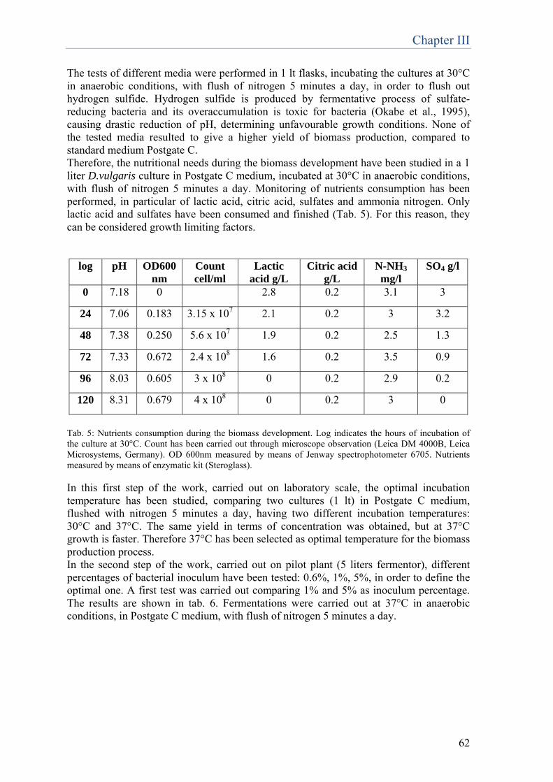
Chapter III
62
The tests of different media were performed in 1 lt flasks, incubating the cultures at 30°C in anaerobic conditions, with flush of nitrogen 5 minutes a day, in order to flush out hydrogen sulfide. Hydrogen sulfide is produced by fermentative process of sulfate-reducing bacteria and its overaccumulation is toxic for bacteria (Okabe et al., 1995), causing drastic reduction of pH, determining unfavourable growth conditions. None of the tested media resulted to give a higher yield of biomass production, compared to standard medium Postgate C. Therefore, the nutritional needs during the biomass development have been studied in a 1 liter D.vulgaris culture in Postgate C medium, incubated at 30°C in anaerobic conditions, with flush of nitrogen 5 minutes a day. Monitoring of nutrients consumption has been performed, in particular of lactic acid, citric acid, sulfates and ammonia nitrogen. Only lactic acid and sulfates have been consumed and finished (Tab. 5). For this reason, they can be considered growth limiting factors.
log pH OD600nm
Count cell/ml
Lactic acid g/L
Citric acid g/L
N-NH3 mg/l
SO4 g/l
0 7.18 0 2.8 0.2 3.1 3
24 7.06 0.183 3.15 x 10⁷ 2.1 0.2 3 3.2
48 7.38 0.250 5.6 x 10⁷ 1.9 0.2 2.5 1.3
72 7.33 0.672 2.4 x 10⁸ 1.6 0.2 3.5 0.9
96 8.03 0.605 3 x 10⁸ 0 0.2 2.9 0.2
120 8.31 0.679 4 x 10⁸ 0 0.2 3 0
Tab. 5: Nutrients consumption during the biomass development. Log indicates the hours of incubation of the culture at 30°C. Count has been carried out through microscope observation (Leica DM 4000B, Leica Microsystems, Germany). OD 600nm measured by means of Jenway spectrophotometer 6705. Nutrients measured by means of enzymatic kit (Steroglass). In this first step of the work, carried out on laboratory scale, the optimal incubation temperature has been studied, comparing two cultures (1 lt) in Postgate C medium, flushed with nitrogen 5 minutes a day, having two different incubation temperatures: 30°C and 37°C. The same yield in terms of concentration was obtained, but at 37°C growth is faster. Therefore 37°C has been selected as optimal temperature for the biomass production process. In the second step of the work, carried out on pilot plant (5 liters fermentor), different percentages of bacterial inoculum have been tested: 0.6%, 1%, 5%, in order to define the optimal one. A first test was carried out comparing 1% and 5% as inoculum percentage. The results are shown in tab. 6. Fermentations were carried out at 37°C in anaerobic conditions, in Postgate C medium, with flush of nitrogen 5 minutes a day.
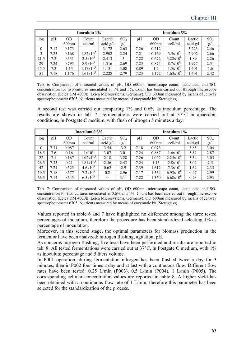
Chapter III
63
Inoculum 1% Inoculum 5% log pH OD
600nm Count cell/ml
Lactic acid g/l
SO4
g/l pH OD
600nm Count cell/ml
Lactic acid g/l
SO4
g/l 0 7.17 0.173 3.172 2.63 7.26 0.212 3.223 2.46 3 7.23 0.168 1.82x10⁷ 2.902 2.24 7.21 0.169 3.5x10⁷ 2.902 2.66
21.5 7.2 0.331 2.5x10⁸ 2.413 3 7.22 0.672 3.22x10⁸ 1.89 2.26 29 7.24 0.795 8.9x10⁸ 1.316 2.69 7.25 0.874 8.7x10⁸ 1.977 2.51
45.5 7.2 1.13 1.17x10⁹ 1.131 3.08 8.89 1.2 1.5x10⁹ 1.401 1.6 51 7.18 1.176 1.61x10⁹ 2.228 2.79 7.23 1.172 1.63x10⁹ 1.401 2.42
Tab. 6: Comparison of measured values of pH, OD 600nm, microscope count, lactic acid and SO4 concentration for two cultures inoculated at 1% and 5%. Count has been carried out through microscope observation (Leica DM 4000B, Leica Microsystems, Germany). OD 600nm measured by means of Jenway spectrophotometer 6705. Nutrients measured by means of enzymatic kit (Steroglass). A second test was carried out comparing 1% and 0.6% as inoculum percentage. The results are shown in tab. 7. Fermentations were carried out at 37°C in anaerobic conditions, in Postgate C medium, with flush of nitrogen 5 minutes a day. Inoculum 0.6% Inoculum 1% log pH OD
600nm Count cell/ml
Lactic acid g/l
SO4
g/l pH OD
600nm Count cell/ml
Lactic acid g/l
SO4
g/l 0 7.31 0.087 3.54 3.2 7.18 0.073 3.85 3.04
18.5 7.6 0.16 1x10⁸ 3.07 3.01 7.24 0.887 1.6x10⁸ 5.62 2.87 22 7.1 0.167 1.02x10⁸ 2.18 3.28 7.26 1.022 2.25x10⁸ 3.34 3.05
26.5 7.33 0.21 1.81x10⁸ 2.56 2.43 7.24 1.15 2.8x10⁸ 3.02 2.5 42 7.21 0.525 4.6x10⁸ 0.62 2.9 7.39 1.612 7.3x10⁸ 1.62 2.35
50.5 7.18 0.577 7.2x10⁸ 0.2 2.96 7.17 1.564 6.93x10⁸ 0.47 2.99 66.5 7.14 0.545 6.5x10⁸ 0 3.11 7.22 1.360 6.68x10⁸ 0.23 2.93
Tab. 7: Comparison of measured values of pH, OD 600nm, microscope count, lactic acid and SO4 concentration for two cultures inoculated at 0.6% and 1%. Count has been carried out through microscope observation (Leica DM 4000B, Leica Microsystems, Germany). OD 600nm measured by means of Jenway spectrophotometer 6705. Nutrients measured by means of enzymatic kit (Steroglass). Values reported in table 6 and 7 have highlighted no difference among the three tested percentages of inoculum, therefore the procedure has been standardized selecting 1% as percentage of inoculation. Moreover, in this second stage, the optimal parameters for biomass production in the fermentor have been analyzed: nitrogen flushing, agitation, pH. As concerns nitrogen flushing, five tests have been performed and results are reported in tab. 8. All tested fermentations were carried out at 37°C, in Postgate C medium, with 1% as inoculum percentage and 5 liters volume. In P001 operation, during fermentation nitrogen has been flushed twice a day for 3 minutes, then in P002 four times a day and at last with a continuous flow. Different flow rates have been tested: 0.25 L/min (P003), 0.5 L/min (P004), 1 L/min (P005). The corresponding cellular concentration values are reported in table 8. A higher yield has been obtained with a continuous flow rate of 1 L/min, therefore this parameter has been selected for the standardization of the process.
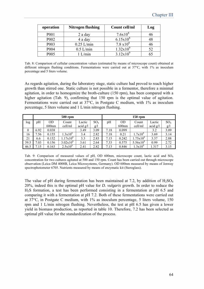
Chapter III
64
operation Nitrogen flushing Count cell/ml Log
P001 2 a day 7.6x10⁸ 46 P002 4 a day 6.15x10⁸ 48 P003 0.25 L/min 7.8 x10⁸ 46 P004 0.5 L/min 1.32x10⁹ 52 P005 1 L/min 3.12x10⁹ 65
Tab. 8: Comparison of cellular concentration values (estimated by means of microscope count) obtained at different nitrogen flushing conditions. Fermentations were carried out at 37°C, with 1% as inoculum percentage and 5 liters volume. As regards agitation, during the laboratory stage, static culture had proved to reach higher growth than stirred one. Static culture is not possible in a fermentor, therefore a minimal agitation, in order to homogenize the broth-culture (150 rpm), has been compared with a higher agitation (Tab. 9), confirming that 150 rpm is the optimal value of agitation. Fermentations were carried out at 37°C, in Postgate C medium, with 1% as inoculum percentage, 5 liters volume and 1 L/min nitrogen flushing. 500 rpm 150 rpm log pH OD
600nm Count cell/ml
Lactic acid g/l
SO4
g/l pH OD
600nm Count cell/ml
Lactic acid g/l
SO4
g/l 0 6.92 0.038 3.49 3.09 7.18 0.099 3.2 3.09
16 7.56 0.155 1.3x10⁸ 3.4 2.82 7.18 0.21 1.7x10⁸ 3.69 3.14 21 6.6 0.152 1.17x10⁸ 3.3 2.83 7.15 0.242 1.75x10⁸ 3.37 2.88
39.5 7.03 0.156 3.02x10⁸ 3.61 2.64 7.33 0.575 5.56x10⁸ 0.99 2.72 46.5 7.15 0.163 2.5x10⁸ 2.41 2.82 7.13 0.846 1.3x10⁹ 1.317 3.15
Tab. 9: Comparison of measured values of pH, OD 600nm, microscope count, lactic acid and SO4 concentration for two cultures agitated at 500 and 150 rpm. Count has been carried out through microscope observation (Leica DM 4000B, Leica Microsystems, Germany). OD 600nm measured by means of Jenway spectrophotometer 6705. Nutrients measured by means of enzymatic kit (Steroglass). The value of pH during fermentation has been maintained at 7.2, by addition of H2SO4 20%, indeed this is the optimal pH value for D. vulgaris growth. In order to reduce the H2S formation, a test has been performed consisting in a fermentation at pH 6.5 and comparing it with a fermentation at pH 7.2. Both of these fermentations were carried out at 37°C, in Postgate C medium, with 1% as inoculum percentage, 5 liters volume, 150 rpm and 1 L/min nitrogen flushing. Nevertheless, the test at pH 6.5 has given a lower yield in biomass production, as reported in table 10. Therefore, 7.2 has been selected as optimal pH value for the standardization of the process.
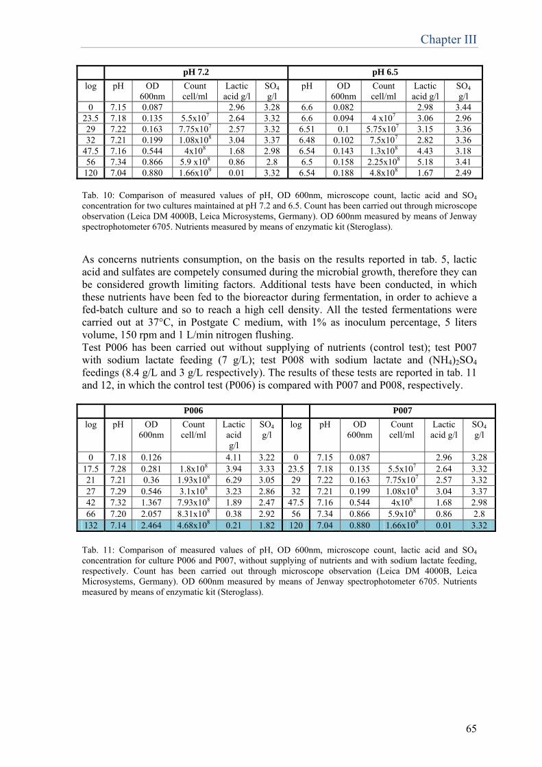
Chapter III
65
pH 7.2 pH 6.5 log pH OD
600nm Count cell/ml
Lactic acid g/l
SO4
g/l pH OD
600nm Count cell/ml
Lactic acid g/l
SO4
g/l 0 7.15 0.087 2.96 3.28 6.6 0.082 2.98 3.44
23.5 7.18 0.135 5.5x10⁷ 2.64 3.32 6.6 0.094 4 x10⁷ 3.06 2.96 29 7.22 0.163 7.75x10⁷ 2.57 3.32 6.51 0.1 5.75x10⁷ 3.15 3.36 32 7.21 0.199 1.08x10⁸ 3.04 3.37 6.48 0.102 7.5x10⁷ 2.82 3.36
47.5 7.16 0.544 4x10⁸ 1.68 2.98 6.54 0.143 1.3x10⁸ 4.43 3.18 56 7.34 0.866 5.9 x10⁸ 0.86 2.8 6.5 0.158 2.25x10⁸ 5.18 3.41
120 7.04 0.880 1.66x10⁹ 0.01 3.32 6.54 0.188 4.8x10⁸ 1.67 2.49 Tab. 10: Comparison of measured values of pH, OD 600nm, microscope count, lactic acid and SO4 concentration for two cultures maintained at pH 7.2 and 6.5. Count has been carried out through microscope observation (Leica DM 4000B, Leica Microsystems, Germany). OD 600nm measured by means of Jenway spectrophotometer 6705. Nutrients measured by means of enzymatic kit (Steroglass). As concerns nutrients consumption, on the basis on the results reported in tab. 5, lactic acid and sulfates are competely consumed during the microbial growth, therefore they can be considered growth limiting factors. Additional tests have been conducted, in which these nutrients have been fed to the bioreactor during fermentation, in order to achieve a fed-batch culture and so to reach a high cell density. All the tested fermentations were carried out at 37°C, in Postgate C medium, with 1% as inoculum percentage, 5 liters volume, 150 rpm and 1 L/min nitrogen flushing. Test P006 has been carried out without supplying of nutrients (control test); test P007 with sodium lactate feeding (7 g/L); test P008 with sodium lactate and (NH4)2SO4
feedings (8.4 g/L and 3 g/L respectively). The results of these tests are reported in tab. 11 and 12, in which the control test (P006) is compared with P007 and P008, respectively. P006 P007 log pH OD
600nm Count cell/ml
Lactic acid g/l
SO4
g/l log pH OD
600nm Count cell/ml
Lactic acid g/l
SO4
g/l
0 7.18 0.126 4.11 3.22 0 7.15 0.087 2.96 3.28 17.5 7.28 0.281 1.8x10⁸ 3.94 3.33 23.5 7.18 0.135 5.5x10⁷ 2.64 3.32 21 7.21 0.36 1.93x10⁸ 6.29 3.05 29 7.22 0.163 7.75x10⁷ 2.57 3.32 27 7.29 0.546 3.1x10⁸ 3.23 2.86 32 7.21 0.199 1.08x10⁸ 3.04 3.37 42 7.32 1.367 7.93x10⁸ 1.89 2.47 47.5 7.16 0.544 4x10⁸ 1.68 2.98 66 7.20 2.057 8.31x10⁸ 0.38 2.92 56 7.34 0.866 5.9x10⁸ 0.86 2.8
132 7.14 2.464 4.68x10⁸ 0.21 1.82 120 7.04 0.880 1.66x10⁹ 0.01 3.32
Tab. 11: Comparison of measured values of pH, OD 600nm, microscope count, lactic acid and SO4 concentration for culture P006 and P007, without supplying of nutrients and with sodium lactate feeding, respectively. Count has been carried out through microscope observation (Leica DM 4000B, Leica Microsystems, Germany). OD 600nm measured by means of Jenway spectrophotometer 6705. Nutrients measured by means of enzymatic kit (Steroglass).
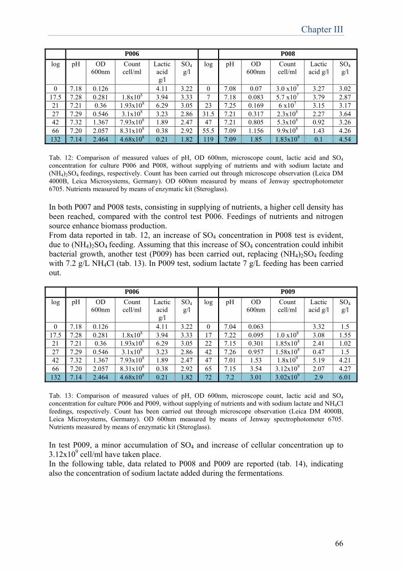
Chapter III
66
P006 P008 log pH OD
600nm Count cell/ml
Lactic acid g/l
SO4
g/l log pH OD
600nm Count cell/ml
Lactic acid g/l
SO4
g/l
0 7.18 0.126 4.11 3.22 0 7.08 0.07 3.0 x10⁷ 3.27 3.02 17.5 7.28 0.281 1.8x10⁸ 3.94 3.33 7 7.18 0.083 5.7 x10⁷ 3.79 2.87 21 7.21 0.36 1.93x10⁸ 6.29 3.05 23 7.25 0.169 6 x10⁷ 3.15 3.17 27 7.29 0.546 3.1x10⁸ 3.23 2.86 31.5 7.21 0.317 2.3x10⁸ 2.27 3.64 42 7.32 1.367 7.93x10⁸ 1.89 2.47 47 7.21 0.805 5.3x10⁸ 0.92 3.26 66 7.20 2.057 8.31x10⁸ 0.38 2.92 55.5 7.09 1.156 9.9x10⁸ 1.43 4.26
132 7.14 2.464 4.68x10⁸ 0.21 1.82 119 7.09 1.85 1.83x10⁹ 0.1 4.54
Tab. 12: Comparison of measured values of pH, OD 600nm, microscope count, lactic acid and SO4 concentration for culture P006 and P008, without supplying of nutrients and with sodium lactate and (NH4)2SO4 feedings, respectively. Count has been carried out through microscope observation (Leica DM 4000B, Leica Microsystems, Germany). OD 600nm measured by means of Jenway spectrophotometer 6705. Nutrients measured by means of enzymatic kit (Steroglass). In both P007 and P008 tests, consisting in supplying of nutrients, a higher cell density has been reached, compared with the control test P006. Feedings of nutrients and nitrogen source enhance biomass production. From data reported in tab. 12, an increase of SO4 concentration in P008 test is evident, due to (NH4)2SO4 feeding. Assuming that this increase of SO4 concentration could inhibit bacterial growth, another test (P009) has been carried out, replacing (NH4)2SO4 feeding with 7.2 g/L NH4Cl (tab. 13). In P009 test, sodium lactate 7 g/L feeding has been carried out. P006 P009 log pH OD
600nm Count cell/ml
Lactic acid g/l
SO4
g/l log pH OD
600nm Count cell/ml
Lactic acid g/l
SO4
g/l
0 7.18 0.126 4.11 3.22 0 7.04 0.063 3.32 1.5 17.5 7.28 0.281 1.8x10⁸ 3.94 3.33 17 7.22 0.095 1.0 x10⁸ 3.08 1.55 21 7.21 0.36 1.93x10⁸ 6.29 3.05 22 7.15 0.301 1.85x10⁸ 2.41 1.02 27 7.29 0.546 3.1x10⁸ 3.23 2.86 42 7.26 0.957 1.58x10⁸ 0.47 1.5 42 7.32 1.367 7.93x10⁸ 1.89 2.47 47 7.01 1.53 1.8x10⁹ 5.19 4.21 66 7.20 2.057 8.31x10⁸ 0.38 2.92 65 7.15 3.54 3.12x10⁹ 2.07 4.27
132 7.14 2.464 4.68x10⁸ 0.21 1.82 72 7.2 3.01 3.02x10⁹ 2.9 6.01
Tab. 13: Comparison of measured values of pH, OD 600nm, microscope count, lactic acid and SO4 concentration for culture P006 and P009, without supplying of nutrients and with sodium lactate and NH4Cl feedings, respectively. Count has been carried out through microscope observation (Leica DM 4000B, Leica Microsystems, Germany). OD 600nm measured by means of Jenway spectrophotometer 6705. Nutrients measured by means of enzymatic kit (Steroglass). In test P009, a minor accumulation of SO4 and increase of cellular concentration up to 3.12x10⁹ cell/ml have taken place. In the following table, data related to P008 and P009 are reported (tab. 14), indicating also the concentration of sodium lactate added during the fermentations.
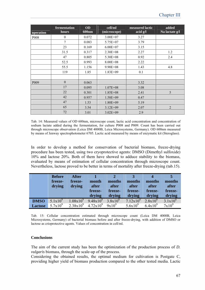
Chapter III
67
operation
fermentation hours
OD 600nm
cell/ml (microscope)
measured lactic acid g/l
added Na lactate g/l
P008 0 0.072 3.00E+07 3.27
7 0.083 5.75E+07 3.79
23 0.169 6.00E+07 3.15
31.5 0.317 2.30E+08 2.27 1.2
47 0.805 5.30E+08 0.92 2.4
52.5 0.993 8.00E+08 2.22
55.5 1.156 9.90E+08 1.43 4.8
119 1.85 1.83E+09 0.1
P009 0 0.063 3.32
17 0.095 1.07E+08 3.08
22 0.301 1.85E+08 2.41 5
42 0.957 1.58E+09 0.47
47 1.53 1.80E+09 5.19
65 3.54 3.12E+09 2.07 2
72 3.01 3.02E+09 2.9 Tab. 14: Measured values of OD 600nm, microscope count, lactic acid concentration and concentration of sodium lactate added during the fermentation, for culture P008 and P009. Count has been carried out through microscope observation (Leica DM 4000B, Leica Microsystems, Germany). OD 600nm measured by means of Jenway spectrophotometer 6705. Lactic acid measured by means of enzymatic kit (Steroglass). In order to develop a method for conservation of bacterial biomass, freeze-drying procedure has been tested, using two cryoprotective agents: DMSO (Dimethyl sulfoxide) 10% and lactose 20%. Both of them have showed to adduce stability to the biomass, evaluated by means of estimation of cellular concentration through microscope count. Nevertheless, lactose proved to be better in terms of mortality after freeze-drying (tab.15). Before
freeze-drying
After freeze-drying
1 month after
freeze-drying
2 months
after freeze-drying
3 months
after freeze-drying
4 months
after freeze-drying
5 months
after freeze-drying
DMSO 5.1x10⁹ 1.08x10⁹ 9.48x10⁸ 3.8x10⁸ 7.12x10⁸ 2.8x10⁸ 3.1x10⁸ Lactose 5.7x10⁹ 2.38x10⁹ 4.72x10⁹ 9x10⁹ 5.6x10⁹ 6.4x10⁹ 7x10⁹ Tab. 15: Cellular concentration estimated through microscope count (Leica DM 4000B, Leica Microsystems, Germany) of bacterial biomass before and after freeze-drying, with addition of DMSO or lactose as crioprotective agents. Values of concentration in cell/ml. Conclusions The aim of the current study has been the optimization of the production process of D. vulgaris biomass, through the scale-up of the process. Considering the obtained results, the optimal medium for cultivation is Postgate C, providing higher yield of biomass production compared to the other tested media. Lactic

Chapter III
68
acid and sulfates can be considered growth limiting factors, therefore feeding of sodium lactate and (NH4)2SO4 has allowed to obtain higher biomass production. However, increase of SO4 concentration could inhibit bacterial growth, therefore (NH4)2SO4 feeding has been replaced with NH4Cl. Three different inocula have been tested: 0.6%, 1%, 5%. The obtained yield for the three tested percentages of inoculum was the same, therefore the procedure has been standardized selecting 1% inoculation. Two different incubation temperatures have been experimented: 30°C and 37°C. The same yield in terms of concentration was obtained, but 37°C speed up the growth. Therefore 37°C has been selected as optimal temperature for the biomass production process. Flush of nitrogen is important in order to flush out hydrogen sulfide, which is toxic for bacteria, determining unfavourable growth conditions. Among the different flow rates tested, a continuous flow rate of 1 L/min allowed the highest yield in terms of biomass production, therefore this parameter has been selected for the standardization of the process. As concerns agitation, 150 rpm is the optimal value of agitation, giving the highest cellular concentration; instead considering pH, 7.2 has been selected as optimal pH value for the standardization of the process. The process parameters for the pilot fermenter can be schematized as follows:
Postgate C medium 1% inoculum 150 rpm 37°C pO2 mantained at 0 with a continuos N2 flux of 1 L/min pH mantained at 7.2 using H2SO4 20% Lactic acid mantained at the concentration of 2 g/l by adding sodium lactate 60% Add NH4Cl at a flux of 0.1 g/L/h
These improvements allowed a significative increase in biomass production, from a concentration of 1*108 cell/ml in 120h of fermentation in flask, to the concentration of 3*109 cell/ml in 72h of fermentation in bioreactor. In conclusion, we can state that in 3 days of fermentation of 5 liters of broth-culture, product for the treatment of 50 m2 of surface is obtained. References PRO Okabe, S., Nielsen, P.H., Jones, W.L. and Characklis., W.G. (1995) Sulfide product inhibition of Desulfovibrio desulfuricans in batch and continuous cultures. Wat. Res. 29 (2), 571-578. SULFIDE DESULFURICANS IN BATCH AND CONTINUOUS CULTURES S. OKABE*~, P. H. NIELSEN~, W. L. JONES and W. G. CHARACKLIS
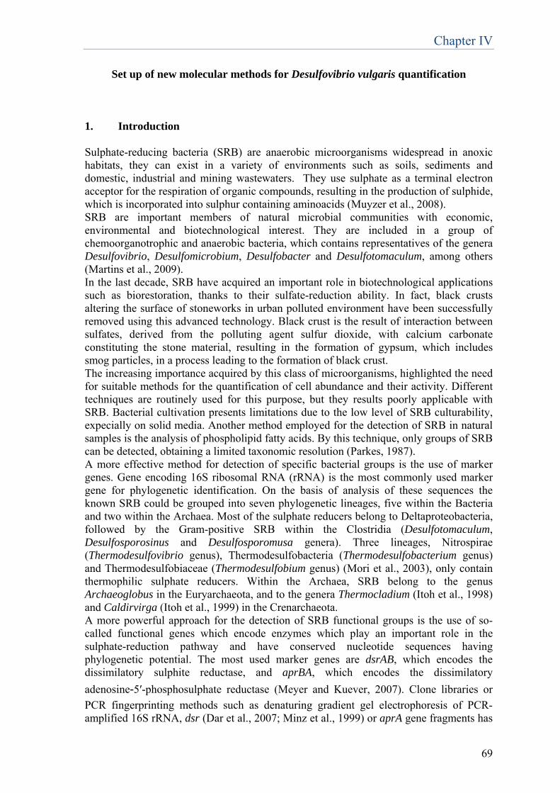
Chapter IV
69
Set up of new molecular methods for Desulfovibrio vulgaris quantification 1. Introduction Sulphate-reducing bacteria (SRB) are anaerobic microorganisms widespread in anoxic habitats, they can exist in a variety of environments such as soils, sediments and domestic, industrial and mining wastewaters. They use sulphate as a terminal electron acceptor for the respiration of organic compounds, resulting in the production of sulphide, which is incorporated into sulphur containing aminoacids (Muyzer et al., 2008). SRB are important members of natural microbial communities with economic, environmental and biotechnological interest. They are included in a group of chemoorganotrophic and anaerobic bacteria, which contains representatives of the genera Desulfovibrio, Desulfomicrobium, Desulfobacter and Desulfotomaculum, among others (Martins et al., 2009). In the last decade, SRB have acquired an important role in biotechnological applications such as biorestoration, thanks to their sulfate-reduction ability. In fact, black crusts altering the surface of stoneworks in urban polluted environment have been successfully removed using this advanced technology. Black crust is the result of interaction between sulfates, derived from the polluting agent sulfur dioxide, with calcium carbonate constituting the stone material, resulting in the formation of gypsum, which includes smog particles, in a process leading to the formation of black crust. The increasing importance acquired by this class of microorganisms, highlighted the need for suitable methods for the quantification of cell abundance and their activity. Different techniques are routinely used for this purpose, but they results poorly applicable with SRB. Bacterial cultivation presents limitations due to the low level of SRB culturability, expecially on solid media. Another method employed for the detection of SRB in natural samples is the analysis of phospholipid fatty acids. By this technique, only groups of SRB can be detected, obtaining a limited taxonomic resolution (Parkes, 1987). A more effective method for detection of specific bacterial groups is the use of marker genes. Gene encoding 16S ribosomal RNA (rRNA) is the most commonly used marker gene for phylogenetic identification. On the basis of analysis of these sequences the known SRB could be grouped into seven phylogenetic lineages, five within the Bacteria and two within the Archaea. Most of the sulphate reducers belong to Deltaproteobacteria, followed by the Gram-positive SRB within the Clostridia (Desulfotomaculum, Desulfosporosinus and Desulfosporomusa genera). Three lineages, Nitrospirae (Thermodesulfovibrio genus), Thermodesulfobacteria (Thermodesulfobacterium genus) and Thermodesulfobiaceae (Thermodesulfobium genus) (Mori et al., 2003), only contain thermophilic sulphate reducers. Within the Archaea, SRB belong to the genus Archaeoglobus in the Euryarchaeota, and to the genera Thermocladium (Itoh et al., 1998) and Caldirvirga (Itoh et al., 1999) in the Crenarchaeota. A more powerful approach for the detection of SRB functional groups is the use of so-called functional genes which encode enzymes which play an important role in the sulphate-reduction pathway and have conserved nucleotide sequences having phylogenetic potential. The most used marker genes are dsrAB, which encodes the dissimilatory sulphite reductase, and aprBA, which encodes the dissimilatory
adenosine‑5′-phosphosulphate reductase (Meyer and Kuever, 2007). Clone libraries or
PCR fingerprinting methods such as denaturing gradient gel electrophoresis of PCR-amplified 16S rRNA, dsr (Dar et al., 2007; Minz et al., 1999) or aprA gene fragments has
Chapter IV
70
been successfully applied to determine the presence and functional diversity of SRB in many different habitats. Recently, a DNA microarray, the SRP-PhyloChip, has been used to detect SRB in natural samples, such as acidic fen soils. However, these methods have the disadvantage that they provide little or no information on the number of SRB cells that are present (Loy et al., 2002). Quantitative real-time PCR is a highly sensitive technique which, when applied to conserved functional genes, can be used to quantify the number of SRB. The dissimilatory sulfite reductase (DSR) gene sequence among the SRB is conserved; therefore, it is possible to design specific primers or probes for this enzyme (Wagner et al., 1998). Kondo et al. (2004) developed PCR primers selective for the gene coding for the α-subunit of DSR (dsrA) of most mesophilic SRB belonging to the δ-Proteobacteria and used a quantitative competitive PCR to rapidly and reproducibly detect and count SRB, as an alternative to the culture-dependent method. This primer set was applied to enumerate SRB in various environments, such as rice field soils, soda lakes and industrial waste water (Kondo et al. 2006, 2007, Kondo & Butani 2007, Leloup et al., 2007, Schippers & Neretin 2006). Moreover, quantitative real-time PCR, when applied to RNA, can also be used to study the expression of this functional gene. Another technique which was used to quantify the number of SRB in environmental samples is fluorescence in situ hybridization (FISH), which also allowed their spatial distribution to be visualized (Muyzer et al., 2008). The aim of this work was the set up of new molecular methods for the quantification of the SRB Desulfovibrio vulgaris subsp. vulgaris ATCC 29579, which is the strain selected for biorestoration processes which is able to reduce sulfates even under low oxygen tension (Cappitelli et al., 2006; Ranalli et al., 1997). The methods applied to estimate cell abundance of this strain were the microscope cell counting and the spectrophotometric measure of culture turbidity by Optical Density (OD600). These methods presented nevertheless some limitations, mainly because i) they did not allow a discrimination between live and dead cells, overestimating the active cellular concentration and ii) were biased by the presence of inorganic precipitates (mainly black iron sulphide salts) generated during cell growth. An alternative methodology characterized by the advantage of enumeration of only viable organisms, is the count by Most Probable Number (MPN), based on cell cultivation on liquid medium to overcome the recalcitrance of SRB to grow on solid media. This method proved to be improper with Desulfovibrio vulgaris, probably because these cells tend to aggregate, therefore serial decimal dilutions performed in MPN are not reliable, giving incoherent results. In the set up of a SRB-based formulation to be applied in stone biorestoration, the main factor underlying the importance of a new, effective method for enumeration of Desulfovibrio vulgaris, is the necessity of vitality monitoring of the freeze-dried product. After cell production the product is freeze-dried in order to stabilize it, allowing for its long-term storage, and the estimation of the abundance of viable cell is of primary importance to define product activity and shelf life. Another important factor to be estimated is the evaluation of the viability of D.vulgaris cells when they are embedded in the delivery system during product application for biorestoration, after several hours of oxygen exposure. In response to this need, in past studies, ATP assay was carried out in order to determine the content of cells in delivery systems (Cappitelli et al., 2006; Ranalli et al., 2005; Alfano et al., 2011), nevertheless in our tests we have found some problems linked to the interference of the delivery system in ATP quantification. In the light of all the above mentioned evidences, the development of a new molecular method for enumeration of live Desulfovibrio vulgaris cells is of primary importance.
Chapter IV
71
2. Materials and methods 2.1 Cells, media and cultural methods In this work, Desulfovibrio vulgaris subsp. vulgaris ATCC 29579 was used. The strain was cultivated in Postgate C medium (KH2PO4 0.50 g/l, NH4Cl 1.00 g/l, Na2SO4 4.50 g/l, CaCl2•2H2O 0.035 g/l, MgSO4 0.030 g/l, Sodium lactate (60%) 7.00 g/l, Yeast Extract 1.00 g/l, FeSO4•7H2O 0.004 g/l, Sodium Citrate•2H2O 0.30 g/l) (Postgate J., 1959; Menert et al., 2004). For medium preparation, pre-reduced water is used and pH is adjusted to 7,5. Anaerobic cultivation methods were adopted. 2.2 Growth estimation through solid and semisolid cultivation The strain Desulfovibrio vulgaris ATCC 29579 is recalcitrant to laboratory cultivation and especially to growth in solid media. In different studies (Jain, D.K., 1995; Iverson, W.P., 1966; Postgate, J., 1963) semisolid cultivation for SRB with 0.75% or less agar was successfully realised. For this reason, in the present work different conditions for solid and semisolid cultivation were tested. The strain was cultivated in Postgate C medium, five different gelling agents for the solidification of the specific medium were tested: gelzan, phytagel, agarose, agar and PVP (polyvinylpyrrolidone) (all Sigma-Aldrich). As concerns semisolid cultivation, they were tested at different concentrations, going from 0.5% to 1%. DNA was extracted from obtained colonies using the cetyltrimethylammonium bromide (CTAB) method described by Jara et al. (2008) with a prior enzymatic cell lysis using both lysozyme and proteinase K and followed by isopropanol precipitation of DNA. DNA samples from isolates were dereplicated by PCR-ITS (Daffonchio et al., 1998). Partial sequencing of the 16S rRNA gene for phylogenetic characterization of bacterial isolates has been carried out using the following primer set: 357F (CCTACGGGAGGCAGCAG) and 907R (CCGTCAATTCCTTTGAGTTT), the reaction mixture (50 μl) was: 1× Buffer (Invitrogen), 1.5 mM of MgCl2, 0.12 mM of dNTP mix, 0.3μM of each primer and 1 U of Taq DNA polymerase (Invitrogen) in a PCR Thermocycler (BioRad, Milano). The PCR included an initial denaturation at 94°C for 4 min followed by 35 cycles consisting of denaturation at 94°C for 45 s, annealing at 55°C for 1 min and extension at 72°C for 2 min and a final extension step at 72°C for 10 min. The PCR product was analyzed by 1.2% (w/v) agarose gel electrophoresis at 100V in 0.5X TBE buffer (Tris 1M, boric acid 13.7 g, EDTA 0.5 M pH 8). Gel images were captured with GelDoc 2000 apparatus (Bio-Rad, Milan, Italy) using the Quantity one software (Bio-Rad). The obtained PCR products were sequenced by Macrogen Inc. (Korea). Sequence identification was performed by use of the BLASTN facility of the National Center for Biotechnology Information (http://www.ncbi.nlm.nih.gov/BLAST/). 2.3 Estimation of sulfate-reducing activity – the silver foil method A silver foil of 0.125-mm thickness (Sigma) was cut into 10- by 10-mm squares, which were then washed extensively with hexane, water, and acetone to remove surface contamination, exposed to 0.1 N HNO3 for 1 min to form an outer layer of silver oxide, and washed extensively with double-distilled water. Then it was immersed into a solution 0.00044 M Na2SO4, to which 0.06 g/l sodium silicate was added in order to obtain a
Chapter IV
72
homogenous coating of sulfate. After the treatment, the foil resulted to be coated by a silver sulfate layer, which, put in contact with SRB, was converted in silver sulfide, consequently visualized by the blackening of the foil. After drying, the silver foil can be inserted in the sample to estimate sulfate-reduction (modified from Sigalevich et al., 2000). 2.4 Estimation of cell viability – Live/Dead bacterial viability test LIVE/DEAD BacLight bacterial viability kit L7012 (Invitrogen, Carlsbad, CA, USA) was used for this test (Gatti et al., 2006). The system was set up on dilutions of a D.vulgaris culture, in order to evaluate its efficiency. The kit includes: Component A (SYTO 9 dye, 3.34 mM, solution in DMSO), Component B (Propidium iodide, 20 mM, solution in DMSO). SYTO 9 stain labels all bacteria in a population – those with intact membranes and those with damaged membranes. In contrast, propidium iodide (PI) penetrates only bacteria with damaged membranes, causing a reduction in the SYTO 9 stain fluorescence, when both dyes are present. Thus, mixing SYTO 9 and PI stains, live bacteria stain fluorescent green, whereas dead bacteria stain fluorescent red. 25 ml of the culture were concentrated by centrifugation at 11000 rpm for 10 minutes, the supernatant was removed and the pellet was resuspended in 2 ml of 0.85% NaCl buffer. Equal volumes of Component A and Component B were combined and mixed thoroughly. 3 μL of the dye mixture were added for each mL of the bacterial suspension, mixed thoroughly and incubated at room temperature in the dark for 15 minutes. 10 μL of the stained bacterial suspension were placed on Thoma Glass and observed using a Leica DM 4000B fluorescent microscope (Leica Microsystems, Germany) provided with 40X objective lens and Leica filtercubes H3 and M2 filters (Leica Microsystems, Germany). Live (green fluorescent, visualized using H3 filter) and dead (red fluorescent, visualized using M2 filter) cells were counted in 10-square areas of the 16 composing the glass. The average number obtained from the ten counts was multiplied to account for the dilution and volume. Each sample was counted in triplicate. 2.5 Estimation of cell viability – ATP assay Total ATP assays to monitor both liquid culture growth and the viability of the bacterial cell suspensions embedded in the delivery system used for the biorestoration processes, were performed using the luciferin-luciferase reaction. ATP Biomass Detection kit (Promicol), luminometer R-Biopharm Rhone Berthold VEGA (Berthold Detection System GmbH) and cuvettes 55x12 (Promicol) were used. In previous works, this method had already been used for D.vulgaris species (Cappitelli et al., 2006; Ranalli et al., 2005; Alfano et al., 2011), but only as an absolute measure. In this study, for the first time, Adenosine triphosphate (ATP) bioluminescence has been used to determine whether there is a linear relationship between cultured cell number and measured luminescence. ATP is present in all metabolically active cells, since they require ATP to remain alive and carry out their specialized functions. Most ATP is found within living cells; cell injury results in a rapid decrease in cytoplasmic ATP (Crouch et al., 1993).
Chapter IV
73
2.6 Estimation of cell viability – fluorescence measurements Fluorescence activity has been previously used to identify Desulfovibrio and has been termed the ‘desulfoviridin test’. This fluorescence is attributed to the prosthetic group of bisulfite reductase, a key enzyme in dissimilatory sulfate reduction. In 2013 (Barton and Carpenter, 2013) fluorescence measurement was proposed as method to quantify sulfate-reducing bacteria. After removal of the cells from the culture, they were treated with NaOH in order to cause precipitation and elimination of metals present in the medium, as they could interfere with fluorescence emitted by desulfoviridin chromophore, generating a quenching effect. Here we propose its employment both for liquid culture and for bacterial cell suspensions embedded in the delivery system. 1 ml of culture was centrifuged at 8000 rpm for 10 min and suspended in physiological saline (0.85% NaCl) to a volume equal to the initial sample. After the addition of 40 μl of 10 N NaOH per 1 ml of suspended cells, the sample was mixed for 30 s and clarified using a microfuge. The centrifugate was measured in a fluorimeter. Triplicate samples were conducted on all tests with an average of these test values presented. The intensity of fluorescence at 535 nm with excitation at 485 nm was recorded for the various measurements. Fluorescent measurements were made with a TECAN infinite F200 PRO microplate reader (TECAN, Switzerland). Nunc MicroWell 96-Well Optical-Bottom Plates with Polymer Base (Thermoscientific, Denmark) were used for all measurements. 2.7 Enumeration of D.vulgaris cells – Real time PCR quantification of dsr gene Quantitative real-time PCR (qPCR) was used to determine the copy numbers of the dsrA gene of D.vulgaris. For dsrA quantification, the dsrA specific primers for mesophilic SRB described elsewhere were used (Kondo et al., 2008; Kondo et al., 2004; Leloup et al., 2007): DSR-1F+ (5’-ACSCACTGGAAGCACGCCGG-3’) and DSR-R (5’-GTGGMRCCGTGCAKRTTGG-3’). PCR SYBR green reactions were prepared by using the “Brilliant II SYBR® Master Mix” kit (Agilent Technologies, Milan, Italy) in 96-well plates on the I-Cycler (Biorad). The reaction mix (25 μl) contained: 12.5 μl of 1 x Brilliant SYBR Green (2.5 mM MgCl2), 0.3 μl of each primer (0.12 μM), and 2 μl of template DNA extract. The thermal cycling program consisted of: 10 min at 95°C for initial denaturation; 50 cycles: 30 s at 95°C, 1 m at 59°C, and 1 m at 72°C; 1 min at 95°C; 1 min at 55°C and 81 cycles 10 sec at 55°C (+0.5°C/cycle) (Merlino et al., 2013). For this assay, a standard curve was realized. Standard DNA consisted of plasmid carrying a dsrAB insert obtained from dsr amplification of DNA from Desulfovibrio vulgaris subsp. vulgaris ATCC 29579 cells. The strain was cultivated in Postgate C liquid medium, reaching the exponential phase of growth. The cells were than pelletted discharging the supernatant and subsequently resuspended with TE. The genomic DNA was extracted using the cetyltrimethylammonium bromide (CTAB) method described by Jara et al., 2008, with a prior enzymatic cell lysis using both lysozyme and proteinase K and followed by isopropanol precipitation of DNA. From this sample, dsrA gene was PCR amplified with primers DSR1F+ and DSR-R as described for real-time PCR analysis. The 221 bp amplified product was ligated into the TOPO-TA cloning vector, using the TOPO-TA cloning kit (Invitrogen, Carlsbad, CA, USA) according to the manufacturer’s instructions. Briefly, E.Coli competent cells were transformed with TOPO-TA cloning vector, white colonies were randomly selected and were screened for cloned inserts of the expected size using the vector primers M13 forward (5’-GTAAAACGACGGCCAG-3’) and reverse (5’-
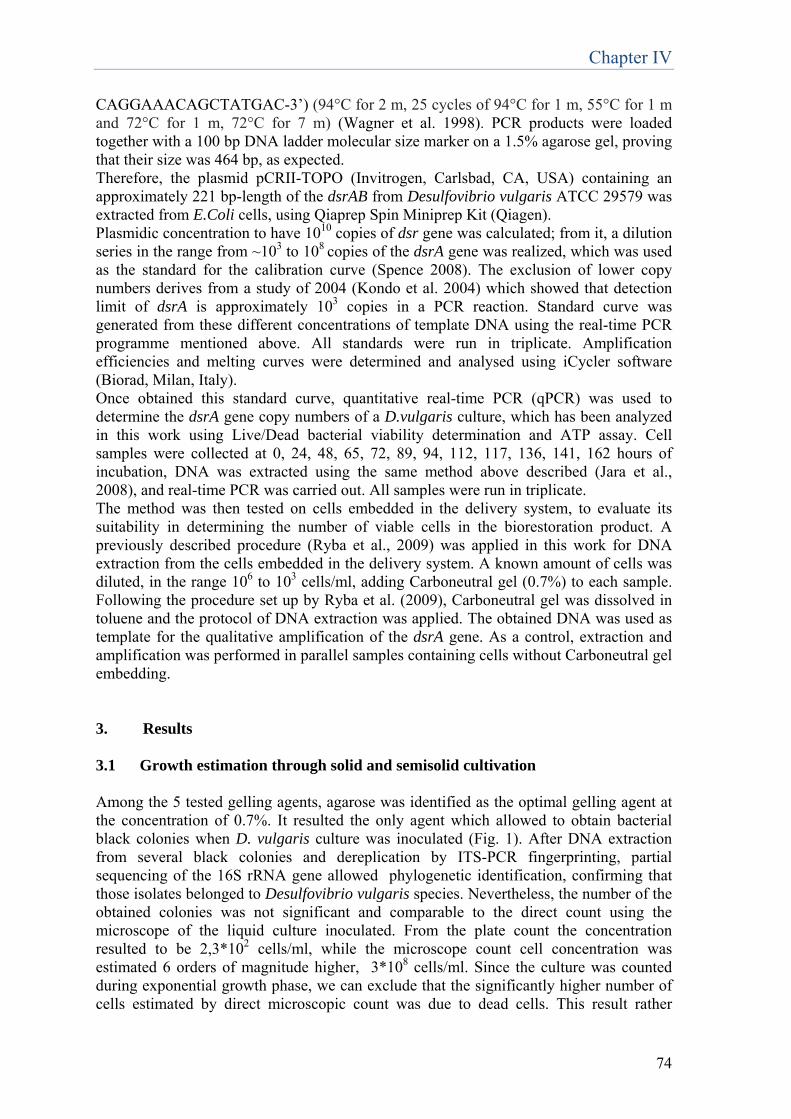
Chapter IV
74
CAGGAAACAGCTATGAC-3’) (94°C for 2 m, 25 cycles of 94°C for 1 m, 55°C for 1 m and 72°C for 1 m, 72°C for 7 m) (Wagner et al. 1998). PCR products were loaded together with a 100 bp DNA ladder molecular size marker on a 1.5% agarose gel, proving that their size was 464 bp, as expected. Therefore, the plasmid pCRII-TOPO (Invitrogen, Carlsbad, CA, USA) containing an approximately 221 bp-length of the dsrAB from Desulfovibrio vulgaris ATCC 29579 was extracted from E.Coli cells, using Qiaprep Spin Miniprep Kit (Qiagen). Plasmidic concentration to have 1010 copies of dsr gene was calculated; from it, a dilution series in the range from ~103 to 108 copies of the dsrA gene was realized, which was used as the standard for the calibration curve (Spence 2008). The exclusion of lower copy numbers derives from a study of 2004 (Kondo et al. 2004) which showed that detection limit of dsrA is approximately 103 copies in a PCR reaction. Standard curve was generated from these different concentrations of template DNA using the real-time PCR programme mentioned above. All standards were run in triplicate. Amplification efficiencies and melting curves were determined and analysed using iCycler software (Biorad, Milan, Italy). Once obtained this standard curve, quantitative real-time PCR (qPCR) was used to determine the dsrA gene copy numbers of a D.vulgaris culture, which has been analyzed in this work using Live/Dead bacterial viability determination and ATP assay. Cell samples were collected at 0, 24, 48, 65, 72, 89, 94, 112, 117, 136, 141, 162 hours of incubation, DNA was extracted using the same method above described (Jara et al., 2008), and real-time PCR was carried out. All samples were run in triplicate. The method was then tested on cells embedded in the delivery system, to evaluate its suitability in determining the number of viable cells in the biorestoration product. A previously described procedure (Ryba et al., 2009) was applied in this work for DNA extraction from the cells embedded in the delivery system. A known amount of cells was diluted, in the range 106 to 103 cells/ml, adding Carboneutral gel (0.7%) to each sample. Following the procedure set up by Ryba et al. (2009), Carboneutral gel was dissolved in toluene and the protocol of DNA extraction was applied. The obtained DNA was used as template for the qualitative amplification of the dsrA gene. As a control, extraction and amplification was performed in parallel samples containing cells without Carboneutral gel embedding. 3. Results 3.1 Growth estimation through solid and semisolid cultivation Among the 5 tested gelling agents, agarose was identified as the optimal gelling agent at the concentration of 0.7%. It resulted the only agent which allowed to obtain bacterial black colonies when D. vulgaris culture was inoculated (Fig. 1). After DNA extraction from several black colonies and dereplication by ITS-PCR fingerprinting, partial sequencing of the 16S rRNA gene allowed phylogenetic identification, confirming that those isolates belonged to Desulfovibrio vulgaris species. Nevertheless, the number of the obtained colonies was not significant and comparable to the direct count using the microscope of the liquid culture inoculated. From the plate count the concentration resulted to be 2,3*102 cells/ml, while the microscope count cell concentration was estimated 6 orders of magnitude higher, 3*108 cells/ml. Since the culture was counted during exponential growth phase, we can exclude that the significantly higher number of cells estimated by direct microscopic count was due to dead cells. This result rather
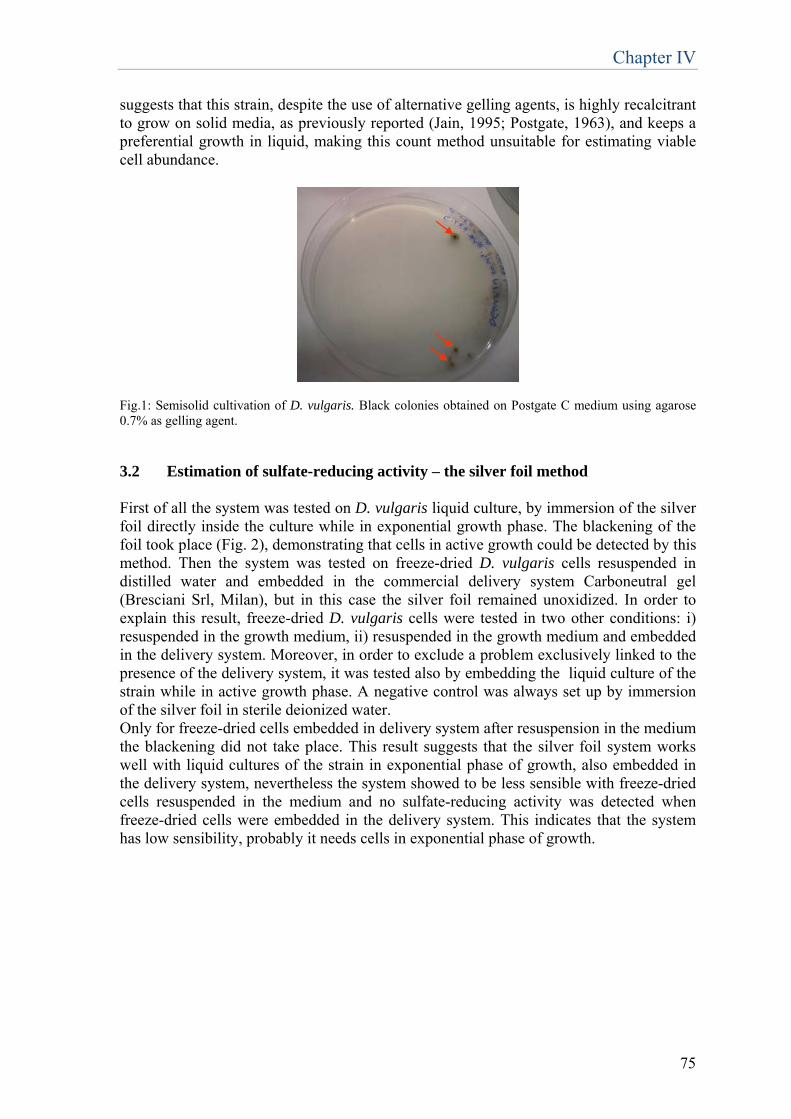
Chapter IV
75
suggests that this strain, despite the use of alternative gelling agents, is highly recalcitrant to grow on solid media, as previously reported (Jain, 1995; Postgate, 1963), and keeps a preferential growth in liquid, making this count method unsuitable for estimating viable cell abundance.
Fig.1: Semisolid cultivation of D. vulgaris. Black colonies obtained on Postgate C medium using agarose 0.7% as gelling agent. 3.2 Estimation of sulfate-reducing activity – the silver foil method First of all the system was tested on D. vulgaris liquid culture, by immersion of the silver foil directly inside the culture while in exponential growth phase. The blackening of the foil took place (Fig. 2), demonstrating that cells in active growth could be detected by this method. Then the system was tested on freeze-dried D. vulgaris cells resuspended in distilled water and embedded in the commercial delivery system Carboneutral gel (Bresciani Srl, Milan), but in this case the silver foil remained unoxidized. In order to explain this result, freeze-dried D. vulgaris cells were tested in two other conditions: i) resuspended in the growth medium, ii) resuspended in the growth medium and embedded in the delivery system. Moreover, in order to exclude a problem exclusively linked to the presence of the delivery system, it was tested also by embedding the liquid culture of the strain while in active growth phase. A negative control was always set up by immersion of the silver foil in sterile deionized water. Only for freeze-dried cells embedded in delivery system after resuspension in the medium the blackening did not take place. This result suggests that the silver foil system works well with liquid cultures of the strain in exponential phase of growth, also embedded in the delivery system, nevertheless the system showed to be less sensible with freeze-dried cells resuspended in the medium and no sulfate-reducing activity was detected when freeze-dried cells were embedded in the delivery system. This indicates that the system has low sensibility, probably it needs cells in exponential phase of growth.
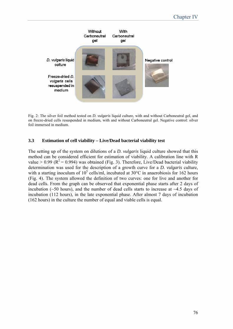
Chapter IV
76
Fig. 2: The silver foil method tested on D. vulgaris liquid culture, with and without Carboneutral gel, and on freeze-dried cells resuspended in medium, with and without Carboneutral gel. Negative control: silver foil immersed in medium. 3.3 Estimation of cell viability – Live/Dead bacterial viability test The setting up of the system on dilutions of a D. vulgaris liquid culture showed that this method can be considered efficient for estimation of viability. A calibration line with R value > 0.99 (R2 = 0.994) was obtained (Fig. 3). Therefore, Live/Dead bacterial viability determination was used for the description of a growth curve for a D. vulgaris culture, with a starting inoculum of 105 cells/ml, incubated at 30°C in anaerobiosis for 162 hours (Fig. 4). The system allowed the definition of two curves: one for live and another for dead cells. From the graph can be observed that exponential phase starts after 2 days of incubation (~50 hours), and the number of dead cells starts to increase at ~4.5 days of incubation (112 hours), in the late exponential phase. After almost 7 days of incubation (162 hours) in the culture the number of equal and viable cells is equal.
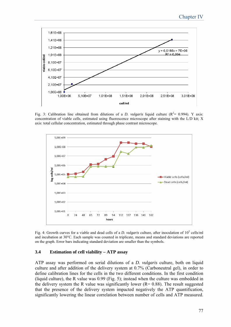
Chapter IV
77
Fig. 3: Calibration line obtained from dilutions of a D. vulgaris liquid culture (R2= 0.994). Y axis: concentration of viable cells, estimated using fluorescence microscope after staining with the L/D kit; X axis: total cellular concentration, estimated through phase contrast microscope.
Fig. 4: Growth curves for a viable and dead cells of a D. vulgaris culture, after inoculation of 105 cells/ml and incubation at 30°C. Each sample was counted in triplicate, means and standard deviations are reported on the graph. Error bars indicating standard deviation are smaller than the symbols. 3.4 Estimation of cell viability – ATP assay ATP assay was performed on serial dilutions of a D. vulgaris culture, both on liquid culture and after addition of the delivery system at 0.7% (Carboneutral gel), in order to define calibration lines for the cells in the two different conditions. In the first condition (liquid culture), the R value was 0.99 (Fig. 5); instead when the culture was embedded in the delivery system the R value was significantly lower (R= 0.88). The result suggested that the presence of the delivery system impacted negatively the ATP quantification, significantly lowering the linear correlation between number of cells and ATP measured.
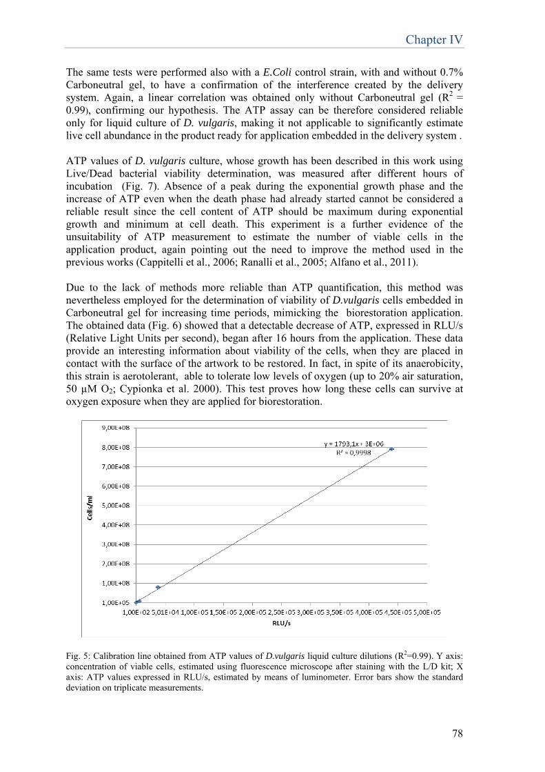
Chapter IV
78
The same tests were performed also with a E.Coli control strain, with and without 0.7% Carboneutral gel, to have a confirmation of the interference created by the delivery system. Again, a linear correlation was obtained only without Carboneutral gel (R2 = 0.99), confirming our hypothesis. The ATP assay can be therefore considered reliable only for liquid culture of D. vulgaris, making it not applicable to significantly estimate live cell abundance in the product ready for application embedded in the delivery system . ATP values of D. vulgaris culture, whose growth has been described in this work using Live/Dead bacterial viability determination, was measured after different hours of incubation (Fig. 7). Absence of a peak during the exponential growth phase and the increase of ATP even when the death phase had already started cannot be considered a reliable result since the cell content of ATP should be maximum during exponential growth and minimum at cell death. This experiment is a further evidence of the unsuitability of ATP measurement to estimate the number of viable cells in the application product, again pointing out the need to improve the method used in the previous works (Cappitelli et al., 2006; Ranalli et al., 2005; Alfano et al., 2011). Due to the lack of methods more reliable than ATP quantification, this method was nevertheless employed for the determination of viability of D.vulgaris cells embedded in Carboneutral gel for increasing time periods, mimicking the biorestoration application. The obtained data (Fig. 6) showed that a detectable decrease of ATP, expressed in RLU/s (Relative Light Units per second), began after 16 hours from the application. These data provide an interesting information about viability of the cells, when they are placed in contact with the surface of the artwork to be restored. In fact, in spite of its anaerobicity, this strain is aerotolerant, able to tolerate low levels of oxygen (up to 20% air saturation, 50 µM O2; Cypionka et al. 2000). This test proves how long these cells can survive at oxygen exposure when they are applied for biorestoration.
Fig. 5: Calibration line obtained from ATP values of D.vulgaris liquid culture dilutions (R2=0.99). Y axis: concentration of viable cells, estimated using fluorescence microscope after staining with the L/D kit; X axis: ATP values expressed in RLU/s, estimated by means of luminometer. Error bars show the standard deviation on triplicate measurements.
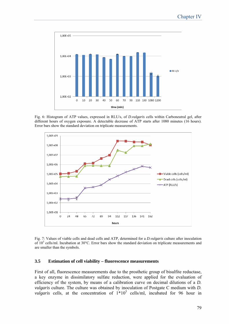
Chapter IV
79
Fig. 6: Histogram of ATP values, expressed in RLU/s, of D.vulgaris cells within Carboneutral gel, after different hours of oxygen exposure. A detectable decrease of ATP starts after 1080 minutes (16 hours). Error bars show the standard deviation on triplicate measurements.
Fig. 7: Values of viable cells and dead cells and ATP, determined for a D.vulgaris culture after inoculation of 105 cells/ml. Incubation at 30°C. Error bars show the standard deviation on triplicate measurements and are smaller than the symbols.
3.5 Estimation of cell viability – fluorescence measurements First of all, fluorescence measurements due to the prosthetic group of bisulfite reductase, a key enzyme in dissimilatory sulfate reduction, were applied for the evaluation of efficiency of the system, by means of a calibration curve on decimal dilutions of a D. vulgaris culture. The culture was obtained by inoculation of Postgate C medium with D. vulgaris cells, at the concentration of 1*105 cells/ml, incubated for 96 hour in
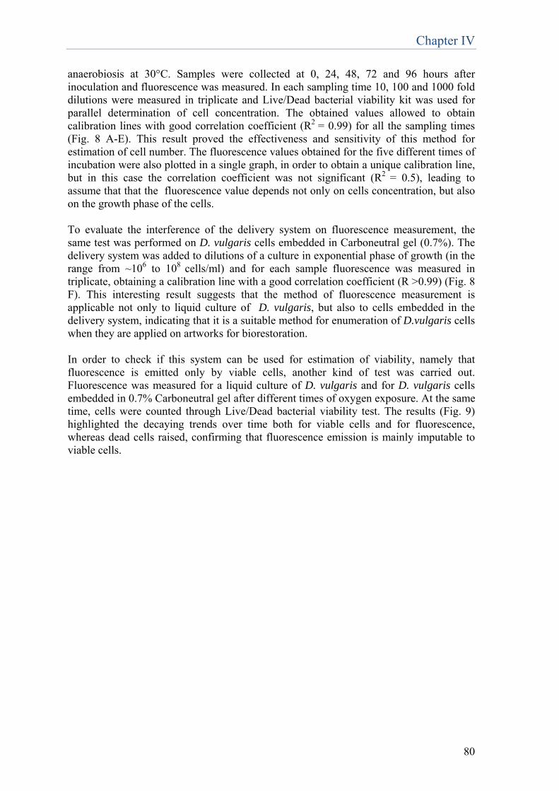
Chapter IV
80
anaerobiosis at 30°C. Samples were collected at 0, 24, 48, 72 and 96 hours after inoculation and fluorescence was measured. In each sampling time 10, 100 and 1000 fold dilutions were measured in triplicate and Live/Dead bacterial viability kit was used for parallel determination of cell concentration. The obtained values allowed to obtain calibration lines with good correlation coefficient (R2 = 0.99) for all the sampling times (Fig. 8 A-E). This result proved the effectiveness and sensitivity of this method for estimation of cell number. The fluorescence values obtained for the five different times of incubation were also plotted in a single graph, in order to obtain a unique calibration line, but in this case the correlation coefficient was not significant (R2 = 0.5), leading to assume that that the fluorescence value depends not only on cells concentration, but also on the growth phase of the cells. To evaluate the interference of the delivery system on fluorescence measurement, the same test was performed on D. vulgaris cells embedded in Carboneutral gel (0.7%). The delivery system was added to dilutions of a culture in exponential phase of growth (in the range from ~106 to 108 cells/ml) and for each sample fluorescence was measured in triplicate, obtaining a calibration line with a good correlation coefficient (R >0.99) (Fig. 8 F). This interesting result suggests that the method of fluorescence measurement is applicable not only to liquid culture of D. vulgaris, but also to cells embedded in the delivery system, indicating that it is a suitable method for enumeration of D.vulgaris cells when they are applied on artworks for biorestoration. In order to check if this system can be used for estimation of viability, namely that fluorescence is emitted only by viable cells, another kind of test was carried out. Fluorescence was measured for a liquid culture of D. vulgaris and for D. vulgaris cells embedded in 0.7% Carboneutral gel after different times of oxygen exposure. At the same time, cells were counted through Live/Dead bacterial viability test. The results (Fig. 9) highlighted the decaying trends over time both for viable cells and for fluorescence, whereas dead cells raised, confirming that fluorescence emission is mainly imputable to viable cells.
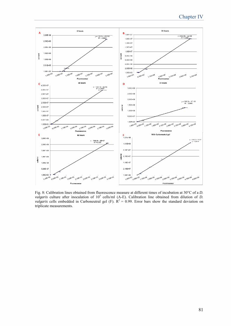
Chapter IV
81
Fig. 8: Calibration lines obtained from fluorescence measure at different times of incubation at 30°C of a D. vulgaris culture after inoculation of 105 cells/ml (A-E). Calibration line obtained from dilution of D. vulgaris cells embedded in Carboneutral gel (F). R2 = 0.99. Error bars show the standard deviation on triplicate measurements.
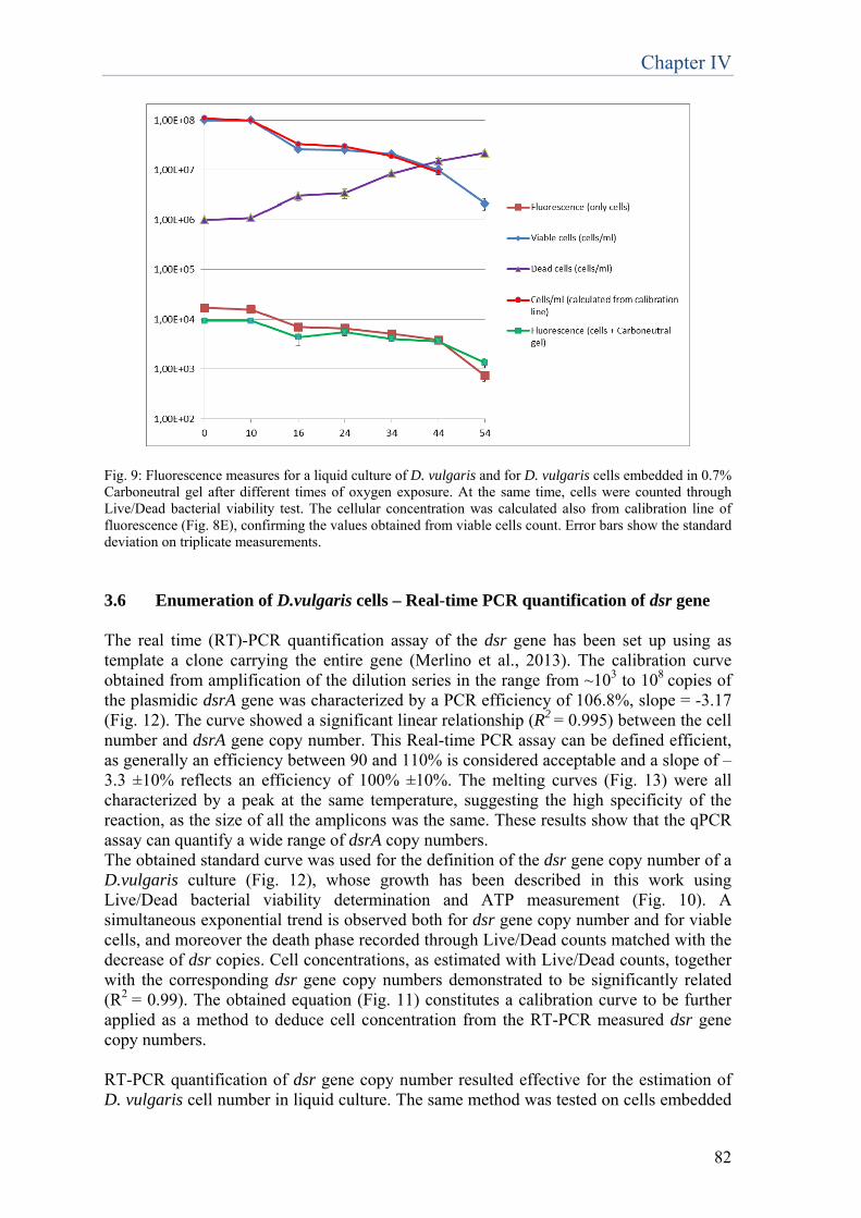
Chapter IV
82
Fig. 9: Fluorescence measures for a liquid culture of D. vulgaris and for D. vulgaris cells embedded in 0.7% Carboneutral gel after different times of oxygen exposure. At the same time, cells were counted through Live/Dead bacterial viability test. The cellular concentration was calculated also from calibration line of fluorescence (Fig. 8E), confirming the values obtained from viable cells count. Error bars show the standard deviation on triplicate measurements.
3.6 Enumeration of D.vulgaris cells – Real-time PCR quantification of dsr gene The real time (RT)-PCR quantification assay of the dsr gene has been set up using as template a clone carrying the entire gene (Merlino et al., 2013). The calibration curve obtained from amplification of the dilution series in the range from ~103 to 108 copies of the plasmidic dsrA gene was characterized by a PCR efficiency of 106.8%, slope = -3.17 (Fig. 12). The curve showed a significant linear relationship (R2 = 0.995) between the cell number and dsrA gene copy number. This Real-time PCR assay can be defined efficient, as generally an efficiency between 90 and 110% is considered acceptable and a slope of –3.3 ±10% reflects an efficiency of 100% ±10%. The melting curves (Fig. 13) were all characterized by a peak at the same temperature, suggesting the high specificity of the reaction, as the size of all the amplicons was the same. These results show that the qPCR assay can quantify a wide range of dsrA copy numbers. The obtained standard curve was used for the definition of the dsr gene copy number of a D.vulgaris culture (Fig. 12), whose growth has been described in this work using Live/Dead bacterial viability determination and ATP measurement (Fig. 10). A simultaneous exponential trend is observed both for dsr gene copy number and for viable cells, and moreover the death phase recorded through Live/Dead counts matched with the decrease of dsr copies. Cell concentrations, as estimated with Live/Dead counts, together with the corresponding dsr gene copy numbers demonstrated to be significantly related (R2 = 0.99). The obtained equation (Fig. 11) constitutes a calibration curve to be further applied as a method to deduce cell concentration from the RT-PCR measured dsr gene copy numbers. RT-PCR quantification of dsr gene copy number resulted effective for the estimation of D. vulgaris cell number in liquid culture. The same method was tested on cells embedded
Chapter IV
83
in the delivery system, to evaluate its suitability in determining the number of viable cells in the biorestoration product. The results showed that, while in control samples (without delivery system) PCR products were obtained for all the tested dilutions, when the same amounts of cells were originally embedded in Carboneutral gel, only for the most concentrated sample (107 cells/ml) a PCR product had been obtained (Fig. 14). This result highlights the potential interference created by the delivery system in reducing the DNA extraction or amplification efficiency. We can conclude that this method, demonstrated efficient to estimate cell number in liquid culture, has low sensibility when D. vulgaris cells are embedded in Carboneutral gel and can be applied only with high cell concentrations, >107 cells/ml. In biorestoration application, cells are applied at a concentration of 4*107 cells/ml, therefore by this method a concentration decay would not be detectable.
Fig. 10: Values of viable cells and dead cells, ATP and dsr copy number, determined for a D.vulgaris culture after inoculation of 105 cells/ml. Incubation at 30°C. Error bars show the standard deviation on triplicate measurements and are smaller than the symbols.
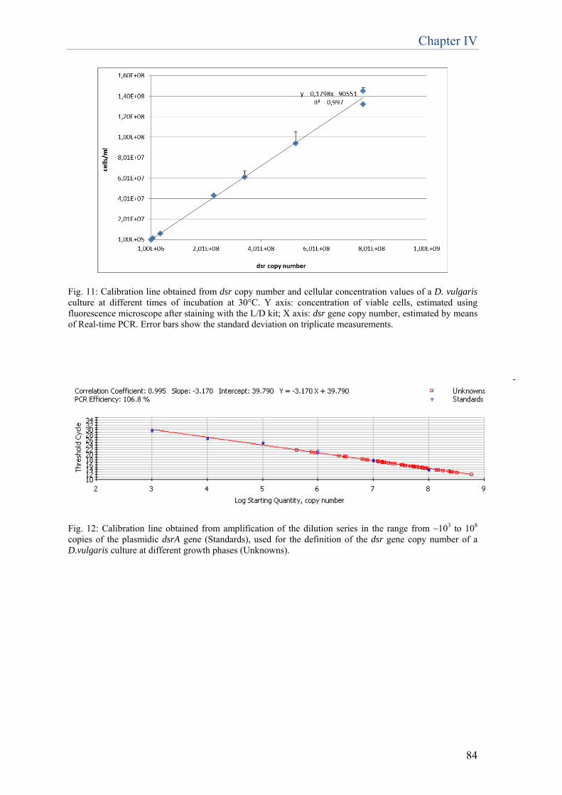
Chapter IV
84
Fig. 11: Calibration line obtained from dsr copy number and cellular concentration values of a D. vulgaris culture at different times of incubation at 30°C. Y axis: concentration of viable cells, estimated using fluorescence microscope after staining with the L/D kit; X axis: dsr gene copy number, estimated by means of Real-time PCR. Error bars show the standard deviation on triplicate measurements.
Fig. 12: Calibration line obtained from amplification of the dilution series in the range from ~103 to 108
copies of the plasmidic dsrA gene (Standards), used for the definition of the dsr gene copy number of a D.vulgaris culture at different growth phases (Unknowns).

Chapter IV
85
Temperature, CelsiusTemperature, Celsius
Fig. 13: Melting curves obtained from amplification of the dilution series in the range from ~103 to 108
copies of the plasmidic dsrA gene.
NC 10 3 M 10 4 10 5 10 6 10 7 10 3 10 4 10 710 5 10 6 M
Without Carboneutral gel With Carboneutral gel
NC 10 3 M 10 4 10 5 10 6 10 7 10 3 10 4 10 710 5 10 6 M
Without Carboneutral gel With Carboneutral gel
NC 10 3 M 10 4 10 5 10 6 10 7 10 3 10 4 10 710 5 10 6 M
Without Carboneutral gel With Carboneutral gel
Fig. 14: Analysis of dsr PCR products by 1.2% (w/v) agarose gel electrophoresis at 100V in 0.5X TBE buffer (Tris 1M, boric acid 13.7 g, EDTA 0.5 M pH 8). dsr PCR products obtained from dilutions of a D.vulgaris liquid culture (without Carboneutral gel) and from the same dilutions with addition of Carboneutral gel (shown in brilliant red). Values of cellular concentration (cells/ml) are reported. NC= negative control. M= marker. Interference created by the delivery system compromise the success of DNA extraction from <107 cells/ml samples.
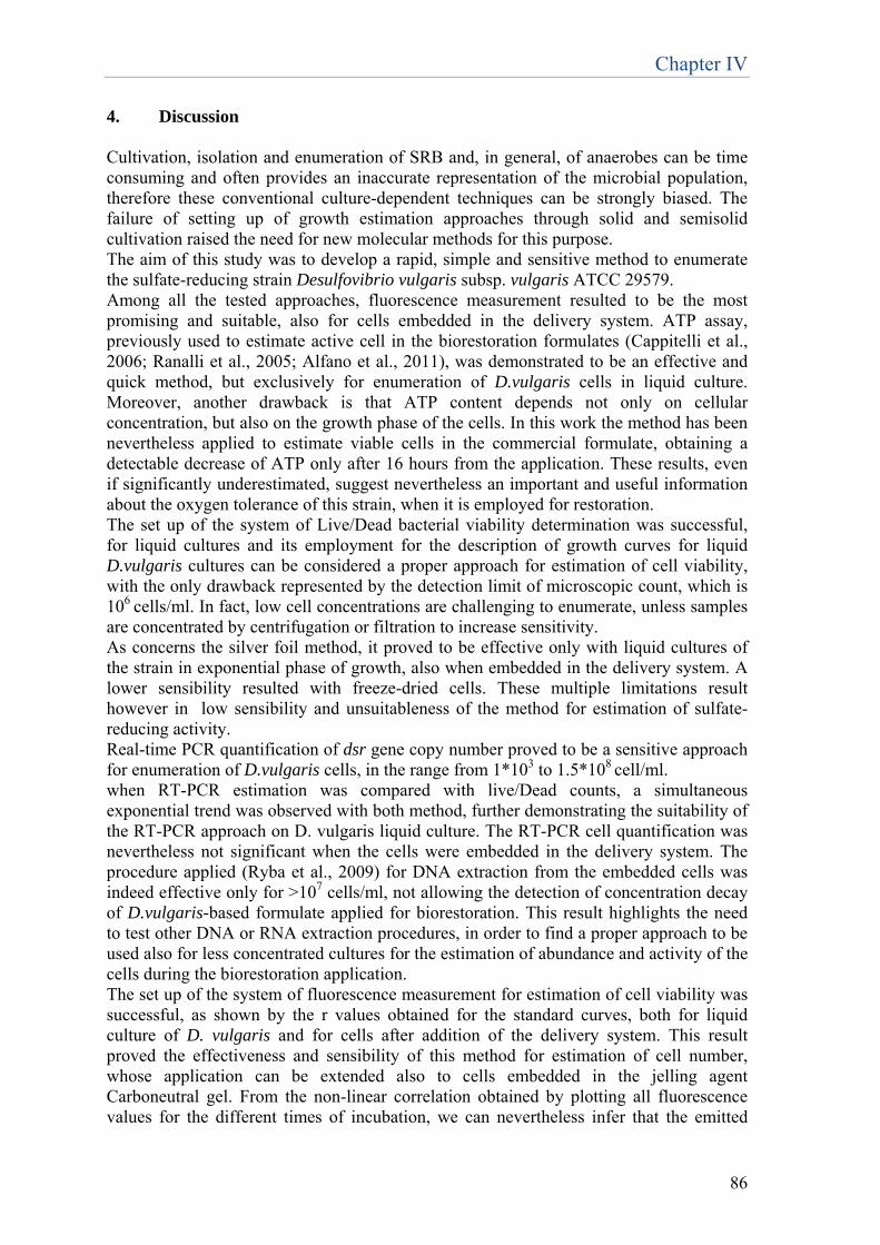
Chapter IV
86
4. Discussion Cultivation, isolation and enumeration of SRB and, in general, of anaerobes can be time consuming and often provides an inaccurate representation of the microbial population, therefore these conventional culture-dependent techniques can be strongly biased. The failure of setting up of growth estimation approaches through solid and semisolid cultivation raised the need for new molecular methods for this purpose. The aim of this study was to develop a rapid, simple and sensitive method to enumerate the sulfate-reducing strain Desulfovibrio vulgaris subsp. vulgaris ATCC 29579. Among all the tested approaches, fluorescence measurement resulted to be the most promising and suitable, also for cells embedded in the delivery system. ATP assay, previously used to estimate active cell in the biorestoration formulates (Cappitelli et al., 2006; Ranalli et al., 2005; Alfano et al., 2011), was demonstrated to be an effective and quick method, but exclusively for enumeration of D.vulgaris cells in liquid culture. Moreover, another drawback is that ATP content depends not only on cellular concentration, but also on the growth phase of the cells. In this work the method has been nevertheless applied to estimate viable cells in the commercial formulate, obtaining a detectable decrease of ATP only after 16 hours from the application. These results, even if significantly underestimated, suggest nevertheless an important and useful information about the oxygen tolerance of this strain, when it is employed for restoration. The set up of the system of Live/Dead bacterial viability determination was successful, for liquid cultures and its employment for the description of growth curves for liquid D.vulgaris cultures can be considered a proper approach for estimation of cell viability, with the only drawback represented by the detection limit of microscopic count, which is 106 cells/ml. In fact, low cell concentrations are challenging to enumerate, unless samples are concentrated by centrifugation or filtration to increase sensitivity. As concerns the silver foil method, it proved to be effective only with liquid cultures of the strain in exponential phase of growth, also when embedded in the delivery system. A lower sensibility resulted with freeze-dried cells. These multiple limitations result however in low sensibility and unsuitableness of the method for estimation of sulfate-reducing activity. Real-time PCR quantification of dsr gene copy number proved to be a sensitive approach for enumeration of D.vulgaris cells, in the range from 1*103 to 1.5*108 cell/ml. when RT-PCR estimation was compared with live/Dead counts, a simultaneous exponential trend was observed with both method, further demonstrating the suitability of the RT-PCR approach on D. vulgaris liquid culture. The RT-PCR cell quantification was nevertheless not significant when the cells were embedded in the delivery system. The procedure applied (Ryba et al., 2009) for DNA extraction from the embedded cells was indeed effective only for >107 cells/ml, not allowing the detection of concentration decay of D.vulgaris-based formulate applied for biorestoration. This result highlights the need to test other DNA or RNA extraction procedures, in order to find a proper approach to be used also for less concentrated cultures for the estimation of abundance and activity of the cells during the biorestoration application. The set up of the system of fluorescence measurement for estimation of cell viability was successful, as shown by the r values obtained for the standard curves, both for liquid culture of D. vulgaris and for cells after addition of the delivery system. This result proved the effectiveness and sensibility of this method for estimation of cell number, whose application can be extended also to cells embedded in the jelling agent Carboneutral gel. From the non-linear correlation obtained by plotting all fluorescence values for the different times of incubation, we can nevertheless infer that the emitted
Chapter IV
87
fluorescence depends not only on cellular concentration, but also on the growth phase of the cells. Therefore, when using calibration lines for determination of cellular concentration from a known fluorescence value, we can compare only results obtained on cells in the same growth phase. The performed test to verify that emission of fluorescence derives only from viable cells, highlighted the decaying trends over time both for viable cells and for fluorescence, whereas dead cells raised. These results were the proof that fluorescence emission is ascribable just to viable cells. In this test, cellular concentration was estimated using Live/Dead bacterial viability determination, but also from calibration line of fluorescence. The two curves obtained were almost coincident, in fact for the values obtained after 16 hours of incubation, the standard deviation between the two values corresponded to 15% of the cellular concentration estimated using calibration line of fluorescence, after 24 hours of incubation corresponded to 10%, after 34 and 44 hours of incubation corresponded to 7%. Among all the tested methods, fluorescence measurement, in spite of its limitations, resulted to be the best molecular approach for the quantification of the SRB Desulfovibrio vulgaris subsp. vulgaris ATCC 29579, as it is the only one adaptable also to cells embedded in the delivery system Carboneutral gel. References Alfano, G., Lustrato, G., Belli, C., Zanardini, E., Cappitelli, F., Mello, E., Sorlini, C., Ranalli, G. (2011). The bioremoval of nitrate and sulfate alterations on artistic stonework: the case-study of Matera Cathedral after six years from the treatment. International Biodeterioration & Biodegradation, 65, 1004-1011. Barton, L.L., Carpenter, C.M. (2013). Suitability of fluorescence measurements to quantify sulfate-reducing bacteria. Journal of Microbiol. Methods. 93, 192-197. Cappitelli, F., Zanardini, E., Ranalli, G., Mello, E., Daffonchio, D., & Sorlini, C. (2006). Improved Methodology for Bioremoval of Black Crusts on Historical Stone Artworks by Use of Sulfate-Reducing Bacteria, Applied and Environmental Microbiology, 72(5), 3733–3737.
Crouch, S.P.M, Kozlowski, R., Slater, K.J. and Fletcher, J. (1993). The use of ATP bioluminescence as a measure of cell proliferation and cytotoxicity. Journal of Immunological Methods.160, 81-88.
Cypionka, H. (2000). Oxygen respiration by Desulfovibrio species. Annu. Rev. Microbiol, 54, 827-48. Daffonchio, D., De Biase, A., Rizzi, A. and Sorlini, C. (1998) Interspecific, intraspecific and interoperonic variability in the 16S rRNA gene of methanogens revealed by length and single-strand conformation polymorphism analysis. FEMS Microbiology Letters, vol. 164, no. 2, pp. 403–410. Dar, S. A., Yao, L., van Dongen, U., Kuenen, J. G. & Muyzer, G. (2007) Analysis of diversity and activity of sulfatereducing bacterial communities in sulfidogenic bioreactors using 16S rRNA and dsrB genes as molecular markers. Appl. Environ. Microbiol. 73, 594–604.
Chapter IV
88
Gatti, M., Bernini, V., Lazzi, C. and Neviani, E. (2006). Fluorescence microscopy for studying the viability of micro-organisms in natural whey starters. Letters in Applied Microbiology 42, 338-343. Iverson, W.P. (1966). Growth of Desulfovibrio on the surface of agar media. Appl. Microbiol. 14 (4), 529-534. Itoh, T., Suzuki, K‑I. & Nakase, T. (1998) Thermocladium modestius gen. nov., sp. nov. a new genus of rodshaped, extremely thermophilic crenarchaeote. Int. J. Syst. Bacteriol. 48, 879–887. Itoh, T., Suzuki, K‑I., Sanches, P. C. & Nakase, T. (1999) Caldivirga maquilingensis gen. nov., sp. nov. a new genus of rod-shaped crenarchaeote isolated from a hot spring in the Philippines. Int. J. Syst. Bacteriol. 49, 1157–1163. Jain, D.K. (1995). Evaluation of the semisolid Postgate’s B medium for enumerating sulfate-reducing bacteria. Journal of Microbiological Methods 22, 27-38. Jara C., Mateo E., Guillamòn J. M., Torija M. J., Mas A. (2008) Analysis of several methods for the extraction of high quality DNA from acetic acid bacteria in wine and vinegar for characteriazation by PCR-based methods. Int. J. Food Micr. 128: 336-341. Kondo, R., Osawa, K., Mochizuki, L., Fujioka Y. & Butani J. (2006). Abundance and diversity of sulphate-reducing bacterioplankton in Lake Suigetsu, a meromictic lake in Fukui, Japan. Plankton Benthos Res 1(4), 165-177. Kondo, R. and Butani, J. (2007). Comparison of the diversity of sulfate-reducing bacterial communities in the water column and the surface sediments of a Japanese meromictic lake. Limnology 8, 131–141. Kondo, R., Shigematsu, K., & Butani, J. (2008). Rapid enumeration of sulphate-reducing bacteria from aquatic environments using real-time PCR. Plankton Benthos Res 3(3), 180-183. Kondo, R., Nedwell, D.B., Purdy, K.J., Silva, S.Q. (2004). Detection and enumeration of sulphate-reducing bacteria in estuarine sediments by competitive PCR. Geomicrobiol J 21: 145-157. Kondo, R., Purdy, K.J., Silva, S.Q., Nedwell, D.B. (2007). Spatial dynamics of sulphate-reducing bacterial compositions in sediment along a salinity gradient in a UK estuary. Microbes Environ 22, 11-19. Leloup, J., Loy, A., Knab, N.J., Borowski, C., Wagner M and Jørgensen B.B. (2007). Diversity and abundance of sulfate-reducing microorganisms in the sulfate and methane zones of a marine sediment, Black Sea. Environmental Microbiology 9(1), 131-142. Loy, A. et al. (2002) Oligonucleotide microarray for 16S rRNA gene-based detection of all recognized lineages of sulfate-reducing prokaryotes in the environment. Appl. Environ. Microbiol. 68, 5064–5081.

Chapter IV
89
Martins, M., Faleiro, M.L., Barros, R.J., Veríssimo, A.R., Barreiros, M.A., Costa, M.C. (2009). Characterization and activity studies of highly heavy metal resistant sulphate-reducing bacteria to be used in acid mine drainage decontamination. J. Hazard. Mater. 166, 706–13. Menert, A., Paalme, V., Juhkam, J., Vilu, R. (2004). Characterization of sulfate-reducing bacteria in yeast industry waste by microcalorimetry and PCR amplification. Thermochimica Acta 420, 89–98. Merlino, G., Rizzi, A., Schievano, A., Tenca, A., Scaglia, B., Oberti, R., Adani, F., Daffonchio, D. (2013) Microbial community structure and dynamics in two-stage vs single-stage thermophilic anaerobic digestion of mixed swine slurry and market biowaste. Water research, 47, 1983-1995. Meyer, B. & Kuever, J. (2007) Phylogeny of the alpha and beta subunits of the dissimilatory adenosine‑5′-phosphosulfate (APS) reductase from sulfate-reducing prokaryotes — origin and evolution of the dissimilatory sulfate-reduction pathway. Microbiology 153, 2026–2044. Minz, D. et al. (1999) Diversity of sulfate-reducing bacteria in oxic and anoxic regions of a microbial mat characterized by comparative analysis of dissimilatory sulfite reductase genes. Appl. Environ. Microbiol. 65, 4666–4671. Mori, K., Kim, H., Kakegawa, T. & Hanada, S. (2003) A novel lineage of sulphate-reducing microorganisms: Thermodesulfobiaceae fam. nov., Thermodesulfobium narugense, gen. nov., sp. nov. a new thermophilic isolate from a hot spring. Extremophiles 7, 283–290. Muyzer, G. & Stams, A. J. M. (2008). The ecology and biotechnology of sulphate-reducing bacteria. Nat. Rev. Microbiol. 6, 441-54. Parkes, R. J. (1987) in Ecology of Microbial Communities (eds Fletcher, M., Gray, T. R. & Jones, J. G.) 147–177 (Cambridge Univ. Press). Postgate, J. (1963). Versatile medium for the enumeration of sulfate-reducing bacteria. Appl. Microbiol. 11, 265-267. Postgate, J. (1959). Sulphate reduction by bacteria. Ann. Rev. Microbiol. 13, 505-520. Ranalli, G., Alfano, G., Belli, C., Lustrato, G., Colombini, M.P., Bonaduce, I., Zanardini, E., Abbruscato, P., Cappitelli, F and Sorlini, C. (2005). Biotechnology applied to cultural heritage: biorestoration of frescoes using viable bacterial cells and enzymes. Journal of Applied Microbiology 98, 73-83. Ranalli, G., Chiavarini, M., Guidetti, V., Marsala, F., Matteini, M., Zanardini, E., Sorlini, C. (1997). The use of microorganisms for the removal of sulfates on artistic stoneworks. Int. Biodet. Biodeg. 40, 255-261.

Chapter IV
90
Ryba, S., Titera, D., Haklova, M., Stopka, P. (2009). A PCR method of detecting American Foulbrood (Paenibacillus larvae) in winter beehive wax debris. Veterinary Microbiology 139, 193-196. Schippers, A. and Neretin, L.N. (2006). Quantification of microbial communities in near-surface and deeply buried marine sediments on the Peru continental margin using real-time PCR. Environ Microbiol 8, 1251-1260. Sigalevich, P., Meshorer, E., Helman, Y. and Cohen, Y. (2000). Transition from Anaerobic to Aerobic Growth Conditions for the Sulfate-Reducing Bacterium Desulfovibrio oxyclinae Results in Flocculation. Appl. Environ. Microbiol. 66 (11) 5005-5012. Spence, C., Whitehead, T.R., and Cotta, M.A. (2008). Development and comparison of SYBR Green quantitative real-time PCR assays for detection and enumeration of sulfate-reducing bacteria in stored swine manure. Journal of Applied Microbiology 105, 2143-2152. Wagner, M., Roger, A.J., Flax, J.L., Brusseau, G.A. and Stahl, D.A. (1998). Phylogeny of Dissimilatory Sulfite Reductases Supports an Early Origin of Sulfate Respiration. J. Bacteriol. 180 (11), 2975–2982.
Chapter V
91
Successful combination of chemical and biological treatments for the cleaning of stone artworks
Published by: Troiano F, Gulotta D, Balloi A, Polo A, Toniolo L, Lombardi E, Daffonchio D, Sorlini C, Cappitelli F (2013) International Biodeterioration & Biodegradation 85, 294–304. Abstract The removal of sulfate-based crusts from stone artworks using sulfate-reducing bacteria (SRB) has been proven to be an effective cleaning procedure. However, some concerns still remain: it is a time-consuming process when the crust is thick, and it is not clear how the biotechnological approach can fit within a complex conservation treatment. To address these challenges, the effects of an SRB strain (Desulfovibrio vulgaris subsp. vulgaris ATCC 29579) coupled with a non-ionic detergent pre-treatment was studied on a stone column affected by black crusts. The coupling of the two treatments removed the black crust without affecting the original sound marble, with 38% reduction in cleaning time. The combined method was later applied to a one-century-old artistic marble statue weathered by sulfate-based crusts and grey deposits. The detergent used alone effectively removed the grey deposit, but not the black crust. However the co-treatment synergy resulted in the complete removal of the black crust layers, with the added advantage, compared to the biocleaning alone, of fewer biological applications and a 70% reduction in total cleaning time, but still retaining all the advantages of the biocleaning approach. 1. Introduction
Stone surfaces on buildings exposed to the urban environment undergo rapid and differentiated deterioration due to chemical and physical attack (Camuffo et al., 1983). Dark grey or black surface layers can be found everywhere on lithoid materials, especially on sculpted elements sheltered from direct rainfall; some alterations derive only from a deposition mechanism, others from the sulfation of marble and soiling deposition acting contemporaneously (Moropoulou et al., 1998; Toniolo et al., 2009). Marble sulfation occurs in the presence of moisture when sulfur dioxide, a major urban atmosphere pollutant, is converted to sulfuric acid, which reacts with marble and other soluble calcareous substrates to form gypsum (Böke et al., 1999). During gypsum crystallization, airborne organic pollutants and carbonaceous particles accumulate on surfaces protected from rainfall and wash-out, and are subsequently trapped in the newly-formed mineral matrix (Moropoulou et al., 1998) to form so-called ‘black crust’ (El-Metwally and Ramadan, 2005). Air pollution also causes the deposition of a variety of organic and inorganic compounds on the surface of historic monuments. Such deposits contain a gypsum component but have greater porosity than black crust because gypsum does not come from the chemical corrosion of the substrate, it comes from an atmospheric deposition mechanism of carbonaceous particles, mixed together with dust, pollen, and spores from the atmosphere (Alessandrini et al., 2002; Toniolo et al., 2009). A cleaning method for the removal of altered layers must respect the chemical-physical nature of the material of the artwork, and its historical and artistic value, but when there are different weathering forms on a stone surface it is difficult to use a single cleaning procedure (Slaton and Normandin, 2005). Indeed, quite often, more than one cleaning method is needed. However, even though it is common in the conservation field to combine cleaning procedures
Chapter V
92
(Bromblet et al., 2003), the cleaning methods themselves can interact with the surface materials and with each other (Sansonetti et al., 2008). Biocleaning has been tested, successfully, on many materials including white and colored marbles (Cappitelli et al., 2007; Gioventù et al., 2011) and porous limestone (Polo et al., 2010), and has proved to be more selective than mechanical, chemical and laser techniques as it efficiently removes black crusts without affecting the stone surface (Cappitelli et al., 2007; Gioventù et al., 2011). Despite being a successful treatment, biocleaning alone has been found to be highly time-consuming in the presence of thick and compact crusts, and its compatibility with other cleaning procedures has not, until now, ever been studied (Gioventù et al., 2011). The aim of the present study was to assess the effects of a chemical and sulfate-reducing bacteria (SRB) co-treatment for the removal of black crusts and grey deposits. The method was first tested on an old outdoor stone column that had been exposed to a polluted environment for a long time. It was then applied to an old artwork, a funeral monument realized by Lina Arpesani in 1921 in memory of ‘Neera’, the poetess Anna Zuccari. The marble statue, a naked young woman supporting a big open book on her head, had stood in the courtyard of the Cimitero Monumentale in Milan (Italy) and therefore had long been exposed to outdoor conditions and the polluted urban atmosphere; in 1958 the statue was moved to the Famedio porch of the same cemetery, where it is currently located in a semi-confined environment. Its state of conservation was good and there was no evidence of structural damage. However, visual inspection revealed two main alterations: a putative grey deposit covering all the surface and black crust-like alterations in the areas formerly protected from the direct rain-wash, i.e. under the right arm and on the face. Considering the putative nature of the weathering forms of the marble statue, two cleaning methods were combined and an assessment made of their interaction and synergic effect: the soft detergent Tween 20 together with a SRB-based biocleaning procedure for the removal of the grey deposit and black crust respectively. Up until then, nothing was known of the interaction between biocleaning and other chemical cleaning procedures like the use of detergents. 2. Materials and methods 2.1 Sampling and material characterization Sampling was by means of a micro-chisel and a steel scalpel for both the stone column and the statue, and was as little invasive as possible. The samples were selected so as to be representative of the surface before and after the different cleaning procedures (see paragraph 2.2): i) surface with a black crust-like alteration before any treatment (Figure 1a); ii) biologically cleaned surface; iii) chemically cleaned surface; iv) surface after the chemical pre-treatment and prior to the subsequent biological cleaning; v) surface after combined chemical and biological cleaning. Samples taken from the column are coded with the letter ‘C’, and samples from the statue with ‘S’ (Table 1). The stereomicroscope observations were performed with a Leica M205C, equipped with a Leica DFC290 digital camera. For optical microscopy, the samples were prepared in polished cross-sections with bi-component epoxy resin. Optical microscopy was carried out with a Leica DMRE Microscope, equipped with a Leica DFC290 digital camera. Environmental Scanning Electron Microscopy (ESEM) for morphological observations of the sample fragments and polished cross-sections was performed with an Environmental

Chapter V
93
Scanning Electron Microscope Zeiss EVO 50 EP equipped with an Oxford INCA 200 - Pentafet LZ4 spectrometer in Secondary Electrons mode. X-ray diffraction (XRD) analyses on powder materials were performed by a Philips PW1830 instrument with Bragg-Brentano and Thin Film geometry, using a Cu anticathode and Kα radiation (λ = 1.54058 Å). Fourier Transformed Infrared Spectroscopy (FTIR) analyses were carried out with a Thermo Nicolet 6700 instrument using a DTGS detector in the spectral range 4000 - 400 cm-1, coupled with a FTIR Thermo Nicolet Continuum microscope using a MCT detector in the spectral range 4000 - 600 cm-1. Sample fragments were ground in an agate mortar, and analyzed after dispersion in KBr pellets (KBr FTIR grade by Sigma-Aldrich). Micro-fragments were analyzed by means of a compressive diamond cell. Average spectra of 128 scans were recorded for each sample.
Fig. 1: (a) is the marble statue before the cleaning; the arrows indicate the following sampling areas: sample S1A for the characterization of the grey deposit, sample S1B for the characterization of the black crust-like alteration. Samples S2-4 were taken after cleaning tests with different methods: sample S2 after the biological treatment, sample S3 after the combined chemical and biological treatment, and sample S4 after the chemical treatment as control. (b) shows the results after the removal of the grey deposit and the black crusts on the whole statue. (c, e) show the details before the cleaning. (d, f) give the details after the cleaning.
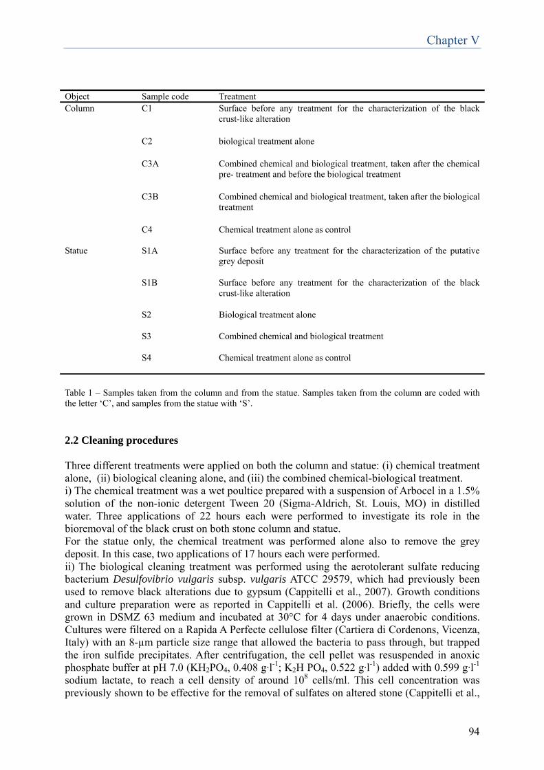
Chapter V
94
Object Sample code Treatment Column C1 Surface before any treatment for the characterization of the black
crust-like alteration
C2 biological treatment alone
C3A Combined chemical and biological treatment, taken after the chemical pre- treatment and before the biological treatment
C3B Combined chemical and biological treatment, taken after the biological treatment
C4 Chemical treatment alone as control
Statue S1A Surface before any treatment for the characterization of the putative grey deposit
S1B Surface before any treatment for the characterization of the black crust-like alteration
S2 Biological treatment alone
S3 Combined chemical and biological treatment
S4 Chemical treatment alone as control
Table 1 – Samples taken from the column and from the statue. Samples taken from the column are coded with the letter ‘C’, and samples from the statue with ‘S’. 2.2 Cleaning procedures Three different treatments were applied on both the column and statue: (i) chemical treatment alone, (ii) biological cleaning alone, and (iii) the combined chemical-biological treatment. i) The chemical treatment was a wet poultice prepared with a suspension of Arbocel in a 1.5% solution of the non-ionic detergent Tween 20 (Sigma-Aldrich, St. Louis, MO) in distilled water. Three applications of 22 hours each were performed to investigate its role in the bioremoval of the black crust on both stone column and statue. For the statue only, the chemical treatment was performed alone also to remove the grey deposit. In this case, two applications of 17 hours each were performed. ii) The biological cleaning treatment was performed using the aerotolerant sulfate reducing bacterium Desulfovibrio vulgaris subsp. vulgaris ATCC 29579, which had previously been used to remove black alterations due to gypsum (Cappitelli et al., 2007). Growth conditions and culture preparation were as reported in Cappitelli et al. (2006). Briefly, the cells were grown in DSMZ 63 medium and incubated at 30°C for 4 days under anaerobic conditions. Cultures were filtered on a Rapida A Perfecte cellulose filter (Cartiera di Cordenons, Vicenza, Italy) with an 8-µm particle size range that allowed the bacteria to pass through, but trapped the iron sulfide precipitates. After centrifugation, the cell pellet was resuspended in anoxic phosphate buffer at pH 7.0 (KH2PO4, 0.408 g·l-1; K2H PO4, 0.522 g·l-1) added with 0.599 g·l-1
sodium lactate, to reach a cell density of around 108 cells/ml. This cell concentration was previously shown to be effective for the removal of sulfates on altered stone (Cappitelli et al.,
Chapter V
95
2006). All the manipulations described above were done under anaerobic conditions in a glove box. The biomass was entrapped in an Arbocel polymeric matrix (CTS, Vicenza, Italy) as the delivery system (Cappitelli et al., 2006). The treatment procedure was as previously reported in Cappitelli et al. (2007). Briefly, the surfaces to be treated were moistened with the phosphate buffer and covered with tissue paper before any applications began. The biological cleaning system was covered with a polyvinylchloride film (Silplast, Italy) to reduce undesirable evaporation. After the treatment, removal of the bioformulate was accomplished by removing the tissue paper and subsequently washing the area with distilled water. Applications of 22 hours were applied on the altered surface of both stone column and statue up to the complete removal of black crust. No treatment with only the delivery system (without entrapped microorganisms) was carried out as this had never removed black crust in previous experiments (Cappitelli et al., 2006; Cappitelli et al., 2007; Polo et al., 2010; Gioventù et al., 2011). iii) The combined chemical-biological treatment consisted of two steps. First, a chemical pre-treatment consisting of a wet poultice prepared with 1.5% solution of Tween 20 in Arbocel as reported above. This poultice was applied to the black crust for two hours, and covered by a polyvinylchloride film. The site was then rinsed and the residual poultice washed off with distilled water while gently wiping with a small brush. The second step, the biological treatment performed as reported above, consisted of 22 hour-applications up to the complete removal of black crust. 3. Results 3.1 Column 3.1.1 Chemical characterization before treatment The preliminary stereomicroscopic observation of sample C1 showed the presence of a diffused and rather compact black colored alteration covering most of the stone surface (Figure 2a). After optical observation of the polished cross-section (Figure 2b), the sample stratigraphy appeared to be composed of a single crust layer (layer 2) overlapping the stone substrate (layer 1). The alteration continued along the entire section, with an irregular shape and thickness varying from less than 50 µm to almost 200 µm. A few red particles embedded in a generally very dark crust matrix were also observed. The mineralogical and compositional characterization of the alteration after XRD and FTIR analysis confirmed the carbonatic nature of the substrate, which was composed of calcite. The alteration composition included gypsum (FTIR peaks at 3543-3405, 1685-1621, 1142-1117, 670-602 cm-1) as main component, together with nitrates (FTIR peak at 1384 cm-1), silicates and quartz (FTIR peaks at 1005, 797-779 cm-1), calcite (FTIR peaks at 1427, 875, 713 cm-1). ESEM-EDX results highlighted a dramatic weathering of the stone substrate (Figure 2c) but no sulfation, according to the EDX spectrum of this area (Figure 2d). Intergranular corrosion was observed along the borders of the calcite grains and the mineral matrix appeared to be deeply disaggregated. Voids and discontinuities were present between the substrate and the black alteration layer (layer 2). The latter was rather heterogeneous and its composition mainly included Si, S, and Ca, together with Al, Fe and Mg (Figure 2e). Some residual traces of a thin discontinuous Fl-enriched layer between the substrate and the crust were also identified. The overall data indicated that the superficial dark alteration on the stone surface was a black crust.
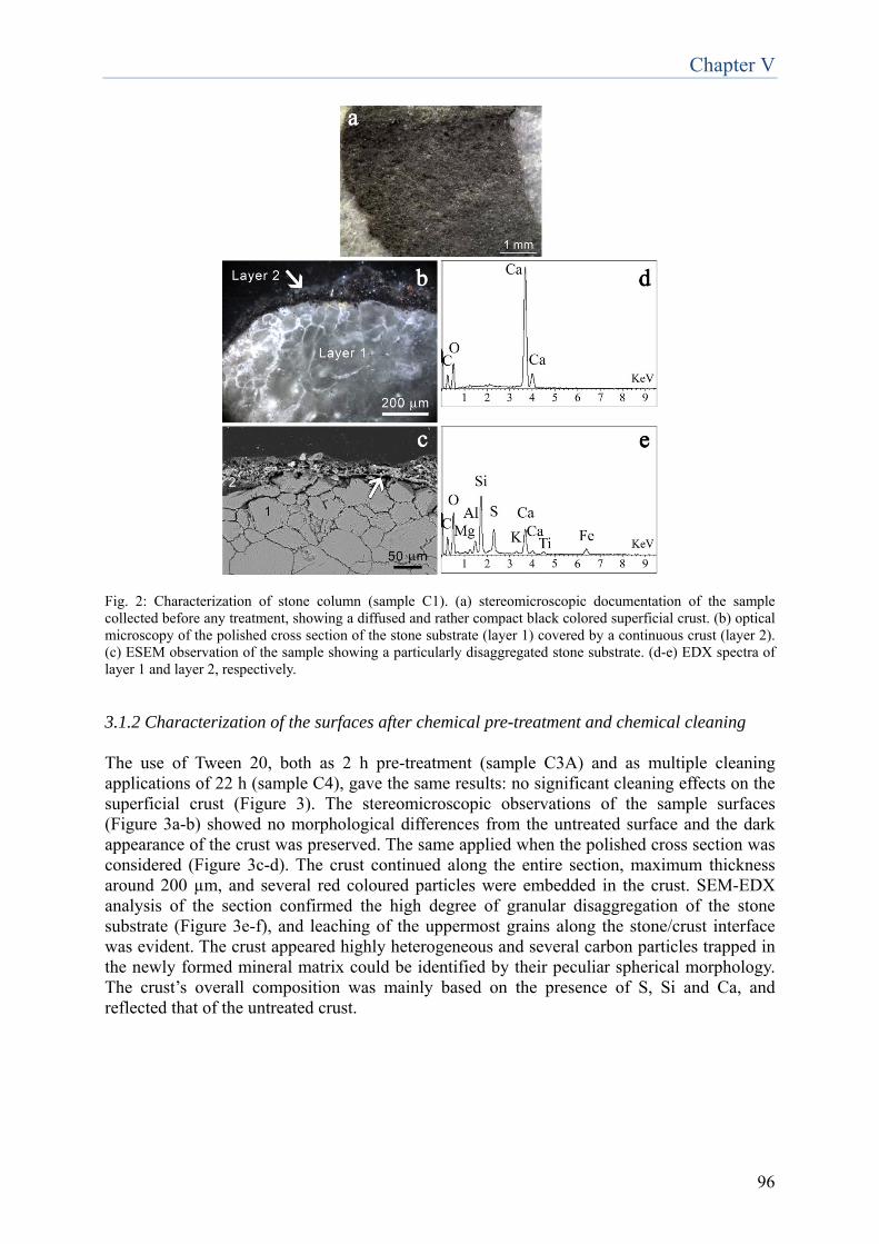
Chapter V
96
Fig. 2: Characterization of stone column (sample C1). (a) stereomicroscopic documentation of the sample collected before any treatment, showing a diffused and rather compact black colored superficial crust. (b) optical microscopy of the polished cross section of the stone substrate (layer 1) covered by a continuous crust (layer 2). (c) ESEM observation of the sample showing a particularly disaggregated stone substrate. (d-e) EDX spectra of layer 1 and layer 2, respectively. 3.1.2 Characterization of the surfaces after chemical pre-treatment and chemical cleaning The use of Tween 20, both as 2 h pre-treatment (sample C3A) and as multiple cleaning applications of 22 h (sample C4), gave the same results: no significant cleaning effects on the superficial crust (Figure 3). The stereomicroscopic observations of the sample surfaces (Figure 3a-b) showed no morphological differences from the untreated surface and the dark appearance of the crust was preserved. The same applied when the polished cross section was considered (Figure 3c-d). The crust continued along the entire section, maximum thickness around 200 µm, and several red coloured particles were embedded in the crust. SEM-EDX analysis of the section confirmed the high degree of granular disaggregation of the stone substrate (Figure 3e-f), and leaching of the uppermost grains along the stone/crust interface was evident. The crust appeared highly heterogeneous and several carbon particles trapped in the newly formed mineral matrix could be identified by their peculiar spherical morphology. The crust’s overall composition was mainly based on the presence of S, Si and Ca, and reflected that of the untreated crust.
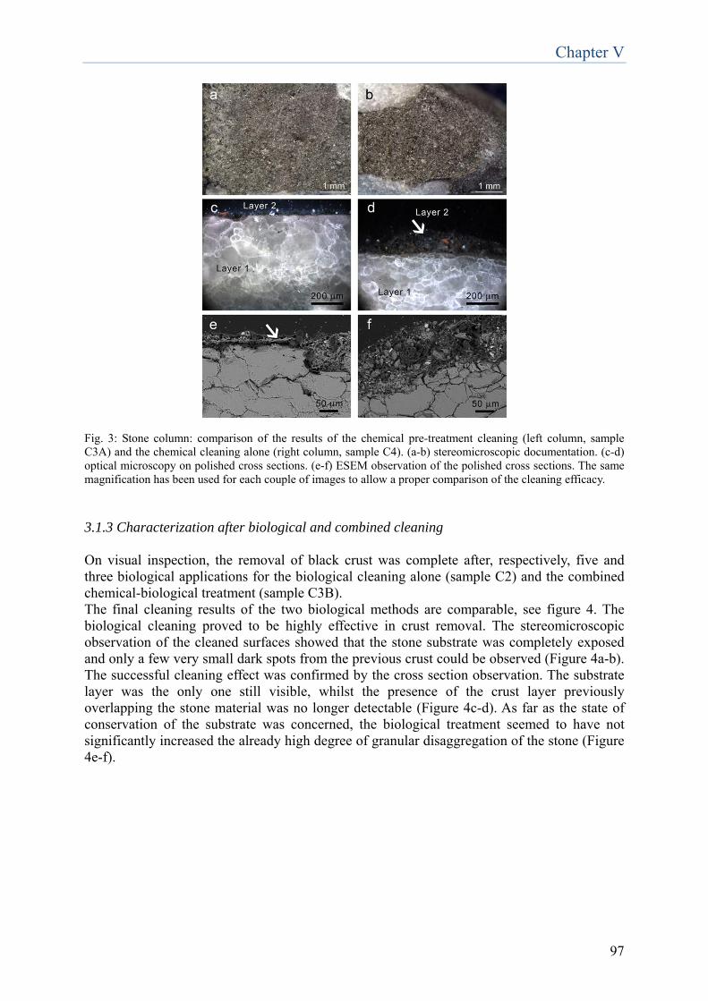
Chapter V
97
Fig. 3: Stone column: comparison of the results of the chemical pre-treatment cleaning (left column, sample C3A) and the chemical cleaning alone (right column, sample C4). (a-b) stereomicroscopic documentation. (c-d) optical microscopy on polished cross sections. (e-f) ESEM observation of the polished cross sections. The same magnification has been used for each couple of images to allow a proper comparison of the cleaning efficacy. 3.1.3 Characterization after biological and combined cleaning On visual inspection, the removal of black crust was complete after, respectively, five and three biological applications for the biological cleaning alone (sample C2) and the combined chemical-biological treatment (sample C3B). The final cleaning results of the two biological methods are comparable, see figure 4. The biological cleaning proved to be highly effective in crust removal. The stereomicroscopic observation of the cleaned surfaces showed that the stone substrate was completely exposed and only a few very small dark spots from the previous crust could be observed (Figure 4a-b). The successful cleaning effect was confirmed by the cross section observation. The substrate layer was the only one still visible, whilst the presence of the crust layer previously overlapping the stone material was no longer detectable (Figure 4c-d). As far as the state of conservation of the substrate was concerned, the biological treatment seemed to have not significantly increased the already high degree of granular disaggregation of the stone (Figure 4e-f).
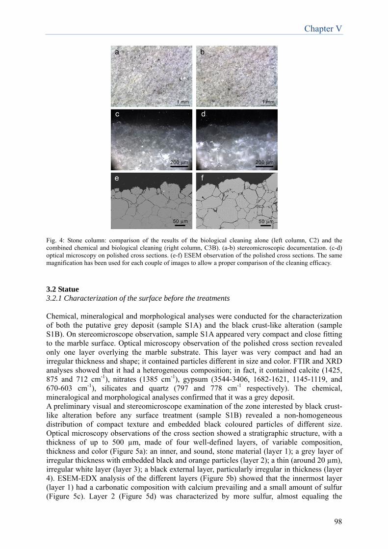
Chapter V
98
Fig. 4: Stone column: comparison of the results of the biological cleaning alone (left column, C2) and the combined chemical and biological cleaning (right column, C3B). (a-b) stereomicroscopic documentation. (c-d) optical microscopy on polished cross sections. (e-f) ESEM observation of the polished cross sections. The same magnification has been used for each couple of images to allow a proper comparison of the cleaning efficacy. 3.2 Statue 3.2.1 Characterization of the surface before the treatments Chemical, mineralogical and morphological analyses were conducted for the characterization of both the putative grey deposit (sample S1A) and the black crust-like alteration (sample S1B). On stereomicroscope observation, sample S1A appeared very compact and close fitting to the marble surface. Optical microscopy observation of the polished cross section revealed only one layer overlying the marble substrate. This layer was very compact and had an irregular thickness and shape; it contained particles different in size and color. FTIR and XRD analyses showed that it had a heterogeneous composition; in fact, it contained calcite (1425, 875 and 712 cm-1), nitrates (1385 cm-1), gypsum (3544-3406, 1682-1621, 1145-1119, and 670-603 cm-1), silicates and quartz (797 and 778 cm-1 respectively). The chemical, mineralogical and morphological analyses confirmed that it was a grey deposit. A preliminary visual and stereomicroscope examination of the zone interested by black crust-like alteration before any surface treatment (sample S1B) revealed a non-homogeneous distribution of compact texture and embedded black coloured particles of different size. Optical microscopy observations of the cross section showed a stratigraphic structure, with a thickness of up to 500 µm, made of four well-defined layers, of variable composition, thickness and color (Figure 5a): an inner, and sound, stone material (layer 1); a grey layer of irregular thickness with embedded black and orange particles (layer 2); a thin (around 20 µm), irregular white layer (layer 3); a black external layer, particularly irregular in thickness (layer 4). ESEM-EDX analysis of the different layers (Figure 5b) showed that the innermost layer (layer 1) had a carbonatic composition with calcium prevailing and a small amount of sulfur (Figure 5c). Layer 2 (Figure 5d) was characterized by more sulfur, almost equaling the
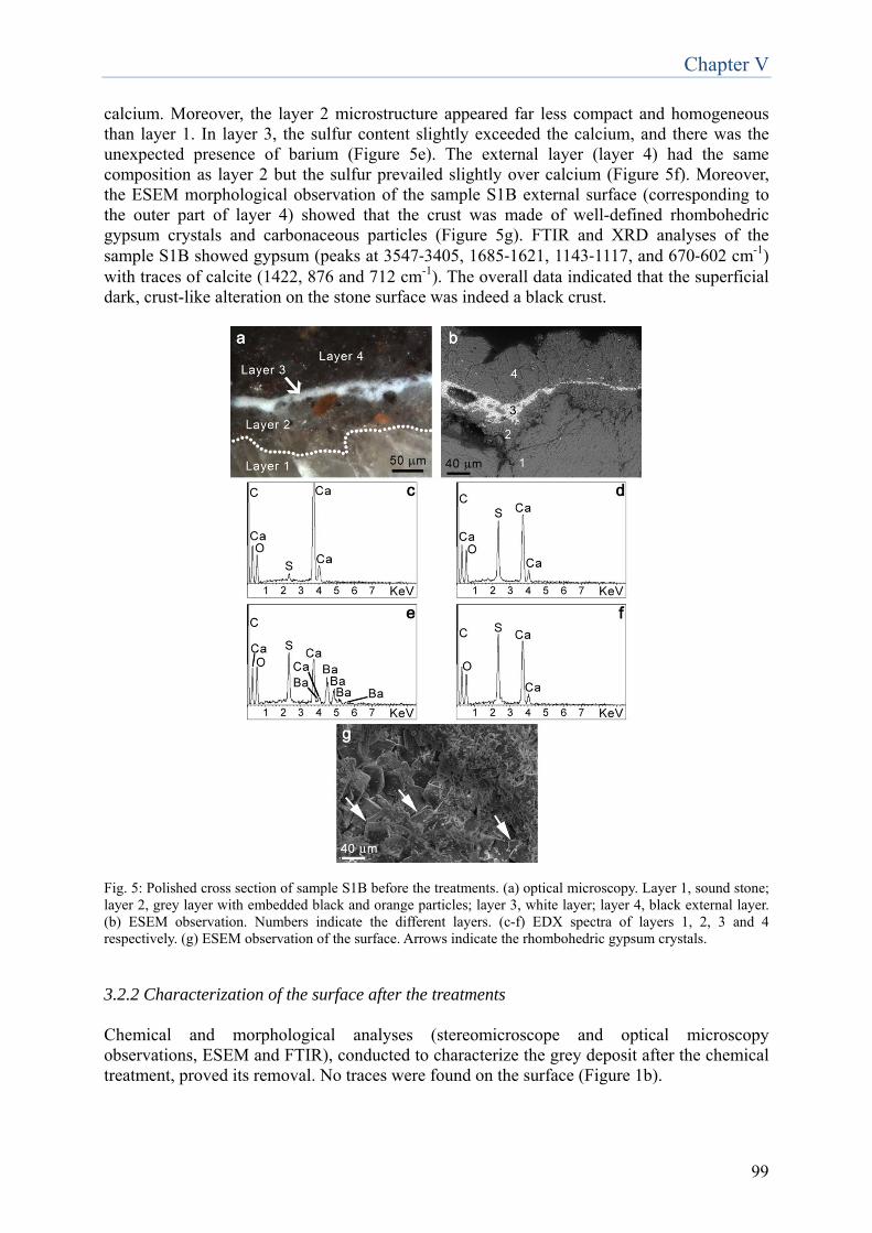
Chapter V
99
calcium. Moreover, the layer 2 microstructure appeared far less compact and homogeneous than layer 1. In layer 3, the sulfur content slightly exceeded the calcium, and there was the unexpected presence of barium (Figure 5e). The external layer (layer 4) had the same composition as layer 2 but the sulfur prevailed slightly over calcium (Figure 5f). Moreover, the ESEM morphological observation of the sample S1B external surface (corresponding to the outer part of layer 4) showed that the crust was made of well-defined rhombohedric gypsum crystals and carbonaceous particles (Figure 5g). FTIR and XRD analyses of the sample S1B showed gypsum (peaks at 3547‐3405, 1685‐1621, 1143‐1117, and 670‐602 cm-1) with traces of calcite (1422, 876 and 712 cm-1). The overall data indicated that the superficial dark, crust-like alteration on the stone surface was indeed a black crust.
Fig. 5: Polished cross section of sample S1B before the treatments. (a) optical microscopy. Layer 1, sound stone; layer 2, grey layer with embedded black and orange particles; layer 3, white layer; layer 4, black external layer. (b) ESEM observation. Numbers indicate the different layers. (c-f) EDX spectra of layers 1, 2, 3 and 4 respectively. (g) ESEM observation of the surface. Arrows indicate the rhombohedric gypsum crystals.
3.2.2 Characterization of the surface after the treatments Chemical and morphological analyses (stereomicroscope and optical microscopy observations, ESEM and FTIR), conducted to characterize the grey deposit after the chemical treatment, proved its removal. No traces were found on the surface (Figure 1b).
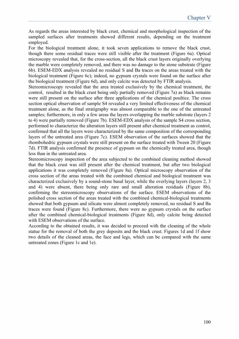
Chapter V
100
As regards the areas interested by black crust, chemical and morphological inspection of the sampled surfaces after treatments showed different results, depending on the treatment employed. For the biological treatment alone, it took seven applications to remove the black crust, though there some residual traces were still visible after the treatment (Figure 6a). Optical microscopy revealed that, for the cross-section, all the black crust layers originally overlying the marble were completely removed, and there was no damage to the stone substrate (Figure 6b). ESEM-EDX analysis revealed no residual S and Ba traces on the areas treated with the biological treatment (Figure 6c); indeed, no gypsum crystals were found on the surface after the biological treatment (Figure 6d), and only calcite was detected by FTIR analysis. Stereomicroscopy revealed that the area treated exclusively by the chemical treatment, the control, resulted in the black crust being only partially removed (Figure 7a) as black remains were still present on the surface after three applications of the chemical poultice. The cross section optical observation of sample S4 revealed a very limited effectiveness of the chemical treatment alone, as the final stratigraphy was almost comparable to the one of the untreated samples; furthermore, in only a few areas the layers overlapping the marble substrate (layers 2 to 4) were partially removed (Figure 7b). ESEM-EDX analysis of the sample S4 cross section, performed to characterize the alteration layers still present after chemical treatment as control, confirmed that all the layers were characterized by the same composition of the corresponding layers of the untreated area (Figure 7c). ESEM observation of the surfaces showed that the rhombohedric gypsum crystals were still present on the surface treated with Tween 20 (Figure 7d). FTIR analysis confirmed the presence of gypsum on the chemically treated area, though less than in the untreated area. Stereomicroscopy inspection of the area subjected to the combined cleaning method showed that the black crust was still present after the chemical treatment, but after two biological applications it was completely removed (Figure 8a). Optical microscopy observation of the cross section of the areas treated with the combined chemical and biological treatment was characterized exclusively by a sound-stone basal layer, while the overlying layers (layers 2, 3 and 4) were absent, there being only rare and small alteration residuals (Figure 8b), confirming the stereomicroscopy observations of the surface. ESEM observations of the polished cross section of the areas treated with the combined chemical-biological treatments showed that both gypsum and silicate were almost completely removed, no residual S and Ba traces were found (Figure 8c). Furthermore, there were no gypsum crystals on the surface after the combined chemical-biological treatments (Figure 8d), only calcite being detected with ESEM observations of the surface. According to the obtained results, it was decided to proceed with the cleaning of the whole statue for the removal of both the grey deposits and the black crust. Figures 1d and 1f show two details of the cleaned areas, the face and legs, which can be compared with the same untreated zones (Figure 1c and 1e).
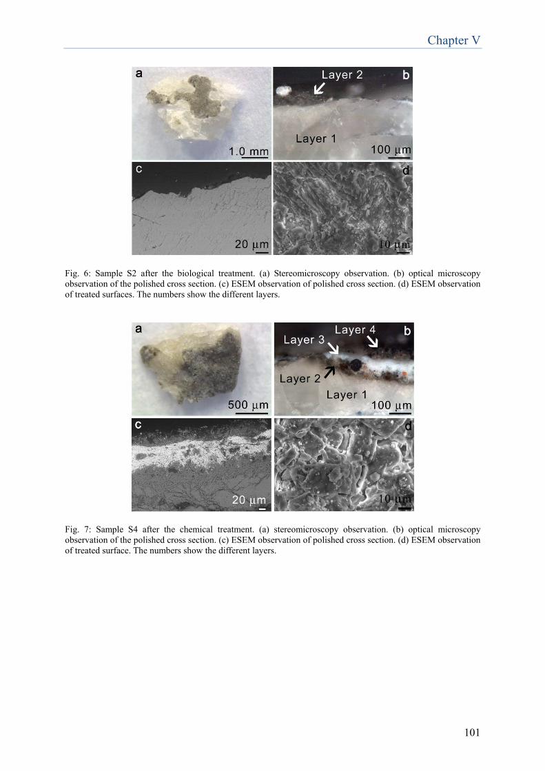
Chapter V
101
Fig. 6: Sample S2 after the biological treatment. (a) Stereomicroscopy observation. (b) optical microscopy observation of the polished cross section. (c) ESEM observation of polished cross section. (d) ESEM observation of treated surfaces. The numbers show the different layers.
Fig. 7: Sample S4 after the chemical treatment. (a) stereomicroscopy observation. (b) optical microscopy observation of the polished cross section. (c) ESEM observation of polished cross section. (d) ESEM observation of treated surface. The numbers show the different layers.
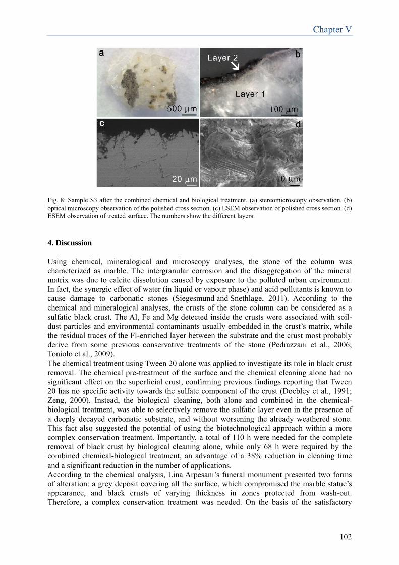
Chapter V
102
Fig. 8: Sample S3 after the combined chemical and biological treatment. (a) stereomicroscopy observation. (b) optical microscopy observation of the polished cross section. (c) ESEM observation of polished cross section. (d) ESEM observation of treated surface. The numbers show the different layers.
4. Discussion Using chemical, mineralogical and microscopy analyses, the stone of the column was characterized as marble. The intergranular corrosion and the disaggregation of the mineral matrix was due to calcite dissolution caused by exposure to the polluted urban environment. In fact, the synergic effect of water (in liquid or vapour phase) and acid pollutants is known to cause damage to carbonatic stones (Siegesmund and Snethlage, 2011). According to the chemical and mineralogical analyses, the crusts of the stone column can be considered as a sulfatic black crust. The Al, Fe and Mg detected inside the crusts were associated with soil-dust particles and environmental contaminants usually embedded in the crust’s matrix, while the residual traces of the Fl-enriched layer between the substrate and the crust most probably derive from some previous conservative treatments of the stone (Pedrazzani et al., 2006; Toniolo et al., 2009). The chemical treatment using Tween 20 alone was applied to investigate its role in black crust removal. The chemical pre-treatment of the surface and the chemical cleaning alone had no significant effect on the superficial crust, confirming previous findings reporting that Tween 20 has no specific activity towards the sulfate component of the crust (Doebley et al., 1991; Zeng, 2000). Instead, the biological cleaning, both alone and combined in the chemical-biological treatment, was able to selectively remove the sulfatic layer even in the presence of a deeply decayed carbonatic substrate, and without worsening the already weathered stone. This fact also suggested the potential of using the biotechnological approach within a more complex conservation treatment. Importantly, a total of 110 h were needed for the complete removal of black crust by biological cleaning alone, while only 68 h were required by the combined chemical-biological treatment, an advantage of a 38% reduction in cleaning time and a significant reduction in the number of applications. According to the chemical analysis, Lina Arpesani’s funeral monument presented two forms of alteration: a grey deposit covering all the surface, which compromised the marble statue’s appearance, and black crusts of varying thickness in zones protected from wash-out. Therefore, a complex conservation treatment was needed. On the basis of the satisfactory
Chapter V
103
results achieved on the stone column, we tested the simultaneous action of the cleaning methods for black crust and grey deposit removal on the statue. The features of the grey deposit are typically connected with the dry deposition (impaction, gravitational setting) of aerosol particles (Torfs and Van Grieken, 1997; Toniolo et al., 2009), different from the wet deposit and sulfation phenomena in an acid environment that causes black crust. A soft chemical treatment using the non-ionic detergent Tween 20 was adopted for the removal of the grey deposit. Neutral or alkaline detergents, used alone or in water solution, are often used for the removal of deposits covering buildings exposed to a polluted atmosphere (Castello, 2008); after two 17 hour-applications the grey deposit was completely removed, as was apparent on visual inspection. As regards the black crust, ESEM revealed the morphology to be rather atypical with respect to the lamellar or acicular morphologies usually described in the literature (Toniolo et al., 2009; Török et al., 2011). The stratigraphy consisted of 4 layers: layer 1 corresponded to sound marble; its main composition was Ca with minor amounts of sulfur, indicating good overall conservation of the stone substrate. Layers 2 and 4 were mainly Ca and S, though in different ratios; the chemical composition of these layers was confirmed as typical of black crusts (Sarmiento et al., 2008). Between layers 2 and 4 there was an intermediate discontinuous layer (layer 3) of Ca, S and Ba, corresponding to a barium sulfate layer, most probably deriving from an undocumented conservation treatment. Indeed, the barium hydroxide technique is well known in the literature (Matteini, 1991) and was performed in the past to desulfate and consolidate stone surfaces. Barium sulfate (BaSO4) was meant to act as a protective layer, being less soluble than CaSO4 even in an acid environment (Price, 1984; Hansen et al., 2003), and thus reducing the solubilization-crystallization sulfate cycles and the risk of both acid attack and penetration of soluble salts (Hansen et al., 2003; Giorgi et al., 2010). At ESEM, layer 3 appeared discontinuous, probably because of the uneven barium sulfate distribution; in fact, below and above layer 3 there were differently formed sulfated layers (layers 2 and 4 respectively). Silicate particles were also found in the black crust structure, presumably of atmospheric origin and most likely ash released by industrial sources (Maravelaki-Kalaitzaki, 2005; Kramar and Mirtič, 2008). Both the biological cleaning alone, and the biological method coupled with the soft chemical pre-treatment using Tween 20, were used for the selective removal of the black crust. Also the chemical treatment using the non-ionic detergent was applied alone to investigate its role in black crust removal. Each method’s contribution to black crust removal was evaluated. After seven 22-hour applications of the biological treatment alone, chemical and morphological analyses revealed that the surface was homogeneous and satisfactorily cleaned. Indeed, the cross-section observation demonstrated that all the layers originally overlying the marble were completely removed. ESEM analysis of the sample S2 cross-section demonstrated that the bacteria also reduced the sulfate bound to barium, the barium sulfate being completely removed by the microbiological method. Indeed, sulfate-reducing bacteria can use barite as a sulfate source for anaerobic respiration (Bolze et al., 1974), and D. vulgaris has been shown to use BaSO4 as a sulfate source (McCready et al., 1980). On our artwork under study, layer 4 most probably originated from an external deposition process, which rendered it less compact and more porous than the black crust that derives from direct sulfation of the stone substrate (Schifter et al., 2001; Ozga et al., 2011). Chemical and morphological analyses confirmed the chemical poultice’s effectiveness in removing the less coherent particulate matter embedded in the crust (little reduction in the thickness of layer 4). Note that barium sulfate was not removed by the chemical treatment as it was still present on sample S4 after the chemical treatment. Despite the fact that the advantages and effectiveness of the biocleaning method have already been well documented (Cappitelli et al., 2006; Cappitelli et al., 2007; Polo et al., 2010;
Chapter V
104
Gioventù et al., 2011), and reiterated also in this work, nobody, till now, has studied the interaction between biocleaning and chemical cleaning treatments. In the present study the chemical pre-treatment consisted of a single application aimed at softening the black crust, past results having shown that chemical treatment removed only the less compact part of the black crust. Tween 20 is commonly used in microbiological laboratories to avoid the binding of bacteria (Courtney and Hasty, 1991) and, being a mild detergent, it does not affect the structural features of membrane proteins (le Maire et al., 2000; Schuck et al., 2003). The chemical pre-treatment was then followed by the biological procedure. In terms of cleaning effectiveness, electron microscope observations, elemental analysis and infrared spectroscopy showed that the combined methods achieved results comparable to those of the biological treatment alone. Using Tween 20 and SRB practical advantages were achieved: a very significant reduction in the number of applications (from seven to two), and a remarkable 70% reduction in the total cleaning time with respect to the biological approach. Despite being a successful treatment, biocleaning alone has been demonstrated to be a highly time-consuming process in the presence of thick and compact crusts (Gioventù et al., 2011). The chemical treatment did not interfere in any way with the biological method, but it did make the biological sulfate removal quicker, likely because it softened the crust, reducing the cleaning time, a parameter conservators must consider when choosing a restoration approach (Gaspar et al., 2003). In addition, when the surface state of conservation is powdery and incoherent, prolonged contact with water can further exacerbate the degradation (Slaton and Normandir, 2005). In such cases, the synergic combination of chemical and biological methods reduces this hazard. In conclusion, to obtain homogeneous and satisfactory cleaning results, we suggest, for appropriate cases, chemical pre-treatment with the soft non-ionic detergent Tween 20, followed by safe and non-invasive biological treatment for a quick removal of black crust. Acknowledgments The authors are grateful to Alberto Gnesutta, descendant of the poetess Anna Zuccari and patron of this work, and the Soprintendenza per i Beni Architettonici e Paesaggistici di Milano, Italy. The authors also thank the conservator Eleonora Gioventù for her help in the cleaning of the sculpture. References Alessandrini, G., Realini, M., Calia, A., Mecchi, A.M., D’Agostino, F., Rizzo, G. (2002) The old town of Palermo (Italy): stone materials and their decay. Protection and Conservation of the Cultural Heritage of the Mediterranean Cities. In: Galán, E., Zezza, F., (Eds.), Proceedings of the 5th International Symposium on the Conservation of Monuments in the Mediterranean Basin, Swets & Zeitlinge, Lisse, pp. 93-99. Böke, H., Göktürk, E.H., Caner-Saltık, E.N., Demirci, Ş. (1999) Effect of airborne particle on SO2–calcite reaction. Applied Surface Science 140, 70-82. Bolze, C.E., Malone, P.G., Smith, M.J. (1974) Microbial mobilization of barite. Chemical Geology 13, 141-143.
Chapter V
105
Bromblet, P., Labouré, M., Orial, G. (2003) Diversity of the cleaning procedures including laser for the restoration of carved portals in France over the last 10 years. Journal of Cultural Heritage 4, 17-26. Camuffo, D., Del Monte, M., Sabbioni, C. (1983) Origin and growth mechanisms of the sulfated crusts on urban limestone. Water, Air, & Soil Pollution 19, 351-359. Cappitelli, F., Toniolo, L., Sansonetti, A., Gulotta, D., Ranalli, G., Zanardini, E., Sorlini, C. (2007) Advantages of using microbial technology over traditional chemical technology in removal of black crusts from stone surfaces of historical monuments. Applied and Environmental Microbiology 73, 5671-5675. Cappitelli, F., Zanardini, E., Ranalli, G., Mello E., Daffonchio, D., Sorlini, C. (2006) Improved methodology for bioremoval of black crusts on historical stone artworks by use of sulfate-reducing bacteria. Applied and Environmental Microbiology 72, 3733-3737. Castello, I. (2008) The restoration of a Roman stone statue exhibited outdoors: a Bacchus in the Boboli Gardens in Florence. OPD restauro 20, 245-262. Courtney, H.S., Hasty, D.L. (1991) Aggregation of group A streptococci by human saliva and effect of saliva on streptococcal adherence to host cells. Infection and Immunity 59, 1661-1666. Doebley, C.E., Lewin, S.Z., Aronson, S. (1991) Detergent and hypochlorites for the cleaning of travertine. APT Bulletin 23, 54-58. El-Metwally, A., Ramadan, A. (2005) The role of air pollutants and sewage waste in acceleration of degradation of the Islamic Cultural Heritage of Cairo. NATO Science Series, IV: Earth and Environmental Sciences 38, 363-370. Gaspar, P., Hubbard, C., McPhail, D., Cummings, A. (2003) A topographical assessment and comparison of conservation cleaning treatments. Journal of Cultural Heritage 4, 294-302. Giorgi, R., Ambrosi, M., Toccafondi, N., Baglioni, P. (2010) Nanoparticles for cultural heritage conservation: calcium and barium hydroxide nanoparticles for wall painting consolidation. Chemistry-A European Journal 16, 9374-9382. Gioventù, E., Lorenzi, P.F., Villa, F., Sorlini, C., Rizzi, M., Cagnini, A., Griffo, A., Cappitelli, F. (2011) Comparing the bioremoval of black crusts on colored artistic lithotypes of the Cathedral of Florence with chemical and laser treatment. International Biodeterioration & Biodegradation 65, 832-839. Hansen, E., Doehne, E., Fidler, J., Larson, J., Martin, B., Matteini, M., Rodriguez-Navarro, C., Pardo, E.S., Price, C., de Tagle, A., Teutonico, J.M., Weiss, N. (2003) A review of selected inorganic consolidants and protective treatments for porous calcareous materials. Reviews in Conservation 4, 13-25. Kramar, S., Mirtič, B. (2008) Characterization of black crusts of Robba´s fountain statues, Ljubljana (Slovenia). RMZ - Materials and Geoenvironment 55, 490-504.

Chapter V
106
le Maire, M., Champeil, P., Möller, J.V. (2000) Interaction of membrane proteins and lipids with solubilizing detergents. Biochimica et Biophysica Acta 1508, 86-111. Maravelaki-Kalaitzaki, P. (2005) Black crusts and patinas on Pentelic marble from the Parthenon and Erechtheum (Acropolis, Athens): characterization and origin. Analytica Chimica Acta 532, 187-198. Matteini, M. (1991) In review: an assessment of Florentine methods of wall painting conservation based on the use of mineral treatments. In Cather, S., (Eds.), The Conservation of Wall Paintings. Proceedings of a symposium organized by the Courtauld Institute of Art and the Getty Conservation Institute, Getty Conservation Institute, Marina del Rey, pp. 137-148. McCready, R.G.L., Bland, C.J., Gonzales, D.E. (1980) Preliminary studies on the chemical, physical and biological stability of Ba/RaSO4 precipitates. Hydrometry 5, 109-116. Moropoulou, A., Bisbikou, K., Torfs, K., Van Grieken, R., Zezza, F., Macri, M. (1998) Origin and growth of weathering crusts on ancient marbles in industrial atmosphere. Atmospheric Environment 32, 967-982. Ozga, I., Bonazza, A., Bernardi, E., Tittarelli, F., Favoni, O., Ghedini, N., Morselli, L., Sabbioni, C. (2011) Diagnosis of surface damage induced by air pollution on 20th-century concrete buildings. Atmospheric Environment 45, 4986-4995. Pedrazzani, R., Alessandri, I., Bontempi, E., Cappitelli, F., Cianci, M., Pantos, E., Toniolo, L., Depero, L. E. (2006) Study of sulphation of Candoglia marble by means of micro X-ray diffraction experiments. Applied physics. A, 83, 689-694. Polo, A., Cappitelli, F., Brusetti, L., Principi, P., Villa, F., Giacomucci, L., Ranalli, G., Sorlini, C. (2010) Feasibility of removing surface deposits on stone using biological and chemical remediation methods. Microbial Ecology 60, 1-14. Price, C.A. (1984) The consolidation of limestone using a lime poultice and limewater. In: Brommelle, N.S., Pye, E.M., Smith, P., Thomson, G., (Eds.), Adhesives and consolidants, Preprints of the contributions to the IIC Paris congress, IIC, London, pp. 160-162. Sansonetti, A., Mecchi, A.M., Poli, T., Realini, M. (2008) Problems in drawing up standards to evaluate effectiveness and harmfulness in cleaning operations. In: Townsend, J.H., Toniolo, L., Cappitelli, F., (Eds.), Conservation Science 2007, Archetype Publications, London, pp. 80-87. Sarmiento, A., Maguregui, M., Martinez-Arkarazo, I., Angulo, M., Castro, K., Olazábal, M.A., Fernández, L.A., Rodríguez-Laso, M.D., Mujika, A.M., Gómez, J., Madariaga, J.M. (2008) Raman spectroscopy as a tool to diagnose the impacts of combustion and greenhouse acid gases on properties of Built Heritage. Journal of Raman Spectroscopy 39, 1042-1049. Schifter, I., Vera, M., Díaz, L., Guzmán, E., Ramos, F., López-Salinas, E. (2001) Environmental implications on the oxygenation of gasoline with ethanol in the metropolitan area of Mexico City. Environmental Science & Technology 35, 1893-1901.
Chapter V
107
Schuck, S., Honsho, M., Ekroos, K., Shevchenko, A., Simons, K. (2003) Resistance of cell membranes to different detergents. Proceedings of the National Academy of Sciences 100, 5795-5800. Siegesmund, S., Snethlage, R. (2011) Stone in Architecture. Properties, Durability. Springer-Verlag, Berlin, Heidelberg. Slaton, D., Normandin, K.C. (2005) Masonry cleaning technologies. Journal of Architectural Conservation 11, 7-31. Toniolo, L., Zerbi, C.M., Bugini, R. (2009) Black layers on historical architecture. Environmental Science and Pollution Research 16, 218-226. Torfs, K., Van Grieken, R. (1997) Chemical relations between atmospheric aerosols, deposition and stone decay layers on historic buildings at the Mediterranean coast. Atmospheric Environment 31, 2179-2192. Török, Á., Licha, T., Simon, K., Siegesmund, S. (2011) Urban and rural limestone weathering; the contribution of dust to black crust formation. Environmental Earth Sciences 63, 675-693. Zeng, Z. (2000) The research of a detergent for washing off the filth from oils, fats and smoke on ancient stone sculptures. Sciences of conservation & archaeology 1, 34-40.

108
Chapter VI
109
Evaluation of effectiveness of biocleaning in sulfates removal from mural paintings of Queen Teodolinda Chapel in Monza Cathedral
Introduction The methodology based on use of bacteria as cleaning agents for stone artworks and monuments is increasingly meeting with great approval by restorers and authorized personnel, thanks to its property of combining effectiveness with respectfulness towards treated surfaces. The use of not invasive techniques toward the substrate and selective toward the alteration results desirable, especially in the case of mural paintings, by their nature characterized by fragility. Therefore, the aim of the present study is the test of applicability of microorganisms also for cleaning of pigmented surfaces. The object of this experimentation is the pictorial cycle decorating Queen Teodolinda Chapel situated in Monza Cathedral. The artwork was subject to a mild conservation intervention, involving the most prestigious Italian research Institutes1 and important sponsors. The carried out experimentation, not exhaustive and still in progress, has to be intended as a first innovative approach to the delicate issue of sulfations removal from mural paintings. Artwork description and its state of conservation The pictorial cycle “Delle Storie della Regina Teodolinda” painted by “La Bottega degli Zavattari”, was accomplished in the first half of 1400 and completed in 1446, as stated in the contract. It is composed by 45 scenes, which narrate courtly life episodes, with a precious description of clothes and armours, presenting an extraordinary description of courtly life of XV century in Milan. The artwork was almost entirely realised using colours diluted in organic media, sometimes using milk of lime as basis or, very seldom, the ‘buon fresco’ technique. This technical choice has allowed a particular chromatic vivaciousness, by means of a broad recourse to gildings carried out with the method “pastiglia a rilievo” of gypsum and glue, whereas the dresses, damasks and armours are made of golden or silver tin foil and veiled with copper resinate and red lacquer. At the same time, the choice of the organic binders constitutes in itself the cause of extreme weakness, exacerbated by decay owing to ageing and manifold undergone restoration interventions.

Chapter VI
110
F
Fig. 1: Pictorial cycle decorating Queen Teodolinda Chapel situated in Monza Cathedral.
The trial with sulfate-reducing bacteria was carried out on two scenes belonging to the pictorial cycle of Queen Teodolinda Chapel: Scene 16 (upper rectangle in Fig. 1) and 33 (down rectangle in Fig. 1). Scene 16 belongs to the group describing the preliminaries to the wedding of Teodolinda and Autari. It represents the episode in which Autari is informed of Teodolinda’s arrival in Lombardic territory by the delegates (Fig. 2). This scene, never treated for cleaning, was characterised by exfoliations and raisings of the pictorial film, caused by previous sulfations, still ongoing at the moment of treatment.
Fig. 2: Scene 16. Scene 33 describes the apparition in Teodolinda’s dream of the Holy Spirit in the form of a dove, suggesting to her the place for the building of her church (Fig. 3). In this scene the superficial salts appeared in a crystalline form, previously treated for sulfate removal by means of packs of sepiolite, deionized water and ion-exchange resins.
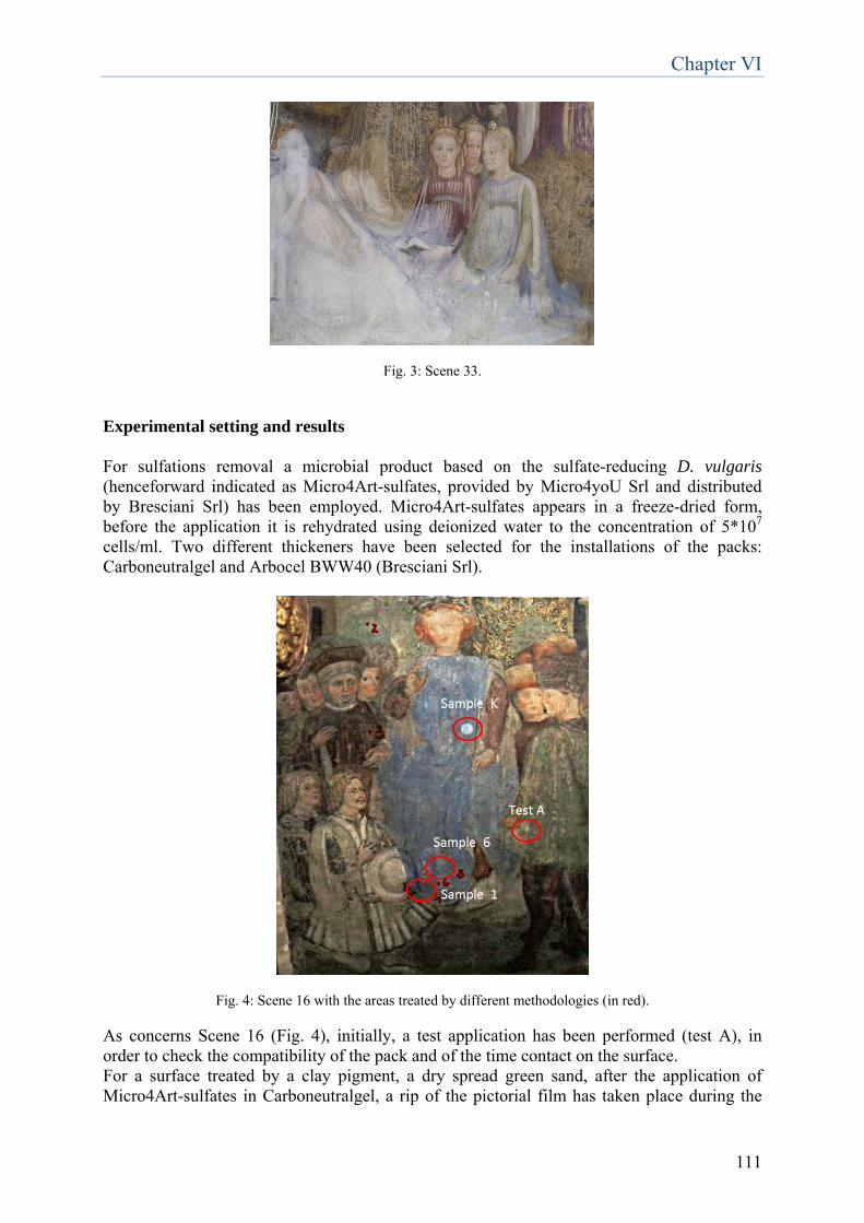
Chapter VI
111
Fig. 3: Scene 33.
Experimental setting and results For sulfations removal a microbial product based on the sulfate-reducing D. vulgaris (henceforward indicated as Micro4Art-sulfates, provided by Micro4yoU Srl and distributed by Bresciani Srl) has been employed. Micro4Art-sulfates appears in a freeze-dried form, before the application it is rehydrated using deionized water to the concentration of 5*107 cells/ml. Two different thickeners have been selected for the installations of the packs: Carboneutralgel and Arbocel BWW40 (Bresciani Srl).
Fig. 4: Scene 16 with the areas treated by different methodologies (in red).
As concerns Scene 16 (Fig. 4), initially, a test application has been performed (test A), in order to check the compatibility of the pack and of the time contact on the surface. For a surface treated by a clay pigment, a dry spread green sand, after the application of Micro4Art-sulfates in Carboneutralgel, a rip of the pictorial film has taken place during the
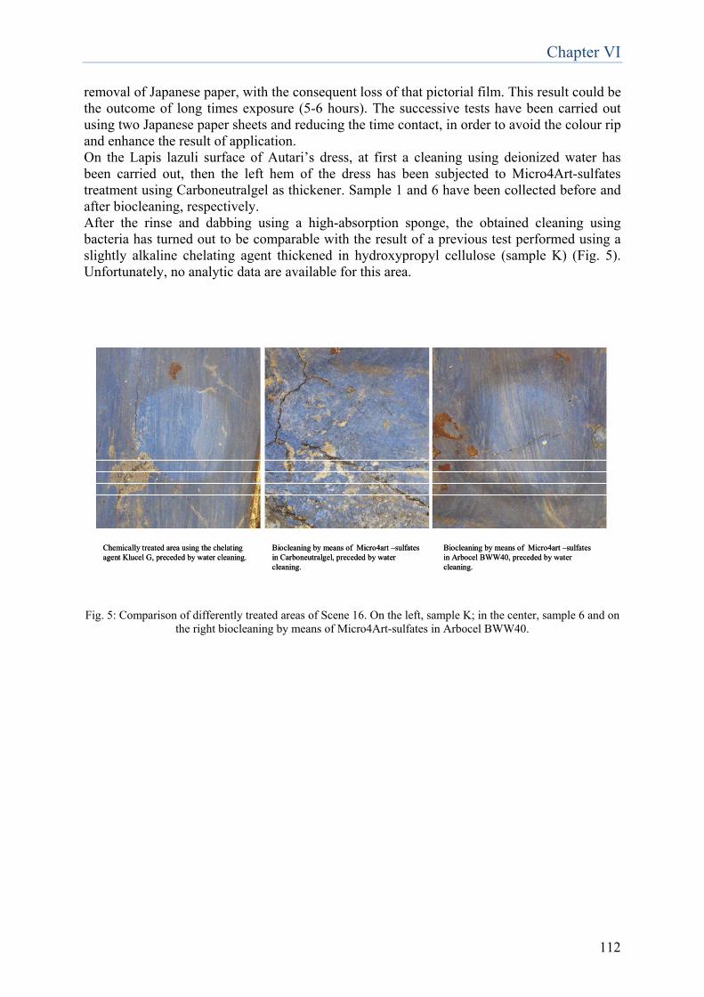
Chapter VI
112
removal of Japanese paper, with the consequent loss of that pictorial film. This result could be the outcome of long times exposure (5-6 hours). The successive tests have been carried out using two Japanese paper sheets and reducing the time contact, in order to avoid the colour rip and enhance the result of application. On the Lapis lazuli surface of Autari’s dress, at first a cleaning using deionized water has been carried out, then the left hem of the dress has been subjected to Micro4Art-sulfates treatment using Carboneutralgel as thickener. Sample 1 and 6 have been collected before and after biocleaning, respectively. After the rinse and dabbing using a high-absorption sponge, the obtained cleaning using bacteria has turned out to be comparable with the result of a previous test performed using a slightly alkaline chelating agent thickened in hydroxypropyl cellulose (sample K) (Fig. 5). Unfortunately, no analytic data are available for this area.
Chemically treated area using the chelating agent Klucel G, preceded by water cleaning.
Biocleaning by means of Micro4art –sulfates in Carboneutralgel, preceded by water cleaning.
Biocleaning by means of Micro4art –sulfates in Arbocel BWW40, preceded by water cleaning.
Chemically treated area using the chelating agent Klucel G, preceded by water cleaning.
Biocleaning by means of Micro4art –sulfates in Carboneutralgel, preceded by water cleaning.
Biocleaning by means of Micro4art –sulfates in Arbocel BWW40, preceded by water cleaning.
Fig. 5: Comparison of differently treated areas of Scene 16. On the left, sample K; in the center, sample 6 and on
the right biocleaning by means of Micro4Art-sulfates in Arbocel BWW40.
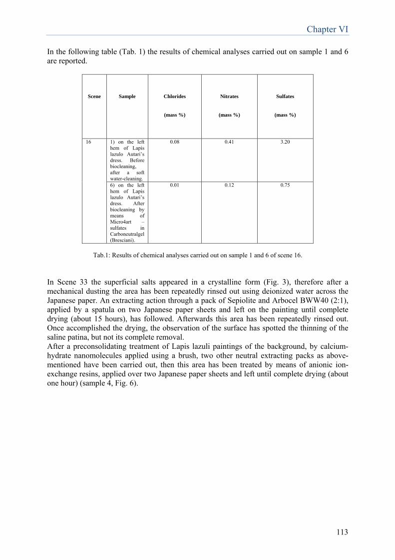
Chapter VI
113
In the following table (Tab. 1) the results of chemical analyses carried out on sample 1 and 6 are reported.
Scene
Sample
Chlorides
(mass %)
Nitrates
(mass %)
Sulfates
(mass %)
16 1) on the left hem of Lapis lazulo Autari’s dress. Before biocleaning, after a soft water-cleaning.
0.08 0.41 3.20
6) on the left hem of Lapis lazulo Autari’s dress. After biocleaning by means of Micro4art –sulfates in Carboneutralgel (Bresciani).
0.01 0.12 0.75
Tab.1: Results of chemical analyses carried out on sample 1 and 6 of scene 16.
In Scene 33 the superficial salts appeared in a crystalline form (Fig. 3), therefore after a mechanical dusting the area has been repeatedly rinsed out using deionized water across the Japanese paper. An extracting action through a pack of Sepiolite and Arbocel BWW40 (2:1), applied by a spatula on two Japanese paper sheets and left on the painting until complete drying (about 15 hours), has followed. Afterwards this area has been repeatedly rinsed out. Once accomplished the drying, the observation of the surface has spotted the thinning of the saline patina, but not its complete removal. After a preconsolidating treatment of Lapis lazuli paintings of the background, by calcium-hydrate nanomolecules applied using a brush, two other neutral extracting packs as above-mentioned have been carried out, then this area has been treated by means of anionic ion-exchange resins, applied over two Japanese paper sheets and left until complete drying (about one hour) (sample 4, Fig. 6).
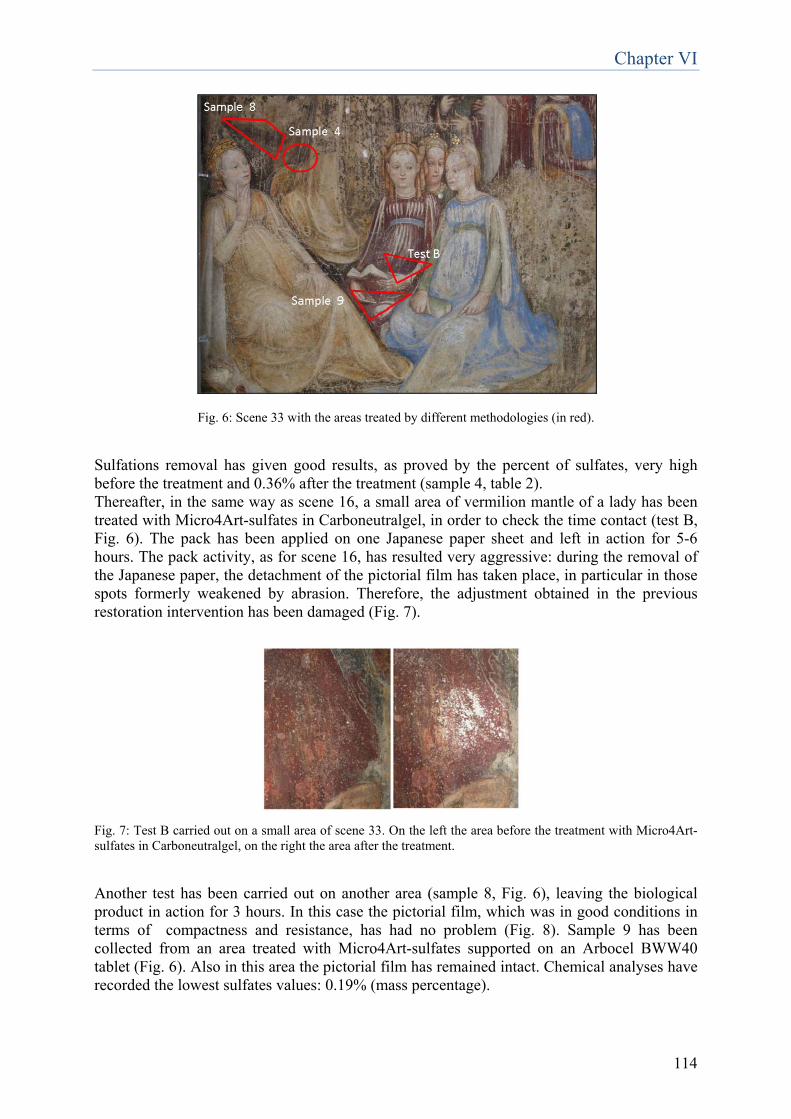
Chapter VI
114
Fig. 6: Scene 33 with the areas treated by different methodologies (in red).
Sulfations removal has given good results, as proved by the percent of sulfates, very high before the treatment and 0.36% after the treatment (sample 4, table 2). Thereafter, in the same way as scene 16, a small area of vermilion mantle of a lady has been treated with Micro4Art-sulfates in Carboneutralgel, in order to check the time contact (test B, Fig. 6). The pack has been applied on one Japanese paper sheet and left in action for 5-6 hours. The pack activity, as for scene 16, has resulted very aggressive: during the removal of the Japanese paper, the detachment of the pictorial film has taken place, in particular in those spots formerly weakened by abrasion. Therefore, the adjustment obtained in the previous restoration intervention has been damaged (Fig. 7). Fig. 7: Test B carried out on a small area of scene 33. On the left the area before the treatment with Micro4Art-sulfates in Carboneutralgel, on the right the area after the treatment. Another test has been carried out on another area (sample 8, Fig. 6), leaving the biological product in action for 3 hours. In this case the pictorial film, which was in good conditions in terms of compactness and resistance, has had no problem (Fig. 8). Sample 9 has been collected from an area treated with Micro4Art-sulfates supported on an Arbocel BWW40 tablet (Fig. 6). Also in this area the pictorial film has remained intact. Chemical analyses have recorded the lowest sulfates values: 0.19% (mass percentage).
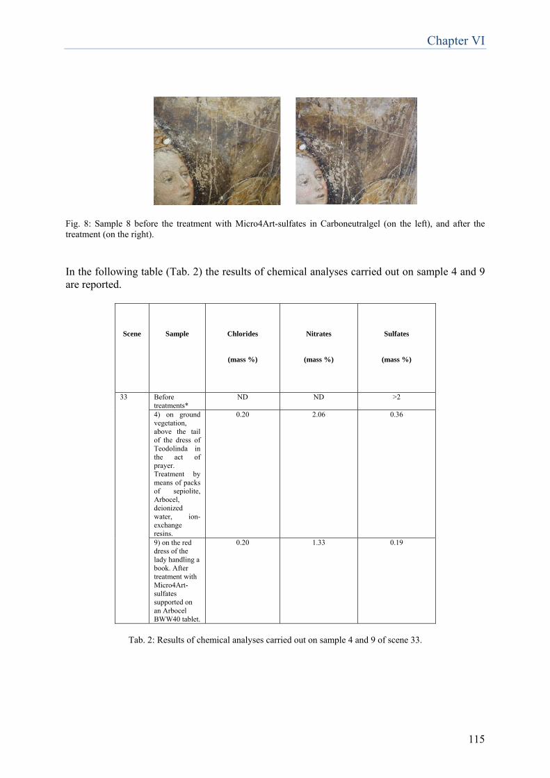
Chapter VI
115
Fig. 8: Sample 8 before the treatment with Micro4Art-sulfates in Carboneutralgel (on the left), and after the treatment (on the right). In the following table (Tab. 2) the results of chemical analyses carried out on sample 4 and 9 are reported.
Scene
Sample
Chlorides
(mass %)
Nitrates
(mass %)
Sulfates
(mass %)
33 Before treatments*
ND ND >2
4) on ground vegetation, above the tail of the dress of Teodolinda in the act of prayer. Treatment by means of packs of sepiolite, Arbocel, deionized water, ion-exchange resins.
0.20 2.06 0.36
9) on the red dress of the lady handling a book. After treatment with Micro4Art-sulfates supported on an Arbocel BWW40 tablet.
0.20 1.33 0.19
Tab. 2: Results of chemical analyses carried out on sample 4 and 9 of scene 33.
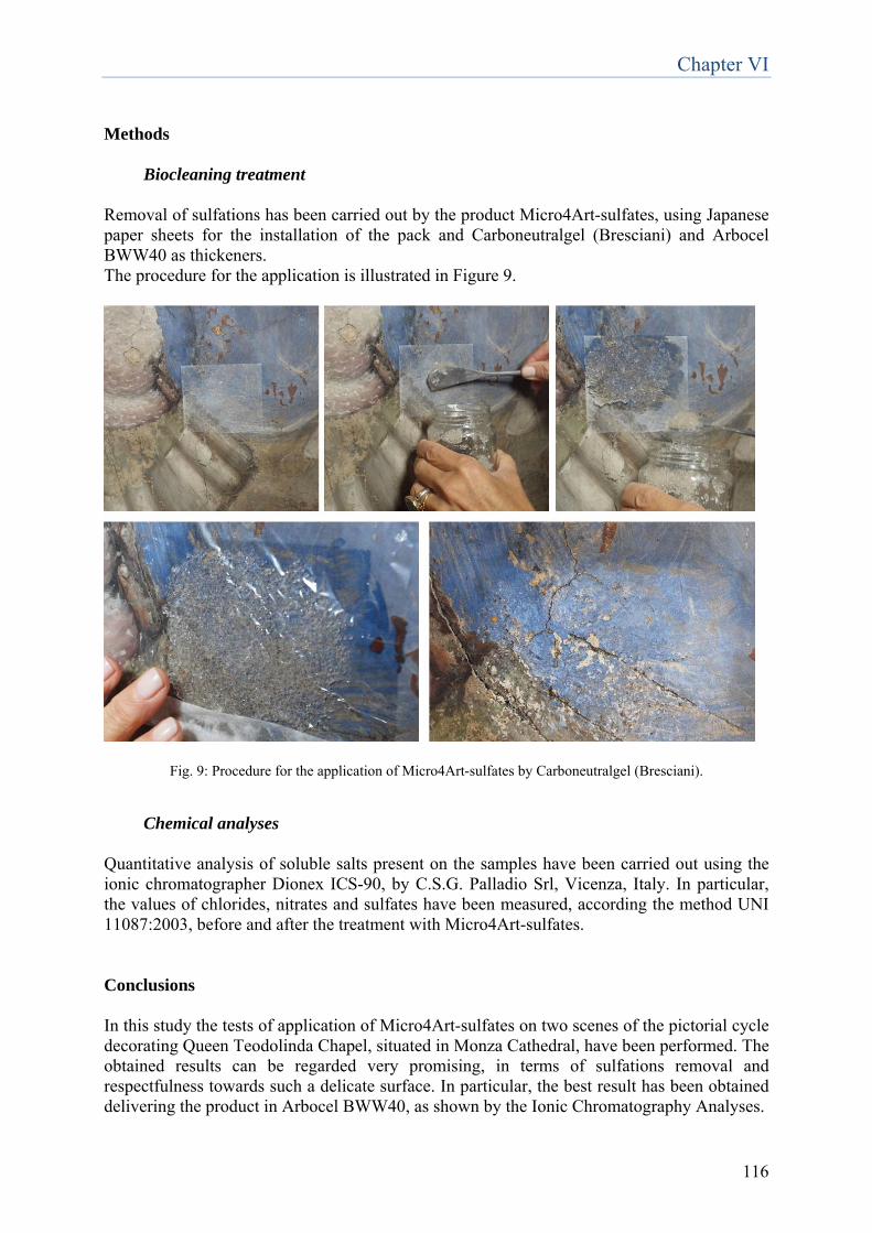
Chapter VI
116
Methods
Biocleaning treatment Removal of sulfations has been carried out by the product Micro4Art-sulfates, using Japanese paper sheets for the installation of the pack and Carboneutralgel (Bresciani) and Arbocel BWW40 as thickeners. The procedure for the application is illustrated in Figure 9.
Fig. 9: Procedure for the application of Micro4Art-sulfates by Carboneutralgel (Bresciani).
Chemical analyses Quantitative analysis of soluble salts present on the samples have been carried out using the ionic chromatographer Dionex ICS-90, by C.S.G. Palladio Srl, Vicenza, Italy. In particular, the values of chlorides, nitrates and sulfates have been measured, according the method UNI 11087:2003, before and after the treatment with Micro4Art-sulfates. Conclusions In this study the tests of application of Micro4Art-sulfates on two scenes of the pictorial cycle decorating Queen Teodolinda Chapel, situated in Monza Cathedral, have been performed. The obtained results can be regarded very promising, in terms of sulfations removal and respectfulness towards such a delicate surface. In particular, the best result has been obtained delivering the product in Arbocel BWW40, as shown by the Ionic Chromatography Analyses.

Chapter VI
117
Nevertheless, this work has to be considered merely preliminary, incomplete, a “first step” in the employment of sulfate-reducing bacteria for restoration of mural paintings, which constitute, as is known, a very complex category of artworks. The experimentation is just at the very beginning, therefore more research must be conducted in order to verify the compatibility with different kinds of materials, such as pigments containing metals as copper and lead, which could be susceptible to the hydrogen sulfide released by sulfate-reducing bacteria, or pigments with oily binder, based on malachite and copper resinate, which are very delicate. Moreover, another interesting issue to analyze is the time exposure, probably it could be further reduced. From the Ionic Chromatography Analyses, an interesting consideration is the reduction not only of sulfates after the application of bacteria, but also of nitrates. In fact the mass % of nitrates has passed from 0.41 to 0.12 in Scene 16 and from 2.06 a 1.33 in Scene 33. These data can be correlated to the ability of nitrate-reduction by sulfate-reducing bacteria, already discussed in previous studies. In fact, D. vulgaris possesses the proteins FprAs which provide constitutive protection against nitric oxide exposure (Silaghi-Dumitrescu et al., 2005). Moreover SRB of the genus Desulfovibrio are able to carry out the dissimilatory nitrate reduction to ammonia, thanks to the presence of NrfA, a gene encoding for a nitrite reductase. NrfA plays an important role in those strains able to use nitrate and nitrite as electron acceptor when sulphate is not freely available and in those bacteria capable of reducing nitrite but not nitrate (Giacomucci et al., 2012). Further investigations and trials for the removal of nitrates using sulfate-reducing bacteria could be interesting. Chlorides and nitrates, because of their high solubility and deliquescence (property of some hygroscopic substances which pass in solution through absorption of water vapor from the environment, holding humidity) constitute very hazardous salts for stone materials composing and cultural heritage. For this reason their acceptability limit is very low and near to the instrumental detectability limit. Instead, for sulfates, the tolerability is higher. In the following table (Tab.3) the tolerability of these salts is reported: Low Significative High Chlorides 0.01-0.09 % 0.10-0.99 % >1 % Nitrates 0.01-0.09 % 0.10-0.99 % >1 % Sulfates 0.1-0.99 % 1-2 % >2 % Tab. 3: Acceptability limits for chlorides, nitrates and sulfates on stone materials composing and cultural heritage. On account of this, the effectiveness of Micro4Art-sulfates in removal of nitrates can be considered another interesting and promising result.
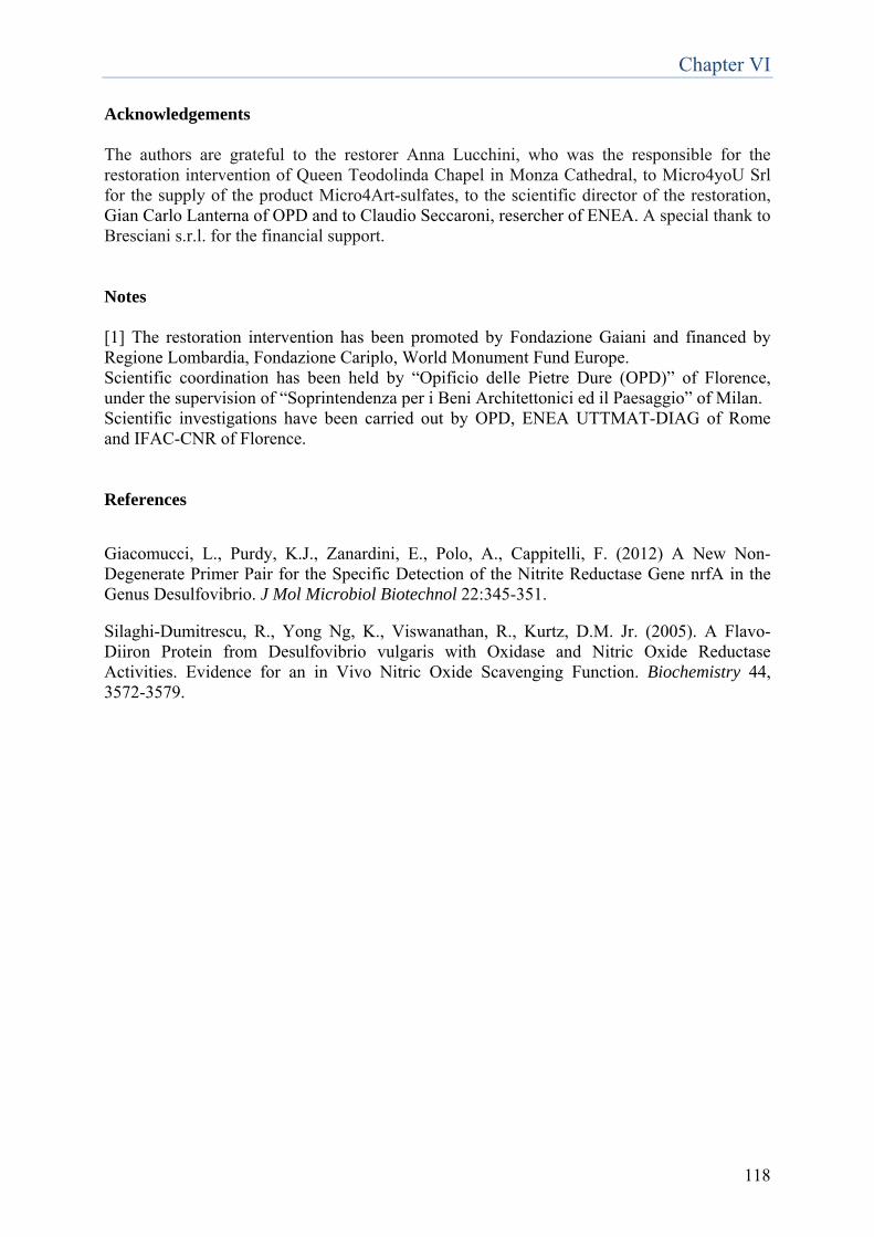
Chapter VI
118
Acknowledgements The authors are grateful to the restorer Anna Lucchini, who was the responsible for the restoration intervention of Queen Teodolinda Chapel in Monza Cathedral, to Micro4yoU Srl for the supply of the product Micro4Art-sulfates, to the scientific director of the restoration, Gian Carlo Lanterna of OPD and to Claudio Seccaroni, resercher of ENEA. A special thank to Bresciani s.r.l. for the financial support. Notes [1] The restoration intervention has been promoted by Fondazione Gaiani and financed by Regione Lombardia, Fondazione Cariplo, World Monument Fund Europe. Scientific coordination has been held by “Opificio delle Pietre Dure (OPD)” of Florence, under the supervision of “Soprintendenza per i Beni Architettonici ed il Paesaggio” of Milan. Scientific investigations have been carried out by OPD, ENEA UTTMAT-DIAG of Rome and IFAC-CNR of Florence. References Giacomucci, L., Purdy, K.J., Zanardini, E., Polo, A., Cappitelli, F. (2012) A New Non-Degenerate Primer Pair for the Specific Detection of the Nitrite Reductase Gene nrfA in the Genus Desulfovibrio. J Mol Microbiol Biotechnol 22:345-351. Silaghi-Dumitrescu, R., Yong Ng, K., Viswanathan, R., Kurtz, D.M. Jr. (2005). A Flavo-Diiron Protein from Desulfovibrio vulgaris with Oxidase and Nitric Oxide Reductase Activities. Evidence for an in Vivo Nitric Oxide Scavenging Function. Biochemistry 44, 3572-3579.
Chapter VII
119
Analysis of the microbial community present on the reverse side of a deteriorated canvas
1. Introduction and aim of the work
The role played by microorganisms in the deterioration of artifacts of our cultural heritage is nowadays well known. Two main factors are involved in the proliferation of microorganisms on monuments and artworks: the chemical nature of the substratum and the environmental conditions. Paintings contain a wide range of organic and inorganic compounds that may be used by many microorganisms for their growth. Firstly, the materials that constitute the painting itself, i.e. the cellulose of the canvas support material, the animal glue and gypsum used to prepare a ground layer, and the linseed oil of the paint layer, are all easily degraded. Secondly, the spectrum of compounds that might provide nutrients for microorganisms is further augmented by dirt, dust and other environmental contaminants deposited on the surface of the paintings, such as hydrocarbons or organic biocides (Lopez-Miras et al., 2013). As concerns environmental conditions, the most important are favourable humidity and temperature, for example fungi are able to grow in an environment with a relative humidity higher than 65 % and temperatures ranging between 20 and 35°C. Effects induced by microorganisms deterioration on paintings can occur on both the obverse and the reverse side. The degree of deterioration on the obverse side depends on the paint medium (acrylic paints, oil paints, distemper or watercolours) and mode of application, while on the reverse side, it depends on the nature of the support (canvas or wood). Bacteria and fungi represent an important part of the community which can accumulate on the surface for a long time as spores. The further growth of these deposited microorganisms can result in the detachment of the paint layer from the support, especially in paintings kept under conditions of high humidity. Moreover, the excretion of aggressive metabolic products (organic or inorganic acids) and the additional production of extracellular enzymes increase the loss of material. The main enzymatic activities involved in the deterioration of paintings caused by microorganisms are due to lipases, esterases, endo-N-acetyl-glucosaminidases (ENGases) (Lopez-Miras et al., 2012). In order to correctly conserve and restore biodeteriorated paintings, a detailed knowledge of the microbial communities associated with these substrates is an important pre-requisite for the definition of the most suitable preservation and restoration strategy. The aim of this study was the investigation of microbial communities adhering to the reverse side of a deteriorated acrylic monochrome painting on canvas and the definition of the optimal biocide for the inhibition of their proliferation, among the following: BIOTIN N, BIOTIN R, NEW DES 50 and AMUCHINA provided by Studio Restauri Formica Srl. To this end, samples were taken from areas on the reverse side where visual inspection revealed signs of biodeterioration, such as yellow-earth/red point areas of different extensions. A strategy combining culture-dependent and culture-independent techniques was chosen for their complementary aspects. In fact, nowadays it is generally accepted that culture-based techniques recover less than 1% of the total microorganisms present in environmental samples. Hence, the study of microbial communities based only on
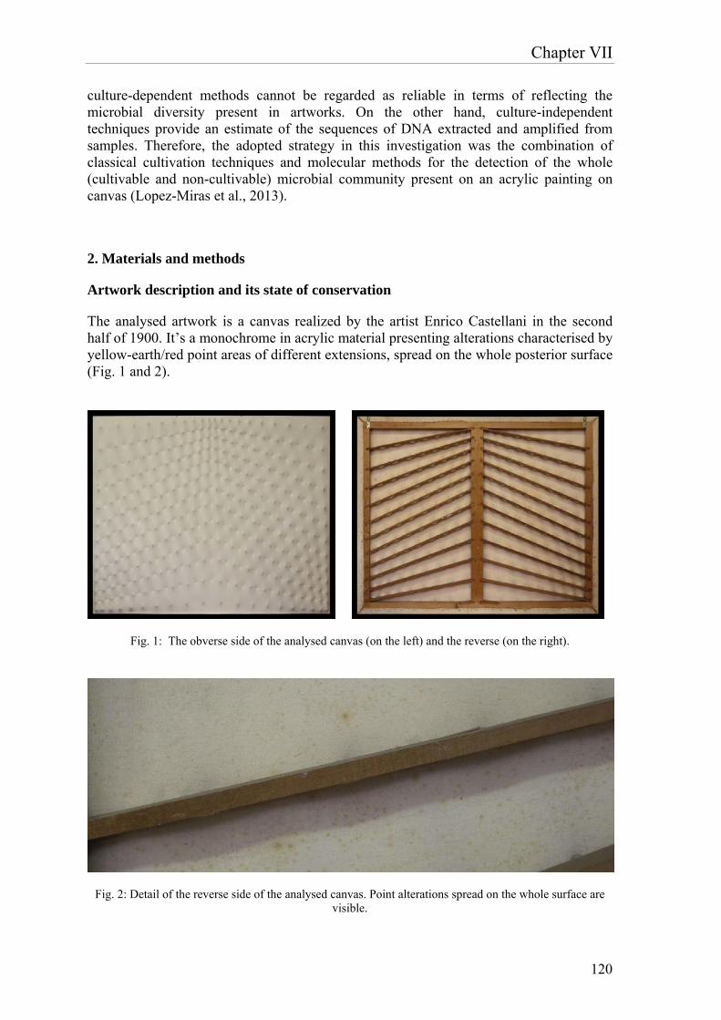
Chapter VII
120
culture-dependent methods cannot be regarded as reliable in terms of reflecting the microbial diversity present in artworks. On the other hand, culture-independent techniques provide an estimate of the sequences of DNA extracted and amplified from samples. Therefore, the adopted strategy in this investigation was the combination of classical cultivation techniques and molecular methods for the detection of the whole (cultivable and non-cultivable) microbial community present on an acrylic painting on canvas (Lopez-Miras et al., 2013).
2. Materials and methods
Artwork description and its state of conservation
The analysed artwork is a canvas realized by the artist Enrico Castellani in the second half of 1900. It’s a monochrome in acrylic material presenting alterations characterised by yellow-earth/red point areas of different extensions, spread on the whole posterior surface (Fig. 1 and 2).
Fig. 1: The obverse side of the analysed canvas (on the left) and the reverse (on the right).
Fig. 2: Detail of the reverse side of the analysed canvas. Point alterations spread on the whole surface are visible.
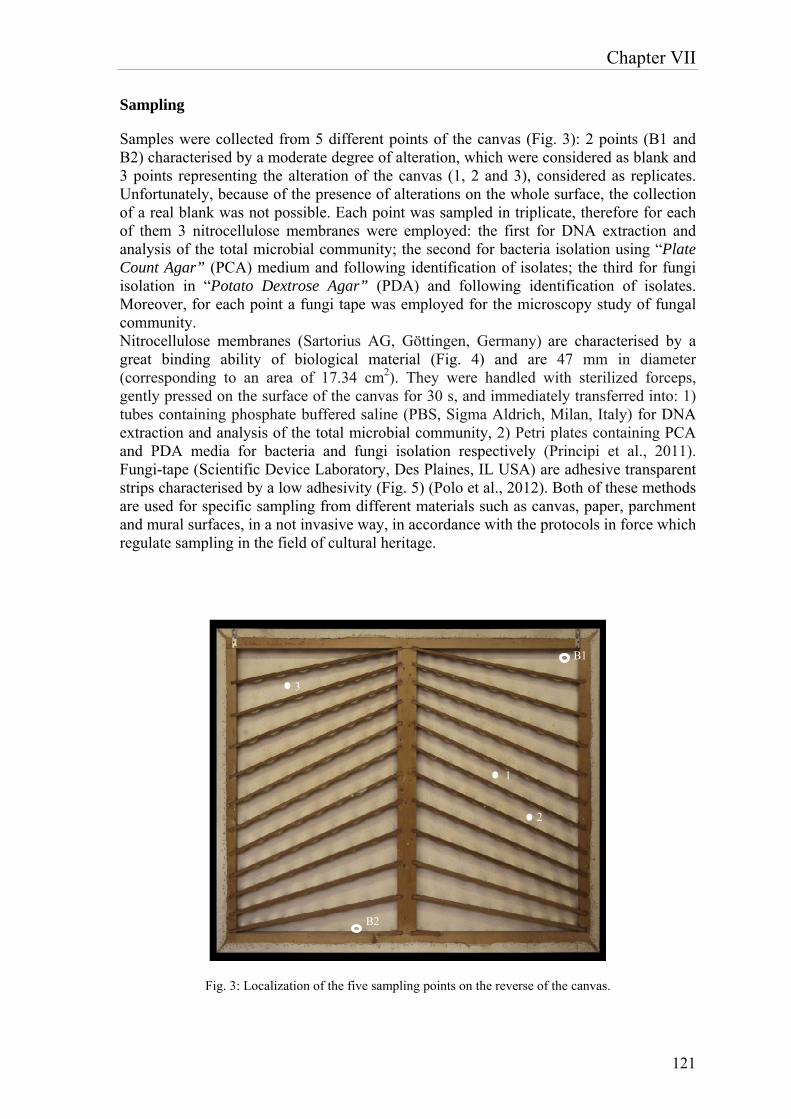
Chapter VII
121
Sampling
Samples were collected from 5 different points of the canvas (Fig. 3): 2 points (B1 and B2) characterised by a moderate degree of alteration, which were considered as blank and 3 points representing the alteration of the canvas (1, 2 and 3), considered as replicates. Unfortunately, because of the presence of alterations on the whole surface, the collection of a real blank was not possible. Each point was sampled in triplicate, therefore for each of them 3 nitrocellulose membranes were employed: the first for DNA extraction and analysis of the total microbial community; the second for bacteria isolation using “Plate Count Agar” (PCA) medium and following identification of isolates; the third for fungi isolation in “Potato Dextrose Agar” (PDA) and following identification of isolates. Moreover, for each point a fungi tape was employed for the microscopy study of fungal community. Nitrocellulose membranes (Sartorius AG, Göttingen, Germany) are characterised by a great binding ability of biological material (Fig. 4) and are 47 mm in diameter (corresponding to an area of 17.34 cm2). They were handled with sterilized forceps, gently pressed on the surface of the canvas for 30 s, and immediately transferred into: 1) tubes containing phosphate buffered saline (PBS, Sigma Aldrich, Milan, Italy) for DNA extraction and analysis of the total microbial community, 2) Petri plates containing PCA and PDA media for bacteria and fungi isolation respectively (Principi et al., 2011). Fungi-tape (Scientific Device Laboratory, Des Plaines, IL USA) are adhesive transparent strips characterised by a low adhesivity (Fig. 5) (Polo et al., 2012). Both of these methods are used for specific sampling from different materials such as canvas, paper, parchment and mural surfaces, in a not invasive way, in accordance with the protocols in force which regulate sampling in the field of cultural heritage.
B1
1
2
3
B2
Fig. 3: Localization of the five sampling points on the reverse of the canvas.
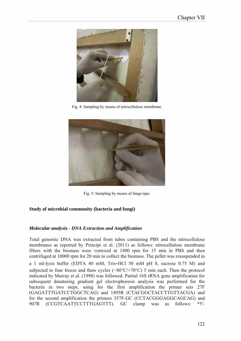
Chapter VII
122
Fig. 4: Sampling by means of nitrocellulose membrane.
Fig. 5: Sampling by means of fungi-tape.
Study of microbial community (bacteria and fungi) Molecular analysis - DNA Extraction and Amplification Total genomic DNA was extracted from tubes containing PBS and the nitrocellulose membranes as reported by Principi et al. (2011) as follows: nitrocellulose membrane filters with the biomass were vortexed at 1400 rpm for 15 min in PBS and then centrifuged at 10000 rpm for 20 min to collect the biomass. The pellet was resuspended in
a 1 ml-lysis buffer (EDTA 40 mM, Tris–HCl 50 mM pH 8, sucrose 0.75 M) and
subjected to four freeze and thaw cycles (−80°C/+70°C) 5 min each. Then the protocol indicated by Murray et al. (1998) was followed. Partial 16S rRNA gene amplification for subsequent denaturing gradient gel electrophoresis analysis was performed for the bacteria in two steps, using for the first amplification the primer sets 27F (GAGATTTGATCCTGGCTCAG) and 1495R (CTACGGCTACCTTGTTACGA) and for the second amplification the primers 357F-GC (CCTACGGGAGGCAGCAG) and 907R (CCGTCAATTCCTTTGAGTTT). GC clamp was as follows: *5′-
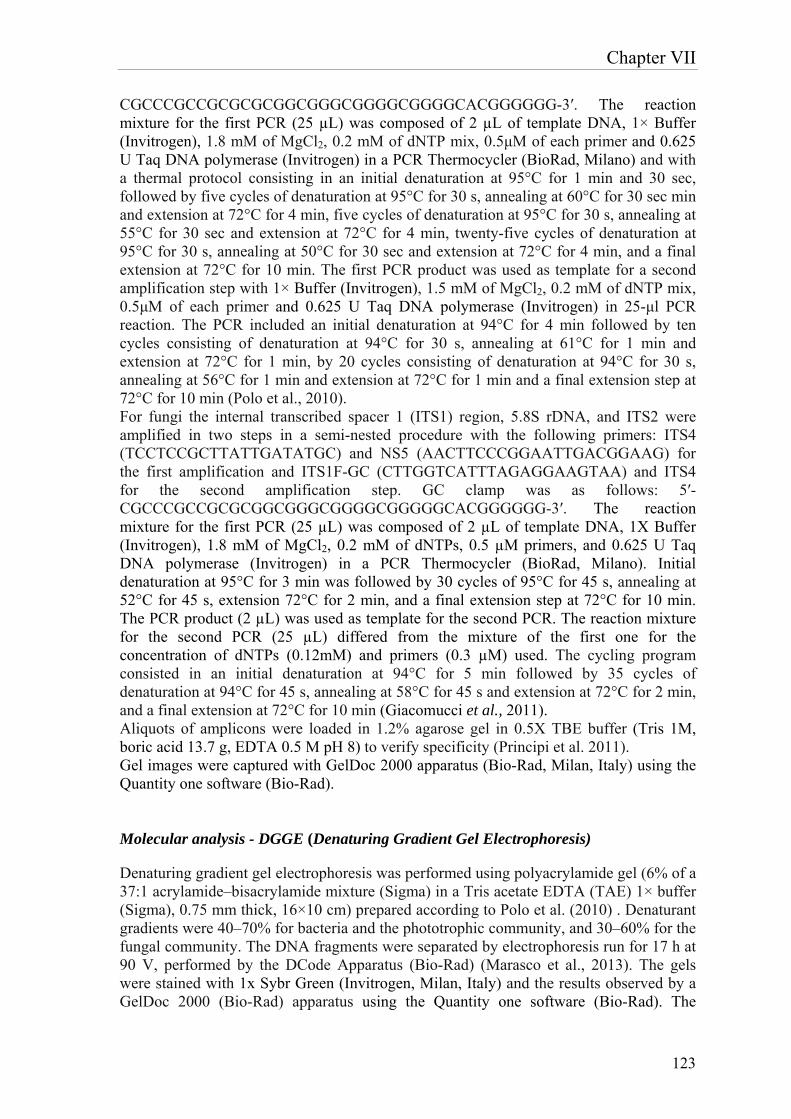
Chapter VII
123
CGCCCGCCGCGCGCGGCGGGCGGGGCGGGGCACGGGGGG-3′. The reaction mixture for the first PCR (25 µL) was composed of 2 µL of template DNA, 1× Buffer (Invitrogen), 1.8 mM of MgCl2, 0.2 mM of dNTP mix, 0.5μM of each primer and 0.625 U Taq DNA polymerase (Invitrogen) in a PCR Thermocycler (BioRad, Milano) and with a thermal protocol consisting in an initial denaturation at 95°C for 1 min and 30 sec, followed by five cycles of denaturation at 95°C for 30 s, annealing at 60°C for 30 sec min and extension at 72°C for 4 min, five cycles of denaturation at 95°C for 30 s, annealing at 55°C for 30 sec and extension at 72°C for 4 min, twenty-five cycles of denaturation at 95°C for 30 s, annealing at 50°C for 30 sec and extension at 72°C for 4 min, and a final extension at 72°C for 10 min. The first PCR product was used as template for a second amplification step with 1× Buffer (Invitrogen), 1.5 mM of MgCl2, 0.2 mM of dNTP mix, 0.5μM of each primer and 0.625 U Taq DNA polymerase (Invitrogen) in 25-μl PCR reaction. The PCR included an initial denaturation at 94°C for 4 min followed by ten cycles consisting of denaturation at 94°C for 30 s, annealing at 61°C for 1 min and extension at 72°C for 1 min, by 20 cycles consisting of denaturation at 94°C for 30 s, annealing at 56°C for 1 min and extension at 72°C for 1 min and a final extension step at 72°C for 10 min (Polo et al., 2010). For fungi the internal transcribed spacer 1 (ITS1) region, 5.8S rDNA, and ITS2 were amplified in two steps in a semi-nested procedure with the following primers: ITS4 (TCCTCCGCTTATTGATATGC) and NS5 (AACTTCCCGGAATTGACGGAAG) for the first amplification and ITS1F-GC (CTTGGTCATTTAGAGGAAGTAA) and ITS4 for the second amplification step. GC clamp was as follows: 5′-CGCCCGCCGCGCGGCGGGCGGGGCGGGGGCACGGGGGG-3′. The reaction mixture for the first PCR (25 µL) was composed of 2 µL of template DNA, 1X Buffer (Invitrogen), 1.8 mM of MgCl2, 0.2 mM of dNTPs, 0.5 µM primers, and 0.625 U Taq DNA polymerase (Invitrogen) in a PCR Thermocycler (BioRad, Milano). Initial denaturation at 95°C for 3 min was followed by 30 cycles of 95°C for 45 s, annealing at 52°C for 45 s, extension 72°C for 2 min, and a final extension step at 72°C for 10 min. The PCR product (2 µL) was used as template for the second PCR. The reaction mixture for the second PCR (25 µL) differed from the mixture of the first one for the concentration of dNTPs (0.12mM) and primers (0.3 µM) used. The cycling program consisted in an initial denaturation at 94°C for 5 min followed by 35 cycles of denaturation at 94°C for 45 s, annealing at 58°C for 45 s and extension at 72°C for 2 min, and a final extension at 72°C for 10 min (Giacomucci et al., 2011). Aliquots of amplicons were loaded in 1.2% agarose gel in 0.5X TBE buffer (Tris 1M, boric acid 13.7 g, EDTA 0.5 M pH 8) to verify specificity (Principi et al. 2011). Gel images were captured with GelDoc 2000 apparatus (Bio-Rad, Milan, Italy) using the Quantity one software (Bio-Rad). Molecular analysis - DGGE (Denaturing Gradient Gel Electrophoresis)
Denaturing gradient gel electrophoresis was performed using polyacrylamide gel (6% of a 37:1 acrylamide–bisacrylamide mixture (Sigma) in a Tris acetate EDTA (TAE) 1× buffer (Sigma), 0.75 mm thick, 16×10 cm) prepared according to Polo et al. (2010) . Denaturant gradients were 40–70% for bacteria and the phototrophic community, and 30–60% for the fungal community. The DNA fragments were separated by electrophoresis run for 17 h at 90 V, performed by the DCode Apparatus (Bio-Rad) (Marasco et al., 2013). The gels were stained with 1x Sybr Green (Invitrogen, Milan, Italy) and the results observed by a GelDoc 2000 (Bio-Rad) apparatus using the Quantity one software (Bio-Rad). The
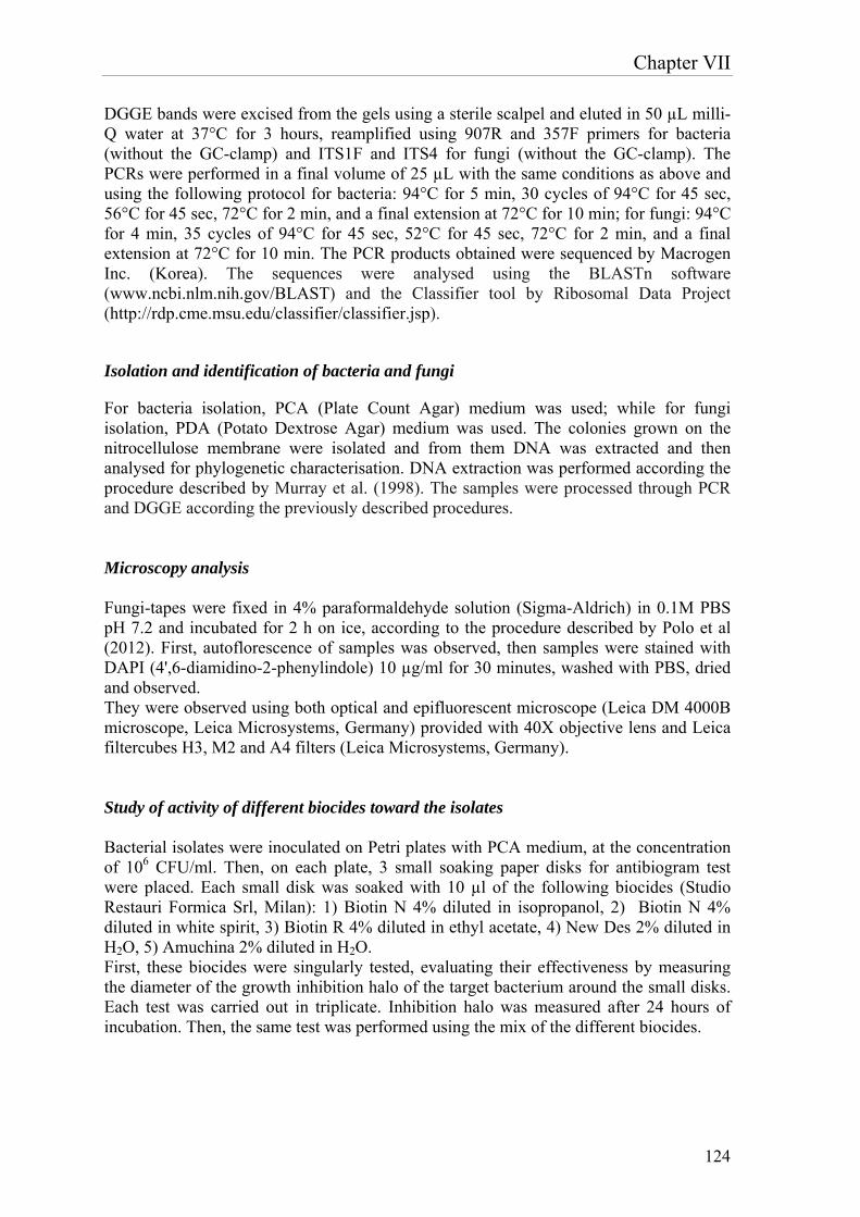
Chapter VII
124
DGGE bands were excised from the gels using a sterile scalpel and eluted in 50 µL milli-Q water at 37°C for 3 hours, reamplified using 907R and 357F primers for bacteria (without the GC-clamp) and ITS1F and ITS4 for fungi (without the GC-clamp). The PCRs were performed in a final volume of 25 µL with the same conditions as above and using the following protocol for bacteria: 94°C for 5 min, 30 cycles of 94°C for 45 sec, 56°C for 45 sec, 72°C for 2 min, and a final extension at 72°C for 10 min; for fungi: 94°C for 4 min, 35 cycles of 94°C for 45 sec, 52°C for 45 sec, 72°C for 2 min, and a final extension at 72°C for 10 min. The PCR products obtained were sequenced by Macrogen Inc. (Korea). The sequences were analysed using the BLASTn software (www.ncbi.nlm.nih.gov/BLAST) and the Classifier tool by Ribosomal Data Project (http://rdp.cme.msu.edu/classifier/classifier.jsp).
Isolation and identification of bacteria and fungi
For bacteria isolation, PCA (Plate Count Agar) medium was used; while for fungi isolation, PDA (Potato Dextrose Agar) medium was used. The colonies grown on the nitrocellulose membrane were isolated and from them DNA was extracted and then analysed for phylogenetic characterisation. DNA extraction was performed according the procedure described by Murray et al. (1998). The samples were processed through PCR and DGGE according the previously described procedures. Microscopy analysis Fungi-tapes were fixed in 4% paraformaldehyde solution (Sigma-Aldrich) in 0.1M PBS pH 7.2 and incubated for 2 h on ice, according to the procedure described by Polo et al (2012). First, autoflorescence of samples was observed, then samples were stained with DAPI (4',6-diamidino-2-phenylindole) 10 µg/ml for 30 minutes, washed with PBS, dried and observed. They were observed using both optical and epifluorescent microscope (Leica DM 4000B microscope, Leica Microsystems, Germany) provided with 40X objective lens and Leica filtercubes H3, M2 and A4 filters (Leica Microsystems, Germany). Study of activity of different biocides toward the isolates Bacterial isolates were inoculated on Petri plates with PCA medium, at the concentration of 106 CFU/ml. Then, on each plate, 3 small soaking paper disks for antibiogram test were placed. Each small disk was soaked with 10 µl of the following biocides (Studio Restauri Formica Srl, Milan): 1) Biotin N 4% diluted in isopropanol, 2) Biotin N 4% diluted in white spirit, 3) Biotin R 4% diluted in ethyl acetate, 4) New Des 2% diluted in H2O, 5) Amuchina 2% diluted in H2O. First, these biocides were singularly tested, evaluating their effectiveness by measuring the diameter of the growth inhibition halo of the target bacterium around the small disks. Each test was carried out in triplicate. Inhibition halo was measured after 24 hours of incubation. Then, the same test was performed using the mix of the different biocides.
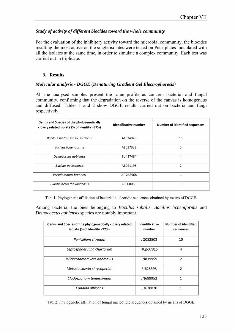
Chapter VII
125
Study of activity of different biocides toward the whole community For the evaluation of the inhibitory activity toward the microbial community, the biocides resulting the most active on the single isolates were tested on Petri plates inoculated with all the isolates at the same time, in order to simulate a complex community. Each test was carried out in triplicate.
3. Results
Molecular analysis - DGGE (Denaturing Gradient Gel Electrophoresis)
All the analysed samples present the same profile as concern bacterial and fungal community, confirming that the degradation on the reverse of the canvas is homogeneus and diffused. Tables 1 and 2 show DGGE results carried out on bacteria and fungi respectively.
Genus and Species of the phylogenetically
closely related isolate (% of identity >97%) Identificative number Number of identified sequences
Bacillus subtilis subsp. spizizenii AF074970 15
Bacillus licheniformis AE017333 5
Deinococcus gobiensis EU427464 4
Bacillus vallismortis AB021198 2
Pseudomonas brenneri AF 268968 1
Burkholderia thailandensis CP000086 1
Tab. 1: Phylogenetic affiliation of bacterial nucleotidic sequences obtained by means of DGGE.
Among bacteria, the ones belonging to Bacillus subtilis, Bacillus licheniformis and Deinococcus gobiensis species are notably important.
Genus and Species of the phylogenetically closely related
isolate (% of identity >97%)
Identificative
number
Number of identified
sequences
Penicillium citrinum JQ082503 10
Leptosphaerulina chartarum HQ607815 4
Wickerhamomyces anomalus JN839959 2
Metschnikowia chrysoperlae FJ623593 2
Cladosporium tenuissimum JN689952 1
Candida albicans JQ678820 1
Tab. 2: Phylogenetic affiliation of fungal nucleotidic sequences obtained by means of DGGE.
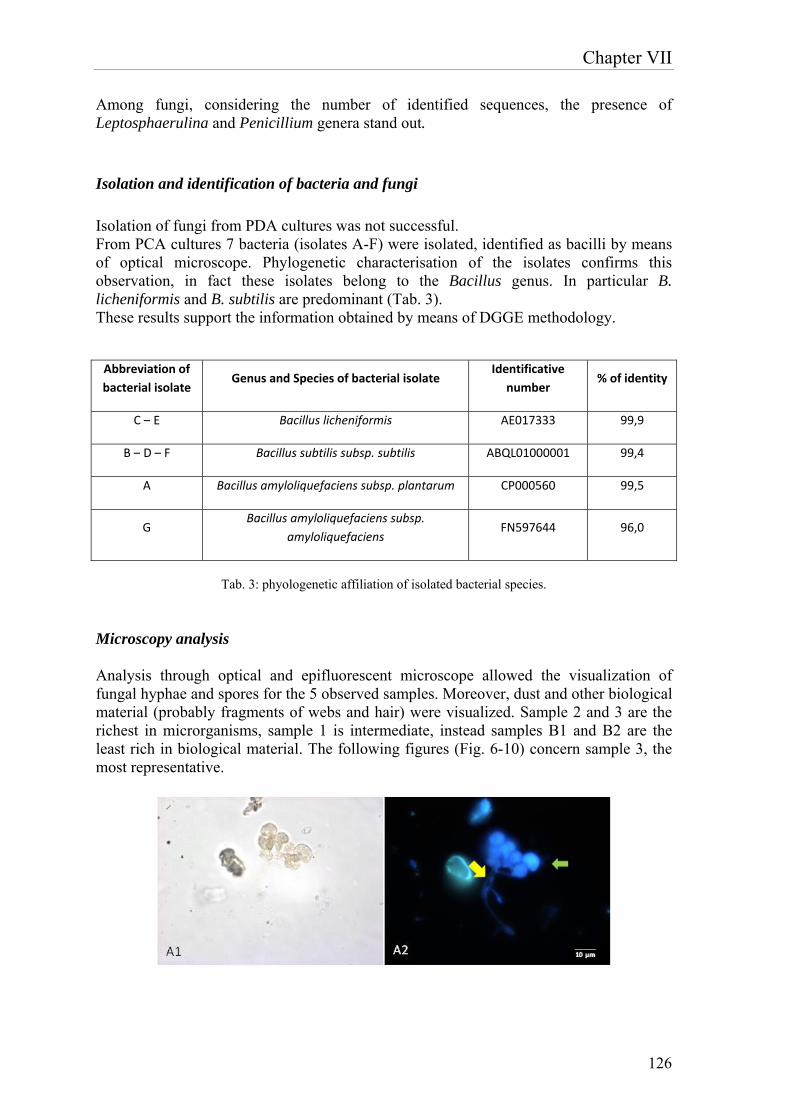
Chapter VII
126
Among fungi, considering the number of identified sequences, the presence of Leptosphaerulina and Penicillium genera stand out. Isolation and identification of bacteria and fungi Isolation of fungi from PDA cultures was not successful. From PCA cultures 7 bacteria (isolates A-F) were isolated, identified as bacilli by means of optical microscope. Phylogenetic characterisation of the isolates confirms this observation, in fact these isolates belong to the Bacillus genus. In particular B. licheniformis and B. subtilis are predominant (Tab. 3). These results support the information obtained by means of DGGE methodology.
Abbreviation of
bacterial isolate Genus and Species of bacterial isolate
Identificative
number % of identity
C – E Bacillus licheniformis AE017333 99,9
B – D – F Bacillus subtilis subsp. subtilis ABQL01000001 99,4
A Bacillus amyloliquefaciens subsp. plantarum CP000560 99,5
G Bacillus amyloliquefaciens subsp.
amyloliquefaciens FN597644 96,0
Tab. 3: phyologenetic affiliation of isolated bacterial species.
Microscopy analysis Analysis through optical and epifluorescent microscope allowed the visualization of fungal hyphae and spores for the 5 observed samples. Moreover, dust and other biological material (probably fragments of webs and hair) were visualized. Sample 2 and 3 are the richest in microrganisms, sample 1 is intermediate, instead samples B1 and B2 are the least rich in biological material. The following figures (Fig. 6-10) concern sample 3, the most representative.
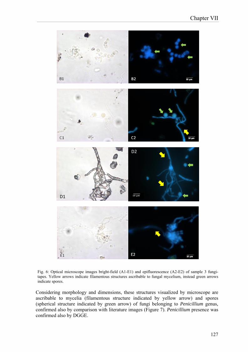
Chapter VII
127
Fig. 6: Optical microscope images bright-field (A1-E1) and epifluorescence (A2-E2) of sample 3 fungi-tapes. Yellow arrows indicate filamentous structures ascribable to fungal mycelium, instead green arrows indicate spores.
Considering morphology and dimensions, these structures visualized by microscope are ascribable to mycelia (filamentous structure indicated by yellow arrow) and spores (spherical structure indicated by green arrow) of fungi belonging to Penicillium genus, confirmed also by comparison with literature images (Figure 7). Penicillium presence was confirmed also by DGGE.
µm
D2
D1 10 µm
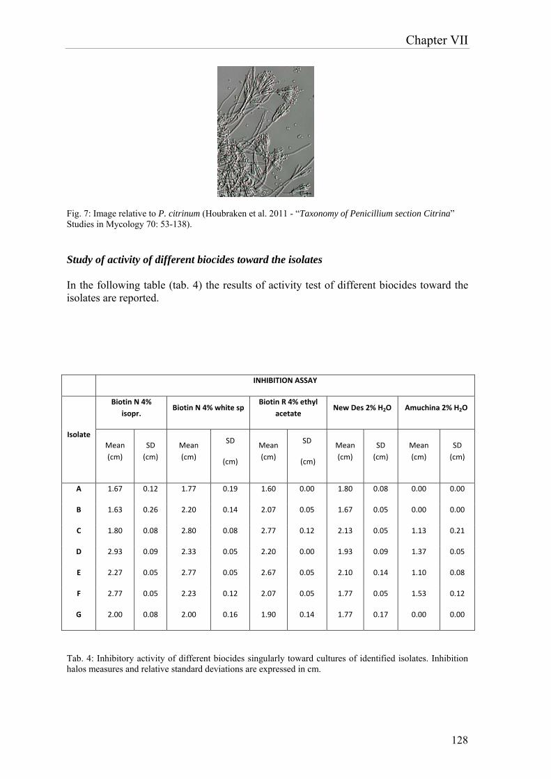
Chapter VII
128
Fig. 7: Image relative to P. citrinum (Houbraken et al. 2011 - “Taxonomy of Penicillium section Citrina” Studies in Mycology 70: 53-138).
Study of activity of different biocides toward the isolates In the following table (tab. 4) the results of activity test of different biocides toward the isolates are reported.
INHIBITION ASSAY
Isolate
Biotin N 4%
isopr. Biotin N 4% white sp
Biotin R 4% ethyl
acetate New Des 2% H2O Amuchina 2% H2O
Mean
(cm)
SD
(cm)
Mean
(cm)
SD
(cm)
Mean
(cm)
SD
(cm)
Mean
(cm)
SD
(cm)
Mean
(cm)
SD
(cm)
A 1.67 0.12 1.77 0.19 1.60 0.00 1.80 0.08 0.00 0.00
B 1.63 0.26 2.20 0.14 2.07 0.05 1.67 0.05 0.00 0.00
C 1.80 0.08 2.80 0.08 2.77 0.12 2.13 0.05 1.13 0.21
D 2.93 0.09 2.33 0.05 2.20 0.00 1.93 0.09 1.37 0.05
E 2.27 0.05 2.77 0.05 2.67 0.05 2.10 0.14 1.10 0.08
F 2.77 0.05 2.23 0.12 2.07 0.05 1.77 0.05 1.53 0.12
G 2.00 0.08 2.00 0.16 1.90 0.14 1.77 0.17 0.00 0.00
Tab. 4: Inhibitory activity of different biocides singularly toward cultures of identified isolates. Inhibition halos measures and relative standard deviations are expressed in cm.
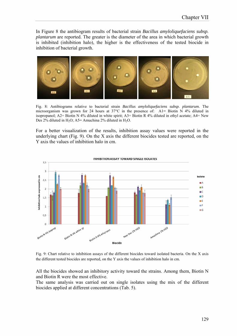
Chapter VII
129
In Figure 8 the antibiogram results of bacterial strain Bacillus amyloliquefaciens subsp. plantarum are reported. The greater is the diameter of the area in which bacterial growth is inhibited (inhibition halo), the higher is the effectiveness of the tested biocide in inhibition of bacterial growth.
Fig. 8: Antibiograms relative to bacterial strain Bacillus amyloliquefaciens subsp. plantarum. The microorganism was grown for 24 hours at 37°C in the presence of: A1= Biotin N 4% diluted in isopropanol; A2= Biotin N 4% diluted in white spirit; A3= Biotin R 4% diluted in ethyl acetate; A4= New Des 2% diluted in H2O; A5= Amuchina 2% diluted in H2O.
For a better visualization of the results, inhibition assay values were reported in the underlying chart (Fig. 9). On the X axis the different biocides tested are reported, on the Y axis the values of inhibition halo in cm.
Fig. 9: Chart relative to inhibition assays of the different biocides toward isolated bacteria. On the X axis the different tested biocides are reported, on the Y axis the values of inhibition halo in cm. All the biocides showed an inhibitory activity toward the strains. Among them, Biotin N and Biotin R were the most effective. The same analysis was carried out on single isolates using the mix of the different biocides applied at different concentrations (Tab. 5).
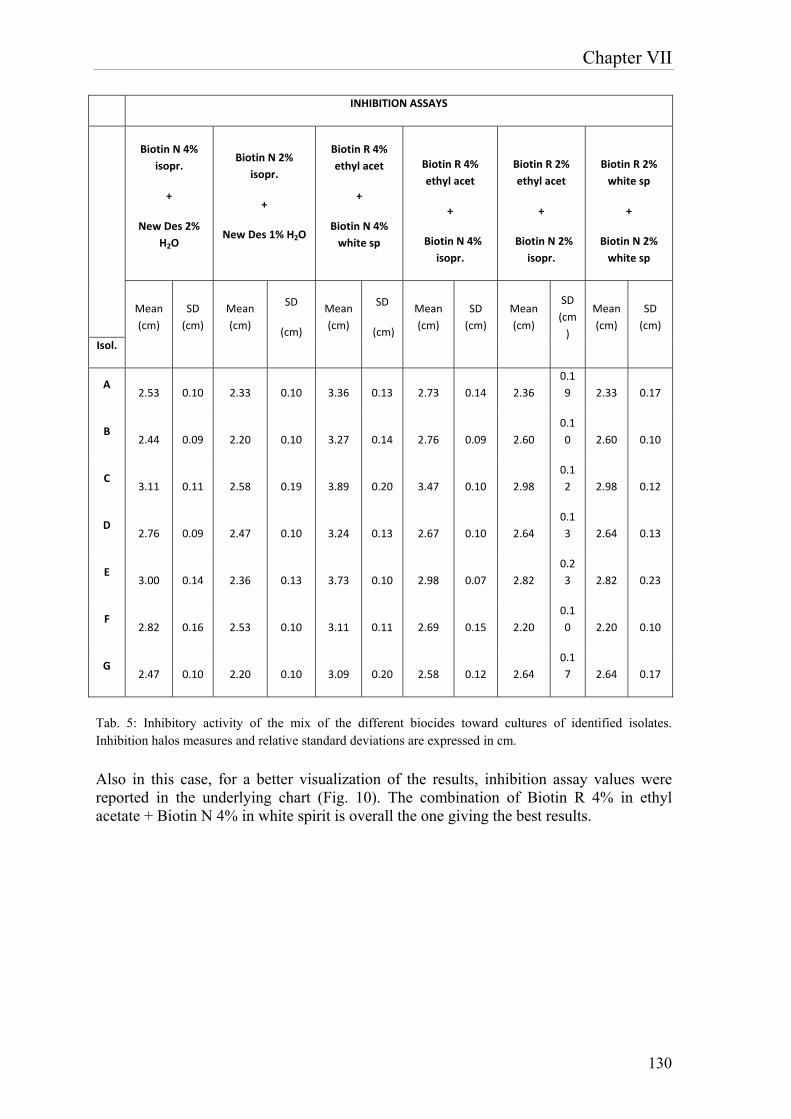
Chapter VII
130
INHIBITION ASSAYS
Biotin N 4%
isopr.
+
New Des 2%
H2O
Biotin N 2%
isopr.
+
New Des 1% H2O
Biotin R 4%
ethyl acet
+
Biotin N 4%
white sp
Biotin R 4%
ethyl acet
+
Biotin N 4%
isopr.
Biotin R 2%
ethyl acet
+
Biotin N 2%
isopr.
Biotin R 2%
white sp
+
Biotin N 2%
white sp
Mean
(cm)
SD
(cm)
Mean
(cm)
SD
(cm)
Mean
(cm)
SD
(cm)
Mean
(cm)
SD
(cm)
Mean
(cm)
SD
(cm
)
Mean
(cm)
SD
(cm)
Isol.
A 2.53 0.10 2.33 0.10 3.36 0.13 2.73 0.14 2.36
0.1
9 2.33 0.17
B 2.44 0.09 2.20 0.10 3.27 0.14 2.76 0.09 2.60
0.1
0 2.60 0.10
C 3.11 0.11 2.58 0.19 3.89 0.20 3.47 0.10 2.98
0.1
2 2.98 0.12
D 2.76 0.09 2.47 0.10 3.24 0.13 2.67 0.10 2.64
0.1
3 2.64 0.13
E 3.00 0.14 2.36 0.13 3.73 0.10 2.98 0.07 2.82
0.2
3 2.82 0.23
F 2.82 0.16 2.53 0.10 3.11 0.11 2.69 0.15 2.20
0.1
0 2.20 0.10
G 2.47 0.10 2.20 0.10 3.09 0.20 2.58 0.12 2.64
0.1
7 2.64 0.17
Tab. 5: Inhibitory activity of the mix of the different biocides toward cultures of identified isolates. Inhibition halos measures and relative standard deviations are expressed in cm.
Also in this case, for a better visualization of the results, inhibition assay values were reported in the underlying chart (Fig. 10). The combination of Biotin R 4% in ethyl acetate + Biotin N 4% in white spirit is overall the one giving the best results.
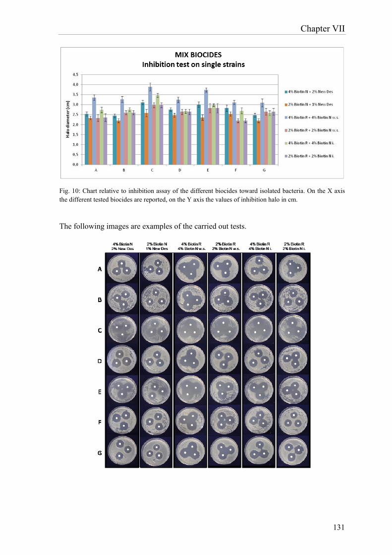
Chapter VII
131
Fig. 10: Chart relative to inhibition assay of the different biocides toward isolated bacteria. On the X axis the different tested biocides are reported, on the Y axis the values of inhibition halo in cm.
The following images are examples of the carried out tests.

Chapter VII
132
Statistical analysis
The results obtained from inhibition tests were analysed through the T- Student Test (Tab. 6). This test is performed between two numerical series, in order to evaluate whether the difference between the means is significant or not. The threshold value is 0.05; for values inferior to 0.05, the difference between the means is statistically significant, for superior values it’s not significant. The analysis proves that inhibition values obtained from the combination Biotin R 4% + Biotin N 4% in white spirit are statistically superior compared with the other mixtures. Moreover, this is overall the most effective combination in inhibition of the single strains.
T‐test
4% Biotin
N + 2%
New Des
2% Biotin
N + 1%
New Des
4% Biotin R
+ 4% Biotin
N w.s.
2% Biotin R
+ 2% Biotin
N w.s.
4% Biotin
R + 4%
Biotin N i.
2% Biotin
R + 2%
Biotin N i.
4% Biotin N + 2% New Des 1.00
2% Biotin N + 1% New Des 0.01 1.00
4% Biotin R + 4% Biotin N w.s. 0.00115 0.00001 1.00
2% Biotin R + 2% Biotin N w.s. 0.38 0.08 0.0003 1.00
4% Biotin R + 4% Biotin N i. 0.50 0.004 0.006 0.15 1.00
2% Biotin R + 2% Biotin N i. 0.38 0.07 0.0003 0.98 0.15 1.00
Tab. 6: Values relative to T-test. Values for p < 0.05 are indicated in red.
Study of activity of different biocides toward the whole community In the following table (tab. 7) the results of activity test of different biocides toward the whole community are reported.
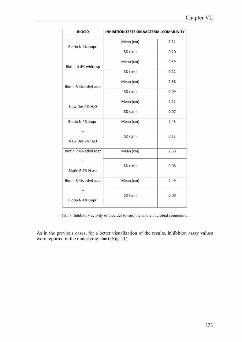
Chapter VII
133
BIOCID INHIBITION TESTS ON BACTERIAL COMMUNITY
Biotin N 4% isopr. Mean (cm) 2.31
SD (cm) 0.20
Biotin N 4% white sp Mean (cm) 2.59
SD (cm) 0.12
Biotin R 4% ethyl acet Mean (cm) 2.58
SD (cm) 0.09
New Des 2% H2O Mean (cm) 2.21
SD (cm) 0.07
Biotin N 4% isopr.
+
New Des 2% H2O
Mean (cm) 2.43
SD (cm) 0.13
Biotin R 4% ethyl acet
+
Biotin R 4% N w.s
Mean (cm) 2.68
SD (cm) 0.08
Biotin R 4% ethyl acet
+
Biotin N 4% isopr.
Mean (cm) 2.49
SD (cm) 0.08
Tab. 7: Inhibitory activity of biocides toward the whole microbial community.
As in the previous cases, for a better visualization of the results, inhibition assay values were reported in the underlying chart (Fig. 11).
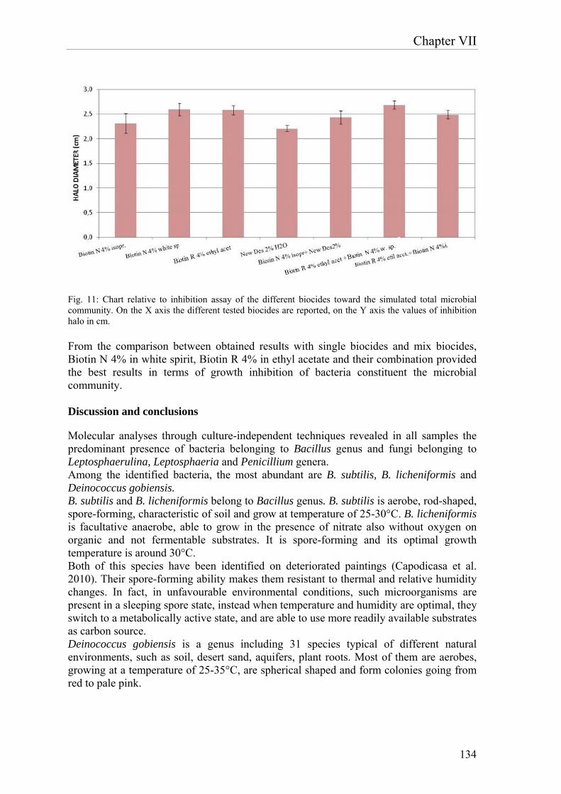
Chapter VII
134
Fig. 11: Chart relative to inhibition assay of the different biocides toward the simulated total microbial community. On the X axis the different tested biocides are reported, on the Y axis the values of inhibition halo in cm. From the comparison between obtained results with single biocides and mix biocides, Biotin N 4% in white spirit, Biotin R 4% in ethyl acetate and their combination provided the best results in terms of growth inhibition of bacteria constituent the microbial community. Discussion and conclusions
Molecular analyses through culture-independent techniques revealed in all samples the predominant presence of bacteria belonging to Bacillus genus and fungi belonging to Leptosphaerulina, Leptosphaeria and Penicillium genera. Among the identified bacteria, the most abundant are B. subtilis, B. licheniformis and Deinococcus gobiensis. B. subtilis and B. licheniformis belong to Bacillus genus. B. subtilis is aerobe, rod-shaped, spore-forming, characteristic of soil and grow at temperature of 25-30°C. B. licheniformis is facultative anaerobe, able to grow in the presence of nitrate also without oxygen on organic and not fermentable substrates. It is spore-forming and its optimal growth temperature is around 30°C. Both of this species have been identified on deteriorated paintings (Capodicasa et al. 2010). Their spore-forming ability makes them resistant to thermal and relative humidity changes. In fact, in unfavourable environmental conditions, such microorganisms are present in a sleeping spore state, instead when temperature and humidity are optimal, they switch to a metabolically active state, and are able to use more readily available substrates as carbon source. Deinococcus gobiensis is a genus including 31 species typical of different natural environments, such as soil, desert sand, aquifers, plant roots. Most of them are aerobes, growing at a temperature of 25-35°C, are spherical shaped and form colonies going from red to pale pink.

Chapter VII
135
In particular, D. gobiensis, is reported in literature as a coccoid bacterium, facultative anaerobe, whose colonies are red. From literature studies, fungi belonging to Leptosphaerulina genus are not implicated in degradation of materials employed in paintings or for the realization of artworks in general. Instead, P. citrium belongs to Penicillium genus, but is different from other fungi of the same genus for its ability to grow at a temperature of about 37°C and for the production of yellow-orange pigments. It is present in different habitats: soil, air of closed environments, roots and leaves of coffee plants (Houbraken et al. 2011). Different species of Penicillium were identified on deteriorated paintings (Capodicasa et al. 2010). In particular Penicillium glabrum and P. chrysogenum were isolated from wooden artworks and described as cellulose and/or lignin degrader (Pangallo et al. 2007). As concerns analysis for identification of cultivable bacteria, the dominance of Bacillus genus was confirmed and in particular of B. subtilis and B. licheniformis species. As concerns identification of fungi, no isolate was obtained, probably because of the uncultivability of these species. In order to further confirm the data obtained by DGGE, the organic material sampled by means of fungi-tape was visualized through optical microscope. A series of cellular aggregates, imputable to Penicillium genus for their morphology and dimensions, was identified. The simultaneous presence of bacilli and penicilli suggests that the deterioration of the canvas is attributable at the beginning to the activity of fungi able to degrade the wooden-cellulosic component, whereas only later bacterial colonisation took place, due to the presence of immediately consumable substances, such as proteins and polysaccharides, derived from fungal metabolism. At last, degradation phases create the environmental-nutritional conditions that are favourable to the following colonization by other bacteria such as D. gobiensis, whose growth could be the cause of yellow-earth/red pigmentation of the alterations. The activity of different biocides was evaluated toward the single isolated bacteria, through the antibiogram test. 4 biocides were compared, they were characterized by 4 different actives: quaternary ammonium salts for NEW DES; Tributyltin Naphthenate and quaternary ammonium salts for BIOTIN N; IPBC (iodiopropynilbuthilcarbammate) and OIT (n-octyl – isothiazolinone) for BIOTIN R; sodium hypochlorite for Amuchina. From the results we can conclude that BIOTIN N and BIOTIN R are equally effective toward all target bacteria, as NEW DES too, even though the latter is less effective toward E (B. licheniformis) and D (B. subtilis) isolates. Amuchina is the least effective product: it doesn’t inhibit the growth of A, G (B. amyloliquefaciens) and B (B. subtilis) isolates and is also less effective than the other biocides toward the other isolates. In the test of the mix of the different biocides at different concentration toward the single strains, the combination of Biotin R 4% in ethyl acetate + Biotin N 4% in white spirit was overall the best, also toward the whole bacterial community (Fig. 11 and 12).
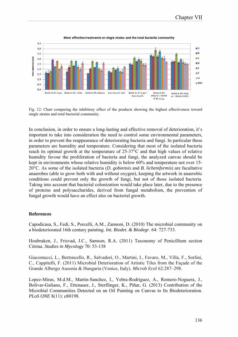
Chapter VII
136
Fig. 12: Chart comparing the inhibitory effect of the products showing the highest effectiveness toward single strains and total bacterial community.
In conclusion, in order to ensure a long-lasting and effective removal of deterioration, it’s important to take into consideration the need to control some environmental parameters, in order to prevent the reappearance of deteriorating bacteria and fungi. In particular these parameters are humidity and temperature. Considering that most of the isolated bacteria reach its optimal growth at the temperature of 25-37°C and that high values of relative humidity favour the proliferation of bacteria and fungi, the analyzed canvas should be kept in environments whose relative humidity is below 60% and temperature not over 15-20°C. As some of the isolated bacteria (D. gobiensis and B. licheniformis) are facultative anaerobes (able to grow both with and without oxygen), keeping the artwork in anaerobic conditions could prevent only the growth of fungi, but not of those isolated bacteria. Taking into account that bacterial colonization would take place later, due to the presence of proteins and polysaccharides, derived from fungal metabolism, the prevention of fungal growth would have an effect also on bacterial growth. References
Capodicasa, S., Fedi, S., Porcelli, A.M., Zannoni, D. (2010) The microbial community on a biodeteriorated 16th century painting. Int. Biodet. & Biodegr. 64: 727-733. Houbraken, J., Frisvad, J.C., Samson, R.A. (2011) Taxonomy of Penicillium section Citrina. Studies in Mycology 70: 53-138 Giacomucci, L., Bertoncello, R., Salvadori, O., Martini, I., Favaro, M., Villa, F., Sorlini, C., Cappitelli, F. (2011) Microbial Deterioration of Artistic Tiles from the Façade of the Grande Albergo Ausonia & Hungaria (Venice, Italy). Microb Ecol 62:287–298. Lopez-Miras, M.d.M., Martin-Sanchez, I., Yebra-Rodriguez, A., Romero-Noguera, J., Bolivar-Galiano, F., Ettenauer, J., Sterflinger, K., Piñar, G. (2013) Contribution of the Microbial Communities Detected on an Oil Painting on Canvas to Its Biodeterioration. PLoS ONE 8(11): e80198.
Chapter VII
137
Lopez-Miras, M.d.M., Piñar, G., Romero-Noguera, J., Bolivar-Galiano, F., Ettenauer, J., Sterflinger, K., Martin-Sanchez, I. (2013) Microbial communities adhering to the obverse and reverse sides of an oil painting on canvas: identification and evaluation of their biodegradative potential. Aerobiologia 29:301–314. Marasco, R., Rolli, E., Fusi, M., Cherif, A., Abou-Hadid, A., El-Bahairy, U., Borin, S., Sorlini, C. and Daffonchio, D. (2013) Plant Growth Promotion Potential Is Equally Represented in Diverse Grapevine Root-Associated Bacterial Communities from Different Biopedoclimatic Environments BioMed Research International ID 491091. Murray, A.E., Preston, C.M., Massana, R., Taylor, L.T., Blakis, A., Wu, K., Delong, E.F. (1998) Seasonal and spatial variability of bacterial and archaeal assemblages in the coastal waters near Anvers Island, Antarctica. Appl Environ Microbiol 64:2585–2595. Pangallo, D., Simonovicova, A., Chovanova K. and Ferianc P. (2007) Wooden art objects and the museum environment: identification and biodegradative characteristics of isolated microflora. Letters in Applied Microbiology 45: 87-94. Polo, A., Cappitelli, F., Brusetti, L., Principi, P., Villa, F., Giacomucci, L., Ranalli, G., Sorlini, C. (2010) Feasibility of removing surface deposits on stone using biological and chemical remediation methods. Microb Ecol 60(1):1–14
Polo, A., Gulotta, D., Santo, N., Di Benedetto, C., Fascio, U., Toniolo, L., Villa, F. and Cappitelli F. (2012) Importance of subaerial biofilms and airborne microflora in the deterioration of stonework: a molecular study. Biofouling 28 (10): 1093–1106. Principi, P., Villa, F., Sorlini,C. and Cappitelli, F. (2011) Molecular Studies of Microbial Community Structure on Stained Pages of Leonardo da Vinci’s Atlantic Codex. Microb Ecol 61:214–222.

138
Chapter VIII
139
Identification of microorganisms deteriorating a paper print of the Monastery of “S. Maria al Carrobiolo” in Monza
1. Introduction and aim of the work
Cellulose is the most abundant organic compound found on Earth (30–50 % of plant dry weight) and represents a major source of energy for microorganisms in natural environment (Pinzari et al., 2011). Microbial deterioration inevitably occurs on paper of different ages and cultural heritage made of this material are not immune to this kind of spoilage. “Biodeterioration” was defined by Hueck as “any undesirable change in the properties of a material caused by the vital activities of organisms”. Microbial degradation of paper causes different kinds of damage, according to the species of organism responsible for the attack. Damage can occur because of mechanical stress or enzymatic action, because moulds can produce a wide range of enzymes (proteinases, gelatinases, cellulases) which are able to destroy the component materials of library and archival collections. Some filamentous fungi frequently associated with paper damage can dissolve cellulose fibres by means of the action of cellulolytic enzymes. Furthermore, fungi produce pigments or organic acids that discolour paper and cause damage to materials of cultural and historical value made from paper. Paper was first made in China around 105 AD, its history can be divided into two phases: 1) before the 19th century, when it was made by hand, using cellulose from linen and cotton rags; 2) from the 19th century, it has been made by machine from wood pulp, and in addition to cellulose it contains also lignin, emicellulose, pectin and sizes such as gelatine, minerals and pigments (Cappitelli et al., 2010). One of the main factors determining the microbial colonization of paper is water availability. As paper is very hygroscopic, the water activity of a paper object is related to environmental parameters such as temperature and relative humidity. Temperatures above 23°C and a relative humidity over 65% increase the risk of microbial colonization. This factor should be considered in range of preventive strategies against biodeterioration. Damage to paper is mainly due to microfungi and to an inferior extent, to heterotrophic bacteria. Identification of these agents is important both for the conservation of the paper artwork and for the health of conservation staff and users. One of the main problems encountered in biological diagnostics of cultural heritage is the carrying out of non-invasive sampling, with the consequent drawback that only microorganisms producing emerging structure on paper will be collected. Moreover samples from this kind of artworks are often few and small, therefore the obtained results are not always representative. A currently used non invasive sampling procedure from paper items involves dry swabs, but they are barely effective. Another non invasive technique involves nitrocellulose membrane filters: sterile circular nitrocellulose membranes 47 mm in diameter are first exposed to the surface being analyzed and then are transferred onto solid culture media to isolate bacteria and fungi. The latter has been selected in this work for sampling of a paper print of the Monastery of “S. Maria al Carrobiolo” in Monza. The aim of the current study is the detection of biodeteriogens responsible for the spoilage of this artwork and determination of their phylogenetic affiliation, by means of a molecular approach.

Chapter VIII
140
2. Materials and methods
Artwork description and its state of conservation
The analyzed artwork is paper print number 10, belonging to a cycle of prints conserved in the Monastery of “S. Maria al Carrobiolo” in Monza, realized in the 17th century. The artwork presented whitenings and small dark spots, on the obverse and on the reverse side, respectively (Fig. 1).
Fig. 1: Arrow A indicates a whitening on the obverse side of Print n°10, arrow B indicates a small dark spot on the reverse side of Print n°10.
Sampling Sampling of microorganisms colonizing the surface of Print n°10 has been carried out by nitrocellulose membranes, in order to fulfill a non-invasive collection of samples. Membranes have been put in contact with the surface for 30 seconds, through the aid of sterile swabs. For each sampling point, 2 membranes have been employed. Then one of the membranes has been placed on a Petri plate containing “Nutrient Broth” (NB) medium for bacteria isolation and following identification of isolates, the other one on a Petri plate containing “Potato Dextrose Agar” (PDA) medium for fungi isolation and following identification of isolates. Samples have been collected from three points of the frame and the body of the obverse side of the print, three points of the reverse side of the print, in correspondence to the most intense chromatic alterations. Another point (4) has been sampled as blank, taken from an area seemingly free from alterations and lastly point 5 has been taken from a detached fragment of the back of the frame. Figure 2 indicates the sampling points.
A B
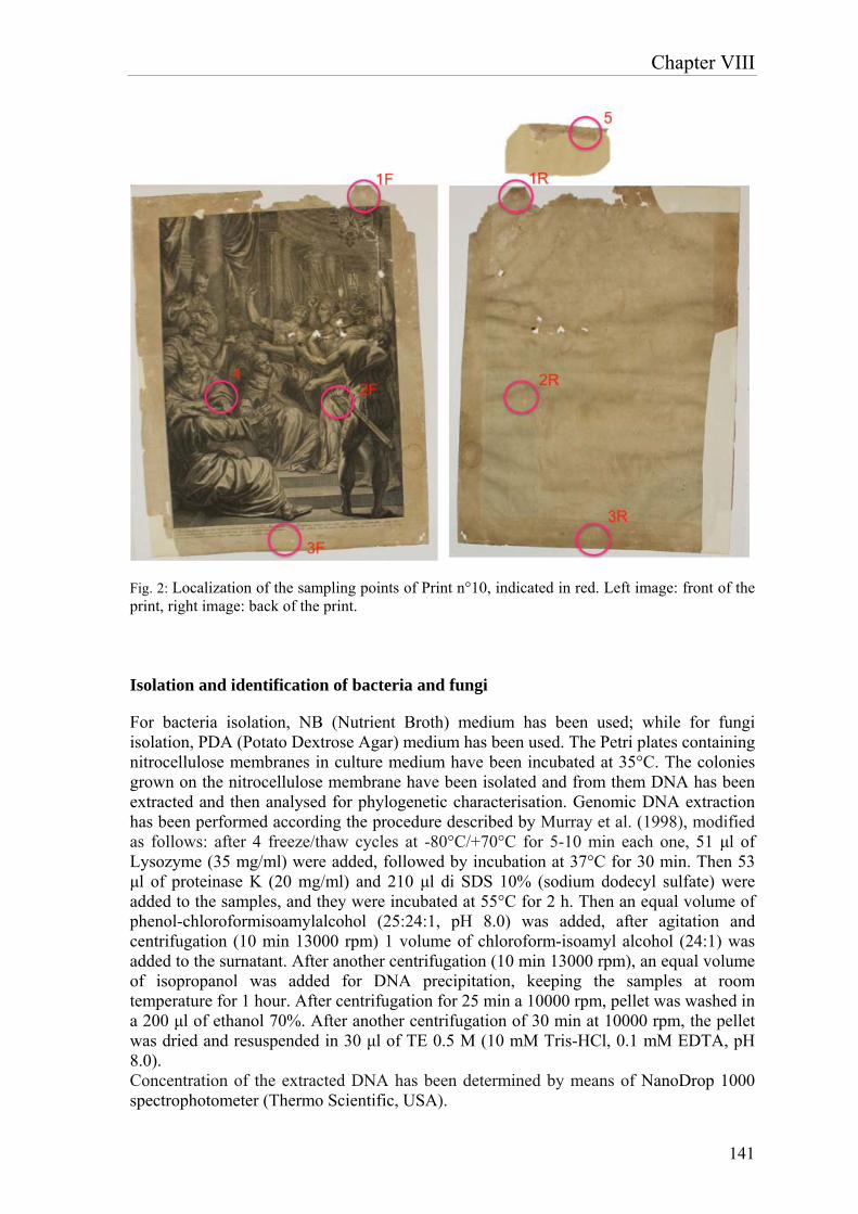
Chapter VIII
141
Fig. 2: Localization of the sampling points of Print n°10, indicated in red. Left image: front of the print, right image: back of the print.
Isolation and identification of bacteria and fungi
For bacteria isolation, NB (Nutrient Broth) medium has been used; while for fungi isolation, PDA (Potato Dextrose Agar) medium has been used. The Petri plates containing nitrocellulose membranes in culture medium have been incubated at 35°C. The colonies grown on the nitrocellulose membrane have been isolated and from them DNA has been extracted and then analysed for phylogenetic characterisation. Genomic DNA extraction has been performed according the procedure described by Murray et al. (1998), modified as follows: after 4 freeze/thaw cycles at -80°C/+70°C for 5-10 min each one, 51 μl of Lysozyme (35 mg/ml) were added, followed by incubation at 37°C for 30 min. Then 53 μl of proteinase K (20 mg/ml) and 210 μl di SDS 10% (sodium dodecyl sulfate) were added to the samples, and they were incubated at 55°C for 2 h. Then an equal volume of phenol-chloroformisoamylalcohol (25:24:1, pH 8.0) was added, after agitation and centrifugation (10 min 13000 rpm) 1 volume of chloroform-isoamyl alcohol (24:1) was added to the surnatant. After another centrifugation (10 min 13000 rpm), an equal volume of isopropanol was added for DNA precipitation, keeping the samples at room temperature for 1 hour. After centrifugation for 25 min a 10000 rpm, pellet was washed in a 200 μl of ethanol 70%. After another centrifugation of 30 min at 10000 rpm, the pellet was dried and resuspended in 30 μl of TE 0.5 M (10 mM Tris-HCl, 0.1 mM EDTA, pH 8.0). Concentration of the extracted DNA has been determined by means of NanoDrop 1000 spectrophotometer (Thermo Scientific, USA).
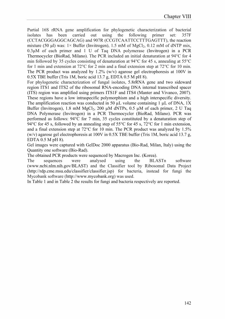
Chapter VIII
142
Partial 16S rRNA gene amplification for phylogenetic characterization of bacterial isolates has been carried out using the following primer set: 357F (CCTACGGGAGGCAGCAG) and 907R (CCGTCAATTCCTTTGAGTTT), the reaction mixture (50 μl) was: 1× Buffer (Invitrogen), 1.5 mM of MgCl2, 0.12 mM of dNTP mix, 0.3μM of each primer and 1 U of Taq DNA polymerase (Invitrogen) in a PCR Thermocycler (BioRad, Milano). The PCR included an initial denaturation at 94°C for 4 min followed by 35 cycles consisting of denaturation at 94°C for 45 s, annealing at 55°C for 1 min and extension at 72°C for 2 min and a final extension step at 72°C for 10 min. The PCR product was analyzed by 1.2% (w/v) agarose gel electrophoresis at 100V in 0.5X TBE buffer (Tris 1M, boric acid 13.7 g, EDTA 0.5 M pH 8). For phylogenetic characterization of fungal isolates, 5.8rRNA gene and two sideward region ITS1 and ITS2 of the ribosomal RNA-encoding DNA internal transcribed spacer (ITS) region was amplified using primers ITS1F and ITS4 (Manter and Vivanco, 2007). These regions have a low intraspecific polymorphism and a high interspecific diversity. The amplification reaction was conducted in 50 µL volume containing 1 µL of DNA, 1X Buffer (Invitrogen), 1.8 mM MgCl2, 200 µM dNTPs, 0.5 µM of each primer, 2 U Taq DNA Polymerase (Invitrogen) in a PCR Thermocycler (BioRad, Milano). PCR was performed as follows: 94°C for 7 min, 35 cycles constituted by a denaturation step of 94°C for 45 s, followed by an annealing step of 55°C for 45 s, 72°C for 1 min extension, and a final extension step at 72°C for 10 min. The PCR product was analyzed by 1.5% (w/v) agarose gel electrophoresis at 100V in 0.5X TBE buffer (Tris 1M, boric acid 13.7 g, EDTA 0.5 M pH 8). Gel images were captured with GelDoc 2000 apparatus (Bio-Rad, Milan, Italy) using the Quantity one software (Bio-Rad). The obtained PCR products were sequenced by Macrogen Inc. (Korea). The sequences were analysed using the BLASTn software (www.ncbi.nlm.nih.gov/BLAST) and the Classifier tool by Ribosomal Data Project (http://rdp.cme.msu.edu/classifier/classifier.jsp) for bacteria, instead for fungi the Mycobank software (http://www.mycobank.org) was used. In Table 1 and in Table 2 the results for fungi and bacteria respectively are reported.
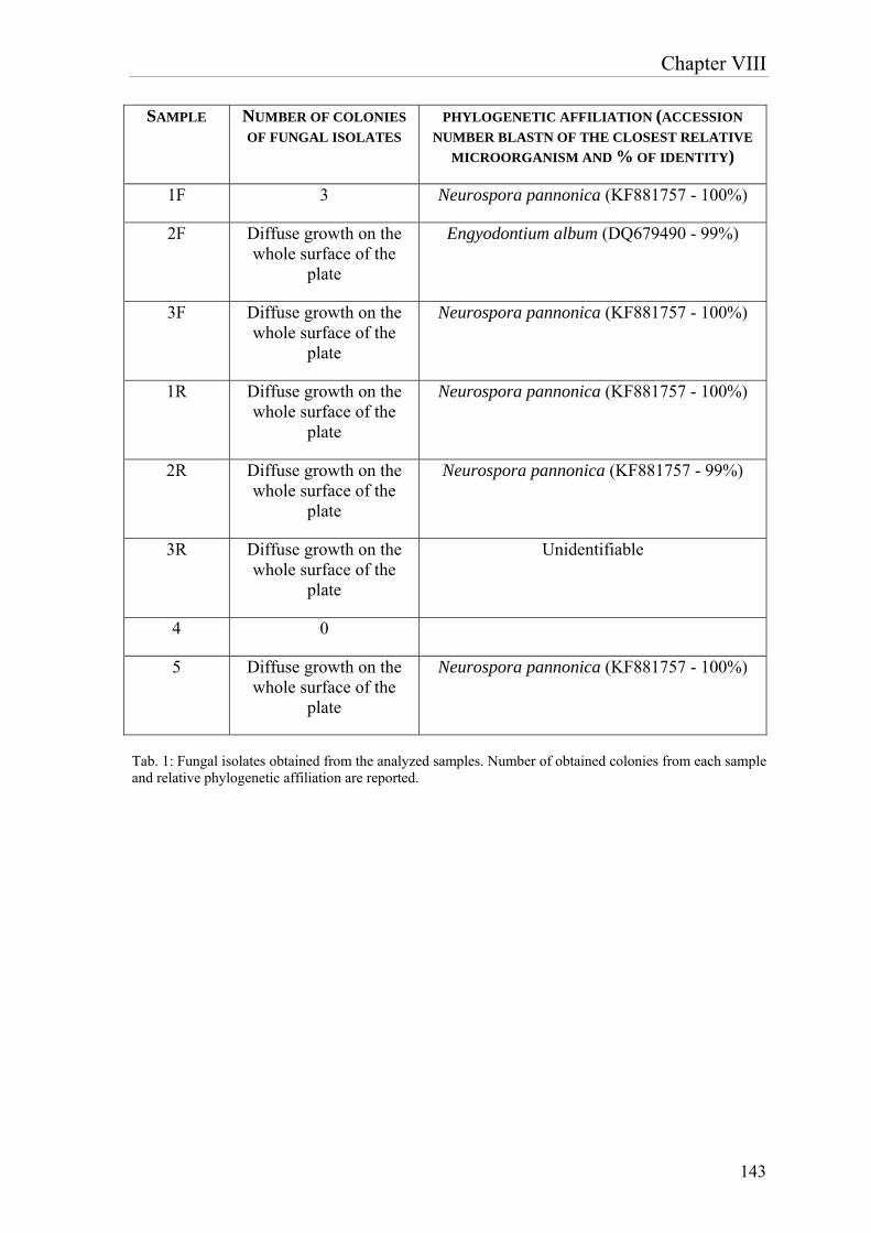
Chapter VIII
143
SAMPLE NUMBER OF COLONIES
OF FUNGAL ISOLATES PHYLOGENETIC AFFILIATION (ACCESSION
NUMBER BLASTN OF THE CLOSEST RELATIVE
MICROORGANISM AND % OF IDENTITY)
1F 3 Neurospora pannonica (KF881757 - 100%)
2F Diffuse growth on the whole surface of the
plate
Engyodontium album (DQ679490 - 99%)
3F Diffuse growth on the whole surface of the
plate
Neurospora pannonica (KF881757 - 100%)
1R Diffuse growth on the whole surface of the
plate
Neurospora pannonica (KF881757 - 100%)
2R Diffuse growth on the whole surface of the
plate
Neurospora pannonica (KF881757 - 99%)
3R Diffuse growth on the whole surface of the
plate
Unidentifiable
4 0
5 Diffuse growth on the whole surface of the
plate
Neurospora pannonica (KF881757 - 100%)
Tab. 1: Fungal isolates obtained from the analyzed samples. Number of obtained colonies from each sample and relative phylogenetic affiliation are reported.
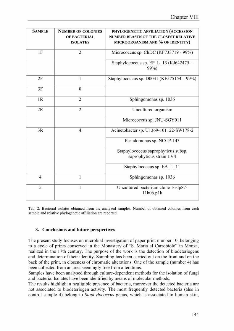
Chapter VIII
144
SAMPLE NUMBER OF COLONIES
OF BACTERIAL
ISOLATES
PHYLOGENETIC AFFILIATION (ACCESSION
NUMBER BLASTN OF THE CLOSEST RELATIVE
MICROORGANISM AND % OF IDENTITY)
1F 2 Micrococcus sp. ChDC (KF733719 - 99%)
Staphylococcus sp. EP_L_13 (KJ642475 – 99%)
2F 1 Staphylococcus sp. D0031 (KF575154 – 99%)
3F 0
1R 2 Sphingomonas sp. 1036
2R 2 Uncultured organism
Micrococcus sp. JNU-SGY011
3R 4 Acinetobacter sp. U1369-101122-SW178-2
Pseudomonas sp. NCCP-143
Staphylococcus saprophyticus subsp. saprophyticus strain LV4
Staphylococcus sp. EA_L_11
4 1 Sphingomonas sp. 1036
5 1 Uncultured bacterium clone 16slp87-11h06.p1k
Tab. 2: Bacterial isolates obtained from the analyzed samples. Number of obtained colonies from each sample and relative phylogenetic affiliation are reported.
3. Conclusions and future perspectives The present study focuses on microbial investigation of paper print number 10, belonging to a cycle of prints conserved in the Monastery of “S. Maria al Carrobiolo” in Monza, realized in the 17th century. The purpose of the work is the detection of biodeteriogens and determination of their identity. Sampling has been carried out on the front and on the back of the print, in closeness of chromatic alterations. One of the sample (number 4) has been collected from an area seemingly free from alterations. Samples have been analysed through culture-dependent methods for the isolation of fungi and bacteria. Isolates have been identified by means of molecular methods. The results highlight a negligible presence of bacteria, moreover the detected bacteria are not associated to biodeteriogen activity. The most frequently detected bacteria (also in control sample 4) belong to Staphylococcus genus, which is associated to human skin,
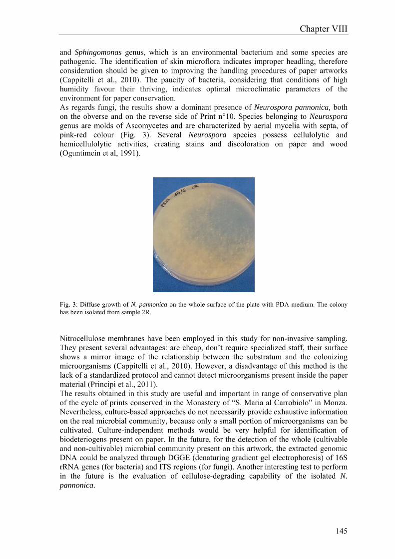
Chapter VIII
145
and Sphingomonas genus, which is an environmental bacterium and some species are pathogenic. The identification of skin microflora indicates improper headling, therefore consideration should be given to improving the handling procedures of paper artworks (Cappitelli et al., 2010). The paucity of bacteria, considering that conditions of high humidity favour their thriving, indicates optimal microclimatic parameters of the environment for paper conservation. As regards fungi, the results show a dominant presence of Neurospora pannonica, both on the obverse and on the reverse side of Print n°10. Species belonging to Neurospora genus are molds of Ascomycetes and are characterized by aerial mycelia with septa, of pink-red colour (Fig. 3). Several Neurospora species possess cellulolytic and hemicellulolytic activities, creating stains and discoloration on paper and wood (Oguntimein et al, 1991).
Fig. 3: Diffuse growth of N. pannonica on the whole surface of the plate with PDA medium. The colony has been isolated from sample 2R.
Nitrocellulose membranes have been employed in this study for non-invasive sampling. They present several advantages: are cheap, don’t require specialized staff, their surface shows a mirror image of the relationship between the substratum and the colonizing microorganisms (Cappitelli et al., 2010). However, a disadvantage of this method is the lack of a standardized protocol and cannot detect microorganisms present inside the paper material (Principi et al., 2011). The results obtained in this study are useful and important in range of conservative plan of the cycle of prints conserved in the Monastery of “S. Maria al Carrobiolo” in Monza. Nevertheless, culture-based approaches do not necessarily provide exhaustive information on the real microbial community, because only a small portion of microorganisms can be cultivated. Culture-independent methods would be very helpful for identification of biodeteriogens present on paper. In the future, for the detection of the whole (cultivable and non-cultivable) microbial community present on this artwork, the extracted genomic DNA could be analyzed through DGGE (denaturing gradient gel electrophoresis) of 16S rRNA genes (for bacteria) and ITS regions (for fungi). Another interesting test to perform in the future is the evaluation of cellulose-degrading capability of the isolated N. pannonica.

Chapter VIII
146
References
Cappitelli, F., Pasquariello, G., Tarsitani, G. and Sorlini, C. (2010) Scripta manent? Assessing microbial risk to paper heritage. Trends in Microbiology. 18(12):538-542. Hueck, H. J. (1968) The biodeterioration of materials – an appraisal. In: “Biodeterioration of materials. Microbiological and allied aspects”, ed. by A. Harry Walters and John J. Elphick, Amsterdam/London, pp. 6–12. Murray, A.E., Preston, C.M., Massana, R., Taylor, L.T., Blakis, A., Wu, K., Delong, E.F. (1998) Seasonal and spatial variability of bacterial and archaeal assemblages in the coastal waters near Anvers Island, Antarctica. Appl Environ Microbiol 64:2585–2595. Oguntimein, G.B., Moo Young, M. (1991) Production and properties of β-glucosidase by Neurospora sitophila. World Journal of Microbiology and Biotechnology. 7(1):4-11. Pinzari, F., Troiano, F., Piñar, G., Sterflinger, K. and Matteo Montanari (2011) The Contribution of Microbiological Research in the Field of Book, Paper and Parchment Conservation. In “New Approaches to Book and Paper Conservation ‑ Restoration”, ed. by Engel, P., Schirò, J., Larsen, R., Moussakova, E. and Kecskeméti I., Verlag Berger Horn/Wien, pp. 575-594. Principi, P., Villa, F., Sorlini, C., Cappitelli, F. (2011) Molecular Studies of Microbial Community Structure on Stained Pages of Leonardo da Vinci’s Atlantic Codex. Microb Ecol 61:214–222.
Conclusions
147
General conclusions and perspectives
The contribution given by biotechnology to restoration and conservation of cultural heritage is raising in the last decades. The integration of biotechnological knowledge with technical-artistical ones, in this field, leads to more and more precise, effective and environmentally-friendly methods for the solution of problems linked to restoration. Biotechnology research in the field of cultural heritage proceeds along two directions: development of innovative restoration methods, based on the employment of new products; development of accurate diagnostic techniques, for the identification and characterization of alterations and biodeteriogens. The first part of the present work was dedicated to the optimization of a microbial product, based on sulfate-reducing bacteria (SRB) belonging to D. vulgaris species, already used for removal of sulfations from cultural assets. Experimentations have been carried out for the optimization of the production process, achieving the scale-up of the biomass production and the development of a method for the long-term conservation of the bacterial biomass. In particular, different growth media, characterized by different electron donors, acceptors and nitrogen source, have been tested. Moreover, the optimal parameters for biomass production have been analyzed: nitrogen flushing, agitation, pH, feeding of nutrients. The process parameters for the pilot fermenter have been defined as follows: 150 rpm (agitation), 37°C, pO2 mantained at 0 with a continuos N2 flux of 1 L/min, pH mantained at 7.2 using H2SO4 20%, lactic acid mantained at the concentration of 2 g/l by adding sodium lactate 60%, adding of NH4Cl at a flux of 0.1 g/L/h. These improvements allowed a significative increase in biomass production, from a concentration of 1*108 cell/ml in 120h of fermentation in flask, to the concentration of 3*109 cell/ml in 72h of fermentation in bioreactor. In conclusion, we can state that in 3 days of fermentation of 5 liters of broth-culture, product for the treatment of 50 m2 of surface is obtained. For the long-term conservation of D. vulgaris biomass, freeze-drying has been carried out, testing the effectiveness of different cryoprotective agents. Among them, the best in terms of cell viability post-rehydration resulted to be lactose, which ensures the stability of the product for a minimum of 6 months. The first part of this thesis focused also on the development of a rapid, simple and sensitive method to enumerate the sulfate-reducing strain Desulfovibrio vulgaris subsp. vulgaris ATCC 29579, both in liquid media and embedded in delivery systems usually employed for biocleaning treatments. Different methods have been tested, among them ATP assay, Live/Dead bacterial viability determination and Real time PCR. All of them resulted to be effective only with D.vulgaris liquid cultures. Another drawback presented by ATP assay is that its content depends not only on cellular concentration, but also on the growth phase of the cells. Instead, Live/Dead bacterial viability determination can be used for concentration superior to 106 cells/ml, due to the detection limit of the method. Real-time PCR quantification of the SRB-specific dsr gene copy number proved to be a sensitive approach for enumeration of D. vulgaris cells, in the range from 1*103 to 1.5*108 cell/ml, nevertheless it is suitable only for D. vulgaris liquid culture, as it was not significant when the cells were embedded in the delivery system. Indeed, in this case it was effective only for a concentration higher than 107 cells/ml, not allowing the detection of concentration decay of D.vulgaris-based formulate applied for biorestoration. The set up of the system of spectrophotometric fluorescence measurement for estimation of cell concentration viability was successful, both for liquid culture of D. vulgaris and for cells after addition of the delivery system. Fluorescence is emitted by the prosthetic group of bisulfite reductase, a key enzyme in dissimilatory sulfate reduction. Another test proved
Conclusions
148
that emission of fluorescence derives only from viable cells, confirming the suitability of the method for enumeration of D. vulgaris cells. In order to reduce time and number of applications required by the employment of D. vulgaris-based product, comparison of different methodologies of application was performed on the funeral monument realized in memory of ‘Neera’, the poetess Anna Zuccari, situated in the Cimitero Monumentale in Milan. The biological cleaning using SRB was tested both alone and coupled with a soft chemical pre-treatment using the non-ionic detergent Tween 20, for the removal of the black crust. Considering cleaning effectiveness, electron microscope observations, elemental analysis and infrared spectroscopy, the combined methods achieved results comparable to those of the biological treatment alone. Additionally, using Tween 20 and SRB practical advantages were obtained: a very significant reduction in the number of applications (from seven to two), and a remarkable 70% reduction in the total cleaning time, compared to the biological approach. The chemical treatment probably softened the crust, accelerating the biological sulfate removal. Moreover, ESEM analysis revealed the presence of a barium sulfate layer in the structure of the black crust, probably derived from an undocumented conservation treatment, which resulted to be completely removed by biological cleaning. The extension of applicability of D. vulgaris-based product to other tipologies of substrates is of key importance for the further development of this technology. Experimentation carried out on two scenes belonging to the pictorial cycle decorating ‘Queen Teodolinda Chapel’ in Monza Cathedral gave interesting and promising results. Indeed, good results in sulfations removal were obtained, in particular delivering D. vulgaris cells in Arbocel BWW40, without damaging such a delicate substrate. However, this work has to be considered preliminary and incomplete. Further research is required in order to verify the compatibility with different kinds of materials, such as pigments containing metals, which could interact with the hydrogen sulfide released by sulfate-reducing bacteria, or pigments with oily binder, which are very delicate. Interestingly, results from Ionic Chromatography Analyses showed the reduction not only of sulfates after the application of bacteria, but also of nitrates. The second part of this thesis sheds light on diagnostic methodologies for the identification and characterisation of biodeteriogens, spoiling two artworks: the first is an acrylic monochrome painting on canvas realised by the artist E. Castellani, the second is a paper print realized in the 17th century, conserved in the Monastery of “S. Maria al Carrobiolo” in Monza. In the first case, a strategy combining culture-dependent and culture-independent techniques was adopted. Results obtained from culture-independent techniques revealed in all samples the predominant presence of bacteria belonging to Bacillus genus and fungi belonging to Leptosphaerulina, Leptosphaeria and Penicillium genera. Among the identified bacteria, the most abundant were B. subtilis, B. licheniformis and Deinococcus gobiensis. B. subtilis and B. licheniformis have been already identified on deteriorated paintings, as different species of Penicillium (Capodicasa et al. 2010). These results were confirmed by culture-dependent techniques, indeed the dominance of Bacillus genus was confirmed and in particular of B. subtilis and B. licheniformis species. Instead as concerns identification of fungi, isolation was unsuccessful, probably because of the uncultivability of these species. Nevertheless, visualization through optical microscope of material sampled by means of fungi-tape confirmed the presence of Penicillium genus. The simultaneous presence of bacilli and penicilli suggests that the deterioration of the canvas is imputable at the beginning to the activity of fungi able to degrade the wooden-cellulosic component. From fungal metabolism derive immediately consumable substances, such as proteins and polysaccharides, originating the consecutive bacterial colonization. At last, degradation

Conclusions
149
phases create the environmental-nutritional conditions that are favourable to the following colonization by other bacteria such as D. gobiensis, whose growth could be the cause of yellow-earth/red pigmentation of the alterations, in fact D. gobiensis forms red-pink colonies. Therefore, the activity of four biocides towards the bacterial isolates was tested through antibiogram test, evaluating their effectiveness by measuring the diameter of the growth inhibition halo of the target bacterium around the small disks soaked with the different biocides. These biocides were: Biotin N, Biotin R, New Des 50 and Amuchina. At first, the test was carried out singularly, then the same test was performed using mixes of the different biocides. An inhibitory activity towards the single isolates was shown by all the tested biocides, but Biotin N and Biotin R resulted to be the most effective. Among the different mixes of biocides tested, the combination of Biotin R 4% in ethyl acetate + Biotin N 4% in white spirit is the one giving the best results. Activity test of the different biocides toward the whole microbial community was performed. Also in this case, Biotin N 4% in white spirit, Biotin R 4% in ethyl acetate and their combination provided the best results in terms of growth inhibition of microorganisms constituent the community. Once identified the microorganisms responsible of deterioration of the acrylic monochrome painting on canvas, determination of the ideal environmental parameters for the prevention of the reappearance of deteriorating bacteria and fungi is possible. Considering that most of the isolated bacteria reach its optimal growth at the temperature of 25-37°C and that high values of relative humidity favour the proliferation of bacteria and fungi, the analyzed canvas should be kept in environments whose relative humidity is below 60% and temperature not over 15-20°C. As some of the isolated bacteria (D. gobiensis and B. licheniformis) are facultative anaerobes, keeping the artwork in anaerobic conditions could not prevent the growth of those bacteria, but only of fungi. Nevertheless, as bacterial colonization depends on proteins and polysaccharides derived from fungal metabolism, prevention of fungal growth would have an effect also on bacterial growth. Diagnostic methodologies have been employed also for the identification and characterisation of biodeteriogens altering a paper print realized in the 17th century, conserved in the Monastery of “S. Maria al Carrobiolo” in Monza. In this case, culture-dependent techniques were adopted. The results highlight a negligible presence of bacteria, moreover the detected bacteria are not associated to biodeteriogen activity. As the bacterial proliferation is favoured by conditions of high humidity, it can be deduced that the current microclimatic parameters of the environment in which the paper is conserved are optimal. As regards fungi, the results show a dominant presence of Neurospora pannonica, both on the obverse and on the reverse side of the print. Several Neurospora species possess cellulolytic and hemicellulolytic activities, creating stains and discoloration on paper and wood (Oguntimein et al, 1991). The results obtained in this study are useful and important, nevertheless, culture-based approaches do not necessarily provide exhaustive information on the real microbial community, because only a small portion of microorganisms can be cultivated. Further research, based on culture-independent methods is required in order to obtain a complete description of biodeteriogens present on paper. In the future, for the detection of the whole (cultivable and non-cultivable) microbial community present on this artwork, the extracted genomic DNA could be analyzed through DGGE (denaturing gradient gel electrophoresis) of 16S rRNA genes (for bacteria) and ITS regions (for fungi). Another interesting test to perform in the future is the evaluation of cellulose-degrading capability of the isolated N. pannonica. The results obtained in the present PhD thesis are tangible evidences of the utility of biotechnologies in the field of cultural heritage. The work described in the first part of the thesis, aimed at the optimization of the microbial product, based on SRB belonging to D.
Conclusions
150
vulgaris species, obtained good results, even though further research is essential in particular for the extension of the methodology to substrates other than stone artworks. Molecular biotechnologies, compared with conventional cultivation methods, provide a more realistic view of microbial populations colonizing artworks, allowing the disclosure of uncultivable species. Therefore, these methodologies allow the planning of an appropriate restoration intervention. Biotechnological techniques are powerful, environmentally friendly and present low-risk to human health. Consequently, a continue dialogue between biotechnologies field and cultural heritage’s one is crucial for the development of more and more effective novel approaches to preserve our historical artworks. However, lack of funds is often a limit for restorers in employment of biotechnological approaches, then in the future the overcoming of these problems is wished. References
Capodicasa, S., Fedi, S., Porcelli, A.M., Zannoni, D. (2010) The microbial community on a biodeteriorated 16th century painting. Int. Biodet. & Biodegr. 64: 727-733. Oguntimein, G.B., Moo Young, M. (1991) Production and properties of β-glucosidase by Neurospora sitophila. World Journal of Microbiology and Biotechnology. 7(1):4-11.
Acknowledgements
151
Acknowledgments First of all, I thank my family and Francesco, for all the love and support they have given to me every moment during these three intense years. Then I would like to thank Professor Daniele Daffonchio and Professor Sara Borin, who gave me the opportunity to work in a high level laboratory. Thanks to Annalisa and Micro4yoU, I will be always grateful for the great chance offered to me, for all the important experiences which have greatly enriched my professional knowledge. I’d like to thank Professor Francesca Cappitelli and her group, for the hospitality in her laboratories, during the first PhD year, and in particular to Andrea and Federica for the collaboration. Thanks to all DD lab and FC lab, for all the beautiful moments spent together, I will always remember them. In particular I want to thank my PhD-mates Violetta, Marta, Michela and Erica, we shared this experience always supporting each other. Thanks to Luigi, Beppe, Marco and Federica, for all the nice moments and the aid in difficult ones. Thanks to Elena Sidoti and Giulia Forotti, who contributed to this work.

Activities
152
Activities performed during the Ph.D.
Training courses, seminars and conferences - 24th November 2011 “Ecogenomics of haloalkaliphilic sulfur bacteria” Prof. Gerard Muyzer, Milan.
- 20th December 2011 “The genetic analysis of gut immunity and gut-microbe symbiosys in Drosophila” Prof. Won-Jae Lee, Milan.
- 26th, 27th January 2012 VII Workshop of the PhD School of “Chemistry, Biochemistry and Ecology of Pesticides”, Milan.
- 16th April 2012 Mini-course on Flow-Cytometry “Theory and principles in flow cytometry” Dr. Stefano Amalfitano, Milan.
- 3rd- 5th May 2012 “Cortona procarioti 2012” Organizers Prof Renato Fani and Prof Paolo Landini, Cortona.
- 22nd May 2012 “Course on CAB Abstract” Prof. Angela Moccia, Milan.
- 20th, 27th June; 4th July 2012 “Corso SAS base per Dottorandi” Prof. Alberto Tamburini, Milan.
- 7th June 2012 “Fungal diversity in Sahara and natural saline systems in the desert”. Prof. Atef Jaouani, Milan.
- 26th, 28th November 2012, 3rd, 10th, 12th December 2012 Theoretical Course "Bacillus thuringiensis an important resource for pest control: ecology, genetics, biotechnology" Prof. Daniele Daffonchio, Milan.
- 21st January 2013 VIII Workshop of the PhD School of “Chemistry, Biochemistry and Ecology of Pesticides”, University of Milan.
- 30th -31st January 2013 Practical course “Isolation of bioactive organic compounds from natural sources” Prof. Angela Bassoli, Milan.
- From 29th January 2013 to 5th March 2013 Theoretical Course “Past and future in crop protection chemicals” Prof. Sabrina Dallavalle, Milan.
- 6th February 2013 Practical course “Purification of bioactive organic compounds from natural sources” Prof. Angela Bassoli, Milan.
- 14th February 2013 Seminar course “Study of microorganisms based on color” Dr. Patricia Sanmartin Sanchez, Milan.
- 6th March 2013 Seminar Course “Systems Paleobiology” and “Deep history of life” Prof Andrew Herbert Knoll, Milan.
- 12th March 2013 Seminar Course “Valorizzazione della ricerca”, corsi competenze trasversali per dottorandi, Milan.
-11th April 2013 Seminar Course “Rispondere ad un bando”, corsi competenze trasversali per dottorandi, Milan.

Activities
153
- 17th April 2013 Seminar Course “Research writing and presentation”, corsi competenze trasversali per dottorandi, Milan.
- 24th May 2013 Seminar Course “Diritto d’autore e open access”, corsi competenze trasversali per dottorandi, Milan.
- 10th July 2013 Seminar Course “The team of microbiology and laboratory activities of SAFE-University of Foggia” Dr. Luciano Beneduce, Milan.
- 15th November 2013 Seminar course “Beyond the biocide: examination of the use of nitric oxide-based treatments in the control of biodecay of cultural materials” Caroline Kyi, PhD candidate, Milan. - 18-19-20th November 2013 Conference “Built Heritage 2013” Politecnico di Milano - 27th - 28th January 2014 IX Workshop of the PhD Course of “Chemistry, Biochemistry and Ecology of Pesticides”, Milan. - 18th February 2014 Seminar course “Proteomics of food allergens and toxins” Prof. Pasquale Ferranti, Milan.
- 21st February 2014 Conference ANFOMAT “Materiali innovativi per dispositivi medicali basati su superfici biofunzionalizzate con proprietà anti-biofilm” , Milan.
- 11th June 2014 Seminar course “Cultures on Culture: A Social Scientific Perspective on Cultural Heritage Restoration Technologies” Grace Kim, PhD student, Milan. - 18th June 2014 Seminar course “A Marie Curie experience as an added value for your CV: The successful story of the ESENCYA project” Dr. Federica Villa, Milan.
- 24th June 2014 Seminar course “Outdoor wooden sculpture - biodeterioration and conservation decision-making” Emma Rouse, Master student, Milan. - 1st-4th July 2014 Conference “Scienza e beni culturali- quale sostenibilità per il restauro?”, Bressanone. - 12th-25th July 2014 FACILIS 2014 Summer School, University of Milan - 11th September 2014 Seminar Course “The microbiome of materials in ancient burial sites” Dr. Alice Dearaujo, Milan.
- 26th September 2014 “MeetMeTonight” Notte Europea dei ricercatori, allestimento dello stand“Batteri restauratori”, Micro4yoU srl. Oral presentations in national /international meetings
- 28th September 2013 “Removal for complex sulphate black crusts in a large scale biorestoration treatment”. A. Balloi; E. Lombardi; F. Troiano; D. Gulotta; A. Polo; E. Gioventù; S. Borin, L. Toniolo; C. Sorlini; F. Cappitelli, D. Daffonchio. Convegno “I batteri nel restauro”, Fondazione Villa Fabris, Vicenza.
Activities
154
- 10th October 2013 “Uso di batteri solfato-riduttori nel biorestauro di dipinti murari”. A. Balloi; E. Lombardi; F. Troiano; D. Gulotta; A. Polo; S. Metaldi; E. Gioventù; L. Toniolo; D. Daffonchio; C. Sorlini; F. Cappitelli. Giornata di studi “Biorestauro nei musei Vaticani”, Musei Vaticani, Città del Vaticano. Papers published in international journals
Troiano F., Gulotta D., Balloi A., Polo A., Toniolo L., Lombardi E., Daffonchio D., Sorlini C, Cappitelli F., 2013. Successful combination of chemical and biological treatments for the cleaning of stone artworks. International Biodeterioration & Biodegradation 85, 294–304. Posters presentation in international conferences
Emanuela Lombardi; Annalisa Balloi; Federica Troiano; Davide Gulotta; Andrea Polo; Eleonora Gioventù; Claudia Sorlini; Francesca Cappitelli; Daniele Daffonchio. “Strategies for increasing the scale of biocleaning treatment through sulfate crust bioremoval”. Conference “Built Heritage 2013” Politecnico di Milano.