bizarre parosteal osteochondromatous …...bizarre parosteal osteochondromatous proliferation with...
TRANSCRIPT

submit.radiology.or.kr J Korean Soc Radiol 2011;65(4):415-419 415
INTRODUCTION
Bizarre parosteal osteochondromatous proliferation (BPOP) is a rare benign lesion arising from the surface of cortical bone which usually occurs in the small bones of distal ex-tremities and shows exophytic growth. Such a lesion can be cured by local excision. BPOP can be confused with some be-nign and malignant diseases such as osteochondroma, paraos-teal osteosarcoma, or chondrosarcoma, both radiologically and pathologically (1). We illustrate imaging features includ-ing simple radiograph and magnetic resonance imaging of BPOP in the proximal phalanx of the hand with unusual cor-tical erosion and bone marrow edema accompanied by soft tissue enhancement.
CASE REPORT
A 10-year-old female presented with a palpable mass in the volar aspect of her right 2nd finger at the level of the proximal
interphalangeal joint. The mass had appeared over 6 months without pain. She did not have underlying joint disease or history of trauma. Physical examination revealed a round, firm, non-tender, fixed mass with intact overlying skin in her 2nd finger.
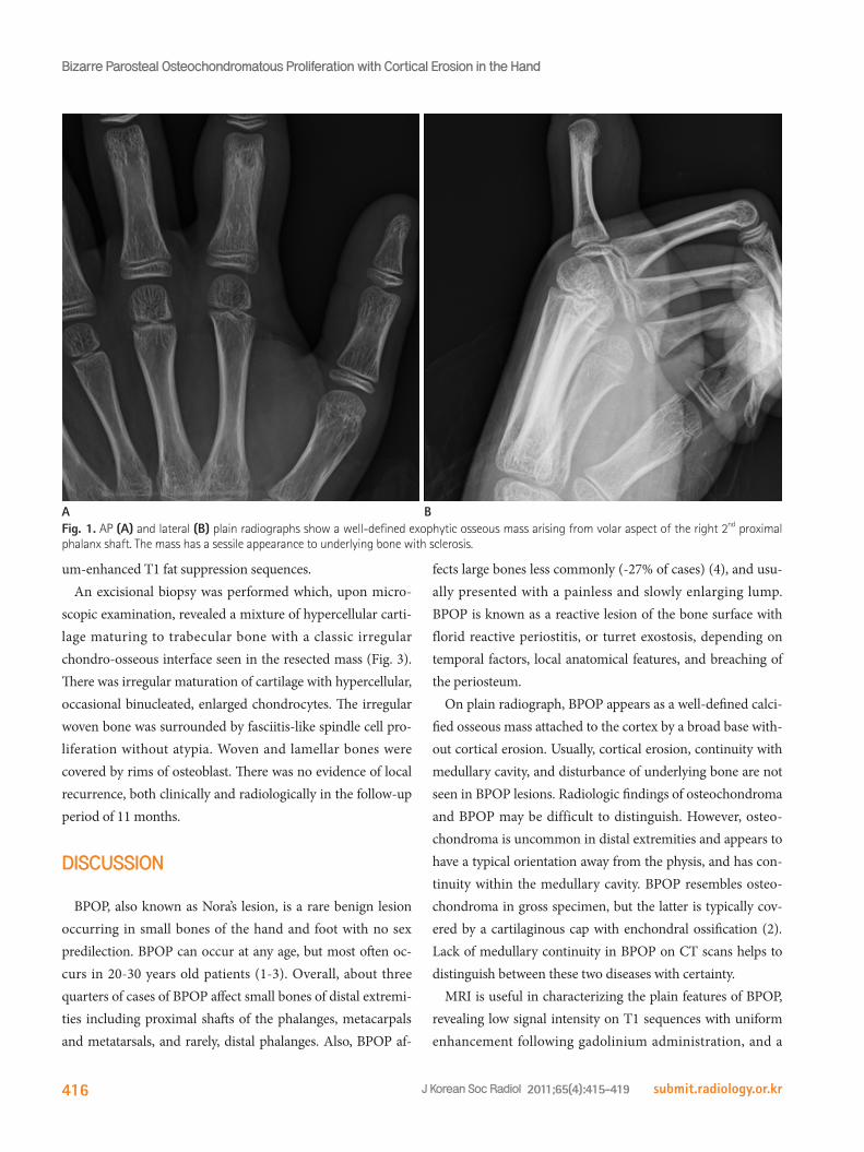
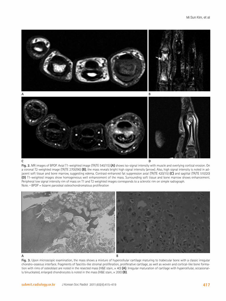
Plain radiographs showed a well-defined exophytic osseous mass arising from volar aspect of the right 2nd proximal pha-langeal shaft (Fig. 1). The mass had a sessile appearance with peripheral sclerosis in the underlying bony cortex and no mineralization in the lesion or continuity with the medulla was evident. Subsequent MRI showed a well-defined ovoid paraosteal mass with erosion in the cortex and medulla of the 2nd proximal phalangeal shaft measuring 3.4 × 3.7 cm. The mass had iso-signal intensity to muscle on T1-weighted imag-es and high signal intensity on T2-weighted images with T1, T2 low signal intensity rim, corresponding to sclerotic rim on simple radiograph (Fig. 2). Surrounding soft tissue and bone marrow edema were seen on T2 weighted images. The lesion and adjacent soft tissue showed well enhanced on gadolini-
Case ReportpISSN 1738-2637J Korean Soc Radiol 2011;65(4):415-419
Received June 29, 2011; Accepted August 4, 2011Corresponding author: Yong Hoon Kim, MDDepartment of Radiology, Inje University College of Medicine, Ilsan Paik Hospital, 2240 Daehwa-dong, Ilsanseo-gu, Goyang 411-706, Korea.Tel. 82-31-910-7628 Fax. 82-31-910-7369E-mail: [email protected]
Copyrights © 2011 The Korean Society of Radiology
Bizarre parosteal osteochondromatous proliferation (BPOP) is a rare osseous lesion occurring in the long bones of distal extremities. Typical imaging findings of BPOP show a well-defined calcified osseous mass without soft tissue swelling, cortical de-struction, and medullary involvement. We experienced rare aggressive magnetic reso-nance imaging findings of BPOP with cortical erosion, bone marrow edema, and adja-cent soft tissue enhancement, which was misdiagnosed as a malignancy or inflammatory lesion.
Index termsBizarre Parosteal Osteochondromatous ProliferationMagnetic Resonance ImagingCortical Invasion
Bizarre Parosteal Osteochondromatous Proliferation with Cortical Erosion in the Hand: A Case Report피질 침범을 동반한 수부의 기괴 방골성 골연골성 증식증: 증례 보고 Mi Sun Kim, MD, Yong Hoon Kim, MD, Yoon Joon Hwang, MD, Jung Wook Seo, MD, Su Young Kim, MD, Byeong Hoon Lee, MD, Ji Young Lee, MDDepartment of Radiology, Inje University College of Medicine, Ilsan Paik Hospital, Goyang, Korea
Bizarre Parosteal Osteochondromatous Proliferation with Cortical Erosion in the Hand
submit.radiology.or.krJ Korean Soc Radiol 2011;65(4):415-419416
fects large bones less commonly (-27% of cases) (4), and usu-ally presented with a painless and slowly enlarging lump. BPOP is known as a reactive lesion of the bone surface with florid reactive periostitis, or turret exostosis, depending on temporal factors, local anatomical features, and breaching of the periosteum.
On plain radiograph, BPOP appears as a well-defined calci-fied osseous mass attached to the cortex by a broad base with-out cortical erosion. Usually, cortical erosion, continuity with medullary cavity, and disturbance of underlying bone are not seen in BPOP lesions. Radiologic findings of osteochondroma and BPOP may be difficult to distinguish. However, osteo-chondroma is uncommon in distal extremities and appears to have a typical orientation away from the physis, and has con-tinuity within the medullary cavity. BPOP resembles osteo-chondroma in gross specimen, but the latter is typically cov-ered by a cartilaginous cap with enchondral ossification (2). Lack of medullary continuity in BPOP on CT scans helps to distinguish between these two diseases with certainty.
MRI is useful in characterizing the plain features of BPOP, revealing low signal intensity on T1 sequences with uniform enhancement following gadolinium administration, and a
um-enhanced T1 fat suppression sequences.An excisional biopsy was performed which, upon micro-
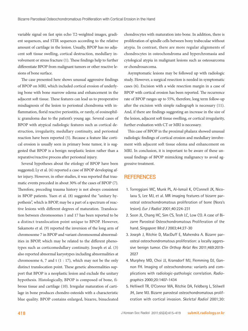
scopic examination, revealed a mixture of hypercellular carti-lage maturing to trabecular bone with a classic irregular chondro-osseous interface seen in the resected mass (Fig. 3). There was irregular maturation of cartilage with hypercellular, occasional binucleated, enlarged chondrocytes. The irregular woven bone was surrounded by fasciitis-like spindle cell pro-liferation without atypia. Woven and lamellar bones were covered by rims of osteoblast. There was no evidence of local recurrence, both clinically and radiologically in the follow-up period of 11 months.
DISCUSSION
BPOP, also known as Nora’s lesion, is a rare benign lesion occurring in small bones of the hand and foot with no sex predilection. BPOP can occur at any age, but most often oc-curs in 20-30 years old patients (1-3). Overall, about three quarters of cases of BPOP affect small bones of distal extremi-ties including proximal shafts of the phalanges, metacarpals and metatarsals, and rarely, distal phalanges. Also, BPOP af-
Fig. 1. AP (A) and lateral (B) plain radiographs show a well-defined exophytic osseous mass arising from volar aspect of the right 2nd proximal phalanx shaft. The mass has a sessile appearance to underlying bone with sclerosis.
A B
Mi Sun Kim, et al
submit.radiology.or.kr J Korean Soc Radiol 2011;65(4):415-419 417
Fig. 3. Upon microscopic examination, the mass shows a mixture of hypercellular cartilage maturing to trabecular bone with a classic irregular chondro-osseous interface. Fragments of fasciitis-like stromal proliferation, proliferative cartilage, as well as woven and cortical-like bone forma-tion with rims of osteoblast are noted in the resected mass (H&E stain, × 40) (A). Irregular maturation of cartilage with hypercellular, occasional-ly binucleated, enlarged chondrocytes is noted in the mass (H&E stain, × 200) (B).
Fig. 2. MR images of BPOP. Axial T1-weighted image (TR/TE 540/15) (A) shows iso-signal intensity with muscle and overlying cortical erosion. On a coronal T2-weighted image (TR/TE 2700/96) (B), the mass reveals bright high signal intensity (arrow). Also, high signal intensity is noted in ad-jacent soft tissue and bone marrow, suggesting edema. Contrast-enhanced fat suppression axial (TR/TE 420/15) (C) and sagittal (TR/TE 510/20) (D) T1-weighted images show homogeneous well enhancement of the mass. Surrounding soft tissue and bone marrow shows enhancement. Peripheral low signal intensity rim of mass on T1 and T2 weighted images corresponds to a sclerotic rim on simple radiograph.Note.-BPOP = bizarre parosteal osteochondromatous proliferation
A
A
C
B
B
D
Bizarre Parosteal Osteochondromatous Proliferation with Cortical Erosion in the Hand
submit.radiology.or.krJ Korean Soc Radiol 2011;65(4):415-419418
chondrocytes with maturation into bone. In addition, there is proliferation of spindle cells between bony trabeculae without atypia. In contrast, there are more regular alignments of chondrocytes in osteochondroma and hyperchromasia and cytological atypia in malignant lesions such as osteosarcoma or chondrosarcoma.
Asymptomatic lesions may be followed up with radiologic study. However, a surgical resection is needed in symptomatic cases (6). Excision with a wide resection margin in a case of BPOP with cortical erosion has been reported. The recurrence rate of BPOP ranges up to 55%, therefore, long term follow-up after the excision with simple radiograph is necessary (11). And, if there are findings suggesting an increase in the size of the lesion, adjacent soft tissue swelling, or cortical irregularity, further evaluation with CT or MRI is necessary.
This case of BPOP in the proximal phalanx showed unusual radiologic findings of cortical erosion and medullary involve-ment with adjacent soft tissue edema and enhancement on MRI. In conclusion, it is important to be aware of these un-usual findings of BPOP mimicking malignancy to avoid ag-gressive treatment.
REFERENCES
1.TorreggianiWC,MunkPL,Al-IsmailK,O’ConnellJX,Nico-
laouS,LeeMJ,etal.MRimagingfeaturesofbizarrepar-
ostealosteochondromatousproliferationofbone(Nora’s
lesion).EurJRadiol2001;40:224-231
2.SoonJL,ChangHC,SimCS,TeohLC,LowCO.AcaseofBi-
zarreParostealOsteochondromatousProliferationofthe
hand.SingaporeMedJ2003;44:27-30
3.JosephJ,RitchieD,MacDuffE,MahendraA.Bizarrepar-
ostealosteochondromatousproliferation:alocallyaggres-
sivebenigntumor.ClinOrthopRelatRes2011;469:2019-
2027
4.MurpheyMD,ChoiJJ,KransdorfMJ,FlemmingDJ,Gan-
nonFH.Imagingofosteochondroma:variantsandcom-
plicationswithradiologic-pathologiccorrelation.Radio-
graphics2000;20:1407-1434
5.HelliwellTR,O’ConnorMA,RitchieDA,FeldbergL,Stilwell
JH,JaneMJ.Bizarreparostealosteochondromatousprolif-
erationwithcortical invasion.SkeletalRadiol2001;30:
variable signal on fast spin echo T2-weighted images, gradi-ent sequences, and STIR sequences according to the relative amount of cartilage in the lesion. Usually, BPOP has no adja-cent soft tissue swelling, cortical destruction, medullary in-volvement or stress fracture (1). These findings help to further differentiate BPOP from malignant tumors or other reactive le-sions of bone surface.
The case presented here shows unusual aggressive findings of BPOP on MRI, which included cortical erosion of underly-ing bone with bone marrow edema and enhancement in the adjacent soft tissue. These features can lead us to preoperative misdiagnosis of the lesion to periosteal chondroma with in-flammation, florid reactive periostitis, or rarely, of eosinophil-ic granuloma due to the patient’s young age. Several cases of BPOP with atypical radiologic features such as cortical de-struction, irregularity, medullary continuity, and periosteal reaction have been reported (5). Because a feature like corti-cal erosion is usually seen in primary bone tumor, it is sug-gested that BPOP is a benign neoplastic lesion rather than a reparative/reactive process after periosteal injury.
Several hypotheses about the etiology of BPOP have been suggested; Ly et al. (6) reported a case of BPOP developing af-ter injury. However, in other studies, it was reported that trau-matic events preceded in about 30% of the cases of BPOP (7). Therefore, preceding trauma history is not always consistent in BPOP patients. Yuen et al. (8) suggested the “unitary hy-pothesis”, which is BPOP, may be a part of a spectrum of reac-tive lesions with different degrees of maturation. Transloca-tion between chromosomes 1 and 17 has been reported to be a distinct translocation point unique to BPOP. However, Sakamoto et al. (9) reported the inversion of the long arm of chromosome 7 in BPOP and variant chromosomal abnormal-ities in BPOP, which may be related to the different pheno-types such as corticomedullary continuity. Joseph et al. (3) also reported abnormal karyotypes including abnormalities at chromosome 6, 7 and t (1 : 17), which may not be the only distinct translocation point. These genetic abnormalities sup-port that BPOP is a neoplastic lesion and exclude the unitary hypothesis. Histologically, BPOP is composed of bone, fi-brous tissue and cartilage (10). Irregular maturation of carti-lage in bone produces chondro-osteoids with a characteristic blue quality. BPOP contains enlarged, bizarre, binucleated
Mi Sun Kim, et al
submit.radiology.or.kr J Korean Soc Radiol 2011;65(4):415-419 419
9.SakamotoA,ImamuraS,MatsumotoY,HarimayaK,Mat-
sudaS,TakahashiY,etal.Bizarreparostealosteochon-
dromatousproliferationwithaninversionofchromosome
7.SkeletalRadiol2011;40:1487-1490
10.ChoiJH,GuMJ,KimMJ,ChoiWH,ShinDS,ChoKH.Fibro-
sarcomainbizarreparostealosteochondromatousprolif-
eration.SkeletalRadiol2001;30:44-47
11.MichelsenH,AbramoviciL,SteinerG,PosnerMA.Bizarre
parostealosteochondromatousproliferation(Nora’slesion)
inthehand.JHandSurgAm2004;29:520-525
282-285
6.LyJQ,Bui-MansfieldLT,TaylorDC.Radiologicdemonstra-
tionoftemporaldevelopmentofbizarreparostealosteo-
chondromatousproliferation.ClinImaging2004;28:216-
218
7.MenesesMF,UnniKK,SweeRG.Bizarreparostealosteo-
chondromatousproliferationofbone(Nora’slesion).AmJ
SurgPathol1993;17:691-697
8.YuenM,FriedmanL,OrrW,CockshottWP.Proliferative
periostealprocessesofphalanges:aunitaryhypothesis.
SkeletalRadiol1992;21:301-303
피질 침범을 동반한 수부의 기괴 방골성 골연골성 증식증: 증례 보고
김미선 · 김용훈 · 황윤준 · 서정욱 · 김수영 · 이병훈 · 이지영
Bizarre parosteal osteochondromatous proliferation (이하 BPOP)은 드문 양성 골 병변으로 손과 발의 작은 뼈에서 발생
한다. BPOP의 특징적인 영상 소견은 골 표면의 경계가 좋은 석회화된 종괴로 인접 연부조직의 종창이나 골피질의 파괴,
골수질의 침범을 동반하지 않는다. 저자는 자기공명영상에서 골피질 미란과 골수 부종, 인접 연부조직의 조영증강과 같
은 공격적인 영상 소견을 보여 악성 종양이나 염증성 병변으로 오진하였던 BPOP 한 예를 경험하여 보고하고자 한다.
인제대학교 의과대학 일산백병원 영상의학과학교실