björn rasch, prof. dr. rer. nat. - uni-saarland.de€¦ · 1 björn rasch, prof. dr. rer. nat....
TRANSCRIPT
1
Björn Rasch, Prof. Dr. rer. nat. Division of Cognitive Biopsychology and Methods, University of Fribourg
Rue P-.A. de Faucigny 2; 1701 Fribourg; Switzerland;
( +41 (0)26 300 76 37 * [email protected]
PERSONAL INFORMATION
Born on the 04.01.1975 in Lüneburg, Germany, married
EDUCATION
2011 Vienia Docendi in Psychology (Habilitation), University of Basel, Switzerland
2008 Doctor of Science (Dr. rer. nat., summa cum laude), University of Trier, Germany
2003 Diploma (equivalent to M.Sc.), Psychology, University of Trier, Germany
PROFESSIONAL APPOINTMENTS
2013 – present Full professor of Cognitive Biopsychology and Methods at the University of Fribourg, Switzerland
2011 - 2013 SNSF Professor of the Swiss National Science Foundation (SNSF) at the University of Zürich, Switzerland
2008 – 2011 Lecturer and Research Scientist, Division of Cognitive Neuroscience (Prof. De Quervain) and Division of Molecular Psychology (Prof. Papassotiropoulos), University of Basel, Switzerland
2003 – 2008 Research Scientist, Institute for Neuroendocrinology (Prof. Born), University of Lübeck, Germany
FELLOWSHIPS AND AWARDS
2011 SNSF professorship of the Swiss National Science Foundation (SNSF)
2008 2-year post-doc scholarship, Deutsche Forschungsgemeinschaft (DFG)
2007 Young Scientist Award (Fachgruppe „Biologische Psychologie und Neuropsychologie“ der Deutschen Gesellschaft für Psychologie (DGPs))
2001 1-year Fulbright scholarship for studying in the U.S.A.
POSITIONS OFFERED
2013 Full Professor for Biological and Clinical Psychology, University of Trier, Germany
2011 Team leader position at the RIKEN Brain Science Institute, Tokyo, Japan (tenure-track)
2011 Assistance professor for "Learning and Plasticity in the old Age" at the University of Zürich, Switzerland
SUPERVISED PHD STUDENTS AND POST DOCS
2011 – present S. Ackermann, M. Cordi, M. Göldi, G. Gvozdanovic, M. Lehmann, M. Lüthi, M. Munz, J. Rihm, T. Schreiner
TEACHING ACTIVITIES
2013 – present Lectures on General Psychology I + II and Research Methods in Psychology, regular seminars
2008 – 2013 Participation in the Master-Modul “Cognitive Psychology and Cognitive Neuroscience”, Univ. of Zurich Regular seminars for psychology students on Memory, Sleep, and neuroscientific methods (fMRI, EEG)

2
INSTITUTIONAL RESPONSIBILITIES
2014 - present Vice-president of the department of psychology, University of Fribourg, Switzerland
2014 - present President of the internal ethical review board of the Departement of Psychology, University of Fribourg
AD HOC REVIEWER
Organizations: German Research Foundation (DFG), Volkswagenstiftung (D), Swiss National Science Foundation (SNSF), Alberta Univ. (USA), BBSRC (UK), Netherlands Organisation of Scientific Research etc.
Journals: Science, Nature Neurosci., Neuron, PNAS, J. Neurosci, Biol. Psychiatry, Current Biology, Biol. Psychology, Neuroimage, Sleep, PlosOne, Psychoneuroendocrinology, etc.
Editor: Special Issue Guest Editor for Neurobiology of Learning and Memory, and Brain and Language, Review Editor for Frontiers in Human Neuroscience,
MEMBERSHIPS
Association for Psychological Sciences, Cognitive Neuroscience Society; Deutsche Gesellschaft für Psychologie (DGPs); Deutsche Gesellschaft für Psychophysiologie und ihre Anwendungen (DGPA), Swiss Society of Neuroscience; Swiss Society of Sleep Research, Sleep Medicine and Chronobiology; Milton Erickson Society for Clinical Hypnosis (MEG), Zurich Centre for interdisciplinary Sleep Research (ZiS)
FUNDING (COMPETETIVE, AS PRINCIPLE INVESTIGATOR)
2014 University of Fribourg CHF: 62.500,-
2012 Subproject in KFSP “Sleep and Health” CHF: 525.000.-
2011 SNSF professorship (memory reactivation and sleep) CHF: 1.600.000.-
2011 SNSF project (neural correlates of self-control) CHF: 268.900.-
2009 German Research Foundation (DFG) EURO: 370.000.-
2009 Freiwillige Akademische Gesellschaft Basel CHF: 62.000.-
2008 University of Basel CHF: 50.000.-
PUBLICATIONS
Total: 50 peer reviewed articles, 12 as first, 19 as last/corresponding author; 2 Books; 2 Book Chapters
Cumulative Impact 343 (ResearchGate); h-Index 18 (Web of Science), > 1400 citations, Average citation per article: 32.4
Five most important publications
Rasch, B., Büchel, C., Gais, S., & Born, J. (2007). Odor cues during slow-wave sleep prompt declarative memory consolidation. Science, 315, 1426-1429.
Rasch, B., Pommer, J., Diekelmann, S., & Born, J. (2008). Pharmacological REM sleep suppression paradoxically improves rather than impairs skill memory. Nature Neuroscience. 12(4). 396-397.
Diekelmann, S., Büchel, C., Born, J. & Rasch, B. (2011). Labile or stable: opposing consequences for memory when reactivated during waking and sleep. Nature Neuroscience. 14(3):381-6.
Rasch, B. & Born, J. (2013). About sleep’s role in memory. Physiological Reviews 93:681-766.
Schreiner, T. & Rasch, B. (2014). Boosting Vocabulary Learning by Verbal Cueing During Sleep. Cerebral Cortex (advanced online publication).

3
List of Publications B. Rasch (Mai 2015)
PEER-REVIEWED ARTICLES
2015
Rihm, J. & Rasch, B. (in press). Replay of conditioned stimuli during late REM and stage N2 sleep influences affective tone rather than emotional memory strength. Neurobiol. Learn. Mem.
Schreiner, T. & Rasch, B. (in press). Cueing vocabulary during daytime wake has no effect on memory. Somnologie.
Cordi, M.; Hirsiger, S.; Merillat, S. & Rasch, B. (2015). Improving sleep and cognition by hypnotic suggestion in the elderly. Neuropsychologia. 69:176-82.
Kleim B., Wilhelm FH., Temp I., Margraf J., Wiederhold BK. & Rasch B. (2015). Letter to the Editor: Simply avoiding reactivating fear memory after exposure therapy may help to consolidate fear extinction memory - a reply. Psychol Med. 45(4):887-8
2014
Ackermann S., Hartmann F., Papassotiropoulos A., de Quervain D.J., & Rasch B. (2014). No Associations between Interindividual Differences in Sleep Parameters and Episodic Memory Consolidation. Sleep. (advanced online publication)
Ackermann S. & Rasch B. (2014). Differential Effects of Non-REM and REM Sleep on Memory Consolidation? Curr Neurol Neurosci Rep. 14(2):430.
Cordi, C., Schlarb, A. & Rasch, B. Deepening sleep by hypnotic suggestion. Sleep. 37(6):1143-52.
Cordi, M., Ackerman, S., Bes, F.W., Hartmann, F., Konrad, B.N., Genzel, L., Pawlowski, M., Steiger, A., Schulz, H., Rasch, B., Dresler, M. (2014). Lunar cycle effect on sleep and the file drawer problem. Current Biology 24(12): R549-50.
Cordi MJ., Diekelmann S., Born J., Rasch B. (2014). No effect of odor-induced memory reactivation during REM sleep on declarative memory stability. Front Syst Neurosci. 8:157
Göder, R. Nissen, C., & Rasch, B. (2014). [Sleep, learning and memory: relevance for psychiatry and psychotherapy.]
Nervenarzt 5(1):50-6.
Helversen, B., Karllson, L, Rasch, B. & Rieskamp, J. (2014)Neural Substrates of Similarity and Rule-based Strategies in Judgment. Frontiers in Human Neuroscience 8:809.
Kleim B, Wilhelm FH, Temp L, Margraf J, Wiederhold BK & Rasch B. (2014). Sleep enhances exposure therapy. Psychol. Med. 44(7):1511-9.
Luksys G, Ackermann S, Coynel D, Fastenrath M, Gschwind L, Heck A, Rasch B, Spalek K, Vogler C, Papassotiropoulos A, de Quervain D. (2014). BAIAP2 Is Related to Emotional Modulation of Human Memory Strength. PLoS One. 2;9(1):e83707.
Rihm, J., Diekelmann, S., Born, J., & Rasch, B. (2014). Reactivating Memories During Sleep by Odors: Odor-Specificity and Associated Changes in Sleep Oscillations. J Cogn Neurosci. 26(8):1806-18.
Schreiner, T. & Rasch, B. (2014). Boosting Vocabulary Learning by Verbal Cueing During Sleep. Cerebral Cortex (advanced online publication).
2013
Ackermann S, Hartmann F, Papassotiropoulos A, de Quervain DJ & Rasch B. (2013). Associations between Basal Cortisol Levels and Memory Retrieval in Healthy Young Individuals. J Cogn Neurosci. 25(11):1896-907.
Ackermann S., Heck A., Rasch B., Papassotiropoulos A., de Quervain DJ. (2013) The BclI polymorphism of the glucocorticoid receptor gene is associated with emotional memory performance in healthy individuals. Psychoneuroendocrinology 38(7):1203-7.
Bosch OG, Rihm JS, Scheidegger M, Landolt HP, Stämpfli P, Brakowski J, Esposito F, Rasch B, Seifritz E. (2013) Sleep deprivation increases dorsal nexus connectivity to the dorsolateral prefrontal cortex in humans. Proc Natl Acad Sci U S A. 26;110(48):19597-602.
Friese, M., Binder, J., Luechinger, R., Boesiger, P. & Rasch, B. (2013). Exerting self control exhausts the prefrontal cortex. PlosOne 8(4):e60385.

4
Papassotiropoulos A., Stefanova E., Vogler C., Gschwind L., Ackermann S., Spalek K., Rasch B., Heck A., Aerni A., Hanser E., Demougin P., Huynh KD., Luechinger R., Klarhöfer M., Novakovic I., Kostic V., Boesiger P., Scheffler K., de Quervain DJ. (2013). A genome-wide survey and functional brain imaging study identify CTNNBL1 as a memory-related gene. Mol Psychiatry 18(2):264.
Rasch, B. & Born, J. (2013). About sleep’s role in memory. Physiological Reviews 93:681-766.
Wascher E, Rasch B, Sänger J, Hoffmann S, Schneider D, Rinkenauer G, Heuer H, Gutberlet I. (2013). Frontal theta activity reflects distinct aspects of mental fatigue. Biol Psychol. 2;96C:57-65.
Wilhelm, I., Rose, M., Imhof, K.I., Rasch, B., Buchel, C. & Born, J. (2013). The sleeping child outplays the adult's capacity to convert implicit into explicit knowledge. Nature Neurosci. 16(4):391-3.
2012
Binder, J., de Quervain, D., Friese, M., Luechinger, R., Boesiger, P., Rasch, B. (2012). Emotion suppression reduces hippocampal activity during successful memory encoding. Neuroimage 63(1):525-32.
Diekelmann S., Biggel S., Rasch B., Born J. (2012) Offline consolidation of memory varies with time in slow wave sleep and can be accelerated by cuing memory reactivations. Neurobiol Learn Mem. 98(2):103-11.
Ackermann, S., Spalek, K., Rasch, B., Gschwind, L., Coynel D., Fastenrath, M., Papassotiropoulos, A., de Quervain, D. (2012). Testosterone levels in healthy men are related to amygdala reactivity and memory performance. Psychoneuroendocrinolgy 37(9):1417-24.
De Quervain, D., Kolassa, T., Ackermann, S., Aerni, A., Boesiger, P., Demougin, P., Elbert, T., Ertl, V., Gschwind, L., Hadziselimovice, N., Hanser, E., Heck, A., Hieber, P., Huynh, P., Klarho fer, M.,Luechinger, R., Rasch, B., Scheffler, K., Spalek, K., Stippich, C., Vogler, C., Vukojevice, V., Stetak, A. & Papassotiropoulos, P. PKC is genetically linked to memory capacity in nontraumatized individuals and to traumatic memory and PTSD in genocide survivors. Proc.Natl.Acad.Sci.U.S.A . 109(22):8746-51.
2011
Rasch, B., Dodt, C., Sayk, F., Mölle, M. & Born, J. (2011). No elevated plasma catecholamine levels during sleep in newly diagnosed, untreated hypertensives. PlosOne 6(6):e21292
Diekelmann, S., Büchel, C., Born, J. & Rasch, B. (2011). Labile or stable: opposing consequences for memory when reactivated during waking and sleep. Nature Neuroscience. 14(3):381-6.
Gais, S., Rasch, B., Dahmen, J.C., Sara, S., Born, J. (2011). The Memory Function of Noradrenergic Activity in Non-REM Sleep. J.Cogn Neurosci., 23(9):2582-92.
Heck A, Vogler C, Gschwind L, Ackermann S, Auschra B, Spalek K, Rasch B, de Quervain D, Papassotiropoulos A (2011). Statistical epistasis and functional brain imaging support a role of voltage-gated potassium channels in human memory. PLoS One. 6(12):e29337.
2010
Rasch, B., Spalek, K., Buholzer, S., Luechinger, R., Boesiger, P., de Quervain, D.J.-F. & Papassotiropoulos, A. (2010). Aversive stimuli lead to differential amygdala activation and connectivity patterns depending on Catechol-O-Methyltransferase Val158Met genotype. Neuroimage. 52(4):1712-9.
Rasch, B., Papassotiropoulos, A. & de Quervain, D. (2010). Imaging genetics of cognitive functions: Focus on episodic memory. Neuroimage. 53(3), 870-7.
Hallschmid, M., Jauch-Chara, K., Korn, O., Mölle, M., Rasch, B., Born, J., Schultes, B. & Kern, W. (2010). Euglycemic infusion of insulin detemir compared to human insulin appears to increase direct current brain potential response and reduces food intake while inducing similar systemic effects. Diabetes. 9, 1101-7.
2009
Rasch, B., Spalek, K., Buholzer, S., Luechinger, R., Boesiger, P., Papassotiropoulos, A., de Quervain, D. (2009). A genetic variation of the noradrenergic system is related to differential amygdala activation during encoding of emotional memories. Proc.Natl.Acad.Sci.U.S.A . 106(45). 19191-6.
Rasch, B., Gais, S. & Born, J. (2009). Impaired off-line consolidation of motor memories after combined blockade of cholinergic receptors during REM sleep-rich sleep. Neuropsychopharmacology. 34(7), 1843-63.
Bly, B.M., Carrion, R.E. & Rasch, B. (2009). Domain-specific learning of grammatical structure in musical and phonological sequences. Mem Cognit., 1, 10-20.
2008

5
Rasch, B., Pommer, J., Diekelmann, S., & Born, J. (2008). Pharmacological REM sleep suppression paradoxically improves rather than impairs skill memory. Nature Neuroscience. 12(4). 396-397.
Rasch, B. & Born, J. (2008). Reactivation and Consolidation of Memory During Sleep. Current Directions in Psychological Science, 17(3), 188-192.
Gais, S., Rasch, B., Wagner, U., & Born, J. (2008). Visual-procedural memory consolidation during sleep blocked by glutamatergic receptor antagonists. J Neurosci., 28, 5513-8.
2007
Rasch, B., Büchel, C., Gais, S., & Born, J. (2007). Odor cues during slow-wave sleep prompt declarative memory consolidation. Science, 315, 1426-1429.
Rasch, B., Dodt, C., Mölle, M., & Born, J. (2007). Sleep-stage-specific regulation of plasma catecholamine concentration. Psychoneuroendocrinology, 32(8-10), 884-891.
Rasch, B. & Born, J. (2007). Maintaining Memories by Reactivation. Current Opinion in Neurobiol. 17(6), 698-703
Perras, B., Berkemeier, E., Rasch, B., Fehm, H. L., & Born, J. (2007). PreproTRH((158-183)) fails to affect pituitary-adrenal response to CRH/vasopressin in man: A pilot study. Neuropeptides, 41, 233-238.
2006
Born, J., Rasch, B., & Gais, S. (2006). Sleep to remember. Neuroscientist, 12, 410-424.
Rasch, B., Born, J., & Gais, S. (2006). Combined blockade of cholinergic receptors shifts the brain from stimulus encoding to memory consolidation. J.Cogn Neurosci., 18, 793-802.
Krug, R., Born, J., & Rasch, B. (2006). A 3-day estrogen treatment improves prefrontal cortex-dependent cognitive function in postmenopausal women. Psychoneuroendocrinology, 31, 965-975.
Wagner, U., Hallschmid, M., Rasch, B., & Born, J. (2006). Brief sleep after learning keeps emotional memories alive for years. Biol Psychiatry, 60, 788-790.
Kozhevnikov, M., Motes, M. A., Rasch, B., Blajenkova, O. (2006). Perspective-Taking vs. Mental Rotation Transformations and How They Predict Spatial Navigation Performance. Applied Cognitive Psychology, 20(3), 397-417.
2002
Levinson, S.C., Kita, S., Haun, D.B. & Rasch, B. (2002). Returning the tables: language affects spatial reasoning. Cognition. 84(2):155-88.
EDITORIALS / BOOKS / CHAPTERS
Rasch, B. & Born, J. (2015). In search of a role of REM sleep in memory formation. Neurobiol. Learn. Mem.
Rasch, B., Friese, M., Hofmann, W.J. & Naumann, E. (2010): Quantitative Methoden, Band I, 3. Auflage. Heidelberg: Springer Verlag.
Rasch, B., Friese, M., Hofmann, W.J. & Naumann, E. (2010): Quantitative Methoden, Band II, 3. Auflage. Heidelberg: Springer Verlag.
Born, J. & Rasch, B. (2005). Psychologie des Schlafs. In: Schulz, H. (Ed.), Kompendium für Schlafmedizin (Kap. II, 9.1 - 9.3). Landsberg/Lech : ecomed.
DISSERTATION
Rasch, B. (2008). Odor-induced memory reactivations during human sleep. University of Trier.
http://ubt.opus.hbz-nrw.de/volltexte/2008/478/

Neuron
Perspective
Sleep and the Price of Plasticity:From Synaptic and Cellular Homeostasisto Memory Consolidation and Integration
Giulio Tononi1,* and Chiara Cirelli1,*1Department of Psychiatry, University of Wisconsin, Madison, WI 53719, USA*Correspondence: [email protected] (G.T.), [email protected] (C.C.)http://dx.doi.org/10.1016/j.neuron.2013.12.025
Sleep is universal, tightly regulated, and its loss impairs cognition. But why does the brain needto disconnect from the environment for hours every day? The synaptic homeostasis hypothesis(SHY) proposes that sleep is the price the brain pays for plasticity. During a waking episode, learningstatistical regularities about the current environment requires strengthening connections throughoutthe brain. This increases cellular needs for energy and supplies, decreases signal-to-noise ratios, and sat-urates learning. During sleep, spontaneous activity renormalizes net synaptic strength and restorescellular homeostasis. Activity-dependent down-selection of synapses can also explain the benefits ofsleep on memory acquisition, consolidation, and integration. This happens through the offline, com-prehensive sampling of statistical regularities incorporated in neuronal circuits over a lifetime. ThisPerspective considers the rationale and evidence for SHY and points to open issues related to sleepand plasticity.
Why we need to sleep seems clear: without sleep, we become
tired, irritable, and our brain functions less well. After a good
night of sleep, brain and body feel refreshed and we are
restored to normal function. However, what exactly is being
restored by sleep has proven harder to explain. Sleep
occupies a large fraction of the day, it occurs from early
development to old age, and it is present in all species
carefully studied so far, from fruit flies to humans. Its hallmark
is a reversible disconnection from the environment, usually
accompanied by immobility. The risks inherent in forgoing
vigilance, and the opportunity costs of not engaging in more
productive behaviors, suggest that allowing the brain to go
periodically ‘‘offline’’ must serve some important function.
Here we review a proposal concerning what this function
might be—the synaptic homeostasis hypothesis or SHY
(Tononi and Cirelli, 2003, 2006). SHY proposes that the
fundamental function of sleep is the restoration of synaptic
homeostasis, which is challenged by synaptic strengthening
triggered by learning during wake and by synaptogenesis
during development (Figure 1). In other words, sleep is ‘‘the
price we pay for plasticity.’’ Increased synaptic strength has
various costs at the cellular and systems level including higher
energy consumption, greater demand for the delivery of
cellular supplies to synapses leading to cellular stress, and
associated changes in support cells such as glia. Increased
synaptic strength also reduces the selectivity of neuronal re-
sponses and saturates the ability to learn. By renormalizing
synaptic strength, sleep reduces the burden of plasticity on
neurons and other cells while restoring neuronal selectivity
and the ability to learn, and in doing so enhances signal-to-
noise ratios (S/Ns), leading to the consolidation and integration
of memories.
12 Neuron 81, January 8, 2014 ª2014 Elsevier Inc.
Synaptic Homeostasis and Sleep FunctionNeurobiological and Informational Constraints
SHY was initially motivated by considering neurobiological
and informational constraints faced by neurons in the wake
state, as outlined in the following section.
Neurons Should Fire Sparsely and Selectively. Energetically, a
neuron is faced with a major constraint: firing is more expensive
than not firing and firing strongly (bursting) is especially expen-
sive (Attwell and Gibb, 2005). Informationally, a neuron is a tight
bottleneck: it can receive a very large number of different input
patterns over thousands of synapses, but through its single
axon it produces only a few different outputs. Simplifying a
bit, a neuron’s dilemma is ‘‘to fire or not to fire’’ or ‘‘to burst or
not to burst.’’ Together, these energetic and informational con-
straints force neurons to fire sparsely and selectively: bursting
only in response to a small subset of inputs while remaining silent
or only firing sporadic spikes in response to a majority of other
inputs (Balduzzi and Tononi, 2013). In line with this requirement
and with theoretical predictions (Barlow, 1985), firing rates are
very low under natural conditions (Haider et al., 2013) and re-
sponses to stimuli are sparse, especially in the cerebral cortex
(Barth and Poulet, 2012).
Neurons Should Detect and Communicate Suspicious Coinci-
dences. Since a neuron must fire sparsely, it should choose well
when to do so. A classic idea is that a neuron should fire for ‘‘sus-
picious coincidences’’—when inputs occur together more
frequently than would be expected by chance (Barlow, 1985).
Suspicious coincidences suggest regularities in the input and
ultimately in the environment, such as the presence and persis-
tence in time of objects, which a neuron should learn to predict.
Importantly, due to sparse firing, excess coincidences of firing
are easier to detect than coincidences of silence (Hashmi

Figure 1. The Synaptic HomeostasisHypothesis
Neuron
Perspective
et al., 2013). Thus, a neuron should integrate across its many in-
puts to best detect suspicious coincidences of firing. Moreover,
it should communicate their detection by firing in response,
assuming that other neurons will also pay attention to firing. A
good strategy to reliably communicate to other neurons would
therefore be to fire most (burst) for the most suspicious coinci-
dences, less so for less suspicious ones, and not at all for all
other inputs. Finally, in order to fire when it detects suspicious
coincidences, a neuron should make sure that the synapses car-
rying them are strong.
Neurons Should Strengthen Synapses inWake,When Interact-
ing with the Environment. A neuron cannot allocate high synaptic
strength to input lines carrying suspicious coincidences once
and for all: neurons must remain plastic and appropriately in-
crease synaptic strength to become selective for novel suspi-
cious coincidences and ensure that they can percolate through
the brain. Clearly, this should happen in wake, and especially
when organsims explore their environment and interact with it,
encounter novel situations, and pay attention to salient events.
There are a variety of plasticity mechanisms that can promote
some form of synaptic potentiation during wake and that are
known to occur during exploration (Clem and Barth, 2006), asso-
ciation learning (Gruart et al., 2006), contextual memory forma-
tion (Hu et al., 2007), fear conditioning (Matsuo et al., 2008;
Rumpel et al., 2005), visual perceptual learning (Sale et al.,
2011), cue-reward learning (Tye et al., 2008), and avoidance
learning (Whitlock et al., 2006). While there are also forms of
learning ‘‘by depression,’’ including reversal learning in the hip-
pocampus (e.g., Dong et al., 2013), some aspects of fear extinc-
Neuron
tion in the amygdala (reviewed in Quirk
et al., 2010), and familiarity recognition
in perirhinal cortex (e.g., Cho et al.,
2000), enduring synaptic depression is
associated more with forgetting what
was previously known than with acquiring
new knowledge (Collingridge et al., 2010).
While potentiating synapses in wake
when the organism is interacting with
the environment is essential, doing so in
sleep, when neural activity is discon-
nected from the environment and the
brain is exposed to its own ‘‘fantasies,’’
may instead be maladaptive. For
example, more than half of nocturnal
awakenings reveal the occurrence of
imaginary scenes or full-fledged dreams,
so it could be dangerous if they gave
rise to new declarative memories (Nir
and Tononi, 2010). Similarly, nondeclara-
tive skills are acquired and refined with
environmental feedback in wake, but if
new learning occurred during sleep
without such feedback, these skills could
easily become corrupted. Indeed, the strengthening of fantasies
is a known problem in neural networks that learn based on a
wake-sleep algorithm in which feedforward (‘‘recognition’’) con-
nections that match feedback (‘‘generative’’) connections are
potentiated in the sleep phase (Hinton et al., 1995).
Neurons Should Renormalize Synapses in Sleep, When They
Can Sample Memories Comprehensively. While neurons should
learn primarily by potentiating synapses in wake, synaptic
strength is a costly resource. One set of reasons is cell biological:
stronger synapses consume more energy, require extra
supplies, and lead to cellular stress (see below). Another reason
is informational and can be termed the plasticity-selectivity
dilemma: when a neuron strengthens additional input lines, a
broader distribution of its input patterns can make it burst,
reducing its ability to capture suspicious coincidences because
it will also begin to fire for chance, spurious coincidences
(Balduzzi and Tononi, 2013; Hashmi et al., 2013). Clearly, as
recognized in many models of learning, neurons must eventually
renormalize total synaptic strength in order to restore cellular
functions as well as selectivity. SHY proposes that renormaliza-
tion through synaptic depression should happen during sleep.
This is because, when the brain goes offline in sleep, the contin-
uously changing patterns of spontaneous activity allows neurons
to obtain a ‘‘comprehensive’’ sampling of the brain’s overall
knowledge of the environment (Figure 2, bottom)—one acquired
over evolution, development, and a lifetime of learning (Tononi
et al., 1996). During a period of wake, instead, an organism is
faced with the ‘‘current’’ sampling of the environment that is
necessarily limited and biased. For example, consider spending
81, January 8, 2014 ª2014 Elsevier Inc. 13

Figure 2. SHY, Wake/Sleep Cycles, and thePlasticity-Stability DilemmaTop: during wake the brain interacts with theenvironment (grand loop) and samples a limitednumber of inputs dictated by current events(current sampling, here represented by a new ac-quaintance). High levels of neuromodulators, suchas noradrenaline released by the locus coeruleus(LC), ensure that suspicious coincidences relatedto the current sampling percolate through the brainand lead to synaptic potentiation. Bottom: duringsleep, when the brain is disconnected from theenvironment on both the sensory and motor sides,spontaneous activity permits a comprehensivesampling of the brain’s knowledge of the environ-ment, including old memories about people,places, etc. Low levels of neuromodulators, com-bined with the synchronous, ON and OFF firingpattern of many neurons during NREM sleepevents such as slow waves, spindles, and sharp-wave ripples, are conducive to synaptic down-selection: synapses belonging to the fittestcircuits, those that were strengthened repeatedlyduring wake and/or are better integrated with oldermemories, are protected and survive. By contrast,synapses belonging to circuits that were onlyrarely activated during wake and/or fit less wellwith old memories, are progressively depressedand eventually eliminated over many wake/sleepcycles. The green lines in the sleeping brain (right),taken from Murphy et al. (2009), illustrate thepropagation of slow waves during NREM sleep, asestablished using high-density EEG and sourcemodeling.
Neuron
Perspective
a day with a new acquaintance (Figure 2, top). By the evening,
neurons in various brain areas will have learned to recognize
the person’s face, voice, posture, clothes, and many other
aspects by strengthening incoming synapses. But it would not
be a good idea if, to renormalize total synaptic strength, synap-
ses underutilized during that particular waking day were to be
weakened and possibly eliminated—otherwise one would
remember the new acquaintance and forget old friends, a prob-
lem known as the plasticity-stability dilemma (Abraham and
Robins, 2005; Grossberg, 1987).
In summary, SHY claims that neurons should achieve some
basic goals with respect to plasticity. (1) New learning should
happen primarily by synaptic potentiation. In this way, firing
that signals suspicious coincidences can percolate throughout
the brain. (2) Synaptic potentiation should occur primarily in
wake, when the organism interacts with its environment, not in
sleep when it is disconnected. In this way, what the organism
learns is controlled by reality and not by fantasy. (3) Renormali-
zation of synaptic strength should happen primarily during sleep,
when the brain is spontaneously active offline, not in wake when
a neuron’s inputs are biased by a particular situation. In this way,
neurons can sample comprehensively the brain’s overall statisti-
cal knowledge of its environment.
Heuristic Rules for Neuronal Plasticity in Wake and
Sleep
Learning by Potentiation in Wake. The actual plasticity mecha-
nisms employed by specific neuronal populations are bound to
be complex, variable, and adaptable to local conditions and
firing patterns (Feldman, 2009). However, learning and commu-
nicating downstream important events that occur during wake
14 Neuron 81, January 8, 2014 ª2014 Elsevier Inc.
can in principle be achieved using a few heuristic rules (Nere
et al., 2012). First, a neuron should pay attention to inputs that
fire strongly, because they could signal the detection of suspi-
cious coincidences by upstream neurons. Furthermore, a strong
input that persists over time could signal the presence of some-
thing (like an object) that remains present longer than expected
by chance. Positive correlations between pre- and postsynaptic
spikes, whether over pairs of spikes (spike-timing-dependent
plasticity [STDP]) or as an average, signal that the neuron must
have detected enough suspicious coincidences, by integrating
over its dendritic tree, to make it fire strongly within a restricted
time frame (tens to hundreds of milliseconds), so they should
be rewarded by increasing synaptic strength. Suspicious coinci-
dences in input firing that occur over a restricted dendritic
domain may be especially important (Legenstein and Maass,
2011; Winnubst and Lohmann, 2012), particularly if they involve
both feedforward and feedback inputs. Such coincidences
suggest the closure of a loop between input and output in which
the neuron may have played a causal role (Hashmi et al., 2013).
They also suggest that the feedforward suspicious coincidences
the neuron has captured, presumably originating in the environ-
ment, can be matched internally, within the same dendritic
domain, by feedback coincidences generated higher up in the
brain, indicating that bottom-up data fit at least in part with
top-down expectations. This is a sign that the brain can model
internally what it captures externally and vice versa—a good
recipe for increasing the matching between its causal structure
and that of the environment (Hinton et al., 1995; Tononi, 2012).
Finally, in this scheme, a neuron should enable the strengthening
of connections only when it is awake and engaged in situations

Neuron
Perspective
worth remembering. This can be signaled globally by neuromo-
dulatory systems that gate plasticity and are active during
wake, especially during salient, unexpected, or rewarding cir-
cumstances.
Renormalization by Down-Selection in Sleep. Various synaptic
rules enforcing activity-dependent depression during sleep are
compatible with the renormalization process predicted by
SHY. In all cases, the end result is a competitive ‘‘down-selec-
tion’’ whereby after sleep, some synapses become less effective
than others. Computer implementations of down-selection
include: a downscaling rule where all synapses decrease in
strength proportionally, but those that end up below a minimal
threshold become virtually ineffective (Hill et al., 2008); a modi-
fied STDP rule by which stronger synapses are depressed less
than weaker ones (Olcese et al., 2010); and a ‘‘protection from
depression’’ rule (Hashmi et al., 2013; Nere et al., 2013). In this
last implementation of down-selection, when a neuron detects
many suspicious coincidences during sleep (thus fires strongly),
rather than potentiating the associated synapses as in the awake
state, it protects them from depression (Figure 2). This compet-
itive down-selection mechanism has the advantage that synap-
ses activated strongly and consistently during sleep survive
mostly unchanged and may actually consolidate, in the classic
sense of becoming more resistant to interference and decay.
By contrast, synapses that are comparatively less activated
are depressed, resulting in the consolidated synapses being
stronger in relative terms. Thus, down-selection ensures the
survival of those circuits that are ‘‘fittest,’’ because they were
strengthened repeatedly during wake or better integrated with
older memories, whereas synapses that were only occasionally
strengthened during wake, or fit less well with old memories,
are depressed and eventually eliminated. The simulations also
show that down-selection during sleep increases S/N and
promotes memory consolidation, gist extraction, and the inte-
gration of new memories with established knowledge, while
ensuring that no new memories are formed in the absence of
reality checks (Nere et al., 2013). Finally, it should be noted
that in the special case of a neuron that received all its inputs
from the same source (or from strongly correlated sources),
down-selection would be ineffective because it could not
enforce any competition among synapses. Neurons ‘‘taken
over’’ by a particular source might be relevant for memories
that are extremely stable, such as traumatic ones.
A few cellular mechanisms could explain why during sleep
strongly activated synapses could depress less or not at all.
For instance, high calcium levels can partially or totally block
calcineurin, a phosphatase that promotes synaptic depression
and whose expression is upregulated in sleep (Cirelli et al.,
2004). Another potential mechanism involves the endogenous
inhibitor of CamKII (CamKIIN), which decreases synaptic
strength by directly impairing the binding of CaMKII to the
NMDA receptor (Sanhueza and Lisman, 2013). The alpha isoform
of CaMKIIN is upregulated during sleep (Cirelli et al., 2004), and
its inhibitory function is reduced by high calcium levels (Gouet
et al., 2012). Alternatively Arc/Arg3.1, an activity-induced imme-
diate-early gene that enters spines and mediates receptor inter-
nalization (Bramham et al., 2010; Okuno et al., 2012), may be
excluded from the spines that need to be protected, while synap-
ses that are activated in isolation are not protected and depress
progressively in the course of sleep. In sleep, the switch to a
mode of plasticity where synaptic potentiation is prevented
and synapses can at most be protected or depressed in an
activity-dependent manner may be signaled globally by a drop
in the level of neuromodulators, such as noradrenaline, hista-
mine, and serotonin, that are high in wake and low in sleep.
Indeed, the radically altered balance of neuromodulators and
trophins such as brain-derived neurotrophic factor (BDNF) dur-
ing sleep can reverse the sign of plastic changes compared to
wake, blocking potentiation and promoting depression (Aicardi
et al., 2004; Harley, 1991; Seol et al., 2007).
The schematic scenario described above is indicative of the
general principles that would allow neurons to learn suspicious
coincidences during wake and renormalize synaptic strength
during sleep. Nevertheless, given the variety and complexity of
plasticity mechanisms, the specific synaptic rules followed
by neurons in order to learn during wake and to renormalize
synapses during sleep are likely to differ in different species,
brain structures, neuronal types, and developmental times
(Tononi and Cirelli, 2012). For instance, it is unclear whether
inhibitory connections also need to be renormalized after
wake. It is also unknown whether invertebrates, such as the fruit
fly, or ancient brain structures, such as the brainstem, use the
same mechanisms of renormalization as the vertebrate cortex
or may not even require activity and oscillations in membrane
potentials. Moreover, while SHY unambiguously predicts that
wake should result in a net increase in synaptic strength and
sleep in a net decrease, it does not rule out that some synaptic
depression may also occur in wake and some potentiation in
sleep.
Sleep and Synaptic Homeostasis: The Evidence
In view of themultiplicity ofmechanisms of synaptic potentiation,
depression, metaplasticity, homeostatic plasticity, and intrinsic
plasticity, it is natural to assume that neurons have many ways
to keep overall synaptic strength balanced (Kubota et al.,
2009). However, for the reasons outlined above, SHY claims
that such a balance is best achieved through an alternation of
net synaptic potentiation in wake and net depression in sleep.
Over the past few years, the core claim of SHY has been in-
vestigated using molecular, electrophysiological, and structural
approaches (Figure 3) that will be discussed in the following
section.
Molecular Evidence. The trafficking of GluA1-containing
AMPA receptors (AMPARs) in and out of the synaptic membrane
is considered a primary mechanism for the occurrence of
synaptic potentiation and depression, respectively (Kessels
and Malinow, 2009). GluA1-containing AMPARs are permeable
to calcium and their expression shows a supralinear relationship
with the area of the postsynaptic density (Shinohara and Hirase,
2009), making them especially powerful in affecting synaptic
strength. Levels of GluA1-containing AMPARs are 30%–40%
higher after wakefulness than after sleep in rats (Vyazovskiy
et al., 2008) and phosphorylation changes of AMPARs, and
of the enzymes CamKII and GSK3b, are also consistent with
net synaptic potentiation during wake and depression during
sleep (Vyazovskiy et al., 2008) (Figure 3A). Similar sleep/wake
changes in AMPAR expression have been found in other studies,
Neuron 81, January 8, 2014 ª2014 Elsevier Inc. 15

Figure 3. Evidence Supporting SHY(A) Experiments in rats andmice show that the number andphosphorylation levels of GluA1-AMPARs increase afterwake (data from rats are from Vyazovskiy et al., 2008).(B, B0, and B00) Electrophysiological analysis of corticalevoked responses using electrical stimulation (in rats, fromVyazovskiy et al., 2008) and TMS (in humans, from Huberet al., 2013) shows increased slope after wake anddecreased slope after sleep. In (B), W0 and W1 indicateonset and end of �4 hr of wake; S0 and S1 indicate onsetand end of �4 hr of sleep, including at least 2 hr of NREMsleep. In (B0 ), pink and blue bars indicate a night of sleepdeprivation and a night of recovery sleep, respectively. (B00)In vitro analysis of mEPSCs in rats and mice showsincreased frequency and amplitude of mEPSCs after wakeand sleep deprivation (SD) relative to sleep (control). Datafrom rats are from Liu et al. (2010).(C and C0) In flies, the number of spines and dendriticbranches in the visual neuron VS1 increase after enrichedwake (ew) and decrease only if flies are allowed to sleep(from Bushey et al., 2011). (C0) Structural studies inadolescent mice show a net increase in cortical spinedensity after wake and sleep deprivation (SD) and a netdecrease after sleep (from Maret et al., 2011).
16 Neuron 81, January 8, 2014 ª2014 Elsevier Inc.
Neuron
Perspective

Neuron
Perspective
for example, the insertion of GluA1-containing AMPAR during
wake (Qin et al., 2005) and their removal during sleep (Lante
et al., 2011), as well as increases and decreases in a molecular
hallmark of synaptic depression, dephosphorylation of GluA1-
containing AMPARs at Ser845 (Kessels and Malinow, 2009),
with time spent in sleep and wake respectively (Hinard et al.,
2012).
Electrophysiological Evidence. The slope of the early (mono-
synaptic) response evoked by electrical stimulation delivered
in vivo is a classical measure of synaptic strength. In rat frontal
cortex, the first negative component of the response evoked
by transcallosal stimulation increases with time spent awake
and decreases with time spent asleep, and the sleep-related
decline correlates with the extent of the decline in slow-wave
activity (Vyazovskiy et al., 2008) (Figure 3B). The slope of the
response evoked in the rat hippocampal CA3 region by electrical
stimulation of the fimbria also declines in the sleep period
following a wake episode (Lubenov and Siapas, 2008). Similarly,
in humans, the slope of the early response evoked in frontal
cortex by transcranial magnetic stimulation (TMS) increases
progressively in the course of 18 hr of continuous wake and
returns to baseline levels after one night of recovery sleep (Huber
et al., 2013) (Figure 3B0). These changes in the slope of evoked
responses occurred after several hours of sleep or wake with
the subjects fully awake when postsleep responses were re-
corded. By contrast, a recent study in head-restrained cats
saw an increase in the cortical response evoked by medial
lemniscus stimulation after sleep (Chauvette et al., 2012).
Notably, the effect was observed after as little as 10 min of sleep
and saturated after two short sleep episodes. While species-
specific differences may exist, electroencephalogram (EEG)
and intracellular recordings in the report suggest that the mem-
brane potential in the ‘‘awake’’ condition immediately postsleep
was hyperpolarized, implying that the enhanced responses were
most likely due to sleep inertia.
Other experiments measure amplitude and frequency of
miniature excitatory postsynaptic currents (mEPSCs) from slices
of frontal cortex (Figure 3B00). Changes in mEPSC frequency
reflect modifications of the presynaptic component of synaptic
transmission, while amplitude changes indicate alterations
in the postsynaptic component. In the cerebral cortex of mice
and rats, both parameters are lower after a few hours of sleep,
higher after a few hours of wake, and decline during recovery
sleep following sleep deprivation (Liu et al., 2010). This suggests
that synaptic efficacy varies between sleep and wake because
of changes at the postsynaptic level, as already indicated
by changes in AMPAR expression (Vyazovskiy et al., 2008),
as well as at the presynaptic level. Consistent with these
findings, the mean firing rates of cortical neurons increase
after prolonged wake (Vyazovskiy et al., 2009), and levels of
glutamate in the rat cortical extrasynaptic space rise progres-
sively during wake and decrease during NREM sleep (Dash
et al., 2009). A study that tested excitatory synapses on hypo-
cretin/orexin neurons of the hypothalamus also found an
increase in both frequency and amplitude of mEPSCs after sleep
deprivation (Rao et al., 2007), suggesting that changes in syn-
aptic efficacy due to sleep/wakemay not be restricted to cortical
areas.
Structural Evidence. Structural correlates of synaptic strength
also support SHY. In Drosophila, protein levels of pre- and post-
synaptic components are high after wake and decline in the
course of sleep (Gilestro et al., 2009). Moreover, the number or
size of synapses in four different neural circuits increases after
a few hours of wake and decreases only if flies are allowed to
sleep (Bushey et al., 2011; Donlea et al., 2009, 2011). For
instance, in the first giant tangential neuron in the visual system,
the number of dendritic spines increases after 12 hr of wake
spent in an enriched environment and returns to pre-enrichment
levels only if the flies are allowed to sleep (Bushey et al., 2011)
(Figure 3C). In mammals, structural synaptic changes due to
sleep and wake have been studied by repeated two-photon
microscopy in transgenic YFP-H mice. With only a few apical
dendrites of layer V pyramidal neurons expressing yellow
fluorescent protein, spines were counted twice within �12–
16 hr, after a period spent mostly asleep or mostly awake (Maret
et al., 2011) (Figure 3C0). In adolescent 1-month-old mice, spines
form and disappear at all times, but spine gain prevails during
wake, resulting in a net increase in spine density, while spine
loss is larger during sleep, resulting in a net spine decrease
(Maret et al., 2011). Another study using younger YFP-H mice
(3 weeks old) also found greater formation of spines and filopo-
dia (possible precursors of mature spines) during the dark
period, when mice are mostly awake, and more elimination of
these protrusions during the light period, when mice are mostly
asleep (Yang and Gan, 2012). These findings confirm that, in
youngmice, a few hours of sleep andwake can affect the density
of cortical synapses. By contrast, spine turnover is limited and is
not impacted by sleep and wake in adult mice (Maret et al.,
2011), suggesting that after adolescence synaptic homeostasis
may be mediated primarily by changes in synaptic strength
rather than number.
While the cellular, electrophysiological, and structural
evidence discussed above largely support SHY, it is important
to bear in mind the limitations of these markers. Changes in
evoked responses or firing rates may also be explained by fast
changes in neuronal excitability due to neuromodulators such
as norepinephrine, although synaptic strength and neuronal
excitability are usually coregulated in the same direction
(Cohen-Matsliah et al., 2010; Kim and Linden, 2007). Moreover,
in vitro changes inmEPSCsmay not reflect what happens in vivo,
structural changes of synapses do not always reflect changes in
efficacy, and changes in the number and/or phosphorylation
levels of AMPARs may not fully capture their functional status.
Thus, more refined approaches, such as Cre-dependent tagging
of activated circuits, will be needed to establish precisely which
synapses strengthen and weaken during and after a specific
learning task, and whether they mostly do so in wake and in
sleep, respectively. Finally, in most of the studies highlighted,
increases in synaptic strength after wake and their renormaliza-
tion after sleep occurred in the absence of specific training
paradigms, merely requiring that the experimental subjects
stay awake. Regardless, it should be kept in mind that, even
without any explicit instruction to learn, at the end of a typical
waking day, we can recollect an extraordinary amount of events,
facts, and scenes, including many irrelevant details (Brady et al.,
2008; Standing, 1973).
Neuron 81, January 8, 2014 ª2014 Elsevier Inc. 17

Figure 4. SHY and Slow-Wave Activity(A) Slow-wave activity (SWA), a quantitative measure of the number and amplitude of slow waves (left), is high in NREM sleep and low in REM sleep and wake(middle). SWA increases with time spent awake and decreases during sleep, thus reflecting sleep pressure (right).(B) In rats kept awake for 6 hr by exposure to novel objects, longer times spent exploring result in greater cortical induction of BDNF during wake, as well as inlarger subsequent increases in SWA at sleep onset (from Huber et al., 2007b).
(legend continued on next page)
18 Neuron 81, January 8, 2014 ª2014 Elsevier Inc.
Neuron
Perspective

Neuron
Perspective
Synaptic Homeostasis and Slow-Wave Activity
In mammals and birds, a reliable marker of sleep need is the
amount of slow-wave activity (SWA, 0.5–4.5 Hz) in the EEG of
NREM sleep. As shown by many experimental and modeling
studies, SWA is highest at sleep onset, decreases with the
time spent asleep, increases further if one stays awake longer,
and is reduced by naps (Figure 4A). SWA occurs when, due to
changes in neuromodulation in NREM sleep, cortical neurons
become bistable and undergo a slow oscillation (<1 Hz) in
membrane potential (Steriade et al., 2001). This consists of a de-
polarized up state, when neurons show sustained firing, and a
hyperpolarized down state, characterized by neuronal silence,
which corresponds to the negative downstroke of EEG slow
waves. Computer simulations show that, for a given level of neu-
romodulatory and inhibitory tone, the amplitude and slope of
EEG slow waves are related to the number of neurons that enter
an up state or a down state near synchronously. In turn,
synchrony is directly related to the number, strength, and distri-
bution of synaptic connections among them (Esser et al., 2007;
Olcese et al., 2010).
SWA as an Index of Synaptic Homeostasis. An important
corollary of SHY is that, to the extent that SWA reflect the
homeostatic regulation of sleep need, it should reflect changes
in synaptic strength. Work in humans and rodents is consistent
with these predictions. For example, the increase in the slope
of cortical evoked field potentials (an electrophysiological sign
of increased synaptic strength) after a period of wakefulness
correlates with SWA values at the onset of the following sleep
period (Vyazovskiy et al., 2008). Furthermore, rats exposed to
an enriched environment experience a diffuse induction of
BDNF (a marker of synaptic potentiation) and show a wide-
spread increase in SWA during subsequent sleep, which is posi-
tively correlated with the amount of the time spent exploring and
with the cortical induction of BDNF (Huber et al., 2007b)
(Figure 4B). By contrast, the increase in sleep SWA after wake
is dampened following noradrenergic lesions, which reduce
levels of BDNF, Arc, and other markers of plasticity (Cirelli
et al., 1996; Cirelli and Tononi, 2000) (Figure 4C).
The link between plasticity, SWA, and sleep is also seen
locally. In rats, SWA increases locally both after learning a
task involving motor cortex (Hanlon et al., 2009) and after
locally infusing BDNF to induce synaptic potentiation (Faraguna
et al., 2008). In humans, learning tasks that involve particular
regions of cortex, i.e., right parietal cortex (Perfetti et al.,
2011), leads to a local increase in sleep SWA and correlates
(C) After bilateral lesions of the LC, expression of plasticity-related genes during w(from Cirelli et al., 1996; Cirelli and Tononi, 2000).(D) During wake, subjects learn to adapt to systematic rotations imposed on theet al., 2000); during subsequent NREMsleep, SWA in the same areas shows a locaHuber et al., 2004).(E) After a subject’s arm is immobilized during the day, motor performance in aevoked by stimulation of the median nerve (SEP) decreases in contralateral sedecrease in SWA (from Huber et al., 2006).(F) Control loop for the homeostatic regulation of connection strength and firing ra(Olcese et al., 2010). Here connection strength (s) affects firing rates and synchron(P) lead to a depression of synaptic strength (ds/dt) that is proportional to f. The revalue of firing rates and synchrony (f). As an example, strong average connectiondepress synapses, to bring the system back to baseline values of connection streable to induce significant plastic changes and the system will reach a self-limitin
with postsleep performance improvement (Figure 4D; (Huber
et al., 2004; see also Kattler et al., 1994; Landsness et al.,
2009). Similarly, visual perceptual learning, which depends on
a restricted population of orientation-selective neurons in lateral
occipital cortex, increases the number of slow waves initiated in
these areas (Mascetti et al., 2013b). High-frequency TMS over
motor cortex also leads to a local increase in the amplitude of
EEG responses, indicative of potentiation of premotor circuits.
The magnitude of this potentiation in wake predicts the local in-
crease in SWA during the subsequent sleep episode (Huber
et al., 2007a). By contrast, arm immobilization leads to motor
performance deterioration with a decrease in somatosensory
and motor-evoked responses over contralateral sensorimotor
cortex (indicative of local synaptic depression) and a decrease
in sleep SWA over the same cortical area (Huber et al., 2006)
(Figure 4E). Sustained increases or decreases of cortical excit-
ability induced by a paired associative stimulation protocol also
result in local SWA increases and decreases, respectively
(Huber et al., 2008), although some studies employing slightly
different protocols failed to detect local changes in SWA (for
details, see Hanlon et al., 2011). Overall, these results support
the idea that sleep may be regulated locally (Krueger and To-
noni, 2011).
SWA as a Contributor to Synaptic Homeostasis. SHY also
suggests that SWA may not simply reflect changes in synaptic
strength but that the underlying slow oscillations may contribute
directly to synaptic renormalization. One scenario is that burst
firing, which is common in slow-wave sleep during transitions
between intracellular up and down states, may lead to a long-
lasting depression of excitatory postsynaptic potentials (Czar-
necki et al., 2007), mainly via postsynaptic mechanisms. Indeed,
repetitive burst firing without synaptic stimulation, or paired with
synaptic stimulation in a way that mimics in vivo conditions,
leads to long-term depression and removal of AMPARs via
serine/threonine phosphatases and protein kinase C signaling
(Lante et al., 2011). Another possible mode for SWA to enforce
synaptic renormalization is by decoupling through synchrony
(Lubenov and Siapas, 2008). In recurrent networks with con-
duction delays, synchronous bursts of activity typical of slow-
wave sleep would lead to net synaptic depression through
STDP mechanisms. For example, if neurons A and B fire sim-
ultaneously and neuron A projects to neuron B, then, due to
conduction delays, the presynaptic spike will arrive after the
postsynaptic spike has occurred, leading to synaptic depres-
sion.
ake is low; during subsequent sleep, SWA is lower than in nonlesioned controls
perceived cursor trajectory, a task that activates right parietal areas (Ghilardil increase, which correlates with postsleep improvements in performance (from
reaching task deteriorates, and the P45 cortical component of the responsensorimotor cortex. In sleep postimmobilization, the same area shows a local
te/synchrony, based on the results of computer simulations of slow-wave sleepy (f) via activity mechanisms (A). During slow-wave sleep, plasticity mechanismssulting integrated value of connection strength (!), in turn, determines the newstrength will lead to high firing rates and synchrony that, in turn, will strongly
ngth. Conversely, when connections are renormalized, activity levels will not beg equilibrium point.
Neuron 81, January 8, 2014 ª2014 Elsevier Inc. 19

Neuron
Perspective
A Control Loop for Synaptic Strength. If stronger synapses
increase SWA and SWA contributes to the decrease of synaptic
strength during sleep, the conditions are in place for implement-
ing a control loop in which synaptic strength is the regulated
variable (Olcese et al., 2010). In this loop (Figure 4F), synapses
are potentiated due to learning in wake, leading to higher
neuronal firing rates and synchrony and thus to high SWA
when entering the sleep mode. On the other hand, strong,
synchronous firing during NREM sleep leads to synaptic depres-
sion. In turn, the progressive weakening of synapses reduces
firing rates and synchrony, slowing the process of activity-
dependent renormalization. Finally, the network reaches an
equilibrium point where synaptic strength is sufficiently low
that firing rates and synchrony are too low to further weaken
connections. Altogether, this control loop ensures that the
decline in synaptic strength and SWA during sleep is exponential
and self-limiting (Olcese et al., 2010), in agreement with experi-
mental data in mammals and birds. Consistent with the exis-
tence of a control loop, the suppression of SWA during the first
3 hr of sleep prevents the homeostatic decline of SWA (Dijk
et al., 1987), suggesting that SWA is both a sensor and an
effector in a homeostatic process occurring during sleep.
SWA and the Specificity of Cortical Connections. In addition to
the total amount of synaptic strength, the specificity of connec-
tions is another factor that may influence neuronal synchroniza-
tion and SWA. Computer simulations show that, for the same
total number and strength of synapses, synchronization is higher
if the connectivity among cortical neurons is homogenous or
random (all neurons tend to receive similar inputs) and lower if
the connectivity reflects functional specialization (different
groups of neurons receive different inputs) (Tononi et al., 1994,
1998). As mentioned earlier, learning in wake can reduce selec-
tivity of firing as neurons start responding to a broader distribu-
tion of inputs (Balduzzi and Tononi, 2013), which in turn leads to a
reduction of specificity, with more neurons firing in response to
the same inputs. Thus, a reduction of selectivity and specificity
may also contribute to increased synchronization and increased
SWA after prolonged wake. By the same token, the restoration of
selectivity and specificity after sleep-dependent renormalization
should decrease SWA by lessening synchronization.
The relationship between connection specificity and neural
synchronization may be especially important during neural
development, including adolescence, when SWA shows a
remarkable decline (Campbell and Feinberg, 2009). In various
periods of development, after the overall anatomical wiring
patterns have been established, a process of synaptic refine-
ment, often activity dependent, leads to an increase in the spec-
ificity of connections not only through synaptic pruning but also
through synaptic redistribution (Sanes and Yamagata, 2009).
Moreover, synapses may be rearranged within distinct dendritic
domains of a single neuron, whereby synapses from correlated
sources become clustered together and those from uncorrelated
sources are eliminated from one dendritic domain and redirected
to another one (Sanes and Yamagata, 2009; Winnubst and
Lohmann, 2012). Characteristically, target cells are initially inner-
vated by several axons from multiple neurons, then lose many
inputs and become innervated more specifically by fewer sour-
ces (Ko et al., 2013). Electrophysiological evidence indicates
20 Neuron 81, January 8, 2014 ª2014 Elsevier Inc.
that after developmental refinement, if two cortical neurons are
connected by one synapse, they are more likely than average
to be connected by further synapses (Ko et al., 2013; Markram
et al., 1997). Thus, the decrease in SWA during adolescence
may reflect not only a decline in cortical synaptic density but
also an increase in the specificity of neuronal connections and,
by extension, cognitive maturation (Buchmann et al., 2011; de
Vivo et al., 2013).
While we highlight SWA in this Perspective, other mechanisms
of synaptic renormalization may also play a role in synaptic
homeostasis in sleep and should be kept in mind as well. For
example, sharp-wave ripples in slow-wave sleep or rest in CA1
hippocampal neurons could lead to a rescaling of synaptic
strength via antidromic spikes that requires L-type calcium
channel activation and functional gap junctions (Bukalo et al.,
2013). Other sleep events grouped by the onset of the ON period
of the slow oscillation, such as spindles and bursts of gamma
activity, may also be involved in the overall effects of NREM
sleep on plasticity (Rasch and Born, 2013). In general, the switch
from a mode of net synaptic potentiation to one of net synaptic
depression is likely mediated by the drop in the level of many
neuromodulators, such as acetylcholine, norepinephrine, sero-
tonin, histamine, and hypocretin during NREM sleep. Neuromo-
dulators can powerfully affect plasticity, including STDP polarity
(Pawlak et al., 2010). Specifically, changes in cholinergic and
noradrenergic modulation during sleep can shift the STDP curve
to favor depression (Isaac et al., 2009; Seol et al., 2007) and
could in turn promote synaptic renormalization in sleep.
Synaptic Homeostasis and the Cellular Benefits of Sleep
If sleep does in fact enforce the renormalization of synaptic
strength, what are the benefits? As mentioned earlier, if learning
during wake produces a net increase in synaptic strength, there
are consequences both at the cellular and at the systems level.
For an average neuron this means higher energy consumption,
larger synapses, greater need for the delivery of cellular supplies
to thousands of synapses, and cellular stress (Figure 1).
Energy. The human brain accounts for 2% of body mass but
uses up to 25% of the whole-body glucose consumption (Sokol-
off, 1960). The averagemetabolic cost per neuron is not only high
but also fixed, as suggested by the fact that the total glucose use
by the brain is a linear function of the number of its neurons
(Herculano-Houzel, 2011). Synaptic activity as a whole accounts
for most of the brain’s energy use (Attwell and Gibb, 2005) due to
the energetically expensive processes of synaptic signaling,
including the release of neurotransmitter vesicles and their
recycling, action potential initiation and propagation, spiking,
and restoration of Na+ and K+ gradients via the Na+/K+
ATPase pump. Thus, a net increase in synaptic strength neces-
sarily comes at the expense of an increase in energy consump-
tion even for the same level of neural activity.
Moreover, despite the various mechanisms that ensure a tight
balance between excitation and inhibition (Haider et al., 2006)
and regulate excitability through intrinsic conductances (van
Welie et al., 2004) and synaptic scaling (Turrigiano, 2012), synap-
tic potentiation can lead to increased probability of firing in the
hippocampus (Buzsaki et al., 2002). Moreover, sustained wake
leads to increased firing rates (Kostin et al., 2010; Vyazovskiy
et al., 2009), while during the course of sleep firing decreases

Neuron
Perspective
in cortex (Vyazovskiy et al., 2009) and hippocampus (Grosmark
et al., 2012). Thus, if synaptic strengths and firing rates were to
grow without check as a result of wake plasticity, they could
eventually become energetically too expensive. It is well estab-
lished that the brain’s energy consumption is ‘‘state dependent,’’
being higher in wake than in sleep, especially slow-wave sleep
(Kennedy et al., 1982; Madsen and Vorstrup, 1991). During this
stage, the second-by-second occurrence of hyperpolarized
down states is poised to reduce the energy consumption asso-
ciated with synaptic activity and make more energy available
for other cellular processes (Cirelli et al., 2004; Mackiewicz
et al., 2007; Mongrain et al., 2010; Vyazovskiy and Harris,
2013). However, few studies have assessed whether the brain’s
energy consumption is also ‘‘history dependent,’’ i.e., whether it
increases in the course of wake and/or decreases in the course
of sleep. The available evidence suggests that this may be the
case, but only when wake is forced beyond its physiological
duration (Braun et al., 1997; Buysse et al., 2004; Shannon
et al., 2013; Vyazovskiy et al., 2004).
Cellular Supplies. Synapses also require many cellular constit-
uents, frommitochondria to synaptic vesicles to various proteins
and lipids synthesized and often delivered over great lengths
(Kleim et al., 2003; McCann et al., 2008). These needs grow
acutely when synaptic strength increases. Indeed, one of the
genes most consistently upregulated in the brain during wake
is the endoplasmic reticulum (ER) chaperone BiP (Hspa5) (Cirelli,
2009). BiP assists in the folding of newly synthesized proteins,
including those produced after learning (Kuhl et al., 1992; Van-
denberghe et al., 2005). BiP also assists in the folding of mis-
folded proteins as part of the unfolded protein response (UPR),
a global ER stress response whose corrective actions aim at
preserving ER functions. For reasons that remain unclear, a
few hours of sleep deprivation are sufficient to trigger the UPR,
whose end result is an overall decrease in protein synthesis
(Naidoo et al., 2005). Thus, the induction of plastic changes
during wake increases the need for protein synthesis, but
when wake is extended beyond its physiological duration, pro-
tein synthesis becomes impaired. With the reduced consump-
tion of energy by synaptic transmission during hyperpolarized
down states, slow-wave sleep may represent an elective time
for brain cells to carry out many housekeeping functions,
including protein translation, the replenishment of calcium in
presynaptic stores, the replenishment of glutamate vesicles,
the recycling of membranes, the resting of mitochondria (Cirelli
et al., 2004; Mackiewicz et al., 2007; Mongrain et al., 2010; Vya-
zovskiy and Harris, 2013), and the clearance of the extracellular
space (Xie et al., 2013).
White Matter and Glia. Finally, imaging studies in humans
show that, as a result of learning, changes in gray and white
matter can occur within a few hours or days even in the adult
brain (Zatorre et al., 2012). Although the underlying cellular
mechanisms are poorly characterized, changes in synaptic
strength, synaptogenesis, and dendritic or axonal sprouting
are often accompanied by astrocytic growth, proliferation of
oligodendrocyte precursor cells, and possibly microvascular
modifications. Whether sleep plays a specific role in the glial
response to learning is unclear but should be explored in future
studies, as many brain transcripts upregulated during sleep are
involved in the synthesis andmaintenance of membranes in gen-
eral and of myelin in particular, and the proliferation of oligoden-
drocyte precursor cells is facilitated by sleep (Bellesi et al., 2013).
Synaptic Homeostasis and theMemoryBenefits of SleepIn this section, we consider how a process of activity-dependent
synaptic down-selection can also be beneficial for neuronal
communication and memory management (Figure 1), thus ac-
counting for many of the positive effects of sleep on memory.
We then contrast down-selection with ‘‘instructive’’ models of
memory consolidation, according to which sleep benefits mem-
ory by potentiating recent memory traces.
Memory and Synaptic Renormalization by Down-
Selection
As illustrated by different computer models, SHY provides a
parsimonious explanation for several of the positive conse-
quences of sleep on memory processes including acquisition,
consolidation, gist extraction, integration, and smart forgetting.
Acquisition. Restoration of the capacity to acquire new
memories is one of the most evident benefits of sleep. For
example, episodic memory retention is substantially impaired if
the training session follows sleep deprivation, despite no change
in reaction time at training, suggesting a decrease in encoding
ability due to sleep loss (Yoo et al., 2007). Similarly, the encoding
of novel images is impaired after a night of mild sleep disruption,
which decreases SWAwithout reducing total sleep time (Van Der
Werf et al., 2009). Conversely, a nap in which slow oscillations
were enhanced by transcranial stimulation, relative to sham
stimulation, enhanced the encoding of pictures, word pairs,
and word lists (Antonenko et al., 2013). Synaptic renormalization
provides a straightforward account of these beneficial effects of
sleep, since the desaturation of synaptic weights (Olcese et al.,
2010), the improvement in energy availability, and the reduction
in cellular stress all lead to an improved ability to learn.
Consolidation. Activity-dependent down-selection of synap-
ses can also explain various aspects of memory consolidation.
At first, it may seem implausible that synaptic weakening could
enhance memory, until one considers that synapses supporting
new memories may depress less than synapses supporting
memories that are weak or less integrated with previous
memories (Figure 1). For example, a sequence-learning para-
digm representative of nondeclarative tasks that benefit from
sleep was implemented in a large-scale model of the corticotha-
lamic system equipped with a STDP-like down-selection rule
(Olcese et al., 2010). When the model learned a sequence of
activations during wake, the learned sequencewas preferentially
reactivated during sleep, and reactivation declined over time, in
line with experimental results (Ji and Wilson, 2007; Kudrimoti
et al., 1999). The simulations showed that, by biasing the
STDP-like plasticity rule toward depression during sleep, weaker
synapses were depressed more than stronger ones, with the
result that S/N increased and learned sequences were better re-
called by the model, in agreement with experimental findings.
Similar results were obtained with a downscaling rule under a
threshold of minimal efficacy (Hill et al., 2008) and with a
down-selection rule that protected the synapses that were
most activated (Nere et al., 2013). In summary, different down-
selection rules implemented in different models consistently
Neuron 81, January 8, 2014 ª2014 Elsevier Inc. 21

Neuron
Perspective
yielded an increase in S/N and performance, potentially ac-
counting for the consolidation of procedural memories. It
remains to be determined whether specific down-selection
mechanisms may be engaged in different species, brain circuits,
and developmental periods, and whether different rules may
offer specific advantages.
Activity-dependent down-selection during sleep also ac-
counted for memory consolidation in a model of paired-asso-
ciate (‘‘declarative’’) learning (Nere et al., 2013). Moreover, the
simulations found that enhancing activation of a particular mem-
ory in the down-selection phase results in a selective enhance-
ment of that memory, in line with experimental results showing
the benefits of cuing during sleep (Antony et al., 2012; Bendor
and Wilson, 2012; Diekelmann et al., 2011; Rasch et al., 2007;
Rudoy et al., 2009). These simulations also examined the effects
of further synaptic potentiation in wake and of potentiation dur-
ing ‘‘reactivation’’ in the sleep mode, followed by downscaling
of connections (Lewis and Durrant, 2011). In both cases, S/N,
performance, and recall showed a decrease rather than the
increase observed with down-selection during sleep. This im-
plies that further potentiation in wake or sleep may result in
‘‘overtraining’’ and saturation of relevant neural circuits, since
both ‘‘signal’’ and ‘‘noise’’ synapses are potentiated. Similar
conclusions have been reached from perceptual learning exper-
iments in humans using the visual texture discrimination task,
one of the best-characterized examples of sleep-dependent
memory consolidation (Karni and Sagi, 1993; Karni et al.,
1994). In this task, perceptual learning is assumed to occur
through synaptic potentiation (Cooke and Bear, 2012) within
the neural circuits specific for the trained background orientation
(Karni and Sagi, 1991). However, performance in wake declines
with overtraining and eventually does not recover even after
sleep, consistent with saturation of both signal and noise synap-
ses and in line with the idea that the benefits provided by sleep
may be due to desaturation (Censor and Sagi, 2008, 2009).
Gist Extraction. Simulations of hierarchically organized net-
works indicate that down-selection can also account for gist
extraction—a prominent feature of memory that appears to be
facilitated by sleep (Inostroza and Born, 2013; Lewis and Dur-
rant, 2011; Rasch and Born, 2013; Stickgold and Walker,
2013). Gist extraction is related to the brain’s penchant for form-
ing more enduring memories of high-level invariants, such as
faces, places, or even maps, than of low-level details and in-
stances of a particular encounter with the environment. In the
simulations, a hierarchically organized network was trained in
the wake mode with stimuli that shared some invariant features
but differed in specific details (Nere et al., 2013). Learning during
wake led to the strengthening of many connections, most of all
those of neurons in higher cortical areas relating to the invariant
concepts. During sleep, connections in higher areas were pro-
tected by strong and frequent reactivations, while synaptic
depression predominantly weakened synapses associated
with details learned by lower cortical areas, in line with the
more frequent origin of sleep slow waves in anterior rather than
posterior cortices (Massimini et al., 2004; Murphy et al., 2009).
A bias for preferential top-down activation during sleep can be
predicted based on multiple factors: (1) the inherent reversal of
the flow of signals from bottom-up to top-down, due to the
22 Neuron 81, January 8, 2014 ª2014 Elsevier Inc.
lack of driving input from low areas associated with the sensory
disconnection of sleep; (2) the large number and long time
constant of feedback connections (due to a higher percentage
of NMDARs (Self et al., 2012); (3) the high likelihood that acti-
vation of higher areas can produce meaningful activation
patterns that percolate top-down through diverging back con-
nections, in line with the evidence suggesting that cognitive
activity during sleep is more akin to imagination than to percep-
tion (Nir and Tononi, 2010); and (4) the low likelihood that random
activations of neurons in lower areas may selectively activate
neurons in higher areas through their specialized convergent
connectivity. Therefore top-down spontaneous activation during
sleep would have a competitive advantage over bottom-up,
random activation of lower areas, which would resemble mean-
ingless ‘‘TV noise’’ and thus would fail to percolate bottom up
through feedforward connections. Conceptually, the process
of preserving the gist and removing the chaff resembles the in-
crease in S/N through which sleep appears to benefit nonde-
clarative memories. The benefits of sleep for gaining insight of
a hidden rule, enhancing the extraction of second-order infer-
ences, and helping abstraction in language-learning children—
all tasks that are conceptually related to gist extraction (Stick-
gold and Walker, 2013)—may also be achieved through similar
mechanisms.
Integration. Another prominent feature of memory is that new
material is better remembered if it fits with previously learned
schemas (Bartlett, 1932), that is, if the new memories are inte-
grated or incorporated with an organized body of old memories
(McClelland et al., 1995). Once again, sleep seems to facilitate
this process (Inostroza and Born, 2013; Lewis and Durrant,
2011; Rasch and Born, 2013; Stickgold and Walker, 2013).
Computer simulations confirm that memory integration can be
obtained through down-selection (Nere et al., 2013), whereby
new and old memories that fit well together are coactivated
strongly and repeatedly during sleep and thus are comparatively
protected, while new memories that fit less well with previous
knowledge are less activated and are competitively down-
selected.
Protection from Interference. Sleep can also benefit declara-
tive memories by sheltering them from interference (Alger
et al., 2012; Ellenbogen et al., 2006; Korman et al., 2007; Sheth
et al., 2012). A simple mechanism by which NREM sleep, like
quiet wake, alcohol, and several drugs, can reduce interference
is by blocking LTP-like potentiation and thus new learning
(Mednick et al., 2011; Wixted, 2004). Another mechanism may
involve the molecular or structural ‘‘stabilization’’ of synapses
tagged during wake, although direct evidence that sleep may
do so is missing. In this context, an interesting possibility is
that learning in wake would promote the early/induction phase
of synaptic potentiation, while sleep would promote the late/
maintenance phase. GluA2-containing AMPARs are strongly
involved in constitutive receptor cycling and synaptic depres-
sion, while GluA1-containing AMPARs are linked to synaptic
potentiation (Kessels and Malinow, 2009). According to current
models, the maintenance of synaptic potentiation requires that
a constant amount of GluA2-containing AMPARs is preserved
at the synaptic membrane, perhaps through the formation of
CamKII-NMDA complexes acting as seeds to keep them

Neuron
Perspective
anchored to the plasma membrane (Sanhueza and Lisman,
2013). Because these complexes are large and are made of
many, partly redundant proteins with different lifespan, the turn-
over of each protein is unlikely to imperil the existence of the
complex and thus of the memory (Sanhueza and Lisman,
2013). Thus, if sleep were to actively maintain previously induced
synaptic potentiation rather than inducing it de novo, it would
likely do so by preventing the removal of synaptic GluA2-
containing AMPARs, rather than by promoting the new insertion
of GluA1-containing AMPARs. The available evidence, however,
suggests that synaptic expression of GluA2-containing AMPARs
goes in the same direction as that of GluA1-containing AMPARs,
i.e., it is higher in wake than in sleep, although the changes do not
reach significance (Vyazovskiy et al., 2008).
Forgetting. Forgetting has been recognized as an important
mechanism for dealing efficiently with the inevitable accumula-
tion of unimportant details (Wixted, 2004). According to a recent
view, forgetting relies heavily on active decay and could involve
the internalization of GluA2-containing AMPARs during sleep
(Hardt et al., 2013). Indeed, computer simulations show that
active forgetting, if performed offline so as to weaken pre-
ferentially memory traces that represent details and are less
integrated with the overall structure of knowledge, is likely to
constitute a major benefit of sleep on memory (Hashmi et al.,
2013), offering a potential solution to the plasticity-stability
dilemma of learning new associations without wiping out previ-
ously learned ones (Abraham and Robins, 2005; Grossberg,
1987). The plasticity-stability dilemma is evident in artificial neu-
ral networks, where increasing connection strengths to store
new associations can lead to ‘‘catastrophic interference’’
(French, 1999). The brain, despite its large memory capacity, is
probably not immune to such problems, and the potential for
sleep to help with this issue has been recognized before (Crick
andMitchison, 1995; Robins andMcCallum, 1999). Down-selec-
tion during sleep provides an efficient and smart means for en-
forcing an overall renormalization of synaptic strength, thereby
avoiding runaway potentiation and catastrophic interference
(Hashmi et al., 2013).
Matching. Another benefit of down-selection becomes
apparent when considering the systematic alternation between
net synaptic potentiation during wake and depression during
sleep (Hashmi et al., 2013). Most neurons in the brain only
communicate with other neurons and not directly with sensory
inputs and motor outputs. However, high levels of neuromodula-
tors during wake alert neurons that they are connected in a
‘‘grand loop’’ with the environment and learning should be
enabled. Conversely in sleep, low levels of neuromodulators
signal disconnection from the environment, leaving only internal
loops operative, and enforce a bias toward smart, selective
forgetting (Figure 2). Over time, the systematic alternation
between ‘‘connected’’ potentiation and ‘‘disconnected’’ depres-
sion should favor the acquisition of activity patterns related to
statistical regularities in the environment that are presumably
adaptive, at the expense of activity patterns that are unrelated
to the environment and are potentially maladaptive. In this
way, sleep can increase the ‘‘matching’’ between the causal
structure of the brain and that of the environment to which it is
adapted. In principle, matching can be assessed by measuring
how much the brain states triggered when interacting with the
environment differ from those triggered when it is exposed to
uncorrelated noise. In a simple model in which changes in
matching could be measured rigorously (Hashmi et al., 2013),
the learning rules for potentiation in wake and down-selection
in sleep led to a progressive increase in matching over repeated
sleep-wake cycles. By contrast, matching decreased if down-
selection occurred in wake or if synaptic potentiation occurred
during sleep, due to the frequent strengthening of spurious
coincidences not sampled from the environment. This result
highlights a potential problem with the idea that sleep may
help memory through ‘‘pseudorehearsal’’—the systematic ‘‘re-
learning’’ of both new and old memories by random reactivation
and synaptic potentiation (Robins and McCallum, 1999). By
contrast, activity-dependent down-selection can lead to the
transfer, transformation, and integration of memories, and to
the stimulation of unused circuits, without the pitfalls of spurious
potentations.
An Alternative View of Sleep-Dependent Memory
Consolidation: Replay-Transfer-Potentiation and Active
System Consolidation
An alternative model suggests that sleep benefits memory
consolidation by selectively strengthening certain synaptic
traces. The original replay-transfer-potentiation model (Born
et al., 2006) was inspired by three main sets of observations.
First, in line with the standard system consolidation framework
(McClelland et al., 1995; Squire et al., 2004), the hippocam-
pus—a fast learner—stores memories for a short time before
they are transferred to the cerebral cortex—a slow learner—for
long-term storage. Second, firing patterns established during
learning in wake are replayed in sleep, especially as accelerated
sequences during sharp-wave ripples in NREM sleep, and im-
pressed upon neocortical circuits. This evidence is often tied
together with work suggesting that the dialogue between hippo-
campus and cortex may reverse in direction between wake and
sleep (Buzsaki, 1998; Chrobak and Buzsaki, 1994), that the
neuromodulatory milieu of sleep may favor outflow from hippo-
campus in some stages of sleep (Hasselmo, 1999), and that
intense activity in the hippocampus during sleep may impinge
upon cortex and modify the firing of cortical neurons (Logothetis
et al., 2012; Siapas and Wilson, 1998). Third, there is strong
evidence that sleep benefits declarative memory consolidation
(Born et al., 2006; Diekelmann and Born, 2010; Stickgold and
Walker, 2013; Wilhelm et al., 2012). Based on these premises,
it is natural to consider the possibility that hippocampal replay
during sleep may ‘‘transfer’’ memory representations from a
short-term store in the hippocampus to long-term stores in the
cortex. Similarly, it is plausible to infer that the activation of
hippocampal circuits during sharp-wave ripples, followed by
spindles and slow waves in the cortex, may be responsible for
memory enhancements after sleep and ‘‘system consolidation’’
(Born et al., 2006). Finally, one can hypothesize that replay during
sleep leads to an enhancement of memories through synaptic
potentiation in the relevant neural circuits, in a process of ‘‘syn-
aptic consolidation.’’
While the replay-transfer-potentiation model is straightfor-
ward and elegant, some of its assumptions are problematic.
Thus, the original idea that memories are transferred from
Neuron 81, January 8, 2014 ª2014 Elsevier Inc. 23

Neuron
Perspective
short-term storage in the hippocampus to long-term storage in
the cortex has lost support, in favor of the notion that an episodic
memory trace is always a hippocampal-neocortical ensemble,
where the role of the hippocampal formation is to index and
bind together sparse cortical representations (Winocur and
Moscovitch, 2011). Over time, memories are likely to be reacti-
vated in multiple contexts, forming multiple related traces that
slowly become integrated into a large body of semantic knowl-
edge and lose their episodic character (Winocur and Mosco-
vitch, 2011). There are also indications that neocortical circuits
may not be ‘‘slow learners’’ after all, but may rapidly achieve
system-level consolidation as long as a new memory can be
easily assimilated into a body of related knowledge (Tse et al.,
2011). It should also be noted that in the down-selection model,
the very uniqueness of the hippocampal, episodic component of
memories would make them unsuitable to gist extraction and
more liable to interference from the superposition of new mem-
ories, leading to an advantage of the new at the expense of the
old in hippocampal circuits. Conversely, the cortical, semantic
component of such memories would benefit from superposition
and gist extraction, as is the case with nondeclarative memories,
leading to an advantage for the signal at the expense of the noise
in cortical circuits.
Moreover, most of the evidence indicates that, during NREM
sleep, synchronous volleys associated with slow waves per-
colate from cortex to hippocampus, rather than the other way
around. Recent studies in animals and humans show that
cortical slow waves typically begin in cortex and only later reach
medial temporal lobe structures and the hippocampus (Isomura
et al., 2006; Molle et al., 2006; Nir et al., 2011). Thus, the interac-
tions between cortex and hippocampus during sleep are most
likely bidirectional (Buhry et al., 2011; Diekelmann and Born,
2010; Ji and Wilson, 2007; Tononi et al., 2006), with up states
in the cortex activating the hippocampus in a feedforward
manner, prompting the hippocampus itself to feedback on the
cortex with sharp-wave ripple complexes.
More recent accounts of how sleep can benefit memory can
be grouped under the general heading of ‘‘active system consol-
idation’’ models, which have modified and elaborated the stan-
dard replay-transfer-potentiation model in several important
ways (Diekelmann and Born, 2010; Inostroza and Born, 2013;
Lewis and Durrant, 2011; Rasch and Born, 2013; Stickgold and
Walker, 2013). First, such models propose that sleep leads to a
system-level transformation of memory representations and
not just to a straightforward transfer from hippocampus to cor-
tex. Moreover, some aspects of the renormalization model,
including the claim that overall synaptic strength decreases
during sleep, have been incorporated in the process of active
system consolidation. For example, it has been proposed that
synapses subject to replay during sleep may first be selectively
potentiated and then globally downscaled (Lewis and Durrant,
2011) or may first be ‘‘tagged’’ for potentiation during NREM
sleep replays in the context of an overall downscaling and then
potentiated during subsequent REM sleep (Rasch and Born,
2013). Active system consolidation models can account for
many experimental data and have inspired numerous experi-
ments (Mascetti et al., 2013a; Rasch and Born, 2013). However,
even in their latest incarnations, such models still differ from the
24 Neuron 81, January 8, 2014 ª2014 Elsevier Inc.
down-selection model on a fundamental issue: whether memory
consolidation and integration during sleep are achieved primarily
by ‘‘instruction’’ or by ‘‘selection.’’
Instruction or Selection?
In both the active system consolidation model and the down-
selection model, spontaneous activity during sleep, especially
slow oscillations, spindle oscillations, and sharp-wave ripples,
trigger plastic processes that ultimately account for the memory
benefits of sleep. There is now evidence that promoting such
oscillations can enhance memory consolidation and disrupting
them can impair it (reviewed in Rasch and Born, 2013). What
remains controversial is the direction of synaptic changes
(potentiation or depression) during sleep and their synaptic and
systems-level consequences. In the active system consolidation
model, ‘‘replays’’ of recent waking activity patterns in NREM
sleep ‘‘instruct’’ learning, determining which connections should
be strengthened selectively or ‘‘tagged’’ for subsequent
strengthening in REM sleep (Diekelmann and Born, 2010; Rasch
and Born, 2013). Such strengthening would explain why sleep
not only enhances declarative memories but also changes their
quality, enabling the integration of newly learned material into
pre-existent schema, the emergence of insight, and even the
formation of false memories. By contrast, in the down-selection
model, spontaneous activity during sleep samples comprehen-
sively the brain’s knowledge basis in a neuromodulatory milieu
that promotes depression. In doing so, spontaneous activity ‘‘se-
lects’’ among pre-existingmemory traces those that are stronger
and fit better with the overall organization of memory, protecting
them preferentially and leading to the ‘‘survival of the fittest,’’
without requiring new learning. Of note, according to the
down-selection model, spontaneous ensemble activation of
corticohippocampal circuits during sleep does not need to be
randombutmay be highly structured, as long as it is comprehen-
sive. For example, slow waves are more global early in the night,
then becomemore local (Nir et al., 2011), suggesting that consol-
idation and integration of memory traces may first be achieved
on a larger-scale and then, progressively, in more restricted
circuits. Moreover, slow waves not only have varying sources
of origin and propagation (Massimini et al., 2004; Murphy et al.,
2009; Nir et al., 2011) but typically only involve a subset of brain
areas (Nir et al., 2011). It could be that certain slowwavesmay be
triggered preferentially by synapses that were recently strength-
ened during wake, thus priming certain circuits for preferential
consolidation. Similarly, instructions to remember certain mate-
rial, administered after learning but before sleep, may prime
certain pathways for more frequent sleep-dependent consolida-
tion. Which of these two frameworks—instruction and selec-
tion—fits better with the available data?
Replay to Reinforce or Play to Select? Active system consoli-
dation models were initially galvanized by the demonstration of
so-called ‘‘replays’’ or reactivations: patterns of neuronal firing
during sleep that bear some resemblance to patterns of activity
during preceding wake. Replays are especially evident during
hippocampal sharp-wave ripples, but they can be demonstrated
also during ‘‘ON’’ periods in cortex, corresponding to the up
state of the slow oscillation. However, we now know that reacti-
vations occur outside of sleep, i.e., in quiet wakefulness (David-
son et al., 2009; Diba and Buzsaki, 2007; Foster and Wilson,

Neuron
Perspective
2006; Karlsson and Frank, 2009; Kudrimoti et al., 1999), during
initial learning (Singer et al., 2013), and during many states of
cortical activation (Bermudez Contreras et al., 2013). This makes
it difficult to understand why, if replays are important for
rehearsing memories, animals should risk being asleep if they
can do so when awake and monitoring the environment.
An equally serious issue is that replays during sleep are
comparatively infrequent, are not very faithful to the original,
are usually played several times faster, and decline rapidly
during the first hour of sleep (Ji and Wilson, 2007; Kudrimoti
et al., 1999; Nadasdy et al., 1999). Rare, noisy reactivations
would not seem ideal for enhancing memories. Moreover, if
replays strengthen the associated memory traces, why should
replays themselves fade rapidly? Above all, what should one
make of the overwhelming proportion of spontaneous activity
that does not constitute replays? Even during quiet wake, firing
sequences of CA1 hippocampal cells do not only replay previous
events but may instead anticipate (‘‘preplay’’) those that will be
triggered by future interactions with the environment, suggesting
that spontaneous firing patterns can be recruited to make plans
and encode newmemories (Dragoi and Tonegawa, 2011; Pfeiffer
and Foster, 2013). In fact, since the brain is spontaneously
active, it can be expected by default to ‘‘replay,’’ ‘‘preplay,’’
and just plain ‘‘play’’ many different combinations from its vast
repertoire of memories (as suggested by dreams), whether old
or recently modified, in wake or in sleep (Gupta et al., 2010).
What is not easy to explain in an instructive model, then, is
how the sleeping brain, disconnected from the environment,
could distinguish the ‘‘right replays’’ from the ‘‘wrong’’ ones
and make sure that only the former are potentiated, thus avoid-
ing the formation of spurious memories. Of note, while enhanced
hippocampal replay could explain why sensory cuing during
sleep enhances memory, the beneficial effects of sensory cuing
during sleep, as well as of precuing through instructions to
remember before sleep, can be explained just as well by
increased down-selection triggered by increased activations
(Nere et al., 2013).
Does Synaptic Potentiation Occur during Sleep? As we have
seen, structural, molecular, and electrophysiological studies
consistently indicate that sleep is accompanied by a net depres-
sion of synaptic strength, although this evidence so far does not
rule out the selective potentiation of a subset of synapses. The
case for synaptic potentiation in sleep rests on several grounds.
One is based on the assumption that phasic events such as
hippocampal sharp-wave ripples and correlated cortical spin-
dles (Siapas and Wilson, 1998; Sirota et al., 2003) may provide
conditions conducive to long-term potentiation (e.g., Buzsaki,
1989; Louie and Wilson, 2001; Pennartz et al., 2004), because
they may result in a large influx of calcium inside dendrites
(Sejnowski and Destexhe, 2000; Steriade and Timofeev, 2003).
However, it was recently shown that antidromic spikes produced
during sharp-wave ripples produce an overall downscaling of
synaptic strength through L-type calcium channel activation
(Bukalo et al., 2013). In vitro and in vivo studies show that elec-
trical stimulation near 10 Hz, which spans the spindle range
(7–14 Hz), can result in either synaptic potentiation or depres-
sion, depending on the intensity of the stimulation and the
pattern of cortical activity (Rosanova and Ulrich, 2005; Werk
and Chapman, 2003; Werk et al., 2006). Moreover, high-fre-
quency stimulation in hippocampus consistently induces synap-
tic potentiation during wake and REM sleep but rarely during
NREM sleep (Bramham and Srebro, 1989; Leonard et al.,
1987). Finally, the most ubiquitous and frequent pattern of activ-
ity during NREM sleep is burst-pause activity at around 0.8 Hz,
corresponding to the up and down states of the slow oscillation,
which leads to synaptic depression (Lante et al., 2011; see also
Czarnecki et al., 2007).
Another reason is provided by imaging studies indicating that
the relative activation of several brain areas increases during
postsleep retest but not at encoding (Mascetti et al., 2013a).
These results are interpreted as evidence for the selective poten-
tiation of connections during sleep. Yet, relative changes in fMRI
responses after sleep could also result from a down-selection
process, whereby certain memory traces are protected more
than others fromdepression, changing the ‘‘synaptic landscape’’
of the brain.
Then there are some molecular studies in rats indicating that
induction of electrical LTP or novel experiences increase cortical
expression of the immediate-early genes zif-268 and Arc during
REM sleep, though not during NREM sleep (Ribeiro et al., 2002,
2007). Reactivation during NREM sleep may set the stage for the
induction of synaptic potentiation during a subsequent REM
sleep episode (Diekelmann and Born, 2010; Rasch and Born,
2013). However, the link between zif-268 and synaptic potentia-
tion remains indirect (Davis et al., 2003; Knapska and Kacz-
marek, 2004). Moreover, recent studies show that after early
induction in response to neuronal activation, Arc enters weakly
stimulated synapses and promotes their depression via endocy-
tosis of AMPARs (Okuno et al., 2012) and/or enters the nucleus
to mediate cell-wide synaptic downscaling by repressing the
transcription of the same receptors (Korb et al., 2013). Other
experiments found that active avoidance learning increases the
density of ponto-geniculo-occipital (PGO) waves during post-
learning REM sleep, and this increase is correlated with the sub-
sequent consolidation of the task (Datta, 2000). The expression
of Arc, P-CREB, BDNF, and zif-268 also increases in several
brain areas 1–6 hr after avoidance learning (Ulloor and Datta,
2005), but whether the induction of these activity-dependent
genes occurs specifically during REM sleep after training, and
whether it is causally linked to the consolidation of the avoidance
task, remains unclear. Altogether, how REM sleep may
contribute to memory consolidation remains an open issue
(see below).
A final reason comes from developmental studies using
monocular deprivation, which triggers first a decrease in the
response of the deprived eye due to synaptic depression, fol-
lowed by an increase in the response of the open eye due to
homosynaptic and/or heterosynaptic potentiation (Smith et al.,
2009). In kittens, an increase in the open eye response occurs
during the 6 hr of sleep following eye closing (Frank et al.,
2001). This result and the identification of a narrow window of
1–2 hr, during which the phosphorylation of CamKII and other
molecular markers of synaptic potentiation increases during
sleep (Aton et al., 2009), poses a challenge to the down-selection
model (Frank, 2012). However, while acute monocular depriva-
tion is a powerful paradigm for investigating the occurrence
Neuron 81, January 8, 2014 ª2014 Elsevier Inc. 25

Neuron
Perspective
and mechanisms of plasticity, it is also nonphysiological. Under
such conditions, wake is accompanied not by the usual net
potentiation but by massive synaptic depression, and it is fol-
lowed by a 40% decrease in slow waves during subsequent
sleep (Miyamoto et al., 2003). What these experiments show,
then, is that under special conditions, synaptic potentiation
can be induced during sleep, consistent with previous evidence
that LTP induction during NREM sleep is difficult but not impos-
sible (Bramham and Srebro, 1989; Leonard et al., 1987). How-
ever, these findings say less about the role played by sleep under
physiological conditions.
Are New Associations Formed during Sleep? If replays and,
more generally, neural activity during sleep were able to poten-
tiate synapses and transform memories, they should also be
able, under the right conditions, to promote the learning of new
associations. However, a consistent feature of declarative mem-
ory consolidation is that, after acquisition, declarative memories
do not improve in absolute terms but always deteriorate. Thus,
the positive effect of sleep on retention is that it slows down
forgetting of certain memories. This observation is clearly more
in line with relative down-selection rather than with absolute
potentiation. Procedural memories do get better in absolute
terms, due either to a net increase in speed of performance, as
in visual discrimination learning, or in accuracy, as in visuomotor
learning, both of which depend on slow-wave sleep (Aeschbach
et al., 2008; Landsness et al., 2009). Yet even in these cases,
computer simulations show that an absolute increase in S/N
can be obtained through down-selection (Hill et al., 2008; Nere
et al., 2013; Olcese et al., 2010).
Three other cases suggesting that sleep may positively
strengthen memory traces and create new associations are
insight, false memories, and sleep learning. The emergence of
insight after sleep was shown using a modified version of the
number reduction task, which subjects can solve slowly, by
applying the instructions they are given at the onset of training,
or quickly, if they realize that there is a hidden rule to reach the
final solution. Subjects who slept were twice more likely to gain
insight of the hidden rule than those who stayed awake (Wagner
et al., 2004), suggesting that sleep may have created new asso-
ciations that may have led to insight. However, fMRI data indi-
cate that insight solutions activate a specific brain region, the
anterior superior temporal gyrus, more than noninsight solutions
and that the same area is already activated during the initial solv-
ing efforts (Jung-Beeman et al., 2004). Thus, it may be that sleep,
rather than creating an insight solution from scratch, simply lets it
emerge more clearly after removing the ‘‘noise’’ around it, as
suggested by the simulations of gist extraction discussed earlier
in this Perspective.
False recall is classically tested using the Deese-Roediger-
McDermott paradigm, in which memorizing a list of related
words (e.g., bed, rest, tired, dream, snooze, nap), elicits high
recall of a ‘‘lure,’’ a word that is semantically associated to the
list of studied words but is never presented at training (e.g.,
sleep). False memories have been shown to increase after sleep
relative to wake in some studies (Darsaud et al., 2011; Payne
et al., 2009), but not in others (Diekelmann et al., 2008; Fenn
et al., 2009). Even when sleep increased false recall, however,
the false memories were already present at the end of training.
26 Neuron 81, January 8, 2014 ª2014 Elsevier Inc.
Once again, it seems that sleep did not create new false mem-
ories from scratch but at most slowed down their forgetting
(Fenn et al., 2009; Payne et al., 2009).
As for sleep learning, well-controlled studies have failed to
show the transfer of any learning for declarative material from
sleep to the following wake (Roth et al., 1988; Wyatt et al.,
1994). If the EEG is monitored carefully, there is virtually no recall
of verbal material presented during sleep (Simon and Emmons,
1956) unless subjects are awakened (Koukkou and Lehmann,
1968; Portnoff et al., 1966). On the other hand, some forms of
nondeclarative memory may be acquired during sleep. For
example, subjects could learn a simple conditioned response
(an EEG K complex triggered by a tone associated with a shock)
and the learned response could be transferred to waking (Beh
and Barratt, 1965). Also, in a recent study in which the sleep
EEGwas carefully monitored (Arzi et al., 2012), subjects did learn
to inhale larger volumes when conditioned with tones paired with
pleasant odors. However, simple conditioning is quite different
from declarative learning (Stickgold, 2012) or even skill learning.
Moreover, we know now that during EEG-defined sleep, individ-
ual brain regions can be in a wake-like state (Nir et al., 2011).
Thus, it is possible that the conditioning procedures may have
induced a local wake-like activation that could not be detected
with traditional EEG.
In summary, while a definitive assessment is still premature,
there is no clear evidence that, under normal conditions, sleep
can lead to new learning or promote synaptic potentiation, nor
that the sleeping brain can distinguish between ‘‘replays’’ that
should be potentiated and ‘‘plays’’ that should not.
Some Open Issues
In the future, systematic optogenetic stimulation of multiple
inputs to a given neuron, simultaneously with calcium imaging
in vivo, may establish more directly what happens to individual
synapses during wake and sleep and adjudicate between the
active system consolidation and down-selection models. More-
over, other important features of sleepwill have to be addressed,
including the role of REM sleep, the effects of sleep deprivation,
and the function of sleep during development, as will be briefly
discussed below.
REM Sleep. While some of the evidence that wake leads to
an increase in synaptic strength and sleep to its homeostatic
renormalization points to a specific role for NREM sleep, many
of the findings suggestive of renormalization were obtained in
relation to total sleep, not just NREM sleep. Moreover, synaptic
homeostasis occurs in invertebrates such as flies, where there is
so far no indication for a distinction between different sleep
stages. It is thus natural to ask how REM sleep may or may not
fit with the hypothesized core function of sleep, and whether
the regular alternation of NREM/REM periods in most mammals
and birds may serve a particular function (Giuditta et al., 1995).
These are classic questions that have implications for both the
ontogeny and the phylogeny of REM sleep (Jouvet, 1998).
It has been argued that REM sleep may not serve an essential
function, at least with respect to memory, since it can be largely
suppressed for months by antidepressant treatment, or even
permanently by brainstem lesions, without obvious ill effects
(Siegel, 2001). On the other hand, REM sleep can have memory
benefits (Graves et al., 2001; Karni et al., 1994), and the PGO

Neuron
Perspective
waves, phasic events that are triggered by burst firing in the pons
but extend to the forebrain, could be one of the underlyingmech-
anisms (Datta, 2000). In rodents, REM sleep is also characterized
by the occurrence of regular theta oscillations in the
hippocampus. Theta oscillations also occur during active explo-
ration in wake, travel across the hippocampal formation (Lube-
nov and Siapas, 2009), and can modulate plasticity in complex
ways (Holscher et al., 1997; Poe et al., 2010). An intriguing
possibility is that REM sleep may promote the insertion of
AMPARs in the synaptic sites that are still effective after renorm-
alization during NREM sleep, thus favoring their consolidation
(Tononi and Cirelli, 2003). Similarly, it may potentiate synapses
that were ‘‘tagged’’ by replays during NREM sleep (Rasch and
Born, 2013). It may also stimulate unused synapses, another
possible function that has been repeatedly attributed to sleep
in general (Kavanau, 1997; Krueger and Obal, 1993, 2003), or
prompt the formation of new synaptic contacts to refresh the
repertoire of circuits available for the acquisition and selection
of new memories.
An alternative possibility is suggested by the observation that
intense spontaneous activity can lead to the cleansing of uncor-
related synapses and to the relative consolidation of correlated
ones (Cohen-Cory, 2002; Zhou et al., 2003). In this view, REM
sleep could lead, with different means and perhaps for different
brain structures, to results similar to the ones postulated here for
NREMsleep in the cerebral cortex ofmammals and birds (Tononi
and Cirelli, 2003). In a recent study (Grosmark et al., 2012), firing
rates of hippocampal CA1 neurons decreased across sleep, as
they do in cortex (Vyazovskiy et al., 2009) but did so in REM sleep
as a function of REM theta power and not, as they do in cortex, in
NREM sleep as a function of SWA (Vyazovskiy et al., 2009). If
these progressive changes in firing rate are indicative of changes
of neuronal excitability and net synaptic strength, they would
suggest that synaptic renormalization is brought about by SWA
during NREM in the cortex and by theta activity during REM
sleep in the hippocampus. In this regard, it is notable that hippo-
campal cells lack the slow oscillation. Unlike cortical cells, hip-
pocampal granule cells and CA3 and CA1 pyramidal cells do
not show OFF states and are not bistable (Isomura et al.,
2006). Considering the likely role of the slow oscillation in pro-
moting synaptic depression during sleep, it may be that synaptic
renormalization in the hippocampus uses different mechanisms,
tied to theta/gamma activities, which are its dominant oscillatory
modes. Indeed, there are several indications that the phase of
theta activity can influence the direction of plasticity (Holscher
et al., 1997; Poe et al., 2010), and the dynamics of theta-gamma
coordination are different in REM sleep and wake (Montgomery
et al., 2008). Finally, as in the cortex, the levels of neuromodula-
tors such as noradrenaline, serotonin, and histamine are high in
wake and low in REM sleep, potentially affecting the direction of
plastic changes. In summary, while REM sleep could contribute
tomemory inmany intriguingways, we still do not know for sure if
it is even necessary, and if so how it would perform its functions.
Sleep Deprivation and Local Sleep in Wake. Acutely extending
wake or chronically curtailing sleep impairs many cognitive func-
tions. The underlying cellular mechanisms remain unclear, but
the recent identification of ‘‘local sleep’’ during wake offers
some clues (Vyazovskiy et al., 2011). Multiarray recordings in
rats show that the longer an animal stays awake, the more its
cortical neurons show brief periods of silence that are essentially
indistinguishable from the OFF periods associated with the slow
oscillations of sleep. These OFF periods are local in that they
occur at different times in different brain regions and are asso-
ciated with a local EEG slow/theta wave (2–6 Hz). Since local
OFF periods in wake are remarkably similar to sleep OFF
periods, they may also result from increased neuronal bistability
caused by an increased drive toward hyperpolarization. In turn,
hyperpolarization could be a local consequence of synaptic
overload caused by intense wake plasticity leading to an imbal-
ance between energy supply and demand, possibly signaled by
a local increase in extracellular adenosine (Brambilla et al., 2005).
At present, it is unknown whether local OFF periods in wake can
carry out some of the restorative functions of sleep, including
synaptic homeostasis. However, the occurrence of local OFF
periods raises interesting new questions. For example, if local
sleep in wake occurred in hypothalamic and brainstem neurons
that exert a central control on arousal, it could help explain the
increased sleepiness and global deficits in arousal and attention
after sleep deprivation, especially for simple, boring tasks (Kill-
gore, 2010; Lim and Dinges, 2010). Intriguingly, overall perfor-
mance in sleep-deprived subjects is highly unstable, oscillating
back and forth from normal levels to catastrophic mistakes
(Doran et al., 2001; Zhou et al., 2011), just as would be expected
given the stochastic, all-or-none occurrence of local sleep. In
addition, the occurrence of local sleep at times in subcortical,
arousal-promoting systems, and at other times in specific
cortical areas, could explain the occasional dissociation be-
tween overall vigilance and specific cognitive functions under
conditions of sleep deprivation (Blatter et al., 2005; Sagaspe
et al., 2007; Sandberg et al., 2011).
Development. If sleep serves synaptic homeostasis, then it
should do so even more prominently during development (Roff-
warg et al., 1966). Childhood and adolescence are times of
concentrated learning, which in itself would make sleep parti-
cularly important. Moreover, development is characterized by
intense synaptic remodeling, with massive synaptogenesis
accompanied by massive synaptic pruning. The increase in the
number of synapses during early development is explosive and
is likely to pose a risk of synaptic overload and associated
cellular burdens for both neurons and glia. Thus, sleep may be
essential for maintaining homeostasis not just in the strength
but also in the number and distribution of synapses. For the usual
reasons, such rebalancing is best achieved offline, when neu-
rons can sample most of their inputs in a comprehensive
manner. It is worth remarking that, during the initial, experi-
ence-independent phases of synaptic formation and refinement
(Sanes and Yamagata, 2009), spontaneous activity during sleep
might serve to restore synaptic homeostasis also in the positive
direction, to avoid the risk that a neuron may end up connected
just to a few sources.
As was mentioned above, in adolescent mice, wake is asso-
ciated with a net increase in the number of synapses and sleep
with a net decrease, although the total number of synapses
does not change appreciably over 24 hr (Maret et al., 2011;
Yang andGan, 2012). Evenwithout changes in number, the addi-
tion and survival of some synapses, and the elimination of others,
Neuron 81, January 8, 2014 ª2014 Elsevier Inc. 27

Neuron
Perspective
can enforce an activity-dependent process of synaptic rear-
rangement that increases the specificity of connections. For
example, shared inputs may be redistributed to segregated
neurons or to distinct dendritic domains within a neuron (Ko
et al., 2013; Sanes and Yamagata, 2009; Winnubst and
Lohmann, 2012). Cycles of synaptogenesis or synaptic strength-
ening in wake, followed by down-selection in sleep, may play a
role in this process and enforce competition for ‘‘survival of the
fittest.’’
By the same token, if down-selection were impaired during
developmental critical periods when a progressive weakening
of short-range connections occurs in association with the
strengthening of long-range connections (Uddin et al., 2010),
there could be irreversible consequences on the wiring and
function of neural circuits. For example, if circuits involving
nearby neurons were activated more frequently than those
involving neurons in a distant cortical area, the former would
be consolidated, while the latter would be lost permanently. In-
creases in short-distance connections at the expense of long-
distance ones have been reported in autism (Zikopoulos and
Barbas, 2010), a developmental disorder with prominent sleep
abnormalities (Reynolds and Malow, 2011), though the cause-
effect relationships are not known. In the future, by mapping
the axonal projections of specific cell types, it should be possible
to determine whether sleep deprivation or restriction in critical
periods during development may alter the refinement of cortical
and other connections. If this were the case, sleep loss early in
life would lead not only to impaired performance but also to a
permanent miswiring of brain circuits.
Conclusions and CaveatsSo far, direct experimental evidence from structural, molecular,
and electrophysiological studies in a variety of species is broadly
consistent with the core idea behind SHY—that normal sleep
allows the brain to reestablish synaptic and cellular homeostasis
challenged by plastic changes occurring during normal wake.
The wealth of data about how sleep benefits learning and mem-
ory are also compatible with SHY, though other interpretations
are certainly possible. Finally, SHY offers a parsimonious ratio-
nale for why the brain needs sleep: to renormalize synaptic
strength based on a comprehensive sampling of its overall
knowledge of the environment, rather than being biased by the
particular inputs of a particular waking day. It is important to
emphasize that SHY is a hypothesis about sleep and plasticity
under natural conditions, not about which plastic changes may
be induced under nonphysiological conditions. Moreover, SHY
does not endorse a specific mechanism for potentiation during
wake and depression during sleep.
While we have discussed the strengths of SHY, there are
many ways in which SHY may turn out to be wholly or at least
partly wrong. A major way is if synaptic homeostasis can be
accomplished sufficiently well in wake. For example, it could
be that at a given time only a small subset of brain circuits are
engaged by behavior, and all other circuits are effectively offline
even in wake. Conceivably, such offline circuits could renormal-
ize synaptic strength even while the organism is behaving. What
is less easily conceivable is how the brain could determine and
control which circuits are being engaged by behavior, and thus
28 Neuron 81, January 8, 2014 ª2014 Elsevier Inc.
expected to potentiate, and which would be offline, and thus
expected to depress. Taken as awhole, SHY provides a possible
and testable explanation for why sleep, a pervasive behavioral
state of environmental disconnection and opportunity cost,
may be universally needed to address the plasticity-selectivity
and plasticity-stability dilemmas faced by the brain.
ACKNOWLEDGMENTS
This work was supported by NIMH (1R01MH091326 and 1R01MH099231 toG.T. and C.C.). We thank present and past members of the laboratory andmany colleagues for helpful discussions.
REFERENCES
Abraham, W.C., and Robins, A. (2005). Memory retention—the synaptic stabil-ity versus plasticity dilemma. Trends Neurosci. 28, 73–78.
Aeschbach, D., Cutler, A.J., and Ronda, J.M. (2008). A role for non-rapid-eye-movement sleep homeostasis in perceptual learning. J. Neurosci. 28, 2766–2772.
Aicardi, G., Argilli, E., Cappello, S., Santi, S., Riccio, M., Thoenen, H., andCanossa, M. (2004). Induction of long-term potentiation and depression isreflected by corresponding changes in secretion of endogenous brain-derivedneurotrophic factor. Proc. Natl. Acad. Sci. USA 101, 15788–15792.
Alger, S.E., Lau, H., and Fishbein, W. (2012). Slowwave sleep during a daytimenap is necessary for protection from subsequent interference and long-termretention. Neurobiol. Learn. Mem. 98, 188–196.
Antonenko, D., Diekelmann, S., Olsen, C., Born, J., and Molle, M. (2013).Napping to renew learning capacity: enhanced encoding after stimulation ofsleep slow oscillations. Eur. J. Neurosci. 37, 1142–1151.
Antony, J.W., Gobel, E.W., O’Hare, J.K., Reber, P.J., and Paller, K.A. (2012).Cuedmemory reactivation during sleep influences skill learning. Nat. Neurosci.15, 1114–1116.
Arzi, A., Shedlesky, L., Ben-Shaul, M., Nasser, K., Oksenberg, A., Hairston,I.S., and Sobel, N. (2012). Humans can learn new information during sleep.Nat. Neurosci. 15, 1460–1465.
Aton, S.J., Seibt, J., Dumoulin, M., Jha, S.K., Steinmetz, N., Coleman, T.,Naidoo, N., and Frank, M.G. (2009). Mechanisms of sleep-dependent consol-idation of cortical plasticity. Neuron 61, 454–466.
Attwell, D., and Gibb, A. (2005). Neuroenergetics and the kinetic design ofexcitatory synapses. Nat. Rev. Neurosci. 6, 841–849.
Balduzzi, D., and Tononi, G. (2013). What can neurons do for their brain?Communicate selectivity with bursts. Theory Biosci. 132, 27–39.
Barlow, H.B. (1985). The twelfth Bartlett memorial lecture: the role of singleneurons in the psychology of perception. Q. J. Exp. Psychol. A 37, 121–145.
Barth, A.L., and Poulet, J.F. (2012). Experimental evidence for sparse firing inthe neocortex. Trends Neurosci. 35, 345–355.
Bartlett, F.C. (1932). Remembering: A Study in Experimental and SocialPsychology. (Cambridge: Cambridge University Press).
Beh, H.C., and Barratt, P.E. (1965). Discrimination and Conditioning DuringSleep as Indicated by the Electroencephalogram. Science 147, 1470–1471.
Bellesi, M., Pfister-Genskow,M., Maret, S., Keles, S., Tononi, G., and Cirelli, C.(2013). Effects of sleep and wake on oligodendrocytes and their precursors.J. Neurosci. 33, 14288–14300.
Bendor, D., and Wilson, M.A. (2012). Biasing the content of hippocampalreplay during sleep. Nat. Neurosci. 15, 1439–1444.
Bermudez Contreras, E.J., Schjetnan, A.G., Muhammad, A., Bartho, P.,McNaughton, B.L., Kolb, B., Gruber, A.J., and Luczak, A. (2013). Formationand reverberation of sequential neural activity patterns evoked by sensorystimulation are enhanced during cortical desynchronization. Neuron 79,555–566.

Neuron
Perspective
Blatter, K., Opwis, K., Munch, M., Wirz-Justice, A., and Cajochen, C. (2005).Sleep loss-related decrements in planning performance in healthy elderlydepend on task difficulty. J. Sleep Res. 14, 409–417.
Born, J., Rasch, B., andGais, S. (2006). Sleep to remember. Neuroscientist 12,410–424.
Brady, T.F., Konkle, T., Alvarez, G.A., and Oliva, A. (2008). Visual long-termmemory has a massive storage capacity for object details. Proc. Natl. Acad.Sci. USA 105, 14325–14329.
Brambilla, D., Chapman, D., and Greene, R. (2005). Adenosine mediation ofpresynaptic feedback inhibition of glutamate release. Neuron 46, 275–283.
Bramham, C.R., and Srebro, B. (1989). Synaptic plasticity in the hippocampusis modulated by behavioral state. Brain Res. 493, 74–86.
Bramham, C.R., Alme, M.N., Bittins, M., Kuipers, S.D., Nair, R.R., Pai, B.,Panja, D., Schubert, M., Soule, J., Tiron, A., and Wibrand, K. (2010). The Arcof synaptic memory. Exp. Brain Res. 200, 125–140.
Braun, A.R., Balkin, T.J., Wesenten, N.J., Carson, R.E., Varga, M., Baldwin, P.,Selbie, S., Belenky, G., and Herscovitch, P. (1997). Regional cerebral bloodflow throughout the sleep-wake cycle. An H2(15)O PET study. Brain 120,1173–1197.
Buchmann, A., Ringli, M., Kurth, S., Schaerer, M., Geiger, A., Jenni, O.G., andHuber, R. (2011). EEG sleep slow-wave activity as a mirror of cortical matura-tion. Cereb. Cortex 21, 607–615.
Buhry, L., Azizi, A.H., and Cheng, S. (2011). Reactivation, replay, and preplay:how it might all fit together. Neural Plast. 2011, 203462.
Bukalo, O., Campanac, E., Hoffman, D.A., and Fields, R.D. (2013). Synapticplasticity by antidromic firing during hippocampal network oscillations. Proc.Natl. Acad. Sci. USA 110, 5175–5180.
Bushey, D., Tononi, G., and Cirelli, C. (2011). Sleep and synaptic homeostasis:structural evidence in Drosophila. Science 332, 1576–1581.
Buysse, D.J., Nofzinger, E.A., Germain, A., Meltzer, C.C., Wood, A., Ombao,H., Kupfer, D.J., and Moore, R.Y. (2004). Regional brain glucose metabolismduring morning and evening wakefulness in humans: preliminary findings.Sleep 27, 1245–1254.
Buzsaki, G. (1989). Two-stage model of memory trace formation: a role for‘‘noisy’’ brain states. Neuroscience 31, 551–570.
Buzsaki, G. (1998). Memory consolidation during sleep: a neurophysiologicalperspective. J. Sleep Res. 7 (Suppl 1 ), 17–23.
Buzsaki, G., Csicsvari, J., Dragoi, G., Harris, K., Henze, D., and Hirase, H.(2002). Homeostatic maintenance of neuronal excitability by burst dischargesin vivo. Cereb. Cortex 12, 893–899.
Campbell, I.G., and Feinberg, I. (2009). Longitudinal trajectories of non-rapideye movement delta and theta EEG as indicators of adolescent brain matura-tion. Proc. Natl. Acad. Sci. USA 106, 5177–5180.
Censor, N., and Sagi, D. (2008). Benefits of efficient consolidation: shorttraining enables long-term resistance to perceptual adaptation induced byintensive testing. Vision Res. 48, 970–977.
Censor, N., and Sagi, D. (2009). Global resistance to local perceptual adapta-tion in texture discrimination. Vision Res. 49, 2550–2556.
Chauvette, S., Seigneur, J., and Timofeev, I. (2012). Sleep oscillations in thethalamocortical system induce long-term neuronal plasticity. Neuron 75,1105–1113.
Cho, K., Kemp, N., Noel, J., Aggleton, J.P., Brown, M.W., and Bashir, Z.I.(2000). A new form of long-term depression in the perirhinal cortex. Nat. Neuro-sci. 3, 150–156.
Chrobak, J.J., and Buzsaki, G. (1994). Selective activation of deep layer (V-VI)retrohippocampal cortical neurons during hippocampal sharp waves in thebehaving rat. J. Neurosci. 14, 6160–6170.
Cirelli, C. (2009). The genetic and molecular regulation of sleep: from fruit fliesto humans. Nat. Rev. Neurosci. 10, 549–560.
Cirelli, C., and Tononi, G. (2000). Differential expression of plasticity-relatedgenes in waking and sleep and their regulation by the noradrenergic system.J. Neurosci. 20, 9187–9194.
Cirelli, C., Pompeiano, M., and Tononi, G. (1996). Neuronal gene expression inthe waking state: a role for the locus coeruleus. Science 274, 1211–1215.
Cirelli, C., Gutierrez, C.M., and Tononi, G. (2004). Extensive and divergenteffects of sleep and wakefulness on brain gene expression. Neuron 41, 35–43.
Clem, R.L., and Barth, A. (2006). Pathway-specific trafficking of nativeAMPARs by in vivo experience. Neuron 49, 663–670.
Cohen-Cory, S. (2002). The developing synapse: construction and modulationof synaptic structures and circuits. Science 298, 770–776.
Cohen-Matsliah, S.I., Motanis, H., Rosenblum, K., and Barkai, E. (2010). Anovel role for protein synthesis in long-term neuronal plasticity: maintainingreduced postburst afterhyperpolarization. J. Neurosci. 30, 4338–4342.
Collingridge, G.L., Peineau, S., Howland, J.G., and Wang, Y.T. (2010). Long-term depression in the CNS. Nat. Rev. Neurosci. 11, 459–473.
Cooke, S.F., and Bear, M.F. (2012). Stimulus-selective response plasticity inthe visual cortex: an assay for the assessment of pathophysiology and treat-ment of cognitive impairment associated with psychiatric disorders. Biol.Psychiatry 71, 487–495.
Crick, F., and Mitchison, G. (1995). REM sleep and neural nets. Behav. BrainRes. 69, 147–155.
Czarnecki, A., Birtoli, B., and Ulrich, D. (2007). Cellular mechanisms of burstfiring-mediated long-term depression in rat neocortical pyramidal cells.J. Physiol. 578, 471–479.
Darsaud, A., Dehon, H., Lahl, O., Sterpenich, V., Boly, M., Dang-Vu, T.,Desseilles, M., Gais, S., Matarazzo, L., Peters, F., et al. (2011). Does sleeppromote false memories? J. Cogn. Neurosci. 23, 26–40.
Dash, M.B., Douglas, C.L., Vyazovskiy, V.V., Cirelli, C., and Tononi, G. (2009).Long-term homeostasis of extracellular glutamate in the rat cerebral cortexacross sleep and waking states. J. Neurosci. 29, 620–629.
Datta, S. (2000). Avoidance task training potentiates phasic pontine-wavedensity in the rat: A mechanism for sleep-dependent plasticity. J. Neurosci.20, 8607–8613.
Davidson, T.J., Kloosterman, F., and Wilson, M.A. (2009). Hippocampal replayof extended experience. Neuron 63, 497–507.
Davis, S., Bozon, B., and Laroche, S. (2003). How necessary is the activation ofthe immediate early gene zif268 in synaptic plasticity and learning? Behav.Brain Res. 142, 17–30.
de Vivo, L., Faraguna, U., Nelson, A.B., Pfister-Genskow,M., Klapperich, M.E.,Tononi, G., and Cirelli, C. (2013). Developmental patterns of sleep slow waveactivity and synaptic density in adolescent mice. Sleep, in press.
Diba, K., and Buzsaki, G. (2007). Forward and reverse hippocampal place-cellsequences during ripples. Nat. Neurosci. 10, 1241–1242.
Diekelmann, S., and Born, J. (2010). The memory function of sleep. Nat. Rev.Neurosci. 11, 114–126.
Diekelmann, S., Landolt, H.P., Lahl, O., Born, J., and Wagner, U. (2008). Sleeploss produces false memories. PLoS ONE 3, e3512.
Diekelmann, S., Buchel, C., Born, J., and Rasch, B. (2011). Labile or stable:opposing consequences for memory when reactivated during waking andsleep. Nat. Neurosci. 14, 381–386.
Dijk, D.J., Beersma, D.G.M., Daan, S., Bloem, G.M., and Van den Hoofdakker,R.H. (1987). Quantitative analysis of the effects of slow wave sleep deprivationduring the first 3 h of sleep on subsequent EEG power density. Eur. Arch.Psychiatry Neurol. Sci. 236, 323–328.
Dong, Z., Bai, Y., Wu, X., Li, H., Gong, B., Howland, J.G., Huang, Y., He, W., Li,T., and Wang, Y.T. (2013). Hippocampal long-term depression mediatesspatial reversal learning in the Morris water maze. Neuropharmacology 64,65–73.
Neuron 81, January 8, 2014 ª2014 Elsevier Inc. 29

Neuron
Perspective
Donlea, J.M., Ramanan, N., and Shaw, P.J. (2009). Use-dependent plasticity inclock neurons regulates sleep need in Drosophila. Science 324, 105–108.
Donlea, J.M., Thimgan, M.S., Suzuki, Y., Gottschalk, L., and Shaw, P.J. (2011).Inducing sleep by remote control facilitates memory consolidation inDrosophila. Science 332, 1571–1576.
Doran, S.M., Van Dongen, H.P., and Dinges, D.F. (2001). Sustained attentionperformance during sleep deprivation: evidence of state instability. Arch.Ital. Biol. 139, 253–267.
Dragoi, G., and Tonegawa, S. (2011). Preplay of future place cell sequences byhippocampal cellular assemblies. Nature 469, 397–401.
Ellenbogen, J.M., Hulbert, J.C., Stickgold, R., Dinges, D.F., and Thompson-Schill, S.L. (2006). Interfering with theories of sleep andmemory: sleep, declar-ative memory, and associative interference. Curr. Biol. 16, 1290–1294.
Esser, S.K., Hill, S.L., and Tononi, G. (2007). Sleep homeostasis and corticalsynchronization: I. Modeling the effects of synaptic strength on sleep slowwaves. Sleep 30, 1617–1630.
Faraguna, U., Vyazovskiy, V.V., Nelson, A.B., Tononi, G., and Cirelli, C. (2008).A causal role for brain-derived neurotrophic factor in the homeostatic regula-tion of sleep. J. Neurosci. 28, 4088–4095.
Feldman, D.E. (2009). Synaptic mechanisms for plasticity in neocortex. Annu.Rev. Neurosci. 32, 33–55.
Fenn, K.M., Gallo, D.A., Margoliash, D., Roediger, H.L., 3rd, and Nusbaum,H.C. (2009). Reduced false memory after sleep. Learn. Mem. 16, 509–513.
Foster, D.J., and Wilson, M.A. (2006). Reverse replay of behaviouralsequences in hippocampal place cells during the awake state. Nature 440,680–683.
Frank, M.G. (2012). Erasing synapses in sleep: is it time to be SHY? NeuralPlast. 2012, 264378.
Frank, M.G., Issa, N.P., and Stryker, M.P. (2001). Sleep enhances plasticity inthe developing visual cortex. Neuron 30, 275–287.
French, R.M. (1999). Catastrophic forgetting in connectionist networks. TrendsCogn. Sci. 3, 128–135.
Ghilardi, M., Ghez, C., Dhawan, V., Moeller, J., Mentis, M., Nakamura, T.,Antonini, A., and Eidelberg, D. (2000). Patterns of regional brain activationassociated with different forms of motor learning. Brain Res. 871, 127–145.
Gilestro, G.F., Tononi, G., and Cirelli, C. (2009). Widespread changes in synap-tic markers as a function of sleep and wakefulness in Drosophila. Science 324,109–112.
Giuditta, A., Ambrosini, M.V., Montagnese, P., Mandile, P., Cotugno, M.,Grassi Zucconi, G., and Vescia, S. (1995). The sequential hypothesis of thefunction of sleep. Behav. Brain Res. 69, 157–166.
Gouet, C., Aburto, B., Vergara, C., and Sanhueza, M. (2012). On the mecha-nism of synaptic depression induced by CaMKIIN, an endogenous inhibitorof CaMKII. PLoS ONE 7, e49293.
Graves, L., Pack, A., and Abel, T. (2001). Sleep and memory: a molecularperspective. Trends Neurosci. 24, 237–243.
Grosmark, A.D., Mizuseki, K., Pastalkova, E., Diba, K., and Buzsaki, G. (2012).REM sleep reorganizes hippocampal excitability. Neuron 75, 1001–1007.
Grossberg, S. (1987). Competitive learning: from interactive activation toadaptive resonance. Cogn. Sci. 11, 23–63.
Gruart, A., Munoz, M.D., and Delgado-Garcıa, J.M. (2006). Involvement of theCA3-CA1 synapse in the acquisition of associative learning in behaving mice.J. Neurosci. 26, 1077–1087.
Gupta, A.S., van der Meer, M.A., Touretzky, D.S., and Redish, A.D. (2010).Hippocampal replay is not a simple function of experience. Neuron 65,695–705.
Haider, B., Duque, A., Hasenstaub, A.R., and McCormick, D.A. (2006).Neocortical network activity in vivo is generated through a dynamic balanceof excitation and inhibition. J. Neurosci. 26, 4535–4545.
30 Neuron 81, January 8, 2014 ª2014 Elsevier Inc.
Haider, B., Hausser, M., and Carandini, M. (2013). Inhibition dominatessensory responses in the awake cortex. Nature 493, 97–100.
Hanlon, E.C., Faraguna, U., Vyazovskiy, V.V., Tononi, G., and Cirelli, C. (2009).Effects of skilled training on sleep slow wave activity and cortical gene expres-sion in the rat. Sleep 32, 719–729.
Hanlon, E.C., Vyazovskiy, V.V., Faraguna, U., Tononi, G., and Cirelli, C. (2011).Synaptic potentiation and sleep need: clues from molecular and electrophys-iological studies. Curr. Top. Med. Chem. 11, 2472–2482.
Hardt, O., Nader, K., and Nadel, L. (2013). Decay happens: the role of activeforgetting in memory. Trends Cogn. Sci. 17, 111–120.
Harley, C. (1991). Noradrenergic and locus coeruleus modulation of the perfo-rant path-evoked potential in rat dentate gyrus supports a role for the locuscoeruleus in attentional and memorial processes. Prog. Brain Res. 88,307–321.
Hashmi, A., Nere, A., and Tononi, G. (2013). Sleep dependent synaptic down-selection (II): single neuron level benefits for matching, selectivity, and speci-ficity. Front. Neurol. 4, 148.
Hasselmo, M.E. (1999). Neuromodulation: acetylcholine and memory consol-idation. Trends Cogn. Sci. 3, 351–359.
Herculano-Houzel, S. (2011). Scaling of brain metabolism with a fixed energybudget per neuron: implications for neuronal activity, plasticity and evolution.PLoS ONE 6, e17514.
Hill, S., Tononi, G., and Ghilardi, M.F. (2008). Sleep improves the variability ofmotor performance. Brain Res. Bull. 76, 605–611.
Hinard, V., Mikhail, C., Pradervand, S., Curie, T., Houtkooper, R.H., Auwerx, J.,Franken, P., and Tafti, M. (2012). Key electrophysiological, molecular, andmetabolic signatures of sleep and wakefulness revealed in primary corticalcultures. J. Neurosci. 32, 12506–12517.
Hinton, G.E., Dayan, P., Frey, B.J., and Neal, R.M. (1995). The ‘‘wake-sleep’’algorithm for unsupervised neural networks. Science 268, 1158–1161.
Holscher, C., Anwyl, R., and Rowan, M.J. (1997). Stimulation on the positivephase of hippocampal theta rhythm induces long-term potentiation that canBe depotentiated by stimulation on the negative phase in area CA1 in vivo.J. Neurosci. 17, 6470–6477.
Hu, H., Real, E., Takamiya, K., Kang, M.G., Ledoux, J., Huganir, R.L., andMalinow, R. (2007). Emotion enhances learning via norepinephrine regulationof AMPA-receptor trafficking. Cell 131, 160–173.
Huber, R., Ghilardi, M.F., Massimini, M., and Tononi, G. (2004). Local sleep andlearning. Nature 430, 78–81.
Huber, R., Ghilardi, M.F., Massimini, M., Ferrarelli, F., Riedner, B.A., Peterson,M.J., and Tononi, G. (2006). Arm immobilization causes cortical plasticchanges and locally decreases sleep slow wave activity. Nat. Neurosci. 9,1169–1176.
Huber, R., Esser, S.K., Ferrarelli, F., Massimini, M., Peterson, M.J., and Tononi,G. (2007a). TMS-induced cortical potentiation during wakefulness locallyincreases slow wave activity during sleep. PLoS ONE 2, e276.
Huber, R., Tononi, G., and Cirelli, C. (2007b). Exploratory behavior, corticalBDNF expression, and sleep homeostasis. Sleep 30, 129–139.
Huber, R., Maatta, S., Esser, S.K., Sarasso, S., Ferrarelli, F., Watson, A.,Ferreri, F., Peterson, M.J., and Tononi, G. (2008). Measures of cortical plas-ticity after transcranial paired associative stimulation predict changes in elec-troencephalogram slow-wave activity during subsequent sleep. J. Neurosci.28, 7911–7918.
Huber, R., Maki, H., Rosanova, M., Casarotto, S., Canali, P., Casali, A.G.,Tononi, G., and Massimini, M. (2013). Human cortical excitability increaseswith time awake. Cereb. Cortex 23, 332–338.
Inostroza, M., and Born, J. (2013). Sleep for preserving and transformingepisodic memory. Annu. Rev. Neurosci. 36, 79–102.
Isaac, J.T., Buchanan, K.A., Muller, R.U., andMellor, J.R. (2009). Hippocampalplace cell firing patterns can induce long-term synaptic plasticity in vitro.J. Neurosci. 29, 6840–6850.

Neuron
Perspective
Isomura, Y., Sirota, A., Ozen, S., Montgomery, S., Mizuseki, K., Henze, D.A.,and Buzsaki, G. (2006). Integration and segregation of activity in entorhinal-hippocampal subregions by neocortical slow oscillations. Neuron 52,871–882.
Ji, D., andWilson, M.A. (2007). Coordinated memory replay in the visual cortexand hippocampus during sleep. Nat. Neurosci. 10, 100–107.
Jouvet, M. (1998). Paradoxical sleep as a programming system. J. Sleep Res.7 (Suppl 1 ), 1–5.
Jung-Beeman, M., Bowden, E.M., Haberman, J., Frymiare, J.L., Arambel-Liu,S., Greenblatt, R., Reber, P.J., and Kounios, J. (2004). Neural activity whenpeople solve verbal problems with insight. PLoS Biol. 2, E97.
Karlsson, M.P., and Frank, L.M. (2009). Awake replay of remote experiences inthe hippocampus. Nat. Neurosci. 12, 913–918.
Karni, A., and Sagi, D. (1991). Where practicemakes perfect in texture discrim-ination: evidence for primary visual cortex plasticity. Proc. Natl. Acad. Sci. USA88, 4966–4970.
Karni, A., and Sagi, D. (1993). The time course of learning a visual skill. Nature365, 250–252.
Karni, A., Tanne, D., Rubenstein, B.S., Askenasy, J.J., and Sagi, D. (1994).Dependence on REM sleep of overnight improvement of a perceptual skill.Science 265, 679–682.
Kattler, H., Dijk, D.J., and Borbely, A.A. (1994). Effect of unilateral somatosen-sory stimulation prior to sleep on the sleep EEG in humans. J. Sleep Res. 3,159–164.
Kavanau, J.L. (1997). Memory, sleep and the evolution of mechanisms ofsynaptic efficacy maintenance. Neuroscience 79, 7–44.
Kennedy, C., Gillin, J.C., Mendelson, W., Suda, S., Miyaoka, M., Ito, M., Naka-mura, R.K., Storch, F.I., Pettigrew, K., Mishkin, M., and Sokoloff, L. (1982).Local cerebral glucose utilization in non-rapid eye movement sleep. Nature297, 325–327.
Kessels, H.W., and Malinow, R. (2009). Synaptic AMPA receptor plasticity andbehavior. Neuron 61, 340–350.
Killgore, W.D. (2010). Effects of sleep deprivation on cognition. Prog. BrainRes. 185, 105–129.
Kim, S.J., and Linden, D.J. (2007). Ubiquitous plasticity and memory storage.Neuron 56, 582–592.
Kleim, J.A., Bruneau, R., Calder, K., Pocock, D., VandenBerg, P.M., MacDon-ald, E., Monfils, M.H., Sutherland, R.J., and Nader, K. (2003). Functional orga-nization of adult motor cortex is dependent upon continued protein synthesis.Neuron 40, 167–176.
Knapska, E., and Kaczmarek, L. (2004). A gene for neuronal plasticity in themammalian brain: Zif268/Egr-1/NGFI-A/Krox-24/TIS8/ZENK? Prog. Neuro-biol. 74, 183–211.
Ko, H., Cossell, L., Baragli, C., Antolik, J., Clopath, C., Hofer, S.B., and Mrsic-Flogel, T.D. (2013). The emergence of functional microcircuits in visual cortex.Nature 496, 96–100.
Korb, E., Wilkinson, C.L., Delgado, R.N., Lovero, K.L., and Finkbeiner, S.(2013). Arc in the nucleus regulates PML-dependent GluA1 transcription andhomeostatic plasticity. Nat. Neurosci. 16, 874–883.
Korman, M., Doyon, J., Doljansky, J., Carrier, J., Dagan, Y., and Karni, A.(2007). Daytime sleep condenses the time course of motor memory consolida-tion. Nat. Neurosci. 10, 1206–1213.
Kostin, A., Stenberg, D., and Porkka-Heiskanen, T. (2010). Effect of sleepdeprivation on multi-unit discharge activity of basal forebrain. J. Sleep Res.19, 269–279.
Koukkou, M., and Lehmann, D. (1968). EEG and memory storage in sleepexperiments with humans. Electroencephalogr. Clin. Neurophysiol. 25,455–462.
Krueger, J.M., and Obal, F. (1993). A neuronal group theory of sleep function.J. Sleep Res. 2, 63–69.
Krueger, J.M., and Obal, F., Jr. (2003). Sleep function. Front. Biosci. 8, d511–d519.
Krueger, J.M., and Tononi, G. (2011). Local use-dependent sleep; synthesis ofthe new paradigm. Curr. Top. Med. Chem. 11, 2490–2492.
Kubota, S., Rubin, J., and Kitajima, T. (2009). Modulation of LTP/LTD balancein STDP by an activity-dependent feedback mechanism. Neural Netw. 22,527–535.
Kudrimoti, H.S., Barnes, C.A., and McNaughton, B.L. (1999). Reactivation ofhippocampal cell assemblies: effects of behavioral state, experience, andEEG dynamics. J. Neurosci. 19, 4090–4101.
Kuhl, D., Kennedy, T.E., Barzilai, A., and Kandel, E.R. (1992). Long-term sensi-tization training in Aplysia leads to an increase in the expression of BiP, themajor protein chaperon of the ER. J. Cell Biol. 119, 1069–1076.
Landsness, E.C., Crupi, D., Hulse, B.K., Peterson, M.J., Huber, R., Ansari, H.,Coen, M., Cirelli, C., Benca, R.M., Ghilardi, M.F., and Tononi, G. (2009). Sleep-dependent improvement in visuomotor learning: a causal role for slow waves.Sleep 32, 1273–1284.
Lante, F., Toledo-Salas, J.C., Ondrejcak, T., Rowan, M.J., and Ulrich, D.(2011). Removal of synaptic Ca2+-permeable AMPA receptors during sleep.J. Neurosci. 31, 3953–3961.
Legenstein, R., and Maass, W. (2011). Branch-specific plasticity enables self-organization of nonlinear computation in single neurons. J. Neurosci. 31,10787–10802.
Leonard, B.J., McNaughton, B.L., and Barnes, C.A. (1987). Suppression ofhippocampal synaptic plasticity during slow-wave sleep. Brain Res. 425,174–177.
Lewis, P.A., and Durrant, S.J. (2011). Overlapping memory replay during sleepbuilds cognitive schemata. Trends Cogn. Sci. 15, 343–351.
Lim, J., and Dinges, D.F. (2010). A meta-analysis of the impact of short-termsleep deprivation on cognitive variables. Psychol. Bull. 136, 375–389.
Liu, Z.W., Faraguna, U., Cirelli, C., Tononi, G., and Gao, X.B. (2010). Directevidence for wake-related increases and sleep-related decreases in synapticstrength in rodent cortex. J. Neurosci. 30, 8671–8675.
Logothetis, N.K., Eschenko, O., Murayama, Y., Augath,M., Steudel, T., Evrard,H.C., Besserve, M., and Oeltermann, A. (2012). Hippocampal-cortical interac-tion during periods of subcortical silence. Nature 491, 547–553.
Louie, K., and Wilson, M.A. (2001). Temporally structured replay of awakehippocampal ensemble activity during rapid eye movement sleep. Neuron29, 145–156.
Lubenov, E.V., and Siapas, A.G. (2008). Decoupling through synchrony inneuronal circuits with propagation delays. Neuron 58, 118–131.
Lubenov, E.V., and Siapas, A.G. (2009). Hippocampal theta oscillations aretravelling waves. Nature 459, 534–539.
Mackiewicz,M., Shockley, K.R., Romer,M.A., Galante, R.J., Zimmerman, J.E.,Naidoo, N., Baldwin, D.A., Jensen, S.T., Churchill, G.A., and Pack, A.I. (2007).Macromolecule biosynthesis: a key function of sleep. Physiol. Genomics 31,441–457.
Madsen, P.L., and Vorstrup, S. (1991). Cerebral blood flow and metabolismduring sleep. Cerebrovasc. Brain Metab. Rev. 3, 281–296.
Maret, S., Faraguna, U., Nelson, A.B., Cirelli, C., and Tononi, G. (2011). Sleepand waking modulate spine turnover in the adolescent mouse cortex. Nat.Neurosci. 14, 1418–1420.
Markram, H., Lubke, J., Frotscher, M., Roth, A., and Sakmann, B. (1997). Phys-iology and anatomy of synaptic connections between thick tufted pyramidalneurones in the developing rat neocortex. J. Physiol. 500, 409–440.
Mascetti, L., Foret, A., Schrouff, J., Muto, V., Dideberg, V., Balteau, E., Deguel-dre, C., Phillips, C., Luxen, A., Collette, F., et al. (2013a). Concurrent synapticand systems memory consolidation during sleep. J. Neurosci. 33, 10182–10190.
Mascetti, L., Muto, V., Matarazzo, L., Foret, A., Ziegler, E., Albouy, G., Sterpe-nich, V., Schmidt, C., Degueldre, C., Leclercq, Y., et al. (2013b). The impact of
Neuron 81, January 8, 2014 ª2014 Elsevier Inc. 31

Neuron
Perspective
visual perceptual learning on sleep and local slow-wave initiation. J. Neurosci.33, 3323–3331.
Massimini, M., Huber, R., Ferrarelli, F., Hill, S., and Tononi, G. (2004). The sleepslow oscillation as a traveling wave. J. Neurosci. 24, 6862–6870.
Matsuo, N., Reijmers, L., and Mayford, M. (2008). Spine-type-specific recruit-ment of newly synthesized AMPA receptors with learning. Science 319, 1104–1107.
McCann, C.M., Tapia, J.C., Kim, H., Coggan, J.S., and Lichtman, J.W. (2008).Rapid and modifiable neurotransmitter receptor dynamics at a neuronalsynapse in vivo. Nat. Neurosci. 11, 807–815.
McClelland, J.L., McNaughton, B.L., and O’Reilly, R.C. (1995). Why there arecomplementary learning systems in the hippocampus and neocortex: insightsfrom the successes and failures of connectionist models of learning andmemory. Psychol. Rev. 102, 419–457.
Mednick, S.C., Cai, D.J., Shuman, T., Anagnostaras, S., and Wixted, J.T.(2011). An opportunistic theory of cellular and systems consolidation. TrendsNeurosci. 34, 504–514.
Miyamoto, H., Katagiri, H., and Hensch, T. (2003). Experience-dependentslow-wave sleep development. Nat. Neurosci. 6, 553–554.
Molle, M., Yeshenko, O., Marshall, L., Sara, S.J., and Born, J. (2006). Hippo-campal sharp wave-ripples linked to slow oscillations in rat slow-wave sleep.J. Neurophysiol. 96, 62–70.
Mongrain, V., Hernandez, S.A., Pradervand, S., Dorsaz, S., Curie, T.,Hagiwara, G., Gip, P., Heller, H.C., and Franken, P. (2010). Separating thecontribution of glucocorticoids and wakefulness to the molecular and electro-physiological correlates of sleep homeostasis. Sleep 33, 1147–1157.
Montgomery, S.M., Sirota, A., and Buzsaki, G. (2008). Theta and gamma coor-dination of hippocampal networks during waking and rapid eye movementsleep. J. Neurosci. 28, 6731–6741.
Murphy, M., Riedner, B.A., Huber, R., Massimini, M., Ferrarelli, F., and Tononi,G. (2009). Source modeling sleep slow waves. Proc. Natl. Acad. Sci. USA 106,1608–1613.
Nadasdy, Z., Hirase, H., Czurko, A., Csicsvari, J., and Buzsaki, G. (1999).Replay and time compression of recurring spike sequences in the hippocam-pus. J. Neurosci. 19, 9497–9507.
Naidoo, N., Giang, W., Galante, R.J., and Pack, A.I. (2005). Sleep deprivationinduces the unfolded protein response in mouse cerebral cortex.J. Neurochem. 92, 1150–1157.
Nere, A., Olcese, U., Balduzzi, D., and Tononi, G. (2012). A neuromorphicarchitecture for object recognition and motion anticipation using burst-STDP. PLoS ONE 7, e36958.
Nere, A., Hashmi, A., Cirelli, C., and Tononi, G. (2013). Sleep-dependentsynaptic down-selection (I): modeling the benefits of sleep onmemory consol-idation and integration. Front. Neurol. 4, 143.
Nir, Y., and Tononi, G. (2010). Dreaming and the brain: from phenomenology toneurophysiology. Trends Cogn. Sci. 14, 88–100.
Nir, Y., Staba, R.J., Andrillon, T., Vyazovskiy, V.V., Cirelli, C., Fried, I., andTononi, G. (2011). Regional slow waves and spindles in human sleep. Neuron70, 153–169.
Okuno, H., Akashi, K., Ishii, Y., Yagishita-Kyo, N., Suzuki, K., Nonaka, M.,Kawashima, T., Fujii, H., Takemoto-Kimura, S., Abe, M., et al. (2012). Inversesynaptic tagging of inactive synapses via dynamic interaction of Arc/Arg3.1with CaMKIIb. Cell 149, 886–898.
Olcese, U., Esser, S.K., and Tononi, G. (2010). Sleep and synaptic renormali-zation: a computational study. J. Neurophysiol. 104, 3476–3493.
Pawlak, V., Wickens, J.R., Kirkwood, A., and Kerr, J.N. (2010). Timing is noteverything: neuromodulation opens the STDP gate. Front. Synaptic Neurosci.2, 146.
Payne, J.D., Schacter, D.L., Propper, R.E., Huang, L.W., Wamsley, E.J.,Tucker, M.A., Walker, M.P., and Stickgold, R. (2009). The role of sleep in falsememory formation. Neurobiol. Learn. Mem. 92, 327–334.
32 Neuron 81, January 8, 2014 ª2014 Elsevier Inc.
Pennartz, C.M., Lee, E., Verheul, J., Lipa, P., Barnes, C.A., and McNaughton,B.L. (2004). The ventral striatum in off-line processing: ensemble reactivationduring sleep and modulation by hippocampal ripples. J. Neurosci. 24, 6446–6456.
Perfetti, B., Moisello, C., Landsness, E.C., Kvint, S., Lanzafame, S., Onofrj, M.,Di Rocco, A., Tononi, G., and Ghilardi, M.F. (2011). Modulation of gamma andtheta spectral amplitude and phase synchronization is associated with thedevelopment of visuo-motor learning. J. Neurosci. 31, 14810–14819.
Pfeiffer, B.E., and Foster, D.J. (2013). Hippocampal place-cell sequencesdepict future paths to remembered goals. Nature 497, 74–79.
Poe, G.R., Walsh, C.M., and Bjorness, T.E. (2010). Cognitive neuroscience ofsleep. Prog. Brain Res. 185, 1–19.
Portnoff, G., Baekeland, F., Goodenough, D.R., Karacan, I., and Shapiro, A.(1966). Retention of verbal materials perceived immediately prior to onset ofnon-REM sleep. Percept. Mot. Skills 22, 751–758.
Qin, Y., Zhu, Y., Baumgart, J.P., Stornetta, R.L., Seidenman, K., Mack, V., vanAelst, L., and Zhu, J.J. (2005). State-dependent Ras signaling and AMPAreceptor trafficking. Genes Dev. 19, 2000–2015.
Quirk, G.J., Pare, D., Richardson, R., Herry, C., Monfils, M.H., Schiller, D., andVicentic, A. (2010). Erasing fear memories with extinction training. J. Neurosci.30, 14993–14997.
Rao, Y., Liu, Z.W., Borok, E., Rabenstein, R.L., Shanabrough, M., Lu, M.,Picciotto, M.R., Horvath, T.L., and Gao, X.B. (2007). Prolonged wakefulnessinduces experience-dependent synaptic plasticity in mouse hypocretin/orexinneurons. J. Clin. Invest. 117, 4022–4033.
Rasch, B., and Born, J. (2013). About sleep’s role in memory. Physiol. Rev. 93,681–766.
Rasch, B., Buchel, C., Gais, S., and Born, J. (2007). Odor cues during slow-wave sleep prompt declarative memory consolidation. Science 315, 1426–1429.
Reynolds, A.M., and Malow, B.A. (2011). Sleep and autism spectrum disor-ders. Pediatr. Clin. North Am. 58, 685–698.
Ribeiro, S., Mello, C.V., Velho, T., Gardner, T.J., Jarvis, E.D., and Pavlides, C.(2002). Induction of hippocampal long-term potentiation during waking leadsto increased extrahippocampal zif-268 expression during ensuing rapid-eye-movement sleep. J. Neurosci. 22, 10914–10923.
Ribeiro, S., Shi, X., Engelhard, M., Zhou, Y., Zhang, H., Gervasoni, D., Lin, S.C.,Wada, K., Lemos, N.A., and Nicolelis, M.A. (2007). Novel experience inducespersistent sleep-dependent plasticity in the cortex but not in the hippocam-pus. Front. Neurosci. 1, 43–55.
Robins, A., and McCallum, S. (1999). The consolidation of learning duringsleep: comparing the pseudorehearsal and unlearning accounts. NeuralNetw. 12, 1191–1206.
Roffwarg, H.P., Muzio, J.N., and Dement, W.C. (1966). Ontogenetic develop-ment of the human sleep-dream cycle. Science 152, 604–619.
Rosanova, M., and Ulrich, D. (2005). Pattern-specific associative long-termpotentiation induced by a sleep spindle-related spike train. J. Neurosci. 25,9398–9405.
Roth, T., Roehrs, T., Zwyghuizen-Doorenbos, A., Stepanski, E., and Wittig, R.(1988). Sleep and memory. Psychopharmacol. Ser. 6, 140–145.
Rudoy, J.D., Voss, J.L., Westerberg, C.E., and Paller, K.A. (2009). Strength-ening individual memories by reactivating them during sleep. Science 326,1079.
Rumpel, S., LeDoux, J., Zador, A., and Malinow, R. (2005). Postsynapticreceptor trafficking underlying a form of associative learning. Science 308,83–88.
Sagaspe, P., Taillard, J., Chaumet, G., Moore, N., Bioulac, B., and Philip, P.(2007). Aging and nocturnal driving: better with coffee or a nap? A randomizedstudy. Sleep 30, 1808–1813.
Sale, A., De Pasquale, R., Bonaccorsi, J., Pietra, G., Olivieri, D., Berardi, N.,and Maffei, L. (2011). Visual perceptual learning induces long-term potentia-tion in the visual cortex. Neuroscience 172, 219–225.

Neuron
Perspective
Sandberg, D., Anund, A., Fors, C., Kecklund, G., Karlsson, J.G., Wahde, M.,and Akerstedt, T. (2011). The characteristics of sleepiness during real drivingat night—a study of driving performance, physiology and subjective experi-ence. Sleep 34, 1317–1325.
Sanes, J.R., and Yamagata, M. (2009). Many paths to synaptic specificity.Annu. Rev. Cell Dev. Biol. 25, 161–195.
Sanhueza, M., and Lisman, J. (2013). The CaMKII/NMDAR complex as amolecular memory. Mol. Brain 6, 10.
Sejnowski, T.J., and Destexhe, A. (2000). Why do we sleep? Brain Res. 886,208–223.
Self, M.W., Kooijmans, R.N., Super, H., Lamme, V.A., and Roelfsema, P.R.(2012). Different glutamate receptors convey feedforward and recurrentprocessing in macaque V1. Proc. Natl. Acad. Sci. USA 109, 11031–11036.
Seol, G.H., Ziburkus, J., Huang, S., Song, L., Kim, I.T., Takamiya, K., Huganir,R.L., Lee, H.K., and Kirkwood, A. (2007). Neuromodulators control the polarityof spike-timing-dependent synaptic plasticity. Neuron 55, 919–929.
Shannon, B.J., Dosenbach, R.A., Su, Y., Vlassenko, A.G., Larson-Prior, L.J.,Nolan, T.S., Snyder, A.Z., and Raichle, M.E. (2013). Morning-evening variationin human brain metabolism and memory circuits. J. Neurophysiol. 109, 1444–1456.
Sheth, B.R., Varghese, R., and Truong, T. (2012). Sleep shelters verbalmemoryfrom different kinds of interference. Sleep 35, 985–996.
Shinohara, Y., and Hirase, H. (2009). Size and receptor density of glutamater-gic synapses: a viewpoint from left-right asymmetry of CA3-CA1 connections.Front. Neuroanat. 3, 10.
Siapas, A.G., and Wilson, M.A. (1998). Coordinated interactions betweenhippocampal ripples and cortical spindles during slow-wave sleep. Neuron21, 1123–1128.
Siegel, J.M. (2001). The REM sleep-memory consolidation hypothesis.Science 294, 1058–1063.
Simon, C.W., and Emmons, W.H. (1956). Responses to material presentedduring various levels of sleep. J. Exp. Psychol. 51, 89–97.
Singer, A.C., Carr, M.F., Karlsson, M.P., and Frank, L.M. (2013). HippocampalSWR activity predicts correct decisions during the initial learning of an alterna-tion task. Neuron 77, 1163–1173.
Sirota, A., Csicsvari, J., Buhl, D., and Buzsaki, G. (2003). Communicationbetween neocortex and hippocampus during sleep in rodents. Proc. Natl.Acad. Sci. USA 100, 2065–2069.
Smith, G.B., Heynen, A.J., and Bear, M.F. (2009). Bidirectional synaptic mech-anisms of ocular dominance plasticity in visual cortex. Philos. Trans. R. Soc.Lond. B Biol. Sci. 364, 357–367.
Sokoloff, L. (1960). Metabolism of the central nervous system in vivo. In Hand-book of Physiology. Neurophysiology, J. Field and H.W. Magoun, eds. (Wash-ington, DC: American Physiological Society), pp. 1843–1864.
Squire, L.R., Stark, C.E., and Clark, R.E. (2004). The medial temporal lobe.Annu. Rev. Neurosci. 27, 279–306.
Standing, L. (1973). Learning 10,000 pictures. Q. J. Exp. Psychol. 25, 207–222.
Steriade, M., and Timofeev, I. (2003). Neuronal plasticity in thalamocorticalnetworks during sleep and waking oscillations. Neuron 37, 563–576.
Steriade, M., Timofeev, I., and Grenier, F. (2001). Natural waking and sleepstates: a view from inside neocortical neurons. J. Neurophysiol. 85, 1969–1985.
Stickgold, R. (2012). To sleep: perchance to learn. Nat. Neurosci. 15, 1322–1323.
Stickgold, R., and Walker, M.P. (2013). Sleep-dependent memory triage:evolving generalization through selective processing. Nat. Neurosci. 16,139–145.
Tononi, G. (2012). Integrated information theory of consciousness: an updatedaccount. Arch. Ital. Biol. 150, 56–90.
Tononi, G., and Cirelli, C. (2003). Sleep and synaptic homeostasis: a hypothe-sis. Brain Res. Bull. 62, 143–150.
Tononi, G., and Cirelli, C. (2006). Sleep function and synaptic homeostasis.Sleep Med. Rev. 10, 49–62.
Tononi, G., and Cirelli, C. (2012). Time to be SHY? Some comments on sleepand synaptic homeostasis. Neural Plast. 2012, 415250.
Tononi, G., Sporns, O., and Edelman, G.M. (1994). A measure for braincomplexity: relating functional segregation and integration in the nervoussystem. Proc. Natl. Acad. Sci. USA 91, 5033–5037.
Tononi, G., Sporns, O., and Edelman, G.M. (1996). A complexity measure forselective matching of signals by the brain. Proc. Natl. Acad. Sci. USA 93,3422–3427.
Tononi, G., Edelman, G.M., and Sporns, O. (1998). Complexity and coherency:integrating information in the brain. Trends Cogn. Sci. 2, 474–484.
Tononi, G., Massimini, M., and Riedner, B.A. (2006). Sleepy dialogues betweencortex and hippocampus: who talks to whom? Neuron 52, 748–749.
Tse, D., Takeuchi, T., Kakeyama, M., Kajii, Y., Okuno, H., Tohyama, C., Bito,H., and Morris, R.G. (2011). Schema-dependent gene activation and memoryencoding in neocortex. Science 333, 891–895.
Turrigiano, G. (2012). Homeostatic synaptic plasticity: local and global mech-anisms for stabilizing neuronal function. Cold Spring Harb. Perspect. Biol. 4,a005736.
Tye, K.M., Stuber, G.D., de Ridder, B., Bonci, A., and Janak, P.H. (2008). Rapidstrengthening of thalamo-amygdala synapses mediates cue-reward learning.Nature 453, 1253–1257.
Uddin, L.Q., Supekar, K., and Menon, V. (2010). Typical and atypical develop-ment of functional human brain networks: insights from resting-state FMRI.Front. Syst. Neurosci. 4, 21.
Ulloor, J., and Datta, S. (2005). Spatio-temporal activation of cyclic AMPresponse element-binding protein, activity-regulated cytoskeletal-associatedprotein and brain-derived nerve growth factor: a mechanism for pontine-wave generator activation-dependent two-way active-avoidance memoryprocessing in the rat. J. Neurochem. 95, 418–428.
Van DerWerf, Y.D., Altena, E., Schoonheim, M.M., Sanz-Arigita, E.J., Vis, J.C.,De Rijke, W., and Van Someren, E.J. (2009). Sleep benefits subsequent hippo-campal functioning. Nat. Neurosci. 12, 122–123.
van Welie, I., van Hooft, J.A., and Wadman, W.J. (2004). Homeostatic scalingof neuronal excitability by synaptic modulation of somatic hyperpolarization-activated Ih channels. Proc. Natl. Acad. Sci. USA 101, 5123–5128.
Vandenberghe, W., Nicoll, R.A., and Bredt, D.S. (2005). Interaction with theunfolded protein response reveals a role for stargazin in biosynthetic AMPAreceptor transport. J. Neurosci. 25, 1095–1102.
Vyazovskiy, V.V., and Harris, K.D. (2013). Sleep and the single neuron: the roleof global slow oscillations in individual cell rest. Nat. Rev. Neurosci. 14,443–451.
Vyazovskiy, V.V., Welker, E., Fritschy, J.M., and Tobler, I. (2004). Regionalpattern of metabolic activation is reflected in the sleep EEG after sleep depri-vation combined with unilateral whisker stimulation in mice. Eur. J. Neurosci.20, 1363–1370.
Vyazovskiy, V.V., Cirelli, C., Pfister-Genskow,M., Faraguna, U., and Tononi, G.(2008). Molecular and electrophysiological evidence for net synaptic potentia-tion in wake and depression in sleep. Nat. Neurosci. 11, 200–208.
Vyazovskiy, V.V., Olcese, U., Lazimy, Y.M., Faraguna, U., Esser, S.K.,Williams, J.C., Cirelli, C., and Tononi, G. (2009). Cortical firing and sleephomeostasis. Neuron 63, 865–878.
Vyazovskiy, V.V., Olcese, U., Hanlon, E.C., Nir, Y., Cirelli, C., and Tononi, G.(2011). Local sleep in awake rats. Nature 472, 443–447.
Wagner, U., Gais, S., Haider, H., Verleger, R., and Born, J. (2004). Sleepinspires insight. Nature 427, 352–355.
Neuron 81, January 8, 2014 ª2014 Elsevier Inc. 33

Neuron
Perspective
Werk, C.M., and Chapman, C.A. (2003). Long-term potentiation of polysyn-aptic responses in layer V of the sensorimotor cortex induced by theta-patterned tetanization in the awake rat. Cereb. Cortex 13, 500–507.
Werk, C.M., Klein, H.S., Nesbitt, C.E., and Chapman, C.A. (2006). Long-termdepression in the sensorimotor cortex induced by repeated delivery of 10 Hztrains in vivo. Neuroscience 140, 13–20.
Whitlock, J.R., Heynen, A.J., Shuler, M.G., and Bear, M.F. (2006). Learninginduces long-term potentiation in the hippocampus. Science 313, 1093–1097.
Wilhelm, I., Prehn-Kristensen, A., and Born, J. (2012). Sleep-dependentmemory consolidation—what can be learnt from children? Neurosci. Bio-behav. Rev. 36, 1718–1728.
Winnubst, J., and Lohmann, C. (2012). Synaptic clustering during develop-ment and learning: the why, when, and how. Front. Mol. Neurosci. 5, 70.
Winocur, G., and Moscovitch, M. (2011). Memory transformation and systemsconsolidation. J. Int. Neuropsychol. Soc. 17, 766–780.
Wixted, J.T. (2004). The psychology and neuroscience of forgetting. Annu.Rev. Psychol. 55, 235–269.
Wyatt, J.K., Bootzin, R.R., Anthony, J., and Bazant, S. (1994). Sleep onset isassociated with retrograde and anterograde amnesia. Sleep 17, 502–511.
34 Neuron 81, January 8, 2014 ª2014 Elsevier Inc.
Xie, L., Kang, H., Xu, Q., Chen, M.J., Liao, Y., Thiyagarajan, M., O’Donnell, J.,Christensen, D.J., Nicholson, C., Iliff, J.J., et al. (2013). Sleep drives metaboliteclearance from the adult brain. Science 342, 373–377.
Yang, G., and Gan, W.B. (2012). Sleep contributes to dendritic spine formationand elimination in the developing mouse somatosensory cortex. Dev. Neuro-biol. 72, 1391–1398.
Yoo, S.S., Hu, P.T., Gujar, N., Jolesz, F.A., andWalker, M.P. (2007). A deficit inthe ability to form new human memories without sleep. Nat. Neurosci. 10,385–392.
Zatorre, R.J., Fields, R.D., and Johansen-Berg, H. (2012). Plasticity in gray andwhite: neuroimaging changes in brain structure during learning. Nat. Neurosci.15, 528–536.
Zhou, Q., Tao, H.W., and Poo, M.M. (2003). Reversal and stabilization ofsynaptic modifications in a developing visual system. Science 300, 1953–1957.
Zhou, X., Ferguson, S.A., Matthews, R.W., Sargent, C., Darwent, D., Kenn-away, D.J., and Roach, G.D. (2011). Dynamics of neurobehavioral perfor-mance variability under forced desynchrony: evidence of state instability.Sleep 34, 57–63.
Zikopoulos, B., and Barbas, H. (2010). Changes in prefrontal axons maydisrupt the network in autism. J. Neurosci. 30, 14595–14609.

Boosting Vocabulary Learning by Verbal Cueing During Sleep
Thomas Schreiner1 and Björn Rasch1,2,3
1University of Zurich, Institute of Psychology, Zurich, Switzerland, 2Zurich Center for Interdisciplinary Sleep Research (ZiS),Zurich, Switzerland and 3Department of Psychology, University of Fribourg, Fribourg, Switzerland
Address correspondence to Björn Rasch, Division of Cognitive Biopsychology and Methods, Department of Psychology, University of Fribourg,Rue P.-A.-Faucigny 2, CH-1701 Fribourg, Switzerland. Email: [email protected]
Reactivating memories during sleep by re-exposure to associatedmemory cues (e.g., odors or sounds) improves memory consolida-tion. Here, we tested for the first time whether verbal cueing duringsleep can improve vocabulary learning. We cued prior learned Dutchwords either during non-rapid eye movement sleep (NonREM) orduring active or passive waking. Re-exposure to Dutch words duringsleep improved later memory for the German translation of the cuedwords when compared with uncued words. Recall of uncued wordswas similar to an additional group receiving no verbal cues duringsleep. Furthermore, verbal cueing failed to improve memory duringactive and passive waking. High-density electroencephalographic re-cordings revealed that successful verbal cueing during NonREMsleep is associated with a pronounced frontal negativity in event-related potentials, a higher frequency of frontal slow waves as wellas a cueing-related increase in right frontal and left parietal oscilla-tory theta power. Our results indicate that verbal cues presentedduring NonREM sleep reactivate associated memories, and facilitatelater recall of foreign vocabulary without impairing ongoing consoli-dation processes. Likewise, our oscillatory analysis suggests thatboth sleep-specific slow waves as well as theta oscillations (typicallyassociated with successful memory encoding during wakefulness)might be involved in strengthening memories by cueing during sleep.
Keywords: high-density EEG, language, sleep, targeted memoryreactivations, vocabulary learning
IntroductionLanguage acquisition is a quintessential human trait and fun-damental for every-day communication (Pinker 2000). Learn-ing a new language depends essentially on the learning of newvocabulary, both for learning the native language as an infantas well as during acquisition of foreign languages in schoolchildren and adults (Shatz 2001). It has been suggested thatsleep may play an important role in language learning (Davisand Gaskell 2009; Margoliash 2010; Margoliash and Schmidt2010) possibly due to its beneficial role on memory consolida-tion (Rasch and Born 2013). Sleep appears to facilitatememory for abstract relations of words of an artificial languagein infants (Gómez et al. 2006) and benefits the integration ofnewly learned words into pre-existing knowledge in bothschool children and adults (Dumay and Gaskell 2007; Hender-son et al. 2012). More specifically, Gais et al. (2006) demon-strated that the ability of high school students to remembervocabulary of a foreign language was enhanced when learningwas followed by sleep when compared with wakefulness.
According to the active system consolidation hypothesis, thebeneficial role of sleep on language acquisition is due to a spon-taneous and repeated reactivation of newly acquired informa-tion during subsequent non-rapid eye movement (NonREM)
sleep, promoting memory stabilization and integration (Diekel-mann and Born 2010; Stickgold and Walker 2013; Genzel et al.2014). In support of the hypothesis, replay activity during sleephas been consistently reported in memory-related brain struc-tures in rodents and humans, particularly in the hippocampus(Pavlides and Winson 1989; Wilson and McNaughton 1994;Peyrache et al. 2009; O’Neill et al. 2010). In animal models oflanguage learning, reactivation of song patterns during sleep inbirds is assumed to be critical for song learning during develop-ment (Dave and Marholiash 2000), although mechanisms ofmemory consolidation during sleep may differ betweenmammals and birds, particularly with respect to system consoli-dation (Rattenborg et al. 2011). Furthermore, a series of recentstudies has shown that experimentally inducing reactivationsduring NonREM sleep by using associated memory cues bene-fits memory consolidation using odors (Rasch et al. 2007; Die-kelmann et al. 2011; Ritter et al. 2012; Rihm et al. 2014), sounds(Rudoy et al. 2009; Dongen et al. 2012), or even melodies(Antony et al. 2012; Schönauer et al. 2013), including the suc-cessful cueing of hippocampal place cells during sleep inrodents (Bendor and Wilson 2012). In spite of the increasingevidence for the beneficial role of cueing during sleep onvarious memory processes (e.g., Oudiette and Paller (2013)), itremains an open question whether words can also be used asmemory cues during sleep.
Based on studies using event-related potentials (ERPs), ithas been suggested that the capacity to establish neural repre-sentations of stimuli in sensory memory during sleep is pre-served (for a review, see Atienza et al. (2001)). For example,previous studies have shown that several ERP components(such as the auditory N1, the mismatch negativity, the P3a and2 sleep-specific components, the N350 and the N550) react to avariable degree to different features of the stimuli presentedduring sleep, such as frequency and significance (e.g., the sub-jects’ own name) (Brualla et al. 1998; Pratt et al. 1999; Perrinet al. 2002). However, it is still unknown whether processingof complex verbal cues during sleep is indeed capable of re-activating associated memories (e.g., the previously learnedtranslation of the foreign word), thereby benefiting the con-solidation of foreign vocabulary. Furthermore, it is still unclearwhether cueing during sleep is purely beneficial or whether it isassociated with “costs” by disturbing ongoing consolidation pro-cesses of uncued memories. Finally, the underlying event-relatedand oscillatory processes of successful reactivations during sleepare basically unknown.
In this study, we directly tested the hypothesis that verbalcueing during postlearning sleep enhances acquisition offoreign vocabulary. We hypothesized that cueing Dutch wordsspecifically improves memory for cued words when comparedwith uncued words without disturbing consolidation of uncuedwords. Furthermore, we predict that the improving effect of
© The Author 2014. Published by Oxford University Press. All rights reserved.For Permissions, please e-mail: [email protected]
Cerebral Cortexdoi:10.1093/cercor/bhu139
Cerebral Cortex Advance Access published June 23, 2014 at U
niversitaet Zuerich on June 28, 2014http://cercor.oxfordjournals.org/
Dow
nloaded from

cueing is sleep-specific and does not occur after cueing duringwaking. In addition, we tested the hypothesis that event-relatedand oscillatory activity associated with cueing during sleep ispredictive for cueing-related gains in vocabulary by recordinghigh-density electroencephalography (EEG) during sleep.
Materials and Methods
SubjectsA total of 68 healthy, right-handed subjects (32 female, mean age =24.61 ± 0.6) with German mother tongue and without Dutch languageskills participated in the study. Seventeen subjects participated in eachof the 4 experimental groups (e.g., main sleep group, control sleepgroup, active waking, and passive waking group). Four subjects had tobe excluded from both sleep groups due to sleeping problems, result-ing in 15 participants in each sleep group (main sleep group: 8 female,mean age = 25.1 ± 1.17 years; control sleep group: 8 female, mean age= 23.87 ± 0.68), 17 subjects in the active waking group (7 female, meanage = 24.7 ± 1.11 years), and 17 subjects in the passive waking group(8 female, mean age = 23.9 ± 0.97 years). Age and gender distributiondid not differ between the experimental groups (both P > 0.75).
None of the participants were taking any medication at the time ofthe experiment and none had a history of any neurological or psychiatricdisorders. All subjects reported a normal sleep-wake cycle and none hadbeen on a night shift for at least 8 weeks before the experiment. Onlysubjects with a normal working memory capacity (i.e., minimumOSPAN score of 20, see task description, page 4) were recruited, due tothe potential impact of working memory capacity on sleep-dependentdeclarative memory consolidation (Fenn and Hambrick 2012). On ex-perimental days, subjects were instructed to get up at 7.00 h and werenot allowed to take in caffeine and alcohol or to nap during daytime.
The study was approved by the ethics committee of the Departmentof Psychology, University of Zurich, and all subjects gave written in-formed consent prior to participating. After completing the whole ex-periment, participants received 120 swiss francs (CHF) (sleep groups)or 100 CHF (wake groups), respectively.
Design and ProcedureParticipants entered the laboratory at 21.00 h. The session started withthe application of the electrodes for standard polysomnography,
including electroencephalographic (EEG; 128 channels, Electrical Geo-desic, Inc.), electromyographic (EMG), and electrocardiographic (ECG)recordings. Prior to the experiment, participants of the sleep groupspent an adaptation night in the sleep laboratory.
In all 4 experimental groups, the learning phase started at ∼22.00 hwith the vocabulary learning task (Dutch–German word pairs, for a de-tailed description see Vocabulary Learning Task section). After com-pleting the learning task, participants of both sleep groups went to bedat 23.00 h and were allowed to sleep for 3 h, whereas participants inthe 2 wake control groups stayed awake (see Fig. 1, for an overview ofthe procedure). During the 3-h retention interval, a selection of theprior learned Dutch words was presented again during sleep stages N2and N3 (slow wave sleep, SWS) in the cueing sleep group and duringactive or passive waking in the wake control groups for a total durationof 90 min (see below for a detailed description of the reactivationphase). In the control sleep group, the same procedure was adminis-tered but the selected Dutch words were not replayed during sleep. At∼2.00 h, subjects of both sleep groups were awakened from sleepstage 1 or 2 and at ∼2.15 h, recall of the vocabulary was tested in all ex-perimental groups.
Vocabulary Learning TaskThe vocabulary learning task consisted of 120 Dutch words and theirGerman translation, randomly presented in 3 learning rounds (wordpairs are listed in the Supplementary Table 1). Dutch words were pre-sented aurally (duration range 400–650 ms) via loudspeakers (70 dBsound pressure level). In the first learning round, each Dutch wordwas followed by a fixation cross (500 ms) and subsequently by a visualpresentation of its German translation (2000 ms). The intertrial intervalbetween consecutive word pairs was 2000–2200 ms. The subjects wereinstructed to memorize as many word pairs as possible. In a secondround, the Dutch words were presented again followed by a questionmark (ranging up to 7 s in duration). The participants were instructedto vocalize the correct German word or to say, “next” (German transla-tion: “weiter”). Afterward, the correct German translation was shownagain for 2000 ms, irrespective of the correctness of the given answer.In the third learning round, the cued recall procedure was repeatedwithout any feedback of the correct German translation. Recall per-formance of the third round (without feedback) was taken as prereten-tion learning performance. In the third round, participants recalled onaverage 60.88 ± 1.1 words (range 40–82 words) of the 120 words cor-rectly, indicating an ideal medium task difficulty (recall performance
Figure 1. Experimental procedure. (a and b) Participants studied 120 Dutch–German word pairs in the evening. Afterward, participants of the main and the control sleep groupsslept for 3 h, whereas 2 other groups stayed awake. During the retention interval, 90 Dutch words (30 prior remembered, 30 prior not remembered and 30 new words) wererepeatedly presented again. Cueing of vocabulary occurred during NonREM sleep, during performance of a working memory task, or during rest. The control sleep group did notreceive any cues during sleep. After the retention interval, participants were tested on the German translation of the Dutch words using a cued recall procedure.
2 Cueing Vocabulary During Sleep • Schreiner and Rasch
at Universitaet Zuerich on June 28, 2014
http://cercor.oxfordjournals.org/D
ownloaded from

50.41%) without any danger of ceiling or floor effects. We observed nodifference in preretention memory performance between the 4 experi-mental groups (main effect of “condition”: F3,60 = 0.86; P = 0.46), no dif-ference in presleep memory performance between later cued anduncued words (main effect “cueing”: F1,60 = 0.001; P = 0.96) and nointeraction between condition and cueing (F3,60 = 0.41; P = 0.74; seeTable 1 for descriptive statistics).
Reactivation of VocabularyIn the reactivation phase during the 3-h retention interval, Dutchwords were presented aurally without the German translation. Thepresentation occurred via loudspeakers (50-dB sound pressure level).Of the 120 words learned before the retention interval, 60 wordswere cued and 60 were not cued during the subsequent retentioninterval. The 60 cued words consisted of 30 words that participantsremembered during the preretention learning phase (cued hits), and30 words that participants did not remember before the retention inter-val (cued misses). The words were individually and randomly chosenfor each participant using an automatic MATLAB algorithm. In add-ition, 30 new words were presented during the retention interval thathad not been included in the preretention learning list, serving as
control stimuli. Thus, in total, 90 Dutch words were presented duringthe retention interval. Presentation occurred every 2.800–3.200 ms in arandomized order for a total of 90 min, resulting in 10–11 exposures toeach word (see Table 2). The rational of repeated cueing during sleepwas derived from previous studies using olfactory cues which were re-peated several times successfully induces memory reactivation duringsleep (Rasch et al. 2007; Diekelmann et al. 2011; Rihm et al. 2014). Fur-thermore, we aimed at obtaining a sufficient number of trials for de-tailed EEG analysis. In the main sleep group, exposure to Dutch wordsoccurred during sleep stages 2 and SWS. Sleep was continuously moni-tored by the experimenter, and the stimulation was interrupted when-ever polysomnographic signs of REM sleep, arousal, or awakeningsoccurred. On average, the presentation of Dutch words during sleepwas interrupted 5.2 ± 0.5 times. In the control sleep group, Dutchwords were also classified as “cued” and “uncued” words using thesame procedure as in the main experiment, but the verbal cues werenot administered during sleep. In the active waking group, cueing ofDutch words occurred during performance on a computerized n-backtask. The 3-h wake retention interval was divided into 30-min periods.In the first, third, and fifth 30-min period, participants performed onthe n-back task (including a total of 27 67-s blocks of 0-back, 1-back,and 2-back blocks, in a randomized order, for more details see task de-scription). Subjects were instructed to focus on the task and were givenfeedback on accuracy after each 30-min period. While subjects accom-plished the n-back task, Dutch words were played in the same manneras in the sleep group, resulting in a total exposure time of 90 min.Between the 3 blocks of word reactivation, subjects completed ques-tionnaires and played an online computer game (Bubble shooter). Inthe passive waking group, Dutch words were played during passivewaking of the participants, allowing full attention on the replayedDutch words. Participants were re-exposed to the Dutch words in thefirst, third, and fifth 30-min period of the 3-h retention interval. Theywere instructed that they would hear some of the Dutch words againand should attentively listen to the words. In the remaining 30-minperiods, the participants performed on the n-back task and filled outquestionnaires, without any auditory stimulation.
Recall of Vocabulary after the Retention IntervalDuring the recall phase, the Dutch words were presented aurally in arandomized order. In addition to the 120 words included in the prere-tention learning list, the 30 control words from the reactivation phaseand 30 entirely new words were tested. After listening to the word, par-ticipants had to indicate whether the word was old (part of the learn-ing material) or new. If the current word was recognized as old, theywere asked to give the German translation.
As index of memory recall of German translations across the reten-tion interval, we calculated the relative difference between the numberof correctly recalled words before and after the retention interval, withthe preretention memory performance set to 100%. For recognition
Table 1Overview of memory performance
Cued Uncued t P
Main sleep groupCued recallLearning 29.87 ± 0.09 33.20 ± 2.54 −1.29 0.22Retrieval 31.40 ± 0.16 31.33 ± 2.17 0.04 0.97Change +1.53 ± 0.79 −1.87 ± 0.70 3.52 0.003**% Change 105.15 ± 2.64 95.43 ± 2.07 3.43 0.004**
RecognitionHits 52.40 ± 0.98 51.20 ± 1.57 1.33 0.80
% Hits 87.33 ± 1.62 85.33 ± 2.62d′ 2.32 ± 0.15 2.32 ± 0.17 0.00 0.99
Control sleep groupCued recallLearning 30 31.93 ± 1.84 −1.04 0.31Retrieval 28.07 ± 0.71 29.27 ± 1.66 −0.77 0.45Change −1.93 ± 0.71 −2.66 ± 0.89 0.79 0.44% Change 93.55 ± 2.37 92.80 ± 3.10 0.24 0.81
RecognitionHits 50 ± 1.24 50.60 ± 1.55 −0.64 0.53% Hits 83.33 ± 2.07 84.33 ± 2.59d′ 2.01 ± 0.13 2.09 ± 0.16 −0.93 0.36
Active waking groupCued recallLearning 30.06 ± 0.10 30.59 ± 2.7 −0.19 0.89Retrieval 25.71 ± 0.83 26.12 ± 2.5 −0.19 0.85Change −4.35 ± 0.84 −4.47 ± 0.63 0.12 0.90% Change 85.53 ± 2.81 84.21 ± 2.16 0.56 0.58
RecognitionHits 50.29 ± 1.05 49.35 ± 1.55 0.79 0.43% Hits 83.83 ± 1.75 82.25 ± 2.59d′ 1.44 ± 0.15 1.39 ± 0.17 0.65 0.52
Passive waking groupCued recallLearning 30.35 ± 0.14 27.82 ± 1.75 1.46 0.16Retrieval 24.24 ± 1.14 22.82 ± 1.78 1.17 0.25Change −6.11 ± 1.41 −5.00 ± 0.59 −0.79 0.44% Change 79.86 ± 4.58 81.25 ± 2.09 −0.35 0.74
RecognitionHits 46.53 ± 1.83 43.71 ± 1.85 2.88 0.01*% Hits 77.54 ± 3.06 72.84 ± 3.08d′ 1.13 ± 0.17 0.95 ± 0.17 2.41 0.02*
Data are means ± SEM; Numbers indicate absolute or relative values of correctly recalled orrecognized words that where presented during the retention interval (cued words, 60 in total) ornot (uncued words, 60 in total). For cued recall testing, number of correctly recalled words duringthe learning phase before and the retrieval phase after the retention interval are indicated. Change(% Change) refers to the absolute (relative) difference in performance between learning andretrieval phases. Hits (% Hits) refers to the absolute (relative) number of correctly recognizedwords as “old” (since % Hits = Hits× 100/60, statistics are redundant). The sensitivity measured′ reflects recognition performance according to signal detection theory based on the proportion ofHits and False Alarms (Macmillan and Creelman 2005). *P< 0.05; **P< 0.01.
Table 2Sleep and reactivation parameter
Main sleep group Control sleep group P
Duration (min)N1 7.76 ± 1.66 5.20 ± 1.46 0.16N2 93.16 ± 5.93 100.27 ± 4.71 0.71SWS 62.26 ± 5.8 57.93 ± 5.37 0.94REM 22.13 ± 3.18 22.07 ± 2.73 0.37WASO 4.66 ± 1.71 0.37 ± 0.14 0.03
Duration (%)N1 4.02 ± 0.84 2.72 ± 0.70 0.31N2 48.70 ± 2.64 53.73 ± 2.95 0.25SWS 33.11 ± 3.26 31.13 ± 2.95 0.72REM 11.38 ± 1.59 11.65 ± 1.36 0.89WASO 2.35 ± 0.82 0.002 ± 0.00 0.01
Number of reactivationsN2 442.86 ± 40.68 –SWS 508.80 ± 54.42 –
Data are means ± SEM. N1, N2: NonREM sleep stages N1 and N2; SWS, slow wave sleep/N3;REM, rapid-eye movement sleep; WASO, wake after sleep onset.
Cerebral Cortex 3
at Universitaet Zuerich on June 28, 2014
http://cercor.oxfordjournals.org/D
ownloaded from
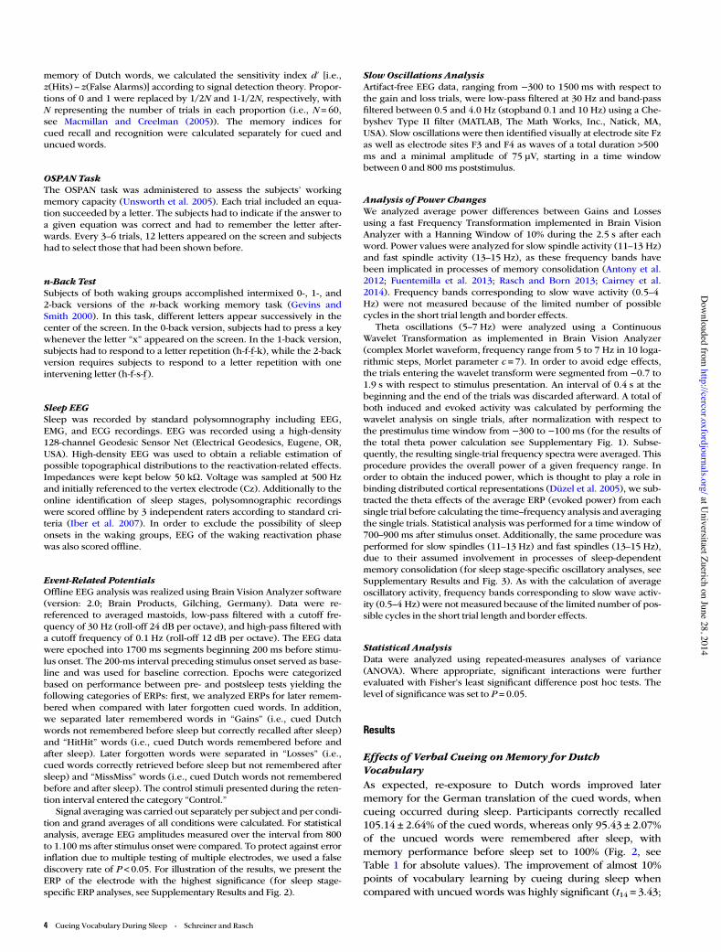
memory of Dutch words, we calculated the sensitivity index d′ [i.e.,z(Hits) – z(False Alarms)] according to signal detection theory. Propor-tions of 0 and 1 were replaced by 1/2N and 1-1/2N, respectively, withN representing the number of trials in each proportion (i.e., N = 60,see Macmillan and Creelman (2005)). The memory indices forcued recall and recognition were calculated separately for cued anduncued words.
OSPAN TaskThe OSPAN task was administered to assess the subjects’ workingmemory capacity (Unsworth et al. 2005). Each trial included an equa-tion succeeded by a letter. The subjects had to indicate if the answer toa given equation was correct and had to remember the letter after-wards. Every 3–6 trials, 12 letters appeared on the screen and subjectshad to select those that had been shown before.
n-Back TestSubjects of both waking groups accomplished intermixed 0-, 1-, and2-back versions of the n-back working memory task (Gevins andSmith 2000). In this task, different letters appear successively in thecenter of the screen. In the 0-back version, subjects had to press a keywhenever the letter “x” appeared on the screen. In the 1-back version,subjects had to respond to a letter repetition (h-f-f-k), while the 2-backversion requires subjects to respond to a letter repetition with oneintervening letter (h-f-s-f).
Sleep EEGSleep was recorded by standard polysomnography including EEG,EMG, and ECG recordings. EEG was recorded using a high-density128-channel Geodesic Sensor Net (Electrical Geodesics, Eugene, OR,USA). High-density EEG was used to obtain a reliable estimation ofpossible topographical distributions to the reactivation-related effects.Impedances were kept below 50 kΩ. Voltage was sampled at 500 Hzand initially referenced to the vertex electrode (Cz). Additionally to theonline identification of sleep stages, polysomnographic recordingswere scored offline by 3 independent raters according to standard cri-teria (Iber et al. 2007). In order to exclude the possibility of sleeponsets in the waking groups, EEG of the waking reactivation phasewas also scored offline.
Event-Related PotentialsOffline EEG analysis was realized using Brain Vision Analyzer software(version: 2.0; Brain Products, Gilching, Germany). Data were re-referenced to averaged mastoids, low-pass filtered with a cutoff fre-quency of 30 Hz (roll-off 24 dB per octave), and high-pass filtered witha cutoff frequency of 0.1 Hz (roll-off 12 dB per octave). The EEG datawere epoched into 1700 ms segments beginning 200 ms before stimu-lus onset. The 200-ms interval preceding stimulus onset served as base-line and was used for baseline correction. Epochs were categorizedbased on performance between pre- and postsleep tests yielding thefollowing categories of ERPs: first, we analyzed ERPs for later remem-bered when compared with later forgotten cued words. In addition,we separated later remembered words in “Gains” (i.e., cued Dutchwords not remembered before sleep but correctly recalled after sleep)and “HitHit” words (i.e., cued Dutch words remembered before andafter sleep). Later forgotten words were separated in “Losses” (i.e.,cued words correctly retrieved before sleep but not remembered aftersleep) and “MissMiss” words (i.e., cued Dutch words not rememberedbefore and after sleep). The control stimuli presented during the reten-tion interval entered the category “Control.”
Signal averaging was carried out separately per subject and per condi-tion and grand averages of all conditions were calculated. For statisticalanalysis, average EEG amplitudes measured over the interval from 800to 1.100 ms after stimulus onset were compared. To protect against errorinflation due to multiple testing of multiple electrodes, we used a falsediscovery rate of P < 0.05. For illustration of the results, we present theERP of the electrode with the highest significance (for sleep stage-specific ERP analyses, see Supplementary Results and Fig. 2).
Slow Oscillations AnalysisArtifact-free EEG data, ranging from −300 to 1500 ms with respect tothe gain and loss trials, were low-pass filtered at 30 Hz and band-passfiltered between 0.5 and 4.0 Hz (stopband 0.1 and 10 Hz) using a Che-byshev Type II filter (MATLAB, The Math Works, Inc., Natick, MA,USA). Slow oscillations were then identified visually at electrode site Fzas well as electrode sites F3 and F4 as waves of a total duration >500ms and a minimal amplitude of 75 µV, starting in a time windowbetween 0 and 800 ms poststimulus.
Analysis of Power ChangesWe analyzed average power differences between Gains and Lossesusing a fast Frequency Transformation implemented in Brain VisionAnalyzer with a Hanning Window of 10% during the 2.5 s after eachword. Power values were analyzed for slow spindle activity (11–13 Hz)and fast spindle activity (13–15 Hz), as these frequency bands havebeen implicated in processes of memory consolidation (Antony et al.2012; Fuentemilla et al. 2013; Rasch and Born 2013; Cairney et al.2014). Frequency bands corresponding to slow wave activity (0.5–4Hz) were not measured because of the limited number of possiblecycles in the short trial length and border effects.
Theta oscillations (5–7 Hz) were analyzed using a ContinuousWavelet Transformation as implemented in Brain Vision Analyzer(complex Morlet waveform, frequency range from 5 to 7 Hz in 10 loga-rithmic steps, Morlet parameter c = 7). In order to avoid edge effects,the trials entering the wavelet transform were segmented from −0.7 to1.9 s with respect to stimulus presentation. An interval of 0.4 s at thebeginning and the end of the trials was discarded afterward. A total ofboth induced and evoked activity was calculated by performing thewavelet analysis on single trials, after normalization with respect tothe prestimulus time window from −300 to −100 ms (for the results ofthe total theta power calculation see Supplementary Fig. 1). Subse-quently, the resulting single-trial frequency spectra were averaged. Thisprocedure provides the overall power of a given frequency range. Inorder to obtain the induced power, which is thought to play a role inbinding distributed cortical representations (Düzel et al. 2005), we sub-tracted the theta effects of the average ERP (evoked power) from eachsingle trial before calculating the time–frequency analysis and averagingthe single trials. Statistical analysis was performed for a time window of700–900 ms after stimulus onset. Additionally, the same procedure wasperformed for slow spindles (11–13 Hz) and fast spindles (13–15 Hz),due to their assumed involvement in processes of sleep-dependentmemory consolidation (for sleep stage-specific oscillatory analyses, seeSupplementary Results and Fig. 3). As with the calculation of averageoscillatory activity, frequency bands corresponding to slow wave activ-ity (0.5–4 Hz) were not measured because of the limited number of pos-sible cycles in the short trial length and border effects.
Statistical AnalysisData were analyzed using repeated-measures analyses of variance(ANOVA). Where appropriate, significant interactions were furtherevaluated with Fisher’s least significant difference post hoc tests. Thelevel of significance was set to P = 0.05.
Results
Effects of Verbal Cueing onMemory for DutchVocabularyAs expected, re-exposure to Dutch words improved latermemory for the German translation of the cued words, whencueing occurred during sleep. Participants correctly recalled105.14 ± 2.64% of the cued words, whereas only 95.43 ± 2.07%of the uncued words were remembered after sleep, withmemory performance before sleep set to 100% (Fig. 2, seeTable 1 for absolute values). The improvement of almost 10%points of vocabulary learning by cueing during sleep whencompared with uncued words was highly significant (t14 = 3.43;
4 Cueing Vocabulary During Sleep • Schreiner and Rasch
at Universitaet Zuerich on June 28, 2014
http://cercor.oxfordjournals.org/D
ownloaded from

P = 0.004). In fact, cueing during sleep even induced a 5% in-crease in memory for cued Dutch words above presleep per-formance levels, and this increase reached a statistical trend(+5.14 ± 2.64%; P = 0.072, one-sample t-test, two-sided). In con-trast, German translations of uncued Dutch words were signifi-cantly more forgotten when compared with recall performancebefore sleep (−4.75 ± 2.07%; P = 0.045). Thus, reactivation of vo-cabulary during sleep did not only prevent forgetting ofGerman translations, but showed a trend of improving memorybeyond baseline levels. On the individual level, 12 of 15 partici-pants benefited from cueing (range +1 to +11 words, for the ab-solute difference between cued and uncued words), whereas 3participants did not (range 0 to −1 words).
To test whether the observed benefits of cueing duringsleep disturbed the consolidation of uncued words or not, weconducted an independent control experiment without pre-senting any verbal cues during sleep after learning (sleepcontrol group). After learning, words were also classified ascued and uncued words using the same algorithm as in themain experiment (see Materials and Methods), but no verbalcues were replayed during sleep. As expected, recall of wordsclassified as cued and uncued did not differ (93.55 ± 2.37 vs.92.80 ± 3.10%; t14 = 0.24; P = 0.81). More importantly, memoryperformance in the sleep control group after sleeping withoutany verbal cues was highly comparable with the recall per-formance for uncued words observed in the main experimentwith verbal cues during sleep (93.55 ± 2.37 vs. 95.43 ± 2.07%;t14 = 0.71; P = 0.48), and was significantly lower when com-pared with memory for cued words (92.80 ± 3.10 vs.105.14 ± 2.64%; t14 = 3.26; P = 0.003, Fig. 2, see Table 1, for ab-solute values).
In the 2 waking groups, cueing did not reveal any beneficialeffect on memory for Dutch vocabulary, neither in the activewaking group (85.53 ± 2.8 vs. 84.2 ± 2.16%, for cued anduncued words, respectively; t16 = 0.56; P = 0.58) nor in thepassive waking group (79.86 ± 4.58 vs. 81.25 ± 2.09%), forcued and uncued words, respectively, t16 =−0.35, P = 0.74; seeTable 1 for absolute values). Thus, even with the availability of
attentive processing resources in the passive waking group, re-exposure to Dutch words during waking failed to improvememory for the German translations.
In addition to sleep-specific improvement by cueing, recallof German translation was generally better in the 2 sleepgroups when compared with the 2 waking control groups, re-flecting the well-known beneficial effect of retention intervalsfilled with sleep when compared with waking on memory con-solidation (main effect condition; F3,60 = 13.06; P < 0.001; seeFig. 2). Post hoc tests revealed that recall performance in bothsleep groups independent of cueing was better when com-pared with the active waking and the passive waking group(t62 = 5.61; P < 0.001).
While cueing during sleep improved memory for Germantranslation of Dutch words as tested by cued recall, we ob-served no sleep-specific benefit of cueing on recognition ofDutch words. The interaction remained nonsignificant(F3,60 = 1.35; P = 0.15). However, sleep improved recognitionof Dutch words independently of cueing (main effect condi-tion; F2,46 = 15.87, P < 0.001): both sleep groups showed a sig-nificantly higher recognition performance (main sleep group:d′ = 2.32 ± 0.13; sleep control group: d′ = 2.04 ± 0.14) whencompared with the active waking group (d′ = 1.42 ± 0.16) andthe passive waking group (d′ = 1.05 ± 0.16; all P < 0.001), whileneither the 2 waking groups (P = 0.10) nor the 2 sleep groups(P = 0.68) differed significantly among each other. In fact, rec-ognition of cued and uncued Dutch words was basically identi-cal in the main sleep group (see Table 1), safely excluding thatrecognition testing prior to cued recall might have confoundedthe reported beneficial effect of cueing during sleep as testedby cued recall. While cueing also did not affect recognition inthe active waking group, cued words were better recognized inthe passive waking group in an exploratory analysis, possiblyreflecting the fact that the participants in the latter group at-tended the cued Dutch words during the retention interval(see Table 1).
Sleep and CueingThe beneficial effect of cueing on memory during NonREMsleep cannot be explained by general alterations in sleep as theeffect was specific for cued when compared with uncuedwords, while the general improving effect of sleep on memorywas present for both word categories. Sleep architecture wasnot altered by cueing, as sleep parameters recorded in themain sleep group did not differ from those of the control sleepgroup (see Table 2). In addition, we did not observe any in-creases in alpha power 1000 ms before (indicative of brief awa-kenings (Rudoy et al. 2009)) and after the auditory stimulationat electrode site Oz, excluding that cueing of words inducedshort lasting arousal responses (alpha power before(2.12 ± 0.41 μV) and after the auditory cue (2.01 ± 0.5 μV), re-spectively, t14 = 0.31, P = 0.75). Still participants of the mainsleep group spent more time awake then subjects of thecontrol sleep group (4.66 vs. 0.55 min; t14 = 2.86, P = 0.013), in-dicating that auditory cueing slightly interrupted sleep. Notethat auditory presentation of words was stop whenever signsof arousal or awakenings were detected. Importantly, perform-ance levels of uncued words in the main sleep group and inthe sleep group without cueing were almost identical, indicat-ing that increases in wake time did not impair ongoing andspontaneous processes of memory consolidation.
Figure 2. Behavioral results. In the main sleep group, memory for cued word pairs(black bar) was significantly improved when compared with uncued pairs (white bar).Recall of uncued word pairs in the main sleep group was comparable with recallperformance of word pairs in the control sleep group, which did not receive any cuesduring sleep. No enhancing effects of cueing on later memory retrieval occurred inboth waking control groups. Retrieval performance is indicated as percentage ofrecalled German translations with performance before sleep set to 100%. Values aremean ± SEM. **P≤ 0.01.
Cerebral Cortex 5
at Universitaet Zuerich on June 28, 2014
http://cercor.oxfordjournals.org/D
ownloaded from

We did not observe any significant associations between thememory advantage induced by cueing (i.e., by subtractingmemory for cued minus uncued words (Antony et al. 2012))and the relative time spent in a certain sleep stage (N1: r = 0.18,P = 0.50; N2: r =−0.360, P = 0.18; SWS: r = 0.18, P = 0.51; REM:r = 0.24, P = 0.93). Cueing was monitored online and wasrestricted to sleep stages N2 and SWS. The total number ofcueings did not differ between N2 and SWS (Table 2), and wedid not observe any significant association between thememory advantage induced by cueing and number of cueingsin N2 or SWS (N2: r =−0.39, P = 0.14; SWS: r = 0.1, P = 0.72; fora more detailed description and analysis see SupplementaryTable 2 and Results). Additionally, EEG offline scoring of thewaking groups revealed no signs of sleep onsets, indicatingthat the subjects of both waking groups were awake through-out the reactivation phase.
Neural Correlates of Cueing During SleepIn order to characterize the process of cueing on a neuralbasis, we analyzed ERPs and oscillatory responses to vocabu-lary cues during sleep. First, we analyzed ERPs for later re-membered when compared with later forgotten cued words.In addition, we separated later remembered words in Gains (i.e., cued Dutch words not remembered before sleep but cor-rectly recalled after sleep) and HitHit words (i.e., cued Dutchwords remembered before and after sleep). Later forgottenwords were separated in Losses (i.e., cued words correctly re-trieved before sleep but not remembered after sleep) and Mis-sMiss words (i.e., cued Dutch words not remembered beforeand after sleep). Please note that the categories Gains andLosses reflect a clear behavioral change after cueing, thereforebest representing the neural pattern associated with processesunderlying successful versus unsuccessful cueing for latermemory retrieval. In contrast, neural correlates of HitHit andMissMiss words are more difficult to interpret, as cueing duringsleep might be ineffective for sufficiently strong memory traces(cases of HitHit) or nonexisting associations (cases of “Los-sLoss”) after encoding before sleep (for the behavioral analysisof Gains and Losses please see Supplementary Results andTable 2).
Remarkably, the EEG analysis of the average ERP amplitudesin the main sleep group clearly revealed a more pronouncednegativity for subsequently remembered versus subsequentlyforgotten cued words at electrode site Fz (t14 =−2.85, P = 0.013).We further explored this difference by separately analyzingGains and “HitHits” as well as Losses and MissMiss. Similar tothe previous analysis, the difference between the ERP responsesassociated with HitHits when compared with “MissMisses”was significant (t14 = 2.45, P = 0.028). More importantly, weobserved the largest negative amplitude associated withcueing of “Gain” words. Neural correlates of Gains represent amemory gain induced by cueing during sleep (i.e., successfulverbal cueing during sleep), and the amplitude was significant-ly increased when compared with all other word categories atelectrode site Fz in a time interval from 800 to 1100 ms afterword onset (F6,84 = 4.52, P = 0.001), all pairwise post hoc testsP < 0.04, see Fig. 3a,b). As Losses are the most suitable controlcategory for Gains (i.e., behavioral change in memory inducedby cueing, relatively similar number of occurrences, etc.), wefocused on the comparison between Gains and Losses in allsubsequent analyses.
The analysis of all electrode revealed that the amplitude dif-ference between Gains and Losses had a stable fronto-centraldistribution (see Fig. 3c) comparable with distributions of sub-sequent memory effects observed during waking (Werkle-Bergner et al. 2006). Furthermore, in a single-trial analysis, wecounted the number of clearly identifiable slow waves (nega-tive amplitude >75 μV with a duration of >500 ms starting in atime window 0–800 ms poststimulus, see Materials andMethods) that followed cueing of Gain words when comparedwith Losses during sleep. This analysis revealed, that Gainswere significantly more often followed by slow oscillations(31.09 ± 3.6% of all cueing trials of Gains) when comparedwith Losses (18.48 ± 3.4% cueing trials of Losses; t14 = 5.35,P < 0.001). This result was found at electrode site Fz, as well asF3 and F4 indicating a stable frontal distribution of this effect.This result is compatible with the assumption that the presenceof a slow oscillation after the presentation of a Dutch wordduring sleep plays an important role for successfully stabilizingthe associated memory trace, reactivated by the memory cuepresented during sleep. As both slow oscillations and sleepspindles are critically involved in processes of memory consoli-dation during sleep (Rasch and Born 2013), we also analyzedpossible differences in average oscillatory power betweenGains and Losses for slow spindles (11–13 Hz) and fastspindle activity (13–15 Hz). However, we did not observeany difference between Gains and Losses in this analysis (allP > 0.10).
We further explored difference between Gains and Lossesin time–frequency space. We controlled for a possible contri-bution of the evoked brain response by subtracting theaverage ERP (evoked power) from each single trial before cal-culating the time–frequency analysis (induced power) (Kli-mesch et al. 1998). In contrast to our expectations, the time–frequency analysis revealed no significant increase in oscilla-tory power in the spindle band related to Gains versusLosses, neither in the fast spindle band (13–15 Hz) nor in theslow spindle band (11–13 Hz). However, sleep stage-specificanalyses revealed a significant increase in slow spindle powerduring SWS (but not during stage N2) in a time window 600–800 ms after the cue (P < 0.05, for details see SupplementaryResults and Fig. 3). Please note that the analysis of powerchanges in the slow oscillations/delta band was not possibledue to the relatively small intertrial interval between verbalcues.
Finally, we also analyzed power changes for the theta band.Theta activity is prevalently linked to successful memory encod-ing during waking (Nyhus and Curran 2010) and poststimulusincreases in induced theta power have been specifically linkedto processes of recollection (Düzel et al. 2005). Interestingly,induced theta power associated with verbal cueing duringsleep differed significantly between conditions (F4,56 = 7.38,P = 0.002). Gains were associated with an increase in inducedtheta power in a time window of 700–900 ms after stimulusonset. The increase in induced theta power was particularlystrong in right frontal as well as left parietal electrodes (e.g.,electrode FC6: t14 = 3.68; P = 0.009), strongly suggesting that atransient increase in theta power is critical for successful cueingduring sleep (see Fig. 3d–f; see Supplementary Fig. 1 for totalpower changes). Interestingly, increases in theta activity forGains when compared with Losses were more pronouncedduring stage 2 sleep, but were also reliably observed duringSWS (see Supplementary Results and Fig. 3).
6 Cueing Vocabulary During Sleep • Schreiner and Rasch
at Universitaet Zuerich on June 28, 2014
http://cercor.oxfordjournals.org/D
ownloaded from

DiscussionOur findings show for the first time that cueing prior learnedforeign vocabulary during sleep improves later recall. Further-more, memory performance for uncued words in the mainsleep group resembled memory performance of participantswho did not receive any verbal cues during sleep, suggestingthat cueing led to a real gain in memory performance. In add-ition, successful cueing during sleep, which resulted in latermemory gains during retrieval testing, was associated with anincreased late negativity and increased theta activity duringNonREM sleep.
The beneficial effect of cueing during sleep is consistent withthe active system consolidation hypothesis, which assumes thatspontaneous memory reactivations during sleep are critical forthe enhancing effect of sleep on memory consolidation. In fact,recent studies have successfully used memory-associated odors,sounds, or melodies (Rasch et al. 2007; Rudoy et al. 2009;Antony et al. 2012) to cue and strengthen memories duringsleep. Here, we go an important step beyond these previousresults by showing that also complex stimuli like foreign vo-cabulary can be successfully used to reactivate memories during
sleep, leading to an enhanced memory for vocabulary the nextday. Importantly, our results are highly relevant for vocabularylearning in an educational setting, because our procedure of re-activating foreign vocabulary could be easily applied to theseevery-day learning contexts. However, as retrieval was tested inthe night after only a few hours of sleep in the current study,future studies should test the memory-improving effects ofcueing during sleep the next day or after several days. In add-ition, it still needs to be determined whether or not the benefi-cial effects of cueing during sleep are possibly accompanied byany detrimental effects on sleep-dependent memory consolida-tion of other material learned during the day. Finally, futurestudies need to examine whether cueing of vocabulary duringsleep indeed facilitates foreign language learning.
In our experiment, we explicitly chose Dutch as a foreignlanguage to achieve sufficiently few learning trials required forour analysis. Due to the close relation of Dutch to German orEnglish, German-speaking participants could more easily learnthe vocabulary and might even be able to correctly guess themeaning of some words. However, guesses cannot explain ourreported improved effect of cueing during sleep, as words
Figure 3. Electrophysiological results. ERPs and oscillatory theta power recorded during cueing in the sleep group were computed for words, for which cueing during sleep led to achange in memory performance. “Gains” reflect cued words not remembered in the presleep test but correctly recalled in the postsleep test. “Losses” refer to cued wordsremembered in the presleep test but not in the postsleep test. Words remembered before and after the retention interval were labeled “HitHit” and words not remembered bothbefore and after the retention interval were labeled “MissMiss.” The new 30 Dutch words formed the “Control” condition. (a and b) Successful cueing was associated with a morepronounced negativity at frontal electrode sites (representative electrode Fz). The rectangle illustrates the time window used for waveform quantification. (c) Scalp map representingthe topographical distribution for the difference between “Gains” and “Losses” in the time window between 800 and 1100 ms, indicating a pronounced frontal distribution (allelectrodes entered the analysis; black dots indicate significant electrodes at P< 0.05, false discovery rate) corrected for multiple comparisons). The following electrodes weresignificant: E4, E5, E6, E11, E12, E13, E16, E19, E20, E23, E24, E28, E29, E35, E112 (see Supplementary Fig. 2 for the exact electrode positions). (d and e) Induced theta power forthe difference between “Gains” and “Losses” (electrode FC6), indicating a distinct increase in induced theta power associated with successful cueing. (f ) Scalp map depicting thedistribution of theta power increase for “Gains” relative to “Losses” in the time window between 700 and 900 ms. The following electrodes were significant: E53, E60, E61, E62,E111, E117 (FC6), E118). **P≤ 0.01.
Cerebral Cortex 7
at Universitaet Zuerich on June 28, 2014
http://cercor.oxfordjournals.org/D
ownloaded from
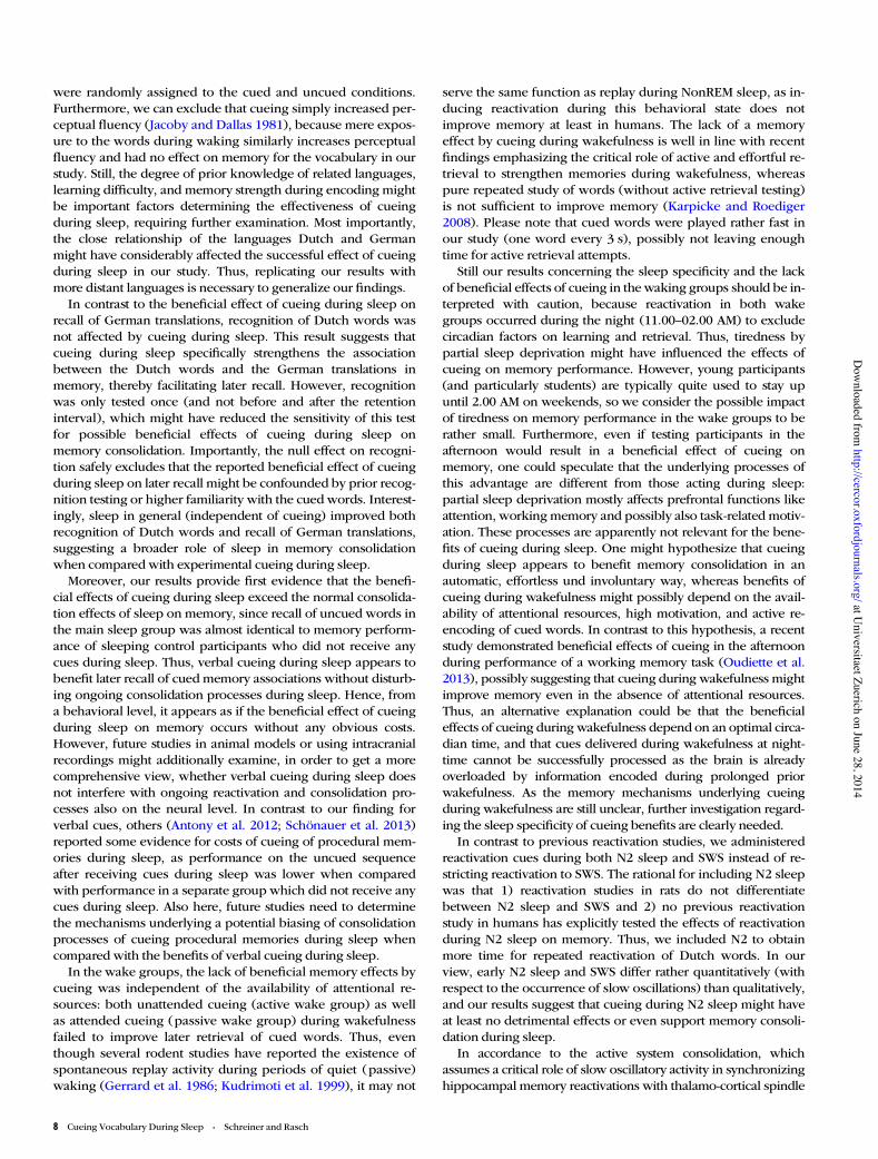
were randomly assigned to the cued and uncued conditions.Furthermore, we can exclude that cueing simply increased per-ceptual fluency (Jacoby and Dallas 1981), because mere expos-ure to the words during waking similarly increases perceptualfluency and had no effect on memory for the vocabulary in ourstudy. Still, the degree of prior knowledge of related languages,learning difficulty, and memory strength during encoding mightbe important factors determining the effectiveness of cueingduring sleep, requiring further examination. Most importantly,the close relationship of the languages Dutch and Germanmight have considerably affected the successful effect of cueingduring sleep in our study. Thus, replicating our results withmore distant languages is necessary to generalize our findings.
In contrast to the beneficial effect of cueing during sleep onrecall of German translations, recognition of Dutch words wasnot affected by cueing during sleep. This result suggests thatcueing during sleep specifically strengthens the associationbetween the Dutch words and the German translations inmemory, thereby facilitating later recall. However, recognitionwas only tested once (and not before and after the retentioninterval), which might have reduced the sensitivity of this testfor possible beneficial effects of cueing during sleep onmemory consolidation. Importantly, the null effect on recogni-tion safely excludes that the reported beneficial effect of cueingduring sleep on later recall might be confounded by prior recog-nition testing or higher familiarity with the cued words. Interest-ingly, sleep in general (independent of cueing) improved bothrecognition of Dutch words and recall of German translations,suggesting a broader role of sleep in memory consolidationwhen compared with experimental cueing during sleep.
Moreover, our results provide first evidence that the benefi-cial effects of cueing during sleep exceed the normal consolida-tion effects of sleep on memory, since recall of uncued words inthe main sleep group was almost identical to memory perform-ance of sleeping control participants who did not receive anycues during sleep. Thus, verbal cueing during sleep appears tobenefit later recall of cued memory associations without disturb-ing ongoing consolidation processes during sleep. Hence, froma behavioral level, it appears as if the beneficial effect of cueingduring sleep on memory occurs without any obvious costs.However, future studies in animal models or using intracranialrecordings might additionally examine, in order to get a morecomprehensive view, whether verbal cueing during sleep doesnot interfere with ongoing reactivation and consolidation pro-cesses also on the neural level. In contrast to our finding forverbal cues, others (Antony et al. 2012; Schönauer et al. 2013)reported some evidence for costs of cueing of procedural mem-ories during sleep, as performance on the uncued sequenceafter receiving cues during sleep was lower when comparedwith performance in a separate group which did not receive anycues during sleep. Also here, future studies need to determinethe mechanisms underlying a potential biasing of consolidationprocesses of cueing procedural memories during sleep whencompared with the benefits of verbal cueing during sleep.
In the wake groups, the lack of beneficial memory effects bycueing was independent of the availability of attentional re-sources: both unattended cueing (active wake group) as wellas attended cueing (passive wake group) during wakefulnessfailed to improve later retrieval of cued words. Thus, eventhough several rodent studies have reported the existence ofspontaneous replay activity during periods of quiet (passive)waking (Gerrard et al. 1986; Kudrimoti et al. 1999), it may not
serve the same function as replay during NonREM sleep, as in-ducing reactivation during this behavioral state does notimprove memory at least in humans. The lack of a memoryeffect by cueing during wakefulness is well in line with recentfindings emphasizing the critical role of active and effortful re-trieval to strengthen memories during wakefulness, whereaspure repeated study of words (without active retrieval testing)is not sufficient to improve memory (Karpicke and Roediger2008). Please note that cued words were played rather fast inour study (one word every 3 s), possibly not leaving enoughtime for active retrieval attempts.
Still our results concerning the sleep specificity and the lackof beneficial effects of cueing in the waking groups should be in-terpreted with caution, because reactivation in both wakegroups occurred during the night (11.00–02.00 AM) to excludecircadian factors on learning and retrieval. Thus, tiredness bypartial sleep deprivation might have influenced the effects ofcueing on memory performance. However, young participants(and particularly students) are typically quite used to stay upuntil 2.00 AM on weekends, so we consider the possible impactof tiredness on memory performance in the wake groups to berather small. Furthermore, even if testing participants in theafternoon would result in a beneficial effect of cueing onmemory, one could speculate that the underlying processes ofthis advantage are different from those acting during sleep:partial sleep deprivation mostly affects prefrontal functions likeattention, working memory and possibly also task-related motiv-ation. These processes are apparently not relevant for the bene-fits of cueing during sleep. One might hypothesize that cueingduring sleep appears to benefit memory consolidation in anautomatic, effortless und involuntary way, whereas benefits ofcueing during wakefulness might possibly depend on the avail-ability of attentional resources, high motivation, and active re-encoding of cued words. In contrast to this hypothesis, a recentstudy demonstrated beneficial effects of cueing in the afternoonduring performance of a working memory task (Oudiette et al.2013), possibly suggesting that cueing during wakefulness mightimprove memory even in the absence of attentional resources.Thus, an alternative explanation could be that the beneficialeffects of cueing during wakefulness depend on an optimal circa-dian time, and that cues delivered during wakefulness at night-time cannot be successfully processed as the brain is alreadyoverloaded by information encoded during prolonged priorwakefulness. As the memory mechanisms underlying cueingduring wakefulness are still unclear, further investigation regard-ing the sleep specificity of cueing benefits are clearly needed.
In contrast to previous reactivation studies, we administeredreactivation cues during both N2 sleep and SWS instead of re-stricting reactivation to SWS. The rational for including N2 sleepwas that 1) reactivation studies in rats do not differentiatebetween N2 sleep and SWS and 2) no previous reactivationstudy in humans has explicitly tested the effects of reactivationduring N2 sleep on memory. Thus, we included N2 to obtainmore time for repeated reactivation of Dutch words. In ourview, early N2 sleep and SWS differ rather quantitatively (withrespect to the occurrence of slow oscillations) than qualitatively,and our results suggest that cueing during N2 sleep might haveat least no detrimental effects or even support memory consoli-dation during sleep.
In accordance to the active system consolidation, whichassumes a critical role of slow oscillatory activity in synchronizinghippocampal memory reactivations with thalamo-cortical spindle
8 Cueing Vocabulary During Sleep • Schreiner and Rasch
at Universitaet Zuerich on June 28, 2014
http://cercor.oxfordjournals.org/D
ownloaded from
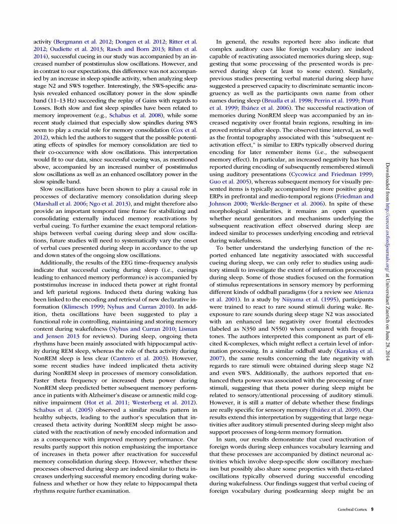
activity (Bergmann et al. 2012; Dongen et al. 2012; Ritter et al.2012; Oudiette et al. 2013; Rasch and Born 2013; Rihm et al.2014), successful cueing in our study was accompanied by an in-creased number of poststimulus slow oscillations. However, andin contrast to our expectations, this difference was not accompan-ied by an increase in sleep spindle activity, when analyzing sleepstage N2 and SWS together. Interestingly, the SWS-specific ana-lysis revealed enhanced oscillatory power in the slow spindleband (11–13 Hz) succeeding the replay of Gains with regards toLosses. Both slow and fast sleep spindles have been related tomemory improvement (e.g., Schabus et al. 2008), while somerecent study claimed that especially slow spindles during SWSseem to play a crucial role for memory consolidation (Cox et al.2012), which led the authors to suggest that the possible potenti-ating effects of spindles for memory consolidation are tied totheir co-occurrence with slow oscillations. This interpretationwould fit to our data, since successful cueing was, as mentionedabove, accompanied by an increased number of poststimulusslow oscillations as well as an enhanced oscillatory power in theslow spindle band.
Slow oscillations have been shown to play a causal role inprocesses of declarative memory consolidation during sleep(Marshall et al. 2006; Ngo et al. 2013), and might therefore alsoprovide an important temporal time frame for stabilizing andconsolidating externally induced memory reactivations byverbal cueing. To further examine the exact temporal relation-ships between verbal cueing during sleep and slow oscilla-tions, future studies will need to systematically vary the onsetof verbal cues presented during sleep in accordance to the upand down states of the ongoing slow oscillations.
Additionally, the results of the EEG time–frequency analysisindicate that successful cueing during sleep (i.e., cueingsleading to enhanced memory performance) is accompanied bypoststimulus increase in induced theta power at right frontaland left parietal regions. Induced theta during waking hasbeen linked to the encoding and retrieval of new declarative in-formation (Klimesch 1999; Nyhus and Curran 2010). In add-ition, theta oscillations have been suggested to play afunctional role in controlling, maintaining and storing memorycontent during wakefulness (Nyhus and Curran 2010; Lismanand Jensen 2013 for reviews). During sleep, ongoing thetarhythms have been mainly associated with hippocampal activ-ity during REM sleep, whereas the role of theta activity duringNonREM sleep is less clear (Cantero et al. 2003). However,some recent studies have indeed implicated theta activityduring NonREM sleep in processes of memory consolidation.Faster theta frequency or increased theta power duringNonREM sleep predicted better subsequent memory perform-ance in patients with Alzheimer’s disease or amnestic mild cog-nitive impairment (Hot et al. 2011; Westerberg et al. 2012).Schabus et al. (2005) observed a similar results pattern inhealthy subjects, leading to the author’s speculation that in-creased theta activity during NonREM sleep might be asso-ciated with the reactivation of newly encoded information andas a consequence with improved memory performance. Ourresults partly support this notion emphasizing the importanceof increases in theta power after reactivation for successfulmemory consolidation during sleep. However, whether theseprocesses observed during sleep are indeed similar to theta in-creases underlying successful memory encoding during wake-fulness and whether or how they relate to hippocampal thetarhythms require further examination.
In general, the results reported here also indicate thatcomplex auditory cues like foreign vocabulary are indeedcapable of reactivating associated memories during sleep, sug-gesting that some processing of the presented words is pre-served during sleep (at least to some extent). Similarly,previous studies presenting verbal material during sleep havesuggested a preserved capacity to discriminate semantic incon-gruency as well as the participants own name from othernames during sleep (Brualla et al. 1998; Perrin et al. 1999; Prattet al. 1999; Ibáñez et al. 2006). The successful reactivation ofmemories during NonREM sleep was accompanied by an in-creased negativity over frontal brain regions, resulting in im-proved retrieval after sleep. The observed time interval, as wellas the frontal topography associated with this “subsequent re-activation effect,” is similar to ERPs typically observed duringencoding for later remember items (i.e., the subsequentmemory effect). In particular, an increased negativity has beenreported during encoding of subsequently remembered stimuliusing auditory presentations (Cycowicz and Friedman 1999;Guo et al. 2005), whereas subsequent memory for visually pre-sented items is typically accompanied by more positive goingERPs in prefrontal and medio-temporal regions (Friedman andJohnson 2000; Werkle-Bergner et al. 2006). In spite of thesemorphological similarities, it remains an open questionwhether neural generators and mechanisms underlying thesubsequent reactivation effect observed during sleep areindeed similar to processes underlying encoding and retrievalduring wakefulness.
To better understand the underlying function of the re-ported enhanced late negativity associated with successfulcueing during sleep, we can only refer to studies using audi-tory stimuli to investigate the extent of information processingduring sleep. Some of those studies focused on the formationof stimulus representations in sensory memory by performingdifferent kinds of oddball paradigms (for a review see Atienzaet al. 2001). In a study by Niiyama et al. (1995), participantswere trained to react to rare sound stimuli during wake. Re-exposure to rare sounds during sleep stage N2 was associatedwith an enhanced late negativity over frontal electrodes(labeled as N350 and N550) when compared with frequenttones. The authors interpreted this component as part of eli-cited K-complexes, which might reflect a certain level of infor-mation processing. In a similar oddball study (Karakas et al.2007), the same results concerning the late negativity withregards to rare stimuli were obtained during sleep stage N2and even SWS. Additionally, the authors reported that en-hanced theta power was associated with the processing of rarestimuli, suggesting that theta power during sleep might berelated to sensory/attentional processing of auditory stimuli.However, it is still a matter of debate whether these findingsare really specific for sensory memory (Ibáñez et al. 2009). Ourresults extend this interpretation by suggesting that large nega-tivities after auditory stimuli presented during sleep might alsosupport processes of long-term memory formation.
In sum, our results demonstrate that cued reactivation offoreign words during sleep enhances vocabulary learning andthat these processes are accompanied by distinct neuronal ac-tivities which involve sleep-specific slow oscillatory mechan-ism but possibly also share some properties with theta-relatedoscillations typically observed during successful encodingduring wakefulness. Our findings suggest that verbal cueing offoreign vocabulary during postlearning sleep might be an
Cerebral Cortex 9
at Universitaet Zuerich on June 28, 2014
http://cercor.oxfordjournals.org/D
ownloaded from

efficient and effortless tool to improve foreign vocabularylearning in educational settings as well as every-day life.
Supplementary MaterialSupplementary material can be found at: http://www.cercor.oxfordjournals.org/.
FundingThis work was supported by a grant from the Swiss NationalFoundation (SNF) (PP00P1_133685) and the Clinical ResearchPriority Program “Sleep and Health” of the University ofZurich.
NotesWe thank Niki Hug, Janina Leeman, and Rebecca Paladini for assist-ance in data collection and analysis, Tobias Egli and Maurice Göldi forhelp in programming and Ines Wilhelm for helpful comments onearlier versions of the manuscript. Conflict of Interest: None declared.
ReferencesAntony JW, Gobel EW, O’Hare JK, Reber PJ, Paller KA. 2012. Cued
memory reactivation during sleep influences skill learning. NatNeurosci. 15:1114–1116.
Atienza M, Cantero JL, Escera C. 2001. Auditory information processingduring human sleep as revealed by event-related brain potentials.Clin Neurophysiol. 112:2031–2045.
Bendor D, Wilson MA. 2012. Biasing the content of hippocampalreplay during sleep. Nat Neurosci. 15:1439–1444.
Bergmann TO, Mölle M, Diedrichs J, Born J, Siebner HR. 2012. Sleepspindle-related reactivation of category-specific cortical regionsafter learning face-scene associations. Neuroimage. 59:2733–2742.
Brualla J, Romero MF, Serrano M, Valdizán JR. 1998. Auditoryevent-related potentials to semantic priming during sleep. Electro-encephalogr Clin Neurophysiol. 108:283–290.
Cairney SA, Durrant SJ, Hulleman J, Lewis PA. 2014. Targeted memoryreactivation during slow wave sleep facilitates emotional memoryconsolidation. Sleep. 37:701–707.
Cantero JL, Atienza M, Stickgold R, Kahana MJ, Madsen JR, Kocsis B.2003. Sleep-dependent theta oscillations in the human hippocam-pus and neocortex. J Neurosci. 23:10897–10903.
Cox R, Hofman WF, Talamini LM. 2012. Involvement of spindles inmemory consolidation is slow wave sleep-specific. Learn Mem.19:264–267.
Cycowicz YM, Friedman D. 1999. The effect of intention to learn novel,environmental sounds on the novelty P3 and old/new recognitionmemory. Biol Psychol. 50:35–60.
Dave A, Marholiash D. 2000. Song replay during sleep and computa-tional rules for sensorimotor vocal learning. Science. 290:812–816.
Davis MH, Gaskell MG. 2009. A complementary systems account ofword learning: neural and behavioural evidence. Philos Trans RSoc Lond B Biol Sci. 364:3773–3800.
Diekelmann S, Born J. 2010. The memory function of sleep. Nat RevNeurosci. 11:114–126.
Diekelmann S, Büchel C, Born J, Rasch B. 2011. Labile or stable: op-posing consequences for memory when reactivated during wakingand sleep. Nat Neurosci. 14:381–386.
Dumay N, Gaskell MG. 2007. Sleep-associated changes in the mentalrepresentation of spoken words. Psychol Sci. 18:35–39.
Düzel E, Neufang M, Heinze H-J. 2005. The oscillatory dynamics of rec-ognition memory and its relationship to event-related responses.Cereb Cortex. 15:1992–2002.
Fenn KM, Hambrick DZ. 2012. Individual differences in workingmemory capacity predict sleep-dependent memory consolidation.J Exp Psychol Gen. 141:404–410.
Friedman D, Johnson R. 2000. Event-related potential (ERP) studies ofmemory encoding and retrieval: a selective review. Microsc ResTech. 51:6–28.
Fuentemilla L, Miró J, Ripollés P, Vilà-Balló A, Juncadella M, CastañerS, Salord N, Monasterio C, Falip M, Rodríguez-Fornells A. 2013.Hippocampus-dependent strengthening of targeted memories viareactivation during sleep in humans. Curr Biol. 23:1769–1775.
Gais S, Lucas B, Born J. 2006. Sleep after learning aids memory recall.Learn Mem. 13:259–262.
Genzel L, Kroes MCW, Dresler M, Battaglia FP. 2014. Light sleep versusslow wave sleep in memory consolidation: a question of globalversus local processes? Trends Neurosci. 37:10–19.
Gerrard JL, Kudrimoti H, McNaughton BL, Barnes CA. 1986. Reactiva-tion of hippocampal ensemble activity patterns in the aging rat.Behav Neurosci. 115:1180–1192.
Gevins A, Smith ME. 2000. Neurophysiological measures of workingmemory and individual differences in cognitive ability and cogni-tive style. Cereb Cortex. 10:829–839.
Gómez RL, Bootzin RR, Nadel L. 2006. Naps promote abstraction inlanguage-learning infants. Psychol Sci. 17:670–674.
Guo C, Voss JL, Paller KA. 2005. Electrophysiological correlates offorming memories for faces, names, and face-name associations.Brain Res. 22:153–164.
Henderson LM, Weighall AR, Brown H, Gareth Gaskell M. 2012. Con-solidation of vocabulary is associated with sleep in children. DevSci. 15:674–687.
Hot P, Rauchs G, Bertran F, Denise P, Desgranges B, Clochon P, Eu-stache F. 2011. Changes in sleep theta rhythm are related to episod-ic memory impairment in early Alzheimer’s disease. Biol Psychol.87:334–339.
Ibáñez A, López V, Cornejo C. 2006. ERPs and contextual semantic dis-crimination: degrees of congruence in wakefulness and sleep.Brain Lang. 98:264–275.
Ibáñez AM, Martín RS, Hurtado E, López V. 2009. ERPs studies of cog-nitive processing during sleep. Int J Psychol. 44:290–304.
Iber C, Ancoli-Israel S, Chesson A, Quan SF. 2007. The AASM manualfor the scoring of sleep and associated events: rules, terminology,and technical specification. Westchester (IL): American Academy ofSleep Medicine.
Jacoby LL, Dallas M. 1981. On the relationship between autobio-graphical memory and perceptual learning. J Exp Psychol Gen.110:306–340.
Karakas S, Cakmak ED, Bekçi B, Aydin H. 2007. Oscillatory responsesrepresenting differential auditory processing in sleep. Int J Psycho-physiol. 65:40–50.
Karpicke JD, Roediger HL. 2008. The critical importance of retrievalfor learning. Science. 319:966–968.
Klimesch W. 1999. EEG alpha and theta oscillations reflect cognitiveand memory performance: a review and analysis. Brain Res Rev.29:169–195.
Klimesch W, Russegger H, Doppelmayr M, Pachinger T. 1998. Amethod for the calculation of induced band power: implications forthe significance of brain oscillations. Electroencephalogr Clin Neu-rophysiol. 108:123–130.
Kudrimoti HS, Barnes CA, McNaughton BL. 1999. Reactivation of hip-pocampal cell assemblies: effects of behavioral state, experience,and EEG dynamics. J Neurosci. 19:4090–4101.
Lisman JE, Jensen O. 2013. The theta-gamma neural code. Neuron.77:1002–1016.
Macmillan NA, Creelman CD. 2005. Detection theory: a user’s guide.Cambridge, UK: Cambridge University Press.
Margoliash D. 2010. Sleep, learning, and birdsong. ILAR J. 51:378–386.Margoliash D, Schmidt MF. 2010. Sleep, off-line processing, and vocal
learning. Brain Lang. 115:45–58.Marshall L, Helgadóttir H, Mölle M, Born J. 2006. Boosting slow oscilla-
tions during sleep potentiates memory. Nature. 444:610–613.Ngo HV, Martinetz T, Born J, Mölle M. 2013. Auditory closed-loop
stimulation of the sleep slow oscillation enhances memory.Neuron. 78:545–553.
Niiyama Y, Fushimi M, Sekine A, Hishikawa Y. 1995. K-complexevoked in NREM sleep is accompanied by a slow negative potential
10 Cueing Vocabulary During Sleep • Schreiner and Rasch
at Universitaet Zuerich on June 28, 2014
http://cercor.oxfordjournals.org/D
ownloaded from

related to cognitive process. Electroencephalogr Clin Neurophysiol.95:27–33.
Nyhus E, Curran T. 2010. Functional role of gamma and theta oscilla-tions in episodic memory. Neurosci Biobehav Rev. 34:1023–1035.
O’Neill J, Pleydell-Bouverie B, Dupret D, Csicsvari J. 2010. Play itagain: reactivation of waking experience and memory. Trends Neu-rosci. 33:220–229.
Oudiette D, Antony JW, Creery JD, Paller KA. 2013. The role ofmemory reactivation during wakefulness and sleep in determiningwhich memories endure. J Neurosci. 33:6672–6678.
Oudiette D, Paller KA. 2013. Upgrading the sleeping brain with tar-geted memory reactivation. Trends Cogn Sci. 17:142–149.
Pavlides C, Winson J. 1989. Influences of hippocampal place cell firingin the awake state on the activity of these cells during subsequentsleep episodes. J Neurosci. 9:2907–2918.
Perrin F, Bastuji H, Garcia-Larrea L. 2002. Detection of verbal discor-dances during sleep. Neuroreport. 13:1345–1349.
Perrin F, Garcia-Larrea L, Mauguiere F, Bastuji H. 1999. A differentialbrain response to the subject’s own name persists during sleep.Clin Neurophysiol. 110:2153–2164.
Peyrache A, Khamassi M, Benchenane K, Wiener SI, Battaglia FP. 2009.Replay of rule-learning related neural patterns in the prefrontalcortex during sleep. Nat Neurosci. 12:919–926.
Pinker S. 2000. Survival of the clearest. Nature. 404:441–442.Pratt H, Berlad I, Lavie P. 1999. “Oddball” event-related potentials and
information processing during REM and non-REM sleep. Clin Neu-rophysiol. 110:53–61.
Rasch B, Born J. 2013. About sleep’s role in memory. Physiol Rev.93:681–766.
Rasch B, Büchel C, Gais S, Born J. 2007. Odor cues during slow-wave sleepprompt declarativememory consolidation. Science. 315:1426–1429.
Rattenborg NC, Martinez-Gonzalez D, Roth TC, Pravosudov VV. 2011.Hippocampal memory consolidation during sleep: a comparison ofmammals and birds. Biol Rev Camb Philos Soc. 86:658–691.
Rihm JS, Diekelmann S, Born J, Rasch B. 2014. Reactivating memoriesduring sleep by odors: odor specificity and associated changes insleep oscillations. J Cogn Neurosci. 23:1–14.
Ritter SM, Strick M, Bos MW, Van Baaren RB, Dijksterhuis A. 2012.Good morning creativity: task reactivation during sleep enhancesbeneficial effect of sleep on creative performance. J Sleep Res.21:643–647.
Rudoy JD, Voss JL, Westerberg CE, Paller KA. 2009. Strengthening indi-vidual memories by reactivating them during sleep. Science.326:1079.
Schabus M, Hoedlmoser K, Pecherstorfer T, Anderer P, Gruber G, Para-patics S, Sauter C, Kloesch G, Klimesch W, Saletu B et al. 2008. In-terindividual sleep spindle differences and their relation tolearning-related enhancements. Brain Res. 1191:127–135.
Schabus M, Hoedlmoser K, Pecherstorfer T, Kloesch G. 2005. Influenceof midday naps on declarative memory performance and motiv-ation. Somnologie. 9:148–153.
Schönauer M, Geisler T, Gais S. 2013. Strengthening procedural mem-ories by reactivation in sleep. J Cogn Neurosci. 26:143–53.
Shatz M. 2001. Psychology of vocabulary acquisition. Int Encycl SocBehav Sci. 16292–16294
Stickgold R, Walker MP. 2013. Sleep-dependent memory triage: evolv-ing generalization through selective processing. Nat Neurosci.16:139–145.
Unsworth N, Heitz RP, Schrock JC, Engle RW. 2005. An automatedversion of the operation span task. Behav Res Methods. 37:498–505.
Van Dongen EV, Takashima A, Barth M, Zapp J, Schad LR, Paller KA.2012. Memory stabilization with targeted reactivation duringhuman slow-wave sleep. Proc Natl Acad Sci USA. 109:10575–10580.2012.
Werkle-Bergner M, Müller V, Li S-C, Lindenberger U. 2006. Cor-tical EEG correlates of successful memory encoding: implica-tions for lifespan comparisons. Neurosci Biobehav Rev. 30:839–854.
Westerberg CE, Mander BA, Florczak SM, Weintraub S, Mesulam M-M,Zee PC, Paller KA. 2012. Concurrent impairments in sleep andmemory in amnestic mild cognitive impairment. J Int NeuropsycholSoc. 18:1–11.
Wilson MA, McNaughton BL. 1994. Reactivation of hippocampal en-semble memories during sleep. Science. 265:676–679.
Cerebral Cortex 11
at Universitaet Zuerich on June 28, 2014
http://cercor.oxfordjournals.org/D
ownloaded from

Although sleep is a systems-level process that affects the whole organism, its most distinctive features are the loss of behavioural control and consciousness. Among the multiple functions of sleep1, its role in the establish-ment of memories seems to be particularly important: as it seems to be incompatible with the brain’s normal processing of stimuli during waking, it might explain the loss of consciousness in sleep. Sleep promotes primarily the consolidation of memory, whereas memory encoding and retrieval take place most effectively during waking. Consolidation refers to a process that transforms new and initially labile memories encoded in the awake state into more stable representations that become integrated into the network of pre-existing long-term memories. Consolidation involves the active re-processing of ‘fresh’ memories within the neuronal networks that were used for encoding them. It seems to occur most effectively off-line, i.e. during sleep, so that encoding and consoli-dation cannot disturb each other and the brain does not ‘hallucinate’ during consolidation2.
The hypothesis that sleep favours memory consolida-tion has been around for a long time3. Recent research in this field has provided important insights into the underlying mechanisms through which sleep serves memory consolidation4–7. In this Review, we first discuss findings from behavioural studies regarding the specific conditions that determine the access of a freshly encoded memory to sleep-dependent consolidation, and regard-ing the way in which sleep quantitatively and qualitatively changes new memory representations. We then consider the role of slow-wave sleep (SWS) and rapid eye move-ment (REM) sleep in memory consolidation (BOX 1). We
finish by comparing two hypotheses that might explain sleep-dependent memory consolidation on a mechanis-tic level, that is, the synaptic homeostasis hypothesis and the active system consolidation hypothesis.
Behavioural studiesNumerous studies have confirmed the beneficial effect of sleep on declarative and procedural memory in various tasks8–10, with practically no evidence for the opposite effect (sleep promoting forgetting)11. Compared with a wake interval of equal length, a period of post-learning sleep enhances retention of declarative information3,12–16 and improves performance in procedural skills13,17–24. Sleep likewise supports the consolidation of emotional information25–27. Effects of a 3-hour period of sleep on emotional memory were even detectable 4 years later28. However, the consolidating effect of sleep is not revealed under all circumstances and seems to be associated with specific conditions29 (see below).
Sleep duration and timing. Significant sleep benefits on memory are observed after an 8-hour night of sleep, but also after shorter naps of 1–2 hours14,19,23,30, and even an ultra-short nap of 6 minutes can improve memory retention16. However, longer sleep durations yield greater improvements, particularly for procedural memo-ries18,21,31. The optimal amount of sleep needed to benefit memory and how this might generalize across species showing different sleep durations is unclear at present.
Some data suggest that a short delay between learning and sleep optimizes the benefits of sleep on memory consolidation. For example, for declarative
University of Lübeck, Department of Neuroendocrinology, Haus 50, 2. OG, Ratzeburger Allee 160, 23538 Lübeck, Germany.Correspondence to J. B.e‑mail: [email protected]‑luebeck.dedoi:10.1038/nrn2762Published online 4 January 2010
Declarative memoryMemories that are accessible to conscious recollection including memories for facts and episodes, for example, learning vocabulary or remembering events. Declarative memories rely on the hippocampus and associated medial temporal lobe structures, together with neocortical regions for long-term storage.
Procedural memoryMemories for skills that result from repeated practice and are not necessarily available for conscious recollection, for example, riding a bike or playing the piano. Procedural memories rely on the striatum and cerebellum, although recent studies indicate that the hippocampus can also be implicated in procedural learning.
The memory function of sleepSusanne Diekelmann and Jan Born
Abstract | Sleep has been identified as a state that optimizes the consolidation of newly acquired information in memory, depending on the specific conditions of learning and the timing of sleep. Consolidation during sleep promotes both quantitative and qualitative changes of memory representations. Through specific patterns of neuromodulatory activity and electric field potential oscillations, slow-wave sleep (SWS) and rapid eye movement (REM) sleep support system consolidation and synaptic consolidation, respectively. During SWS, slow oscillations, spindles and ripples — at minimum cholinergic activity — coordinate the re-activation and redistribution of hippocampus-dependent memories to neocortical sites, whereas during REM sleep, local increases in plasticity-related immediate-early gene activity — at high cholinergic and theta activity — might favour the subsequent synaptic consolidation of memories in the cortex.
s l e e p
R E V I E W S
114 | FEbRuARy 2010 | VoluME 11 www.nature.com/reviews/neuro
© 20 Macmillan Publishers Limited. All rights reserved10
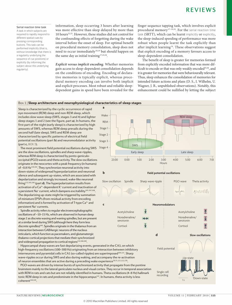
REM sleep
Nature Reviews | Neuroscience
Slow oscillation Spindle Sharp wave-ripple PGO wave Theta activity
Field potential oscillationsb
a
c
d
Neuromodulators
Slow oscillations
SWSStage 4
Stage 3
Stage 2
Stage 1
REM
Wake
23:00 0:00 1:00 2:00 3:00 4:00 5:00 6:00 7:00
Late sleepEarly sleep
Hours
Acetylcholine Acetylcholine
Noradrenaline/serotonin
Noradrenaline/serotonin
Cortisol Cortisol
Field potential
Single cellrecording
1 s
Up-state
Down-state
Serial reaction time taskA task in which subjects are required to rapidly respond to different spatial cues by pressing corresponding buttons. This task can be performed implicitly (that is, without knowledge that there is a regularity underlying the sequence of cue positions) or explicitly (by informing the subject about this underlying regularity).
information, sleep occurring 3 hours after learning was more effective than sleep delayed by more than 10 hours32,33. However, these studies did not control for the confounding effects of forgetting during the wake interval before the onset of sleep. For optimal benefit on procedural memory consolidation, sleep does not need to occur immediately18,19 but should happen on the same day as initial training17,22,24.
Explicit versus implicit encoding. Whether memories gain access to sleep-dependent consolidation depends on the conditions of encoding. Encoding of declara-tive memories is typically explicit, whereas proce-dural memory encoding can involve both implicit and explicit processes. Most robust and reliable sleep-dependent gains in speed have been revealed for the
finger sequence tapping task, which involves explicit procedural memory17–19,24. For the serial reaction time task (SRTT), which can be learnt implicitly or explicitly, the sleep-induced speeding of performance was more robust when people learnt the task explicitly than after implicit learning34. These observations suggest that explicit encoding of a memory favours access to sleep-dependent consolidation.
The benefit of sleep is greater for memories formed from explicitly encoded information that was more dif-ficult to encode or that was only weakly encoded35,36, and it is greater for memories that were behaviourally relevant. Thus, sleep enhances the consolidation of memories for intended future actions and plans (D. S., I. Wilhelm, u. Wagner, J. b., unpublished observations). Notably, this enhancement could be nullified by letting the subject
Box 1 | sleep architecture and neurophysiological characteristics of sleep stages
Sleep is characterized by the cyclic occurrence of rapid eye movement (REM) sleep and non-REM sleep, which includes slow wave sleep (SWS, stages 3 and 4) and lighter sleep stages 1 and 2 (see the figure, part a). In humans, the first part of the night (early sleep) is characterized by high amounts of SWS, whereas REM sleep prevails during the second half (late sleep). SWS and REM sleep are characterized by specific patterns of electrical field potential oscillations (part b) and neuromodulator activity (part c, BOX 3).
The most prominent field potential oscillations during SWS are the slow oscillations, spindles and sharp wave-ripples, whereas REM sleep is characterized by ponto-geniculo-occipital (PGO) waves and theta activity. The slow oscillations originate in the neocortex with a peak frequency (in humans) of ~0.8 Hz130,164. They synchronize neuronal activity into down-states of widespread hyperpolarization and neuronal silence and subsequent up-states, which are associated with depolarization and strongly increased, wake-like neuronal firing132,165,166 (part d). The hyperpolarization results from activation of a Ca2+-dependent K+ current and inactivation of a persistent Na+ current, which dampens excitability165,167,168. The depolarizing up-state might be triggered by summation of miniature EPSPs (from residual activity from encoding information) and is formed by activation of T-type Ca2+ and persistent Na+ currents.
Spindle activity refers to regular electroencephalographic oscillations of ~10–15 Hz, which are observed in human sleep stage 2 as discrete waxing and waning spindles, but are present at a similar level during SWS (although here they form less discrete spindles)169. Spindles originate in the thalamus from an interaction between GABAergic neurons of the nucleus reticularis, which function as pacemakers, and glutamatergic thalamo-cortical projections that mediate their synchronized and widespread propagation to cortical regions132,168,169.
Hippocampal sharp waves are fast depolarizing events, generated in the CA3, on which high-frequency oscillations (100–300 Hz) originating from an interaction between inhibitory interneurons and pyramidal cells in CA1 (so-called ripples) are superimposed104,121. Sharp wave-ripples occur during SWS and also during waking, and accompany the re-activation of neuron ensembles that are active during a preceding wake experience70,71,121,122,170.
PGO-waves are driven by intense bursts of synchronized activity that propagate from the pontine brainstem mainly to the lateral geniculate nucleus and visual cortex. They occur in temporal association with REM in rats and cats but are not reliably identified in humans. Theta oscillations (4–8 Hz) hallmark tonic REM sleep in rats and predominate in the hippocampus141. In humans, theta activity is less coherent144,145.
R E V I E W S
NATuRE REVIEWS | NeuroscieNce VoluME 11 | FEbRuARy 2010 | 115
© 20 Macmillan Publishers Limited. All rights reserved10

Implicit learningLearning without being aware that something is being learned.
Explicit learningLearning while being aware that something is being learned.
Memory systemsDifferent types of memory, such as declarative and non-declarative memory, are thought to be mediated by distinct neural systems, the organization of which is still a topic of debate.
execute the intended behaviour before sleep. Similarly, subjects who had been trained on two different finger-tapping sequences showed greater sleep-dependent gains in performance for the sequence for which they expected to be rewarded for optimal performance at re-testing after sleep37. Thus, a motivational tagging of memories, which probably relies on the function of the prefrontal cortex38, might signal behavioural effort and relevance and mediate the preferential consolidation of these memories.
In summary, a great number of studies indicate that sleep supports the consolidation of memory in all major memory systems, but preferentially those that are explicitly encoded and that have behavioural relevance to the indi-vidual. There is growing evidence that explicit encoding, even in procedural tasks, involves a dialogue between the prefrontal cortex and the hippocampus38–40, which also integrates intentional and motivational aspects of the task. Activity of this circuit may be crucial in mak-ing a memory susceptible to sleep-dependent memory consolidation.
Sleep changes memory representations quantitatively and qualitatively. Consolidation of memory during sleep can produce a strengthening of associations as well as qualitative changes in memory representations. Strengthening of a memory behaviourally expresses itself as resistance to interference from another similar task (‘stabilization’) and as an improvement of performance (‘enhancement’) that occurs at re-testing, in the absence of additional practice during the retention interval. The stabilizing effects of sleep have been observed in declarative41 and procedural19 memory tasks. Similarly, enhancements in performance after sleep have been shown for declarative information13,14,20 and in proce-dural tasks13,17,18,21,22,31. However, it is still controversial to what extent these improvements reflect actual per-formance ‘gains’ induced by sleep, because the measured gains depend on the pre-sleep performance used as a reference, which itself can be subject to rapid changes after training42,43.
There is a long-standing debate about whether sleep passively protects memories from decay and interfer-ence or actively consolidates fresh memory represen-tations44 (for a review see Ref. 45). Importantly, a lack of enhancement of memory performance after sleep does not preclude an active role of sleep in memory consolidation. There is strong evidence for an active con-solidating influence of sleep from behavioural studies, which indicate that sleep can lead to qualitative changes in memory46–48. For example, in one study, subjects learned single relations between different objects which, unknown to the subject, relied on an embedded hierar-chy47. When learning was followed by sleep, subjects at a re-test were better at inferring the relationship between the most distant objects, which had not been learned before. likewise, after sleep subjects more easily solved a logical calculus problem that they were unable to solve before sleep or after corresponding intervals of wakeful-ness46. of note, sleep facilitated the gain of insight into the problem only if adequate encoding of the task was ensured before sleep.
Interacting or competing memory systems? The behav-ioural findings described above show that sleep can ‘re-organize’ newly encoded memory representations, enabling the generation of new associations and the extraction of invariant features from complex stimuli, and thereby eventually easing novel inferences and insights. Re-organization of memory representations during sleep also promotes the transformation of implicit into explicit knowledge, as was shown in an SRTT which was implicitly trained but in which explicit knowledge about the underlying sequence was exam-ined during the re-test48. Following post-training sleep, subjects were better at explicitly generating the SRTT sequence. Interestingly, subjects who developed explicit sequence knowledge no longer showed the improve-ment in implicit procedural skill (that is, faster reaction times) that is normally observed after sleep, suggesting that procedural and declarative memory systems interact during sleep-dependent consolidation.
Contrasting with this view of interacting memory systems, it has also been proposed that disengagement of memory systems is an essential characteristic of sleep-dependent consolidation49. This idea derives mainly from experiments showing that declarative learning of words immediately after training of a procedural skill can block off-line improvement in that skill if the subject does not sleep between learning and re-testing, but not if the subject sleeps between learning and re-testing50. This suggests that memory systems compete and reciprocally interfere during waking, but disengage during sleep, allowing for the independent consolidation of memories in different systems. The two views might be reconciled by assuming a sequential contribution of interaction and disengagement processes to consolidation, which might be associated with different sleep stages (REM sleep and SWS), as discussed below.
Influence of sleep stages on consolidationEarly studies in rats and humans investigating whether different sleep stages have different roles in memory consolidation mainly focused on REM sleep and the consequences of REM sleep deprivation (REMD) by repeatedly waking subjects at the first signs of REM sleep. However, this approach is of limited value for logi-cal reasons and because the repeated awakenings cause stress, which itself influences memory function51,52. overall, these studies have provided mixed results52–55. of note is a recent study showing that pharmacologi-cal suppression of REM sleep by administration of anti-depressant drugs (selective noradrenaline or serotonin re-uptake inhibitors) did not impair consolidation of procedural memory56, which is in agreement with clini-cal observations that antidepressant treatment does not affect memory function57. However, such substances also exert direct effects on synaptic plasticity and synaptic forms of consolidation that could compensate for a loss of REM sleep58.
Some studies performed in rats showed that REMD is only effective during specific periods after learning — the so-called ‘REM sleep windows’54. During post-learning sleep, increases in the amount and intensity
R E V I E W S
116 | FEbRuARy 2010 | VoluME 11 www.nature.com/reviews/neuro
© 20 Macmillan Publishers Limited. All rights reserved10

Transitory sleepShort transitory periods of sleep in rats that, based on eeG criteria, can neither be classified as ReM sleep or SWS.
of REM sleep occur several hours or even days after learning, depending on the kind of task and amount of initial training54, and memory is particularly impaired if REMD coincides with these periods. of note, the mem-ory tasks used in rats are typically emotionally loaded. As there is evidence that REM sleep preferentially benefits the consolidation of emotional aspects of a memory25,27, this could partly account for the strong REMD effect observed in many animal studies53,55.
Studies in humans have compared the effects on consolidation between sleep periods with different proportions of SWS and REM sleep. In humans, SWS and REM sleep dominate the early and late part of noc-turnal sleep, respectively (BOX 1). SWS-rich, early sleep consistently benefits the consolidation of declarative memories12,13,59, whereas REM-rich sleep benefits non-declarative types of memory (that is, procedural and emotional aspects of memory)13,25,59. These results are con-sistent with the ‘dual-process hypothesis’, which assumes that SWS facilitates declarative, hippocampus-depend-ent memory and REM sleep supports non-declarative, hippocampus-independent memory6.
other studies have shown that SWS can also improve procedural skill (that is, non-declarative) memories31,60,61 and that REM sleep can also improve declarative memory62,63. Although these divergent find-ings could reflect that stimuli used in memory tasks are often not of one type of memory system, they agree with the ‘sequential hypothesis’, which argues that the optimum benefits of sleep on the consolidation of both declarative and non-declarative memory occur when SWS and REM sleep take place in succession31,64. Thus, overnight improvements in visual texture discrimina-tion correlated with both the amount of SWS in the first quarter of sleep and the amount of REM sleep in the last quarter21. Texture discrimination also improved following a short midday nap of 60–90 minutes con-taining solely SWS, but more so if the nap included both SWS and REM sleep23. Also, memory consolida-tion seems to be impaired by disruptions of the natural SWS–REM sleep cycle that left the time spent in these sleep stages unchanged65.
Intermediate sleep stages (non-REM sleep stage 2 in humans, transitory sleep in rats) can also contribute to memory consolidation66,67. For example, pharmaco-logical suppression of REM sleep in humans produced an unexpected overnight improvement in procedural skill that was correlated with increased non-REM sleep stage 2 spindle activity (see below)56. Such findings high-light the fact that it is not a particular sleep stage per se that mediates memory consolidation, but rather the neuro physiological mechanisms associated with those sleep stages, and that some of these mechanisms are shared by different sleep stages.
Core features of off-line consolidationSince the publication of Hebb’s seminal book68, memory formation has been conceptualized as a process in which neuronal activity reverberating in specific circuits pro-motes enduring synaptic changes. building on this, it is widely accepted that the consolidation process that takes
place off-line after encoding relies on the re-activation of neuronal circuits that were implicated in the encod-ing of the information. This would promote both the gradual redistribution and re-organization of memory representations to sites for long-term storage (that is, system consolidation; BOX 2) and the enduring synaptic changes that are necessary to stabilize memories (syn-aptic consolidation). The conditions that enable these two processes during sleep differ strongly between SWS and REM sleep.
Re-activation of memory traces during sleep. The finding that in rats the spatio-temporal patterns of neuronal firing that occur in the hippocampus during explora-tion of a novel environment or simple spatial tasks are re-activated in the same order during subsequent sleep was an important breakthrough in memory research69–74 (fIG. 1a, see Ref. 75 for methodological considerations on the identification of neuronal re-activations). Such neuronal re-activation of ensemble activity mostly occurs during SWS (it is rarely observed during REM sleep76,77) and during the first hours after learning (but see Ref. 78), and typically only in a minority of recorded neurons69–74. Moreover, unlike re-activations that occur during wakefulness, re-activations during SWS almost always occur in the order in which they were expe-rienced79. Compared with activity during encoding phases, re-activations during SWS seem to be noisier, less accurate and often happen at a faster firing rate71. They are also observed in the thalamus, the striatum and the neocortex72–74,78. Sleep-dependent signs of re-activation in brain regions implicated in prior learning were also shown in human neuroimaging studies80,81.
The first evidence for a causal role of re-activation during SWS in memory consolidation came from a study in humans learning spatial locations in the presence of an odour15. Re-exposure to the odour during SWS, but not REM sleep, enhanced the spatial memories (fIG. 1b) and induced stronger hippocampal activation than during wakefulness, indicating that during SWS hippocampal networks are particularly sensitive to inputs that can re-activate memories (fIG. 1c). It is assumed that the re-activations during system consolidation stimulate the redistribution of hippocampal memories to neocorti-cal storage sites, although this has not been directly demonstrated yet82,83.
Synaptic consolidation. In addition to system consoli-dation (BOX 2), consolidation involves the strengthening of memory representations at the synaptic level (syn-aptic consolidation)84,85. long-term potentiation (lTP) is considered a key mechanism of synaptic consolida-tion, but it is unclear whether memory re-activation during sleep promotes the redistribution of memories by inducing new lTP (at long-term storage sites not involved at encoding) or whether re-activation merely enhances the maintenance of lTP that was induced during encoding.
lTP can be induced in the hippocampus during REM sleep but less reliably so during SWS86. lTP induc-tion in the hippocampus or neocortex during SWS is
R E V I E W S
NATuRE REVIEWS | NeuroscieNce VoluME 11 | FEbRuARy 2010 | 117
© 20 Macmillan Publishers Limited. All rights reserved10

Nature Reviews | Neuroscience
Long-term store (slow learning)
Encoding
Encoding Consolidation
Temporary store (fast learning)
Immediate early genesGenes that encode transcription factors that are induced within minutes of raised neuronal activity without requiring a protein signal. Immediate-early gene activation is, therefore, used as an indirect marker of neuronal activation. The immediate early genes Arc and Egr1 (zif268) are associated with synaptic plasticity.
Hebbian plasticityRefers to the functional changes at synapses that increase the efficacy of synaptic transmission and occurs when the presynaptic neuron repeatedly and persistently stimulates the postsynaptic neuron.
Spike-time dependent plasticityRefers to the functional changes at synapses that alter the efficacy of synaptic transmission depending on the relative timing of pre- and postsynaptic firing (‘spiking’). The synaptic connection is strengthened if the presynaptic neuron fires shortly before the postsynaptic neuron, but is weakened if the sequence of firing is reversed. probably temporally restricted to the up-states of the
slow oscillation and its concurrent phenomena of rip-ples and spindles87,88 (see BOX 1 and below). Indeed, in neocortical slices, stimulation that mimicked neuronal activity during SWS could induce long-term depression (lTD)89 or lTP87 depending on the pattern of stimula-tion (rhythmic bursts or spindle-like trains, respectively). lTP maintenance in the rat hippocampus, but not in the medial prefrontal cortex, was impaired if induction was followed by REMD90. In humans, sleep strengthened lTP-like plasticity that had been induced in the neocor-tex by transcranial magnetic stimulation (TMS) prior to sleeping91.
Globally (meaning measured in whole-brain or large cortical samples) sleep suppresses the molecular sig-nals that mediate lTP-related synaptic remodelling but enhances lTD-related signalling, and this effect seems to be mediated by SWS92–95. This observation, however, does not preclude that lTP occurs during sleep (during SWS or REM sleep) in specific regions, for example in those that were engaged in memory encoding prior to sleeping. In rats, both induction of hippocampal lTP and exposure to a novel tactile experience during wak-ing increased the expression of the plasticity-related immediate early genes (IEGs) Arc and Egr1 (which are implicated in lTP) during subsequent sleep, mainly in cortical areas that were the most activated by the novel experience, and this effect seemed to be mediated by REM sleep96–98. Investigations in visual cortex in cats and humans have demonstrated that sleep-dependent
plasticity depends on the activation of glutamatergic NMDA (N-methyl-d-aspartate) receptors and associ-ated cAMP-dependent protein kinase A (PKA), and on AMPA (α-amino-3-hydroxy-5-methyl-4-isoxazole propionic acid) receptor activation, that is, the post-synaptic machinery that is crucial for the induction and maintenance of lTP99–102. These findings indicate that local, off-line re-activation of specific glutamatergic circuits supports both lTP induction and maintenance, and the molecular processes underlying synaptic con-solidation. Moreover, these processes probably occur preferentially during REM sleep, although they are likely to be triggered by the re-activations that occur during prior SWS (see below). Evidence about how lTP induction and maintenance is linked to specific sleep stages is presently scarce, but based on the available data it is tempting to speculate that SWS supports the re-activation of new memories (system consolidation) and thus, could initialize lTP and prime the relevant networks for synaptic consolidation during subsequent REM sleep. This idea seems to be supported by elec-troencephalographic (EEG) rhythms that characterize these sleep stages.
sleep-specific field potential oscillationsSleep stages are characterized by specific electrical field potential rhythms that temporally coordinate information transfer between brain regions and might support Hebbian and spike-time-dependent plasticity103,104.
Box 2 | The two-stage model of memory consolidation
A key issue of long-term memory formation, the so-called stability–plasticity dilemma, is the problem of how the brain’s neuronal networks can acquire new information (plasticity) without overriding older knowledge (stability). Many aspects of events experienced during waking represent unique and irrelevant information that does not need to be stored long term. The two-stage model of memory offers a widely accepted solution to this dilemma2,7,85,152 (see the figure). The model assumes two separate memory stores: one store allows learning at a fast rate and serves as an intermediate buffer that holds the information only temporarily; the other store learns at a slower rate and serves as the long-term store. Initially, new events are encoded in parallel in both stores. In subsequent periods of consolidation, the newly encoded memory traces are repeatedly re-activated in the fast-learning store, which drives concurrent re-activation in the slow-learning store, and thereby new memories become gradually redistributed such that representations in the slow-learning, long-term store are strengthened. Through the repeated re-activation of new memories, in conjunction with related and similar older memories, the fast-learning store acts like an internal ‘trainer’ of the slow-learning store to gradually adapt the new memories to the pre-existing network of long-term memories. This process also promotes the extraction of invariant repeating features from the new memories. As both stores are used for encoding information, in order to prevent interference, the re-activation and redistribution of memories take place off-line (during sleep) when no encoding occurs. Because in this model consolidation involves the redistribution of representations between different neuronal systems that is, the fast- and slow-learning stores, it has been termed ‘system consolidation’. For declarative memories, the fast- and slow-learning stores are represented by the hippocampus and neocortex, respectively. Figure modified, with permission, from Ref. 85 © (2005) Macmillan Publishers Ltd. All rights reserved.
R E V I E W S
118 | FEbRuARy 2010 | VoluME 11 www.nature.com/reviews/neuro
© 20 Macmillan Publishers Limited. All rights reserved10

Nature Reviews | Neuroscience
x = 11 y = -15
3.5
6.0
t21
c
a
b
Run Sleep Run Sleep
Sleep RetrievalLearning
Cortex Hippocampus
Cel
l num
ber
Cel
l num
ber
0
7
0
7
0
5
0
5
1 s0.5 s 0.2 s1 s
WakeREM
Stage 1Stage 2Stage 3Stage 4
During SWS
Reca
lled
card
loca
tions
No odour Odour No odour Odour No odour Odour0
80
90
100
20:00 24:00 04:00 08:00
During REM During waking%
0
80
90
100%
0
80
90
100%***
Odour/vehicle
Odour
Odour re-exposure
Retrieval performance
Odour
Time of day
Field potentials associated with SWS. Neocortical slow oscillations, thalamo-cortical spindles and hippocampal ripples have been associated with memory consolidation during SWS (BOX 1). The neocortical slow oscillations (of <1 Hz), by globally inducing up- and down-states of neu-ronal activity, are thought to provide a supra-ordinate temporal frame for the dialogue between the neocortex and subcortical structures that is necessary for redistrib-uting memories for long-term storage8,105,106. The ampli-tude and slope of the slow oscillations are increased when SWS is preceded by specific learning experi-ences60,107,108 and decreased when the encoding of infor-mation was prevented109. These changes occur locally, in the cortical regions that were involved in encoding, and can also be induced in humans by potentiating synap-tic circuits through TMS91,110,111. Inducing slow oscilla-tions during non-REM sleep by transcranial electrical stimulation using slow (0.75 Hz) but not fast (5 Hz) oscillating potential fields improved the consolidation of hippocampus-dependent but not hippocampus-independent (procedural) memories112, indicating that slow oscillations have a causal role in the consolidation of hippocampus-dependent memories.
Thalamo-cortical spindles seem to prime cortical networks for the long-term storage of memory repre-sentations. Repeated spindle-associated spike discharges can trigger lTP87 and synchronous spindle activity occurs preferentially at synapses that were potentiated during encoding113. Studies in rats and humans showed increases in spindle density and activity during non-REM sleep and SWS after learning of both declarative tasks and procedural motor skills20,108,114–118. In some studies these increases correlated with the post-sleep memory improvement30,119,120 and were localized to the cortical areas that were activated during encoding, for example, in the prefrontal cortex after encoding of dif-ficult word pairs117,119, the parietal cortex after a visuo-spatial task120 and the contralateral motor cortex after finger motor-skill learning30.
Hippocampal sharp wave-ripples accompany the sleep-associated re-activation of hippocampal neuron ensembles that were active during the preceding awake experience70,71,121,122. The occurrence of sharp wave-ripples is facilitated in previously potentiated synap-tic circuits123 and sharp wave-ripples might promote synaptic potentiation88,124. During an individual ripple event only a small subpopulation of pyramidal cells fire — the subpopulation varies between successive ripples, indicating modulation of select neuronal circuits121,125. In rats, learning of odour–reward associations pro-duced a robust increase in the number and size of ripple events for up to two hours during subsequent SWS126. In humans (epileptic patients) the consolidation of picture memories that were acquired before a nap correlated with the number of ripples recorded from the peri- and entorhinal cortex, which are important output regions of the hippocampus127. Selective disruption of ripples by electrical stimulation during the post-learning rest periods in rats impaired formation of long-lasting spatial memories128, suggesting that ripples have a causal role in sleep-associated memory consolidation.
Figure 1 | Memory re-activation during slow wave sleep (sWs). a | In awake rats running on a circular track (Run), neurons in the sensory cortex and hippocampus fire in a characteristic sequential pattern. Each row represents an individual cell and each mark in the upper parts of the diagrams indicates a spike; the curves in the lower parts indicate the respective average firing patterns of the cells. During subsequent slow wave sleep (SWS) (Sleep), temporal firing sequences observed in the cell assemblies during running re-appear both in the cortex and in the hippocampus72. b | Human subjects learned a two-dimensional object location task on a computer while an odour was presented as a context stimulus. Re-exposure to the odour specifically during subsequent SWS enhanced retention performance (recalled card locations) when tested the next day. There was no enhancement in retention when no association was formed between object locations and odour (that is, odour presentation during SWS but not during learning) or when odour re-exposure occurred during rapid eye movement (REM) sleep or waking15. c | When participants slept in an fMRI scanner after learning in the presence of odour, re-exposure to the odour during SWS activated the left anterior hippocampus (left) and neocortical regions like the retrosplenial cortex (right), which was not observed without odour presentation during prior learning7. Part a is modified, with permission, from Ref. 72 © 2007 Macmillan Publishers Ltd. All rights reserved; part b is modified, with permission, from Ref. 15 © 2007 American Association for the Advancement of Science; part c modified, with permission, from Ref. 7 © 2007 Elsevier.
R E V I E W S
NATuRE REVIEWS | NeuroscieNce VoluME 11 | FEbRuARy 2010 | 119
© 20 Macmillan Publishers Limited. All rights reserved10

Up- and down-statesThe slow oscillations that predominate eeG activity during SWS are characterized by alternating states of neuronal silence with an absence of spiking activity and membrane hyperpolarization in all cortical neurons (‘down-state’) and strongly increased wake-like firing of large neuronal populations and membrane depolarization (‘up-state’).
Interestingly, there is a fine-tuned temporal relation-ship between the occurrence of slow oscillations, spin-dles and sharp wave-ripples during SWS that coordinates the bidirectional information flow between the neocor-tex and the hippocampus. With some exceptions (which are probably due to methodological differences129) a consistent finding in humans, cats, rats and mice is that spindle activity and ripples increase during the up-state and become suppressed during the down-state of a slow oscillation105,129–132. The top–down control of neuronal activity by neocortical slow oscillations probably extends to activity in other brain regions that are also relevant to memory consolidation, such as the noradrenergic burst activity of the locus coeruleus133,134. Sharp wave-ripple complexes are also temporally coupled to sleep spindles105,135,136, with individual ripple events becom-ing nested in individual spindle troughs135. It has been suggested that such ripple-spindle events provide a mechanism for a fined-tuned hippocampal-neocortical information transfer, whereby ripples and associated hippocampal memory re-activations feed exactly into the excitatory phases of the spindle cycle8,105,137,138. In this scenario, the feed-forward control of slow oscil-lations over ripples and spindles enables transferred information to reach the neocortex during widespread depolarization (during the up-state), that is, a state that favours the induction of persistent synaptic changes, eventually resulting in the storage of the information in the cortex. The extent to which the grouping effect of the slow oscillation on hippocampal activity is associated with transfer of memory-specific information in the opposite direction (from cortex to hippocampus), is currently unclear.
Field potentials associated with REM sleep. Ponto-geniculo-occipital (PGo) waves and the EEG theta rhythm seem to support REM sleep-dependent consoli-dation processes (BOX 1). The significance of PGo-waves for memory consolidation is indicated by findings in rats of a robust increase in REM sleep PGo-wave density for 3–4 hours following training on an active avoid-ance task67,139,140. The increase was proportional to the improvement in post-sleep task performance, and was associated with increased activity of plasticity-related IEGs and brain-derived neurotrophic factor (bdnf) in the dorsal hippocampus within 3 hours following training140.
The theta (4–8 Hz) oscillations that characterize REM sleep in rats are also thought to contribute to consolida-tion, based mainly on the finding that theta activity during waking occurs during the encoding of hippocampus- dependent memories141. However, evidence for this assumption is scarce. There is evidence of neuronal re-play of memories in the hippocampus during REM sleep-associated theta activity76,77. Place cells encoding a familiar route were re-activated preferentially during the troughs of theta oscillations during post-training REM sleep, whereas cells encoding novel sites fired during the peaks77. As lTP induction in hippocampal CA1 cells during theta activity depends on the phase of burst activ-ity142, this finding is consistent with the idea that REM
sleep de-potentiates synaptic circuits that encode famil-iar events but potentiates synaptic circuits that encode novel episodes77. In humans, neocortical theta activity was enhanced during REM sleep following learning of word pairs62. Theta activity specifically over the right prefrontal cortex was correlated with the consolidation of emotional memories27. by contrast, mice exhibited reduced REM sleep theta activity after fear condition-ing143. Thus, although overall there is some evidence for an involvement of theta activity in memory processing during sleep, its specific contribution to consolidation is obscure at present.
Theta activity occurring in conjunction with activity in other EEG frequencies points to another important feature that is relevant to memory processing: during REM sleep, EEG activity in a wide range of frequencies, including theta, shows reduced coherence between lim-bic-hippocampal and thalamo-cortical circuits than dur-ing SWS or waking144,145. likewise, >40 Hz gamma band activity shows reduced coherence between CA3 and CA1 during tonic REM sleep146. These findings suggest that memory systems become disengaged during REM sleep49, possibly as a pre-requisite for establishing effec-tive local processes of synaptic consolidation in these systems (see below).
synaptic homeostasis versus system consolidationThere are currently two hypotheses for the mecha-nisms underlying the consolidation of memory during sleep (fIG. 2). The synaptic homeostasis hypothesis11,147 assumes that consolidation is a by-product of the glo-bal synaptic downscaling that occurs during sleep. The active system consolidation hypothesis proposes that an active consolidation process results from selective re-activation of memories during sleep2,8. The two models are not mutually exclusive; indeed, the hypoth-esized processes probably act in concert to optimize the memory function of sleep.
Synaptic homeostasis. According to the synaptic home-ostasis hypothesis, information encoding during wake-fulness leads to a net increase in synaptic strength in the brain. Sleep would serve to globally downscale synaptic strength to a level that is sustainable in terms of energy and tissue volume demands and that allows for the re-use of synapses for future encoding92,94. Slow oscillations are associated with downscaling: they show maximum amplitudes at the beginning of sleep when overall syn-aptic strength is high, due to information uptake dur-ing encoding prior to sleep, and decrease in amplitude across SWS cycles as a result of the gradual synaptic de-potentiation. Memories become relatively enhanced as downscaling is assumed to be proportional in all syn-apses, nullifying weak potentiation and thus improv-ing the signal-to-noise ratio for the synapses that were strongly potentiated during prior waking147 (fIG. 2a).
However, there is no clear evidence on how slow oscillations might induce synaptic downscaling. The low levels of excitatory neurotransmitters during SWS (BOX 3) and the sequence of depolarization (up-states) and hyperpolarization (down-states) of slow oscillations
R E V I E W S
120 | FEbRuARy 2010 | VoluME 11 www.nature.com/reviews/neuro
© 20 Macmillan Publishers Limited. All rights reserved10

Nature Reviews | Neuroscience
Waking – Synaptic potentiation
SWS REM
Sleep – Synaptic downscalinga
b
W=100
W=80W=120
W=150W=5
W=100W=100
Time
Synaptic plasticityNeocortexSlow oscillations
ThalamusSpindles
HippocampusSharp wave-ripples
Ca2+
CaMKIIPKA
IEG
AMPAR
NMDAR
LTP
Synchronousfeedback
Syna
ptic
str
engt
h
Synchronizing feed-forwardeffect
at a frequency of <1 Hz might specifically promote the de-potentiation of synapses148. Indeed, slow oscillations and the associated activation of T-type Ca2+ channels seem to favour lTD over lTP89; however, thalamo-cortical spindles and hippocampal ripples nesting in depolarizing up-states of slow oscillations support lTP87,88,124.
In addition, although the expression of markers of synaptic potentiation (such as plasticity-related IEGs) is globally reduced after a period of sleep, it is increased in specific regions, particularly if sleep was preceded by a learning experience78,96,98, indicating that synaptic poten-tiation might still take place during sleep. Consistent with downscaling, some neuroimaging studies (which measure relative changes in brain activation) have shown reduced task-related activity in cortical regions after sleep (e.g. Ref. 149), but these reductions were accom-panied by increases in activity in other regions82,83,149,150. Also, global synaptic downscaling implicates that weakly encoded memories are forgotten, which contrasts with behavioural evidence indicating either no or, under certain conditions, a greater benefit from sleep for weakly than strongly encoded memories35,36. Therefore,
downscaling per se does not explain key features of sleep-dependent consolidation. However, the synaptic downs-caling model explains a second memory-related function of sleep, namely that sleep pro-actively facilitates the encoding of new information during subsequent wake-fulness through the de-potentiation of synapses that had become saturated during preceding wakefulness (this topic is beyond the scope of this Review)151.
Active system consolidation. This concept originated from the standard two-stage model of consolidation pro-posed for declarative memory2,7,85,121,152 (BOX 2; fIG. 2b), but might also account for consolidation in other memory systems8. It is assumed that in the waking brain events are initially encoded in parallel in neocortical networks and in the hippocampus. During subsequent periods of SWS the newly acquired memory traces are repeatedly re-activated and thereby become gradually redistributed such that connections within the neocortex are strength-ened, forming more persistent memory representations. Re-activation of the new representations gradually adapt them to pre-existing neocortical ‘knowledge networks’,
Figure 2 | synaptic homeostasis versus active system consolidation. The synaptic homeostasis hypothesis (a) proposes that due to encoding of information during waking, synapses become widely potentiated (large yellow nerve ending), resulting in a net increase in synaptic strength (W = synaptic weight). The small nerve ending represents a new synapse and the unfilled nerve ending is not activated and therefore does not increase in weight. The slow oscillations during subsequent SWS serve to globally downscale synaptic strength (burgundy nerve endings). Thereby, weak connections are eliminated, whereas the relative strength of the remaining connections is preserved. Thus, a memory is enhanced as a consequence of an improved signal-to-noise ratio after downscaling. The active system consolidation model (b) assumes that events during waking are encoded in both neocortical and hippocampal networks. During subsequent slow wave sleep (SWS), slow oscillations drive the repeated re-activation of these representations in the hippocampus, in synchrony with sharp wave-ripples and thalamo-cortical spindles (synchronizing feed-forward effect of the slow oscillation up-state). By synchronizing these events the slow oscillations support the formation of ripple-spindle events, which enable an effective hippocampus-to-neocortex transfer of the re-activated information. Arrival of the hippocampal memory output at cortical networks, coinciding with spindle activity during the depolarizing slow oscillation up-state predisposes these networks to persisting synaptic plastic changes (for example, expression of immediate early genes (IEG) through Ca2+/calmodulin-dependent protein kinase II (CaMKII) and protein kinase A (PKA) activation) that are supported primarily by subsequent rapid eye movement (REM) sleep. AMPAR, α-amino-3-hydroxy-5-methyl-4-isoxazole propionic acid receptor; LTP, long-term potentiation; NMDAR, N-methyl-d-aspartate receptor. Part a is modified, with permission, from Ref. 147 © 2006 Elsevier; part b is modified, with permission, from Ref. 5 © 2006 Sage publications.
R E V I E W S
NATuRE REVIEWS | NeuroscieNce VoluME 11 | FEbRuARy 2010 | 121
© 20 Macmillan Publishers Limited. All rights reserved10

thereby promoting the extraction of invariant repeat-ing features and qualitative changes in the memory representations2,7.
Corroborating this concept, studies showed that memory re-activation during post-learning SWS and hippocampal ripples accompanying this re-activation have a causal role in consolidation15,128. Re-activation in hippocampal networks seems to be enabled by the low cholinergic tone that characterizes SWS153–155 (BOX 3). Moreover, there is evidence that the re-activation and redistribution of memories during SWS is regulated by a dialogue between the neocortex and the hippoc-ampus that is essentially under feed-forward control of the slow oscillations, which provide a temporal frame.
The depolarizing cortical up-states repetitively drive the re-activation of memory traces in hippocampal circuits in parallel with thalamo-cortical spindles and activity from other regions (for example, noradrenergic locus coeruleus bursts, see BOX 3). This enables synchronous feedback from these structures to the neocortex during the slow oscillation up-state, which is probably a pre-requisite for the formation of more persistent traces in neocortical networks8,106. Consistent with this concept, neuronal re-activations in the timeframe of cortical slow oscillations have been demonstrated, in which hippocam-pal re-play leads re-activation in the neocortex72,122 (and also in other structures like the striatum156). Moreover, slow oscillations drive the ripples that accompany hip-pocampal re-activation, thus allowing for the formation of spindle-ripple events as a mechanism for effective hippocampus-to-neocortex information transfer105,137,138 (fIG. 2b). Spindles reaching the neocortex during slow oscillation up-states probably act to prime specific neu-ronal networks, for example, by stimulating Ca2+ influx, for subsequent synaptic plastic processes87,157.
The concept of active system consolidation during SWS integrates a central finding from behavioural stud-ies, namely that post-learning sleep not only strength-ens memories but also induces qualitative changes in their representations and so enables the extraction of invariant features from complex stimulus materials, the forming of new associations and, eventually, insights into hidden rules46–48. The concept of a redistribution of memories during sleep has been corroborated by human brain imaging studies82,83,149,150,158. Interestingly, in these studies, hippocampus-dependent memories were particularly redistributed to medial prefrontal cortex regions82,83,122 that also contribute to the generation of slow oscillations159,160. These regions not only have a key role in the recall and binding of these memories once they are stored for the long term85, but also, together with the hippocampus, form a loop that supports the explicit encoding of information. As mentioned above, behavioural data indicate that sleep does not benefit all memories equally, but seems to preferentially consoli-date explicitly encoded information34. In this context, the prefrontal–hippocampal system might provide a selec-tion mechanism that determines which memory enters sleep-dependent consolidation.
A role for ReM sleep in synaptic consolidationThe active system consolidation hypothesis leaves open one challenging issue: although it explains a re-activation-dependent temporary enhancement and integration of newly encoded memories into the network of pre-existing long-term memories, active system consoli-dation alone does not explain how post-learning sleep strengthens memory traces and stabilizes underlying synaptic connections in the long term. Hence, sleep pre-sumably also supports a synaptic form of consolidation for stabilizing memories and this could be the function of REM sleep.
The view that synaptic consolidation is promoted by REM sleep is supported by the molecular and elec-trophysiological events that characterize this stage.
Box 3 | Neuromodulators
The specific neurochemical milieu of neurotransmitters and hormones differs strongly between slow wave sleep (SWS) and rapid eye movement (REM) sleep. Some of these neuromodulators contribute to memory consolidation. Interestingly, the most prominent contributions to memory processing seem to originate from the cholinergic and monoaminergic brainstem systems that are also involved in the basic regulation of sleep171.
sWsCholinergic activity is at a minimum during SWS; this is thought to enable the spontaneous re-activation of hippocampal memory traces and information transfer to the neocortex by reducing the tonic inhibition of hippocampal CA3 and CA1 feedback neurons8,154,155. Accordingly, increasing cholinergic tone during SWS-rich sleep (using physostigmine) blocked the sleep-dependent consolidation of hippocampus-dependent word-pair memories153. Conversely, blocking the high cholinergic tone in awake subjects improved consolidation but impaired the encoding of new information172, suggesting that acetylcholine serves as a switch between modes of brain activity, from encoding during wakefulness to consolidation during SWS154,155. This dual function of acetylcholine seems to be complemented by glucocorticoids (cortisol in humans), the release of which is also at a minimum during SWS. Glucocorticoids block the hippocampal information flow to the neocortex, and if the level of glucocorticoids is artificially increased during SWS, the consolidation of declarative memories is blocked173,174.
Noradrenergic activity is at an intermediate level during SWS, and seems to be related to slow oscillations. In rats, phasic burst firing in the locus coeruleus (the brain’s main source of noradrenaline) can be entrained by slow oscillations in the frontal cortex, with a phase-delay of ~300 ms133. It is possible that such bursts enforce plasticity-related immediate early gene (IEG) activity in the neocortex93,95, and thereby support at the synaptic level the stabilization of newly formed memory representations. In humans, the consolidation of odour memories was impaired after pharmacological suppression of noradrenergic activity during SWS-rich sleep and improved after increasing noradrenaline availability (S. Gais, B. Rasch, J.C. Dahmen, S.J. Sara and J. B., unpublished observations).
reM sleepCholinergic activity during REM sleep is similar or higher than during waking. This high cholinergic activity might promote synaptic consolidation by supporting plasticity-related IEG activity162 and the maintenance of long-term potentiation163. Accordingly, blocking muscarinic receptors in rats by scopolamine during REM sleep impaired memory in a radial arm maze task175. In humans, blocking cholinergic transmission during REM-rich sleep prevented gains in finger motor skill176. Conversely, enhancing cholinergic tone during post-training REM-rich sleep improved consolidation of a visuo-motor skill177.
Noradrenergic and serotonergic activity reaches a minimum during REM sleep, but it is unclear whether this contributes to consolidation. It has been proposed that the release from inhibitory noradrenergic activity during REM sleep enables the re-activation of procedural and emotional aspects of memory (in cortico-striatal and amygdalar networks, respectively), thus supporting memory consolidation154,178. However, enhancing noradrenergic activity during post-learning REM sleep in humans failed to impair procedural memory consolidation56.
R E V I E W S
122 | FEbRuARy 2010 | VoluME 11 www.nature.com/reviews/neuro
© 20 Macmillan Publishers Limited. All rights reserved10

Nature Reviews | Neuroscience
Waking SWS REM sleep
Sequential
Time
Long-term store
Encoding
Temporary store
Active systemconsolidation
Synapticconsolidation
Although any links between sleep phases of short dura-tion and gene expression are difficult to demonstrate for methodological reasons, several studies suggest that REM sleep, unlike SWS, is associated with an upregu-lation of plasticity-related IEG activity (RefS 97,98,139). The upregulation depends on learning experience dur-ing prior wakefulness and is localized to brain regions involved in prior learning97,98,139. Interestingly, this IEG activity is correlated with EEG spindle activity during preceding SWS98. Spindles (which, as discussed above, represent a candidate mechanism that tags networks for the neocortical storage of memories during system con-solidation) per se do not induce IEG activity, but might prime particular brain areas for it, possibly by enhanc-ing Ca2+ concentrations in select subgroups of cortical neurons87,157. The activity of plasticity-related early genes depends on cholinergic tone161,162, which is enhanced to wake-like levels during REM sleep (BOX 3). Cholinergic activation strengthens the maintenance of lTP in the hip-pocampus-medial prefrontal cortex pathway163, a main route for transferring memories during SWS-dependent system consolidation82,83,122,136. Electrophysiological sig-natures of REM sleep, such as PGo waves, are increased during post-learning sleep and might promote IEG activity and memory consolidation140. EEG recordings
indicate that during REM sleep brain activation is as high as during waking, but less coherent between differ-ent regions and noisier144–146. This high level of activation could act non-specifically to amplify local synaptic plas-ticity in an environment that, compared with the awake state, is almost entirely unbiased by external stimulus inputs. The disentangled, localized nature of synaptic consolidation might also explain why REM sleep alone fails to improve declarative memory consolidation: this process essentially relies on the integration of features from different memories in different memory sys-tems and corresponding information transfer between widespread brain areas, that is, SWS-dependent system consolidation.
Conclusions and future directionsSWS and REM sleep have complementary functions to optimize memory consolidation (fIG. 3). During SWS — characterized by slow oscillation-induced widespread synchronization of neuronal activity — active system consolidation integrates newly encoded memories with pre-existing long-term memories, thereby inducing con-formational changes in the respective representations. System consolidation (which preferentially affects explic-itly encoded, behaviourally relevant information) acts in
Figure 3 | sequential contributions of sWs and reM sleep to memory consolidation in a two-stage memory system. During waking, memory traces are encoded in both the fast-learning, temporary store and the slow-learning, long-term store (in the case of declarative memory these are represented by the hippocampus and neocortex, respectively). During subsequent slow wave sleep (SWS), active system consolidation involves the repeated re-activation of the memories newly encoded in the temporary store, which drives concurrent re-activation of respective representations in the long-term store together with similar associated representations (dotted lines). This process promotes the re-organization and integration of the new memories in the network of pre-existing long-term memories. System consolidation during SWS acts on the background of a global synaptic downscaling process (not illustrated) that prevents saturation of synapses during re-activation (or during encoding in the subsequent wake-phase). During ensuing rapid eye movement (REM) sleep, brain systems act in a ‘disentangled’ mode that is also associated with a disconnection between long-term and temporary stores. This allows for locally encapsulated processes of synaptic consolidation, which strengthen the memory representations that underwent system consolidation (that is, re-organization) during prior SWS (thicker lines). In general, memory benefits optimally from the sequence of SWS and REM sleep. However, declarative memory, because of its integrative nature (it binds features from different memories in different memory systems), benefits more from SWS-associated system consolidation, whereas procedural memories, because of their specificity and discrete nature, might benefit more from REM sleep-associated synaptic consolidation in localized brain circuits. Figure modified, with permission, from Ref. 85 © 2005 Macmillan Publishers Ltd. All rights reserved.
R E V I E W S
NATuRE REVIEWS | NeuroscieNce VoluME 11 | FEbRuARy 2010 | 123
© 20 Macmillan Publishers Limited. All rights reserved10
concert with global synaptic downscaling, which serves mainly to preclude the saturation of synaptic networks. Ensuing REM sleep — characterized by de-synchroniza-tion of neuronal networks, which possibly reflects a disen-gagement of memory systems — might act to stabilize the transformed memories by enabling undisturbed synaptic consolidation. Although REM sleep has been suspected for a long time to have a key role in memory consolida-tion, research has paid little attention to the fact that REM sleep naturally follows SWS. This points to complementing
contributions of sequential SWS and REM sleep to mem-ory consolidation — an idea that was originally proposed in the sequential hypothesis64. This Review revives this idea by indicating an essential role of SWS in system con-solidation that might be complemented by the synaptic consolidation taking place during REM sleep. However, direct evidence of this is scarce at present65. Specifying the role of REM sleep, as an integral part of this sequence, in synaptic consolidation will undoubtedly pose a particular challenge to future research.
1. Siegel, J. M. Sleep viewed as a state of adaptive inactivity. Nature Rev. Neurosci. 10, 747–753 (2009).
2. McClelland, J. L., McNaughton, B. L. & O’Reilly, R. C. Why there are complementary learning systems in the hippocampus and neocortex: insights from the successes and failures of connectionist models of learning and memory. Psychol. Rev. 102, 419–457 (1995).
3. Jenkins, J. G. & Dallenbach, K. M. Obliviscence during sleep and waking. Am. J. Psychol. 35, 605–612 (1924).
4. Stickgold, R. Sleep-dependent memory consolidation. Nature 437, 1272–1278 (2005).
5. Born, J., Rasch, B. & Gais, S. Sleep to remember. Neuroscientist 12, 410–424 (2006).
6. Maquet, P. The role of sleep in learning and memory. Science 294, 1048–1052 (2001).
7. Rasch, B. & Born, J. Maintaining memories by reactivation. Curr. Opin. Neurobiol. 17, 698–703 (2007).
8. Marshall, L. & Born, J. The contribution of sleep to hippocampus-dependent memory consolidation. Trends Cogn. Sci. 11, 442–450 (2007).
9. Robertson, E. M., Pascual-Leone, A. & Miall, R. C. Current concepts in procedural consolidation. Nature Rev. Neurosci. 5, 576–582 (2004).
10. Smith, C. Sleep states and memory processes in humans: procedural versus declarative memory systems. Sleep Med. Rev. 5, 491–506 (2001).
11. Crick, F. & Mitchison, G. The function of dream sleep. Nature 304, 111–114 (1983).
12. Barrett, T. R. & Ekstrand, B. R. Effect of sleep on memory. 3. Controlling for time-of-day effects. J. Exp. Psychol. 93, 321–327 (1972).
13. Plihal, W. & Born, J. Effects of early and late nocturnal sleep on declarative and procedural memory. J. Cogn. Neurosci. 9, 534–547 (1997).The first paper to show that SWS preferentially consolidates declarative memories, whereas REM sleep primarily supports procedural memories.
14. Tucker, M. A. et al. A daytime nap containing solely non-REM sleep enhances declarative but not procedural memory. Neurobiol. Learn. Mem. 86, 241–247 (2006).
15. Rasch, B., Buchel, C., Gais, S. & Born, J. Odor cues during slow-wave sleep prompt declarative memory consolidation. Science 315, 1426–1429 (2007).Odours associated with the encoding of visuo-spatial memories were used as cues during post-learning SWS to re-activate the memories. The memory enhancement produced by this re-activation compellingly demonstrates a causal role of re-activations for sleep-dependent consolidation.
16. Lahl, O., Wispel, C., Willigens, B. & Pietrowsky, R. An ultra short episode of sleep is sufficient to promote declarative memory performance. J. Sleep Res. 17, 3–10 (2008).
17. Fischer, S., Hallschmid, M., Elsner, A. L. & Born, J. Sleep forms memory for finger skills. Proc. Natl Acad. Sci. USA 99, 11987–11991 (2002).
18. Walker, M. P. et al. Sleep and the time course of motor skill learning. Learn. Mem. 10, 275–284 (2003).
19. Korman, M. et al. Daytime sleep condenses the time course of motor memory consolidation. Nature Neurosci. 10, 1206–1213 (2007).
20. Gais, S., Molle, M., Helms, K. & Born, J. Learning-dependent increases in sleep spindle density. J. Neurosci. 22, 6830–6834 (2002).
21. Stickgold, R., Whidbee, D., Schirmer, B., Patel, V. & Hobson, J. A. Visual discrimination task improvement: A multi-step process occurring during sleep. J. Cogn. Neurosci. 12, 246–254 (2000).
22. Stickgold, R., James, L. & Hobson, J. A. Visual discrimination learning requires sleep after training. Nature Neurosci. 3, 1237–1238 (2000).
23. Mednick, S., Nakayama, K. & Stickgold, R. Sleep-dependent learning: a nap is as good as a night. Nature Neurosci. 6, 697–698 (2003).
24. Walker, M. P., Brakefield, T., Hobson, J. A. & Stickgold, R. Dissociable stages of human memory consolidation and reconsolidation. Nature 425, 616–620 (2003).
25. Wagner, U., Gais, S. & Born, J. Emotional memory formation is enhanced across sleep intervals with high amounts of rapid eye movement sleep. Learn. Mem. 8, 112–119 (2001).
26. Payne, J. D., Stickgold, R., Swanberg, K. & Kensinger, E. A. Sleep preferentially enhances memory for emotional components of scenes. Psychol. Sci. 19, 781–788 (2008).
27. Nishida, M., Pearsall, J., Buckner, R. L. & Walker, M. P. REM sleep, prefrontal theta, and the consolidation of human emotional memory. Cereb. Cortex 19, 1158–1166 (2009).
28. Wagner, U., Hallschmid, M., Rasch, B. & Born, J. Brief sleep after learning keeps emotional memories alive for years. Biol. Psychiat. 60, 788–790 (2006).
29. Diekelmann, S., Wilhelm, I. & Born, J. The whats and whens of sleep-dependent memory consolidation. Sleep Med. Rev. 13, 309–321 (2009).
30. Nishida, M. & Walker, M. P. Daytime naps, motor memory consolidation and regionally specific sleep spindles. PLoS ONE. 2, e341 (2007).
31. Gais, S., Plihal, W., Wagner, U. & Born, J. Early sleep triggers memory for early visual discrimination skills. Nature Neurosci. 3, 1335–1339 (2000).
32. Gais, S., Lucas, B. & Born, J. Sleep after learning aids memory recall. Learn. Mem. 13, 259–262 (2006).
33. Talamini, L. M., Nieuwenhuis, I. L., Takashima, A. & Jensen, O. Sleep directly following learning benefits consolidation of spatial associative memory. Learn. Mem. 15, 233–237 (2008).
34. Robertson, E. M., Pascual-Leone, A. & Press, D. Z. Awareness modifies the skill-learning benefits of sleep. Curr. Biol. 14, 208–212 (2004).By comparing effects of post-learning sleep on an implicitly and explicitly learned motor skill, this study showed that sleep preferentially benefits the consolidation of explicitly encoded memories.
35. Drosopoulos, S., Schulze, C., Fischer, S. & Born, J. Sleep’s function in the spontaneous recovery and consolidation of memories. J. Exp. Psychol. Gen. 136, 169–183 (2007).
36. Kuriyama, K., Stickgold, R. & Walker, M. P. Sleep-dependent learning and motor-skill complexity. Learn. Mem. 11, 705–713 (2004).
37. Fischer, S. & Born, J. Anticipated reward enhances offline learning during sleep. J. Exp. Psychol. Learn. Mem. Cogn. 35, 1586–1593 (2009).
38. Miller, E. K. The prefrontal cortex and cognitive control. Nature Rev. Neurosci. 1, 59–65 (2000).
39. Schendan, H. E., Searl, M. M., Melrose, R. J. & Stern, C. E. An FMRI study of the role of the medial temporal lobe in implicit and explicit sequence learning. Neuron 37, 1013–1025 (2003).
40. Wagner, A. D. et al. Building memories: remembering and forgetting of verbal experiences as predicted by brain activity. Science 281, 1188–1191 (1998).
41. Ellenbogen, J. M., Hulbert, J. C., Stickgold, R., Dinges, D. F. & Thompson-Schill, S. L. Interfering with theories of sleep and memory: sleep, declarative memory, and associative interference. Curr. Biol. 16, 1290–1294 (2006).
42. Hotermans, C., Peigneux, P., Maertens de, N. A., Moonen, G. & Maquet, P. Early boost and slow
consolidation in motor skill learning. Learn. Mem. 13, 580–583 (2006).
43. Rickard, T. C., Cai, D. J., Rieth, C. A., Jones, J. & Ard, M. C. Sleep does not enhance motor sequence learning. J. Exp. Psychol. Learn. Mem. Cogn. 34, 834–842 (2008).
44. Wixted, J. T. The psychology and neuroscience of forgetting. Annu. Rev. Psychol. 55, 235–269 (2004).
45. Ellenbogen, J. M., Payne, J. D. & Stickgold, R. The role of sleep in declarative memory consolidation: passive, permissive, active or none? Curr. Opin. Neurobiol. 16, 716–722 (2006).
46. Wagner, U., Gais, S., Haider, H., Verleger, R. & Born, J. Sleep inspires insight. Nature 427, 352–355 (2004).An experimental demonstration that sleep promotes insight into a logical problem, which can be considered a behavioural proof that memory representations undergo qualitative changes during sleep.
47. Ellenbogen, J. M., Hu, P. T., Payne, J. D., Titone, D. & Walker, M. P. Human relational memory requires time and sleep. Proc. Natl Acad. Sci. USA 104, 7723–7728 (2007).
48. Fischer, S., Drosopoulos, S., Tsen, J. & Born, J. Implicit learning -- explicit knowing: a role for sleep in memory system interaction. J. Cogn. Neurosci. 18, 311–319 (2006).
49. Robertson, E. M. From creation to consolidation: a novel framework for memory processing. PLoS Biol. 7, e19 (2009).
50. Brown, R. M. & Robertson, E. M. Off-line processing: reciprocal interactions between declarative and procedural memories. J. Neurosci. 27, 10468–10475 (2007).
51. Born, J. & Gais, S. REM sleep deprivation: the wrong paradigm leading to wrong conclusions. Behav. Brain Sci. 23, 912–913 (2000).
52. Horne, J. A. & McGrath, M. J. The consolidation hypothesis for REM sleep function: stress and other confounding factors — a review. Biol. Psychol. 18, 165–184 (1984).
53. Hennevin, E., Hars, B., Maho, C. & Bloch, V. Processing of learned information in paradoxical sleep: relevance for memory. Behav. Brain Res. 69, 125–135 (1995).
54. Smith, C. The REM sleep window and memory processing in Sleep and brain plasticity (eds. Maquet, P., Smith, C. & Stickgold, R.) 117–133 (Oxford University Press, New York, 2003).
55. Smith, C. Sleep states and memory processes. Behav. Brain Res. 69, 137–145 (1995).
56. Rasch, B., Pommer, J., Diekelmann, S. & Born, J. Pharmacological REM sleep suppression paradoxically improves rather than impairs skill memory. Nature Neurosci. 12, 396–397 (2009).
57. Vertes, R. P. & Siegel, J. M. Time for the sleep community to take a critical look at the purported role of sleep in memory processing. Sleep 28, 1228–1229 (2005).
58. Calabrese, F., Molteni, R., Racagni, G. & Riva, M. A. Neuronal plasticity: A link between stress and mood disorders. Psychoneuroendocrinology(2009).
59. Plihal, W. & Born, J. Effects of early and late nocturnal sleep on priming and spatial memory. Psychophysiology 36, 571–582 (1999).
60. Huber, R., Ghilardi, M. F., Massimini, M. & Tononi, G. Local sleep and learning. Nature 430, 78–81 (2004).Using high-density EEG in humans the experiments reveal a local increase in slow wave activity (SWA) over motor cortical areas during sleep after learning a motor skill, which was correlated with the sleep-induced gain in skill. The experiments show that the homeostatic regulation of SWA is locally influenced by prior learning and suggest that this activity contributes to consolidation.
R E V I E W S
124 | FEbRuARy 2010 | VoluME 11 www.nature.com/reviews/neuro
© 20 Macmillan Publishers Limited. All rights reserved10
61. Aeschbach, D., Cutler, A. J. & Ronda, J. M. A role for non-rapid-eye-movement sleep homeostasis in perceptual learning. J. Neurosci. 28, 2766–2772 (2008).
62. Fogel, S. M., Smith, C. T. & Cote, K. A. Dissociable learning-dependent changes in REM and non-REM sleep in declarative and procedural memory systems. Behav. Brain Res. 180, 48–61 (2007).
63. Rauchs, G. et al. Consolidation of strictly episodic memories mainly requires rapid eye movement sleep. Sleep 27, 395–401 (2004).
64. Giuditta, A. et al. The sequential hypothesis of the function of sleep. Behav. Brain Res. 69, 157–166 (1995).
65. Ficca, G. & Salzarulo, P. What in sleep is for memory. Sleep Med. 5, 225–230 (2004).
66. Nader, R. & Smith, C. A role for stage 2 sleep in memory processing in Sleep and brain plasticity (eds. Maquet, P., Smith, C. & Stickgold, R.) 87–98 (Oxford University Press, New York, 2003).
67. Datta, S. Avoidance task training potentiates phasic pontine-wave density in the rat: A mechanism for sleep-dependent plasticity. J. Neurosci. 20, 8607–8613 (2000).
68. Hebb, D. O. The organization of behavior: A neuropsychological theory.(John Wiley & Sons, New York, 1949).
69. Pavlides, C. & Winson, J. Influences of hippocampal place cell firing in the awake state on the activity of these cells during subsequent sleep episodes. J. Neurosci. 9, 2907–2918 (1989).
70. Wilson, M. A. & McNaughton, B. L. Reactivation of hippocampal ensemble memories during sleep. Science 265, 676–679 (1994).A pioneering study revealing that in rats spatial–temporal patterns of neuronal firing in the hippocampus during learning are re-activated in the same order during subsequent SWS.
71. Nadasdy, Z., Hirase, H., Czurko, A., Csicsvari, J. & Buzsaki, G. Replay and time compression of recurring spike sequences in the hippocampus. J. Neurosci. 19, 9497–9507 (1999).
72. Ji, D. & Wilson, M. A. Coordinated memory replay in the visual cortex and hippocampus during sleep. Nature Neurosci. 10, 100–107 (2007).The first study to report that neuronal ensembles in the hippocampus and neocortex become re-activated in parallel during SWS in temporal frames corresponding to the slow oscillation.
73. Euston, D. R., Tatsuno, M. & McNaughton, B. L. Fast-forward playback of recent memory sequences in prefrontal cortex during sleep. Science 318, 1147–1150 (2007).
74. Lansink, C. S. et al. Preferential reactivation of motivationally relevant information in the ventral striatum. J. Neurosci. 28, 6372–6382 (2008).
75. Tatsuno, M., Lipa, P. & McNaughton, B. L. Methodological considerations on the use of template matching to study long-lasting memory trace replay. J. Neurosci. 26, 10727–10742 (2006).
76. Louie, K. & Wilson, M. A. Temporally structured replay of awake hippocampal ensemble activity during rapid eye movement sleep. Neuron 29, 145–156 (2001).
77. Poe, G. R., Nitz, D. A., McNaughton, B. L. & Barnes, C. A. Experience-dependent phase-reversal of hippocampal neuron firing during REM sleep. Brain Res. 855, 176–180 (2000).
78. Ribeiro, S. et al. Long-lasting novelty-induced neuronal reverberation during slow-wave sleep in multiple forebrain areas. PLoS Biol. 2, e24 (2004).
79. Foster, D. J. & Wilson, M. A. Reverse replay of behavioural sequences in hippocampal place cells during the awake state. Nature 440, 680–683 (2006).
80. Peigneux, P. et al. Are spatial memories strengthened in the human hippocampus during slow wave sleep? Neuron 44, 535–545 (2004).
81. Maquet, P. et al. Experience-dependent changes in cerebral activation during human REM sleep. Nature Neurosci. 3, 831–836 (2000).
82. Gais, S. et al. Sleep transforms the cerebral trace of declarative memories. Proc. Natl Acad. Sci. USA 104, 18778–18783 (2007).Using functional brain imaging the authors show that sleep leads to a redistribution of memory traces from the hippocampus to neocortical sites for long-term storage.
83. Takashima, A. et al. Declarative memory consolidation in humans: a prospective functional magnetic resonance imaging study. Proc. Natl Acad. Sci. USA 103, 756–761 (2006).
84. Dudai, Y. The neurobiology of consolidations, or, how stable is the engram? Annu. Rev. Psychol. 55, 51–86 (2004).
85. Frankland, P. W. & Bontempi, B. The organization of recent and remote memories. Nature Rev. Neurosci. 6, 119–130 (2005).
86. Bramham, C. R. & Srebro, B. Synaptic plasticity in the hippocampus is modulated by behavioral state. Brain Res. 493, 74–86 (1989).
87. Rosanova, M. & Ulrich, D. Pattern-specific associative long-term potentiation induced by a sleep spindle-related spike train. J. Neurosci. 25, 9398–9405 (2005).
88. King, C., Henze, D. A., Leinekugel, X. & Buzsaki, G. Hebbian modification of a hippocampal population pattern in the rat. J. Physiol. 521, 159–167 (1999).
89. Czarnecki, A., Birtoli, B. & Ulrich, D. Cellular mechanisms of burst firing-mediated long-term depression in rat neocortical pyramidal cells. J. Physiol. 578, 471–479 (2007).
90. Romcy-Pereira, R. & Pavlides, C. Distinct modulatory effects of sleep on the maintenance of hippocampal and medial prefrontal cortex LTP. Eur. J. Neurosci. 20, 3453–3462 (2004).
91. Bergmann, T. O. et al. A local signature of LTP- and LTD-like plasticity in human NREM sleep. Eur. J. Neurosci. 27, 2241–2249 (2008).
92. Vyazovskiy, V. V., Cirelli, C., Pfister-Genskow, M., Faraguna, U. & Tononi, G. Molecular and electrophysiological evidence for net synaptic potentiation in wake and depression in sleep. Nature Neurosci. 11, 200–208 (2008).The authors demonstrate that physiological markers of synaptic strength increase during waking and decrease during sleep. The results provide strong evidence for global synaptic downscaling during sleep.
93. Cirelli, C. & Tononi, G. Differential expression of plasticity-related genes in waking and sleep and their regulation by the noradrenergic system. J. Neurosci. 20, 9187–9194 (2000).
94. Dash, M. B., Douglas, C. L., Vyazovskiy, V. V., Cirelli, C. & Tononi, G. Long-term homeostasis of extracellular glutamate in the rat cerebral cortex across sleep and waking states. J. Neurosci. 29, 620–629 (2009).
95. Cirelli, C., Gutierrez, C. M. & Tononi, G. Extensive and divergent effects of sleep and wakefulness on brain gene expression. Neuron 41, 35–43 (2004).
96. Ribeiro, S., Goyal, V., Mello, C. V. & Pavlides, C. Brain gene expression during REM sleep depends on prior waking experience. Learn. Mem. 6, 500–508 (1999).
97. Ribeiro, S. et al. Induction of hippocampal long-term potentiation during waking leads to increased extrahippocampal zif-268 expression during ensuing rapid-eye-movement sleep. J. Neurosci. 22, 10914–10923 (2002).An important study showing that hippocampal activity during waking produces an increase in plasticity-related IEG expression in specific cortical areas during subsequent REM sleep, supporting a role of REM sleep in synaptic consolidation.
98. Ribeiro, S. et al. Novel experience induces persistent sleep-dependent plasticity in the cortex but not in the hippocampus. Front. Neurosci. 1, 43–55 (2007).
99. Frank, M. G., Jha, S. K. & Coleman, T. Blockade of postsynaptic activity in sleep inhibits developmental plasticity in visual cortex. Neuroreport 17, 1459–1463 (2006).
100. Frank, M. G., Issa, N. P. & Stryker, M. P. Sleep enhances plasticity in the developing visual cortex. Neuron 30, 275–287 (2001).
101. Aton, S. J. et al. Mechanisms of sleep-dependent consolidation of cortical plasticity. Neuron 61, 454–466 (2009).
102. Gais, S., Rasch, B., Wagner, U. & Born, J. Visual-procedural memory consolidation during sleep blocked by glutamatergic receptor antagonists. J. Neurosci. 28, 5513–5518 (2008).
103. Buzsaki, G. & Draguhn, A. Neuronal oscillations in cortical networks. Science 304, 1926–1929 (2004).
104. Buzsaki, G. Rhythms of the brain (Oxford University Press, New York, 2006).
105. Sirota, A., Csicsvari, J., Buhl, D. & Buzsaki, G. Communication between neocortex and hippocampus during sleep in rodents. Proc. Natl Acad. Sci. USA 100, 2065–2069 (2003).First study in rats and mice that demonstrated a temporally fine-tuned relationship between slow oscillations, spindles and sharp-wave ripples possibly underlying the transfer of information
between the hippocampus and neocortical regions.
106. Sirota, A. & Buzsaki, G. Interaction between neocortical and hippocampal networks via slow oscillations. Thalamus. Relat Syst. 3, 245–259 (2005).
107. Molle, M., Marshall, L., Gais, S. & Born, J. Learning increases human electroencephalographic coherence during subsequent slow sleep oscillations. Proc. Natl Acad. Sci. USA 101, 13963–13968 (2004).
108. Molle, M., Eschenko, O., Gais, S., Sara, S. J. & Born, J. The influence of learning on sleep slow oscillations and associated spindles and ripples in humans and rats. Eur. J. Neurosci. 29, 1071–1081 (2009).
109. Huber, R. et al. Arm immobilization causes cortical plastic changes and locally decreases sleep slow wave activity. Nature Neurosci. 9, 1169–1176 (2006).
110. Huber, R. et al. TMS-induced cortical potentiation during wakefulness locally increases slow wave activity during sleep. PLoS ONE. 2, e276 (2007).
111. Huber, R. et al. Measures of cortical plasticity after transcranial paired associative stimulation predict changes in electroencephalogram slow-wave activity during subsequent sleep. J. Neurosci. 28, 7911–7918 (2008).
112. Marshall, L., Helgadottir, H., Molle, M. & Born, J. Boosting slow oscillations during sleep potentiates memory. Nature 444, 610–613 (2006).By applying electrical stimulation (at the slow oscillation frequency) to healthy humans the authors induced slow oscillation activity during non-REM sleep and improved the retention of memories in humans. These findings provide first evidence for a causal contribution of slow oscillations to sleep-dependent memory consolidation.
113. Werk, C. M., Harbour, V. L. & Chapman, C. A. Induction of long-term potentiation leads to increased reliability of evoked neocortical spindles in vivo. Neuroscience 131, 793–800 (2005).
114. Schabus, M. et al. Sleep spindles and their significance for declarative memory consolidation. Sleep 27, 1479–1485 (2004).
115. Fogel, S. M. & Smith, C. T. Learning-dependent changes in sleep spindles and Stage 2 sleep. J. Sleep Res. 15, 250–255 (2006).
116. Eschenko, O., Molle, M., Born, J. & Sara, S. J. Elevated sleep spindle density after learning or after retrieval in rats. J. Neurosci. 26, 12914–12920 (2006).
117. Schmidt, C. et al. Encoding difficulty promotes postlearning changes in sleep spindle activity during napping. J. Neurosci. 26, 8976–8982 (2006).
118. Morin, A. et al. Motor sequence learning increases sleep spindles and fast frequencies in post-training sleep. Sleep 31, 1149–1156 (2008).
119. Clemens, Z., Fabo, D. & Halasz, P. Overnight verbal memory retention correlates with the number of sleep spindles. Neuroscience 132, 529–535 (2005).
120. Clemens, Z., Fabo, D. & Halasz, P. Twenty-four hours retention of visuospatial memory correlates with the number of parietal sleep spindles. Neurosci. Lett. 403, 52–56 (2006).
121. Buzsaki, G. Two-stage model of memory trace formation: a role for “noisy” brain states. Neuroscience 31, 551–570 (1989).
122. Peyrache, A., Khamassi, M., Benchenane, K., Wiener, S. I. & Battaglia, F. P. Replay of rule-learning related neural patterns in the prefrontal cortex during sleep. Nature Neurosci. 12, 919–926 (2009).
123. Behrens, C. J., van den Boom, L. P., de, H. L., Friedman, A. & Heinemann, U. Induction of sharp wave-ripple complexes in vitro and reorganization of hippocampal networks. Nature Neurosci. 8, 1560–1567 (2005).
124. Buzsaki, G., Haas, H. L. & Anderson, E. G. Long-term potentiation induced by physiologically relevant stimulus patterns. Brain Res. 435, 331–333 (1987).
125. Csicsvari, J., Hirase, H., Mamiya, A. & Buzsaki, G. Ensemble patterns of hippocampal CA3-CA1 neurons during sharp wave-associated population events. Neuron 28, 585–594 (2000).
126. Eschenko, O., Ramadan, W., Molle, M., Born, J. & Sara, S. J. Sustained increase in hippocampal sharp-wave ripple activity during slow-wave sleep after learning. Learn. Mem. 15, 222–228 (2008).
127. Axmacher, N., Elger, C. E. & Fell, J. Ripples in the medial temporal lobe are relevant for human memory consolidation. Brain 131, 1806–1817 (2008).
R E V I E W S
NATuRE REVIEWS | NeuroscieNce VoluME 11 | FEbRuARy 2010 | 125
© 20 Macmillan Publishers Limited. All rights reserved10

128. Girardeau, G., Benchenane, K., Wiener, S. I., Buzsaki, G. & Zugaro, M. B. Selective suppression of hippocampal ripples impairs spatial memory. Nature Neurosci. 12, 1222–1223 (2009).The authors showed that the suppression of hippocampal ripples by electrical pulses during post-learning rest in rats impaired the consolidation of a hippocampus-dependent spatial task. These experiments provide direct evidence for an involvement of hippocampal sharp-wave ripples in the off-line consolidation of memory.
129. Isomura, Y. et al. Integration and segregation of activity in entorhinal-hippocampal subregions by neocortical slow oscillations. Neuron 52, 871–882 (2006).
130. Molle, M., Marshall, L., Gais, S. & Born, J. Grouping of spindle activity during slow oscillations in human non-rapid eye movement sleep. J. Neurosci. 22, 10941–10947 (2002).
131. Molle, M., Yeshenko, O., Marshall, L., Sara, S. J. & Born, J. Hippocampal sharp wave-ripples linked to slow oscillations in rat slow-wave sleep. J. Neurophysiol. 96, 62–70 (2006).
132. Steriade, M. Grouping of brain rhythms in corticothalamic systems. Neuroscience 137, 1087–1106 (2006).
133. Lestienne, R., Herve-Minvielle, A., Robinson, D., Briois, L. & Sara, S. J. Slow oscillations as a probe of the dynamics of the locus coeruleus-frontal cortex interaction in anesthetized rats. J. Physiol. Paris 91, 273–284 (1997).
134. Eschenko, O. & Sara, S. J. Learning-dependent, transient increase of activity in noradrenergic neurons of locus coeruleus during slow wave sleep in the rat: brain stem-cortex interplay for memory consolidation? Cereb. Cortex 18, 2596–2603 (2008).
135. Siapas, A. G. & Wilson, M. A. Coordinated interactions between hippocampal ripples and cortical spindles during slow-wave sleep. Neuron 21, 1123–1128 (1998).
136. Wierzynski, C. M., Lubenov, E. V., Gu, M. & Siapas, A. G. State-dependent spike-timing relationships between hippocampal and prefrontal circuits during sleep. Neuron 61, 587–596 (2009).
137. Buzsaki, G. Memory consolidation during sleep: a neurophysiological perspective. J. Sleep Res. 7 (Suppl 1), 17–23 (1998).
138. Molle, M. & Born, J. Hippocampus whispering in deep sleep to prefrontal cortex — for good memories? Neuron 61, 496–498 (2009).
139. Ulloor, J. & Datta, S. Spatio-temporal activation of cyclic AMP response element-binding protein, activity-regulated cytoskeletal-associated protein and brain-derived nerve growth factor: a mechanism for pontine-wave generator activation-dependent two-way active-avoidance memory processing in the rat. J. Neurochem. 95, 418–428 (2005).
140. Datta, S., Li, G. & Auerbach, S. Activation of phasic pontine-wave generator in the rat: a mechanism for expression of plasticity-related genes and proteins in the dorsal hippocampus and amygdala. Eur. J. Neurosci. 27, 1876–1892 (2008).
141. Buzsaki, G. Theta oscillations in the hippocampus. Neuron 33, 325–340 (2002).
142. Holscher, C., Anwyl, R. & Rowan, M. J. Stimulation on the positive phase of hippocampal theta rhythm induces long-term potentiation that can be depotentiated by stimulation on the negative phase in area CA1 in vivo. J. Neurosci. 17, 6470–6477 (1997).
143. Hellman, K. & Abel, T. Fear conditioning increases NREM sleep. Behav. Neurosci. 121, 310–323 (2007).
144. Axmacher, N., Helmstaedter, C., Elger, C. E. & Fell, J. Enhancement of neocortical-medial temporal EEG correlations during non-REM sleep. Neural Plast. 2008, e563028 (2008).
145. Cantero, J. L. et al. Sleep-dependent theta oscillations in the human hippocampus and neocortex. J. Neurosci. 23, 10897–10903 (2003).
146. Montgomery, S. M., Sirota, A. & Buzsaki, G. Theta and gamma coordination of hippocampal networks during waking and rapid eye movement sleep. J. Neurosci. 28, 6731–6741 (2008).
147. Tononi, G. & Cirelli, C. Sleep function and synaptic homeostasis. Sleep Med. Rev. 10, 49–62 (2006).
148. Kemp, N. & Bashir, Z. I. Long-term depression: a cascade of induction and expression mechanisms. Prog. Neurobiol. 65, 339–365 (2001).
149. Fischer, S., Nitschke, M. F., Melchert, U. H., Erdmann, C. & Born, J. Motor memory consolidation in sleep shapes more effective neuronal representations. J. Neurosci. 25, 11248–11255 (2005).
150. Orban, P. et al. Sleep after spatial learning promotes covert reorganization of brain activity. Proc. Natl Acad. Sci. USA 103, 7124–7129 (2006).
151. Van Der Werf, Y. D. et al. Sleep benefits subsequent hippocampal functioning. Nature Neurosci. 12, 122–123 (2009).
152. Marr, D. Simple memory: a theory for archicortex. Philos. Trans. R. Soc. Lond. B Biol. Sci. 262, 23–81 (1971).
153. Gais, S. & Born, J. Low acetylcholine during slow-wave sleep is critical for declarative memory consolidation. Proc. Natl Acad. Sci. USA 101, 2140–2144 (2004).
154. Hasselmo, M. E. Neuromodulation: acetylcholine and memory consolidation. Trends Cogn. Sci. 3, 351–359 (1999).
155. Hasselmo, M. E. & McGaughy, J. High acetylcholine levels set circuit dynamics for attention and encoding and low acetylcholine levels set dynamics for consolidation. Prog. Brain Res. 145, 207–231 (2004).
156. Lansink, C. S., Goltstein, P. M., Lankelma, J. V., McNaughton, B. L. & Pennartz, C. M. Hippocampus leads ventral striatum in replay of place-reward information. PLoS Biol. 7, e1000173 (2009).
157. Sejnowski, T. J. & Destexhe, A. Why do we sleep? Brain Res. 886, 208–223 (2000).
158. Sterpenich, V. et al. Sleep promotes the neural reorganization of remote emotional memory. J. Neurosci. 29, 5143–5152 (2009).
159. Murphy, M. et al. Source modeling sleep slow waves. Proc. Natl Acad. Sci. USA 106, 1608–1613 (2009).
160. Massimini, M., Huber, R., Ferrarelli, F., Hill, S. & Tononi, G. The sleep slow oscillation as a traveling wave. J. Neurosci. 24, 6862–6870 (2004).
161. von der Kammer, H. et al. Muscarinic acetylcholine receptors activate expression of the EGR gene family of transcription factors. J. Biol. Chem. 273, 14538–14544 (1998).
162. Teber, I., Kohling, R., Speckmann, E. J., Barnekow, A. & Kremerskothen, J. Muscarinic acetylcholine receptor stimulation induces expression of the activity-regulated cytoskeleton-associated gene (ARC). Brain Res. Mol. Brain Res. 121, 131–136 (2004).
163. Lopes Aguiar, C. et al. Muscarinic acetylcholine neurotransmission enhances the late-phase of long-term potentiation in the hippocampal-prefrontal cortex pathway of rats in vivo: a possible involvement of monoaminergic systems. Neuroscience 153, 1309–1319 (2008).
164. Achermann, P. & Borbely, A. A. Low-frequency (< 1 Hz) oscillations in the human sleep electroencephalogram. Neuroscience 81, 213–222 (1997).
165. Destexhe, A., Hughes, S. W., Rudolph, M. & Crunelli, V. Are corticothalamic ‘up’ states fragments of wakefulness? Trends Neurosci. 30, 334–342 (2007).
166. Luczak, A., Bartho, P., Marguet, S. L., Buzsaki, G. & Harris, K. D. Sequential structure of neocortical spontaneous activity in vivo. Proc. Natl Acad. Sci. USA 104, 347–352 (2007).
167. Bazhenov, M., Timofeev, I., Steriade, M. & Sejnowski, T. J. Model of thalamocortical slow-wave sleep oscillations and transitions to activated states. J. Neurosci. 22, 8691–8704 (2002).
168. Timofeev, I. & Bazhenov, M. Mechanisms and biological role of thalamocortical oscillations in Trends in Chronobiology Research (ed. Columbus, F.) 1–47 (Nova Science Publishers, Inc, 2005).
169. DeGennaro, L. & Ferrara, M. Sleep spindles: an overview. Sleep Med. Rev. 7, 423–440 (2003).
170. Karlsson, M. P. & Frank, L. M. Awake replay of remote experiences in the hippocampus. Nature Neurosci. 12, 913–918 (2009).
171. Pace-Schott, E. F. & Hobson, J. A. The neurobiology of sleep: genetics, cellular physiology and subcortical networks. Nature Rev. Neurosci. 3, 591–605 (2002).
172. Rasch, B. H., Born, J. & Gais, S. Combined blockade of cholinergic receptors shifts the brain from stimulus encoding to memory consolidation. J. Cogn. Neurosci. 18, 793–802 (2006).
173. de Kloet, E. R., Vreugdenhil, E., Oitzl, M. S. & Joels, M. Brain corticosteroid receptor balance in health and disease. Endocr. Rev. 19, 269–301 (1998).
174. Wagner, U. & Born, J. Memory consolidation during sleep: interactive effects of sleep stages and HPA regulation. Stress 11, 28–41 (2008).
175. Legault, G., Smith, C. T. & Beninger, R. J. Post-training intra-striatal scopolamine or flupenthixol impairs radial maze learning in rats. Behav. Brain Res. 170, 148–155 (2006).
176. Rasch, B., Gais, S. & Born, J. Impaired off-line consolidation of motor memories after combined blockade of cholinergic receptors during REM sleep-rich sleep. Neuropsychopharmacology 34, 1843–1853 (2009).
177. Hornung, O. P., Regen, F., Danker-Hopfe, H., Schredl, M. & Heuser, I. The relationship between REM sleep and memory consolidation in old age and effects of cholinergic medication. Biol. Psychiatry 61, 750–757 (2007).
178. Walker, M. P. The role of sleep in cognition and emotion. Ann. N. Y. Acad. Sci. 1156, 168–197 (2009).
AcknowledgmentsWe apologize to those whose work was not cited because of space constraints. We thank Drs. B. Rasch, L. Marshall, I. Wilhelm, M. Hallschmid, E. Robertson and S. Ribeiro for help-ful discussions and comments on earlier drafts. This work was supported by a grant from the Deutsche Forschungsgemeinschaft (SFB 654 ‘Plasticity and Sleep’).
Competing interests statementThe authors declare no competing financial interests.
DATABAsesEntrez Gene: http://www.ncbi.nlm.nih.gov/gene Arc | Egr1UniProtKB: http://www.uniprot.org Bdnf |
FURTHeR INFORMATIONJan Born’s homepage: http://www.kfg.uni-luebeck.de
All liNks Are Active iN the oNliNe pdf
R E V I E W S
126 | FEbRuARy 2010 | VoluME 11 www.nature.com/reviews/neuro
© 20 Macmillan Publishers Limited. All rights reserved10

Talk 1: Sleep and Memory
Memory formation is a highly dynamic process. According to a widely held concept, the
formation of long-term memories relies on a redistribution of newly acquired memory
representations from temporary stores to neuronal networks supporting long-term
storage. It is assumed that this process of system consolidation takes place
preferentially during sleep as an "off-line" period during which memories are
spontaneously reactivated and redistributed in the absence of interfering external
inputs. In my talk, I would like to provide evidence showing that sleep is beneficial for
the formation of memories. In addition, I aim at discussing different mechanisms
proposed to underlie the sleep-related memory benefits: the active system consolidation
hypothesis, the synaptic down selection hypothesis and the opportunistic theory of
sleep.
Talk 2: Improving memories by reactivation during sleep
In part 2 if my talk, I will present recent evidence supporting the notion that memories
are spontaneously reactivated during sleep and that induced reactivations during sleep
by cueing improves memory consolidation during sleep, but not during wakefulness.
Furthermore, I will discuss possible oscillatory mechanisms underlying the stabilizing
effect of targeted memory reactivations on memory consolidation during sleep.