blair bell, b.s. - history-of-obgyn.com
TRANSCRIPT
TECHNIQUE.
The Technique of the Closure of Laparotomy Incisions."
By Professor W. BLAIR BELL, M.D., B.S. (London).
(From the Department of Gynaecology and Obstetrics, The University, Liverpool .)
INTRODUCTION. OF recent years the general surgeon and the gynaxologist have, perhaps, not devoted to the c1osur;e of the abdominal wound the same amount of thought and care that they have given to intra- abdominal procedures.
With the advent of "Suturing in layers " a false sense of security seems to havle settled upon abdominal surgeons, and the haunting fear, so prevalent thirty years ago, that incisional hernia might follow at some remote date the performance uf an abdominal operation from which the patient had made an otherwise perfect recovery, gradually ceased to disturb them.
To confine the issue in the present communication, I set aside from consideration those rare instances of hernia following post- operative rupture of thre wound, which are usually treated imme- diately, and those, for which suppuration in the wound is held responsible, although in the latter circumstance the complete breaking-down of the wound is rarely followed by hernia owing to the ext'ensive intra-abdominal adhesions that are formed.
A large proportion of all occurrences of incisional hernia is, then, the result of imperfect suture of the laparotomy wound. It is certainly remarkable that this should be so to-day, when we are wont to pride ourselves that in such matters our technique is well- nigh perfect, and can be performed efficiently by the beginner.
The fact that this part of the operation is sometimes left to those who hlelp us is not so much an indication of the unimportance of the procedure, as evidlence of the fact that, being repeated in an identical manner in every abdominal operation performed, it has become wearisome to those who consider that the method has been
Received January 13th, 1926.
J Obs Gyn Brit Emp 1926 V-33
history-of-obgyn.com
Closure of Laparotomy I ticisions 3 0 I
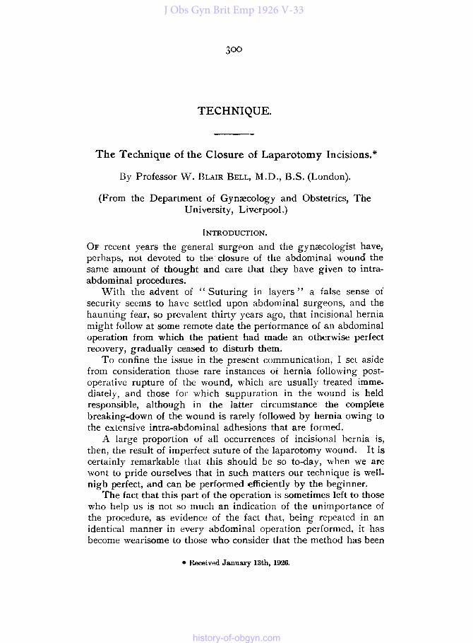
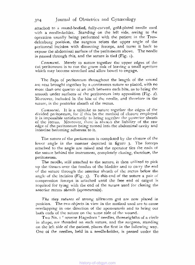
Fig. I . Insertion of the first stitch to close the upper angle of the divided peritoneum.
history-of-obgyn.com
302 Journal of Obstetrics and Gynaxology
perfected so completely that no improvement is possible for the time being. This may be the reason why the technique of suture has r(emained more or less at a standstill for many years. Never- theless, incisional hernias are very common, although they are not always seen by the surgeons concerned.
My excuse for these remarks is that although 1 believe, like other surgeons, that hernia rarely follows my own operations--an opinion that is confirmed by onle of my surgical colleagues who has been investigating the subject-I have never felt entirely satisfied with my methods of closure, and I have endeavouned, regardless of the apparent immunity of my patients to postoperative herniae, gradually to improve the technique in every possible way-that is, in regard to the comfort of the patient, the cosmetic result, and, above all, the subsequent soundness of the scar.
I take it that all will agree that the following are the essential iequirements, apart from asepsis, of the perfect procedure :-
( I ) Avoidance of apertures through the sutured peritoneum. ( 2 ) Freedom from intra-abdominal adhesions to the back of
( 3 ) Obliteration of all dead spaces. (4) Overlapping closure of the aponeurosis with suitable
material. ( 5 ) Stay sutures that will keep the aponeurosis closed as it is
sutured, and which when tied will approximate thc deeper part of the wound throughout, and will not cut the skin.
(6) Neat closure of the skin edges to secure a cosmetic result. All incisions not made vlertically in the midline of the abdom'en
have some disadvantage : nerve fibres are severed, important muscular structures ape injured, or, fro111 an operative point of view, access 1s impeded, vision is limited, or aseptic technique is rendered difficult. I shall, therefore, describe the method I employ at present to close the part of the abdominal wall that is weakest and is sub- jected to the greatest strain, namely, the median subumbilical ; and I shall endeavour to indicae how the procedures meet the require- ments stated. I am wiell aware that in many particulars the methods described may be well-known; but I have not seen them all employed togethter. I have, therefore, put the wholle procedure of the closure of the abdominal wound into one picture in order that others may consider how far the details are an improvement or the reverse on the methods they themselves practise, and with what results.
CLOSURE OF THE MEDIAN SUBUMBILICAL INCISION. The suture material employed for
the closure of the peritoneum is No. o plain catgut, and this is
the scar.
Suture of the peritoneum.
history-of-obgyn.com
Closure of I_aparotomy Incisions 303
U L K .
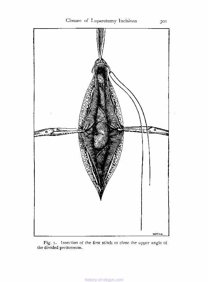
Fig. 2. Closure of the peritoneum. The way in which the under surfaces of the peritoneum are brought into apposition and the posterior sheaths of the recti approximated is shown.
history-of-obgyn.com
304 Journal of Obstetrics and Gynzcofogy
attached to a round-bodied, fully-curved, gold-plated needle used with a needlle-holder. Standing on the left side, owing to the operation usually being performed with tbe patient in the Tren- delenburg position, the surgeon seizes the upper angle of the peritoneal incision with dissecting forceps, and turns it back to expose the abdominal surface of the peritoneum above. The needle is passed through this, and the suturte is tifed (Fig. I ) .
Merely to suture together the upper edges of the cut peritoneurn is to run the grave risk of leaving a small aperture which may become stretched and allow bowel to engage.
Comment.
The flaps of peritoneum throughout the length of the wound are next brought together by a continuous suture so placed, with no more than one quarter of an inch between each bite, as to bring the smooth under surfaces of the peritoneum into apposition (Fig. 2).
Ntoreover, includsed in the bite of the needle, and therefore in the suture, is the posterior sheath of the rectus.
It is a mistake to suture together the edges of the divided peiitoneum, for iC this be. the method of closure employed it is impossible satisfactorily to bring together the posterior sheath of the rectus. Moreover, there is always the liability of the raw edge of the peritoneum being turned into the abdominal cavity and intestine becoming adherent to it.
Comment .
The suture ol the peritoneum is completed by the closure of the lower angle in the manner depicted in figure 3. The forceps attached to the angle are raised and tbe operator ties the ends of the suture behind the instrument, completely closing, therefore, the peritoneum.
The needle, still attached to the suture, is then utilized to pick u p the tissues over the fundus of the bladder and to carry the end of the suture through the antcerior sheath of the rectus below the angle of the incision (Fig. 3). T o this end of the suture a pair o f compression forceps is attached until the friee end of catgut i5 required for tying with the end of the suture used for closing the anterior rectus sheath (aponeurosis) .
The stay sutures of strong silkworm gut are now placed in position. The two objects in view in the method used are to causse overlapping in one direction ot the aponleurosis and to bring out both ends of the suture on tbe same side of the wound.
Two No. I ' reverse Hagedorn ' needles, three-eighths of a circle in shape, are threaded on each suture, and the surgeon, standing on the left side of thle patient, places the first in the following way. One of the needles, held in a nleedle-holder, is passed under the
history-of-obgyn.com
Closure of Laparo tomy 1 ncisions 9 5
Fig. 3. Closing of the peritoneum. The method of closing the lower angle is illustrated.
history-of-obgyn.com

306 Journal of Obstetrics and Gynaecology
aponeurosis of the left side and out through the intervening tissues m d skin on the same side, the skin beiiig pierced about two inches bejond the edge of the wound. The other neledle, likewise held in the needle-holder, is made to pich-up the aponeurosis on the other side o f the wound with a bite parallel to the edge. It is then passed under the aponeurosis on the lclt side and out through the skin in the same way as, and close to the point of exit of the first needle. I t will be seen that the result of drawing tightly the two ends of this suture is to cause the aponeurosis on the left side of the wound t o overlap that on the right side (Fig. 4).
The second suture, the ends of which will pierce the skin on the right side of the wound, is placed in the following manner. The needle held in a needle-holder is made to pierce thle fat and skin from within outwards. The second needle, again held in a needle-holder, is passed through the edge of the aponeurosis on the opposite (left) side from above downwards. Next, a bite, parallel to the edge, is taken of the aponeurosis on the right side, and then the nleedle is made to pierce th'e edge of the aponeurosis on the left side from below upwards, and is afterwards carried to the right side of the wound where it is passed through the fat and skin close to the track of the first needle. It will be seen that when this suture is drawn tightly the aponeurosis of the left sidle is once more made to overlap that of the right side (Fig. 4).
The rest of the silkworm gut stay suturles-there are usually four in all, that is, two on either sidle-are placed alternately in the \\ ay described.
This method of employing stay sutures is a slight modification of that practised by Chipman of Montreal.
Comment. It will be noted that this method of placing the stay sutures has the great advantage over the ordinary 'through and throu h ' sutures, in that they cause overlapping of the aponeurosis,
dead spaces. A ' heaping-up' of the wound niay be observed when the sutures have been tied.
and cf raw together the deeper parts of the vound, preventing all
The aponeurosis, held in an overlapping position by the silk- worm sutures, is readily closed with a continuous sutui;e of tanned (20-day) No. 2 catgut, in the way shown in figure 5. The end of the suture is first tied beyond the upper angle of the incision in the aponeurosis and to the end of the peritoneal suture. A bite is then taken parallel to the edge of the rectus sheath on the right side and the needle is carried on through the edge of the left rectus sheath which is pierced from below upwards. This method of suture is con tinued througilciut the whole length of the incised aponeurosis, and, at thje end, the suture is locked by passing the needle through the
history-of-obgyn.com

Closure of Laparotomy Incisions 307
Fig. 4. Method of insertion of the stay sutiire of silkworm gut to secure overlapping of the aponeurosis.
history-of-obgyn.com
308
final loop. T h e two ends of catgat sutures, the one closing thr peritoneurn and the other the aponeurosis, are then tied together (Fig. 6).
The accurate apposition of the overlapped aponeu- rosis is secured bv a continuous suture. The tying together of the lower ends of the peritoneal a n d aponeurotic sutures obliterates the dead space of Retzius.
Journal of Obstetrics and Gynzecology
Comment.
Skin suture. Provided th'ere is no possibility of infection, the subcutaneous suture gives the best result in a majority of cases. b4eral clips are useful when the skin is very flaccid and tbe patient thin : and, in the face of probable sepsis in the wound from intra- peritoneal infection, interrupted fine silkworm sutures should be u sled .
As I have said, in a majority of cases, howevier, the subcutaneous suture is most useful, provided the proper material is used in the best way.
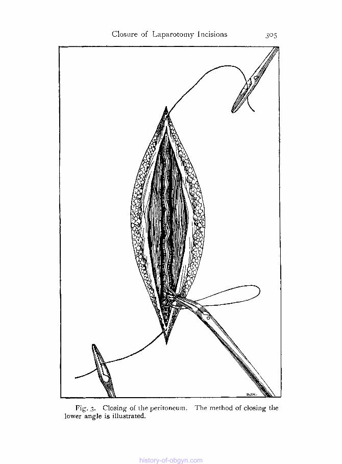
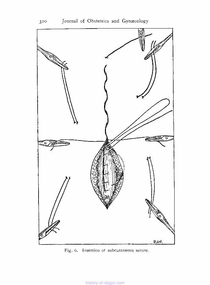
Silkworm gut, threaded on two No. I 'reverse Hagedorn ' needles, three-eighths of a circle in shape, is used. One needle is 1)asse.d through the fat and sk in from within outwards at the upper angle of the wound and then detached from thesilkm~orm gut, the end o f which is held temporarily in forceps. The other needle is made to pas5 closely beneath the skin taking small bites first on one side thrn on the other until the middle of the wound is reached (Fig. 6) . At this point the second needle is removed from the suture, which 1s left loose for the moment. A second silkworm suture threaded o n one needle is nov taltrn, and the skin on the lower half of the wound is brought together in the same m7av as in the upper half, :tnd the needhe is made to pierce the skin from within outwards a little distance beyond the lower angle of the incision. The two ends of the sutures lying free in the middle of the wound are now tied together, and the long ends are left projecting beyond the ai2posed skin ledges.
Next, perforated lead shot of large size are taken, and are thrc>aded on to the ends of the sutures projecting through the skin hcvond the upper and lower limits of the incision. These shot are crushed with forceps after the suturmes have been pulled taut (Fig. 7).
Comment. Sillin~orrn cut is much preferable to catgut for mhcutaneoiis suttlre-material, for tlw latter always causes exudation of serum.
To remove the subcutanfeous suture described above, the long ends projecting from the middle of the wound are seized in forceps, the knot is drawn-out and cut-off. The upper and lower ends with the shot attached are then readily withdrawn,
history-of-obgyn.com

Fig. j. Suture of the aponeurosis, showing the overlapping of this structure.
history-of-obgyn.com
3 JO Journal of Obstetrics and Gynaxology
history-of-obgyn.com
Closure of Laparotomy Incisions 31 1
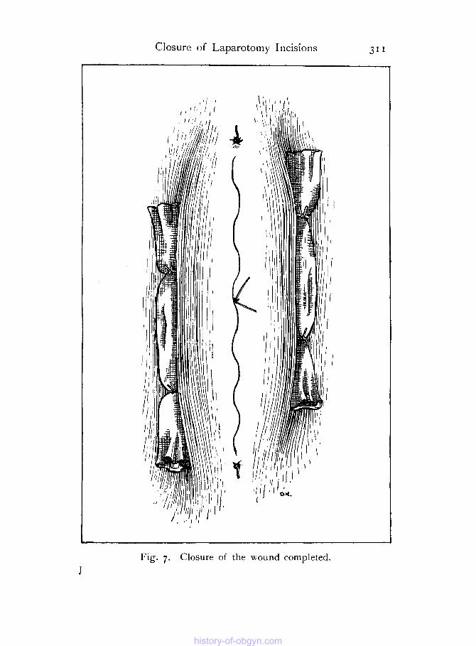
Fig. 7. Closure of the wound completed. J
history-of-obgyn.com
3 1 2 Journal of Obstetrics and Gynaecology
Finally, the stay sutures are pulled tightly and tied on either side over rolls of cyanide gauze (Fig. 7).
If, however, drainage has been established through a stab-wound outside the rectus muscle on *either side, or there be a possibility of sepsis in the wound itself, the stay sutures should be tied over large rubber drainage tubes, made less compressible by the insertion of one or more smaller drainage tubes placed one inside the othcr in nest-formation.
Directly the stay sutures are pulled taut and are tied over gauze or tubing the ' heaping-up ' of the wound IS to be observed. It will be notcd that no longer is i t possible for bleeding or dead spaces to occur in the deeper parts.
Comment.
These sutures are easily removed, if care has been taken in the placing of them to avoid locking i n the wound. I t is necessary only to cut one thread after the overlying roll of gauze has been raised and an alcoholic solution of iodine applied.
These, then, are the methods employed. I have endeavoured to explain the reason for, and advantage of, each step. Although I have described the procedure in respect of the subumbilical median incision, the method is applicable to all vertical incisions through the abdominal wall, and with slight modifications to incisions in any direction.
history-of-obgyn.com