bleeding due to disruption of a cargo-specific er-to-golgi transport complex
TRANSCRIPT

Bleeding due to disruption of a cargo-specific ER-to-Golgi transport complexBin Zhang1, Michael A Cunningham2, William C Nichols3, John A Bernat4, Uri Seligsohn5, Steven W Pipe6,John H McVey7, Ursula Schulte-Overberg8, Norma B de Bosch9, Arlette Ruiz-Saez9, Gilbert C. White10,EGD Tuddenham7, Randal J. Kaufman2,11 & David Ginsburg1,4,11
Departments of 1Internal Medicine and 2Biological Chemistry, University of Michigan, Ann Arbor, Michigan 48109-0650, USA. 3Children’s Hospital Medical Center,Cincinnati, Ohio 45229, USA. 4Department of Human Genetics, University of Michigan, Ann Arbor, Michigan 48109-0650, USA. 5The Chaim Sheba Medical Center,Tel-Hashomer and Sackler Faculty of Medicine, Tel Aviv, Israel. 6Department of Pediatrics, University of Michigan, Ann Arbor, Michigan 48109-0650, USA. 7MRCClinical Sciences Center, Imperial College, London W12 ONN, UK. 8Charite Medical Center, Humboldt University of Berlin, Germany. 9Centro Nacional de Hemofilia,Banco Municipla de Sangre, Caracas, Venezuela. 10Department of Medicine, University of North Carolina, Chapel Hill, North Carolina 27599, USA. 11Howard HughesMedical Institute, University of Michigan, Ann Arbor, Michigan 48109-0650, USA. Correspondence should be addressed to D.G. (e-mail: [email protected]).
L E T T E R S
220 VOLUME 34 | NUMBER 2 | JUNE 2003 NATURE GENETICS
Mutations in LMAN1 (also called ERGIC-53) result in combineddeficiency of factor V and factor VIII (F5F8D), an autosomalrecessive bleeding disorder characterized by coordinatereduction of both clotting proteins1. LMAN1 is a mannose-binding type 1 transmembrane protein localized to theendoplasmic reticulum–Golgi intermediate compartment(ERGIC; refs. 2,3), suggesting that F5F8D could result from adefect in secretion of factor V and factor VIII (ref. 4). Correctlyfolded proteins destined for secretion are packaged in the ERinto COPII-coated vesicles5, which subsequently fuse to formthe ERGIC. Secretion of certain abundant proteins suggests adefault pathway requiring no export signals (bulk flow; refs.6,7). An alternative mechanism involves selective packaging ofsecreted proteins with the help of specific cargo receptors8–13.The latter model would be consistent with mutations in LMAN1causing a selective block to export of factor V and factor VIII.But ∼ 30% of individuals with F5F8D have normal levels ofLMAN1, suggesting that mutations in another gene may also beassociated with F5F8D14,15. Here we show that inactivatingmutations in MCFD2 cause F5F8D with a phenotypeindistinguishable from that caused by mutations in LMAN1.MCFD2 is localized to the ERGIC through a direct, calcium-dependent interaction with LMAN1. These findings suggest thatthe MCFD2-LMAN1 complex forms a specific cargo receptorfor the ER-to-Golgi transport of selected proteins.
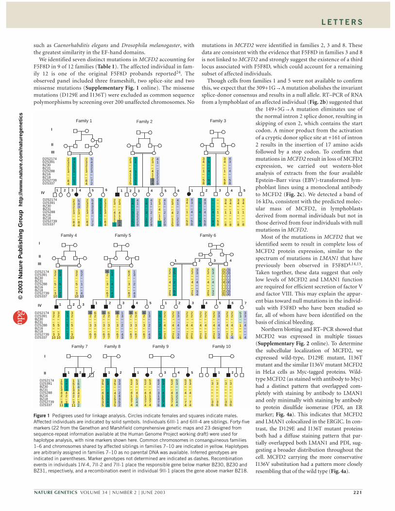
We initially screened other potential components of the ER-Golgisecretory machinery16–18 as candidates for a second gene associatedwith F5F8D without success. We next carried out whole-genome link-age analysis by homozygosity mapping19, similar to the strategy used toidentify LMAN1 (refs. 4,20). We prepared DNA from 19 individualswith F5F8D in ten pedigrees (Fig. 1) and genotyped a total of 382 poly-morphic markers spanning all 22 autosomes. The largest increase inhomozygosity occurred between D2S391 and D2S337 on chromosome
2p21–2p16.3 (maximum lod score of 3.59 for this interval by MAP-MAKER/HOMOZ; ref. 21). Additional genotyping of probands andfamily members placed the gene in the interval of ∼ 2.6 cM betweenmarkers D2S391 and D2S2739. Fine mapping with additional markersdeveloped from the draft human genome sequence22 narrowed thecandidate region to a non-recombinant interval of 1.8 cM betweenmarkers BZ31 and BZ18 (Fig. 2a), with a maximum lod score of 6.4 atD2S2227, by multi-point linkage analysis.
The two affected siblings in family 8 inherited different allele com-binations from their parents (Fig. 1). In addition, although family 3 isconsanguineous, the affected individual (IV1) is heterozygousthroughout the region, and two of the four unaffected siblings (IV4and IV5) are homozygous with respect to an allele shared with theaffected individual (Fig. 1 and data not shown). These results suggestthat the gene associated with F5F8D in family 8 (and probably alsofamily 3) is not located in this region of the genome.
The candidate genetic interval between BZ31 and BZ18 is∼ 2.4 Mb in the draft human genome sequence with four major gaps(Fig. 2). This region contains ∼ 20 known and predicted genes (Fig.2a) and a number of partial expressed-sequence tags (ESTs), none ofwhich have sequence homology to known components of the ER orGolgi. Direct DNA sequencing of individual candidate genes identi-fied mutations in the predicted exons and intron-exon junctions ofHUMTRANSC. This partial cDNA was previously reported basedon its unusually long 3′ untranslated region containing a transpo-son-like human repeat element, THE1 (ref. 23). This gene, nowrenamed MCFD2 (multiple coagulation factor deficiency 2), spansfour exons encompassing ∼ 19 kb in the human genome and con-tains a 145-amino-acid open reading frame, predicting a protein of16 kDa (Fig. 2a). Notable features include a predicted signal peptideat the N terminus and two calmodulin-like EF-hands for putativecalcium binding at the C terminus (Fig. 2a). Clear orthologs ofMCFD2 exist in multiple mammalian and other vertebrate species(Fig. 3). Possible orthologs are also found in invertebrates (Fig. 3),
©20
03 N
atu
re P
ub
lish
ing
Gro
up
h
ttp
://w
ww
.nat
ure
.co
m/n
atu
reg
enet
ics
L E T T E R S
NATURE GENETICS VOLUME 34 | NUMBER 2 | JUNE 2003 221
such as Caenorhabditis elegans and Drosophila melanogaster, withthe greatest similarity in the EF-hand domains.
We identified seven distinct mutations in MCFD2 accounting forF5F8D in 9 of 12 families (Table 1). The affected individual in fam-ily 12 is one of the original F5F8D probands reported24. Theobserved panel included three frameshift, two splice-site and twomissense mutations (Supplementary Fig. 1 online). The missensemutations (D129E and I136T) were excluded as common sequencepolymorphisms by screening over 200 unaffected chromosomes. No
mutations in MCFD2 were identified in families 2, 3 and 8. Thesedata are consistent with the evidence that F5F8D in families 3 and 8is not linked to MCFD2 and strongly suggest the existence of a thirdlocus associated with F5F8D, which could account for a remainingsubset of affected individuals.
Though cells from families 1 and 5 were not available to confirmthis, we expect that the 309+1G→A mutation abolishes the invariantsplice-donor consensus and results in a null allele. RT–PCR of RNAfrom a lymphoblast of an affected individual (Fig. 2b) suggested that
the 149+5G→A mutation eliminates use ofthe normal intron 2 splice donor, resulting inskipping of exon 2, which contains the startcodon. A minor product from the activationof a cryptic donor splice site at +161 of intron2 results in the insertion of 17 amino acidsfollowed by a stop codon. To confirm thatmutations in MCFD2 result in loss of MCFD2expression, we carried out western-blotanalysis of extracts from the four availableEpstein–Barr virus (EBV)-transformed lym-phoblast lines using a monoclonal antibodyto MCFD2 (Fig. 2c). We detected a band of16 kDa, consistent with the predicted molec-ular mass of MCFD2, in lymphoblastsderived from normal individuals but not inthose derived from four individuals with nullmutations in MCFD2.
Most of the mutations in MCFD2 that weidentified seem to result in complete loss ofMCFD2 protein expression, similar to thespectrum of mutations in LMAN1 that havepreviously been observed in F5F8D4,14,15.Taken together, these data suggest that onlylow levels of MCFD2 and LMAN1 functionare required for efficient secretion of factor Vand factor VIII. This may explain the appar-ent bias toward null mutations in the individ-uals with F5F8D who have been studied sofar, all of whom have been identified on thebasis of clinical bleeding.
Northern blotting and RT–PCR showed thatMCFD2 was expressed in multiple tissues(Supplementary Fig. 2 online). To determinethe subcellular localization of MCFD2, weexpressed wild-type, D129E mutant, I136Tmutant and the similar I136V mutant MCFD2in HeLa cells as Myc-tagged proteins. Wild-type MCFD2 (as stained with antibody to Myc)had a distinct pattern that overlapped com-pletely with staining by antibody to LMAN1and only minimally with staining by antibodyto protein disulfide isomerase (PDI, an ERmarker; Fig. 4a). This indicates that MCFD2and LMAN1 colocalized in the ERGIC. In con-trast, the D129E and I136T mutant proteinsboth had a diffuse staining pattern that par-tially overlapped both LMAN1 and PDI, sug-gesting a broader distribution throughout thecell. MCFD2 carrying the more conservativeI136V substitution had a pattern more closelyresembling that of the wild type (Fig. 4a).
Family 3
I
II
III
IV1 2 43 5
34––1––58
75––4––94
43––4––58
34––1––58
34––1––58
––––1––58
34––1–––8
34––1–––8
34––1–––8
34––1–––8
75––4––94
43––4––58
43––4––58
––––4––94
D2S2174D2S391BZ30BZ31D2S288BZ16BZ18D2S2739D2S337
D2S2174D2S391BZ30BZ31D2S288BZ16BZ18D2S2739D2S337
Family 2
1 2 3 4 5 6
23––41115
14––31153
41––22229
23–4–11–3
23––4–115
41––2–229
41––2–229
41––2-229
41––22229
23––41134
14––31153
14––3-153
23––41113
Family 1
2 31 4 5 6
23213––84
43111––17
54323––72
23213––84
54313––84
23213––84
23213––84
23213––84
54323––72
43111––17
Family 5 Family 6
1 2 3 4
I
II
III
IV1 2 3 4 5 6 7
Family 4
31 2
53––5––22
32––5––3
10
32––5––3
10
32––5––3
10
32––5––3
10
32––5––3
10
53––5––22
1 2 3 4 5
62––3––67
62––3––67
14––4––42
13––2––43
15––2––54
33––4––34
53––4––32
22––4––25
53––1–––5
22––4––24
D2S2174D2S391BZ30BZ31D2S288BZ16BZ18D2S2739D2S337
D2S2174D2S391BZ30BZ31D2S288BZ16BZ18D2S2739D2S337
52––1––4
10
62––3––67
62––3––67
62––3––67
13––2––43
13––2––43
22––4––25
22––4––25
22––4––25
22––4––25
22––4––25
22––4––25
22––4––25
22––4––25
22––4––25
33––4––34
15––2––54
22––4––25
(2)(2)(–)(–)
(–)(–)
(4)
(2)(5)
(3)(3)
(4)
(3)(4)
33––4––34
33––4–––4
62––3––67
62––3––67
62––3––67
62––3––67
62––3––67
II
I
Family 8 Family 9 Family 10Family 7
1 1 1 12 2 2 23 4 5
D2S2174D2S391BZ30BZ31D2S288BZ16BZ18D2S2739D2S337
35––41535
35––41535
33––24352
52––41374
52––41374
52––4––74
52––41374
53––41374
53––42274
62––31476
53––42244
53––41244
53––4–
74
–3–––––36
–3–––––36
–3–––––36
–3–––––36
151342–3
11
164342–32
151342–3
11
343442–38
13––43343
23––41134
(–)(–)
(–)(–)
Figure 1 Pedigrees used for linkage analysis. Circles indicate females and squares indicate males.Affected individuals are indicated by solid symbols. Individuals 6III-1 and 6III-4 are siblings. Forty-fivemarkers (22 from the Genethon and Marshfield comprehensive genetic maps and 23 designed fromsequence-repeat information available at the Human Genome Project working draft) were used forhaplotype analysis, with nine markers shown here. Common chromosomes in consanguineous families1–6 and chromosomes shared by affected siblings in families 7–10 are indicated in yellow. Haplotypesare arbitrarily assigned in families 7–10 as no parental DNA was available. Inferred genotypes areindicated in parentheses. Marker genotypes not determined are indicated as dashes. Recombinationevents in individuals 1IV-4, 7II-2 and 7II-1 place the responsible gene below marker BZ30, BZ30 andBZ31, respectively, and a recombination event in individual 9II-1 places the gene above marker BZ18.
©20
03 N
atu
re P
ub
lish
ing
Gro
up
h
ttp
://w
ww
.nat
ure
.co
m/n
atu
reg
enet
ics

L E T T E R S
222 VOLUME 34 | NUMBER 2 | JUNE 2003 NATURE GENETICS
MCFD2 was readily detected in wild-type but not MCFD2-nulllymphoblasts (Fig. 2c) or LMAN1-null lymphoblasts (Fig. 2c). Wedid, however, detect a trace amount of MCFD2 after immunopre-cipitation from a large number of LMAN1-null cells (Fig. 2c), con-sistent with a lack of retention or degradation of MCFD2 in theabsence of LMAN1. MCFD2 lacks the C-terminal KDEL retrievalsignal found in many other soluble ER-resident proteins25 thatmediates recycling back to the ER throughbinding to the KDEL receptor26,27. Thesedata suggest that MCFD2 is retained in theER and intermediate compartment throughits interaction with LMAN1. But MCFD2does not seem to be required for the ER exitand recycling of LMAN1, as both wild-typeand MCFD2-deficient lymphoblasts had sim-ilar LMAN1 staining patterns (Fig. 4b).
Given the colocalization of MCFD2 andLMAN1 to the ERGIC and the identicalF5F8D phenotypes resulting from mutationsin MCFD2 and LMAN1, we next examinedthe potential for direct interaction betweenthese gene products. Antibody to Mycdetected Myc-tagged MCFD2 in transfectedcells that co-immunoprecipitated withendogenous and overexpressed LMAN1(Fig. 5a,b). Similarly, antibody to LMAN1co-precipitated Myc-tagged MCFD2 as wellas endogenous MCFD2 (Fig. 5a,b). In con-trast to these observations with wild-typeMCFD2, LMAN1 did not co-immunoprecip-itate with either D129E or I136T mutant
MCFD2 (Fig. 5a), although the proteins were synthesized at highlevels. The more conservative I136V substitution reduced, but didnot eliminate, interaction with LMAN1 (Fig. 5a).
MCFD2 contains EF-hand domains that may bind calcium.Treatment of cell lysates with the calcium-specific chelator ethylene gly-col bis (b-aminoethylether) tetraacetic acid (EGTA) before immuno-precipitation destroyed the LMAN1 interaction with Myc-tagged
MCFD2
a
1 2 3 4
Domain structure
EF-1 EF-2signal
D2S
2174
D2S
391
D2S
2227
D2S
337
D2S
378
D2S
2739
D2S
288
D2S
288
D2S
2227
BZ
16
BZ
18
BZ
7
Transcripts
*
Gaps
BZ
31
Missense
Splice Deletion
BZ
12
1 cM
200 nt
LMAN1
WT
MCFD215
20
5060
MCFD215
20
LMAN1MCFD2
WT
a+d
a+c
b+d
a+d
a+c
b+d
WT 149+5GA
a b c d
1 2 3 4
0.1
0.20.30.4
1.5
0.6
MW(kb)
b c
Figure 2 Identification of MCFD2 and mutational analysis. (a) Positionalcloning of MCFD2. The nonrecombinant interval of ∼ 1.8 cM is shown inred. Sequence gaps in the public genome assembly are indicated by bluerectangles. Transcripts localized to this interval are depicted byalternating black and gray bars. MCFD2 is indicated with an asterisk. Thecoding region of MCFD2 is illustrated in gray with 5′ and 3′ untranslatedregions in green. Intron sizes are not drawn to scale. Splice mutations aredepicted underneath the corresponding exons (blue diamonds). Thepredicted signal peptide of MCFD2 is indicated in yellow, and the two EF-hand domains in dark blue. The green triangles indicate the locations ofthe 2 missense mutations and red squares indicate frameshift mutations.(b) Analysis of 149 +5G→A mutation in family 12. RT–PCR was doneusing total RNA prepared from control lymphoblasts and lymphoblastsfrom the affected individual. The locations of primers a–d are illustratedin a schematic of the mRNA corresponding to exons 1–4 of MCFD2.RT–PCR products were analyzed by agarose gel electrophoresis, as shownin the top panel. Direct sequencing of the products generated from mRNAfrom the affected individual with primers a+c and a+d shows skipping ofexon 2, whereas the product from primers b+d contains an insertion of161 bp using an alternate donor-splice site in intron 2. MW, molecularweight marker. (c) Cell extracts (from 2 × 10⁵ cells) prepared from twonormal control individuals (WT), two affected individuals with nullmutations in LMAN1 (LMAN1–, derived from families A19 and A12; ref.14) and four individuals with mutations in MCFD2 (MCFD2–; from left toright: family 11, family 7 II-1, family 12, family 6 IV-4) were subject toimmunoblotting with a monoclonal antibody against LMAN1 (top) or amonoclonal antibody against MCFD2 (middle). Cell extracts (from 5 ×10⁶ cells) were immunoprecipitated with Sepharose beads conjugated torabbit antisera to MCFD2 and detected on a western blot by a monoclonalantibody against MCFD2 (bottom). Numbers to the left indicate themolecular weight (kDa) of pre-stained markers.
MTMRS-L-----LRTPFL----CGL-LWAFCAPGARAEEPAASFSQ----PGSMG-LDKNTVHDQEHIMEHLEGV-INKP1 H. sapiensMRSLR-L-----LRIPFL----CGL-LWAFCAPGARAEEPGASSSH----HGSMG-LDKNTVHDQEHIMEHLEGV-INKP1 B. taurusMATLQ-L-----LRAPLL----CVL-LWVFCAPGARAHDHGADV-H----HGSVG-LDKSTVHDQEHIMEHLEGV-IDQP1 M. musculusMAPVS-F------RSPLLL---CLVAAWL---GPVPAEEPAAES------PPSHGRLDKNLVQDKDHIMEHLEGV-IEKP1 G. gallusMASHR-SS-----MTSIL-C--CLVSSLFFLSALAEEHLHPHVEEG----EHGGARFGKSVVQDKDHIMEHLDGV-VDKP1 X. laevisMRLRE-ISGQLRLWSWFLVCSGCMLSVWA---HGGQHGEAVGSNQV----PPPITRLDRNMVQDREHIMEHLEGI-VEKQ1 D. rerioMAANILVVSCLIL-GSF----AHQPQ--QFPGSNQQQPQQGGQAEQAQHAQPGQQQFGGEQARDEHHIKEHLDGK-VD-P1 C. elegansM-CN--L-SNL-L--NFI---ICIAS---F--S-QNFDATLAVKRGPHHPRGETRRVDQHLTHEEHRIDDDLKDMGVQAN1 D. melanogaster
EAEMSPQELQLHYFKMHDYDGNNLLDGLELSTAITHVHKEEGS------------EQAPLMSEDELINIIDGVLRDDDKN64 H. sapiensEAEMSPQELQLHYFKMHDYDGNNLLDGLELSTAITHVHKEEGS------------EQAP-MNEDELINLIDGVLRDDDKN64 B. taurusEAEMSPQELQLHYFKMHDYDGNSLLDGLELSIAITHVHKEEGS------------EQAPVMSEDELVSIIDGVLRDDDKN63 M. musculusESEMSPQELQLHYFKMHDYDGNNLLDGLELATAISHVHKEEGG------------ENTQAMKEEELISLIDDVLRDDDKN61 G. gallusETEMSPQELQLHYFKMHDYDGNNLLDGLELATAITHVHKS-AE------------EPVQTMKEEDLISLIDDVLRDDDKN67 X. laevisESEMTPQELQLHYFKMHDYDGNNLLDGLELATAITHVHREERG------------GDSQPMREEDLINLIDDVLRDDDKN72 D. rerioTANMTPEQLQFHYFNMHDLDKNGKLDGVELIKAITHFHAENPGPQHTQNNANANHQPPPLPSEVELETMIDSILKDDDFN72 C. elegansLDDLSEEEKIFYMFKAHDNDNNNALDGLEMIQSAMH-HNYDYFKNNERDAYLQNATDE-L--E-HFIEAIDKFLLIADDN65 D. melanogaster
NDGYIDYAEFAKSLQ132 H. sapiensNDGYIDYAEFAKSLQ131 B. taurusNDGYIDYAEFAKSLQ131 M. musculusNDGYIDYAEFAKSLE129 G. gallusNDGYIDYAEFARSLE134 X. laevisNDGYIDYAEFATSLE140 D. rerioADGFIDYGEFLKAQKLREDQARSHQEQMQKAGGTQ152 C. elegansNDGLLHYPEFVKAITGGKEQPNVDRNILR140 D. melanogaster
Figure 3 Alignment of the predicted amino-acid sequences of MCFD2 orthologs from othervertebrate and invertebrate species: Homo sapiens, Bos taurus, Mus musculus, Gallus gallus,Xenopus laevis, Danio rerio, Caenorhabditis elegans and Drosophila melanogaster. Regions withsequence identity are shaded. Arrows indicate the two amino-acid positions affected by missensemutations in individuals with F5F8D. The wild-type sequences at both D129 and I136 are highlyconserved across all species analyzed.
©20
03 N
atu
re P
ub
lish
ing
Gro
up
h
ttp
://w
ww
.nat
ure
.co
m/n
atu
reg
enet
ics

L E T T E R S
NATURE GENETICS VOLUME 34 | NUMBER 2 | JUNE 2003 223
MCFD2 (Fig. 5b) and recalcification of the EGTA-treated lysates recon-stituted the MCFD2 association with LMAN1 (Fig. 5b). The D129Eand I136T missense mutations both occur at highly conserved posi-tions in the second EF-hand domain of MCFD2 (Fig. 3). In addition, allfive of the remaining mutations in MCFD2 that we identified (Table 1)would result in frameshifts proximal to this domain. Taken together,these results suggest an essential role for the second EF-hand ofMCFD2 in the calcium-dependent interaction with LMAN1 and thesubsequent function of the MCFD2–LMAN1 complex in the transportof factor V and factor VIII.
Our data indicate that formation of the calcium-dependentMCFD2–LMAN1 complex is an essential step in ER-to-Golgi transportfor a specific subset of glycoproteins including factor V and factor VIII.These data also provide the first direct evidence for a cargo-specific sort-ing receptor complex in the ERGIC. Ca2+–MCFD2 may serve as thecofactor that specifically recruits correctly folded factor V and factor
VIII in the ER lumen. Alternatively, MCFD2 may be required for theseparation of factor V and factor VIII from LMAN1 in the ERGIC,perhaps in response to the dissociation of COPII coats from transportvesicles. A subset of the other EF-hand-containing proteins couldpotentially define a class of adaptors or chaperones that might servesimilar functions for other secreted proteins. The phenotypes associ-ated with mutations in MCFD2 and LMAN1 are indistinguishable andassociated only with deficiency of plasma coagulation factor V and fac-tor VIII, though a selective delay in secretion of procathepsin C hasbeen observed in HeLa cells overexpressing a dominant-negative formof LMAN1 (ref. 28). This incomplete block to production of factor Vand factor VIII, together with the limited subset of proteins clinicallyaffected by deficiency of LMAN1 or MCFD2 make this pathway apotentially attractive therapeutic target for anticoagulation, particu-larly in light of the prothrombotic risk associated with factor V Leidenand with high levels of factor VIII (ref. 1).
METHODSSubjects. We obtained blood samples from all individuals after informedconsent (study protocol approved by the University of MichiganInstitutional Review Board). Our study population consisted of ten fami-lies, including 19 affected individuals in whom no mutations in LMAN1were detected (Fig. 1). Families 1, 2, 3, 7 and 8 correspond to previouslyreported families A5, A7, A13, A21 and A28, respectively14. Families 4, 5, 9and 10 correspond to previously reported families 13, 16, 14 and 19 respec-tively15. Family 6 is a large Turkish family that has not been previouslyreported. We derived an EBV-immortalized lymphoblast line from individ-ual IV:4 of this family. Western-blot analysis showed a normal level ofLMAN1 in this line (Fig. 2c). Two EBV-immortalized lymphoblast linesderived from the probands of family 11 (previously reported family A29;ref. 14) and of family 12 (the first reported family with F5F8D; ref. 24) werenot included in the genome scan but were included in the subsequent muta-tional analysis. Both lymphoblasts had normal levels of LMAN1 (ref. 14;Fig. 2c). The probands of families 1–6 are the progeny of consanguineousmarriages. There is no known consanguinity in families 7–12. We carriedout a genome-wide linkage scan and fine mapping with additional markersas previously described20,22.
RT–PCR, RACE and northern-blot analysis. We prepared total cellular RNAfrom EBV-transformed lymphoblasts and HeLa cells and determined the 5′end of the mRNA by rapid amplification of cDNA ends (RACE) with aFirstChoice RLM-RACE kit (Ambion). We used a PCR product containing thecoding sequence of the MCFD2 cDNA amplified from a human MOLT4 T-cellcDNA library22 to hybridize to a FirstChoice Human Northern Blot(Ambion). We also screened MCFD2 expression by PCR on cDNA obtainedfrom a multiple tissue cDNA panel (Clontech). We used specific probes for 16
Table 1 Mutations in MCFD2 in families with F5F8D
Nucleotide change Amino-acid change Family
149 +5G→A Donor splice site 10, 12
309 +1G→A Donor splice site 1, 5
103delC Frameshift at residue 35 7
249delT Frameshift at residue 83 6
263–270delTTGATGGC Frameshift at residue 88 11
C387→G D129E 4
T407→C I136T 9
MycLMAN1LMAN1+ Myc
WT
D129E
I136V
PDI+ Myc
I136T
a
WT
PDI LMAN1 Mergeb
MCFD2–
Figure 4 Intracellular localization of MCFD2. (a) HeLa cells were transfectedwith pcDNA3.1-Myc-His vectors expressing wild-type MCFD2 (WT), D129Eand I136T mutant proteins or the I136V substitution. Cells were stained 24 hafter transfection with a rabbit antibody to Myc and either a monoclonalantibody to LMAN1 or a monoclonal antibody to PDI. Protein localization wasdetected by immunofluorescence confocal microscopy using an antibody tomouse IgG conjugated to fluorescein isothiocyanate and an antibody to rabbitIgG conjugated to Texas red. Each group of four panels shows, from left toright, the green fluorescence from antibody to LMAN1, the red fluorescencefrom antibody to Myc, the merged image from the two and the merged imagefrom antibody to PDI (green) and antibody to Myc (red), respectively. (b) EBV-transformed lymphoblasts were stained with a rabbit antibody to LMAN1 anda monoclonal antibody to PDI. Protein localization was detected byimmunofluorescence confocal microscopy using an antibody to mouse IgGconjugated to fluorescein isothiocyanate and an antibody to rabbit IgGconjugated to Texas red. Each group of three panels shows, from left to right,the green fluorescence from antibody to PDI, the red fluorescence fromantibody to LMAN1 and the merged image from the two, respectively.
©20
03 N
atu
re P
ub
lish
ing
Gro
up
h
ttp
://w
ww
.nat
ure
.co
m/n
atu
reg
enet
ics

L E T T E R S
224 VOLUME 34 | NUMBER 2 | JUNE 2003 NATURE GENETICS
GenBank accession numbers. HUMTRANSC, M23161. The corrected full-length cDNA sequence of MCFD2 has been deposited with accession numberAF537214.
Note: Supplementary information is available on the Nature Genetics website.
ACKNOWLEDGMENTSWe thank E. Smith for assistance in generating monoclonal antibodies, L. Changand J. Liu for assistance with the confocal microscope and S.J. Weiss and A. Saltielfor comments on the manuscript. This work was supported in part by grants fromthe US National Institutes of Health to D.G., W.C.N. and R.J.K. B.Z. is a JudithGraham Pools Postgraduate Fellow of the National Hemophilia Foundation.M.A.C is the recipient of a research fellowship from the Heart and StrokeFoundation of Canada. D.G. and R.J.K. are investigators of the Howard HughesMedical Institute.
COMPETING INTERESTS STATEMENTThe authors declare that they have no competing financial interests.
Received 16 December 2002; accepted 1 April 2003Published online 28 April 2003; doi:10.1038/ng1153
1. Ginsburg, D. Hemophilia and Other Disorders of Hemostasis. in Emery and Rimoin’sPrinciples and Practice of Medical Genetics vol. II. 1926–1958 (ChurchillLivingstone, New York, 2002).
2. Arar, C. et al. ERGIC-53, a membrane protein of the endoplasmic reticulum-Golgiintermediate compartment, is identical to MR60, an intracellular mannose-specificlectin of myelomonocytic cells. J. Biol. Chem. 270, 3551–3553 (1995).
3. Itin, C., Roche, A.C., Monsigny, M. & Hauri, H.P. ERGIC-53 is a functional man-nose-selective and calcium-dependent human homologue of leguminous lectins.Mol. Biol. Cell 7, 483–493 (1996).
4. Nichols, W.C. et al. Mutations in the ER-Golgi intermediate compartment proteinERGIC-53 cause combined deficiency of coagulation factors V and VIII. Cell 93,61–70 (1998).
5. Schekman, R. & Orci, L. Coat proteins and vesicle budding. Science 271,1526–1533 (1996).
6. Wieland, F.T., Gleason, M.L., Serafini, T.A. & Rothman, J.E. The rate of bulk flowfrom the endoplasmic reticulum to the cell surface. Cell 50, 289–300 (1987).
7. Martinez-Menarguez, J.A., Geuze, H.J., Slot, J.W. & Klumperman, J. Vesicular tubu-lar clusters between the ER and Golgi mediate concentration of soluble secretoryproteins by exclusion from COPI-coated vesicles. Cell 98, 81–90 (1999).
8. Malkus, P., Jiang, F. & Schekman, R. Concentrative sorting of secretory cargo pro-teins into COPII-coated vesicles. J. Cell Biol. 159, 915–921 (2002).
9. Kuehn, M.J., Herrmann, J.M. & Schekman, R. COPII–cargo interactions direct pro-tein sorting into ER-derived transport vesicles. Nature 391, 187–190 (1998).
10. Muniz, M., Morsomme, P. & Riezman, H. Protein sorting upon exit from the endo-plasmic reticulum. Cell 104, 313–320 (2001).
11. Nehls, S. et al. Dynamics and retention of misfolded proteins in native ER mem-branes. Nat. Cell Biol. 2, 288–295 (2000).
12. Springer, S. et al. The p24 proteins are not essential for vesicular transport inSaccharomyces cerevisiae. Proc. Natl. Acad. Sci. USA 97, 4034–4039 (2000).
individual candidate genes to hybridize northern blots of 20 µg total RNAextracted from EBV-immortalized cell lines of two unaffected and fouraffected individuals. Primer sequences are available on request.
Construction of expression vectors. To construct a histidine-tagged recombi-nant protein expression vector, we cloned a DNA fragment containing theMCFD2 coding region amplified from the MOLT4 T-cell cDNA library22 intopET15b (Novagen). For expression in mammalian cells, we amplified wild-type cDNA from the above human cDNA library and cloned intopcDNA3.1/Myc-His (Invitrogen). We constructed mutated cDNA expressionvectors by amplification from the above plasmid using 3′ primers containingthe mutations corresponding to the amino-acid substitutions D129E, I136T orI136V. Primer sequences are available on request.
Antibodies. We generated rabbit antisera to purified His-tagged MCFD2through a commercial supplier (Invitrogen) and affinity-purified MCFD2 anti-serum using a His-tagged MCFD2–coupled Amino-Link column (Pierce).Purified IgG was coupled to CNBr-activated Sepharose 4B beads (Amersham)according to manufacturer’s instructions. We also generated monoclonal anti-bodies using His-tagged MCFD2 at the University of Michigan Hybridoma CoreFacility. Hybridoma clones were screened by direct ELISA using plates coatedwith purified His-tagged MCFD2 and confirmed by western-blot analysis.
Immunofluorescence. We carried out immunofluorescence analysis of COS-1cells transfected with MCFD2 expression vectors and EBV-transformed lym-phoblasts as described previously4, except that permeabilization was achievedby incubating the slides in 0.5% Triton X-100 for 5 min. Images were visual-ized and captured using an Olympus Fluoview confocal microscope. Primaryantibodies were a polyclonal rabbit antibody to Myc (sc-789, Santa CruzBiotechnology), a monoclonal antibody to LMAN1 (G1/93, a gift from H.P.Hauri, University of Basel, Switzerland), a polyclonal rabbit antibody toLMAN1 (anti-rat P58, a gift from R.F. Pettersson, Karolinska Institute,Sweden) and a monoclonal antibody to PDI (ABR). Secondary antibodieswere goat antibody to mouse IgG conjugated to fluorescein isothiocyanate andgoat antibody to rabbit IgG conjugated to Texas red (Molecular Probes).
Metabolic labeling, immunoprecipitation and protein analysis. We trans-fected COS-1 cells using either the DEAE/Dextran method29 or Fugene 6reagent (Roche) according to manufacturer’s instructions and metabolicallylabeled cells 26 h after transfection for 30 min with [35S]-methionine/cysteine(Amersham, 250 µCi ml–1 in methionine/cysteine-free medium). We carriedout immunoprecipitations using equal tricholoracetic acid (TCA)-precip-itable counts of labeled cell extracts as described previously30 or using unla-beled cells followed by western-blot analysis.
Figure 5 MCFD2 interacts with LMAN1.(a) COS-1 cells were either mock-transfected ortransfected with plasmids expressing Myc-taggedwild-type MCFD2, Myc-tagged D129E MCFD2,Myc-tagged I136V MCFD2 or Myc-tagged I136TMCFD2 and metabolically labeled with [35S]-methionine/cysteine. Cell extracts wereimmunoprecipitated (IP) with an antibody to Myc(M) or an antibody to LMAN1 (L). Arrows (fromtop to bottom) denote LMAN1, Myc-taggedMCFD2 and endogenous MCFD2, respectively.(b) COS-1 cells were co-transfected with aplasmid expressing wild-type Myc-tagged MCFD2and a plasmid expressing LMAN1. Cell extractswere immunoprecipitated (IP) with an antibodyto Myc (M) or an antibody to LMAN1 (L). Whereindicated (+), EGTA (2.5 mM) was added to thecell extract before the addition of antibodies.Calcium chloride (Ca, 10 mM) was added tohalf of the EGTA-treated lysate, cleared bycentrifugation, before the addition of antibodies.Arrows (from top to bottom) denote LMAN1 andMyc-tagged MCFD2, respectively.
WT
EGTA + +M L M L M L M L M LIP
WT
97
66
46
30
14
1 2 3 4 5 6 7 8 9 10
WT D129E I136V I136T
a b
9766
46
30
14
M L M L M L M L
1 2 3 4 5 6 7 8
IP
CaMCFD2
+ + WT
MCFD2 – –
– –– – – –– – – – – –
– –
©20
03 N
atu
re P
ub
lish
ing
Gro
up
h
ttp
://w
ww
.nat
ure
.co
m/n
atu
reg
enet
ics

L E T T E R S
NATURE GENETICS VOLUME 34 | NUMBER 2 | JUNE 2003 225
13. Belden, W.J. & Barlowe, C. Role of Erv29p in collecting soluble secretory proteinsinto ER-derived transport vesicles. Science 294, 1528–1531 (2001).
14. Neerman-Arbez, M. et al. Molecular analysis of the ERGIC-53 gene in 35 familieswith combined factor V-factor VIII deficiency. Blood 93, 2253–2260 (1999).
15. Nichols, W.C. et al. ERGIC-53 gene structure and mutation analysis in 19 combinedfactors V and VIII deficiency families. Blood 93, 2261–2266 (1999).
16. Fiedler, K. & Simons, K. Characterization of VIP36, an animal lectin homologous toleguminous lectins. J. Cell Sci. 109 (Pt 1), 271–276 (1996).
17. Pipe, S.W., Morris, J.A., Shah, J. & Kaufman, R.J. Differential interaction of coagu-lation factor VIII and factor V with protein chaperones calnexin and calreticulin. J.Biol. Chem. 273, 8537–8544 (1998).
18. Hebert, D.N., Foellmer, B. & Helenius, A. Glucose trimming and reglucosylationdetermine glycoprotein association with calnexin in the endoplasmic reticulum. Cell81, 425–433 (1995).
19. Lander, E.S. & Botstein, D. Homozygosity mapping: a way to map human recessivetraits with the DNA of inbred children. Science 236, 1567–1570 (1987).
20. Nichols, W.C. et al. Linkage of combined factors V and VIII deficiency to chromo-some 18q by homozygosity mapping. J. Clin. Invest. 99, 596–601 (1997).
21. Kruglyak, L., Daly, M.J. & Lander, E.S. Rapid multipoint linkage analysis of reces-sive traits in nuclear families, including homozygosity mapping. Am. J. Hum. Genet.56, 519–527 (1995).
22. Levy, G.G. et al. Mutations in a member of the ADAMTS gene family cause throm-botic thrombocytopenic purpura. Nature 413, 488–494 (2001).
23. Deka, N. et al. Repetitive nucleotide sequence insertions into a novel calmodulin-related gene and its processed pseudogene. Gene 71, 123–134 (1988).
24. Oeri, J., Matter, M., Isenschmid, H., Hauser, F. & Koller, F. Angeborener mangel anfaktor V (parahaemophilie) verbunden mit echter haemophilie A bein zwei brudern.Med. Probl. Paediatr. 1, 575–588 (1954).
25. Munro, S. & Pelham, H.R. A C-terminal signal prevents secretion of luminal ER pro-teins. Cell 48, 899–907 (1987).
26. Lewis, M.J., Sweet, D.J., & Pelham, H.R. The ERD2 gene determines the specificityof the luminal ER protein retention system. Cell 61, 1359–1363 (1990).
27. Semenza, J.C., Hardwick, K.G., Dean, N. & Pelham, H.R. ERD2, a yeast generequired for the receptor-mediated retrieval of luminal ER proteins from the secre-tory pathway. Cell 61, 1349–1357 (1990).
28. Vollenweider, F., Kappeler, F., Itin, C. & Hauri, H.P. Mistargeting of the lectinERGIC-53 to the endoplasmic reticulum of HeLa cells impairs the secretion of alysosomal enzyme. J. Cell Biol. 142, 377–389 (1998).
29. Sussman, D.J. & Milman, G. Short-term, high-efficiency expression of transfectedDNA. Mol. Cell Biol. 4, 1641–1643 (1984).
30. Moussalli, M. et al. Mannose-dependent endoplasmic reticulum (ER)-Golgi interme-diate compartment-53-mediated ER to Golgi trafficking of coagulation factors V andVIII. J. Biol. Chem. 274, 32539–32542 (1999).
©20
03 N
atu
re P
ub
lish
ing
Gro
up
h
ttp
://w
ww
.nat
ure
.co
m/n
atu
reg
enet
ics