bleiholder, a., mühle, m., hechler, t., bevins, s
TRANSCRIPT

Originally published as: Bleiholder, A., Mühle, M., Hechler, T., Bevins, S., vandeWoude, S., Denner, J., Löchelt, M. Pattern of seroreactivity against feline foamy virus proteins in domestic cats from Germany (2011) Veterinary Immunology and Immunopathology, 143 (3-4), pp. 292-300. DOI: 10.1016/j.vetimm.2011.06.007
This is an author manuscript. The definitive version is available at: http://www.sciencedirect.com/

Pattern of seroreactivity against feline foamy virus proteins in domestic cats from Germany
Anne Bleiholdera, Michael Mühle
b, Torsten Hechler
a, 1, Sarah Bevins
c, Sue vandeWoude
c, Joachim
Dennerb, Martin Löchelt
a
a German Cancer Research Center, Focus Infection and Cancer, Heidelberg, Germany
b Robert Koch Institute, Berlin, Germany
c Colorado State University, Fort Collins, USA
Abstract
The prevalence of feline foamy virus (FFV, spumaretrovirinae) in naturally infected domestic cats ranges between 30 and 80% FFV positive animals depending on age, sex and geographical region analyzed. Two serotypes have been reported for FFV designated FUV7-like and F17/951-like. Serotype-specific neutralization has been shown to correlate with sequence divergence in the surface (SU) domain of the envelope protein (Env). We analyzed a serum collection of 262 domestic cat sera from Germany using a GST-capture ELISA setup screening for Gag and Bet specific antibodies and identified 39% FFV positive animals. Due to the heterogeneity of the serological samples, cut-offs for Gag and Bet reactivity had to be experimentally determined since application of calculated cut-off values yielded some false-positive results; the new cut-off values turned out to be also fully applicable to a previous study. Using the already established FUV7 ElpSU antigen and the newly cloned and produced F17/951 ElpSU antigen, both consisting of the corresponding ectodomains of the envelope leader protein (Elp) and SU protein, we aimed at the detection of Env-specific antibodies and discrimination between the two known FFV serotypes within the diagnostic FFV ELISA. We validated the ElpSU antigens using cat reference sera of known serotype and screened with this assay domestic cat sera from Germany. Use of the FUV7- and F17/951 ElpSU antigens in ELISA resulted in the detection of Env-specific antibodies in both cat reference sera and sera from domestic cats in Germany, but failed to allow serotyping at the same time.
1. Introduction
Foamy viruses (FVs), also known as spumaretroviruses, are a distinct subfamily within the Retroviridae with distinguishing features in their replication pathway and a complex genomic organisation ( [Yu et al., 1996], [Neumann-Haefelin et al., 1993], [Linial, 1999], [Bastone et al., 2003] and [Rethwilm, 2003]). FV infections are persistent and infected animals show a sustained antibody response against Gag and Bet that is used for serological identification of infected hosts via ELISA and/or immunoblotting ( [Alke et al., 2000], [Hahn et al., 1994], [Heneine et al., 2003], [Khan and Kumar, 2006], [Saib, 2003] and [Williams and Khan, 2010]). Virus can commonly be isolated from infected cats, cattle and non-human primates ( [Alke et al., 2000], [Heneine et al., 2003], [Khan and Kumar, 2006], [Romen et al., 2007], [Saib, 2003] and [Williams and Khan, 2010]); however, no disease was associated with infections and thus, FVs are therefore considered apathogenic ( [Linial, 2000] and [Saib, 2003]). In addition, zoonotic infections of human beings by simian FVs have been described but are not associated with an overt disease ( [Heneine et al., 2003] and [Khan, 2009]).
Based on serum neutralization assays and later confirmed by molecular sequencing, 11 different serogroups have been recognized among simian FVs (SFV; [Hooks and Gibbs, 1975], [McClure et al., 1994], [Bieniasz et al., 1995] and [Schweizer and Neumann-Haefelin, 1995]). Interestingly, more than one SFV serotype has been found to infect one species of primates and co-infection of a single animal with different SFVs has been reported ( [Hooks et al., 1972] and [Leendertz et al., 2008]). In cats, two distinct serotypes of FFV have been identified due to differential neutralization patterns ( [Hackett and Manning, 1971], [Mochizuki and Konishi, 1979] and [Flower et al., 1985]). FFV serotypes 951, F17 and others ( [Riggs et al., 1969] and [Helps and Harbour, 1997]) represent the F17/951-like serotype, whereas the FUV7-like viruses comprise the second serotype. Sequence comparison of the two FFV serotypes revealed a very high overall amino acid sequence homology of the Gag, Pol, Bel1/Tas and

Bet proteins whereas Env displayed high conservation of Elp and the N-terminal part of SU plus the whole TM (Winkler et al., 1998). In contrast, the C-terminal end of SU displayed only 57% amino acid identity between the two serotypes whereas within each serotype, homology of this region was again high, with 97% identity (Winkler et al., 1998). This sequence divergence in Env SU, limited to a defined region, has been recently shown to correlate with serotype-specific neutralization of FFV isolates (Zemba et al., 2000). In contrast to SFVs, super-infection by different serotypes does not seem to be the case for FFV, but only a small number of cats have been analyzed and such rare events may have therefore been overlooked (Winkler et al., 1998). Currently, no data is available for bovine and equine foamy viruses concerning different serotypes.
One goal of this study was to determine the prevalence of FFV in German domestic cats using field sera that had been collected and provided by practicing veterinarians and also to determine with these sera the robustness of our recently established ELISA-based FFV screening system. In addition, we studied whether utilization of the divergent Env sequences of the two FFV serotypes FUV7 and F17/951 allows serotyping by GST-ELISA technology. This would open the possibility of detecting within one assay not only Gag-, Bet- and Env-specific antibodies but also of determining the FFV serotype.
2. Materials and methods
2.1. Molecular cloning and recombinant proteins
The ectodomain of the Elp-SU part of FFV serotype F17/951 was amplified by PCR with primers FUV-F17 Elp-SU-s (5′-CGTATCGAATTCTCAATGGAAAGAAGCAATAACAC-3′) and FUV-F17 ElpSU-as (5′-AGCAGTGTCGACTTGTCTTCTACCTTTCTTTCTTTC-3′) introducing restriction sites for EcoRI and SalI, respectively, and plasmid pczFFVenv as template (Picard-Maureau et al., 2003). PCR was done using hot start high fidelity DNA polymerase (Roche, Mannheim, Germany) at 94 °C for 2 min plus 30 cycles of 94 °C for 30 s, 54 °C for 1 min and 72 °C for 2 min. The PCR product was digested with EcoRI and SalI cleaving at the introduced sites, purified by gel electrophoresis and cloned into the correspondingly treated pGEX4T3tag derivative (Sehr et al., 2002). The F17/951 Elp-SU ectodomain was fused in frame between the 5′ GST domain and the 3′ SV40-tag (KPPTPPPEPET). Clones were identified by restriction enzyme digestion and DNA sequencing.
For fusion protein expression, E. coli BL21 or E. coli BL21 Rosetta cells were transformed with pGEX-X-tag plasmids and recombinant proteins (Gag, Bet, FUV-7 ElpSU, F17/951 ElpSU) were purified as described ( [Sehr et al., 2001] and [Sehr et al., 2002]).
2.2. Cat sera used in this study
German domestic cat sera were sampled in veterinary practices under unknown conditions and sent to a diagnostic laboratory under standard postal conditions; aliquots of 262 cat sera of unknown FFV immune status were kindly provided by Dr. Janine Hübner and Susanne Kolb (LABOKLIN, Bad Kissingen, Germany).
Cat reference sera 10, 12, 14, 24, and 26 were collected from homeless cats that had previously been pets in different households in Adelaide, South Australia in 1996/1997. The FFV serotype of the respective viruses was determined as published (Winkler et al., 1998). As positive control in ELISA and immunoblots, the serum from an experimentally FFV FUV-infected female cat (cat 8014) was used (Alke et al., 2000).
Sera from 24 specific pathogen free (SPF) domestic cats were used to determine background reactivity of sera drawn, stored and shipped under optimal conditions. Twelve samples were obtained from the Colorado State University SPF cat breeding colony and an additional 12 samples originated from SPF cats housed at CSU following purchase from a commercial SPF USDA Class A vendor. Blood was collected via cephalic vein on conscious animals following protocols approved by the CSU Institutional Animal Care and Use Committee. Serum was collected following centrifugation and stored at −80 °C until submitted for FFV serologic analysis.

2.3. GST capture ELISA and cut-off definition
ELISAs were performed as previously described ( [Sehr et al., 2001], [Sehr et al., 2002] and [Romen et al., 2006]). 96-well titer plates were coated with glutathione casein, pre-adsorbed with blocking buffer (0.2% (w/v) casein in PBS, 0.05% (v/v) Tween-20) and then 100 μl cleared E. coli lysates containing the GST-tag or GST-X-tag fusion proteins (0.25 μg total protein in blocking buffer) were added.
Cat sera were pre-incubated in blocking buffer containing 2 μg/μl total protein from GST-tag-expressing E. coli BL21 at a dilution of 1:50. Pre-adsorbed sera were incubated for 1 h at RT in the coated plates, washed and incubated for 1 h at RT with Protein A–peroxidase conjugate (Sigma–Aldrich, Germany). Substrate reaction and quantification were done as described (Sehr et al., 2001). All incubations were performed with a volume of 100 μl per well.
For each serum the background absorbance against GST-tag was determined and subtracted from the absorbance of the FFV-GST fusion proteins to calculate its specific reactivity against FFV antigens. Measurements were done in duplicates on different plates and the mean value of the specific reactivity of the duplicate was taken as the readout.
Cut-off values were calculated from the Gag values of all 262 samples as 2 × (meanGag + 3 SD). Positive outliers were excluded and the procedure was repeated until the calculated cut-off value did no longer change after excluding positive outliers. The Bet cut-off was calculated from the group of Gag negative sera as 2 × (meanBet + 3 SD).
2.4. Quality control of recombinant antigens by anti-SV40-tag titration
All recombinantly expressed antigens contain a C-terminal SV40-tag which can be detected by α-SV40-tag antibody. To ensure similar expression levels and purification of full-length antigens, an anti-SV40-tag titration was performed using the GST capture ELISA system: 1 μg/μl cleared E. coli lysate containing the GST-X-tag fusion proteins and a lysate with a SV40-tagged GST were diluted in 3-fold dilution steps and incubated in glutathione casein coated 96-well microtiter plates. Detection of full-length antigen was performed by incubation with a murine anti-SV40-tag antibody (1:5000) followed by detection with goat-anti-mouse peroxidase conjugate (1:10 000). Substrate reaction and quantification were done as described above. Antigen lysates were used for serology only if the measured ELISA reactivity was in a similar range for 0.25 μg/μl total E. coli lysate.
2.5. Detection of Gag protein by immunoblot analysis
10 μg total cell lysate of FFV-infected CRFK cells harvested 2 days after infection and uninfected control cells were separated by SDS-PAGE and served as antigen for immunoblot analyses. Cat sera were used at 1:1000 dilutions (v/v in 3% casein hydrolysate, 0.01% Tween 20, PBS) to detect FFV Gag in lysates of infected CRFK cells.
3. Results
3.1. Expression of recombinant FFV Gag and Bet fusion proteins for GST-ELISA
The FFV structural protein Gag and the accessory Bet protein were expressed as fusion proteins with an N-terminal glutathione-S-transferase (GST) domain and a C-terminal SV40-tag which allows detection of the full-length protein ( [Sehr et al., 2001], [Sehr et al., 2002] and [Romen et al., 2006]).
The E. coli BL21-expressed fusion proteins were assessed for their quality as antigens prior to use in this serological study. Therefore, the FFV Gag and Bet antigen as well as a SV40-tagged glutathione-S-transferase were titrated in a GST-capture ELISA setup for the presence of comparable amounts of full-length diagnostic antigen which were exclusively detected by an anti-SV40-tag antibody. This assay confirmed the presence of antigens as full-length proteins and, as indicated by the black arrow,

similar reactivity for 0.25 μg total E. coli lysate; the amount routinely applied to analyze cat sera (Fig. 1A). This test rules out that differences in Gag and Bet reactivity detected in this serological study are merely due to unequal antigen expression and purification.
3.2. Serological screening of 262 domestic cat sera from Germany for Gag and Bet reactivity and determination of cut-off values
Sera of 262 German domestic cats were assayed for antibodies against FFV Gag and Bet using a GST-capture ELISA as previously described (Romen et al., 2006). A clear distinction of FFV Gag-positive and -negative reactivity was not apparent when displaying the individual reactivity as dot-plot diagram (Fig. 2), as was the case in a previous study (Romen et al., 2006). Thus, cut-off values for Gag and Bet were newly calculated as described in Section 2. To validate these calculated cut-off values for Gag (OD450 = 0.08) and Bet (OD450 = 0.03), selected sera were re-evaluated by immunoblot analysis. In total 34 cat sera displaying Gag reactivity around the calculated Gag cut-off value, as well as clearly FFV-positive and FFV-negative sera as determined by ELISA were used for immunoblotting. Cat sera were used at 1:1000 dilutions to detect the Gag proteins (52 kDa precursor and 48 kDa processed Gag) in 10 μg lysate of FFV-infected CRFK cells; examples are shown for some of the sera (Fig. 3). Cat sera were scored FFV-negative when Gag-specific antibodies were undetectable by immunoblotting. Experimental re-evaluation resulted in a Gag cut-off value of OD450 = 0.24 with sera within or above this value being Gag positive and sera below this value being scored Gag negative (Fig. 2). The experimentally determined Gag cut-off was twice (2.14 fold) as high as the cut-off which had been used in the previous study (0.112 OD450, Romen et al., 2006). This new cut-off clearly separates a group of low-level Gag reactive sera that are negative in immunoblotting from those that are Gag positive in both assays (Fig. 2). Since Bet is not consistently detectable by cat sera in immunoblot assays (Alke et al., 2000), we could not experimentally determine the Bet cut-off value. Thus, we correspondingly adjusted the cut-off for Bet from 0.046 OD450 to 0.098 OD450 using the same factor used for the new Gag cut-off. Using the re-evaluated cut-off values, 103 out of 262 domestic cat sera showed FFV antibodies, of which 102 reacted with Gag and 83 with Bet (Table 1). More than two third (82/102) of the antibody-positive sera showed reactivity against Gag and Bet, 20 were positive for Gag only and 1 sera reacted only with Bet but not with Gag. In summary, 39% of the examined domestic cat sera from Germany were positive for FFV.
3.3. Production of the F17/951 ElpSU antigen
The previously described ElpSU antigen contains the ectodomains of the Elp-SU-protein of the FFV serotype FUV7 (Romen et al., 2006). We wanted to determine whether this diagnostic antigen and the corresponding domain of the F17/951 Env allow serotyping by ELISA reactivity of cat sera. Therefore, the ectodomain of the ElpSU part of F17/951 env (Zemba et al., 2000) was amplified by PCR, cloned into the pGEX4T3tag expression vector and recombinantly expressed as GST-fusion protein with a C-terminal SV40-tag. ElpSU antigens of both serotypes were assayed for their quality in the same way as the FFV Gag and Bet antigens to experimentally confirm equal full-length antigen expression and purification (Fig. 1B).
3.4. Analysis of F17/951 and FUV7 ElpSU antigens using cat reference sera
To determine whether the FUV7- and F17/951 ElpSU antigens display serotype-specific reactivity, FFV Gag, Bet and ElpSU ELISAs using feline reference sera 10, 12, 14, 24, and 26 of known serotypes from naturally FFV-infected Australian cats were performed (Fig. 4). The FUV7- or F17/951- typing of these different FFV isolates had been previously determined by neutralization assays and serotype-specific PCR (Winkler et al., 1998). Detection of Gag, Bet and Env reactivity was compared with the reactivity of an experimentally FUV7-infected cat (cat 8014, positive control). The level of Env reactivity of the reference sera did not correlate with the serotype of the infecting FFV isolate: the FUV7-derived diagnostic antigen was similarly recognized in the ELISA by sera from 951-infected cats (cats 14 and 26) and vice versa, the 951-derived antigen was also recognized by sera from FUV7-infected cats (cats 10 and 12).

3.5. Screening for antibodies against FUV7- and F17/951 ElpSU antigens in sera from domestic cats from Germany
To further corroborate these data, 32 sera from the German domestic cats tested above were additionally assayed for antibodies against FFV Env using the FUV7- and F17/951 ElpSU antigens. Env reactivity was detected in about 35% of Gag- and Bet-positive sera (data not shown). Again, no difference in Env reactivity was detectable using the FUV7- and F17/951 ElpSU antigens (Fig. 5). In summary, the use of the entire F17/951 and FUV Elp-SU ectodomain did not allow discrimination between the two distinct FFV serotypes in this GST-capture ELISA setup.
Parallel to the detection of FFV Gag-, Bet- and ElpSU-specific antibodies described here, a study was performed using the same cat sera but the ectodomain of the FFV transmembrane (TM) protein as antigen in ELISA and Western blot assays. Antibodies against this viral protein were also consistently detected and confirmed the specificity of the FFV GST-ELISAs described in this study (Mühle et al., 2011).
3.6. Prediction of potentially antigenic regions in ElpSU antigens
In order to get deeper insights into the number and the conformity of potentially antigenic regions in the FUV7 and F17/951 ElpSU antigens, we used the program antigenic available on the EMBOSS homepage. The program based on the method of Kolaskar and Tongaonkar (1990) predicts potentially antigenic regions of protein sequences and ranks them by score. Antigenic predicted 21 potential antigenic regions for the FUV7 ElpSU antigen and 19 for F17 ElpSU (Table 2). Four potentially antigenic regions with high scores matched for both ElpSU antigens: one is predicted for Elp and three are predicted for the N-terminal region of the SU ectodomain. These four regions are located in the conserved part of the ElpSU antigens (Fig. 6), whereas other predicted epitopes that show no accordance were located in the divergent part of SU.
4. Discussion
Sera from 262 domestic cats from Germany were screened for the presence of FFV Gag- and Bet-specific antibodies using GST-capture ELISAs. The seroprevalence of FFV in German domestic cats was found to be 39%. This result is in accordance with previous studies on the detection of FFV in domestic cats from Vietnam, Australia and Switzerland with 30–80% FFV positive animals depending on age, sex and geographic region ( [Miyazawa et al., 1998], [Winkler et al., 1999], [Nakamura et al., 2000] and [Romen et al., 2006]).
In contrast to a previous study using the FFV GST ELISA (Romen et al., 2006), Gag reactivity of the cat sera studied here did not display a clear biphasic distribution of FFV-positive and -negative reactants (Fig. 2). We thus recalculated the FFV Gag cut-off values using all 262 sera but this approach did not result in reasonable values as compared with the previous study (Romen et al., 2006). The problems with cut-off definition of the German sera may be due to differences in serum quality: cat sera analyzed by Romen et al. (2006) were directly collected in a Swiss veterinary hospital at Zurich Veterinary School and showed a clear difference in reactivity of Gag-positive and -negative samples whereas the cat samples (serum and plasma) analyzed in this study were sampled in different veterinary practices and sent to a central diagnostic laboratory for serological diagnosis. Thus, the fact that these cat sera did not display a clear grouping of Gag-positive and negative samples may be related to individual differences in sampling or sample preparation, storage and shipping. In line with this assumption, 24 sera drawn under optimal conditions from specified pathogen-free (SPF) and FFV-negative cats housed at Colorado State University showed FFV Gag and Bet reactivity between 0–0.017 OD450 for Gag and 0–0.009 OD450 for Bet (data not shown), which is even below the background levels reported for the cat sera taken directly at the Zurich Veterinary School (Romen et al., 2006). In order to solve the problem with Gag cut-off definition, we re-analyzed cat sera displaying reactivity in the range of 0.07 and 0.27 OD450 (above and below the calculated cut-off) and also included clearly Gag-positive and -negative sera as determined by ELISA. In total we re-analyzed 34 cat sera in immunoblot analyses since Gag is the diagnostic antigen and can be detected easily by this method. Based on the FFV Gag immunoblot reactivity (see Fig. 3) the new and experimentally determined Gag cut-off of 0.24 OD450 was therefore used to distinguish between Gag

positive and negative sera. Remarkably, when this new cut-off value of 0.24 OD450 is applied to the data presented in the previous study (Romen et al., 2006), the results concerning FFV infection status would not be changed at all even though this new cut-off was more than twice the previous cut-off (0.112 OD450). The new cut-off value of 0.24 OD450 will be used for future studies since it proved valuable for different and independently analyzed sets of cat sera.
Since Bet is not consistently detected by cat sera in immunoblot assays (Alke et al., 2000) it was not possible to experimentally determine the Bet cut-off as was done for Gag reactivity. Application of the published Bet cut-off (0.046 OD450; Romen et al., 2006) would increase the number of sera positive for Bet but clearly negative for Gag from one to five. However, only one of these sera actually displayed high reactivity against Bet (0.4 OD450) and it is not clear whether this Bet reactivity is FFV-specific or whether this animal did not mount reactivity against Gag or may have lost it over time. Since the experimentally determined Gag cut-off turned out to be more than twice the cut-off of the previous study (Romen et al., 2006) we decided to correspondingly apply this to re-set the Bet cut-off value.
The current study confirms that Gag is the diagnostic antigen of choice to detect FFV infections by ELISA-based serology. As also described before for FFV and BFV ( [Romen et al., 2006] and [Romen et al., 2007]) Bet reactivity is not consistently detectable in Gag-positive sera and thus of lower diagnostic value.
By using diagnostic ElpSU antigens from both FUV7- and F17/951-like viruses we aimed at a serological discrimination of the two known FFV serotypes FUV7 and 951 (with F17 being a 951-like virus strain). Validation of both ElpSU antigens with reference sera of FUV7 or 951 serotypes revealed similar recognition of both antigens without even minor differences in serological reactivity. Both ElpSU antigens cover the ectodomains of Elp and SU and are C-terminally fused to the GST moiety. The quaternary structure of the resulting fusion protein is most likely different from the wild-type envelope protein. Therefore it might be possible that serotype-specific Env antibodies do not recognize their epitopes since they are either not accessible or not present in the given conformational context of the ElpSU antigens used in this GST-capture ELISA setup.
In order to explore why the ElpSU antigens were recognized but not in a distinguishable manner, we used the program antigenic to predict potentially antigenic regions in the ElpSU antigens. Antigenic predicted among others four possible antigenic regions present in both ElpSU antigens, two of them ranking among the first three in the score order and therefore most likely highly antigenic. Interestingly, these four antigenic regions are located in parts of the ElpSU antigens which are conserved between both serotypes. We conclude that similar recognition of both ElpSU antigens in our ELISA setup is most likely due to the recognition of shared antigenic regions present in both antigens. Removal of the conserved parts of the ElpSU antigens may in fact allow serotyping but could probably impair the diagnostic value of such shortened antigens which may simply lack the major antigenic sites or epitopes recognized by FFV-specific antibodies. In summary, ELISAs performed with the ElpSU antigens failed to discriminate between the FUV7 and 951 serotypes, therefore we conclude that utilization of a serotype-specific PCR assay (Winkler et al., 1998) or neutralization assay (Zemba et al., 2000) so far remain the methods of choice to distinguish between FFV serotypes.
Conflict of interest
The authors declare that there is no conflict of interest.
Acknowledgments
We thank Susanne Kolb and Dr. Janine Hübner from LABOKLIN, Bad Kissingen, Germany for kindly providing the cat sera. We also thank Lutz Gissmann for continuous support and help with the cut-off definition. This study was in part supported by the Volkswagen-Stiftung (to J.D. and M.L.) and by NSF grant EID-0723676 (to S.V.).

References
Alke, A., Schwantes, A., Zemba, M., Flügel, R.M., Löchelt, M., 2000. Characterization of the humoral immune response and virus replication in cats experimentally infected with feline foamy virus. Virology 275, 170–176. Bastone, P., Truyen, U., Löchelt, M., 2003. Potential of zoonotic transmission of non-primate foamy viruses to humans. J. Vet. Med. B: Infect. Dis. Vet. Public Health 50, 417–423. Bieniasz, P.D., Rethwilm, A., Pitman, R., Daniel, M.D., Chrystie, I., McClure, M.O., 1995. A comparative study of higher primate foamy viruses, including a new virus from a gorilla. Virology 207, 217–228. Flower, R.L., Wilcox, G.E., Cook, R.D., Ellis, T.M., 1985. Detection and prevalence of serotypes of feline syncytial spumaviruses. Arch. Virol. 83, 53–63. Hackett, A.J., Manning, J.S., 1971. Comments on feline syncytia-forming virus. J. Am. Vet. Med. Assoc. 158 (Suppl. 2), 948. Hahn, H., Baunach, G., Bräutigam, S., Mergia, A., Neumann-Haefelin, D., Daniel, M.D., McClure, M.O., Rethwilm, A., 1994. Reactivity of primate sera to foamy virus Gag and Bet proteins. J. Gen. Virol. 75, 2635–2644. Helps, C.R., Harbour, D.A., 1997. Comparison of the complete sequence of feline spumavirus with those of the primate spumaviruses reveals a shorter gag gene. J. Gen. Virol. 78, 2549–2564. Heneine, W., Schweizer, M., Sandstrom, P., Folks, T., 2003. Human infection with foamy viruses. Curr. Top. Microbiol. Immunol. 277, 181–196. Hooks, J.J., Gibbs Jr., C.J., Cutchins, E.C., Rogers, N.G., Lampert, P., Gajdusek, D.C., 1972. Characterization and distribution of two new foamy viruses isolated from chimpanzees. Arch. Gesamte Virusforsch. 38, 38–55. Hooks, J.J., Gibbs Jr., C.J., 1975. The foamy viruses. Bacteriol. Rev. 39, 169–185. Khan, A.S., 2009. Simian foamy virus infection in humans: prevalence and management. Expert Rev. Anti-Infect. Ther. 7, 569–580. Khan, A.S., Kumar, D., 2006. Simian foamy virus infection by whole-blood transfer in rhesus macaques: potential for transfusion transmission in humans. Transfusion 46, 1352–1359. Kolaskar, A.S., Tongaonkar, P.C., 1990. A semi empirical method for the predicition of antigenic determinants on protein antigens. FEBS Lett. 276, 172–174. Leendertz, F.H., Zirkel, F., Couacy-Hymann, E., Ellerbrok, H., Morozov, V.A., Pauli, G., Hedemann, C., Formenty, P., Jensen, S.A., Boesch, C., Junglen, S., 2008. Interspecies transmission of simian foamy virus in a natural predator–prey system. J. Virol. 82, 7741–7744. Linial, M.L., 1999. Foamy viruses are unconventional retroviruses. J. Virol. 73, 1747–1755. Linial, M.L., 2000. Why aren’t foamy viruses pathogenic? Trends Microbiol. 8, 284–289. McClure, M.O., Bieniasz, P.D., Schulz, T.F., Chrystie, I.L., Simpson, G., Aguzzi, A., Hoad, J.G., Cunningham, A., Kirkwood, J., Weiss, R.A., 1994. Isolation of a new foamy retrovirus from orangutans. J. Virol. 68, 7124–7130. Miyazawa, T., Ikeda, Y., Maeda, K., Horimoto, T., Tohya, Y., Mochizuki, M., Vu, D., Vu, G.D., Cu, D.X., Ono, K., Takahashi, E., Mikami, T., 1998. Seroepidemiological survey of feline retrovirus infections in domestic and leopard cats in northern Vietnam in 1997. J. Vet. Med. Sci. 60, 1273–1275. Mochizuki, M., Konishi, S., 1979. Feline syncytial virus spontaneously detected in feline cell cultures. Nippon Juigaku Zasshi 41, 351–362. Mühle, M., Bleiholder, A., Kolb, S., Hübner, J., Löchelt, M., Denner, J., 2011. Immunological properties of the transmembrane envelope protein of the feline foamy virus and its use for serological screening. Virology 412, 333–340. Nakamura, K., Miyazawa, T., Ikeda, Y., Sato, E., Nishimura, Y., Nguyen, N.T., Takahashi, E., Mochizuki, M., Mikami, T., 2000. Contrastive prevalence of feline retrovirus infections between northern and southern Vietnam. J. Vet. Med. Sci. 62, 921–923. Neumann-Haefelin, D., Fleps, U., Renne, R., Schweizer, M., 1993. Foamy viruses. Intervirology 35, 196–207. Picard-Maureau, M., Jarmy, G., Berg, A., Rethwilm, A., Lindemann, D., 2003. Foamy virus envelope glycoprotein-mediated entry involves a pHdependent fusion process. J. Virol. 77, 4722–4730. Rethwilm, A., 2003. The replication strategy of foamy viruses. Curr. Top. Microbiol. Immunol. 277, 1–26. Riggs, J.L., Oshirls, Taylor, D.O., Lennette, E.H., 1969. Syncytium-forming agent isolated from domestic cats. Nature 222, 1190–1191. Romen, F., Pawlita, M., Sehr, P., Bachmann, S., Schröder, J., Lutz, H., Löchelt, M., 2006. Antibodies against Gag are diagnostic markers for feline foamy virus infections while Env and Bet reactivity is undetectable in a substantial fraction of infected cats. Virology 345, 502–508.

Romen, F., Backes, P., Materniak, M., Sting, R., Vahlenkamp, T.W., Riebe, R., Pawlita, M., Kuzmak, J., Löchelt, M., 2007. Serological detection systems for identification of cows shedding bovine foamy virus via milk. Virology 364, 123–131. Saib, A., 2003. Non-primate foamy viruses. Curr. Top. Microbiol. Immunol. 277, 197–211. Schweizer, M., Neumann-Haefelin, D., 1995. Phylogenetic analysis of primate foamy viruses by comparison of pol sequences. Virology 207, 577–582. Sehr, P., Zumbach, K., Pawlita, M., 2001. A generic capture ELISA for recombinant proteins fused to glutathione S-transferase: validation for HPV serology. J. Immunol. Methods 253, 153–162. Sehr, P., Müller, M., Hopfl, R., Widschwendter, A., Pawlita, M., 2002. HPV antibody detection by ELISA with capsid protein L1 fused to glutathione S-transferase. J. Virol. Methods 106, 61–70. Williams, D.K., Khan, A.S., 2010. Role of neutralizing antibodies in controlling simian foamy virus transmission and infection. Transfusion 50, 200–207. Winkler, I.G., Flügel, R.M., Löchelt, M., Flower, R.L., 1998. Detection and molecular characterisation of feline foamy virus serotypes in naturally infected cats. Virology 247, 144–151. Winkler, I.G., Löchelt, M., Flower, R.L., 1999. Epidemiology of feline foamy virus and feline immunodeficiency virus infections in domestic and feral cats: a seroepidemiological study. J. Clin. Microbiol. 37, 2848–2851. Yu, S.F., Baldwin, D.N., Gwynn, S.R., Yendapalli, S., Linial, M.L., 1996. Human foamy virus replication: a pathway distinct from that of retroviruses and hepadnaviruses. Science 271, 1579–1582. Zemba, M., Alke, A., Bodem, J., Winkler, I.G., Flower, R.L., Pfrepper, K., Delius, H., Flügel, R.M., Löchelt, M., 2000. Construction of infectious feline foamy virus genomes: cat antisera do not cross-neutralize feline foamy virus chimera with serotype-specific Env sequences. Virology 266, 150–156.
Tables and Figures
Table 1. FFV antibody pattern in domestic cats from Germany.
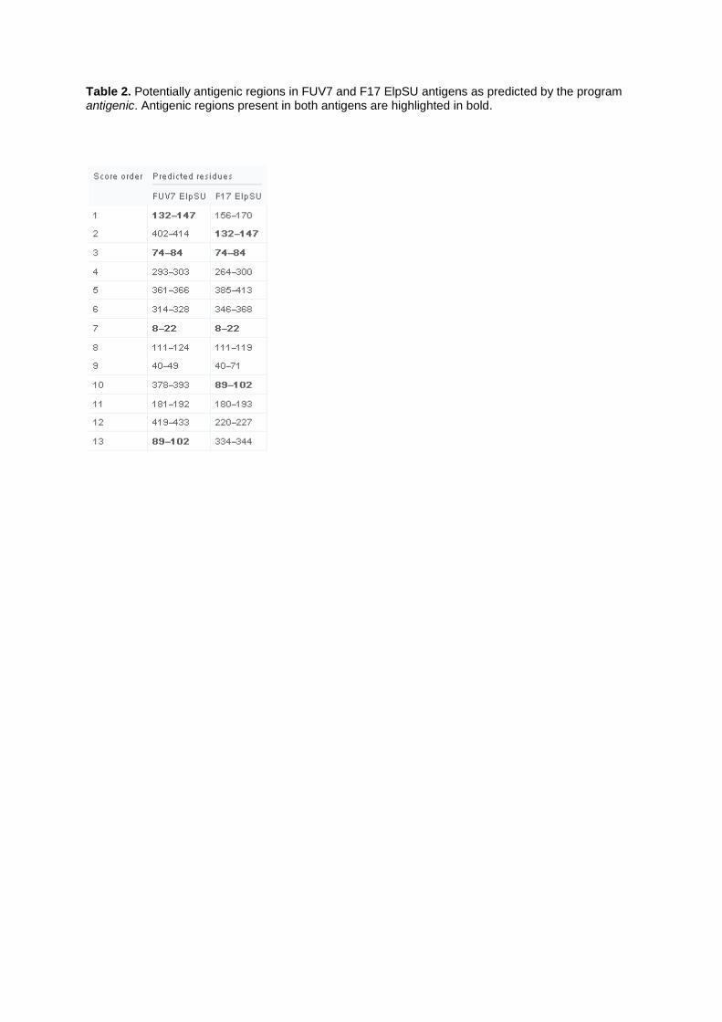
Table 2. Potentially antigenic regions in FUV7 and F17 ElpSU antigens as predicted by the program antigenic. Antigenic regions present in both antigens are highlighted in bold.

Figure 1. Titration of FFV Gag and Bet (A) and F17/951- and FUV-7 ElpSU (Env) antigens (B). FFV antigens were recombinantly expressed in E. coli BL21 as fusion proteins with an N-terminal GST-domain and a C-terminal SV40-tag. 1 μg total E. coli lysate containing the FFV antigens (as indicated) was titrated in 1:3 dilution steps including a SV40-tagged GST-protein. Full-length antigen was detected by an anti-SV40-tag-antibody. Black arrows indicate the amount of FFV antigen initially used for GST-ELISA serology (0.25 μg).
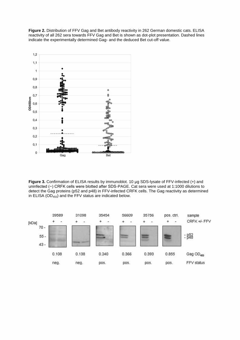
Figure 2. Distribution of FFV Gag and Bet antibody reactivity in 262 German domestic cats. ELISA reactivity of all 262 sera towards FFV Gag and Bet is shown as dot-plot presentation. Dashed lines indicate the experimentally determined Gag- and the deduced Bet cut-off value.
Figure 3. Confirmation of ELISA results by immunoblot. 10 μg SDS-lysate of FFV-infected (+) and uninfected (−) CRFK cells were blotted after SDS-PAGE. Cat sera were used at 1:1000 dilutions to detect the Gag proteins (p52 and p48) in FFV-infected CRFK cells. The Gag reactivity as determined in ELISA (OD450) and the FFV status are indicated below.

Figure 4. Validation of FUV7- and F17/951 ElpSU antigens with cat reference sera. Cats 10, 12, 14, 24 and 26 were Australian cats naturally infected with FFV strains FUV7 and 951 as indicated. Cats 13 and 21 were not infected. FFV infection had been confirmed by serology, virus reisolation and diagnostic PCR (Winkler et al., 1998). Serum from the experimentally FFV FUV7 infected cat 8014 served as positive control, serum from a FFV-negative cat served as negative control. ELISA reactivity shows no discrimination between F17/951- and FUV-7 ElpSU antigens in the GST-ELISA setting.
Figure 5. ELISA reactivity of 11 representative German domestic cat sera against FFV Gag, Bet and F17/951- and FUV-7 ElpSU. As positive control, the serum from the experimentally FFV FUV7-infected cat 8014 was used and a serum from an uninfected cat served as negative control. The F17/951- and FUV-7 ElpSU antigen were similarly recognized by all FFV Env-positive cat sera.

Figure 6. Amino acid alignment of FUV7 and F17/951 ElpSU antigens. Black bars indicate potentially antigenic regions present in both antigens as predicted by the program antigenic. Numbers above the bars indicate the score order of predicted antigenic regions. Asterisks below the aligned sequences mark identical residues, colons mark conservative sequence changes and dots mark semi-conserved sequence changes.