block of l-type calcium channels by charged dihydropyridines
TRANSCRIPT
Block of L-Type Calcium Channels by Charged Dihydropyridines
Sensitivity to Side of Application and Calcium
R. S. KASS, J . P. ARENA, a n d S. CHIN
From the Department of Physiology, University of Rochester Medical Center, Rochester, New York 14642
A B S T R A C T We have studied block of L-type calcium channels by intracellular and extracellular application of the ionized dihydropyridine derivatives amlodipine and SDZ 207-180. We find that extracellular application of either drug causes voltage- dependent block of calcium channels. However, neither drug is effective when applied intracellularly. The insensitivity of calcium channels to intracellular drug is not due to the low concentrations of cytosolic calcium, because voltage-dependent block by ionized amlodipine, SDZ 207-180, and the neutral drug nisoldipine persists under conditions in which Ca 0 is buffered by EGTA. In fact, the time course of the development of block by the ionized but not neutral drug molecules studied, is slower in the presence than in the absence of calcium. Our results indicate that the DHP binding site of the L-type calcium channel is close to the extracellular surface of the cell membrane and that ionized DHP molecules may interact with the receptor in a manner that is uniquely affected by calcium.
I N T R O D U C T I O N
Specific 1,4-dihydropyridine (DHP) ligands have been used as key molecular probes of L-type calcium channels and as powerful therapeutic tools to treat a wide range of cardiovascular disorders (reviewed by Janis and Triggle, 1990). These compounds have been used to isolate and purify the channel protein (Flockerzi et al., 1986; reviewed by Catterall, 1988; Hosey and Lazdunski, 1988). DHP's bind with high affinity to the ~-subuni t of the DHP receptor protein (Galizzi et al., 1986; Sharp et al., 1987; Sieber et al., 1987). However, although the primary structures of rabbit skeletal and heart muscle DHP receptors have been cloned (Tanabe et al., 1987; Mikami et al., 1989) and functional activity of the L-type calcium channel has been expressed (Tanabe et al., 1988; Mikami et al., 1989; Perez-Reyes et al., 1989) from the cloned cDNAs, the molecular identity and location of the DHP binding site have not yet been determined.
Address reprint requests to Dr. Robert S. Kass, Department of Physiology, University of Rochester Medical Center, Rochester, NY 14642.
J. GEN. PHYSIOL. © The Rockefeller University Press • 0022-1295/91/07/0063/13 $2.00 Volume 98 July 1991 63-75
63
Dow
nloaded from http://rupress.org/jgp/article-pdf/98/1/63/1249837/63.pdf by guest on 22 February 2022

64 T H E J O U R N A L O F G E N E R A L P H Y S I O L O G Y - V O L U M E 98 • 1991
We have shown previously that the charged form of amlodipine, a tertiary DHP
molecule, is useful as a probe for the location of the DHP b ind ing site in the cell m e m b r a n e (Kass and Arena, 1989). In the present investigation we have used a combinat ion of charged and neutral DHP compounds to test whether the DHP
b ind ing site, like that for phenylalkamines, is accessible from the inner surface of the cell membrane . We have de te rmined whether block of monovalent ions, like divalent
ions, is voltage dependen t ; and, additionally, have investigated the dependence of
channel block on the absence or presence of calcium.
M E T H O D S
Single ventricular myocytes were isolated from either ventricle of adult guinea pigs using a method similar to that of Mitra and Morad (1985) which has been previously described (Arena and Kass, 1988).
Recording methods were as described by Hamill et al. ( 1981 ) for the whole cell configuration. Patch pipettes were made from Gold Seal Accu-fill 90 Micropets (Clay Adams, Inc., Parsippany, NJ). The resistance of the pipettes was typically 1-3 MI~ when filled with 140 mM CsC1. Series resistance compensation was used in all experiments and was adjusted to give the fastest possible capacity transients without producing ringing. Membrane currents were measured with and voltage was controlled by either a Yale Mark IV (fabricated in this laboratory from parts available at New Haven, CT) or an Axopatch 1C (Axon Instruments, Inc., Foster City, CA) voltage clamp. Cell capacitance was determined directly by capacity compensation of these circuits. Data were sampled once every 0.3 ms and filtered at 1-2 kHz with an eight pole Bessel filter (Frequency Devices, Inc., Haverhill, MA). In experiments designed to dialyze cells with drug-containing solutions, pipette resistances were chosen to be < 2 MI~ and drugs were added to the intracellular solutions at the concentrations indicated for each experiment.
Solutions and Drugs
Solutions were chosen to eliminate K channel currents. Thus, the standard pipette solution contained in millimolar: 60 CsCI, 50 aspartic acid, 68 CsOH, 1 MgCI 2, 1 CaCl z, 11 ethylene- glycol-bis-N,N,N',N'tetraacetic acid (EGTA), 5 K~TP, 10 HEPES (pH 7.4). The standard bath solution contained: 130 mM NaCI, 4.8 CsCI, 5 glucose, 5 HEPES (pH 7.4). In experiments designed to measure divalent ion currents, 2 mM MgClz and divalent charge carriers at indicated concentrations were added to the basic solution. In experiments designed to measure monovalent currents the following were added to the basic extracellular solution: 2 EGTA plus 50 ~M MgCI 2 (for the measurement of Na currents) or 2 ethylenediaminetetraacetic acid (EDTA) and 500 ~M MgCI z (for the measurement of outward currents). Sodium channels were eliminated by 10-50 p~M tetrodotoxin (TTX) (Behring Diagnostics, LaJolla, CA) and by replacement of Na by N-methyl-n-glucamine in some experiments.
The structures of the three DHP compounds used in this study are shown in Fig. 1. Nisoldipine and SDZ-207-180 were dissolved in polyethylene glycol 400 (PEG) to make a concentrated (1 raM) stock and diluted in the bath to the final concentration. PEG at the concentration used (<0.01%) has been shown to have no effects of its own on Ca channel currents (Kass, 1982). Amlodipine was dissolved in water as a concentrated (1 raM) stock solution. Amlodipine concentrations were chosen as previously described (Burges et al., 1985, 1987; Kass and Arena, 1989). SDZ-207-180 was a gift of Sandoz, Ltd., Basle, Switzerland; nisoldipine was a gift from Miles Laboratories, New Haven, CT; and amlodipine was a gift from Pfizer Central Research, Sandwich, UK.
Dow
nloaded from http://rupress.org/jgp/article-pdf/98/1/63/1249837/63.pdf by guest on 22 February 2022

KASS ET AL. Block of L-Type Ca Channels by Charged Dihydropyridines 65
Voltage Protocols
Membrane potential regulates DHP modulation of/ca: block is promoted at positive voltages and relieved at negative voltages (for example, see Sanguinetti and Kass, 1984). The time course of the development or removal of block can be determined by applying a series of test voltages from different holding potentials. In the present study voltage protocols described in detail and illustrated in Kass and Arena (1989) were used to determine onset and recovery of block. Drug onset was measured by applying a depolarizing "train" protocol in which the holding potential was changed from - 8 0 to - 4 0 or - 5 0 mV and test voltage pulses were applied once every 5 s. Test voltages were either 0 mV (Na + currents), + 10 mV (Ca z+ and Ba 2+ currents), or +40 to +60 mV (Cs ÷ and K ÷ currents) and test pulse duration was 200 ms. This protocol was previously shown to maximize the onset of voltage-dependent block by ionized
c ~ o ~ ---oc~s
AMU3DII~TE
c ~ o ~ c.--~, ,~ r - -c - - -oc~m
NISOLDIPII~
FIGURE l. Structures of the three DHP molecules used in this study: amlodipine, nisoldipine, and SDZ 207-180. The location of the charged groups are indicated for pH 7.4.
. / o o
/cxo~ c ~ l~l---c~o(c~.~,(c,.).-
SDZ-207-150
DHP compounds (Kass and Arena, 1989). After development of block recovery was measured using train protocols in which the holding potential was changed from - 4 0 to - 8 0 mV and pulses were applied to the same test potentials, but test pulse duration was either 20 or 40 ms to minimize pulse-induced inactivation.
In experiments that required recording currents from potentials negative to - 6 0 mV, 50-100-ms prepulses were applied to - 4 0 mV to inactivate sodium channel and T-type calcium channel currents (Bean, 1985; Marchetti and Brown, 1988). Thus, in this paper current referred to as Ca channel current (/ca) corresponds to L-type Ca channel current according to the terminology suggested by Nilius et al. (1985).
DHP Receptor: Agonism and Antagonism
Most DHP compounds are capable of causing enhancement or block of/Ca depending on cell membrane potential (Hess et al., 1984; Sanguinetti et al., 1986; Kass, 1987). Binding and
Dow
nloaded from http://rupress.org/jgp/article-pdf/98/1/63/1249837/63.pdf by guest on 22 February 2022

66 T H E J O U R N A L OF GENERAL PHYSIOLOGY • V O L U M E 9 8 • 1 9 9 1
electrical data have provided evidence that these effects are due to interactions with more than one binding site (Brown et al., 1986; Kokubun et al., 1986; Wei et al., 1986; Williams et al., 1985; Hamilton et al., 1987). The binding site probed in this study is associated with inhibition of channel activity. We rarely measured agonistic responses to SDZ 207-180 applied under conditions of negative ( -80 to -100 mV) holding potentials, but under similar conditions observed enhancement of currents by amlodipine at pH 7.4 in ~30% of our experiments. Further investigations will be needed to test whether electrical evidence with charged DHP derivatives can support the view that distinct binding sites exist for agonism and antagonism of L-channels.
Curve Fitting and Statistical Procedures
Where appropriate, experimental data were fitted with functions of one or two exponentials plus an arbitrary baseline using procedures previously described (see Sanguinetti and Kass, 1984). All data in the text and figures are means -+ SEM. T-tests were used to determine statistical significance between groups (Rosner, 1986). Multiple comparisons were made using the pooled estimate of variance from a one-way ANOVA.
R E S U L T S
The DHP Binding Site Is Accessed from the Outside but Not the Inside of the Cell
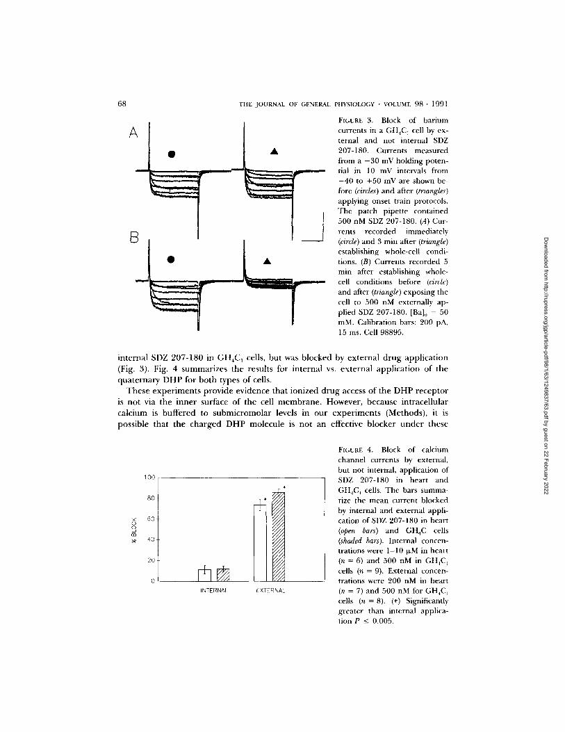
To test for an intracellular location of the DHP binding site, we measured the sensitivity o f calcium channel current to internal and external application of the permanent ly charged drug SDZ-207-180. The charge on this compound will restrict it from diffusing out of the cell across the lipid membrane (Hille, 1977). As a control for internal dialysis, we first measured the effects of internally applied D890, a permanent ly charged phenylalkamine derivative that has been shown to block I~:~, from the inside of cardiac cells (Heschler et al., 1982; Lee and Tsien, 1984). We used pulse-dependent block dur ing train protocols to assay drug activity and, as shown in Fig. 2 A, there is a progressive increase in block as this compound diffuses into the cell. In contrast, we found that SDZ 207-180 had little effect when applied internally. This can be seen in Fig. 2 B, which compares pulse-dependent block after 20 min of dialysis with 10 I~M intracellular SDZ 207-180 to a 3-min exposure to 200 nM SDZ 207-180 applied externally to the same cell. We found a similar pat tern when we compared intracellular with extracellular application of ionized amlodipine (data not illustrated): currents were only blocked by extracellularly applied drug.
Because o f the large size of isolated heart cells, we were concerned that diffusional exchange between the cell interior and patch pipette might be limited within the time frame of our experiments. Rates of diffusional exchange can be estimated for heart cells (Cx ~ 1 0 0 pF) by the methods of Pusch and Neher (1988). For pipette resistances on the order of 3 MI) (typical for these experiments), and for a molecular mass of 350 D, the diffusion time constant is 15 rain. To confirm the insensitivity of Ic:a to internal application of SDZ 207-180, we compared our results obtained in heart cells with experiments carried out in GH4C1 pituitary tumor cells. These cells are small (mean Cr = 12.65 + 1.2 pF, N = 12) and the predicted diffusional time constant is 40 s (Pusch and Neher, 1988). For these cells, we found that internally applied D890 (100 IxM) completely blocked lea, consistent with the predictions of Pusch and Neher. However, as was the case for heart cells, Ic, was not blocked by
Dow
nloaded from http://rupress.org/jgp/article-pdf/98/1/63/1249837/63.pdf by guest on 22 February 2022

KASS ET AL. Block of L-Type Ca Channels by Charged Dihydropyridines 67
A INTERNAL D890 IN HEART
' "
• " - . . - .
N 0 . 5 0 . mm~mm~mm..~ A"-"'= "--.-,,--,L ~ , L
I I ~ I I ...-... i1...~
z 0.25- - - ,L~= ,__
0 .00 , J i 0 20 40 60
TIME IN SECONDS
[3 SDZ INTERNAL AND EXTERNAL APPLICATION IN HEART
sso,
~. 45o
i 350
250-
~'@[email protected]@--'@-'-@~.@_..@.._@.... @.... @_._ @._.@[email protected]
/ A ~ A \
150 , I , 0 20 40 60
TIME tN SECONDS
FIGURE 2. Effects of internal and external application of quaternary Ca channel blockers in heart cells. Currents were measured during the application of onset train protocols with permanently charged drugs added to the whole-cell patch pipettes. (A) D890 (100 i~M) was included in the pipette. Currents were measured during onset trains applied 5, 10, and 15 rain after establishing whole-cell recording conditions. Currents at start of each train were 954, 871, and 754 pA, respectively. Cell 26081. Charge carrier: 2 mM Ca. (B) SDZ 207-180 was (1 I,M) added to pipette and no pulse-dependent block developed. The solid circles are currents measured during an onset train applied 23 rain after establishing whole-cell conditions. The cell was then exposed to an external solution containing 200 nM SDZ 207-180. The solid triangles are currents recorded in response to the same onset train protocol 3 min after changing to this external solution. Cell 11081.
Dow
nloaded from http://rupress.org/jgp/article-pdf/98/1/63/1249837/63.pdf by guest on 22 February 2022
68
A II
p,._ I]
B ]" ~ l l l I
THE JOURNAL OF GENERAL PHYSIOLOGY • VOLUME 98" 1991
A
--7
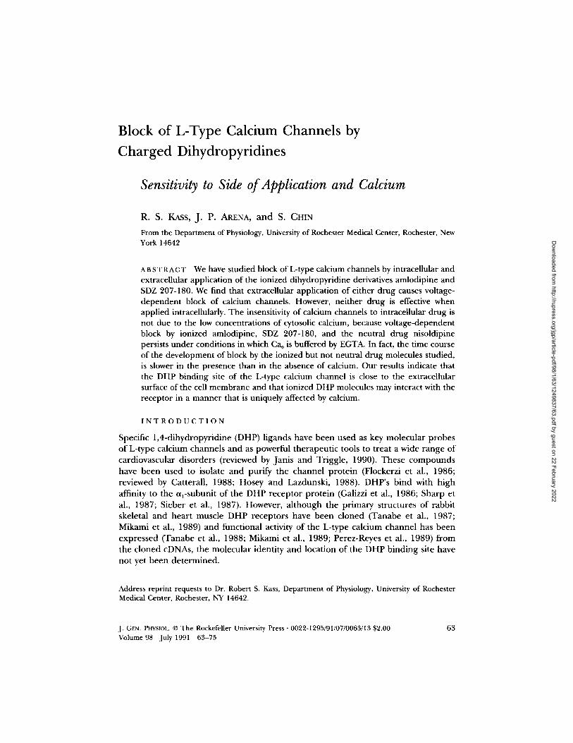
FIGURE 3. Block of barium currents in a GH4C ] cell by ex- ternal and not internal SDZ 207-180. Currents measured from a - 3 0 mV holding poten- tial in 10 mV intervals from - 4 0 to +50 mV are shown be- fore (circles) and after (triangles) applying onset train protocols. The patch pipette contained 500 nM SDZ 207-180. (A) Cur- rents recorded immediately (circle) and 3 rain after (triangle) establishing whole-cell condi- tions. (B) Currents recorded 5 min after establishing whole- cell conditions before (circle) and after (triangle) exposing the cell to 500 nM externally ap- plied SDZ 207-180. [Ba]0 = 50 mM. Calibration bars: 200 pA, 15 ms. Cell 98895.
in t e rna l SDZ 207-180 in GH4C 1 cells, bu t was b locked by e x t e r n a l d r u g app l i ca t ion
(Fig. 3). Fig. 4 s u m m a r i z e s t he resul ts for i n t e rna l vs. e x t e r n a l app l i ca t ion o f the
q u a t e r n a r y D H P for b o t h types o f cells.
T h e s e e x p e r i m e n t s p r o v i d e ev idence tha t i on i zed d r u g access o f the D H P r e c e p t o r
is no t via the i n n e r surface o f the cell m e m b r a n e . However , because in t race l lu la r
ca lc ium is b u f f e r e d to s u b m i c r o m o l a r levels in o u r e x p e r i m e n t s (Methods) , it is
poss ible tha t the c h a r g e d D H P m o l e c u l e is no t an effect ive b locke r u n d e r these
100
80-
60
40
20
INTERNAL
///l
z/ix 1//1 z//z
ll/l IIII IIII z/X/ 11/I I]]I IIIi
till [I// fill
IIII
rzz¢
EXTERNAL
FIGURE 4. Block of calcium channel currents by external, but not internal, application of SDZ 207-180 in heart and GH4C n cells. The bars summa- rize the mean current blocked by internal and external appli- cation of SDZ 207-180 in heart (open bars) and GH4C cells (shaded bars). Internal concen- trations were 1-10 ~M in heart (n = 6) and 500 nM in GH4C 1 cells (n = 9). External concen- trations were 200 nM in heart (n = 7) and 500 nM for GH4C ] cells (n = 8). (*) Significantly greater than internal applica- tion P _< 0.005.
Dow
nloaded from http://rupress.org/jgp/article-pdf/98/1/63/1249837/63.pdf by guest on 22 February 2022

KASS Err AL. Block of L-Type Ca Channels by Charged Dihydropyridines 69
conditions. We tested for this possibility by measur ing the influence of calcium on the
voltage dependence of SDZ 207-180 block.
Influence of Calcium on DHP Block and Recovery: Contrast between Neutral and Charged Drugs
Fig. 5 shows examples of exper iments in which SDZ 207-180 block was studied u n d e r condit ions in which calcium carried the charge and in which Ca 0 was buffered and Na or Cs were the charge carriers. Records are shown in the absence of drug, and dur ing the onset of SDZ 207-180 block of currents. This c o m p o u n d blocks in a voltage- d e p e n d e n t m a n n e r when calcium is the charge carrier, and when Ca 0 is buffered and
C a +2 Na +
0 20 40 60 TIME IN SECONDS
FIGURE 5. Influence of calcium on voltage-dependent block by SDZ 207-180. Current traces illustrate Ca, Na, and Cs ions carrying the charge during onset train protocols in the presence of 200 nM SDZ 207-180. Traces are shown at the start of the train protocol, 50 s, and 100 s after applying pulses (Na, Cs), and 100 and 200 s after starting the onset train (Ca). Monovalent currents were measured in the presence of EGTA and EDTA (Methods). The plot shows normalized currents measured in the absence (open symbols) and presence (solid symbols) of 200 nM SDZ 207-180 as a function of time after start of the train protocol. The permeant ions were Ca (squares), Cs (circles), and Na (triangles). The tonic block at start of train protocols for each charge carrier was 13% (Ca), 0% (Na), and 14% (Cs). Calibration bars: 18 ms; 400 pA (Ca), 1.3 nA (Na), and 3.14 nA (Cs). Cells: 11391 (Ca); 111491 (Na); 22AG94 (Cs).
Dow
nloaded from http://rupress.org/jgp/article-pdf/98/1/63/1249837/63.pdf by guest on 22 February 2022
70 THE JOURNAL OF GENERAL PHYSIOLOGY • VOLUME 9 8 . 1991
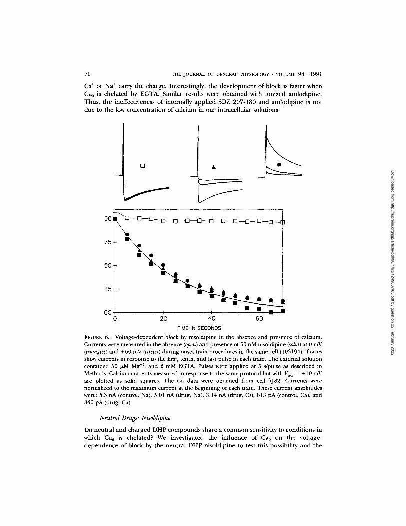
Cs ÷ or Na + carry the charge. Interestingly, the development of block is faster when Ca0 is chelated by EGTA. Similar results were obtained with ionized amlodipine. Thus, the ineffectiveness of internally applied SDZ 207-180 and amlodipine is not due to the low concentra t ion of calcium in our intracellular solutions.
L
301
75-
50~
0
f
2 5 6 • i •
• J n n O0 ~ I ,
0 20 40 60
TIME IN SECONDS
FIGURE 6. Voltage-dependent block by nisoldipine in the absence and presence of calcium. Currents were measured in the absence (open) and presence of 50 nM nisoldipine (solid) at 0 mV (triangles) and +60 mV (circles) during onset train procedures in the same cell (103194). Traces show currents in response to the first, tenth, and last pulse in each train. The external solution contained 50 I~M Mg +z, and 2 mM EGTA. Pulses were applied at 5 s/pulse as described in Methods. Calcium currents measured in response to the same protocol but with V,e~, = + 10 mV are plotted as solid squares. The Ca data were obtained from cell 7J82. Currents were normalized to the maximum current at the beginning of each train. These current amplitudes were: 5.3 nA (control, Na), 5.01 nA (drug, Na), 3.14 nA (drug, Cs), 813 pA (control, Ca), and 840 pA (drug, Ca).
Neutral Drugs: Nisoldipine
Do neutral and charged DHP compounds share a common sensitivity to condit ions in which Ca0 is chelated? We investigated the influence of Ca0 on the voltage- dependence of block by the neutral DHP nisoldipine to test this possibility and the
Dow
nloaded from http://rupress.org/jgp/article-pdf/98/1/63/1249837/63.pdf by guest on 22 February 2022

KASS ET AL. Block of L-Type Ca Channels by Charged Dihydropyridines 71
results are presented in Fig. 6. Under conditions in which Ca0 is chelated, block of monovalent currents by nisoldipine is voltage-dependent, and the time course of block onset is affected neither by the marked difference in test pulse voltage nor in the ionic species carrying the charge. However, in contrast to the actions of the ionized molecules, the onset of nisoldipine block is unchanged when Ca0 is not buffered and calcium is the charge carrier.
D I S C U S S I O N
The major new result presented in this paper is that the permanently charged compound SDZ 207-180 and ionized amlodipine are much more potent when applied extracellularly compared with intracellular application. In addition, we find that the development of voltage-dependent block by ionized and neutral DHP molecules persists under conditions when calcium is buffered to micromolar concen- trations, but that the onset of block by the two ionized drugs is slower in the presence than in the absence of calcium.
Extracellular Access to the DHP Binding Site
Our experiments in which we compared intracellular and extracellular application of ionized amlodipine and SDZ 207-180 in heart and GH4C1 cells provide evidence that these two charged DHP compounds cannot reach the DHP receptor via an intracel- lular pathway. This finding contrasts with the accessibility of the permanently charged drug D890 to the phenylalkamine receptor which occurs via an intracellular pathway in guinea pig ventricular cells (Heschler et al., 1982), but not in vascular smooth muscle cells of the rabbit (Leblanc and Hume, 1989). Our result is, however, consistent with data reported by Iijima et al. (1984) for intracellular application of the DHP nicardipine. The difference in intracellular accessibility of the two permanently charged drug types supports the view that the binding sites for DHPs and phenyl- alkamines on the ~l-subunit of the DHP receptor protein are physically separate entities (Glossmann et al., 1984).
Location of the DHP Binding Site
Valdivia and Coronado (1990) have reported that SDZ 207-180 can interact with a high affinity DHP binding site when applied to either side of a planar lipid bilayer into which skeletal muscle transverse tubule Ca channels had been incorporated. They suggested that the 10 methylene groups that separate the quaternary ammo- nium from the DHP ring in this molecule (Fig. 1) are sufficiently long to allow the DHP moiety to be uncharged and reach the DHP binding site through the lipid bilayer regardless of the side of application. If this view is correct, then our results, which show an asymmetrical response in both intact heart and G H 4 C J cells, suggest that the high affinity DHP binding site is closer to the extracellular membrane face than the intracellular face and is just beyond the reach of the methylene spacer chain when applied from the intracellular side of the membrane. These results are consistent with conclusions reached from investigations of the sensitivity amlodipine to changes in external hydrogen ion concentrations (Kass and Arena, 1989).
Dow
nloaded from http://rupress.org/jgp/article-pdf/98/1/63/1249837/63.pdf by guest on 22 February 2022

72 T H E JOURNAL OF GENERAL PHYSIOLOGY • VOLUME 9 8 . 1991
Calcium Ions Modify Block by Ionized DHP Derivatives
Our results cannot be explained by a change in blocking activity of SDZ 207-180 due to the low levels of intracellular calcium because we find that voltage dependence of block by neutral and ionized DHP molecules is preserved under conditions in which calcium is buffered to micromolar concentrations by either EGTA or EDTA (see also Hess et al., 1986; Hadley and Hume, 1987). However, under conditions in which calcium is the permeant ion, block by ionized amlodipine and SDZ 207-180 develops at a slower rate than under conditions when Ca0 is buffered and monovalent ions carry the charge. Block by the neutral molecule nisoldipine is not affected by chelating Ca0. Recovery from block by these two ionized DHPs, but not nisoldipine, is also slower when calcium is the charge carrier (data not shown).
These results are similar to those of Carbone and Lux (1988a, b, 1989) who reported that t0-Conotoxin (¢o-CTX) blockade of high threshold (L-type) calcium channels differed markedly depending on the type of ion carrying charge through the open channel pore. Those authors suggested that 00-CTX binding was influenced by charge carrier-induced channel conformational changes. Permeating charge species-induced differences in channel conformation are expected since channel permeation is determined by two intrapore binding sites with varying affinity for monovalent and divalent ions (Almers and McCleskey, 1984; Hess et al., 1986). In fact, Prod'hom et al. (1989) have reported that charge carrier-induced conforma- tional changes underlie the modulation of proton-induced fluctuations in L-channel conductance levels, and suggested that Ca forms a stabilizing complex with the calcium channel protein and limits the flexibility of the channel protein that exists when other ion species permeate the channel. Our results are consistent with this view of the channel, but cannot rule out the possibility that it is an interaction between the charged head groups of amlodipine or SDZ 207-180 with extracellular calcium that interacts with the drug-bound state of the channel. Future experiments with other ionized drug molecules will be useful in distinguishing these possibilities.
SUMMARY
In summary, our results have shown that charged DHP derivatives are very useful as probes of the L-type channel in intact cell membranes. We have found that ionized amlodipine and the permanently charged derivative SDZ 207-180 are useful in determining the location of the DHP binding site that causes inhibition of channel activity. Studies of other DHP derivatives with different charge locations will further define the location of this site and provide insight into possible conformational changes L-channels undergo for varied permeant ions.
Original version received 5 February 1990 and accepted version received 15 November 1990.
R E F E R E N C E S
Almers, W., and E. W. McClesky. 1984. Non-selective conductance in calcium channels of frog muscle: calcium selectivity in a single-file pore. Journal of Physiology. 353:585-608.
Arena, J. P. and R. S. Kass. 1988. Block of heart potassium channels by clofilium and its tertial 7 analogs: relationship between dtaig structure and type of channel blocked. Molecular Pharmacolo~. 34:60-66.
Dow
nloaded from http://rupress.org/jgp/article-pdf/98/1/63/1249837/63.pdf by guest on 22 February 2022

KASS ET AL. Block of L-Type Ca Channels by Charged Dihydropyridines 73
Bean, B. P. 1985. Two kinds of calcium channels in canine atrial cells. Differences in kinetics,
selectivity, and pharmacology.Journal of General Physiology. 86:1-30.
Brown, A. M., D. L. Kunze, and A. Yatani. 1986. Dual effects of dihydropyridines on whole cell and
unitary calcium currents in single ventricular cells of guinea-pig.Journal of Physiology. 379:495-514.
Burges, R. A., A. J. Carter, D. F. Gardiner, and A. J. Higgins. 1985. Amlodipine, a new dihydropyridine calcium channel blocker with slow onset and long duration of action. British Journal of Pharmacology. 85:281P.
Burges, R. A., D. G. Gardiner, M. Gwih, A. J. Higgins, K. J. Blackburn, S. F. Campbell, P. E. Cross,
and J. K. Stubbs. 1987. Calcium channel blocking properties of amlodipine in vascular smooth muscle and cardiac muscle In Vitro: evidence for voltage modulation of vascular dihydropyridine
receptors. Journal of Cardiovascular Pharmacology. 9:110-119. Carbone, E., and H. D. Lux. 1988a. ¢0-Conotoxin blockade distinguishes Ca from Na permeable
states in neuronal calcium channels. Pfliigers Archive 413:14-22.
Carbone, E., and H. D. Lux, 1988b. Sodium currents through neuronal calcium channels: kinetics
and sensitivity to calcium antagonists. In The Calcium Channel: Structure, Function, and
Implications. M. Morad, W. Nayler, S. Kazda, and M. Schramm, editors. Springer-Verlag,
Heidelberg. 115-126. Carbone, E., and H. D. Lux. 1989. Modulation of Ca channels in peripheral neurons. Annals of the
New York Academy of Sciences. 560:346-357. Catterall, William A. 1988. Structure and function of voltage-sensitive ion channels. Science. 242:50-
60. Flockerzi, V., H. J. Oeken, F. Hofmann, D. Pelzer, A. Cavalie, and W. Trautwein. 1986. Purified
dihydropyridine-binding site from skeletal muscle T-tubules is a functional calcium channel.
Nature. 323:66-68. Galizzi, J. P, M. Borsotto, J. Barhanin, M. Fosset, and M. Lazdunski. 1986. Characterization and
photoaffinity labeling of receptor sites for Ca channel inhibitors d-cis-Diltiazem, Bepridil, Des-
methoxyverapamil, and (+)-PN 200-110 in skeletal muscle transverse tubule membranes.Journal of Biological Chemistry. 144:211-215.
Glossmann, H., D. R. Ferry, A. Goll, J. Striessnig, and G. Zernig. 1984. Calcium channels:
introduction into their molecular pharmacology. In Cardiovascular Effects of Dihydropyridine-
Type Calcium Antagonists and Agonists. A. Fleckenstein, C. Van Breemen, R. Gross, and F.
Hoffmeister, editors. Springer-Verlag, Heidelberg. 113-139. Hadley, R. W., and J. R. Hume. 1987. An intrinsic potential-dependent inactivation mechanism
associated with calcium channels in guinea-pig myocytes. Journal of Physiology. 389:205-222. Hamill, O. P., A. Marty, E. Neher, B. Sakmann, and F. J. Sigworth. 1981. Improved patch-clamp
techniques for high-resolution current recording from cells and cell-free membrane patches.
Pfliigers Archiv. 391 : 85-100. Hamilton, S. L., A. Yatani, K. Brush, A. Schwartz, and A. M. Brown. 1987. A comparison between the
binding and electrophysiological effects of dihydropyridines on cardiac membranes. Molecular Pharmacology. 31:221-231.
Hescheler, J., Pelzer, D., Trube, G., and W. Trautwein. 1982. Does the organic calcium channel blocker D600 act from inside or outside on the cardiac cell membrane? Pfliigers Archiv. 393:287-
291. Hess, P., J. B. Lansman, and R. W. Tsien. 1984. Different modes of gating behaviour favoured by
dihydropyridine agonists and antagonists. Nature. 311:538-544.
Hess, P., J. B. Lansman, and R. W. Tsien. 1986. Calcium channel selectivity for divalent and
monovalent cations. Voltage and concentration dependence of single channel current in ventricu- lar heart cells. Journal of General Physiology. 88:293-319.
Dow
nloaded from http://rupress.org/jgp/article-pdf/98/1/63/1249837/63.pdf by guest on 22 February 2022

74 THE JOURNAL OF GENERAL PHYSIOLOGY • VOLUME 98 • 199l
Hille, B. 1977. Local anesthetics: hydrophilic and hydrophobic pathways for the drug-receptor
reaction. Journal of General Physiology. 69:497-515.
Hosey, M. H., and M. Lazdunski. 1988. Calcium channels: molecular pharmacology, structure and
regulation. Journal of Membrane Biology. 104:81-105.
Iijima, T., T. Yanagisawa, and N. Taira. 1984. Increase in the slow inward current by intracellularly
applied nifedipine and nicardipine in single ventricular cells of the guinea-pig heart. Journal of. Molecular and Cellular Cardiology. 16:1173-1177.
Janis, R. A., and D. J. Triggle. 1991. Drugs acting on calcium channels. In The Calcium Channel: Its
Properties, Function, Regulation and Clinical Relevance. L. Hurwitz, L. D. Partridge, and J. K.
Leach, editors. CRC Press, Boca Raton, FL.
Kass, R. S. 1982. Nisoldipine: a new, more selective calcium current blocker in cardiac Purkinje
fibers.Journal of Pharmacology and Experimental Therapeutics. 223:446-456.
Kass, R. S. 1987. Voltage-dependent modulation of cardiac calcium channel current by optical
isomers of Bay K8644: implications for channel gating. Circulation Research Supplement. 61 :II-I 15.
Kass, R. S., and J. P. Arena. 1989. Influence of pH 0 on calcium channel block by amlodipine, a
charged dihydropyridine compound: implications for location of the dihydropyridine receptor.
Journal of General Physiology. 93:1109-1127.
Kass, R. S., and D. S. Krafte. 1987. Negative surface charge density near heart calcium channels.
Relevance to block by dihydropyridines. Journal of General Physiology. 89:629-644.
Kokubun, S., B. Prod'hom, C. Becker, H. Porzig, and H. Reuter. 1986. Studies on Ca channels in
intact cardiac cells: voltage-dependent effects and cooperative interactions of dihydropyridine
enantiomers. Molecular Pharmacology. 30:751-584.
Leblanc N., and J. R. Hume. 1989. D600 block of L-type Ca ~÷ channel in vascular smooth muscle
cells: comparison with permanently charged derivative, D890. American Journal of" Physiology. 257:C689-C695.
Lee, K. S., and R. W. Tsien. 1983. Mechanism of calcium channel blockade by verapamil, D600,
dihiazem and nitrendipine in single dialyzed heart cells. Nature. 302:790-794.
Lee, K. S., and R. W. Tsien. 1984. High selectivity of Ca channels in single dialyzed heart cells of the
guinea pig.Journal of Physiology. 354:253-272.
Marchetti, C., and A. M. Brown. 1988. Protein kinase activator 1-oleoyl-2-acetyl-sn-glycerol inhibits
two types of calcium currents in GH3 cells. American Journal of Physiology (Cell). 23:C206-C210.
Mikami, A., K. lmoto, T. Tanabe, T. Niidome, Y. Mori, H. Takeshima, S. Narumiya, and Shosaku
Numa. 1989. Primary Structure and tunctional expression of the cardiac dihydropyridine-sensitive
calcium channel. Nature. 340:230-233.
Mitra, R., and M. Morad. 1985. A uniform enzymatic method for dissociation of myocytes from hearts
and stomachs of vertebrates. American Journal of Physiology. 249:H 1056-H 1060. Nilius, B., P. Hess, J. B. Lansman, and R. W. Tsien. 1985. A novel type of cardiac calcium channel in
ventricular cells. Nature. 316:443-446.
Perez-Reyes, E., H. S. Kim, A. E. Lacxerda, W. Horne, X. Wei, D. Rampe, K. P. Campbell, A. M.
Brown, and L. Birnbaumer. 1989. Induction of calcium currents by the expression of the cq-subunit
of the dihydropyridine receptor from skeletal muscle. Nature. 340:233-236.
Prod'hom, B., D. Pietrobon, and P. Hess. 1989. Interactions of protons with single open L-type
calcium channels. Location of protonation site and dependence of proton-induced current
fluctuations on concentration and species of permeant ion.Journal of General Physiology. 94:23-42. Pusch, M., and E. Neher. 1988. Rates of diffusional exchange between small cells and a measuring
patch pipette. Pfliigers Archiv. 411:204-211.
Dow
nloaded from http://rupress.org/jgp/article-pdf/98/1/63/1249837/63.pdf by guest on 22 February 2022

KASS ~T AL. Block of L-Type Ca Channels by Charged Dihydropyridines 75
Sanguinetti, M. C., and R. S. Kass. 1984. Voltage-dependent block of calcium channel current in the calf cardiac Purkinje fiber by dihydropyridine calcium channel antagonists. Circulation Research. 55:336-348.
Sanguinetti, M. C., D. S. Krafte, and R. R. Kass. 1986. Bay K8644: voltage-dependent modulation of Ca channel current in heart cells. Journal of General Physiology. 88:369-392.
Sharp, A. H., T. Imagawa, A. T. Leung, and K. P. Campbell. 1987. Identification and characterization of dihydropyridine-binding subunit of the skeletal muscle dihydropyridine receptor. Journal of Biological Chemistry. 262:12309-12315.
Sieber, M., W. Nastainczyk, V. Zubor, W. Wernet, and F. Hormann. 1987. The 165-KDa peptide of the purified skeletal muscle dihydropyridine receptor contains the known regulatory sites of the calcium channel. European Journal of Biochemistry. 176:117-122.
Tanabe, T., K. Beam, J. A. Powell, and S. Numa. 1988. Restoration of excitation-contraction coupling and slow calcium current in dysgenic muscle by dihydropyridine receptor complementary DNA. Nature. 336:134-139.
Tanabe, T., H. Takeshima, A. Mikami, V. Fiockerzi, H. Takahashi, K. Kangawa, M. Kojima, H. Matsuo, T. Hiose, and S. Numa. 1987. Primary structure of the receptor for calcium channel blockers from skeletal muscle. Nature. 328:313-318.
Tsien, R. W., P. Hess, E. W. McCleskey, and R. L. Rosenberg. 1987. Calcium channels: mechanisms of selectivity, permeation, and block. Annual Review of Biophysics and Biophysical Chemistry. 16:265- 290.
Valdivia, H., and R. Coronado. 1990. Internal and external effects of dihydropyridines in the calcium channel of skeletal muscle.Journal of General Physiology. 95:1-27.
Wei, X. Y., E. M. Luchowski, A. Rutledge, C. M. Su, and D. M. Triggle. 1986. Pharmacological and radioligand binding analysis of the actions of 1,4-dihydropyridine activator-antagonist pairs in smooth muscle. Journal of Pharmacology and Experimental Therapeutics. 239:144-153.
Williams, J. S., I. L. Grupp, G. Grupp, P. L. Vaghy, L. Dumont, A. Schwartz, A. Yatani, S. Hamilton, and A. M. Brown. 1985. Profile of the oppositely acting enantiomers of the dihydropyridine 202-791 in cardiac preparations: receptor binding, electrophysiological and pharmacological studies. Biochemical and Biophysical Research Communications. 131:13-21.
Dow
nloaded from http://rupress.org/jgp/article-pdf/98/1/63/1249837/63.pdf by guest on 22 February 2022