blocking tumor-educated msc paracrine activity halts...
TRANSCRIPT

Cancer Therapy: Preclinical
Blocking Tumor-Educated MSC ParacrineActivity Halts Osteosarcoma ProgressionS. Rubina Baglio1, Tonny Lagerweij2, Maria P�erez-Lanz�on1, Xuan Dung Ho3,4,5,Nicolas L�eveill�e6, Sonia A. Melo7, Anne-Marie Cleton-Jansen8,Ekaterina S. Jordanova9, Laura Roncuzzi10, Michelina Greco10,Monique A.J. van Eijndhoven1, Giulia Grisendi11, Massimo Dominici11,Roberta Bonafede1,12, Sinead M. Lougheed13, Tanja D. de Gruijl13, Nicoletta Zini14,15,Silvia Cervo16,17, Agostino Steffan16,17, Vincenzo Canzonieri16,18, Aare Martson4,19,Katre Maasalu4,19, Sulev K€oks3,20, Tom Wurdinger2,21, Nicola Baldini10,22, andD. Michiel Pegtel1
Abstract
Purpose: Human osteosarcoma is a genetically heterogeneousbonemalignancywith poor prognosis despite the employment ofaggressive chemotherapy regimens. Because druggable drivermutations have not been established, dissecting the interactionsbetween osteosarcoma cells and supporting stroma may provideinsights into novel therapeutic targets.
Experimental Design: By using a bioluminescent orthotopicxenograft mouse model of osteosarcoma, we evaluated the effectof tumor extracellular vesicle (EV)–educated mesenchymal stemcells (TEMSC) on osteosarcoma progression. Characterizationand functional studies were designed to assess the mechanismsunderlying MSC education. Independent series of tissue speci-mens were analyzed to corroborate the preclinical findings, andthe composition of patient serumEVswas analyzed after isolationwith size-exclusion chromatography.
Results: We show that EVs secreted by highly malignantosteosarcoma cells selectively incorporate a membrane-associ-ated form of TGFb, which induces proinflammatory IL6 pro-
duction by MSCs. TEMSCs promote tumor growth, accompa-nied with intratumor STAT3 activation and lung metastasisformation, which was not observed with control MSCs. Impor-tantly, intravenous administration of the anti-IL6 receptorantibody tocilizumab abrogated the tumor-promoting effectsof TEMSCs. RNA-seq analysis of human osteosarcoma tissuesrevealed a distinct TGFb-induced prometastatic gene signature.Tissue microarray immunostaining indicated active STAT3 sig-naling in human osteosarcoma, consistent with the observa-tions in TEMSC-treated mice. Finally, we isolated pure popula-tions of EVs from serum and demonstrated that circulatinglevels of EV-associated TGFb are increased in osteosarcomapatients.
Conclusions:Collectively, our findings suggest that TEMSCspromote osteosarcoma progression and provide the basisfor testing IL6- and TGFb-blocking agents as new therapeuticoptions for osteosarcoma patients. Clin Cancer Res; 23(14);3721–33. �2017 AACR.
1Department of Pathology, Cancer Center Amsterdam, VU University Med-ical Center, Amsterdam, the Netherlands. 2Department of Neurosurgery,Cancer Center Amsterdam, VU University Medical Center, Amsterdam, theNetherlands. 3Department of Pathophysiology, University of Tartu, Tartu,Estonia. 4Department of Traumatology and Orthopedics, University ofTartu, Tartu, Estonia. 5Department of Oncology, Hue College of Medicineand Pharmacy, Hue University, Hue, Vietnam. 6Laboratory for ExperimentalOncology and Radiobiology, Center for Experimental Molecular Medicine(CEMM), Academic Medical Center (AMC), Amsterdam, the Netherlands.7Instituto de Investigac~ao e Inovac~ao em Sa�ude, Universidade do Porto (i3S)and Institute of Molecular Pathology and Immunology of the University ofPorto (IPATIMUP), 4200 Porto, Portugal. 8Department of Pathology, LeidenUniversity Medical Center, Leiden, the Netherlands. 9Department of Obstet-rics and Gynecology, Center for Gynecological Oncology Amsterdam, VUUniversity Medical Center, Amsterdam, the Netherlands. 10OrthopaedicPathophysiology and Regenerative Medicine Unit, Istituto Ortopedico Riz-zoli, Bologna, Italy. 11Division of Oncology, Department of Medical andSurgical Sciences for Children and Adults, University-Hospital of Modenaand Reggio Emilia, Modena, Italy. 12Department of Neurosciences, Biomed-icine and Movement Sciences. University of Verona, Verona, Italy. 13Depart-ment of Medical Oncology, Cancer Center Amsterdam, VU UniversityMedical Center, Amsterdam, the Netherlands. 14CNR—National ResearchCouncil of Italy, Institute of Molecular Genetics, Bologna, Italy. 15Laboratory
of Musculoskeletal Cell Biology, Rizzoli Orthopaedic Institute, Bologna,Italy. 16CRO-Biobank, CRO Aviano National Cancer Institute, Aviano, Italy.17Clinical Cancer Pathology, CRO Aviano National Cancer Institute, Aviano,Italy. 18Division of Pathology, CRO Aviano National Cancer Institute, Aviano,Italy. 19Clinic of Traumatology and Orthopaedics, Tartu University Hospital,Tartu, Estonia. 20Department of Reproductive Biology, Estonian Universityof Life Sciences, Tartu, Estonia. 21Department of Neurology, MassachusettsGeneral Hospital and Harvard Medical School, Boston, Massachusetts.22Department of Biomedical and Neuromotor Sciences, University of Bolo-gna, Bologna, Italy.
Note: Supplementary data for this article are available at Clinical CancerResearch Online (http://clincancerres.aacrjournals.org/).
Corresponding Authors: Serena Rubina Baglio, VU University Medical Center,de Boelelaan 1117, 1081HV Amsterdam, North Holland, the Netherlands. Phone:31-20-4444052; E-mail: [email protected]; Nicola Baldini, Istituto OrtopedicoRizzoli, via di Barbiano, 1/10, Bologna 40136, Italy. Phone: 39-051-6366748;E-mail: [email protected]; and D. Michiel Pegtel, VU University Medical Center,de Boelelaan 1117, 1081HV Amsterdam, North Holland, the Netherlands. Phone:31-20-4444052; E-mail: [email protected]
doi: 10.1158/1078-0432.CCR-16-2726
�2017 American Association for Cancer Research.
ClinicalCancerResearch
www.aacrjournals.org 3721
on August 21, 2019. © 2017 American Association for Cancer Research. clincancerres.aacrjournals.org Downloaded from
Published OnlineFirst January 4, 2017; DOI: 10.1158/1078-0432.CCR-16-2726
on August 21, 2019. © 2017 American Association for Cancer Research. clincancerres.aacrjournals.org Downloaded from
Published OnlineFirst January 4, 2017; DOI: 10.1158/1078-0432.CCR-16-2726
on August 21, 2019. © 2017 American Association for Cancer Research. clincancerres.aacrjournals.org Downloaded from
Published OnlineFirst January 4, 2017; DOI: 10.1158/1078-0432.CCR-16-2726
IntroductionOsteosarcoma is a very aggressive bone tumor, which mainly
affects children and adolescents. Lung metastases are present inapproximately 20% of osteosarcoma patients at diagnosis andrepresent the main cause of death. However, undetectable micro-metastases seem to be present in at least 80% of patients at initialdiagnosis, and they are mostly resistant to the aggressive chemo-therapy regimen used for osteosarcoma (1). As a consequence, the5-year survival rate in the presence of metastatic disease does notexceed 20% (2). The rarity and heterogeneity of osteosarcoma,together with the chaotic genomic rearrangements and exception-ally frequent chromotripsis (3), are major obstacles in the searchfor molecular therapeutic targets. Indeed, no improvements inosteosarcoma survival have been achieved in the last 30 years (2).Recent studies pointed to a defining role for the local and systemicenvironment in osteosarcoma initiation and progression (4, 5).Osteosarcoma develops during the adolescent growth spurt atsites of rapid bone growth, and preferentially affects male indi-viduals that are taller for their age (6, 7). Intercepting the envi-ronmental factors sustaining osteosarcoma may halt or evenreversemalignant progression, thereby providing novel therapeu-tic options.
The tumor microenvironment takes part in virtually everyaspect of cancer development and progression (8). Mesenchymalstem cells (MSC) are established contributors to malignant dis-semination in multiple cancer types, including breast cancer,brain tumors, colon cancer, and osteosarcoma (9–12). MSCs areadult stem cells that home to sites of inflammation, where inresponse to environmental cues they can differentiate into cancer-supporting cells. Cancer-associatedMSCsprovide essential factorsfor malignant progression (13). MSC-derived CCL5 promotesmetastasis formation in breast and prostate cancer (9, 14), IL6released by MSCs supports tumor growth and angiogenesis incolorectal cancer (15), and MSC-derived stromal-derived growthfactor-1 (SDF-1) favors epithelial–mesenchymal transition (EMT)andmetastasis in prostate cancer (16). However, how tumor cellsinfluence MSC behavior to favor metastatic progression is notunderstood.
Tumor cells are prolific producers of extracellular vesicles(EV), including a significant proportion of vesicles of endoso-
mal origin called exosomes (17). Exosomes are 40 to 100 nmvesicles carrying a bioactive cargo of the cell of origin, includingproteins, lipids, and regulatory RNAs (18). In addition, tumorcells shed membrane vesicles from their surface that are difficultto distinguish from exosomes based on protein content (19).Therefore, we will refer to the heterogeneous population ofvesicles released by cancer cells using the term EVs. Oncereleased, EVs can be taken up by surrounding cells or carriedto distant sites via the blood or lymph circulation and influencetarget cell behavior (18, 20, 21). Cancer EVs can transportfunctional RNAs that promote angiogenesis (22), oncoproteinsinvolved in premetastatic niche formation (20), and heat shockproteins (HSP) that can suppress antitumor immune responses(23). Circulating tumor EVs can be detected in cancer patientsand have remarkable diagnostic and prognostic potential (24)and may predict response to treatment (25).
We provide evidence that osteosarcoma-produced EVs trigger aprometastatic inflammatory loop by altering the physiology ofMSCs. We reveal that EVs from metastatic osteosarcoma cellscarry a membrane-associated form of TGFb that educates humanMSCs to produce IL6 in vitro. When injected in a preclinicalmouse model, "tumor-educated" MSCs (TEMSC) promote oste-osarcoma growth and lung metastasis formation. Importantly,coadministration of a therapeutic IL6 receptor (IL6R) antibodyabolishes the cancer-promoting effects of TEMSCs. Our studyreveals IL6 and TGFb as rational targets for therapeutic interven-tion in osteosarcoma patients.
Materials and MethodsClinical specimens
Tissue microarrays from paraffin-embedded tumor tissue werepreviously constructed (26). All specimens were handled accord-ing to the ethical guidelines described in Code for Proper Sec-ondary Use of Human Tissue in The Netherlands of the DutchFederationofMedical Scientific Societies. Immunohistochemistrywas performed as described previously (27). The phospho-STAT3(Tyr705)(D3A7) antibody (Cell Signaling Technology) was usedat a 1:400 dilution. Lung carcinoma tissue was used as a positivecontrol for titration. Cores from 103 tumor tissues were scored bystaining intensity and percentage of positive cells (average scoresfrom 3 cores/tumor were calculated), and the percentage ofpSTAT3-positive tumors was calculated (cutoff value, 5% positivecells/tumor tissue). The analysis was performed by two operatorsindependently.
Osteosarcoma tissues for RNA-seq analysis were collected from18 patients in Vietnam who had histologically confirmed osteo-sarcoma and were allocated for surgery. Tumor and normal bonesamples were collected from the removed bone immediately afterthe operation. Samples were stored at �80�C until RNA extrac-tion. Protocols were approved from the ethics committee onbiomedical research of the Hue University hospital. All the parti-cipants or representative of patients signed the informed consent.
Serum samples used in this study were prepared and stored byCRO-Biobank (CRONationalCancer Institute, Aviano, Italy). TheCRO-Biobank project has been approved by the CRO Institution-al Ethics Committee and all participants provided writteninformed consent. Briefly, blood samples were collected in SerumZ tubes (Monovette, Sarstedt), placed on ice, and centrifuged at2,608� g for 10minutes at room temperature. Aliquots of serumwere then stored at �80�C.
Translational Relevance
Osteosarcoma is a highly aggressive bone tumor of child-hood and adolescence for which alternative therapeuticoptions are urgently needed. We demonstrate that osteosar-coma cells release TGFb-rich extracellular vesicles (EV), induc-ing a prometastatic phenotype characterized by high IL6production inmesenchymal stem cells (MSC). Administrationof the IL6R antibody tocilizumab prevents lung metastasisformation induced by the tumor-educated MSCs in an ortho-topic xenograft model of osteosarcoma. We found evidence ofactive TGFb and stroma-dependent IL6 signaling in osteosar-coma patients, who in addition display high circulating levelsof EV-associated TGFb compared with control individuals.Our study provides a rationale for the use of IL6R antibodies,possibly in combination with TGFb blocking agents, as a newtherapeutic strategy to stop osteosarcoma progression.
Baglio et al.
Clin Cancer Res; 23(14) July 15, 2017 Clinical Cancer Research3722
on August 21, 2019. © 2017 American Association for Cancer Research. clincancerres.aacrjournals.org Downloaded from
Published OnlineFirst January 4, 2017; DOI: 10.1158/1078-0432.CCR-16-2726

All clinical samples used in this study were used in compliancewith the Declaration of Helsinki.
Cell cultureHuman adipose tissue samples were obtained from the depart-
ment of Plastic Surgery of the Tergooi Hospital (Hilversum, theNetherlands) after institutional Ethical committee approval andwritten informed consent. MSCs were isolated as previouslydescribed (28). GFP-positive adipose-derived MSCs wereobtained from the Department of Medical and Surgical Sciencesfor Children and Adults (University of Modena and ReggioEmilia, Italy; ref. 29). MSCs were expanded in alpha-MEM(Lonza), supplemented with 5% platelet lysate and 10 U/mLheparin (Leo Pharma).MG63,HOS, and 143Bosteosarcoma cellswere cultured in IMDM supplemented with 10% FBS. Primaryhuman fibroblasts were a kind gift from J.M. Middeldorp (VUUniversity Medical Center, Amsterdam, the Netherlands) andwere cultured in DMEM 10% FBS. Primary osteosarcoma cellswere kindly provided by V.W. van Beusechem (VU UniversityMedical Center, Amsterdam, the Netherlands) and cultured inEMEM 10% FBS. All media were supplemented with 100 U/mLpenicillin and 100 mg/mL streptomycin (Gibco).
The endolysosomal compartment of osteosarcoma celllines was characterized by confocal laser scanning microscopyand transmission electron microscopy (TEM) as previouslydescribed (28).
For cell-cycle analysis, MSCs were exposed to osteosarcomaor control EVs for 48 hours (two EV treatments) and incubatedovernight with nocodazole (250 ng/mL) to arrest them inG2–M. The following day cells were collected, washed, andresuspended in PBS containing 0.6%NP-40, 50 mg/mL RNaseAand 50 mg/mL propidium iodide for 10 minutes, and analyzedusing a FACSCalibur flow cytometer (BD Biosciences). For theosteogenic differentiation assay MSCs were cultured in thepresence of 0.1 mol/L ascorbic acid and 10�8 mol/L dexameth-asone. At cell confluence, 10 mmol/L b-glycerophosphate wasadded to the cultures, and after one week mineral depositionwas assessed by Alizarin red staining.
To evaluate whether IL6 induction in MSCs was dependent onthe EV RNA, MSCs were seeded in 12-well plates at a density of70,000 cells per well. The day after cells were transfected withEV-RNA or 20 ng poly(I:C) (Sigma-Aldrich; positive control)using Lipofectamine 2000 (Life Technologies), or treated withmatching amounts of osteosarcoma EVs. Forty-eight hours aftertransfection, MSCs were lysed in TRIzol (Life Technologies) forIL6 expression analysis.
To assess whether IL6 induction was dependent on TGFbsignaling,MSCswerepreincubated for 30minuteswith the activinreceptor-like kinase (ALK) receptor inhibitor SB-431542 (Sigma-Aldrich) at a concentration of 10 mmol/L and then exposed toosteosarcoma EVs or 5 ng/mL soluble human TGFb (ProSpec). Asecond EV/TGFb treatment was performed after 24 hours. At 24and 48 hours cells were lysed in TRizol for mRNA expressionanalysis. TheMSC conditionedmediumwas harvested to performIL6 ELISA.
Exosome isolation and characterizationEVs were isolated from the culture supernatant using differ-
ential centrifugation (28). EV preparations and correspondingcells of origin were characterized by Western blot analysis forenriched EV proteins as CD63 and CD81 and by TEM, and the
EV RNA profile was analyzed using the Bioanalyzer (Agilent) aspreviously described (28).
Vesicle internalization after staining with PKH67 (Sigma-Aldrich) was assessed by fluorescence microscopy and FACSanalysis. To inhibit EV internalization, cells were pre-incubatedfor 30 minutes with 50 mmol/L dynasore (Sigma-Aldrich).
EVs from serum were purified by size-exclusion chromatog-raphy (SEC) as previously described (30). Fractions 9 and 10were considered as EV-enriched fractions, and subjected toTGFb protein quantification by ELISA.
Cytometric bead array and ELISAQuantification of a panel of inflammatory cytokines was per-
formed using the BD Cytometric Bead Array (CBA) HumanInflammatory Cytokines Kit (BD Biosciences) following theman-ufacturer's instructions.
IL6 and TGFb protein concentration were assessed using theHuman IL-6 and Human TGF-beta 1 DuoSet ELISA (R&D Sys-tems) respectively. Quantification of soluble IL6R was carried outusing the Human IL-6 R alpha Quantikine ELISA Kit (R&DSystems), according to the manufacturer's instructions.
RNA isolation and qPCRTotal RNA was isolated using TRizol Reagent (Life Technol-
ogies). Exosome preparations were pre-treated with RNase A(Sigma-Aldrich) at a final concentration of 400 ng/mL at 37�Cfor 1 hour to degrade unprotected RNAs. IL6 mRNA expressionwas analyzed using SYBR Green PCR master mix in a Light-Cycler 480 Real-time PCR System (Roche) with the followingprimer sets:
IL6-F: 50-AGTGAGGAACAAGCCAGAGC-30; IL6-R: 50-CATTTG-TGGTTGGGTCAGG-30.
The mRNA expression of TLR3, TLR7, TLR8, and TLR9 wasanalyzed using theUniversal ProbeLibrary system (RocheAppliedScience). Probes and primers were selected using the web-basedProbeFinder software. Results were normalized to GAPDH.
Animal experimentsAnimal experiments were performed in accordance with the
Dutch law on animal experimentation with the approval of theCommittee on animal experimentation of the VU Universitymedical center (Amsterdam, the Netherlands).
For orthotopic tumor xenografting, a single-cell suspension ofexponentially growing luciferase-positive 143B cells was injectedinto the tibia of nude mice. Briefly, 6-weeks old athymic nude-Foxn1nu mice (Harlan; n ¼ 6/treatment arm) received bupre-nofine s.c. (0.05 mg/kg) and were anesthetized with isoflurane(2%–3% in oxygen). After anesthesia, the knee was flexed beyond90�, a skin incision was made to expose the tibia and a pinholewas made using a 0.8 mm drill. A volume of 1 mL cell suspension(approximately 2 � 105 cells) was injected into the hole using a25-gauge needle. The hole was closed with tissue glue to preventbackflow, and the skin was closed with sutures. The anti-IL6Rantibody tocilizumab (100 mg/mouse, i.p.) was administered atday 1 and every other day until the experimental endpoint.Control animals were treated with PBS. Onemillion GFP-positiveMSCs educated or not educated with osteosarcoma EVs wereinjected intravenously (100 mL) at day 2. Tumor growth wasmonitored by bioluminescence imaging (BLI). Briefly, 150 mL ofD-luciferin (0.03 g/L, Gold Biotechnology) were injected intra-peritoneally, and 10 minutes after administration mice were
Osteosarcoma EVs Trigger Prometastatic IL6 Production by MSCs
www.aacrjournals.org Clin Cancer Res; 23(14) July 15, 2017 3723
on August 21, 2019. © 2017 American Association for Cancer Research. clincancerres.aacrjournals.org Downloaded from
Published OnlineFirst January 4, 2017; DOI: 10.1158/1078-0432.CCR-16-2726

anesthetized with isoflurane and positioned in the IVIS camera.The bioluminescence signal was determinedwith the IVIS LuminaCCD camera. Mice were monitored daily for discomfort andweight loss. When the first animal presented moderate to severesymptoms of discomfort (weight loss of >15%or tumor diameter>15 mm), all animals were sacrificed. The duration of the experi-ments was approximately 3 weeks. The bioluminescence signal ofthe ex vivo tissues was measured with the IVIS, and tissues wereformalin-fixed or cryopreserved for histological analysis. Twoanimal experiments with 6 mice per experimental group wereperformed. In Fig. 2, results from both experiments werepooled. Figure 5 shows the results of the second experiment only.
Immunohistochemical and immunofluorescent analysisImmunohistochemical analysis of formalin-fixed paraffin-
embedded (FFPE) mouse tissue (lung) slides was performedaccording to standard protocols. Briefly, heat-mediated antigenretrieval was performed using citrate buffer. Slides were incubatedwith the Vimentin antibody (V9; Santa Cruz Biotechnology)diluted 1:150, and counterstained with hematoxylin.
To assess the presence of GFP-positive MSCs in tumor andbone marrow tissues, mouse tibias were decalcified in EDTA pH7.2–7.4. Antigen retrieval was performed using citrate buffer.Tissue slides were stained with a rabbit polyclonal anti-GFPantibody (Abcam, ab290) in a 1:900 dilution, and counterstainedwith DAPI.
For the pSTAT3 staining of FFPE osteosarcoma xenograft slides,antigen retrieval was performed with a TRIS-EDTA buffer pH 9,and slides were incubated with the phospho-STAT3(Tyr705;D3A7; 1:100) and with the goat-anti–rabbit-Alexa 546 (LifeTechnologies; 1:200) antibodies and counterstained with DAPI.Images were acquired with an LSM700 confocal laser scanningmicroscope equipped with an LCI Plan-Neofluar 25x/0.8 ImmKorr DIC M27 objective (Zeiss). Positivity was determined usingthe LSM Image Browser (version 4.2.0.121, Zeiss).
RNA sequencingBone samples (40–50 mg) were grinded into powder with
nitrogen in a mortar and lysed using TRizol. Total RNA wasextracted with the RNeasy Fibrous Tissue Mini Kit (Qiagen)according to the manufacturer's protocol. The quality of totalRNA was assessed with Agilent 2100 Bioanalyzer using the RNA6000 Nano Kit (Agilent Technologies). Fifty ng of total RNA wereamplifiedby applyingOvationRNA-Seq SystemV2 (NuGen). Theresulting cDNAs were pooled in equal amounts and the DNAfragment library was prepared with SOLiD System chemistry (LifeTechnologies). Sequencing was performed using SOLiD 5500Wplatform and DNA sequencing chemistry (Life Technologies).Raw reads (75 bp) were color-space mapped to the humangenome hg19 reference using the Maxmapper algorithm imple-mented in the Lifescope software (Life Technologies). Mapping tomultiple locations was permitted. The quality thresholdwas set to10, giving the mapping confidence was more than 90. Reads withscore less than 10 were filtered out. Average mapping quality was30. Analysis of the RNA content and gene-based annotation wasdone within whole transcriptome workflow. For statistical anal-ysis DeSeq2 package for R was used (31).
Statistical analysisStatistical analysis was performed using the IBM SPSS statistics
software. Data were expressed as means � standard deviation orstandard error of the mean (SEM). Two-tailed t test and one-way
ANOVAwere applied to assess the effects of independent variableson quantitative results. The post-hoc Fisher's Least SignificantDifference (LSD) test was applied to highlight the differencesbetween individual groups. P values � 0.05 were consideredstatistically significant.
ResultsOsteosarcoma cells release exosome-like EVs that areefficiently internalized by MSCs
To study the EV population released by osteosarcoma cells, weused two non-metastatic (MG63 and HOS) and one metastatic(143B) osteosarcoma cell lines. We first analyzed the cellularendosomal compartment by immunofluorescent staining forCD63. We found high punctate expression of CD63, localizedboth in acidic and nonacidic vesicles throughout the cell body asdetermined by lysotracker costaining (Fig. 1A, top). By transmis-sion electron microscopy (TEM) we revealed the presence ofmultiple 500nm late endosomeswith internal vesicular structuresof 40 to 100 nm (Fig. 1A, bottom), resembling multivesicularbodies (MVB).
We then isolated osteosarcoma EVs using differential centrifu-gation. The purity of the preparations was confirmed by TEM(Fig. 1B) and Western blot analysis for the exosomal proteinsCD63 and CD81 (Fig. 1C). TEM analysis of cell compartmentsand purified vesicles suggests that osteosarcoma cells release agreater amount of EVs compared to their normal counterparts(bone marrow-MSCs) we previously analyzed (28).
To investigate whether osteosarcoma EVs can interact withMSCs, we labeled purified vesicle preparations with a fluorescentlipid dye (PKH67) before incubation with MSCs. After 24 hours,we observed EV internalization by fluorescence microscopy andFACS analysis (Fig. 1D; Supplementary Fig. S1). To identify non-malignant vesicles that could be used as controls for followingassays we evaluated the ability of MSCs to internalize humanfibroblast (hF) and MSC EVs. We found that the uptake efficien-cies of osteosarcoma EVs (96.9%–99.2% positive cells) andcontrol EVs (93.3%–99.2% positive cells) were highly similar(Supplementary Fig. S1). These observations suggest that besidesphagocytic cells, as dendritic cells and macrophages (32, 33),human primaryMSCs efficiently capture and internalize EVs fromvarious cell types, including tumor (osteosarcoma) cells.
Tumor-educatedMSCs promote osteosarcoma growth and lungmetastasis formation
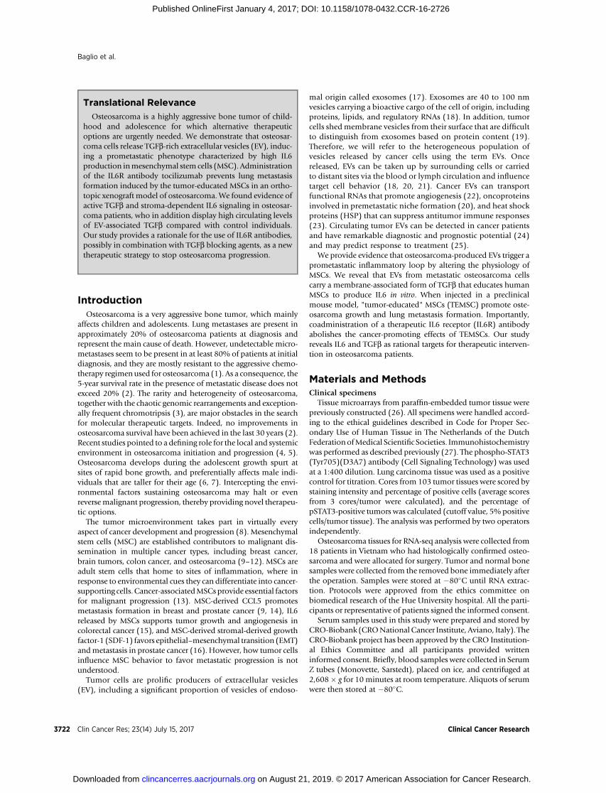
To investigate whether osteosarcoma EVs alter the physiol-ogy of MSCs such that they promote tumor progression, wedeveloped a bioluminescent orthotopic xenograft mouse mod-el of osteosarcoma. Human primary GFP-positive MSCs wereexpanded and exposed for 48 hours to EVs purified frommetastatic 143B cells. Metastatic luciferase-positive 143B cellswere inoculated in the tibia of nude mice, and after 2 days theosteosarcoma-bearing mice were subjected to a single systemicadministration of "tumor-educated" MSCs (TEMSC, Fig. 2A).Mice receiving non-educated MSCs or no MSCs were used ascontrol groups. Tumor growth was monitored by biolumines-cence imaging (BLI). As early as day 10 after inoculation weobserved increased tumor growth in mice that received TEMSCscompared with the control groups. The difference in tumorvolume became increasingly prominent at the following timepoints (Fig. 2B and C).
Baglio et al.
Clin Cancer Res; 23(14) July 15, 2017 Clinical Cancer Research3724
on August 21, 2019. © 2017 American Association for Cancer Research. clincancerres.aacrjournals.org Downloaded from
Published OnlineFirst January 4, 2017; DOI: 10.1158/1078-0432.CCR-16-2726

The dynamics of the tumor microenvironment seem to havefeatures of a wound-healing process (34), including the recruit-ment of MSCs (35). We searched for the presence of GFP-positiveMSCs in decalcified mouse tibias 4 days after systemic injection,and found GFP-positive cells within the tumor mass and in theadjacent bone marrow tissue of mice receiving TEMSCs andcontrol MSCs (Fig. 2D).
Next, we investigated whether TEMSCs promote osteosarco-ma metastatic progression. Ex vivo BLI of lungs, liver, and spleen(Fig. 2E) revealed that metastatic dissemination exclusivelyoccurred in the lungs, the most common metastatic sites inosteosarcoma patients. The number of lung metastases acrossthe experimental groups was determined by BLI and naked eyeevaluation. Strikingly, administration of TEMSCs significantlyincreased the number of metastases compared with the controlgroups (Fig. 2F). The presence of metastases in the lungs wasconfirmed by human vimentin staining (Fig. 2G). No GFP-positive MSCs were detected in the lung tissue at the experi-mental endpoint.
Collectively these observations demonstrate that osteosarco-ma EVs prompt MSCs to acquire a protumorigenic and pro-metastatic phenotype in vivo.
Osteosarcoma EVs induce IL6 production and stimulate cell-cycle progression in human primary MSCs
Because MSCs have specialized immunomodulatory func-tions (36), we wondered whether osteosarcoma EVs affectcytokine production by MSCs. We used a multiplexed bead-based assay to profile the cytokine production of MSCs upon
treatment with EVs. Interestingly, we observed that educationof human primary MSCs with EVs from the metastatic 143Bcells increased the production of IL6 and IL8 when comparedwith control EVs (Fig. 3A; Supplementary Fig. S2A). We noticedthat osteosarcoma cell lines and primary osteosarcoma cellsrelease IL8, but do not produce detectable levels of IL6 (Fig. 3B;Supplementary Fig. S2B and S2C). Therefore, we postulatedthat MSCs may act as tumor-supporting stroma cells by sup-plying exogenous IL6 in vivo. We confirmed that 143B EVseducate MSCs to produce IL6 both at the mRNA (Supplemen-tary Fig. S2D) and protein level (Fig. 3C). The responsiveness oftarget cells to IL6 depends either on the expression of thesurface IL6 receptor (IL6R) or on the availability of a solubleform of the IL6R (sIL6R). Although the expression of the surfaceIL6R is limited to few cell types in vivo, we found that bothosteosarcoma cells and MSCs produce sIL6R (Fig. 3D). Theproduction of sIL6R by MSCs was however not influenced bytreatment with osteosarcoma EVs (Supplementary Fig. S2E).Taken together, these findings demonstrate that osteosarcomacells release EVs inducing IL6 production in MSCs, and canrespond to MSC-derived IL6 in a cell-autonomous fashion.
Because IL6 is implicated in proliferation and stemness main-tenance ofMSCs (37), we investigated the effects of osteosarcomaEVs on cell-cycle progression and osteogenic differentiation ofthese cells.We culturedMSCs in the presence of osteosarcoma EVsfor 48 hours and then treated cells with nocodazole overnight toprevent mitosis. FACS analysis showed that 143B EVs determinedgreater accumulation of cells in the G2–M phase compared withcontrol EVs or untreated condition (set at 0; Fig. 3E and F),
143BMG63 HOS
MSC
+PK
H67
EV
hF EVs: 94% 143B EVs: 98%
D
A C
100 nm
CD63
/lyso
som
esM
VBs
EVs
100 nm100 nm
B
Coun
ts
PKH67 PKH67
CD63
CD81
143BMG63 HOS
Figure 1.
Osteosarcoma cell lines release exosome-like vesicles that can be internalized by MSCs. A, CD63 (green) and lysosome (red) fluorescence staining (top),and ultrastructure of MVB-like endosomes (bottom) in MG63, HOS and 143B osteosarcoma cell lines (scale bar, 100 nm). B, TEM micrographs of EVsisolated from osteosarcoma cell lines (scale bar, 100 nm). C, Western blot analysis for CD63 and CD81 in osteosarcoma cells and corresponding EVs.D, Internalization of PKH67-labeled EVs by MSCs assessed by fluorescence microscopy (top) and FACS analysis (bottom).
Osteosarcoma EVs Trigger Prometastatic IL6 Production by MSCs
www.aacrjournals.org Clin Cancer Res; 23(14) July 15, 2017 3725
on August 21, 2019. © 2017 American Association for Cancer Research. clincancerres.aacrjournals.org Downloaded from
Published OnlineFirst January 4, 2017; DOI: 10.1158/1078-0432.CCR-16-2726
suggesting that osteosarcoma EVs accelerate the transition fromG1 to G2–M. To study whether EVs affect the differentiation ofMSCs we cultured early passage MSCs in osteogenic conditionsand evaluated the formation ofmineral nodules with Alizarin redstaining (Supplementary Fig. S2F). No differences were observedin response to osteosarcoma EV treatment, suggesting that oste-osarcoma EVs promote cell-cycle progression, but do not affectthe osteogenic differentiation ability of MSCs.
Osteosarcoma EV-associated TGFb induces IL6 expression inMSCs
Osteosarcoma EVs induce IL6 release and cell-cycle progres-sion in MSCs, but the mechanism underlying these effects isunclear. One possibility is that osteosarcoma EVs transferinflammatory small RNAs that are recognized by intracellularsensors within the endosomal compartments of the MSCs (32,38, 39). We extracted RNA from osteosarcoma EVs and ana-lyzed their small RNA profile using the Bioanalyzer. The smallRNA profile showed characteristic exosomal RNA peaks
between 20 and 70 nt (Fig. 4A). RNA-seq analysis revealedhigh abundance of polymerase III transcripts (data not shown),which can induce inflammatory responses in recipient cells(32, 39). We then determined the expression of endosomalToll-like receptors (TLR) in MSCs (Fig. 4B), and transfectedcells with the RNA isolated from osteosarcoma EVs (EV-RNA).Although MSCs strongly responded to the TLR3 agonist poly(I:C), no effect on IL6 expression was observed in response toisolated EV-RNA (Fig. 4C, left). However, a single treatmentwith matching amounts of intact tumor EVs prompted a 2-foldincrease in IL6 mRNA expression (Fig. 4C, right). These obser-vations suggest that EV components other than RNAs induceIL6 production in recipient MSCs.
One emerging concept is "direct signaling" of EVs wherefactors located at the surface of EVs can change the physiologyof target cells (40, 41). To investigate this possibility, weblocked EV endocytosis in MSCs using dynasore and subse-quently incubated MSCs with PKH67-labeled 143B EVs for24 hours. Although dynasore treatment decreased 143B EV
2
3
4
5
6
7
8
9
108
No MSC TEMSCMSC
Bone marrow
BA
F
C
E
D
No MSC MSC Human vimen�nHE
G
Lung
Live
rSp
leen
GFP + M
SC, DAPI
Osteosarcomacells
OsteosarcomaEVs
MSCsGFP+
OsteosarcomacellsFLuc+
20151050
*
*
*
0
2.0 × 10⁹
no MSCMSCTEMSC
DayBL
I (p/
s)
1.5 × 10⁹
1.0 × 10⁹
0.5 × 10⁹
Lung metastases
*
No MSC MSC TEMSC0
5
10
15
20
Num
ber o
f lun
g no
dule
s
Tumor growth
Tumor
n.s.
TEMSC
Figure 2.
Tumor EV-educated MSCs promote tumor growth and metastasis formation in a bioluminescent orthotopic xenograft model of osteosarcoma. A,Schematic representation of the experimental design: luciferase-positive human metastatic osteosarcoma (143B) cells were inoculated in the tibia ofimmunocompromised mice; human GFP-positive MSCs were educated with osteosarcoma (143B)-released EVs for 48 hours, and educated (TEMSC) ornon-educated MSCs were systemically injected in the osteosarcoma-bearing mice. Mice receiving no MSCs were used as controls. B, Tumor growthmeasured by bioluminescence imaging (BLI) over the experimental time-frame in mice receiving TEMSCs, control MSCs or no MSCs (two independentexperiments, n ¼ 6/group/experiment; � , P < 0.05 TEMSC vs. MSC, LSD test). C, Representative BLI images of mice receiving TEMSCs, control MSCs orno MSCs. D, Immunofluorescence staining of GFP-positive MSCs in the bone marrow and tumor tissue of MSC-receiving mice. E, Representative ex vivo BLIimages of lung, liver and spleen of mice receiving TEMSCs, control MSCs or no MSCs. F, Lung metastasis number assessed by ex vivo BLI in thedifferent experimental groups (two independent experiments, n ¼ 6/group/experiment; � , P < 0.05, LSD test). G, HE and human vimentin staining of lungmetastasis in osteosarcoma-bearing mice.
Baglio et al.
Clin Cancer Res; 23(14) July 15, 2017 Clinical Cancer Research3726
on August 21, 2019. © 2017 American Association for Cancer Research. clincancerres.aacrjournals.org Downloaded from
Published OnlineFirst January 4, 2017; DOI: 10.1158/1078-0432.CCR-16-2726

internalization by more than 60%, EV-mediated induction ofIL6 MSCs was not affected (Fig. 4D), suggesting that this is notfully dependent on EV internalization by MSCs.
It has been recently shown that several growth factors, suchas TNFa, FGF, and TGFb, can be detected in association withEVs (42–44). TGFb is a pleiotropic cytokine highly expressed byhigh-grade osteosarcoma (45) and presumably functions as anautocrine growth factor for osteosarcoma (46). Apart from thewell-established actions of soluble TGFb, a vesicle-associatedform of TGFb has been implicated in the stimulation ofcytokine production and cancer progression (44). We quanti-fied TGFb in osteosarcoma EVs by ELISA and found highprotein levels in osteosarcoma EVs compared to non-malignantfibroblast control vesicles (143B EVs: 593.9 � 29.5 pg/mL; hFEVs: 146.1 � 15.5 pg/mL; Fig. 4E). Importantly, blocking TGFbsignaling in MSCs by means of a TGFb type I receptor (ALK)
inhibitor strongly decreased EV-mediated IL6 induction inMSCs (Fig. 4F; Supplementary Fig. S3A and S3B). Addition ofrecombinant soluble TGFb (sTGFb) to MSC cultures did notreproduce IL6 induction in the MSCs (Fig. 4F; SupplementaryFig. S3A and S3B). These observations implicate the EV-asso-ciated form of TGFb in the prometastatic inflammatory loopestablished by osteosarcoma EVs.
Blocking IL6 signaling abrogates the protumorigenic effectsof TEMSCs in vivo
We then asked whether an anti-IL6R antibody (tocilizumab)could reverse the effects of TEMSCs on tumor progression. Micebearing bioluminescent osteosarcoma xenografts were injectedwith TEMSCs 2 days after tumor cell orthotopic inoculation.Tocilizumab (100 mg/mouse) was administered intraperitoneallyone day after tumor cell inoculation and every other day until the
CA BhF
EV-
MSC
103102101 104100
143B
EV-
MSC
IL8IL1BIL6IL10TNF
IL12p70
FL2-Height
IL8IL1BIL6IL10TNF
IL12p70
sIL6
R (p
g/m
L)
0
200
400
600
D sIL6R in CM
Rela
�ve
% c
ells
in G
1/G 2
–M
EEVshF
143B EVs
Coun
ts
FL2
G1
G2–M
G1
G2–M
Coun
ts
F
IL6 protein in CM and EVs
IL6
(pg/
mL)
EVsCM
0
2,000
4,000
6,000
8,000
10,000
1,200
900
600
300
01,200
900
600
300
01,0008006004002000
0.0
0.5
1.0
1.5
2.0
2.5
IL6 protein in MSC-CM
IL6
Prot
ein
fold
indu
c�on
*
–10
–5
0
5
10
G1
G2–M
143B EVsEVshF
*
Figure 3.
Osteosarcoma EVs induce IL6 production and promote cell-cycle progression in human MSCs. A, Multiplexed bead-based protein detection of inflammatorycytokines in the culture supernatant of MSCs exposed to human fibroblast (hF) or 143B EVs. B, IL6 protein detection in the conditioned medium (CM) ofosteosarcoma cell lines, primary osteosarcoma cells (Prim-OS) and control fibroblasts (hF), and indicated EVs by ELISA. C, IL6 protein detection in theconditioned medium (CM) of MSCs exposed to indicated EVs. The graph shows data from three independent experiments, data are expressed as foldinduction relative to the untreated control (set at 1; � , P < 0.05 143B EVs vs. untreated and 143B EVs vs. hF EVs, t test). D, sIL6R protein detection inthe conditioned medium of indicated cell types. E, Cell-cycle distribution of MSCs treated with 143B or control EVs upon mitotic arrest. For capturingcells in G2–M, before FACS analysis EV-treated or untreated MSCs were incubated overnight with nocodazole. F, Relative distribution of MSCs in G1
and G2–M phases. For each condition, the difference between EV-treated and untreated cells was calculated and plotted (� , P < 0.05 143B EV vs.untreated control, t test).
Osteosarcoma EVs Trigger Prometastatic IL6 Production by MSCs
www.aacrjournals.org Clin Cancer Res; 23(14) July 15, 2017 3727
on August 21, 2019. © 2017 American Association for Cancer Research. clincancerres.aacrjournals.org Downloaded from
Published OnlineFirst January 4, 2017; DOI: 10.1158/1078-0432.CCR-16-2726

end of the experiment. Mice receiving noneducated MSCs and noMSCs (not shown) were used as control groups. We found thattocilizumab reduced tumor growth as early as day 10 afterinoculation with osteosarcoma cells (Fig. 5A and B). Of note,mice receiving tocilizumab treatment displayed BLI signalsthat overlapped with the control group receiving noneducatedMSCs.
It is well-established that the prooncogenic effects of IL6are mediated by STAT3, which links inflammation to cancer(47). We observed that TEMSCs induce an increase in nuclearpSTAT3 in tumor tissues, which was prevented by the concur-rent administration of tocilizumab (Fig. 5C and D, Supple-mentary Fig. S3C). Most importantly, the administration oftocilizumab reverted the prometastatic effects of TEMSCs in vivo(Fig. 5E and F). Collectively, these data show that tumor EVsactivate a prometastatic IL6/STAT3 signaling axis in osteosar-coma by engaging MSCs (Fig. 5G). However, we cannot rule outa possible contribution of mouse IL6 to cancer progression inour model, as the anti-IL6 receptor antibody would also
prevent the potential cross-reaction between mouse IL6 andhuman IL6R.
Osteosarcoma patients present active IL6/STAT3 and TGFbsignaling and elevated levels of circulating EV-associated TGFb
To confirm the role of IL6/STAT3 signaling in primary osteo-sarcoma tissues, we first analyzed the IL6 mRNA expression in84 pre-treatment high-grade osteosarcoma diagnostic biopsiesusing apublicly available dataset (48) in theR2GenomicsAnalysisand Visualization Platform (http://r2.amc.nl). In accordance withour in vitro data, IL6 mRNA levels in osteosarcoma biopsies werelow compared to osteoblasts, MSCs and bone tissue (Fig. 6A).However, the immunohistochemical analysis of osteosarcomatissue microarrays (TMA) revealed pSTAT3 nuclear staining in65% of biopsies (n ¼ 103, cutoff value: 5% positive cells/tumortissue; Fig. 6B), suggesting that STAT3 activation in tumor tissues ismost likely determined by an exogenous source of IL6.
To confirm these data, we performed RNA-seq analysis ofosteosarcoma tissues and surrounding bone of 18 osteosarcoma
ED
EVs 91.5%EVs + dyn 30.2%
Coun
ts
F
B TLRs Endosomal CA IL6 mRNA
0
2
4
6
8
10
Rela
�ve
expr
essio
n
IL6 mRNA
0.0
0.5
1.0
1.5
2.0
2.5
TGFβ
TGFβ
Con
cent
ra�o
n (p
g/m
L)
Rela
�ve
expr
essio
n
BasalALK inh
IL6 mRNA 24h
0.0
0.5
1.0
1.5
2.0
2.5
Rela
�ve
expr
essio
n
IL6 Induc�onPKH67-EV uptake
Rela
�ve
expr
essio
n
0.005
0.010
1.5 × 10⁻⁵
7.5 × 10⁻⁶
0143B
EVs
HO
S EV
s
[nt]
[FU]
80
100
60
40
200
[nt]
[FU]
80100
604020
0
120140
Rela
�ve
expr
essio
n
**
0.0
0.5
1.0
1.5
2.0
2.5 * *
0
200
400
600
800
EV small RNA profile
Figure 4.
A membrane-associated form of TGFb on the surface of osteosarcoma EVs induces IL6 expression in MSCs. A, Bioanalyzer small RNA profile showingenrichment of 20–70 nucleotide RNA species in indicated osteosarcoma EVs. B, Relative expression level of endosomal TLRs in MSCs analyzed byqPCR. Transcript levels are normalized to GAPDH. C, Relative expression levels of IL6 in MSCs transfected with osteosarcoma (143B) EV-RNA orpoly(I:C) (positive control; left), or treated with matching amounts of intact 143B EVs (right). Transcript levels are normalized to GAPDH and expressed asfold increase relative to the untreated control. D, Assessment of internalization of PKH67-labeled 143B EVs in the presence or absence of dynasore byFACS (left). Relative expression levels of IL6 in MSCs treated with 143B EVs in the presence or absence of dynasore (right). Transcript levels are normalizedto GAPDH and expressed as fold increase relative to the experimental controls (untreated or dynasore-treated). Three independent experiments wereperformed (� , P < 0.05, LSD test). E, TGFb protein detection in control fibroblasts (hF) and osteosarcoma (MG63, HOS and 143B) EVs by ELISA. F, Relativeexpression levels of IL6 in MSCs treated with soluble TGFb (sTGFb) or 143B EVs in the presence or absence of a TGFb type I receptor (ALK) inhibitor(SB-431542) at 24 hours. Transcript levels are normalized to GAPDH and expressed as fold increase relative to the untreated control. Three independentexperiments were performed (� , P < 0.05, LSD test).
Baglio et al.
Clin Cancer Res; 23(14) July 15, 2017 Clinical Cancer Research3728
on August 21, 2019. © 2017 American Association for Cancer Research. clincancerres.aacrjournals.org Downloaded from
Published OnlineFirst January 4, 2017; DOI: 10.1158/1078-0432.CCR-16-2726

patients (Supplementary Table S1). Again, IL6 expression wasrelatively low in tumors and does not significantly differ from thatof normal bone (mean rpm: 11.71 � 3.4 vs. 9.6 � 2.9; Supple-mentary Fig. S4A). Curiously, TGFb mRNA was also not differ-entially expressed between normal and tumor tissues (Supple-mentary Fig. S4B), whereas multiple TGFb-induced genes werestrongly upregulated (Fig. 6C and D; Supplementary Fig. S4C andS4D; Supplementary Table S2). Among these, COL11A1 andTGFbI were the top two upregulated genes in the analysis (log2FC 1.51, P ¼ 1.06E�14 and log2 FC 1.40, P ¼ 1.35E�11,respectively). Gene set enrichment analysis (GSEA) of the topupregulated genes in osteosarcoma tumors (log2 FC > 1, P <0.0001) showed an overlap with 4 extracellularmatrix genes earlyinduced by TGFb in fibroblasts (49). These include three collagengenes identified as definite TGFb/SMAD3 targets (COL3A1,COL6A1, COL6A3), andMMP14 (Fig. 6D). In support of the roleof TGFb in osteosarcoma, elevated TGFb mRNA in high-gradeosteosarcoma biopsies is associated with a decrease inmetastasis-free survival of osteosarcoma patients (Supplementary Fig. S4E).
Finally, we examined whether osteosarcoma patients haveincreased levels of EV-bound TGFb in circulation. To this end,we purified EVs from patient serum (Supplementary Table S3)using size-exclusion chromatography and quantified TGFb levelsby ELISA. Our analysis revealed that serum levels of EV-associatedTGFb are significantly higher in osteosarcoma patients comparedto healthy control individuals (277.5� 35.3 vs. 119.3� 39.1 pg/mL; Fig. 6E). To evaluate how well TGFb levels discriminatebetween osteosarcoma patients and healthy individuals, we usedan ROC curve and obtained an AUC score of 0.88 (Fig. 6F). Thesedata suggest that these TGFb-carrying vesicles, arguably of tumororigin, might act on stromal and tumor cells to sustain cancerprogression.
DiscussionTumor-secreted EVs promote metastasis formation in various
mouse models. Using a bioluminescent orthotopic xenograftmouse model of osteosarcoma, we show that tumor cells instruct
IL6
MSC TEMSCTEMSC
+ IL6R Ab A
E
B
Lung
Live
rSp
leen
*
*
*
0 5 10 15 200
1 × 10⁹
2 × 10⁹
3 × 10⁹ MSCTEMSCTEMSC + IL6R Ab
Day
Tum
or g
row
th (B
LI)
0
5
10
15
20
Num
ber o
f lun
g no
dule
s
F**
G
**
pSTAT3
0
10
20
30
Rela
�ve
% p
osi�
ve c
ells
MSC
C
D
MSC
Osteosarcoma cell
EV-AssociatedTGFβ
Tumor growthMetastasis
TocilizumabTEMSC TEMSC+ IL6R Ab
MSC TEMSC TEMSC+ IL6R Ab
pSTA
T3, D
API
Figure 5.
Treatment with an anti-IL6R antibody abrogates the protumorigenic and prometastatic effects of TEMSCs. A, BLI analysis of tumor growth over theexperimental time-frame in mice receiving non-educated MSCs, TEMSCs, or TEMSCs and IL6R Ab (tocilizumab; n ¼ 6/group). The anti-IL6R antibodywas administered starting from day 1 (green arrow) every other day until the end of the experiment (� , P < 0.05, TEMSC vs. MSC and TEMSC vs. TEMSC þ IL6RAb, LSD test). B, Representative BLI images (top) and tumor pictures (bottom) of mice receiving non-educated MSCs, TEMSCs, or TEMSCs and IL6Rantibody. C, Representative images of nuclear pSTAT3 (red) by immunofluorescence staining in indicated tumors; nuclei are stained with DAPI (gray).D, Quantification of pSTAT3 nuclear staining in tumor slices. Images from 3 mice/group were analyzed. Results are expressed as the relative percentageof pSTAT3-positive nuclei over the total amount of (DAPI-stained) nuclei (� , P < 0.05, LSD test). E, Lung metastasis number assessed by ex vivo BLI inthe different experimental groups (n ¼ 6/group; � , P � 0.05, LSD test). F, Representative ex vivo BLI images of lung, liver, and spleen of mice receivingnon-educated MSCs, TEMSCs, or TEMSCs and IL6R antibody. G, Schematic representation of the proposed model. Osteosarcoma cells release EVs decoratedwith a membrane-bound form of TGFb that increases IL6 production in MSCs. MSC-derived IL6 increases tumor growth and metastasis formation in micebearing osteosarcoma tumors. The protumorigenic effects of IL6-producing MSCs can be abrogated by IL6 signaling blocking agents (tocilizumab).
Osteosarcoma EVs Trigger Prometastatic IL6 Production by MSCs
www.aacrjournals.org Clin Cancer Res; 23(14) July 15, 2017 3729
on August 21, 2019. © 2017 American Association for Cancer Research. clincancerres.aacrjournals.org Downloaded from
Published OnlineFirst January 4, 2017; DOI: 10.1158/1078-0432.CCR-16-2726

MSCs to activate the oncogenic IL6/STAT3 signaling axis, whichis consistent with MSC receptiveness to local stimuli (9–12, 16).We demonstrate that the osteosarcoma-secreted EVs carry func-tional TGFb molecules that interact with MSCs and alter theirbehavior to promote tumor growth and metastasis formation.Our observations suggest that blocking IL6 and TGFb signalingmight represent a valid therapeutic strategy for osteosarcoma.
Tumor-derived EVs can contribute to malignant progressionby aiding in the formation of the premetastatic niche (20, 50,51). This process may involve the education of bone marrow–
derived and/or local specialized cells that shape a favorableenvironment at the metastatic site for malignant cells to seedand grow (20, 50, 51). Moreover, others have shown thatstromal cell EVs also participate in tumorigenesis by providinga favorable environment (52). We propose a third protumori-
genic EV-mediated mechanism by which tumor cell-secretedEVs act on defined subset of stromal cells directly at the primarytumor site establishing a local proinflammatory loop. We showthat short-time ex vivo conditioning of MSCs by tumor EVs issufficient to increase metastasis formation in vivo. The MSCcontribution to metastasis formation is largely dependent onincreased IL6 expression, leading to the activation of the STAT3oncogene in the primary bone tumor.
In various cancer types, chronic or even short activation of theIL6/STAT3 axis is a key event in cancer development and pro-gression (53). IL6/STAT3 signaling supports cancer cell prolifer-ation, metastasis formation, tumor immunosuppression, andcancer stem cell self-renewal (47). In addition, overexpression ofIL6 and its receptor (IL6R) is observed frequently in multiplecancer types (54). Osteosarcoma patients seem to have high IL6
A
C
151413121110
987
Osteosarcoma biopsyOsteosarcoma resec�onOsteosarcoma cellBoneOsteoblastMSC2l
og IL
6IL6 mRNA expression B pSTAT3 staining, TMA
DTGFβI COL11A1 MMP14COL3A1 COL6A1
rpm
10
100
1,000
10,000
100
1,000
10,000COL6A3
rpm
100
1,000
10,000
100,000
10
100
1,000
10
100
1,000
10,000
10
100
1,000
10,000
E FEV-Associated TGFβ ROC: EV-Associated TGFβ
100% − Specificity %
Sens
i�vi
ty %
AUC: 0.88P = 0.004
0 20 40 60 80 1000
20
40
60
80
100
TGFβ
1 Co
ncen
tra�
on (p
g/m
L) *
OSCtrl0
200
400
600
Figure 6.
Osteosarcoma patients have activated STAT3 and TGFb signaling and elevated levels of circulating TGFb associated with EVs. A, IL6 mRNA expressionin pretreatment high-grade osteosarcoma diagnostic biopsies and indicated cell lines and tissues (Kuijjer dataset, R2: Genomics Analysis andVisualization Platform; http://r2.amc.nl). B, Representative image of immunohistochemical staining of pSTAT3 in pre-chemotherapy osteosarcomatissue microarrays (TMA). C and D, Normalized read counts of indicated TGFb-induced genes in osteosarcoma tumors compared to surrounding normalbone tissue as analyzed by RNA-seq (n¼ 18 patients). Data are expressed as reads per million (rpm). E, Quantification of TGFb protein levels in serum EVs fromosteosarcoma patients and control donors by ELISA (n ¼ 10/group, � , P < 0.05, t test) and F, relative ROC curve (AUC: 0.88, P ¼ 0.004); OS, osteosarcoma.
Baglio et al.
Clin Cancer Res; 23(14) July 15, 2017 Clinical Cancer Research3730
on August 21, 2019. © 2017 American Association for Cancer Research. clincancerres.aacrjournals.org Downloaded from
Published OnlineFirst January 4, 2017; DOI: 10.1158/1078-0432.CCR-16-2726

serum levels compared with control individuals (55, 56), andsustain activated intra-tumor STAT3 signaling as we demonstratein this study (Fig. 6B). Surprisingly, osteosarcoma tumor cells invivo, primary osteosarcoma cells cultured in vitro as well as mostosteosarcoma cell-lines that we studied express low to virtuallyundetectable levels of IL6 (Figs. 3B and 6A; and SupplementaryFig. S4A). These observations, combinedwith the activated STAT3signaling observed in osteosarcoma tumors, suggests that anexogenous source of IL6 must be involved, strengthening thenotion that tumors progress with the support of the microenvi-ronment (8).
The complexity and heterogeneity of EVs complicates theidentification of biomolecules that modify the physiology ofrecipient cells. We and others previously showed that both tumorEV-protected small RNAs and virus-derived small RNAs caninduce inflammatory responses in target cells by triggering intra-cellular RNA sensors (32, 38, 39). Although we found that MSCsexpress functional endosomal TLRs, they are unresponsive toisolated tumor EV-RNA suggesting that IL6 induction in responseto osteosarcoma EVs is mediated by alternative mechanisms.Multiple EV-associated proteins including proto-oncogenes andHSPs have been implicated in the intercellular communicationnetworks that support cancer progression (20, 23). Depending ontheir localization EV-associated proteins might not require inter-nalization by target cells to signal. However, until now, conclu-sions on EV function and tropism have been mostly drawn basedon vesicle uptake by recipient cells (20, 21, 33, 50–52). Wedemonstrate that osteosarcoma EVs alter MSC physiology inde-pendently of internalization, by carrying membrane-associatedTGFb to the surface of MSCs, where TGFb can interact with itsreceptor (Fig. 4D–F).
TGFb is a key molecule in many metastasis models (57), andhas a central role in the communication between cancer andstromal cells leading to disease progression (58). In osteosarcomaTGFb has been previously implicated as an autocrine growthfactor. Indeed, TGFb mRNA expression in osteosarcoma tissuesassociates with high-grade tumors (45) and negatively correlateswith metastasis-free survival (R2: Genomics Analysis and Visual-ization Platform, Kuijjer dataset). We show that in osteosarcomapatients metastasis-associated TGFb-induced genes are overex-pressed in the tumor tissue compared with the surroundingnormal bone (Supplementary Table S2). Intriguingly, we couldnot detect differential expression of TGFb, at least at the mRNAlevel (Supplementary Fig. S4B). This suggests that TGFb, secretedin a latent form, is activated within the tumor mass more so thanin the surroundingnormal bone tissue. Analternative explanationis that the levels of EV-bound TGFb, rather than the total amountof TGFb protein, ultimately determines downstream target geneexpression. In fact, we demonstrate that malignant osteosarcomaEVs carry high levels of membrane-bound TGFb compared withEVs secreted by non-transformed cells (Fig. 4E), which corre-sponds with their ability to educate MSCs to produce IL6(Fig. 3C). Importantly, soluble TGFb did not reproduce the effectsof the EV-associated form on the MSCs (Fig. 4F; SupplementaryFig. S3A and S3B), a finding supported by recent independentstudies (44, 59). Thus, a growth factor in association with EVs hasdistinct signaling properties than its soluble form (40, 41). Wepropose that the conformation acquired by TGFb on the EVsurface, the combination with other EV-associated factors, or thepresence of costimulatory signals on tumor EVs (59), mightenhance or alter the TGFb signaling properties.
To demonstrate the clinical significance of vesicle-associatedTGFb in osteosarcoma, we quantified the levels of EV-boundTGFb in human serum. We found that osteosarcoma patientshave much higher levels of EV-associated TGFb compared withhealthy control individuals. Arguably, multiple cell types, includ-ing immune cells, might be exposed to the high levels of local orsystemic EV-bound TGFb in osteosarcoma patients. Although theuse of a xenograft mouse model allows to study the interactionsbetween cell types of human origin in vivo, it limits the possibilityto investigate the contribution of immune components such astumor-infiltrating T cells to cancer progression. Further studiesusing syngeneic mouse models need to be performed to obtain amore complete picture of tumor–stromal cell interactions inosteosarcoma, and to evaluate the potential role of EV-associatedTGFb and IL6 in tumor immune escape.
Currently, adolescent osteosarcoma patients receive one of themost aggressive treatment regimens, whereas prognosis in thepresence of metastases remains discouraging (2). This is the firststudy addressing the role of EV-mediated tumor–stroma commu-nication in osteosarcoma. We describe the establishment of aprometastatic inflammatory loop initiated by osteosarcoma EVsthat can be disrupted to inhibit osteosarcoma progression. This isrelevant because IL6 and TGFb inhibitors are novel attractivetargets for anti-cancer therapy (60, 61). In particular, the anti-IL6R antibody used in this study (tocilizumab), already approvedfor the treatment of rheumatic diseases, has been evaluated withencouraging results in a phase I trial for recurrent ovarian cancer(NCT01637532), and will be tested for the treatment of pancre-atic cancer (NCT02767557) and Chronic Lymphocytic Leukemia(NCT02336048). Osteosarcoma is a rare tumor of childhoodand adolescence, which complicates large clinical studies stress-ing the need for preclinical models. Although it is unlikely thatIL6-blocking antibodies, used as single therapeutic agents, mayresult in patient response, combination with current chemother-apy treatment may improve osteosarcoma survival and allow tolower the dosage of chemotherapeutic drugs, reducing toxicity.Moreover, our findings suggest that combination of IL6-blockingagents with TGFb inhibitorsmight halt osteosarcoma progressionwhile reducing resistance.
Disclosure of Potential Conflicts of InterestD.M. Pegtel is an employee of and holds ownership interest (including
patents) in Exbiome BV and is a consultant/advisory board member forTakeda Pharmaceuticals. No potential conflicts of interest were disclosedby the other authors.
Authors' ContributionsConception and design: S.R. Baglio, T. Lagerweij, X.D. Ho, T.D. de Gruijl,K. Maasalu, N. Baldini, D.M. PegtelDevelopment of methodology: S.R. Baglio, L. Roncuzzi, N. Zini, A. Martson,D.M. PegtelAcquisition of data (provided animals, acquired and managed patients,provided facilities, etc.): S.R. Baglio, T. Lagerweij, M.P. Lanz�on, X.D. Ho,N. L�eveill�e, S.A. Melo, A.-M. Cleton-Jansen, E.S. Jordanova, M. Dominici,R. Bonafede, S. Cervo, A. Steffan, V. Canzonieri, A. Martson, K. Maasalu,S. K€oks, T. WurdingerAnalysis and interpretation of data (e.g., statistical analysis, biostatistics,computational analysis): S.R. Baglio, T. Lagerweij, M.P. Lanz�on, X.D. Ho,E.S. Jordanova, A. Martson, K. Maasalu, N. Baldini, D.M. PegtelWriting, review, and/or revision of the manuscript: S.R. Baglio, T. Lagerweij,M.P. Lanz�on, E.S. Jordanova, L. Roncuzzi, M. Dominici, T.D. de Gruijl,K. Maasalu, T. Wurdinger, N. Baldini, D.M. PegtelAdministrative, technical, or material support (i.e., reporting or organizingdata, constructing databases): T. Lagerweij, M.P. Lanz�on, M. Greco,
Osteosarcoma EVs Trigger Prometastatic IL6 Production by MSCs
www.aacrjournals.org Clin Cancer Res; 23(14) July 15, 2017 3731
on August 21, 2019. © 2017 American Association for Cancer Research. clincancerres.aacrjournals.org Downloaded from
Published OnlineFirst January 4, 2017; DOI: 10.1158/1078-0432.CCR-16-2726

M.A.J. van Eijndhoven, G. Grisendi, R. Bonafede, S.M. Lougheed, S. Cervo,V. Canzonieri, A. Martson, K. Maasalu, S. K€oksStudy supervision: D.M. PegtelOther (funding): N. Baldini
AcknowledgmentsThe authors thank H.W.M. Niessen (VU University Medical Center, Amster-
dam, the Netherlands) for the provision of humanMSCs; J.M. Middeldorp (VUUniversity Medical Center, Amsterdam, the Netherlands) for the provision ofhuman fibroblasts; V.W. van Beusechem (VU University Medical Center,Amsterdam, the Netherlands) for providing primary osteosarcoma cells; A.Avan and E. Giovannetti (VU University Medical Center, Amsterdam, theNetherlands) for their support in the in vivo experimentation; G. Bonuccelli(Istituto Ortopedico Rizzoli, Bologna, Italy) for her assistance in tissue staining;J. Letterio (UH Cleveland Medical Center, Cleveland, OH) for constructivediscussion.
Grant SupportS.R. Baglio was supported by a fellowship by Associazione Italiana per la
Ricerca sul Cancro (AIRC) co-funded by the European Union, by the VeronesiFoundation and by the L'Oréal UNESCO "For Women in Science." This projecthas received funding from the European Union’s Horizon 2020 research andinnovation programme under theMarie Sklodowska-Curie grant agreement No660200 (to S.R. Baglio). In addition, this workwas supported by grants from theDutch Cancer Society (KWF, grant No VU2012-5510; to D.M. Pegtel), and theItalian Association for Cancer Research (AIRC, grant No 15608; to N. Baldini).
The costs of publication of this article were defrayed in part by thepayment of page charges. This article must therefore be hereby markedadvertisement in accordance with 18 U.S.C. Section 1734 solely to indicatethis fact.
Received October 30, 2016; revised December 16, 2016; accepted December19, 2016; published OnlineFirst January 4, 2017.
References1. Messerschmitt PJ, Garcia RM, Abdul-Karim FW, Greenfield EM, Getty PJ.
Osteosarcoma. J Am Acad Orthop Surg 2009;17:515–27.2. Kansara M, Teng MW, Smyth MJ, Thomas DM. Translational biology of
osteosarcoma. Nat Rev Cancer 2014;14:722–35.3. Stephens PJ, Greenman CD, Fu B, Yang F, Bignell GR, Mudie LJ, et al.
Massive genomic rearrangement acquired in a single catastrophic eventduring cancer development. Cell 2011;144:27–40.
4. Rubio R, Abarrategi A, Garcia-Castro J, Martinez-Cruzado L, Suarez C,Tornin J, et al. Bone environment is essential for osteosarcoma develop-ment from transformed mesenchymal stem cells. Stem Cells 2014;32:1136–48.
5. Alfranca A, Martinez-Cruzado L, Tornin J, Abarrategi A, Amaral T, de AlavaE, et al. Bone microenvironment signals in osteosarcoma development.Cell Mol Life Sci 2015;72:3097–113.
6. Ottaviani G, Jaffe N. The epidemiology of osteosarcoma. Cancer Treat Res2009;152:3–13.
7. Longhi A, Pasini A, Cicognani A, Baronio F, Pellacani A, Baldini N, et al.Height as a risk factor for osteosarcoma. J Pediatr Hematol Oncol2005;27:314–8.
8. Hanahan D, Weinberg RA. Hallmarks of cancer: the next generation. Cell2011;646–74.
9. Karnoub AE, Dash AB, Vo AP, Sullivan A, Brooks MW, Bell GW, et al.Mesenchymal stem cells within tumour stroma promote breast cancermetastasis. Nature 2007;449:557–63.
10. Behnan J, Isakson P, Joel M, Cilio C, Langmoen IA, Vik-Mo EO, et al.Recruited brain tumor-derivedmesenchymal stem cells contribute to braintumor progression. Stem Cells 2014;32:1110–23.
11. Shinagawa K, Kitadai Y, Tanaka M, Sumida T, Onoyama M, Ohnishi M,et al. Stroma-directed imatinib therapy impairs the tumor-promoting effectof bone marrow-derived mesenchymal stem cells in an orthotopic trans-plantation model of colon cancer. Int J Cancer 2012;132:813–23.
12. Xu W, Bian Z, Fan Q, Li G, Tang T. Human mesenchymal stem cells(hMSCs) target osteosarcoma and promote its growth and pulmonarymetastasis. Cancer Lett 2009;281:32–41.
13. Barcellos-de-Souza P, Gori V, Bambi F, Chiarugi P. Tumor microenviron-ment: bone marrow-mesenchymal stem cells as key players. BiochimBiophys Acta 2013;1836:321–35.
14. Luo J, Ok Lee S, Liang L, Huang C-K, Li L, Wen S, et al. Infiltrating bonemarrow mesenchymal stem cells increase prostate cancer stem cell pop-ulation andmetastatic ability via secreting cytokines to suppress androgenreceptor signaling. Oncogene 2013;33:2768–78.
15. Huang W-H, Chang M-C, Tsai K-S, Hung M-C, Chen H-L, Hung S-C.Mesenchymal stem cells promote growth and angiogenesis of tumors inmice. Oncogene 2013;32:4343–54.
16. Jung Y, Kim JK, ShiozawaY,Wang J,Mishra A, Joseph J, et al. Recruitment ofmesenchymal stem cells into prostate tumours promotes metastasis. NatCommun 2013;4:1795.
17. Trajkovic K, Hsu C, Chiantia S, Rajendran L, Wenzel D, Wieland F, et al.Ceramide triggers budding of exosome vesicles into multivesicular endo-somes. Science 2008;319:1244–7.
18. Skog J,W€urdinger T, vanRijn S,MeijerDH,Gainche L, Sena-EstevesM, et al.Glioblastoma microvesicles transport RNA and proteins that promotetumour growth and provide diagnostic biomarkers. Nat Cell Biol 2008;10:1470–6.
19. Kowal J, Arras G, Colombo M, Jouve M, Morath JP, Primdal-Bengtson B,et al. Proteomic comparison defines novel markers to characterizeheterogeneous populations of extracellular vesicle subtypes. Proc NatlAcad Sci U S A 2016;113:E968–77.
20. Peinado H, Ale�ckovi�c M, Lavotshkin S, Matei I, Costa-Silva B, Moreno-Bueno G, et al. Melanoma exosomes educate bone marrow progenitorcells toward a pro-metastatic phenotype through MET. Nat Med 2012;18:883–91.
21. Zomer A, Maynard C, Verweij FJ, Kamermans A, Sch€afer R, Beerling E, et al.In vivo imaging reveals extracellular vesicle-mediated phenocopying ofmetastatic behavior. Cell 2015;161:1046–57.
22. Zhuang G, Wu X, Jiang Z, Kasman I, Yao J, Guan Y, et al. Tumour-secretedmiR-9 promotes endothelial cell migration and angiogenesis by activatingthe JAK-STAT pathway. EMBO J 2012;31:3513–23.
23. Chalmin F, Ladoire S, Mignot G, Vincent J, Bruchard M, Remy-Martin J-P,et al. Membrane-associated Hsp72 from tumor-derived exosomes med-iates STAT3-dependent immunosuppressive function of mouse andhuman myeloid-derived suppressor cells. J Clin Invest 2010;120:457–71.
24. Melo SA, Luecke LB, Kahlert C, Fernandez AF, Gammon ST, Kaye J, et al.Glypican-1 identifies cancer exosomes and detects early pancreatic cancer.Nature 2015;523:177–82.
25. ShaoH, Chung J, Lee K, Balaj L,Min C, Carter BS, et al. Chip-based analysisof exosomal mRNA mediating drug resistance in glioblastoma. Nat Com-mun 2015;6:6999.
26. Mohseny AB, Szuhai K, Romeo S, Buddingh EP, Briaire-de Bruijn I, de JongD, et al. Osteosarcoma originates from mesenchymal stem cells in conse-quence of aneuploidization and genomic loss of Cdkn2. J Pathol 2009;219:294–305.
27. Baranski Z, Booij TH, Cleton-Jansen A-M, Price LS, van de Water B, Bov�eeJVMG, et al. Aven-mediated checkpoint kinase control regulates prolifer-ation and resistance to chemotherapy in conventional osteosarcoma.J Pathol 2015;236:348–59.
28. Baglio SR, Rooijers K, Koppers-Lalic D, Verweij FJ, P�erez Lanz�onM, Zini N,et al. Human bone marrow- and adipose-mesenchymal stem cells secreteexosomes enriched in distinctive miRNA and tRNA species. Stem Cell ResTher 2015;6:127.
29. Grisendi G, Bussolari R, Cafarelli L, Petak I, Rasini V, Veronesi E, et al.Adipose-derivedmesenchymal stem cells as stable source of tumor necrosisfactor-related apoptosis-inducing ligand delivery for cancer therapy. Can-cer Res 2010;70:3718–29.
30. van Eijndhoven MAJ, Zijlstra JM, Groenewegen NJ, Drees EEE, vanNiele S, Baglio SR, et al. Plasma vesicle miRNAs for therapy responsemonitoring in Hodgkin lymphoma patients. JCI Insight 2016;1:e89631.
31. Love MI, Huber W, Anders S. Moderated estimation of fold change anddispersion for RNA-seq data with DESeq2. Genome Biol 2014;15:550.
Clin Cancer Res; 23(14) July 15, 2017 Clinical Cancer Research3732
Baglio et al.
on August 21, 2019. © 2017 American Association for Cancer Research. clincancerres.aacrjournals.org Downloaded from
Published OnlineFirst January 4, 2017; DOI: 10.1158/1078-0432.CCR-16-2726

32. Baglio SR, van Eijndhoven MAJ, Koppers-Lalic D, Berenguer J, Lough-eed SM, Gibbs S, et al. Sensing of latent EBV infection throughexosomal transfer of 50pppRNA. Proc Natl Acad Sci U S A 2016;113:E587–9.
33. Pucci F, Garris C, Lai CP, Newton A, Pfirschke C, Engblom C, et al. SCSmacrophages suppress melanoma by restricting tumor-derived vesicle-Bcell interactions. Science 2016;352:242–6.
34. Dvorak HF. Tumors: wounds that do not heal. Similarities between tumorstroma generation and wound healing. N Engl J Med 1986;315:1650–9.
35. Reagan MR, Kaplan DL. Concise review: mesenchymal stem cell tumor-homing: detection methods in disease model systems. Stem Cells 2011;29:920–7.
36. BernardoME, FibbeWE.Mesenchymal stromal cells: sensors and switchersof inflammation. Cell Stem Cell 2013;13:392–402.
37. Pricola KL, Kuhn NZ, Haleem-Smith H, Song Y, Tuan RS. Interleukin-6maintains bone marrow-derived mesenchymal stem cell stemness by anERK1/2-dependent mechanism. J Cell Biochem 2009;108:577–88.
38. Fabbri M, Paone A, Calore F, Galli R, Gaudio E, Santhanam R, et al.MicroRNAs bind to Toll-like receptors to induce prometastatic inflamma-tory response. Proc Natl Acad Sci U S A 2012;109:E2110–6.
39. Boelens MC, Wu TJ, Nabet BY, Xu B, Qiu Y, Yoon T, et al. Exosome transferfrom stromal to breast cancer cells regulates therapy resistance pathways.Cell 2014;159:499–513.
40. Cossetti C, Iraci N, Mercer TR, Leonardi T, Alpi E, Drago D, et al. Extra-cellular vesicles from neural stem cells transfer IFN-g via Ifngr1 to activateStat1 signaling in target cells. Mol Cell 2014;56:193–204.
41. Szczepanski MJ, Szajnik M, Welsh A, Whiteside TL, Boyiadzis M. Blast-derived microvesicles in sera from patients with acute myeloid leukemiasuppress natural killer cell function viamembrane-associated transforminggrowth factor-b1. Haematologica 2011;96:1302–9.
42. ZhangH-G, LiuC, SuK, SuK, Yu S, Zhang L, et al. Amembrane formof TNF-alpha presented by exosomes delays T-cell activation-induced cell death. JImmunol 2006;176:7385–93.
43. Seelenmeyer C, Stegmayer C, Nickel W. Unconventional secretion offibroblast growth factor 2 and galectin-1 does not require shedding ofplasma membrane-derived vesicles. FEBS Lett 2008;582:1362–8.
44. Webber J, Steadman R, Mason MD, Tabi Z, Clayton A. Cancer exosomestrigger fibroblast to myofibroblast differentiation. Cancer Res 2010;70:9621–30.
45. Franchi A, Arganini L, Baroni G, Calzolari A, Capanna R, Campanacci D,et al. Expression of transforming growth factor beta isoforms in osteosar-coma variants: association of TGF beta 1 with high-grade osteosarcomas. JPathol. 1998;185:284–9
46. Kloen P, Jennings CL, Gebhardt MC, Springfield DS, Mankin HJ. Expres-sion of transforming growth factor-beta (TGF-beta) receptors, TGF-beta 1
and TGF-beta 2 production and autocrine growth control in osteosarcomacells. Int J Cancer 1994;58:440–5.
47. YuH, LeeH,HerrmannA, Buettner R, Jove R. Revisiting STAT3 signalling incancer: new and unexpected biological functions. Nat Rev Cancer 2014;14:736–46.
48. KuijjerML,Hogendoorn PCW, Cleton-Jansen A-M.Genome-wide analyseson high-grade osteosarcoma:making sense of a genomicallymost unstabletumor. Int J Cancer 2013;133:2512–21.
49. Verrecchia F, Chu ML, Mauviel A. Identification of novel TGF-beta/Smad gene targets in dermal fibroblasts using a combined cDNAmicroarray/promoter transactivation approach. J Biol Chem 2001;276:17058–62.
50. Costa-Silva B, Aiello NM, Ocean AJ, Singh S, Zhang H, Thakur BK, et al.Pancreatic cancer exosomes initiate pre-metastatic niche formation in theliver. Nat Cell Biol 2015;17:816–26.
51. Hoshino A, Costa-Silva B, Shen T-L, Rodrigues G, Hashimoto A, TesicMarkM, et al. Tumour exosome integrins determine organotropic metastasis.Nature 2015;527:329–35.
52. Zhang L, Zhang S, Yao J, Lowery FJ, Zhang Q, Huang W-C, et al. Micro-environment-induced PTEN loss by exosomal microRNA primes brainmetastasis outgrowth. Nature 2015;527:100–4.
53. Iliopoulos D, Hirsch HA, Struhl K. An epigenetic switch involving NF-kappaB, Lin28, Let-7 MicroRNA, and IL6 links inflammation to celltransformation. Cell 2009;139:693–706.
54. Guo Y, Xu F, Lu T, Duan Z, Zhang Z. Interleukin-6 signaling pathway intargeted therapy for cancer. Cancer Treat Rev 2012;38:904–10.
55. Xiao H, Chen L, Luo G, SonH, Prectoni JH, ZhengW. Effect of the cytokinelevels in serum on osteosarcoma. Tumour Biol 2014;35:1023–8.
56. Rutkowski P, Kamin´ ska J, Kowalska M, Ruka W, Steffen J. Cytokineand cytokine receptor serum levels in adult bone sarcoma patients:correlations with local tumor extent and prognosis. J Surg Oncol2003;84:151–9.
57. Padua D, Massagu�e J. Roles of TGFbeta in metastasis. Cell Res 2009;19:89–102.
58. Calon A, Lonardo E, Berenguer-Llergo A, Espinet E, Hernando-MomblonaX, Iglesias M, et al. Stromal gene expression defines poor-prognosis sub-types in colorectal cancer. Nat Genet 2015;47:320–9.
59. Webber JP, Spary LK, Sanders AJ, Chowdhury R, Jiang WG, Steadman R,et al. Differentiation of tumour-promoting stromal myofibroblasts bycancer exosomes. Oncogene 2015;34:290–302.
60. Rossi J-F, Lu Z-Y, Jourdan M, Klein B. Interleukin-6 as a therapeutic target.Clin Cancer Res 2015;21:1248–57.
61. Neuzillet C, Tijeras-Raballand A, Cohen R, Cros J, Faivre S, Raymond E,et al. Targeting the TGFb pathway for cancer therapy. Pharmacol Ther2015;147:22–31.
www.aacrjournals.org Clin Cancer Res; 23(14) July 15, 2017 3733
Osteosarcoma EVs Trigger Prometastatic IL6 Production by MSCs
on August 21, 2019. © 2017 American Association for Cancer Research. clincancerres.aacrjournals.org Downloaded from
Published OnlineFirst January 4, 2017; DOI: 10.1158/1078-0432.CCR-16-2726

Correction
Correction: Blocking Tumor-Educated MSCParacrine Activity Halts OsteosarcomaProgression
In this article (Clin Cancer Res 2017;23:3721–33), which was published in theJuly 15, 2017, issue of Clinical Cancer Research (1), the corresponding authorinformed the journal that incorrect or duplicate images were included in Figure5F. The images were added accidentally by the AACR during the productionprocess for the article. The conclusions put forth in the article remain unchanged.The published article has been revised to reflect corrections to Figure 5F. Thepublisher regrets this error.
Reference1. Baglio SR, Lagerweij T, P�erez-Lanz�on M, Ho XD, L�eveill�e N, Melo SA, et al. Blocking tumor-
educated MSC paracrine activity halts osteosarcoma progression. Clin Cancer Res2017;23:3721–33.
Published online February 1, 2018.doi: 10.1158/1078-0432.CCR-17-3198�2018 American Association for Cancer Research.
ClinicalCancerResearch
Clin Cancer Res; 24(3) February 1, 2018724

2017;23:3721-3733. Published OnlineFirst January 4, 2017.Clin Cancer Res S. Rubina Baglio, Tonny Lagerweij, Maria Pérez-Lanzón, et al. Osteosarcoma ProgressionBlocking Tumor-Educated MSC Paracrine Activity Halts
Updated version
10.1158/1078-0432.CCR-16-2726doi:
Access the most recent version of this article at:
Material
Supplementary
http://clincancerres.aacrjournals.org/content/suppl/2017/01/04/1078-0432.CCR-16-2726.DC1
Access the most recent supplemental material at:
Cited articles
http://clincancerres.aacrjournals.org/content/23/14/3721.full#ref-list-1
This article cites 59 articles, 11 of which you can access for free at:
Citing articles
http://clincancerres.aacrjournals.org/content/23/14/3721.full#related-urls
This article has been cited by 2 HighWire-hosted articles. Access the articles at:
E-mail alerts related to this article or journal.Sign up to receive free email-alerts
Subscriptions
Reprints and
To order reprints of this article or to subscribe to the journal, contact the AACR Publications Department at
Permissions
Rightslink site. Click on "Request Permissions" which will take you to the Copyright Clearance Center's (CCC)
.http://clincancerres.aacrjournals.org/content/23/14/3721To request permission to re-use all or part of this article, use this link
on August 21, 2019. © 2017 American Association for Cancer Research. clincancerres.aacrjournals.org Downloaded from
Published OnlineFirst January 4, 2017; DOI: 10.1158/1078-0432.CCR-16-2726