blood flagellates or haemoflagellates
TRANSCRIPT
Medical Parasitology
Dr : Ashraf Gaber Timsah Lecturer of Parasitology
Damiatta Faculty of Medicine
Al-Azhar university
•BLOOD FLAGELLATES (Haemoflagellates)
• (Leishmania and Trypanosoma)
BLOOD FLAGELLATES (Haemoflagellates)
(Leishmania and Trypanosoma)
• Haemoflagellates are term used for flagellated parasite which live in the blood or tissues of man.
There are 2 types occur in man:
• Leishmania with morphological form amastigote in man.
• Trypanosoma with morphological form trypomastigote. Other forms may found in insect vectors and culture media as epimategote and promastigote.
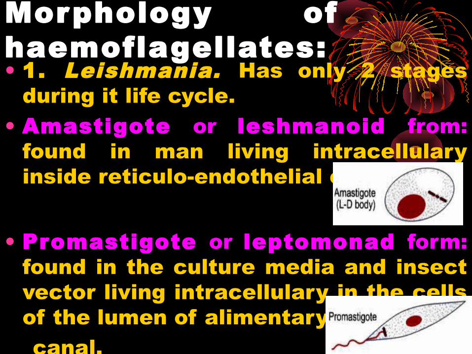
Morphology ofhaemoflagellates:• 1. Leishmania. Has only 2 stages
during it life cycle. • Amastigote or leshmanoid from:
found in man living intracellulary inside reticulo-endothelial cell.
• Promastigote or leptomonad form: found in the culture media and insect vector living intracellulary in the cells of the lumen of alimentary
canal.
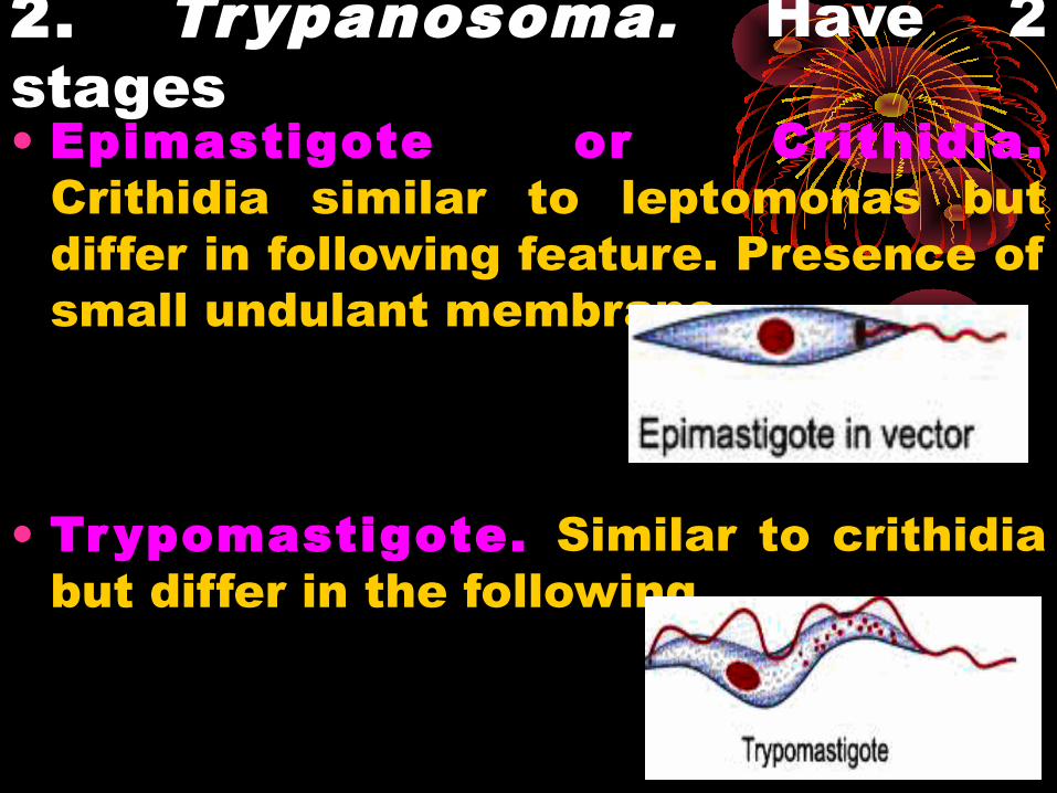
2. Trypanosoma. Have 2 stages• Epimastigote or Crithidia.
Crithidia similar to leptomonas but differ in following feature. Presence of small undulant membrane.
• Trypomastigote. Similar to crithidia but differ in the following.
Leishmaniaiasis
Leishmaniasis is the collective name for a number of diseases caused by protozoan flagellates of genous leishmania , which have diverse clinical manifestations
•The leishmaniasis is endemic in 88 countries on five continents—Africa, Asia, Europe, North America and South America.•350 million people at risk. •12 million people are affected by leishmaniasis •1.5-2 million new cases of leishmaniasis estimated to occur annually.• 500 000 new cases of VL which occur annually
GEOGRAPHICAL DISTRIBUTION
Morphology: As mentioned before Leishmania
occur in 2 forms during its life cycle. The first is Leishmaniod form or amastigote in man and reservoir host and the second is leptomonad form or promastigote in insect vector and culture medi
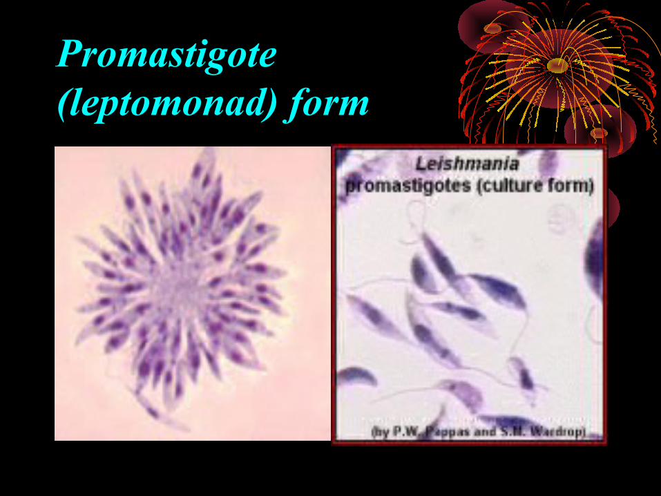
Promastigote(leptomonad) form
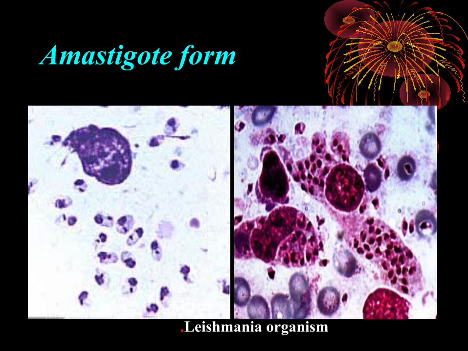
Amastigote form
Leishmania organism.
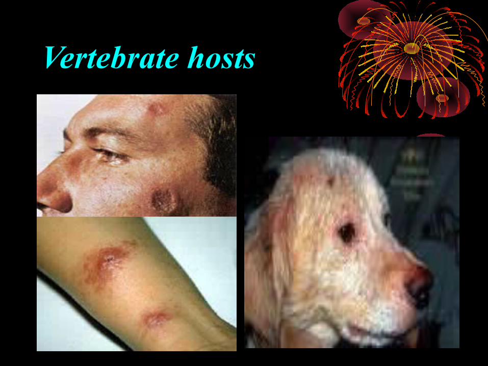
Vertebrate hosts
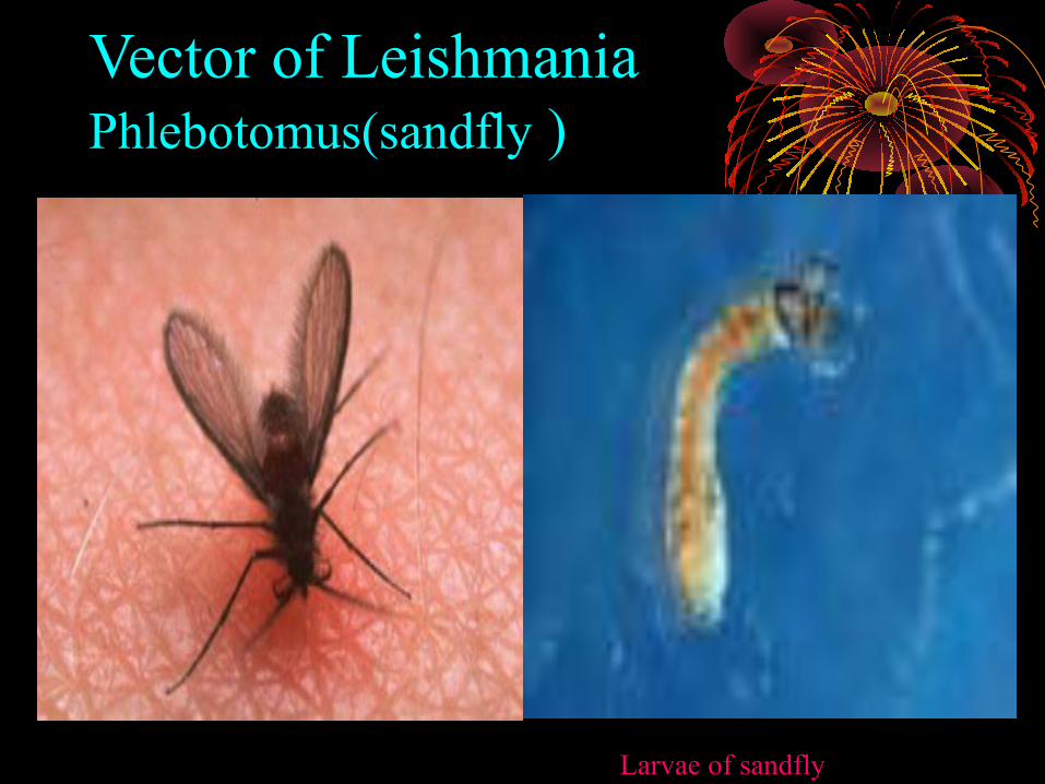
Vector of Leishmania Phlebotomus(sandfly )
Larvae of sandfly
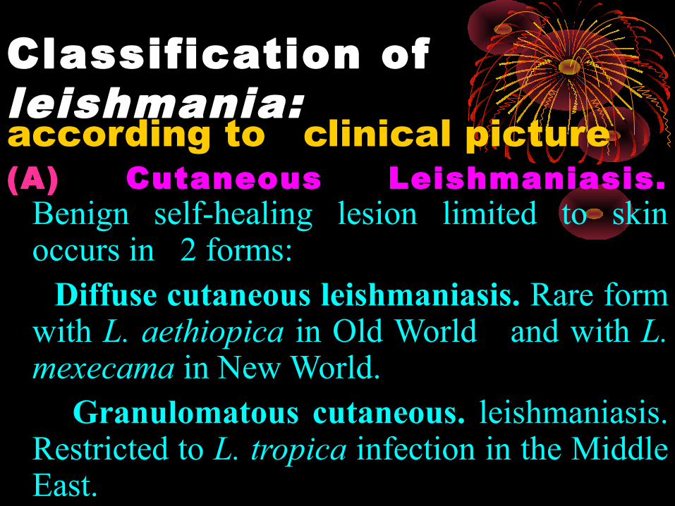
Classification of leishmania: according to clinical picture(A) Cutaneous Leishmaniasis.
Benign self-healing lesion limited to skin occurs in 2 forms:
Diffuse cutaneous leishmaniasis. Rare form with L. aethiopica in Old World and with L. mexecama in New World.
Granulomatous cutaneous. leishmaniasis. Restricted to L. tropica infection in the Middle East.
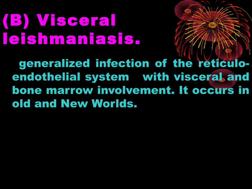
(B) Visceral leishmaniasis. generalized infection of the reticulo-
endothelial system with visceral and bone marrow involvement. It occurs in old and New Worlds.
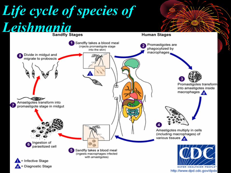
Life cycle of species of Leishmania
1. Leishmaniasis is transmitted by the bite of female phlebotomine sandflies. The sandflies inject the infective stage, promastigotes, during blood meals.2. Promastigotes that reach the puncture wound are phagocytized by macrophages.3.They transform into amastigotes.4. Amastigotes multiply in infected cells and affect different tissues.5. Sandflies become infected during blood meals on an infected host.6. In the sandfly's midgut, the parasites differentiate into promastigotes.7. They multiply and migrate to the proboscis.
Mode of infection:
biting of human by female sand fly and inoculation of promastigote in their blood which changed in to amastigote.
Cutaneous Leishmaniasis (CL) Classified according to their geographical distribution into:
(A) Old world cutaneous leishmaniasis (OWCL). Caused by leishmania tropica complex which include.
1- L tropica. Causes dry, urban or oriental sore.
• Dry painless ulcers 25–70 mm in diameter which are self-healing usually after 1–2 years but often leave disfiguring scars.
• Present in Mediterranean region, Middle East, Asia and Africa in people living in big cities.
• Dogs are reservoir hosts.• The patient is immune to reinfection. Rarely there may
develop single or multiple unhealing lesions, often on exposed parts specially face.
2- L. major. Infection is often referred to as wet or rural ××××oriental sore.
• Present in middle east, Asia and in rural area of Africa. The early papule is often multiple, inflamed and resembles a boil of 5–10 mm in diameter which rapidly develops into a large ulcer which is self-healing within 3–6 months. Lesion occurs in the lower limbs with moist serous exudates and tends to ulcerate early with secondary bacterial infection. L. major infections protect against reinfection and also against infection with L. tropica.
3- L. aethiopica. Can cause diffuse cutaneous leishmaniasis• A cutaneous lesion is produced that is similar to
typical oriental sore with healing in 1–3 years. L. aethiopica can cause diffuse cutaneous leishmaniasis (DCL) in patients with little or no cell mediated immunity against the parasite. This condition characterized by the formation of disfiguring nodules over the surface of the body resembling lepromatous leprosy.
• L. aethiopica can also cause mucocutaneous leishmaniasis.
(B)New world cutaneous leishmaniasis (NWCL). Present in the central and south
America. Rodents, cats and dogs are reservoir
hosts. Vector by Lutzomyia species. It is
caused by
•L. Mexicana. Causes chiclero’s ulcer or ‘bay sore’ • Lesions of the body tend to be self-
healing but those on the ear may last up to 30 years and entirely destroy the ear pinna and cartilage. It occurs in forest worker who collect the chicle gum.
• L. peruviana. Mainly infect children. Single or few lesions are painless, L .tropica. The infection is known locally as ‘uta’. It occurs at high altitudes in dry valleys.
• L. guyanensis. May give rise to painless dry single ulcers or multiple lesions scattered all over the body. The disease is often referred to as ‘forest yaws’ (‘pianbois’).
• L. panamensis. Causes single or few skin ulcers which are not self-healing. Lymphatic involvement is common, resulting in secondary nodules.
• L.pifanoi. lesion starts single and spread slowly like lepromatous leprosy and does not ulcerate or heal.

NB. Other type of leishmania is mucocutaneous leishmaniasis (MCL) or ‘Espundia’. MCL is caused by New World
Leishmania species, L. braziliensis, L. panamensis and occasionally by L. guyanensis.
MCL is the most severe and destructive form of cutaneous leishmaniasis in South America. Lesions are similar in development to those of oriental sore and the resulting ulcers may become very large and long-lasting.
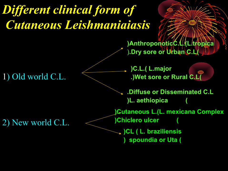
Different clinical form of Cutaneous Leishmaniaiasis
1) Old world C.L.
2) New world C.L.
AnthroponoticC.L.(L.tropica((Dry sore or Urban C.L.(
C.L.( L.major((Wet sore or Rural C.L.(
Diffuse or Disseminated C.L. (L. aethiopica(
Cutaneous L.(L. mexicana Complex( ( Chiclero ulcer(
CL ( L. braziliensis( ( spoundia or Uta(
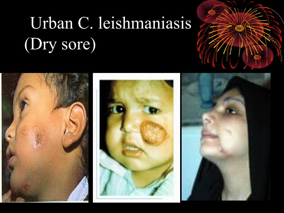
Urban C. leishmaniasis (Dry sore)

Lesions of C.L.( Wet sore)
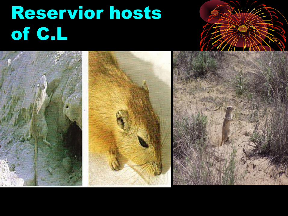
Reservior hosts of C.L

Different form of C. leishmaniasis
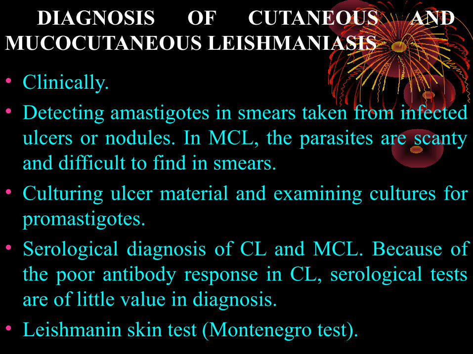
DIAGNOSIS OF CUTANEOUS AND MUCOCUTANEOUS LEISHMANIASIS
• Clinically.
• Detecting amastigotes in smears taken from infected ulcers or nodules. In MCL, the parasites are scanty and difficult to find in smears.
• Culturing ulcer material and examining cultures for promastigotes.
• Serological diagnosis of CL and MCL. Because of the poor antibody response in CL, serological tests are of little value in diagnosis.
• Leishmanin skin test (Montenegro test).
Visceral leishmaniasis (kala-Azar, black fever or Dum-dum
fever)• This disease caused by leishmania donovani
complex which is.
• L. donovani. Present in India, China, Africa, East and scattered area of central Africa. Common in young adults from 10-25 years.
• L. infantum (L. chagasi). In the Mediterranean region, Europe, Middle east and parts of Africa common below the age of four years.
• L. amazonensis. Central and south America. can affect children.
• Reservoir hosts: dogs except in India man is only reservoir and rodents.
• Vector: are Lutzomia species of sand fly.
Life cycle and pathology:
• Life cycle as before in cutaneous leishmaniasis but in visceral leishmaniasis the amastigotes multiply in the macrophages of the spleen, liver, bone marrow, lymph glands, mucosa of the small intestine and other tissues of the reticuloendothelial system. Blood monocytes are also infected. In cutaneous leishmaniasis the parasites multiply in skin macrophages (histocytes).
The affected organ shows.
• Erythropiosis. Become depressed and life span of granulocytes and erythrocytes is reduced granulocytopenia and anaemia.
• Spleen. demonstrates atrophy of white pulp with necrosis and fibrosis.
• The liver. show atrophy of liver cell with cloudy swelling and fatty degeneration.
• Some liver function remains normal but there is hypoalbuminaemia with oedema and ascites.
• Intestine. Atrophy of velli and crypts
CLINICAL PICTURE: This is the most severe form of leishmaniasis, adults
and children are being affected.
Incubation period 4 to 6 months.
• Local lesion in small non ulcerating cutaneous nodule (leishmanioma) that precedes systemic manifestation.
• fever (double daily rise with chills and sweating).
• Diarrhea and dysentery are common.
• Splenomegaly (hard, huge and not tender), Hepatomegaly and generalized lymphadenopathy.
• Pancytopenia (anaemia, leucopenia, lymphocytosis and thrombocytopenia).
• Epistaxis (nose bleed) and bleeding from the gums.
• Hypeprigmentation especially on hand, feet, forehead and abdomen.
• Jaundice and oedema
Post kala-azar dermal leishmaniasis (PKDL). • In India and occasionally in East Africa, a cutaneous
form of leishmaniasis can occur about 2 years after treatment and recovery from visceral leishmaniasis or incomplete treatment. This is referred to as post kala-azar dermal leishmaniasis. It appears as Hypopigmented patches or may develop as nodules and resemble those of lepromatous leprosy, fungal infections or other skin disorders. Occasionally there is ulceration of the lips and tongue. Amastigotes are present in the papules and nodules of this lesion.
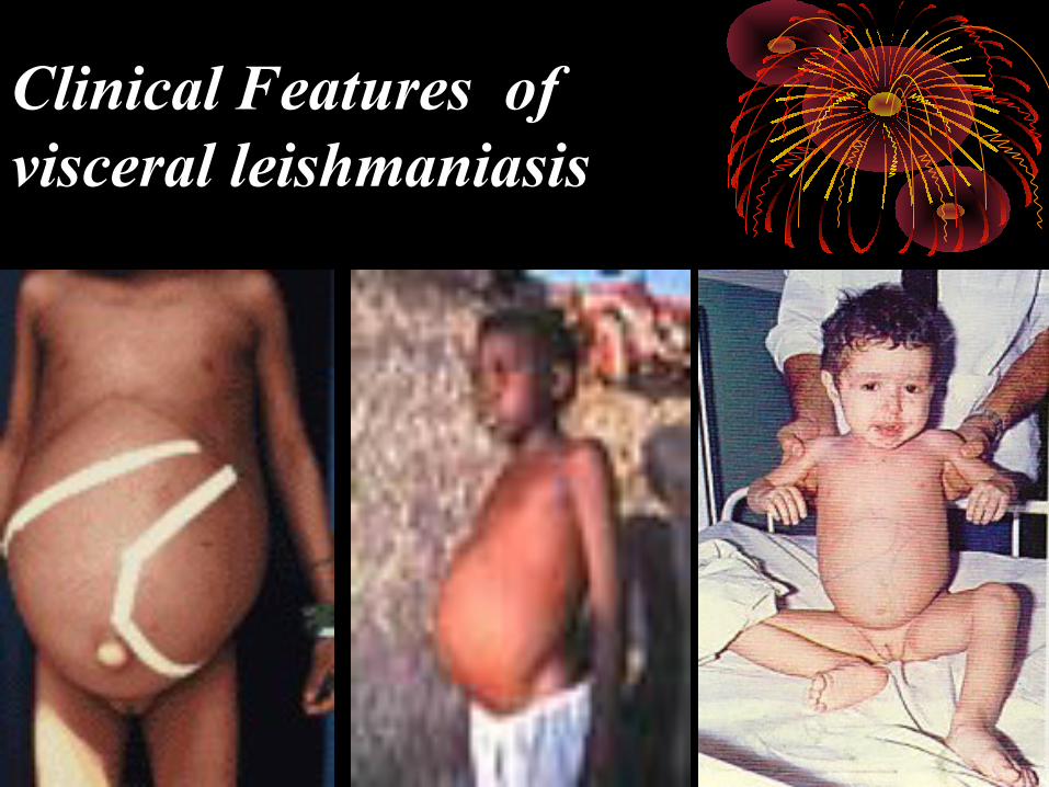
Clinical Features of visceral leishmaniasis
Complication:
• As immunosuppression and secondary infection as pulmonary T.B.Progressive loss of weight and weakness in limbs and chest wall. Death occur from month to 1 or 2 years due to intercurrent infection
• Relapse: Recurrence of clinical attack similar to primary disease resulting from in sufficient treatment or intercurrent infection.
Diagnosis• Clinicl picture.
• Detection of the parasite
-Sample: Bone marrow, spleen and lymph node aspirations.
-Detection methods: the sample taken may be Smeared on to a microscopic slide and stained by leishman OR Giemsa stain for (amastigote). Amastigotes are typically intramonocytic (the nucleus and kinetoplast staining purple).
-Cultured on NNN media for (promastigotes):
-PCR technique for detection parasite DNA.
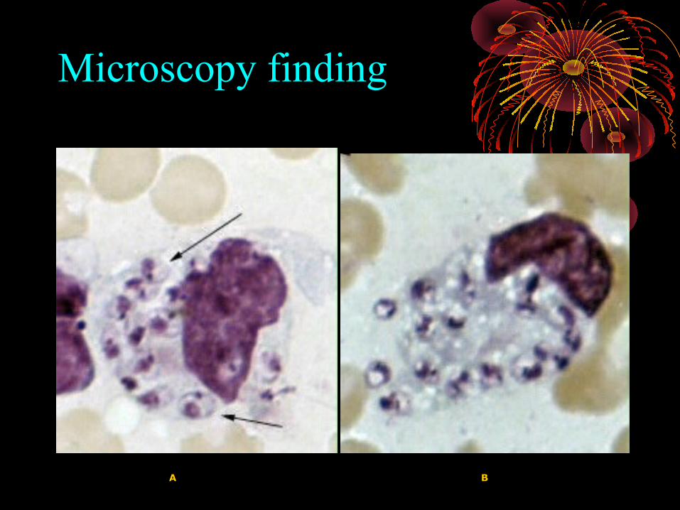
Microscopy finding
A B
• Immunological diagnosis: detect circulating specific antibodies IgG. • Indirect fluorescent antibody technique (IFAT, the
most commonly used).• Immuno-electrophoresis.• Indirect haemagglutination test
• Immunoblot, ELISA and fast-ELISA. • The leishmanin test (Montengro): The leishmanin
skin test measures delayed-type hypersensitivity.
Treatment• Visceral leishmaniasis:
• pentavalent antimonial (Pentostam) at a dose of 20 mg /kg per day for 28 day.
• pentamidine. 2–4 mg/kg intramuscular every other day up to 15 doses.
• Allopurinol : 20 mg/kg daily in 3 divided dose orally for AIDS patients.
• Amphotericin B. used in first intention, with five daily injections (3 mg/kg per injection), and a final injection on the 10th day.
• Correcting nutritional deficiencies and ttt of secondary bacterial infection.

• Localized cutaneous leishmaniasis:
• Diffuse cutaneous Leishmaniasis. Combination of paromomycin and antimonial.
• Mucocutaneous Leishmaniasis. Systemic treatment of the primary cutaneous lesion
by pentavalent antimonial, 20-day course of i.m. injections, 20 mg /kg per day.
prevention and control of leishmaniasis.
• Early detection and treatment of infected persons.
• Vector control.
• Destruction of stray dogs and infected domestic dogs.
• vaccination in endemic.