blood progesterone level before caesarean section in relation to
TRANSCRIPT

Blood progesterone level before caesarean section in relation to retained
placenta in cattle
Research Project Veterinary MedicineUniversity of Utrecht
Drs. J.J. Vrolijk0462349
January 2008
Supervisors:
Dr. R. JorritsmaDr. F.H. Jonker

Progesterone in relation to retained placenta.Drs. J.J. Vrolijk
2
Table of contents
Abstract.............................................................................................................................. 3
Introduction....................................................................................................................... 4
Retained placenta ............................................................................................................ 4
Corpus luteum................................................................................................................. 7
Possible immune related effects of progesterone.......................................................... 11
Induction of parturition ................................................................................................. 12
Hypothesis........................................................................................................................ 13
Materials and Methods................................................................................................... 15
Results .............................................................................................................................. 16
Discussion......................................................................................................................... 18
References........................................................................................................................ 20
Progesterone in relation to retained placenta.Drs. J.J. Vrolijk
3
Abstract
Retaining of foetal membranes is one of the most common complications associated with the reduction in milk yield and impaired fertility in dairy cattle. In order to predict the occurrence of this disease in cattle after a caesarean section (CS), peripheral blood progesterone (P4) levels pre-partum were measured and gestation length was noted in 60 healthy cows. The CS was carried out in favour of the veterinary university education program. Samples were taken at 08.00 h in the morning on several days pre-partum. 43 cows retained their placenta for more than 12 hours (group 1) and 17 had a normal placental delivery (<12 h) (group 2). To evaluate the difference with the normal pre-partum progesterone pattern we used a control group (group 3, n=21). This group did not have a CS and had no retained placenta (RP). Our results show that on the day of the CSsignificant higher (P<0.01) P4 values were seen in group 1. This confirms our hypothesis that a low blood P4 level is required for a normal placental delivery. The predictability of the occurrence of RP was high (100%) when the cows had a P4 level above 2.0 ng/ml on the day of the CS.
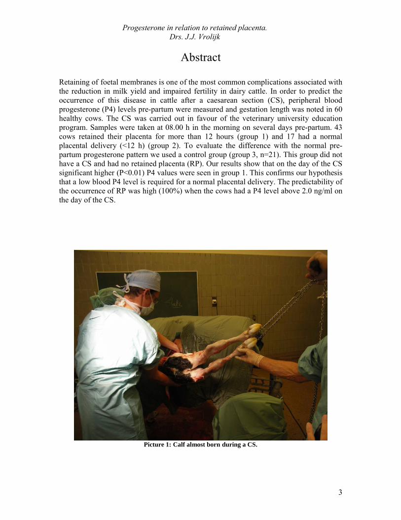
Picture 1: Calf almost born during a CS.

Progesterone in relation to retained placenta.Drs. J.J. Vrolijk
4
Introduction
Retained placenta
Retained placenta (RP) is a serious problem in dairy cattle. Directly after parturition, the foetal cotyledones and the maternal caruncles have to separate from each other. Normally this process is finished within 4-6 hours after birth and the secundinae expelled spontaneously. (Senger 2003) A placental delivery between 6-12 hours is called a delayed but still normal delivery. If the placenta is not expelled from the uterus within 12 hours after parturition, it is called a RP. Incidences of RP on farms between 5–10% in post-partum cows are reported. (Leblanc 2008)
Presence of RP increases the risk for developing an endometritis or salpingitis, because the retained secundinae create ideal circumstances for many bacteria to grow very fast. This also has an economic consequence because it results in a prolonged interval from parturition to conception, lower production and possibly infertility. A further cause of economic loss is veterinary treatment, which is necessary when a cow becomes ill due to the RP. The costs of a cow with RP, inclusive veterinary treatment, are estimated around $285 per cow in 1997. (Laven and Peters 1997)
There is almost no fundamental research that describes the normal process of placental separation. It is clear that immunological, hormonal and structural changes play an important role in the aetiology, but the exact influence of these processes is unknown. These processes are called the basic processes of delivery. (Laven and Peters 1997) Until now it is not fully understood which mechanism causes a delay in placental dehiscence and expulsion.
An immunological reaction against the foetal tissue is necessary after parturition; this reaction is mediated by several proteins and hormones. A relation between low leukocyte activity and a RP was observed by different authors. (Heuweiser and Grunert 1986) (Paisley 1986) Interleukin-8 (IL-8) is a neutrophil activating cytokine and is produced by a wide variety of cell types in presence of inflammatory stimuli. Neutrophils are strongly attracted to areas of high IL-8 concentrations. It is therefore not surprising that cows with a RP had lower levels of IL-8 in the period from 2 weeks pre-partum until 2 weeks post-partum. The lower neutrophil function may not be the result of the RP, but may also be the cause of the RP. (Kimura 2002) The low activity is possibly mediated by chemotaxis. (Gunnink 1984)
Periparturient changes in hormonal activity are other important mechanisms that facilitate the parturition and the expulsion of the placenta. Estradiol-17β, produced by the placenta, increase rapidly from twenty days pre-partum to two days post-partum. (Symons 1972) The production of prostaglandin F2α (PGF2α), stimulated by cortisol, causes luteolysis and because of that a drop in blood progesterone (P4) takes place. (Goff 1996) This fall of P4 is slightly from 15 days pre-partum and decreases faster from day 2.5 pre-partum
Progesterone in relation to retained placenta.Drs. J.J. Vrolijk
5
towards delivery. (Smith 1973) Different studies have been done to search for abnormal hormone patterns in the period pre-partum in association with retaining of the placenta. Wischral et al. have measured P4 from 5 days pre-partum and saw no differences in its pattern between cows with and without RP. Farzaneh et al. have determined P4 in cows only one time before and one time after parturition. Based on these two values they saw no differences between normal and RP cows. (Wischral 2001) (Farzaneh 2006) Older studies measured a little higher P4 level in RP cows short before parturition, but no author did find a significant increase. (Aghte 1974) (Chew 1977) (Peter and Bosu 1987) All these studies were done with cows that calved spontaneously (without induction of parturition) and after parturition they were divided in two groups, based on the occurrence of RP or not. Eiler 1997 did find that P4 inhibits the uterine collagenases and this may the reason why a low P4 is required for a correct placental expulsion. (Eiler 1997)
The RP could also be due to a wrong structural anatomy of the placentome at parturition, this can have different reasons: an immature placentome by a shortened gestation or induced parturition, an advanced involution by a prolonged gestation, necrosis of the villi by allergic reactions or oedema of the villi by a torsio uterus. (Grunert 1986) In conclusion it is clear that the three basis processes of delivery all have influence on the occurrence of RP on their own way.
Epidemiological studies revealed that several factors that influence the occurrence of retained placenta and are thought to have some influence on one of the basic processes in placental separation, mentioned above: (table 1)

Progesterone in relation to retained placenta.Drs. J.J. Vrolijk
6
Table 1: Factors associated with retained placenta. (Laven and Peters 1997)

Progesterone in relation to retained placenta.Drs. J.J. Vrolijk
7
Corpus luteum
In cattle, progesterone is mainly produced by the corpus luteum (CL). The CL is a heterogenous tissue and besides endothelial cells, large and small steroidogenic luteal cells, it also consists of fibroblasts, smooth muscle cells and immune cells (O’Shea et al. 1989). Another source of progesterone production is the placental tissue.
After ovulation, the CL is formed out of the wall of the ruptured follicle; it grows and vascularizes rapidly because of a paracrine loop between the vascular endothelial cells (which produce nitric oxide) and the granulose-lutein cells (which mainly producevascular endothelial growth factor (VEGF)). This ensures a coordinated regulation of luteal vasodilation and angiogenesis. (Berisha and Schams 2005)
Luteinizing hormone (LH) and growth hormone (GH) from the pituitary gland are the most important hormones for the development and function of the CL. But also the local angiogenic growth factors, prostaglandins and the hormone oxytocine are positiveregulators of development of the CL and both also important in stimulating steriodogenic cells to produce P4. (Fig.1) (Ferreira-Dias and Skarzynski 2008)
Figure 1: Hypothetical model of the regulation of the development of the CL.(Ferreira-Dias and Skarzynski 2008)
The CL grows by hypertrophy of the large luteal cells and by hyperplasia of the small luteal cells, but also the other supporting cells increase in number. The capacity of the newly formed CL to produce P4 depends on the numbers of steroidogenic cells and the degree of vascularisation. (Ferreira-Dias and Skarzynski 2008) In small luteal cells, production of progesterone is stimulated by LH through the protein kinase A pathway. The large luteal cells of ruminants produce large quantities of progesterone that are independent of LH stimulation. The process of progesterone production utilizes

Progesterone in relation to retained placenta.Drs. J.J. Vrolijk
8
cholesterol as precursor. Circulating lipoproteins are the major source of cholesterol for luteal cell. (Senger 2003) The first steps of the production are located on the inner mitochondrial membrane and the last steps take places in the endoplasmatic reticulum. Progesterone then diffuses out of the luteal cell and into the bloodstream to be transported to target tissues. (Diaz 2002) The production of P4 has many physiological effects: (Senger 2003) (Schreibl et al. 2000)
P4 stimulates the uterine glands in the endometrium to secrete fluids and proteins.This is essential for survival and implantation of the blastocyst.
P4 promotes the alveolar development in the mammary gland. P4 gives negative feedback to the GnRH neurons inside the hypothalamus.
Because of this feedback GH, LH and follicle stimulating hormone (FSH) are suppressed.
P4 inhibits the contractions of the myometrium. P4 suppresses cell-mediated components of the immune system and activity of
natural killer cells.
Recent studies have reported that P4 is one of the most important factors that support the maintenance of the CL. Intra-luteal P4 stimulates its own production and suppresses apoptosis in the luteal cells. (Okuda 2004) P4 is the dominant hormone during pregnancy, the high level of P4 during pregnancy is called the “progesterone block”. (Berisha and Schams 2005)Until 250 days of gestation the P4 concentration, measured in the peripheral blood plasma, stays steady around 7-8 ng/ml. (Goff 1997)
After nine months of gestation the fetus triggers the onset of parturition by initiating a cascade of complex endocrine and biochemical events. Because of the limitation of space inside the uterus, the fetus becomes stressed. Because of the release of adrenal corticotrophin (ACTH) by the fetus in response to stress, the fetal adrenal cortex becomes stimulated to produce cortisol. Fetal cortisol has different effects. It promotes the synthesis of three placental enzymes that convert P4 to estradiol and thereby removes the P4 block. This results in the initiation of myometrial contractions and more stress for the fetus. The effect is a positive feedback loop that remains until the calf is born. (Senger 2003)

Progesterone in relation to retained placenta.Drs. J.J. Vrolijk
9
Figure 2: Hypothetical model of the structural and functionalregression of the CL. (Ferreira-Dias and Skarzynski 2008)
Stimulating the endometrium to synthesize PGF2α is another effect of fetal cortisol. PGF2α indirectly stimulates the structural and functional regression of the CL, also called luteolysis. (Senger 2003) Influenced by PGF2α, different cytokines reduce the P4 production and induce apoptosis and PGF2α production by luteal cells. PGF2α has also effects on the release of endothelin-1 (EDN1) and nitric oxide (NO). This cytokines mediate the luteolytic process. EDN1 and NO both inhibiting P4 secretion and decreases blood flow through the CL. (Fig 2.) (Ferreira-Dias and Skarzynski 2008)
As described above, the normal decrease of the blood P4 level at the end of gestation is believed to occur into two steps. The first step is due to the utilization of P4 in the foetal part of the placenta, due to the enzymatic conversion of P4 to oestrogen, triggered by fetal cortisol. (Kindahl 2004) A reduction of 20% of the blood P4 concentration is seen from four weeks pre-partum to parturition. The second step is more abrupt and is seen during the last 2-3 days prior to parturition. The P4 falls now from 4-5 ng/ml to 0.2-0.5 ng/ml. (Fig. 3) (Edquist 1978) (Peter 1987) (Hafez 2000) This last step is interpreted as the result of the process of pre-partal luteolysis and is intiated by cortisol release from the calf. During this luteolysis the process of enzymatic conversion of P4 is still going on. (Kindahl 2004)

Progesterone in relation to retained placenta.Drs. J.J. Vrolijk
10
Figure 3: Peripheral plasma progesterone pattern in normal calving cows around parturition (n=28). (Peter 1987)

Progesterone in relation to retained placenta.Drs. J.J. Vrolijk
11
Possible immune related effects of progesterone
Lymphocytes
Progesterone regulates uterine immune function in by inhibition of peripheral immune function. This is achieved in several ways. Well known is the inhibitory effect of P4 on lymphocyte activation. In vitro, P4 can completely inhibit the proliferation of lymphocytes, but the concentrations P4 in vivo in the uterus are not high enough to have influence on this proliferation. So there is only an inhibition of the activation of lymphocytes. (Hansen 2007) P4 suppress specific components of the immune system and also natural killer cell activity, while it has a mainly positive influence on other nonspecific components. (Scheibl 2000) In addition, there are many other actions of progesterone on uterine immune function, mediated by locally produced molecules. One example is in sheep, the molecule that mediates some of the inhibitory effects of progesterone is an endometrial glycoprotein called uterine milk protein (UTMP), a member of the serpin superfamily of proteinase inhibitors. UTMP, also called uterine serpin, can inhibit T-cell proliferation and natural killer cell activation. Cattle, goats and pigs also secrete uterine serpin from the endometrium, but its role in immune function in these species has not been documented. (Hansen 1998)
Apoptosis
During gestation, intraluteal P4 suppresses luteal apoptosis indirectly through the inhibition of Fas and caspase-3 mRNA espression. P4 has to decline before the phase of apoptosis will start. (Okuda 2004) Placental maturation, which means reduction of maternal epithelial cells by apoptosis, is a prerequisite for the release of fetal membrane. It is a more time-consuming process than induction of labour and expulsion of the fetus. Lack of time, when parturition is induced, consequently leads to placental retention. (Boos 2003)
P4 is also an inhibitor of uterine collagenase. This enzyme plays an important role in the structural changes of the collagen network insides the placentomes. These changes are important for a correct expulsion of the fetal membrames. (Eiler 1992) Ishikawa et al. tried to predict a RP by measurement of the P4 concentration and the peripheral interleukin-6 (IL-6) concentration, which is mainly produced by T-helper 2 (Th2) cells. P4 induced conversion from a Th0 to a Th2 cell and the release of cytokines, such as IL-6 and IL-4 in throphoblasts. They thought that the shift from Th2 to Th1 cell dominance is very important for elimination of the fetal membranes. They found that low levels of IL-6 and P4 pre-partum tended to affect RP. (Ishikawa 2004)

Progesterone in relation to retained placenta.Drs. J.J. Vrolijk
12
Induction of parturition
When the calving process is induced by hormone treatment, the normal process of hormone regulation is influenced by exogenous hormones and the time to delivery is shortened. The most used drugs are dexamethason and PGF2α or its analogues. (Guérin 2004) This treatment is used when the calving period has to take place early in theseason, the cow is ill and they would save the calf, but it is also used to do a CS on a planned time. One main disadvantage is a high incidence of RP following induction. The reason for this might be the immaturaty of the placentomes. Evans et al. showed that after induction, the P4 concentration remains high a few hours longer and this may prevent the maturation of the placentomes. (Evans 1976)
Several studies were done to find a way to induce parturition with a good predictability of the moment of parturition and to reduce the occurrence of RP. Besides the usual treatment of dexamethason or PGF2α, also other hormones were tried with different success. See table below:
Treatment: Occurrence ofRP:
Predictability of parturition:
Study:
Dexamethason +++ ++ (Gross and Williams 1986) (Mansell 2006)
PGF2α +++ +++ (Musah 1987) (Rasmussen 1996)
Dexamethason + PGF2α (<1h post-partum)
+ ++ (Gross and Williams 1986)
Dexamethason + porcine relaxin
+ +++ (Musah 1987)(Smith 1995)
PGF2α + porcine relaxin ++ +++ (Musah 1987)Estradiol 17β + + (Rasmussen 1996)PGF2α + Estradiol 17β +++ +++ (Rasmussen 1996)Table 2: Differences of the most used drugs for induction of parturition. Especially the results of Musah et al. are obvious. They saw a much lesser occurrence of RP when relaxin was administered. Other authors also saw a good predictability with the use of porcine relaxin in combination with dexamethason, but a decrease in occurrence or RP was not seen. It is possible that this divergence is caused by the way of supplementation of the drug. Musah et al. administered the relaxin into the cervical os during late gestation and the cows in the survey of Smith et al. received relaxin intramuscular during late gestation. (Smith 1996) (Musah 1987)

Progesterone in relation to retained placenta.Drs. J.J. Vrolijk
13
Guérin et al. found that when 20,000 U of bacterial collagenase was injected into the uterine artery during the surgical period of a caesarean section (CS). The average period of retention of fetal membranes was reduced to 40 hours in the treated cows and was 114 hours in the control cows (P<0.001). (Guérin 2004)

Progesterone in relation to retained placenta.Drs. J.J. Vrolijk
14
Hypothesis
As described above, P4 plays an important role in the maintenance of pregnancy in different ways. Most important are the suppression of parts of the immune system and the influence on placentome maturation. Our expectation is that a high blood level of P4 (above 2,0 ng/ml) on the day of the CS, gives more RP, because when the P4 level do not decrease before the operation the suppressing influence on the immune system prevents a right expulsion of the fetal membrames. Another mechanism that does not work optimal when P4 remains high is the maturation of placentomes. (Boos 2003) In this study the hypothesis that the blood progesterone level before a CS has influence on the occurrence of retained placenta will be tested. We also will investigate the influence of the length of gestation, based on the result of other studies. The gestation length is important to create time for the maturation of placentomes. (Boos 2003) (Okuda 2004)

Progesterone in relation to retained placenta.Drs. J.J. Vrolijk
15
Materials and Methods
Sixty cows from the clinic of the Veterinairy University of Utrecht were studied in a cohort study from December 2005 till December 2008. All cows were intended to be used in the regular teaching programme to undergo a CS.
From these cows venous blood samples were collected from the caudal vein at 08:00 hours in the morning into heparinised vacutainer tubes. In this heparinised blood the level of P4 was measured by the DPC Coat-A-Count Progesterone Radio Immune Assay (Diagnostics Products Corp.) in the Universitair Veterinair Diagnostic Laboratory(UVDL). The intra-assay and inter-assay coefficients of variation were 7% and 11%, respectively. P4 was measured in the pre-partum period (from 16 days before calving till parturition) to estimate the best time for the CS. The cows that have been measured before the start of this survey were only tested on Monday, Tuesday and Friday. Only on these days the radio immune assay was carried out by the laboratorium. Because of this limitation a difference in frequency and number of measurements was seen between the individual animals. During this survey we measured some cows more times to create a better P4 pattern.
On day -2 at 22:00 hours the partus was induced by 20ml (1ml/50 kg BW) dose Dexamethason (Voreen®) intravenous. The day of induction is determined by the availability of the students following clinical rotations for the CS teaching program two day after induction. On day 0 between 9 – 12 h the operation did take place. All Caesarean sections were carried out by two students following clinical rotations and one veterinarian. The duration was about 2.5 hours. After the CS the cows were observed and took care by students following clinical rotations and professional animal fosters duringthe post-partum period. The occurrence of RP was established by observation until 12 hours post-partum. Placental retention was defined as a lack of expulsion within 12 hours after calving. The cows that develop a RP were placed in the RP group (group 1, n= 43) and when the placenta expelled correctly within 12 hours they did belong to the non-RP group (group 2, n= 17). Group 1 and 2 both have had a CS.
Another interesting group is the group with cows that calved normally and without occurrence of RP, but did have had a P4 determination (group 3). This group exists of 21 cows. These cows were fed and housed under the same conditions as the CS group. In this group we expect a normal P4 pattern, not influenced by induction of CS. We use these cows as a control group.
The data were statistically analysed with SAS for Windows software version 9.1.

Progesterone in relation to retained placenta.Drs. J.J. Vrolijk
16
Results
The mean gestational length was 277,2 (± 4,6) days until the cows were operated.43 out of the total 60 cows (72%) retained their placenta for a period longer than 12 hours after the CS, and were called the RP group. The mean time of RP delivery was after 6,4 (± 4.1)days. 17 cows (28%) had a normal placental delivery and form the non-RP group. De mean gestational length of the RP group was 1,3 day shorter, but did not differ significant from the non-RP group (276.9 ± 4,2 and 278,2 ± 5,5) respectively. The mean gestational length of the control group was 278.1 (± 4,9)
Table 3:Mean P4 values on pre-partum days in 3 different groups: group 1 have had a CS and occurrence of RP, group 2 have had a CS and a placental delivery within 12 hours, group 3 calved naturally and have had a placental delivery within 12 hours.
* Significant difference between groups on Day 0 (P < 0.05)
We have placed all the P4 measurements during the last 5 days pre-partum into groups. The results are shown in table 3. Because the progesterone test was not carried out daily, some animals were tested more times then others and on different days pre-partum. (Day 0: n= 32, day -1: n= 24, day -2: n= 35, day -3: n= 19, day -4: n= 18) This happened in all groups. The control group, with normally calving cows, has lower mean values during the whole period and also on the day of parturition (day 0). The cows which have had a CS(group 1 and 2) have higher mean values in the days before the operation. Group 2 is until day -2 the group with the highest values of P4, but the values decreased very rapidly in the last 2 days. On day -2 the mean values arise in each group compared to day -3. .
Based on a statistical Wilcoxon rank sum test, only the mean value of the RP group on day 0 shows a significant difference (P < 0.01), when compared with both other groups on day 0.
In other studies we saw that the normal blood level of P4 before parturition is declining, this happened very rapidly around a concentration of 2.0 ng/ml. (Edquist 1978) (Peter 1987) (Kindahl 2004) Therefore our cows, that have had a CS (n=20), were divide in two groups; cows with a P4 level above 2.0 ng/ml at the last day of pregnancy (day 0) and the cows in the other group had P4 levels lower than 2.0 ng/ml. By using a 2x2 contingency
Group:Mean P4 (st.dev.) on day pre-partum
0 -1 -2 -3 -4RP (group 1)
2.08 (± 1.36)*N=15
3.81 (± 1.21) N= 14
4.29 (± 1.26) N= 18
3.89 (± 1.22) N= 8
4.34 (± 0.79) N= 9
Non-RP (group 2)
0.45 (± 0.20) N= 7
3.85 (± 0.95) N= 5
4.70 (± 0.98) N= 8
4.48 (± 1.75) N= 5
4.73 (± 0.10) N= 2
Control (group 3)
0.46 (± 0.19) N= 10
3.12 (± 2.05) N= 5
3.80 (± 0.97) N= 9
3.60 (± 0.58) N= 6
3.93 (± 0.58) N= 7

Progesterone in relation to retained placenta.Drs. J.J. Vrolijk
17
table the two groups we further divide in RP or non- RP. Based on a statistical Fisher’s exact test, the cows with a P4 level above 2.0 ng/ml have significant (P> 0.01) higher occurrence of RP. When the value is above 2.0 ng/ml the chance on occurrence of RP was 100% and when it is below 2.0 ng/ml the chance is only 36%.
Table 4: 2x2 contingency table of P4 measurements on the day of the CS.
Value on day of CS (day 0):
Occurrence of retained placenta:
Above 2.0 ng/ml.
Under 2.0 ng/ml.
Total:
Yes 9 4 13
No 0 7 7
Total: 9 11 20

Progesterone in relation to retained placenta.Drs. J.J. Vrolijk
18
Discussion
Our hypothesis that the gestation length may have influence on the occurrence of RP is not supported by our data. We expected, based on other surveys, when there is a longer gestation period it will lead to more maturation of the placentomes and a decrease in appearance of RP. (Boos 2003) The mean gestation length was not significant different between the RP group and non-RP group. The cows in the RP group have had a few days shorter mean gestation length.
In this study 43 out of 60 cows (72%) with a CS, have developed a RP. In the literature much is written about percentages of RP after induction (50-80%) (Musah 1987) and also about RP after a CS (13,6% RP) (Joosten 1987), but we could not find data about this combination. It is clear that both are important risk factors for RP. (Laven and Peter 1997) These risk factors could explain the high incidence of RP in this study and the difference with RP incidence of cows on farms (5-10%), which were not induced and not operated. (Leblanc 2008)
In the groups studied in this survey, there was only a significant difference in P4 level on the day of parturition (day 0). The RP group had significant higher mean values on this day compared to the non-RP and the control group. On other days pre-partum, there were not found significant differences between groups, but the variation between the individual values was high.
Observations of P4 concentrations during the pre-partum period in relation to RP are controversial, because some authors observed higher P4 values in the pre-partum days (Aghte 1974) (Chew 1977) (Peter and Bosu 1987) and others did not discover significant differences between animals with or without RP. (Wischral 2001) (Farzaneh 2006) We observed a higher mean P4 value on the day of parturition in the RP group. This is in harmony with findings of Grunnert et al.; they also found a higher P4 value on the day of parturition in RP cows that have had a CS. (Grunnert 1989) When looking at the uterine immune function and the role of P4 on the maturation process of the placentomes, these may be reasons for a high occurrence of RP when the P4 value is high on day 0. (Okuda 2004) (Ishikawa 2004) The situation created in this study is not always the same as observed in veterinary practice. Very often a CS has been accomplished when the parturition process already has started. In beef cows a planned sectio after induction still occurs, this is a more similar situation.
The results of Fisher’s exact test in cows with a CS showed a significant difference (P<0.01), when they were split up in groups above and under a P4 concentration of 2.0 ng/ml. Thus our hypothesis that there was a higher occurrence of RP when the value of blood P4 was above this value is confirmed. The positive predictive value was 100 %; this means that all cows with a high P4 level on the day of CS have had a RP. Otherwise the negative predictive value was low (64%), so when the P4 value is under 2.0 ng/ml. on the day of the CS, this is no good indication to predict the occurrence of RP. Normal calving cows also have a low blood P4 level on the day of parturition and sometimes in these cows there is also occurrence of RP (Joosten 1987) (Peter and Bosu 1987). An

Progesterone in relation to retained placenta.Drs. J.J. Vrolijk
19
explanation for this could be that there are more risk factors, like hypocalcaemia, fatty liver or a lack of selenium or vitamin E, that influence the aetiology of RP. (Laven and Peter 1997)
In cows measured more times, we saw a wide individual variety of P4 level between days. In several cows, but not all of them, it was obvious that the blood P4 level arises with more than 1 ng/ml on day -2 in comparison with day -3. This moment of measurement is before a possible influence of the induction with Dexamethason. These findings are in agreement with results of other research, they also observed a short rise in P4 pattern of normal calving cows, but did not pay attention to it. (See also figure 1) (Peter 1987)(Evans 1976)(Aghte 1974)(Smith 1973)(Kindahl 2002) It is not known which physiological or endocrinological mechanisms make this possible. This event is not seen in during the luteolysis in cyclic cows. Based on this fact, our theory is that this rise in progesterone release may originate from the placental tissue. Around day -2 the placenta starts the loosening process. Futher research is needed to find out if this rise ofP4 occurs in more cows and what may be the function of it.
In conclusion, the overall results of this study show that measurement of P4 concentrations is a possible tool to predict the occurrence of RP after the operation, but only when measured on the day of the CS. When a RP is expected, a treatment could be started earlier. Measurement of P4 on earlier days pre-partum is not very reliable because of the wide variation between individual cows and daily variation. It will give a little indication but not more than that. Some rough lines could be made, based on the results of this study:
1. If the P4 level is above 2.0 ng/ml, it will take more than 24 hours until the cow will calve spontaneously.
2. If the P4 level is above 2.0 ng/ml and a CS will be carried out on that day, the risk for the occurrence of RP is 100%.
3. If the P4 level is less than 2.0 ng/ml, the cow will calve spontaneously within 48 hours.
4. When the P4 level is low before the CS, there is a 36 % chance that RP will happen.
In order to prevent the occurrence of RP on the veterinary university, a possible decision could be to wait longer with the operation, when the P4 remains above the 2.0 ng/ml. This is not always possible because of practical reasons. Another possible tool is to give the cows, after the dexamethason treatment, a treatment with PGF2α. This hormone should be given around the parturition moment. (Gross 1986) The PGF2α will help to breakdown the rest of the CL, so the inhibiting effect of P4 is smaller and a better expulsion of the placenta can take place. Guérin et al. did find that an injection of bacterial collagenases into the uterine artery during the CS possibly could be used as therapy. This may could be used when cows have a P4 level above 2.0 ng/ml before theyundergo a CS. (Guérin 2004) This method is still not available and not registered in the Netherlands.

Progesterone in relation to retained placenta.Drs. J.J. Vrolijk
20
References
1. Aghte O., Kolm H.P. Oestrogen and progesteron levels in the blood plasma of cows with normal parturition or with retained placenta. J. Reprod. Fert. 1974 43; 163-166
2. Arthur G.H., Noakes D.E., Pearson H., Parkinson T.J. Veterinairy reproduction and obstetrics. 7th edition 1996 ISBN 0-7020-1785-X
3. Schams D., Berisha B. Regulation of Corpus Luteum Function in Cattle – an Overview Reprod Dom Anim 2004 39, 241–251
4. Boos A., Janssen V., Mülling C. Proliferation and apoptosis in bovine placentomes during pregnancy and around induced and spontaneous parturition as well as in cows retaining the fetal membranes. Reproduction. 2003 Oct;126(4):469-80
5. Chew B. P., Keller H. F., Erb R. E., Malven P. V. Periparturient concentrations of prolactin, progesteron and the estrogens in blood plasma of cows retaining and not retaining fetal membrames. J Anim Sci 1977. 44:1055-1060.
6. Davies C.J., Hill J.R., Edwards J.L., Schrick F.N., Fisher P.J., Eldridge J.A., Schlafer D.H. Major histocompatibility antigen expression on thebovine placenta: its relationship to abnormal pregnancies and retained placenta.Animal Reproduction Science 2004 82–83 267–280
7. Diaz F.J., Anderson L.E., Wu Y.L., Rabot A., Tsai S.J., Wiltbank M.C.Regulation of progesterone and prostaglandin F2alpha production in the CL. Mol Cell Endocrinol. 2002 May 31;191(1):65-80.
8. Evans L.E., Wagner W.C. Bovine plasma oestrogens, progesterone and glucocorticoids during dexamethason induced parturition. Acta endocrinologica 1976 81;385-397
9. Eiler H, Hopkins FM. Bovine retained placenta: effects of collagenase and hyaluronidase on detachment of placenta. Biol Reprod. 1992 Apr; 46(4):580-5.
10. Farzaneh N., Mohri M., Moghaddam Jafari A., Honarmand K., Mirshokraei P. Peripartal serum biochemical, haematological and hormonal changes associated with retained placenta in dairy cows. Comp. Clin. Pathol. 2006 15: 27–30
11. Goff J.P., Horst R.L. Physiological changes at parturition and their relationship to metabolic disorders. J. Dairy Sci. 1997 80;1260-1268
12. Gross T.S., Williams W.F., Moreland T.W. Prevention of the retained fetal membrane syndrome (retained placenta) during induced calving in dairy cattle.Theriogenology 1986 26 3 365-370
13. Grunert E., Ahlers D., Heuwieser W. The role of endogenous estrogens in the maturation process of the bovine placenta. Theriogenology. 1989 May;31(5):1081-91
14. Guérin P., Thiébault J.J., Delignette-Muller M.L., Badinand F., Bosc L., Ménézo Y. Effect of injecting collagenase into the uterine artery during a caesarean section on the placental separation of cows induced to calve with dexamethasone. Vet Rec. 2004 Mar 13;154(11):326-8.

Progesterone in relation to retained placenta.Drs. J.J. Vrolijk
21
15. Gunnink J.W. Retained placenta and leucocytic activity. Vet Q. 1984 Apr;6(2):49-51
16. Hafez E.S.E., Hafez B. Reproduction in farm animals. 7th edition 2000 ISBN 0-683-30577-8
17. Hansen P.J., Regulation of immune cells in the uterus during pregnancy in ruminants. Journal Animal Sciences 2007 85 E30-E31
18. Hansen P.J., Regulation of uterine immune function by progesterone—lessons from the sheep Journal of Reproductive Immunology 1998 40; 63–79
19. Ishikawa Y, Nakada K, Hagiwara K, Kirisawa R, Iwai H, Moriyoshi M, Sawamukai Y. Changes in interleukin-6 concentration in peripheral blood of pre- and post-partum dairy cattle and its relationship to post-partum reproductive diseases. J Vet Med Sci. 2004 Nov 66(11);1403-8
20. Joosten, I., Van Eldik, P., Elving, L. and Van der Mey, G.J.W. Factors related to the etiology of retained placenta in dairy cattle. Anim. Reprod. Sci. 1987 14: 251-262.
21. Kimura K., Goff J.P., Kehrli M.E. Jr., Reinhardt T.A. Decreased neutrophil function as a cause of retained placenta in dairy cattle. J Dairy Sci. 2002 Mar; 85(3):544-50
22. Kindahl H., Kornmatitsuk B., Gustafsson H. The cow in endocrine focus before and after calving. Reprod Dom Anim 2004 39, 217–221
23. Kindahl H., Kornmatitsuk B., Konigsson K., Gustafsson H. Endocrine changes in late bovine pregnancy with special emphasis on fetal well-being. Domestic animal endocrinology 2002 23 321-328
24. Laven R. A., Peters A. R Bovine retained placenta: aetiology, pathogenesisand economic loss Veterinary Record 1996 139, 465-471
25. LeBlanc SJ. Post-partum uterine disease and dairy herd reproductive performance: a review. Vet J. 2008 Apr 176(1):102-14
26. Musah A.I., Schwabe C., Willham R.L., Anderson L.L. Induction of parturition, progesterone secretion, and delivery of placenta in beef heifers given relaxin with cloprostenol or dexamethason. Biology of reproduction 1987 37, 797-803
27. Okuda K., Korzekwa A., Shibaya M., Murakami S., Nishimura R., Tsubouchi M., Woclawek-Potocka I., Skarzynski D.J. Progesterone is a suppressor of apoptosis in bovine luteal cells. Biol Reprod. 2004 Dec; 71(6):2065-71
28. Paisley L.G., Mickelsen W.D., Anderson P.B. Mechanisms and therapy for retained fetal membranes and uterine infections of cows: a review.Theriogenology 1986 25 no. 2, 353-381
29. Peter A.T., Bosu W.T. Peripartal endocrine changes associated with retained placenta in dairy cows. Theriogenology. 1987 Sep; 28(3):383-94
30. Rasmussen F.E., Wiltbank M.C.,Christensen J.O., Grummer R.R. Effects of fenprostalene and estradiol-17β benzoate on parturition and retained placenta in dairy cows and heifers. J Dairy Science 1996 79: 227-234
31. Schams D., Berisha B. Regulation of Corpus Luteum Function in Cattle – an Overview Reprod Dom Anim 2004 39, 241–251
32. Senger P.L. Pathways to pregnancy and partitution.

Progesterone in relation to retained placenta.Drs. J.J. Vrolijk
22
2nd edition 2003 ISBN 0-9657648-1-833. Scheibl P., Zerbe H. Effect of progesterone on the immune system in
consideration of bovine placental retention Deutsche tierärztliche Wochenschrift Scheibl yr.2000 vol.107 iss.6 pg.221 -7
34. Skarzynski D.J., Ferreira-Dias G., Okuda K. Regulation of luteal function and corpus luteum regression in cows: hormonal control, immune mechanisms and intercellular communication Reprod Domest Anim. 2008 Jul;43 Suppl 2:57-65
35. Smith D.E., Hixon D.L., Moore D.W., Van Kirk E.A., Alexander B.M.,Anthony R.V., Moss G.E., Effects of porcine relaxin on induced parturition in beef heifers. Domestic animal endocrinology 1996 Vol. 13(6):469-476
36. Smith V.G., Edgerton L.A., Hafs H.D., Convey E.M. Bovine serum oestrogens, progerstins and glucocorticoids during late pregnancy, parturition and early lactation. J. Animal Science 1973 36 vol.2;391-396
37. Symons A.M. Levels of oestrogen and progesterone in the plasma of the cow during the last month of pregnancy. J. Endocr. 1973 56;327-328
38. Takag M., Fujimoto S., Ohtan M., Miyamoto A., Wijagunawardane M. P. B., Acosta T. J., Miyazawa K., Sato K. Bovine retained placenta: hormonal concentrations in fetal and maternal placenta. Placenta. 2002 May; 23(5):429-37
39. Wischral A, Verreschi IT, Lima SB, Hayashi LF, Barnabe RC. Pre-parturition profile of steroids and prostaglandin in cows with or without foetal membrane retention. Anim Reprod Sci. 2001 Sep 15;67(3-4):181-8