bme 560 medical imaging: x-ray, ct, and nuclear methods radiation biology part 1
TRANSCRIPT

BME 560Medical Imaging: X-ray, CT, and
Nuclear Methods
Radiation Biology Part 1
Today
• Energy Transfer to Tissues• Cell Effects• Units and Terms
Energy Transfer to Tissues
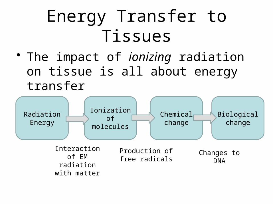
• The impact of ionizing radiation on tissue is all about energy transfer
Radiation Energy
Ionization of molecules
Chemical change
Biological change
Interaction of EM radiation with
matter
Production of free radicals
Changes to DNA
Energy Transfer to Tissues
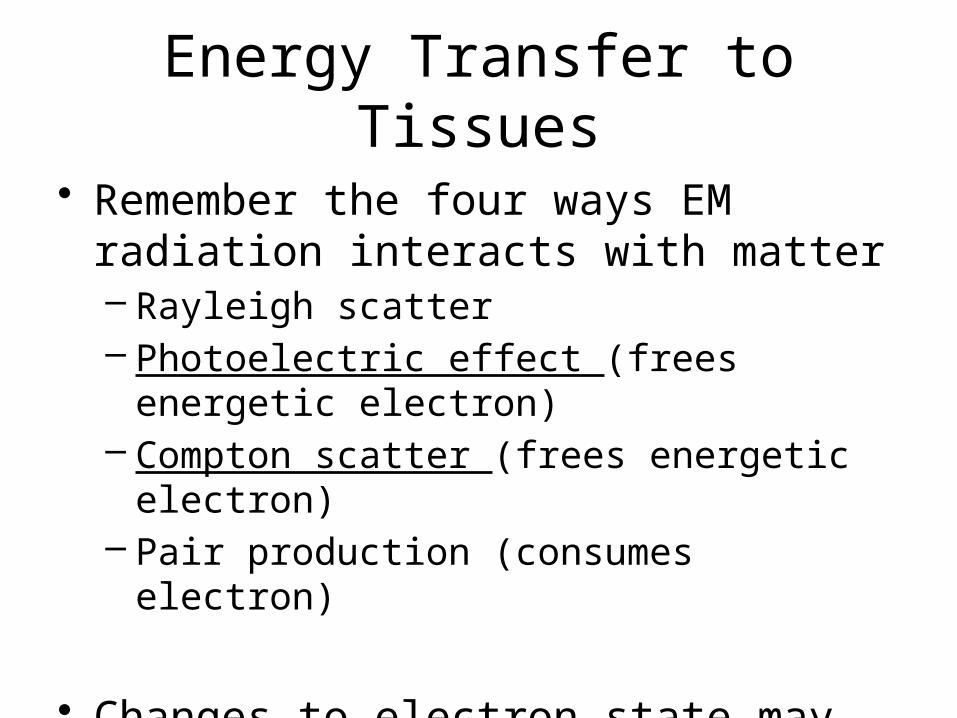
• Remember the four ways EM radiation interacts with matter– Rayleigh scatter– Photoelectric effect (frees energetic electron)– Compton scatter (frees energetic electron)– Pair production (consumes electron)
• Changes to electron state may result in ionized particles
Energy Transfer to Tissues
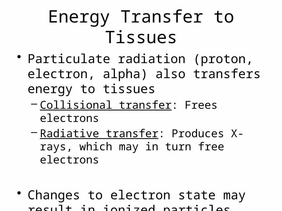
• Particulate radiation (proton, electron, alpha) also transfers energy to tissues– Collisional transfer: Frees electrons– Radiative transfer: Produces X-rays, which may in
turn free electrons
• Changes to electron state may result in ionized particles
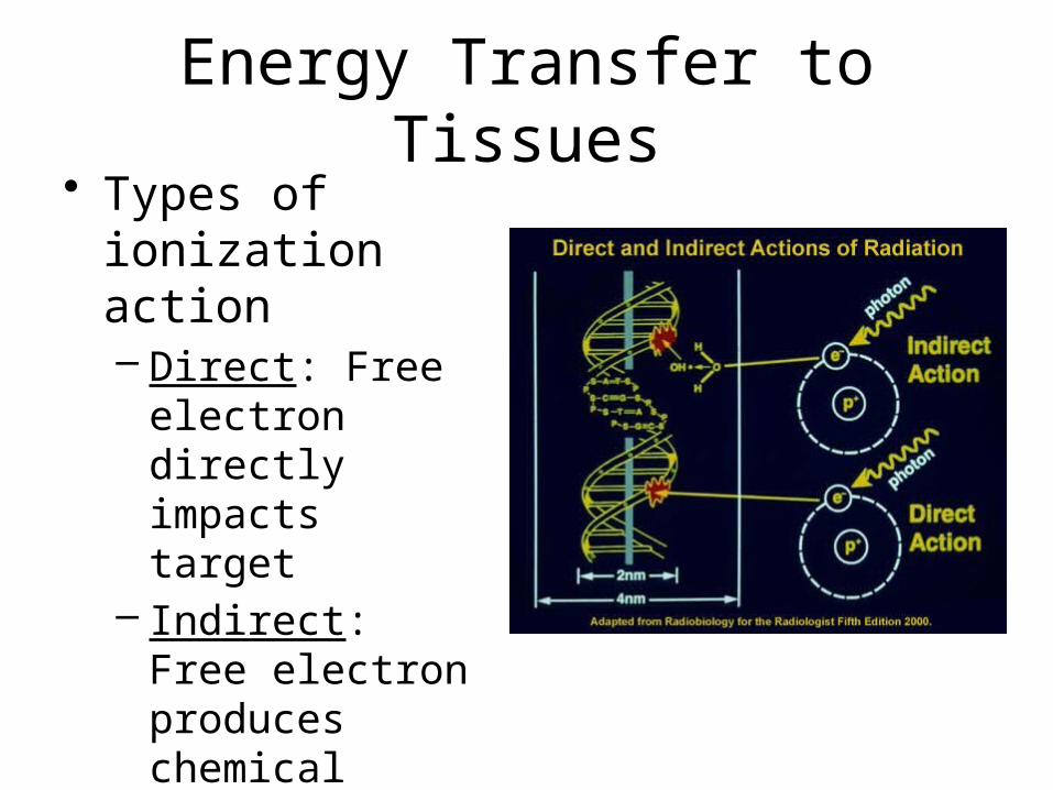
Energy Transfer to Tissues• Types of ionization
action– Direct: Free
electron directly impacts target
– Indirect: Free electron produces chemical change in another molecule that reacts with target

Energy Transfer to Tissues
• Free radicals: Highly reactive ions, atoms, or molecules– Reactive oxygen species (ROS) do a majority (2/3)
of the damage produced by ionizing radiation• Hydroxyl radical OH0
• Hydrogen peroxide H2O2
H2O H2O+ + e- H+ + OH0
OH0 + OH0 H2O2
ionization Very reactive!
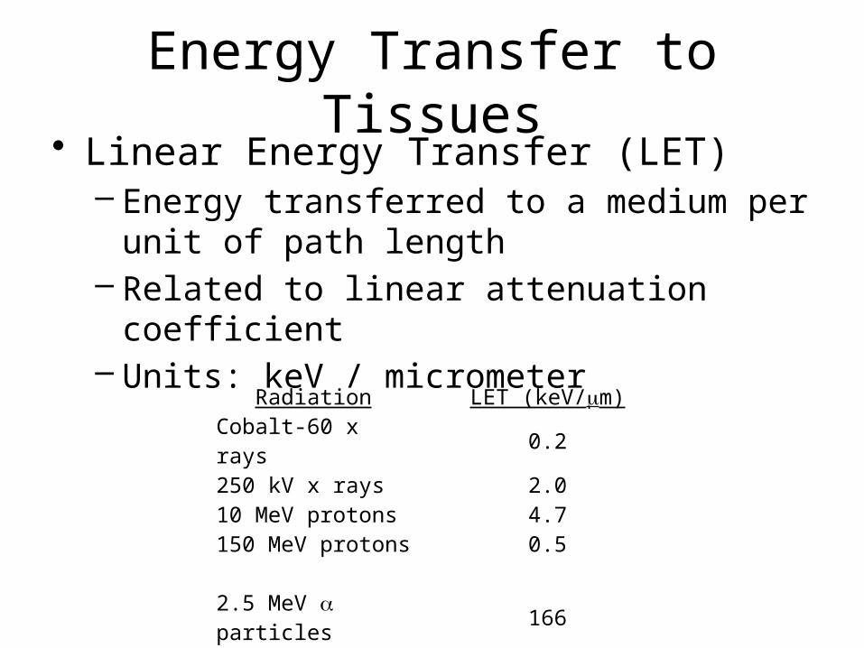
Energy Transfer to Tissues• Linear Energy Transfer (LET)
– Energy transferred to a medium per unit of path length
– Related to linear attenuation coefficient– Units: keV / micrometer
Radiation LET (keV/mm)Cobalt-60 x rays 0.2250 kV x rays 2.010 MeV protons 4.7150 MeV protons 0.5
2.5 MeV a particles 1662 GeV Fe ions 1000
Cell Effects
• DNA is believed to be the main pathway for biological impact of radiation
• Other parts of a cell may be damaged by ionization, but– Cells can self-repair many forms of damage,– Or, if necessary, self-kill (apoptosis)

Cell Effects
• Ionization products result in breakage of the DNA helix
• Most DNA breaks can be self-repaired
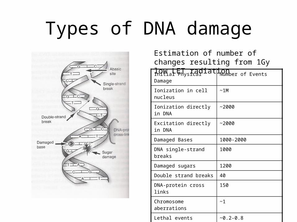
Types of DNA damage
Initial Physical Damage Number of Events
Ionization in cell nucleus ~1M
Ionization directly in DNA ~2000
Excitation directly in DNA ~2000
Damaged Bases 1000-2000
DNA single-strand breaks 1000
Damaged sugars 1200
Double strand breaks 40
DNA-protein cross links 150
Chromosome aberrations ~1
Lethal events ~0.2-0.8
Estimation of number of changes resulting from 1Gy low LET radiation
Cell Effects
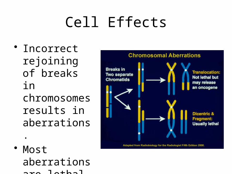
• Incorrect rejoining of breaks in chromosomes results in aberrations.
• Most aberrations are lethal to the daughter cell
Cell Effects
• Surviving daughters have DNA mutations.• Most mutations do not cause a significant
functional change.• Many mutations with functional change will
result in the cell killing itself or being killed by its neighbors,
• But some will produce a survivable, reproducible cell with altered function. The mutated gene is an oncogene.

Cell Effects
• Surviving daughters have DNA mutations.• Most mutations do not cause a significant functional change.• Many mutations with functional change will result in the cell
killing itself or being killed by its neighbors,• But some will produce a survivable, reproducible cell with
altered function. The mutated gene is an oncogene.• A mutated cell mush retain its ability to divide indefinitely in
order to produce a colony sufficient to maintain cancerous growth.
Cell Effects
• To be dangerous, the cell must:– Suffer an ionization event– Suffer DNA damage– Be unable to repair DNA damage– Suffer a nonlethal chromosomal aberration– Divide successfully (and maintain this ability)– Suffer a functional change that does not trigger
apoptosis or killing by neighbors
Cell Effects
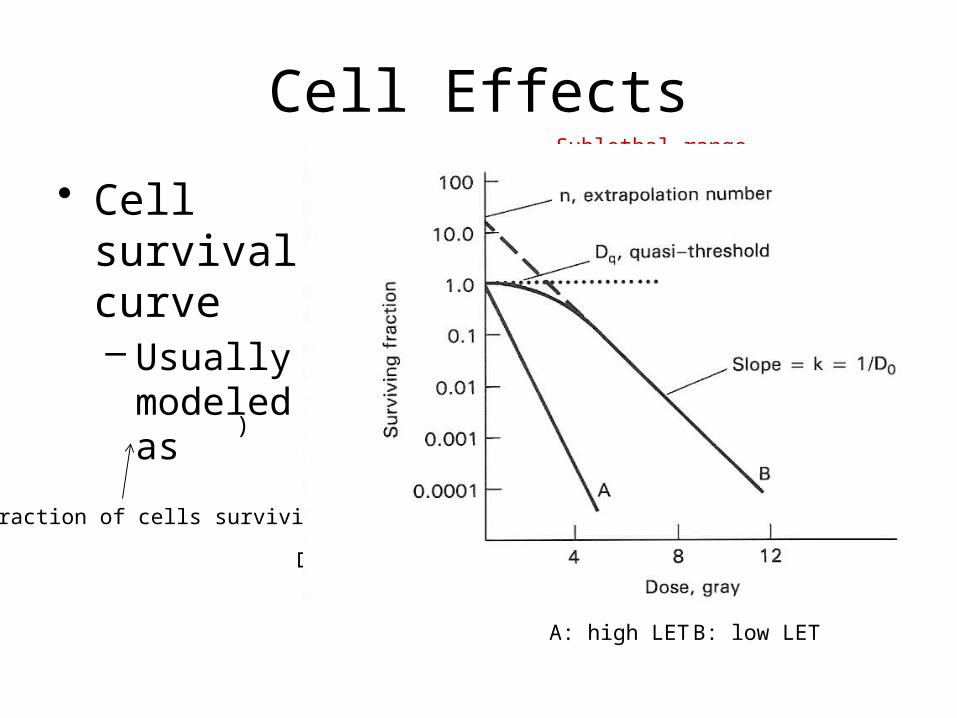
• Cell survival curve– Usually
modeled as
)
Fraction of cells surviving
Dose
A: high LET B: low LET
Sublethal range
“Shoulder”
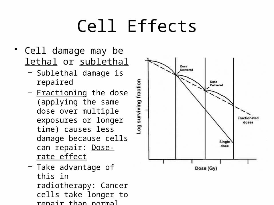
Cell Effects• Cell damage may be lethal
or sublethal– Sublethal damage is repaired– Fractioning the dose
(applying the same dose over multiple exposures or longer time) causes less damage because cells can repair: Dose-rate effect
– Take advantage of this in radiotherapy: Cancer cells take longer to repair than normal tissue

Cell Effects• Cell survival is a function of
cell type and radiation type– Non- or slowly proliferating
cells (nerve, muscle, secretory) are less susceptible to radiation damage.
– Highly-proliferating cells (epithelial, stem cells) are more susceptible.
• This leads to the concept of radiation “quality”– How effective is the radiation type at
causing biological damage?
Units and Terms
• Linear Energy Transfer (LET)– Unit: keV per micrometer (energy per length)– A function of radiation type, energy, medium
(generally water or water-equivalent for tissues)– Survival curve depends on LET.
– Equal doses of different types of radiation do not give equal biological effect – they have different LET.
Units and Terms
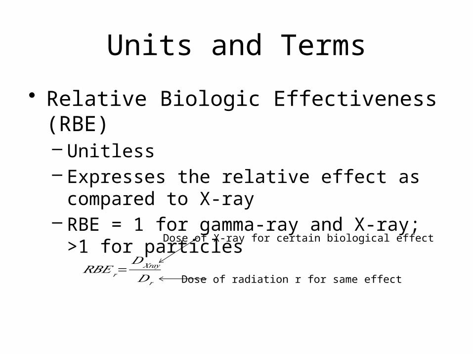
• Relative Biologic Effectiveness (RBE)– Unitless– Expresses the relative effect as compared to X-ray– RBE = 1 for gamma-ray and X-ray; >1 for
particles
𝑅𝐵𝐸𝑟=𝐷𝑋𝑟𝑎𝑦
𝐷𝑟
Dose of X-ray for certain biological effect
Dose of radiation r for same effect
Units and Terms
• Radiation Exposure– Amount of radiation producing a specified amount
of ionized particles in air– Unit: coulomb/kg (producing 1 C of charge in 1
KG of air)– Old unit: Roentgen (1 C/kg = 3876 R)– Only applies to EM radiation– Does not correlate with biological effects
Units and Terms
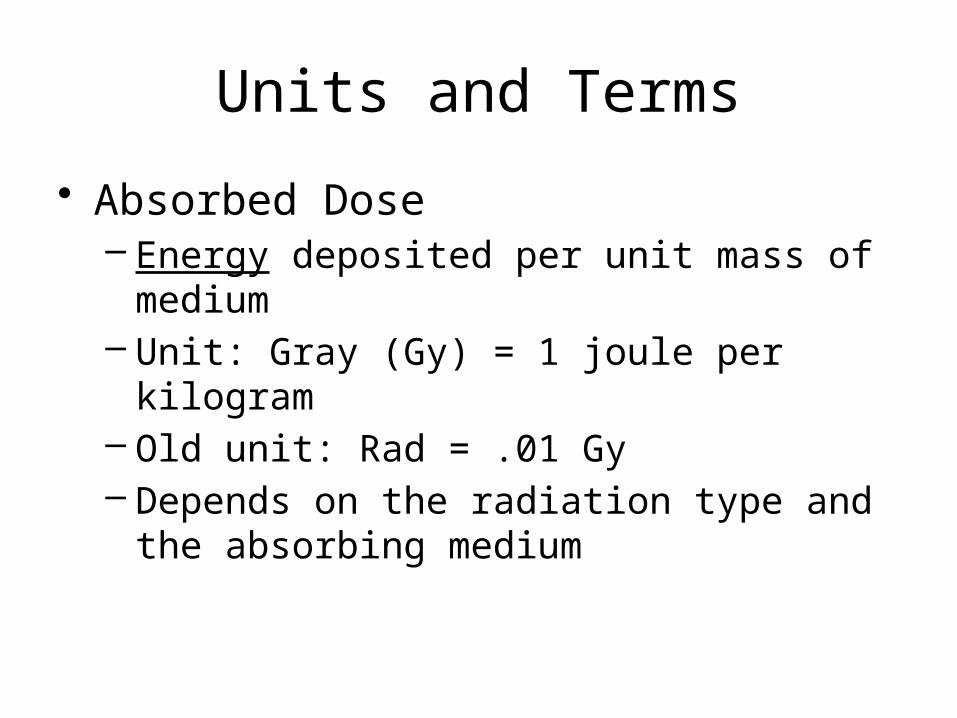
• Absorbed Dose– Energy deposited per unit mass of medium– Unit: Gray (Gy) = 1 joule per kilogram– Old unit: Rad = .01 Gy– Depends on the radiation type and the absorbing
medium

Units and Terms
• Relate Absorbed Dose to Exposure– Exposure is defined in air– Absorbed dose can be any medium– If medium is air, 1 R ≈ .87 rad; 1 Gy ≈ .0297 C/kg– For other materials,
𝐷𝑜𝑠𝑒 (𝐺𝑦 )≈33.67( 𝜇𝜌 )
𝑚𝑎𝑡𝑒𝑟𝑖𝑎𝑙
(𝜇𝜌 )𝑎𝑖𝑟
×𝐸𝑥𝑝𝑜𝑠𝑢𝑟𝑒 ( 𝐶𝑘𝑔
)

Units and Terms
• Kerma– Kinetic Energy Released in Material– Unit: Gray (Gy) = 1 joule per kilogram– Accounts for secondary effects of energetic
electrons (interactions elsewhere or Bremsstrahlung) where absorbed dose does not

Units and Terms
• Radioactivity– Disintegrations per unit time– Unit: Becquerel (Bq) = 1 decays per second– Unit: Curie (Ci) = 3.7 x 10¹⁰ decays per second– Applies to radioisotopes
Units and Terms
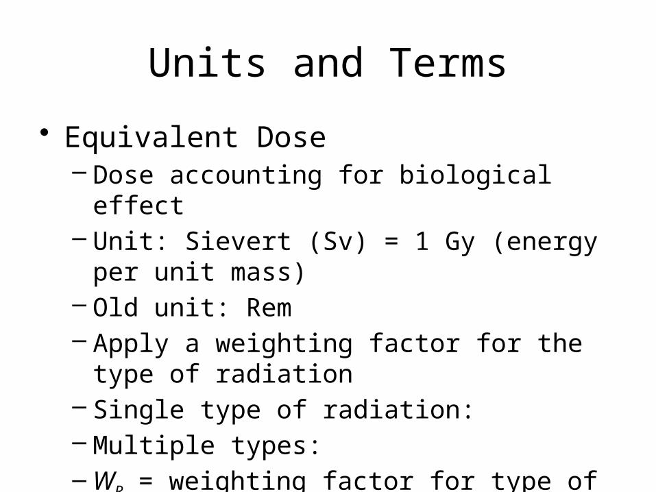
• Equivalent Dose– Dose accounting for biological effect– Unit: Sievert (Sv) = 1 Gy (energy per unit mass)– Old unit: Rem– Apply a weighting factor for the type of radiation– Single type of radiation: – Multiple types: –WR = weighting factor for type of radiation
– DR = absorbed dose in Gy
Units and Terms
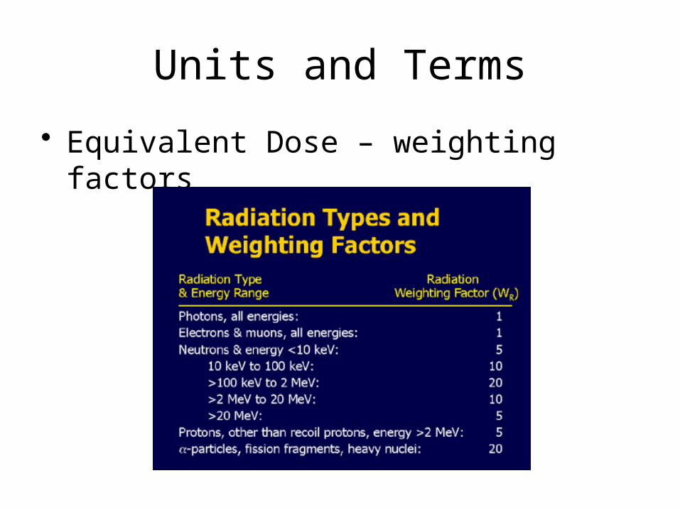
• Equivalent Dose – weighting factors
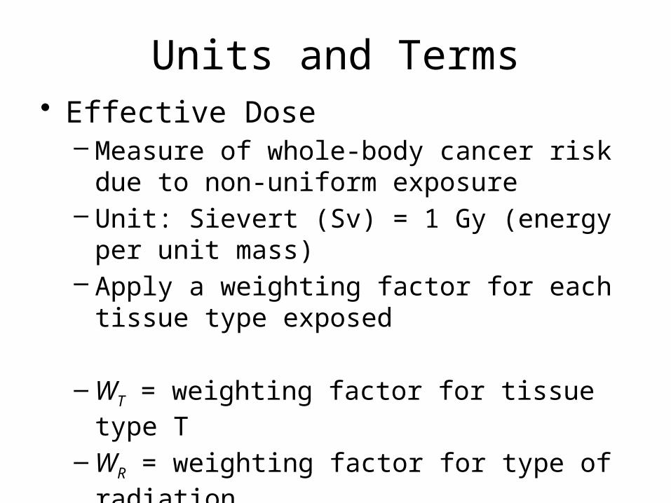
Units and Terms• Effective Dose
– Measure of whole-body cancer risk due to non-uniform exposure
– Unit: Sievert (Sv) = 1 Gy (energy per unit mass)– Apply a weighting factor for each tissue type
exposed
–WT = weighting factor for tissue type T
–WR = weighting factor for type of radiation
– DRT = absorbed dose in Gy for tissue T
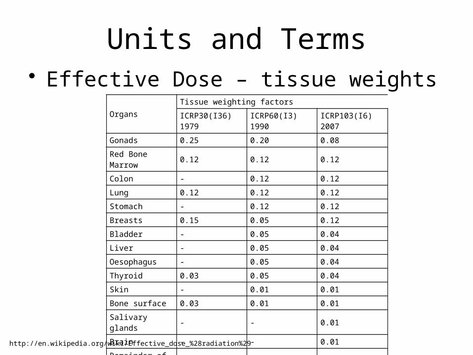
Units and Terms• Effective Dose – tissue weights
Organs
Tissue weighting factors
ICRP30(I36)1979
ICRP60(I3)1990
ICRP103(I6)2007
Gonads 0.25 0.20 0.08
Red Bone Marrow 0.12 0.12 0.12
Colon - 0.12 0.12
Lung 0.12 0.12 0.12
Stomach - 0.12 0.12
Breasts 0.15 0.05 0.12
Bladder - 0.05 0.04
Liver - 0.05 0.04
Oesophagus - 0.05 0.04
Thyroid 0.03 0.05 0.04
Skin - 0.01 0.01
Bone surface 0.03 0.01 0.01
Salivary glands - - 0.01
Brain - - 0.01
Remainder of body 0.30 0.05 0.12
Total 1.00 1.00 1.00
http://en.wikipedia.org/wiki/Effective_dose_%28radiation%29
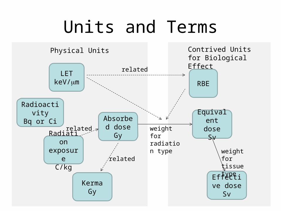
Units and Terms
LETkeV/mm RBE
Radiation exposure
C/kg
Absorbed doseGy
KermaGy
RadioactivityBq or Ci Equivalent
doseSv
Effective doseSv
Physical Units Contrived Units for Biological Effect
related
weight for radiation type
weight for tissue typerelated
related
Units and Terms
• Example problem– A hand X-ray is taken at 20 keV. Let
Wbone=Wmuscle=.002. Find the effective dose for an exposure of 2.5 x 10⁻⁴ C/kg.

Reference
• Hall, E. J., Center for Radiological Research, Columbia University, “Web-Rad-Train,” http://www.columbia.edu/~ejh1/web-rad-train/index.html