body representations in the human brain revealed by ... · pdf fileillusions and their...
TRANSCRIPT

R
Bic
Ea
Ob
c
d
e
a
ARRAA
KMKBMSCSF
C
h0
Neuroscience Research 104 (2016) 16–30
Contents lists available at ScienceDirect
Neuroscience Research
jo ur nal home p age: www.elsev ier .com/ locate /neures
eview article
ody representations in the human brain revealed by kinestheticllusions and their essential contributions to motor control andorporeal awareness
iichi Naitoa,b,c,∗, Tomoyo Moritaa,d, Kaoru Amemiyaa,e
Center for Information and Neural Networks (CiNet), National Institute of Information and Communications Technology (NICT), 1-4 Yamadaoka, Suita,saka 565-0871, JapanGraduate School of Medicine, Osaka University, 2-15 Yamadaoka, Suita, Osaka 565-0871, JapanGraduate School of Frontier Biosciences, Osaka University, 1-3 Yamadaoka, Suita, Osaka 565-0871, JapanGraduate School of Engineering, Osaka University, 2-1 Yamadaoka, Suita, Osaka 565-0871, JapanThe Japan Society for the Promotion of Science, 5-3-1 Koujimachi, Chiyoda, Tokyo 102-0083, Japan
r t i c l e i n f o
rticle history:eceived 1 September 2015eceived in revised form 22 October 2015ccepted 23 October 2015vailable online 10 November 2015
eywords:uscle spindle
inesthesiaody representationotor control
a b s t r a c t
The human brain can generate a continuously changing postural model of our body. Somatic (proprio-ceptive) signals from skeletal muscles and joints contribute to the formation of the body representation.Recent neuroimaging studies of proprioceptive bodily illusions have elucidated the importance of threebrain systems (motor network, specialized parietal systems, right inferior fronto-parietal network) in theformation of the human body representation.
The motor network, especially the primary motor cortex, processes afferent input from skeletal mus-cles. Such information may contribute to the formation of kinematic/dynamic postural models of limbs,thereby enabling fast online feedback control. Distinct parietal regions appear to play specialized roles inthe transformation/integration of information across different coordinate systems, which may subservethe adaptability and flexibility of the body representation. Finally, the right inferior fronto-parietal net-
ensory-motor associationorporeal awarenesself-consciousnessunctional magnetic resonance imaging
work, connected by the inferior branch of the superior longitudinal fasciculus, is consistently recruitedwhen an individual experiences various types of bodily illusions and its possible roles relate to corporealawareness, which is likely elicited through a series of neuronal processes of monitoring and accumulatingbodily information and updating the body representation. Because this network is also recruited whenidentifying one’s own features, the network activity could be a neuronal basis for self-consciousness.
© 2015 Elsevier Ireland Ltd and the Japan Neuroscience Society. All rights reserved.
ontents
1. Introduction . . . . . . . . . . . . . . . . . . . . . . . . . . . . . . . . . . . . . . . . . . . . . . . . . . . . . . . . . . . . . . . . . . . . . . . . . . . . . . . . . . . . . . . . . . . . . . . . . . . . . . . . . . . . . . . . . . . . . . . . . . . . . . . . . . . . . . . . . . . . . 172. Kinesthetic illusion as a useful tool to elucidate neuronal representation of the human body . . . . . . . . . . . . . . . . . . . . . . . . . . . . . . . . . . . . . . . . . . . . . . . . . . . . . . . 17
2.1. Motor network activation during kinesthetic illusion . . . . . . . . . . . . . . . . . . . . . . . . . . . . . . . . . . . . . . . . . . . . . . . . . . . . . . . . . . . . . . . . . . . . . . . . . . . . . . . . . . . . . . . . . . 182.2. Roles of M1/PMD in kinesthetic illusion . . . . . . . . . . . . . . . . . . . . . . . . . . . . . . . . . . . . . . . . . . . . . . . . . . . . . . . . . . . . . . . . . . . . . . . . . . . . . . . . . . . . . . . . . . . . . . . . . . . . . . . . 182.3. Convergence of kinesthetic information in medial-wall motor regions . . . . . . . . . . . . . . . . . . . . . . . . . . . . . . . . . . . . . . . . . . . . . . . . . . . . . . . . . . . . . . . . . . . . . . . 212.4. Application of kinesthetic illusion for neuro-rehabilitation . . . . . . . . . . . . . . . . . . . . . . . . . . . . . . . . . . . . . . . . . . . . . . . . . . . . . . . . . . . . . . . . . . . . . . . . . . . . . . . . . . . . 212.5. Brief summary . . . . . . . . . . . . . . . . . . . . . . . . . . . . . . . . . . . . . . . . . . . . . . . . . . . . . . . . . . . . . . . . . . . . . . . . . . . . . . . . . . . . . . . . . . . . . . . . . . . . . . . . . . . . . . . . . . . . . . . . . . . . . . . . . . . 22
3. Specialized parietal systems . . . . . . . . . . . . . . . . . . . . . . . . . . . . . . . . . . . . . . . . . . . . . . .
3.1. Integration of somatic information from multiple body parts in h3.2. Expansion of the body representation by incorporating external o
∗ Corresponding author at: 2A6 1-4 Yamadaoka, Suita, Osaka 565-0871, Japan. Tel.: +81E-mail addresses: [email protected] (E. Naito), [email protected] (T. M
ttp://dx.doi.org/10.1016/j.neures.2015.10.013168-0102/© 2015 Elsevier Ireland Ltd and the Japan Neuroscience Society. All rights res
. . . . . . . . . . . . . . . . . . . . . . . . . . . . . . . . . . . . . . . . . . . . . . . . . . . . . . . . . . . . . . . . . . . . . . . . . . . . . 22igher-order somatosensory areas . . . . . . . . . . . . . . . . . . . . . . . . . . . . . . . . . . . . . . . . . 22bjects in the left inferior parietal lobule . . . . . . . . . . . . . . . . . . . . . . . . . . . . . . . . . 23
80 9098 3256; fax: +81 06 7174 8612.orita), [email protected] (K. Amemiya).
erved.

E. Naito et al. / Neuroscience Research 104 (2016) 16–30 17
3.3. Visual dominance over kinesthesia is computed in the posterior parietal cortex . . . . . . . . . . . . . . . . . . . . . . . . . . . . . . . . . . . . . . . . . . . . . . . . . . . . . . . . . . . . . 253.4. Brief summery . . . . . . . . . . . . . . . . . . . . . . . . . . . . . . . . . . . . . . . . . . . . . . . . . . . . . . . . . . . . . . . . . . . . . . . . . . . . . . . . . . . . . . . . . . . . . . . . . . . . . . . . . . . . . . . . . . . . . . . . . . . . . . . . . . . 25
4. Activation of the right inferior frontoparietal cortices during a kinesthetic illusion . . . . . . . . . . . . . . . . . . . . . . . . . . . . . . . . . . . . . . . . . . . . . . . . . . . . . . . . . . . . . . . . . . 254.1. Roles of the right inferior frontoparietal cortices in the formation of the body representation . . . . . . . . . . . . . . . . . . . . . . . . . . . . . . . . . . . . . . . . . . . . . . . 264.2. The right SLF III network, self-identification, and self-consciousness . . . . . . . . . . . . . . . . . . . . . . . . . . . . . . . . . . . . . . . . . . . . . . . . . . . . . . . . . . . . . . . . . . . . . . . . . . 274.3. Brief summary . . . . . . . . . . . . . . . . . . . . . . . . . . . . . . . . . . . . . . . . . . . . . . . . . . . . . . . . . . . . . . . . . . . . . . . . . . . . . . . . . . . . . . . . . . . . . . . . . . . . . . . . . . . . . . . . . . . . . . . . . . . . . . . . . . . 28
5. Conclusions . . . . . . . . . . . . . . . . . . . . . . . . . . . . . . . . . . . . . . . . . . . . . . . . . . . . . . . . . . . . . . . . . . . . . . . . . . . . . . . . . . . . . . . . . . . . . . . . . . . . . . . . . . . . . . . . . . . . . . . . . . . . . . . . . . . . . . . . . . . . . 28Acknowledgements . . . . . . . . . . . . . . . . . . . . . . . . . . . . . . . . . . . . . . . . . . . . . . . . . . . . . . . . . . . . . . . . . . . . . . . . . . . . . . . . . . . . . . . . . . . . . . . . . . . . . . . . . . . . . . . . . . . . . . . . . . . . . . . . . . . . .28
. . . . . .
1
tahtcopaajiwmibtetpi(
bvtrimbaonafipst
ibrutsapbf‘t
References . . . . . . . . . . . . . . . . . . . . . . . . . . . . . . . . . . . . . . . . . . . . . . . . . . . . . . . . . . . .
. Introduction
The human brain creates an internal representation of the bodyhat assists in controlling physical movement. The presence ofnatomical and functional somatotopy, which is referred to as aomunculus, is the most vivid example of this, and can be seen inhe primary somatosensory cortex (SI) and in the primary motorortex (M1). Somatosensory information originating from one’swn body plays a very important role in motor control. For exam-le, patients with impaired proprioceptive input (somatic signalsbout spatial position and movement of limbs) may not be able toccurately perform a reaching movement toward a target locatedust 10 cm away (Ghez et al., 1995). When such patients have visualnput about the movement of their hands and arms, only then
ill they be capable of performing an accurate reaching move-ent. However, removal of this visual information will result in
mpairment of reaching performance again. Similar behavior haseen reported when such patients were made to perform a thumb-o-finger opposition task (Rothwell et al., 1982). These lines ofvidence indicate that somatic signals are extremely important inhe control of movement and motor learning. Indeed, non-humanrimates with a disrupted SI, and thus, impaired somatosensory
nputs to the M1, have difficulty in learning new movementsPavlides et al., 1993).
Somatic signals are also essential for recognizing one’s ownody. Humans normally recognize their own bodies mainly throughisual and somatic sensations. The visual system can be usedo acquire information about distant areas that have no directelationship to the individual. In contrast, somatic sensations arenduced by various sensory receptors that are present in the skin,
uscles, and joints, and they normally originate from one’s ownody. Hence, somatic sensations allow us to conceive of ourselvess the source of incoming sensations and as separate entities fromther agents and the external world. Somatic sensations that origi-ate from sensory receptors in the muscles and joints are referred tos proprioception, and these sensations are qualitatively differentrom skin (cutaneous/tactile) sensations. Proprioception is involvedn the perception of positional changes and movements of bodyarts, such as the hands and feet, while the main function of skinensations is to extract the feel of materials and objects, such ashose touched by our hand (Naito, 2004a).
Although it has yet to be clearly defined, the concept of bodymage refers to the image of oneself, and generally encompassesoth mental and psychological factors. In contrast, the body schemaefers to a model of one’s posture (body configuration) that ispdated constantly with new sensory information elicited by pos-ural changes (Head and Holmes, 1911). Consequently, the bodychema is a neural representation of the body that involves motornd posture control. As described, proprioception largely encom-asses the perception of positional changes and movements of
ody parts; thus, it is the most essential sensory modality for theormation of the body representation. Proprioception is Latin forone’s own perception’, and this sensation has long conceived of ashe source of physical self-perception. Hence, we believe that this. . . . . . . . . . . . . . . . . . . . . . . . . . . . . . . . . . . . . . . . . . . . . . . . . . . . . . . . . . . . . . . . . . . . . . . . . . . . 28
sensation must be deeply involved in corporeal awareness, whichcould be the basis of self-consciousness.
Recent neuroimaging studies dealing with kinesthetic illusions(see below) have unveiled the neuronal representation of thehuman body representation. In this chapter, we focus mainly onproprioception, and introduce the importance of three brain sys-tems (the motor network, specialized parietal systems, and theright inferior fronto-parietal network) in the formation of thehuman body representation, which has been revealed by a seriesof our kinesthetic illusion studies. We also discuss and speculatehow proprioceptive input could lead to corporeal awareness andself-consciousness.
2. Kinesthetic illusion as a useful tool to elucidate neuronalrepresentation of the human body
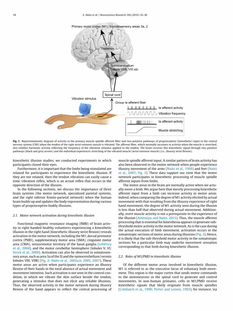
Muscles and joints contain proprioceptors that sense the move-ment and position of limbs (the hands and feet). Among these,the receptors in the (intrafusal) muscles are called muscle spin-dles, and the signals are transmitted to the central nervous system(CNS), mainly through group Ia primary muscle spindle afferentfibers (Fig. 1). When the muscles are stretched, the activity of mus-cle spindle afferents normally increases (Fig. 1), and this activity cancarry information about the direction and speed of limb movement(Burke et al., 1976, 1988; Edin and Vallbo, 1988, 1990; Ribot-Ciscarand Roll, 1988). Thus, movement sensations that largely depend onmuscle spindle afferent input are called kinesthesia. Proprioceptiveinput to the CNS is generally known to comprise the following twomain pathways: the spinal cord-thalamus-cerebral cortex pathwayand the spinal cord-cerebellum-thalamus-cerebral cortex pathway(Fig. 1). Hence, the brain can receive kinesthetic information aboutthe direction and speed of limb movement through Ia muscle affer-ent fibers.
Interestingly, the activity of muscle spindle afferents alsoincreases in response to vibration stimuli of specific frequencies(around 80 Hz) (Fig. 1). Thus, it is possible to employ this propertyto elicit a clear illusory movement sensation, such that vibratedlimbs (hands and feet) feel as though they are moving when theyare not (kinesthetic illusion: Goodwin et al., 1972; Roll and Vedel,1982; Roll et al., 1989). For example, when a vibration stimulus isapplied to the tendon of the wrist extensor muscle, an illusory wristflexion is elicited (Fig. 1). This involves no movement of the hand andno intention to move their hand, but it is possible to experience aclear movement sensation as if their hand is moving. In other words,during kinesthetic illusion, people can experience postural changeof a limb, which is an indispensable element in the formation ofthe body representation (body schema). Further importantly, thismethod enables the experience of not only simple limb movementsbut also various types of bodily illusions (see below). Hence, thisillusion is a useful tool to experimentally manipulate and elucidate
the neuronal basis of the human body representation.Because vision normally supersedes proprioception, the visualinformation about the unmoving limbs significantly attenuatesthe illusions (Hagura et al., 2007). Thus, in the majority of our
18 E. Naito et al. / Neuroscience Research 104 (2016) 16–30
Fig. 1. Representationtic diagram of activity in the primary muscle spindle afferent fiber and two putative pathways of proprioceptive (kinesthetic) input to the centraln ed. Tha applip vibrat
kp
rtto
babt
2
iiacaeSslmflmdgTfl
ervous system (CNS) when the tendon of the right wrist extensor muscle is vibratlso exhibits harmonic activity reflecting the frequency of the vibration stimulusathways (black and grey arrows) and the individual experiences stretching of the
inesthetic illusion studies, we conducted experiments in whicharticipants closed their eyes.
Furthermore, it is important that the limbs being stimulated areelaxed for participants to experience the kinesthetic illusion. Ifhey are not relaxed, then the tendon vibration can easily cause aonic vibration reflex, which is an actual reflex that occurs in thepposite direction of the illusion.
In the following sections, we discuss the importance of threerain systems (the motor network, specialized parietal systems,nd the right inferior fronto-parietal network) when the humanrain builds up and updates the body representation during variousypes of proprioceptive bodily illusions.
.1. Motor network activation during kinesthetic illusion
Functional magnetic resonance imaging (fMRI) of brain activ-ty in right-handed healthy volunteers experiencing a kinestheticllusion in the right hand (kinesthetic illusory wrist flexion) revealsctivation in the motor network, including the M1, dorsal premotorortex (PMD), supplementary motor area (SMA), cingulate motorrea (CMA), sensorimotor territory of the basal ganglia (Lehéricyt al., 2004), and the motor cerebellar hemisphere (lobules V, VI;trick et al., 2009). Activation can also be observed in somatosen-ory areas, such as area 3a of the SI and the spinocerebellum (vermisobules VIII, VIIB) (Fig. 2: Naito et al., 2002a,b, 2005, 2007). These
otor areas are active when participants experience an illusoryexion of their hands in the total absence of actual movement andovement intention. Such activation is not seen in the control con-
ition, in which we vibrate the skin surface beside the tendon,enerating a stimulus that does not elicit any reliable illusions.hus, the observed activity in the motor network during illusoryexion of the hand appears to reflect the central processing of
e afferent fiber, which normally increases in activity when the muscle is stretched,ed to the tendon. The brain receives this kinesthetic signal through two putativeed muscle (wrist extensor muscle) (i.e., illusory wrist flexion).
muscle spindle afferent input. A similar pattern of brain activity hasalso been observed in the motor network when people experienceillusory movement of the arms (Naito et al., 1999) and feet (Naitoet al., 2007; Fig. 3). These data support our view that the motornetwork participates in kinesthetic processing of muscle spindleafferent inputs from limbs.
The motor areas in the brain are normally active when we actu-ally move a limb. We argue here that merely processing kinestheticafferent input from a limb can increase activity in motor areas.Indeed, when comparing the degree of M1 activity elicited by actualmovement with that resulting from the illusory experience of righthand movement, the degree of M1 activity seen during the illusionis less than half that observed during actual movement. Addition-ally, overt muscle activity is not a prerequisite to the experience ofthe illusion (Amemiya and Naito, 2015). Thus, the muscle afferentprocessing that is essential for kinesthesia appears to generate sub-threshold motor activity in the motor network. As is the case duringthe actual execution of limb movement, activation occurs in thesomatotopic sections of motor areas during illusions (Fig. 3). Hence,it is likely that the sub-threshold motor activity in the somatotopicsections for a particular limb may underlie movement sensationcorresponding to that limb during kinesthetic illusion.
2.2. Roles of M1/PMD in kinesthetic illusion
Of the different motor areas involved in kinesthetic illusion,M1 is referred to as the executive locus of voluntary limb move-ment. This region is the major cortex that sends motor commands
to the motoneurons in the spinal cord to generate and controlmovements. In non-human primates, cells in M1/PMD receivekinesthetic signals that likely originate from muscle spindles(Colebatch et al., 1990; Porter and Lemon, 1993), for instance, via
E. Naito et al. / Neuroscience Research 104 (2016) 16–30 19
Fig. 2. M1-centered motor network activity during illusory flexion of the right hand. During the illusion, activation can be seen in the motor network, including the primarymotor cortex (M1), dorsal premotor cortex (PMD), supplementary motor area (SMA), cingulate motor cortex (CMA), putamen/globus pallidas (BG), and motor cerebellarhemisphere (Lobule V, dentate). Activation also occurs in the somatosensory areas, such as putative somatosensory area 3a and the spinocerebellum (Lobule VIIB, VIII). The lightblue arrows represent functional and anatomical connections with M1. Note that the M1-BG circuit is formed by M1 → BG → thalamus → M1, and the M1-cerebellum circuiti thalak ty in t
sFe(p
siavbatetd
hc[(ea
s formed by M1 → pontine nucleus → cerebellar hemisphere → dentate nucleus →inesthetic input. Muscle afferent processing generates sub-threshold motor activi
omatosensory area 3a (Phillips et al., 1971; Huerta and Pons, 1990;ig. 2). Indeed, tendon vibration of triceps brachii muscles can elicitxcitatory responses in M1/PMD cells at a mean latency of 21 msColebatch et al., 1990). Thus, the M1 involves muscle afferentrocessing, which is essential for kinesthesia.
Considering that muscle spindle afferents are capable ofignaling the direction and speed of limb movement (see above),t is likely that somatotopically organized M1/PMD activity duringn illusion (Fig. 3) represents information about the direction andelocity of the illusory limb movement. [Additional parietal contri-ution is also suggested when the brain has to specify the directionnd speed of illusory movement in an ambiguous situation whenwo antagonistic muscles are simultaneously vibrated (Romaigueret al., 2003).] Thus, the fundamental information underlying kines-hesia is likely processed in the M1/PMD-centered motor networkuring an illusion.
The importance of the human M1/PMD in kinesthetic processingas been corroborated by several studies. First, the degree of motor-ortical (M1) excitability is correlated with the degree of illusion
the extent to which people experience an illusory hand movementi.e., wrist angle) within a certain period of vibration time] (Naitot al., 2002b). Likewise, the degree of motor-cortical excitabilitylso reflects the amount of illusory aftereffect [the extent to whichmus → M1, which is simplified in the present diagram. The white arrows indicatehe M1-centered motor network, which likely produces latent motor commands.
people experience movement sensation as if the vibrated hand isgoing back to its original position after the cessation of vibrationstimulus] (Kito et al., 2006). Second, contralateral M1/PMD activityduring an illusion has been found to decrease in proportion to thedegree to which an illusion is attenuated by vision (i.e., the degreeto which the experience of illusory movement is attenuated whenparticipants look at their unmoving hand] (Hagura et al., 2007).Finally, focal damage in the hand section of M1/PMD can severelyimpair the experience of illusory movement of the contralateralhand (Naito et al., 2010). These findings strongly suggest that M1is not simply a center for motor execution, but also an importantnode that may contribute to the somatic perception of limb move-ment (i.e., kinesthesia) by receiving and processing muscle afferentsignals. Hence, the M1 could warrant its title as the primary motor-sensory cortex.
It is still under debate whether the human M1/PMD is purelyinvolved in kinesthesia per se. Some studies have reported thattranscranial magnetic stimulation (TMS) over M1 and PMD mayproduce movement sensations in the nerve blocked arm and leg
(Christensen et al., 2010) and that TMS over M1 of traumaticamputees who report the presence of a phantom limb can evokethe movement sensation in the phantom limb (Cohen et al., 1991;Pascual-Leone et al., 1996; Mercier et al., 2006). On the other hand,
20 E. Naito et al. / Neuroscience Research 104 (2016) 16–30
Fig. 3. Somatotopical sections of multiple motor areas active during illusory movement of the right hand (yellow), left hand (green), right foot (light blue), and left foot(pink). Clear somatotopy is seen in M1 and the motor cerebellar hemisphere (A–C). Somatotopical organization is less apparent in the medial-wall motor regions (D and E).
M
trt
cStdamdbCi(sbls(
odified from Naito et al. (2007).
here is a report that M1 activity is not necessary when people expe-ience visually induced illusory hand movement when they watchhe video of their own moving hand (Kaneko et al., 2015).
M1/PMD has been found to form efficient sensory-motor cir-uits with muscles (Fetz et al., 1980; Cheney and Fetz, 1984).pecifically, cortico-motoneuronal cells in M1/PMD that respondo passive movement of the elbow or wrist toward a particularirection (extension or flexion) also show excitatory activity (1) forctive movement toward the same direction as that of the passiveovement; Type 1, (2) for active movement toward the opposite
irection; Type 2, or (3) in both directions. Importantly, the num-er of cells in these three groups is almost equal (Fetz et al., 1980;heney and Fetz, 1984). The cells that exhibit excitatory activ-
ty during active movements are associated with motor outputgeneration of muscle activity) and the cells that fire during pas-ive movement likely respond to the stretching of muscles. This isecause the muscle spindle is most sensitive to specify a direction of
imb movement among various types of mechanoreceptors (joint,low, and fast adapting cutaneous) that react to passive movementBurke et al., 1988).
Some cells respond to both active wrist flexion and passivewrist flexion (Type 1: same preferred directions for active andpassive movements). The activity of such cell is sent to motoneu-rons to facilitate wrist flexion during active flexion. On the otherhand, the activity of the cell is induced by the afferent inputfrom the stretching extensor muscle during the wrist flexion.Thus, these cells appear to be capable of facilitating wrist flexionwhile receiving muscle afferent input from the stretching extensor(antagonistic) muscle. It is likely that this circuit mediates kines-thetic illusory wrist flexion, because muscle spindle afferent inputfrom a vibrated wrist extensor muscle elicits illusory wrist flexionmovement (Fig. 1). Indeed, during illusory wrist flexion, motor-cortical excitability for the wrist flexor muscle is elevated comparedwith that for the extensor muscle (Kito et al., 2006). This pattern ofmotor-cortical excitation is similar to that observed during actualwrist flexion. Hence, this may support our view that sub-thresholdmotor activity is generated in the motor network when people
experience kinesthetic illusory limb movement as if the movementis actually happening (veridical sensation of limb movement). Theunderlying neuronal operation seems to be the generation of latent
E. Naito et al. / Neuroscience Re
Fig. 4. Medial-wall activations in the left hemisphere (x = −6; panel E in Fig. 3) dur-ing kinesthetic illusions in all limbs. Illusory movement of the right hand or footactivates the caudal part of the contralateral CMA (yellow section) and SMA (bluesection), respectively. Activation is seen in the rostral section of the SMA regardlessof whether participants experience an illusion in the right or left hand (bimanualsection: red section). Activity in the rostral part of the CMA increases when partici-pants experience illusory movement of the contralateral hand or foot (contralaterallimb section: green section). In the more rostral section of the CMA, activation occursregardless of whether participants experience illusory movement of the left or righthands and feet (common section: white section). Kinesthetic information specificto each limb appears to converge from the caudal to rostral direction (indicated bywhite arrows).
Modified from Naito et al. (2007).
mt
paf(loiotlacsttmect
amceusTwa
cle activity gradually emerges if the tendon is vibrated for a longperiod of time, as kinesthetic signals progressively spread to the
otor commands to the agonistic muscles associated with the kines-hetic illusory movement (Naito, 2004b).
In contrast, cells that respond to both active wrist flexion andassive wrist extension (Type 2: opposite preferred directions) canctivate the wrist flexor muscle when this muscle is stretched. Thisunction is implemented in the transcortical long-loop reflex circuitCheney and Fetz, 1984). When a hand muscle is stretched, long-atency (50–90 ms from the stretch onset) EMG responses can bebserved in the stretched muscle. We assume that this circuit isnvolved in the generation of the tonic vibration reflex, which ispposite to the kinesthetic illusion as a tendon vibration is appliedo unrelaxed muscles as discussed above. This is an important bio-ogical defensive mechanism to prevent muscle damage caused byn unexpected and noxious stretch. While the spinal reflex cir-uit is automatic, the transcortical long-loop circuit may reflectome degree of voluntary action, as M1 (executive locus of volun-ary movement) mediates this circuit. The greater contribution ofhe transcortical long-loop circuit to fast feedback control of limb
ovement has been demonstrated in a recent study (Pruszynskit al., 2011). M1 cells can integrate sensory information into motorommands within a very short time period (about 50 ms) in ordero appropriately react to mechanical perturbations.
Finally, in addition to M1/PMD, medial-wall motor areas (SMAnd CMA; Figs. 2–4) can also be considered executive loci of limbovement because these areas contain a considerable number of
orticospinal neurons, both for distal and proximal muscles (Het al., 1995). Interestingly, CMA cells active during object manip-lation also respond to perturbations applied to the object at thehort latency of approximately 45 ms (Cadoret and Smith, 1995).his further implies that CMA may form transcortical circuits
ith its controlling muscles. Thus, several cortical motor areasppear to form efficient transcortical circuits with their associated
search 104 (2016) 16–30 21
muscles where kinesthetic (muscle afferent) signals could bequickly converted and integrated into motor commands.
2.3. Convergence of kinesthetic information in medial-wall motorregions
As seen in M1 and the motor cerebellar hemisphere, illusorymovement of a particular limb activates the corresponding soma-totopical sections (Fig. 3A–C). However, kinesthetic informationspecific to each limb seems to be integrated in the medial-wallmotor areas (Naito et al., 2007). As shown in Fig. 4, illusorymovement of the hand or foot activates the caudal part of thecontralateral CMA and SMA respectively (yellow and blue sec-tions in Fig. 4). These sections are highly specific to contralateralhand or foot movement, as observed in the M1 and cerebellum. Incontrast, topographical organization becomes less apparent in themore rostral sections of the SMA and CMA. For example, activa-tion can be observed in the rostral section of the SMA regardless ofwhether participants experience illusory movement of the right orleft hand (bimanual section: red section). Additionally, activation ofthe rostral part of the CMA, located just beneath the bimanual sec-tion of the SMA, is common when participants experience illusorymovement of the contralateral hand or foot (contralateral limb sec-tion: green section). Finally, in the rostral CMA, neuronal activationincreases regardless of whether participants experience illusorymovement of the left or right hands and feet (common section:white section).
Although we are limited by the specific time course of neuralinformation available when using fMRI, if we consider that kines-thetic information involves bottom-up processing, it is possible thatkinesthetic information specific to each limb converges from thecaudal to the rostral part of medial-wall motor areas (cf. Nachevet al., 2008). Interestingly, despite detailed investigation, no areashave been found that commonly activate during illusions of theright-hand and left-foot. Additionally, no areas have been foundthat activate in common during illusions of any three limbs. Consid-ering that the SMA serves a bimanual function in motor control,the kinesthetic representation of multiple limbs in the medial-wallmotor regions could be beneficial when coordinating movementsbetween limbs.
2.4. Application of kinesthetic illusion for neuro-rehabilitation
As described, kinesthetic illusions elicit veridical somatic sen-sations of limb movement (as if the limb is actually moving) byactivating the motor network that is normally involved in thevoluntary control of limb movement (Fig. 2). Importantly, peo-ple can experience this illusion without accompanying actual limbmovement. Thus, kinesthetic illusion has potential for use as aneuro-rehabilitation tool. There could be two main approachesto the therapeutic use of kinesthetic illusion: (1) the kinesthetic-guidance of limb movement in the restoration of its sensory-motorfunction and (2) the prevention of reduced cortical motor repre-sentation due to learned non-use.
In general, it is very difficult to verbally teach someone howto move his/her limbs because kinesthetic/motor experience is notlinguistic in nature. Kinesthetic illusion can produce a very real andveridical somatic sensation of limb movement, and thus can be anintuitive way to provide kinesthetic guidance about limb move-ment. As mentioned above, the generation of overt muscle activityis not a prerequisite for illusory limb movement. However, mus-
motor network underlying voluntary limb movement (Fig. 2), thusincreasing the likelihood of emergent muscle activity.

2 ce Re
aspmuialIsmeiFt2sh
oibae(mbrutc
ri
2
prtmttktimtrmumlaM
3
mTts
2 E. Naito et al. / Neuroscien
This potential transition to voluntary movement is important,s kinesthetic illusions may be able to elicit urge-to-move sen-ations in limbs in a bottom-up manner. This outcome seemsossible as kinesthetic illusions broadly activate the medial-wallotor regions where electrical stimulation is known to elicit the
rge-to-move sensation (Fried et al., 1991; Lim et al., 1994). This,n combination with top-down approaches, i.e. motor intentionnd motor imagery, may represent an efficacious way to stimu-ate motor activity in a therapeutic setting (Naito et al., 2013).ndeed, motor imagery and kinesthetic illusion share neuronal sub-trates in the motor network (PMD, SMA, and cerebellum), andotor imagery has been found to augment kinesthetic illusory
xperience (Naito et al., 2002a; Thyrion and Roll, 2009). Kinestheticllusory experience may also be augmented by other approaches.or example, visual information about hand flexion makes kines-hetic illusory hand flexion seem more real and vivid (Hagura et al.,009). Additionally, other types of somatosensory inputs, e.g. pres-ure and skin deformation, which are normally associated withand flexion, may also augment the illusory experience.
If one remains completely immobile for a relatively long periodf time, the brain will learn not to move the body. This phenomenons generally referred to as learned non-use. Learned non-use haseen found to reduce central motor representations in M1, PMD,nd SMA (Liepert et al., 1995; Roll et al., 2012). However, if a personxperiences kinesthetic illusions during the immobilization periodi.e., 5 days), the central representations in multiple motor areas
ay be preserved (Roll et al., 2012). Thus, kinesthetic illusion maye efficacious in preventing the disruption of cortical motor rep-esentations resulting from learned non-use. One advantage of these of illusion is that it requires no actual movements; therefore,his technique can be used even when physical movements areompletely restricted by a cast or other factors.
These perspectives are being introduced into current neuro-ehabilitation field (Naito et al., 2013), and we expect their potencyn the restoration of sensory-motor functions after brain injuries.
.5. Brief summary
During kinesthetic illusions, people experience changes in theosition of their limbs, which is an important element of the bodyepresentation. Kinesthetic (muscle afferent) signals are widely dis-ributed and processed in M1-centered cortical and sub-cortical
otor networks. Information processed in these regions concernshe direction and extent of movement of individual limbs, andhus, M1 activity often corresponds well with the degree of theinesthetic percept (i.e., illusory angle, strength of somatic sensa-ion) during illusory limb movement. Accordingly, M1 could be anmportant brain node in kinesthesia (somatic perception of limb
ovement). In this way, the motor network likely contributes tohe formation of the human body representation, and the bodyepresentation represented in this network is most likely a kine-atic/dynamic postural model of a limb, which can be quickly
tilized for fast feedback control (Scott, 2004). Common use of theotor network for voluntary control and somatic perception of
imb movement implies an inseparable connection between actionnd perception (i.e., the duality of action and perception in the1-centered motor control domain).
. Specialized parietal systems
In the previous section, we described the importance of the
otor network in the kinesthetic processing of a single limb.he motor network contributes to the formation of a limb posi-ion model, which is tightly coupled with motor control. In thisection, we discuss several essential issues associated with the
search 104 (2016) 16–30
formation of the body representation, such as (1) how the brainintegrates somatic information from different body parts to com-pute a postural model (spatial configuration) of the body, (2)how the brain expands our body representation by incorporat-ing external objects or tools, and (3) how the brain integratesvisual and kinesthetic information to identify the exact positionof a limb. We introduce the notion that distinct parietal regionsplay specialized roles in the transformation/integration of infor-mation across different coordinate systems (e.g., body-centered,body-parts-centered, eye/head-centered, object-centered), all ofwhich may subserve the adaptability and flexibility of the bodyrepresentation.
3.1. Integration of somatic information from multiple body partsin higher-order somatosensory areas
Somatosensory receptors are present in all parts of the body.However, no receptors are able to sense the positional relation-ship of multiple limbs or the entire body posture simultaneously.Hence, to generate such a body representation, the brain needs tointegrate somatic information originating from each body part. Thisintegration is performed by hierarchical information processingin the somatosensory areas of the brain (cf. Bodegard et al.,2001).
The Pinocchio illusion is the most famous bodily illusion show-ing that the formation of a body representation encompassingmultiple parts of the body requires the integration of somatic infor-mation (Lackner, 1988). In the Pinocchio illusion, a person withtheir eyes closed touches their nose with his/her hand. The tendonof their biceps brachii muscle is then vibrated to elicit an illusoryextension of the arm. The person may not only feel that their armis extending, but also that their nose is elongating. Because thenose cannot physically elongate, this is a bodily illusion, experi-enced as a result of the neural integration of tactile informationfrom the hand that is touching the nose with the proprioceptive(kinesthetic) information from the extended arm (i.e., the angle ofthe elbow joint feels as though it is increasing). As illustrated bythis illusion, the body representation is very adaptable and flex-ibly represented in the brain. This is an important aspect of thebody representation, as the brain must update the internal rep-resentation of the body, depending on various factors, such asdevelopment, aging, illness, accidents, or fatigue (de Vignemontet al., 2005).
We employed this bodily illusion in an fMRI experiment(Ehrsson et al., 2005). In this experiment, participants lay in anfMRI scanner with both hands at the sides of the body (aroundthe waist/hip). We then vibrated the tendons of the wrist exten-sor muscles of both hands. In this situation, a person may feel notonly that both hands are flexing but also that the waist is shrinkingwhere the hands make contact with the body. This percept is inaccordance with the perceptual logic of illusory hand movements(Fig. 5A). When we examined the brain regions specifically associ-ated with this waist-shrinking illusion, we found activation in thecortices lining the postcentral sulcus (Fig. 5B) [the caudalmost partof the postcentral gyrus (cytoarchitectonic area 2) and the cortexrostral to the intraparietal cortex (putative area 5)]. The activitiesin these regions were basically bilateral and were well correlatedwith the degree of the waist-shrinking illusion.
Similar to the Pinocchio illusion, the waist-shrinking illusion isexperienced as a result of the integration of tactile informationfrom both hands as they touched the waist with proprioceptiveinformation conveying the distance between the two hands, which
was manipulated by the illusory flexion of both hands. In primates,areas 2 and 5 are higher-order somatosensory areas (Duffy andBurchfiel, 1971; Sakata et al., 1973; Iwamura, 1998). And theseareas can also be considered as anterior parietal association areas
E. Naito et al. / Neuroscience Re
Fig. 5. Waist-shrinking illusion (A) and brain activity specific to waist-shrinking illu-sion (B). (A) When both hands are at the sides of the body (around the waist/hip) andthe tendons of the wrist extensor muscles of both hands are vibrated, a person maynot only feel that both hands are flexing, but also that their waist is shrinking wheretheir hands make contact with their body. This experience is in accordance with theperceptual logic of illusory hand movements. (B) During the waist-shrinking illu-sion, activation is seen in the cortices lining the postcentral sulcus [the caudalmostpart of the postcentral gyrus (cytoarchitectonic area 2) and the cortex rostral to theintraparietal cortex (putative area 5)]. The amount of activity in these regions isc
smitfica1sbtfHba
the IPL activity specific to the hand-object illusion appears to beleft-side dominant.
orrelated with the degree of the waist-shrinking illusion (see Ehrsson et al., 2005).
ince human area 2 becomes activated by merely viewing handovements in the context of imitation (Oouchida et al., 2004). Cells
n these areas are active when different limbs and other parts ofhe body are touched or moved, i.e., they have complex receptiveelds that include several body parts. For example, some cells dis-harge when the hand, arm, or torso is touched (Taoka et al., 1998),nd many cells have bilateral hand receptive fields (Iwamura et al.,994). Importantly, such cells are not found in primary somatosen-ory areas 3a, 3b, or 1. Thus, the neuronal populations in suchrain areas have the capacity to integrate tactile and propriocep-ive information from different body parts, and contribute to theormation of a postural model encompassing multiple body parts.ence, human areas 2 and 5 seem to integrate information between
ody-centered and body-parts-centered coordinate systems (seelso Naito et al., 2008).search 104 (2016) 16–30 23
3.2. Expansion of the body representation by incorporatingexternal objects in the left inferior parietal lobule
The expansion of the body representation by the incorporationof external objects is a fascinating issue in body representationresearch. Accordingly, this issue has been addressed by manyresearchers in both monkey (Iriki et al., 1996; Maravita and Iriki,2004) and human (Ganesh et al., 2014) studies focused on objectmanipulation and tool-use.
We are constantly surrounded by various external objects. Aslong as we do not directly interact with these objects, they are con-sidered to have no relationship to us. However, once we approachan external object such that it relates to our body, we incorporatethe object into our own body-centered/body-parts-centered coor-dinate systems through proprioception of our body positions andmovements. Through this process, the brain incorporates the objectinto our body representation (Object embodiment) as an extensionof the body.
Recently, our research team discovered a new type of bodilyillusion wherein individuals are able to experience the real somaticsensation of moving an external object by the hand in the completeabsence of actual movements. When a person with their eyes closedplaces his/her hand around a ball, and the tendon of the wrist exten-sor muscle is vibrated (Fig. 6A), the person has the experience of thetouched ball moving together with their hand, which is undergo-ing illusory flexion (hand-object illusion; Naito and Ehrsson, 2006).This is an example of a perceptual illusion resulting from the inte-gration of kinesthetic information about the hand movement withinformation about the object touched by the hand.
When we examined the specific brain activity underlying thehand-object illusion, we found activity in the inferior frontal cor-tex (IFG; cytoarchitectonic area 44) and the inferior parietal lobule(IPL; area PF) of the left hemisphere (Fig. 6B). In particular, left IPLactivity appears to be common to both the left and right hands,and this region does not usually show robust activity when peo-ple merely experience simple illusory hand movement. Thus, theleft IPL seems to play a key role in integrating kinesthetic informa-tion about the movement of the hand with information about theobject touched by the hand (i.e., integrating information from thebody-parts-centered and object-centered coordinate systems). Theleft IPL region (area PF) could be distinct from the human-specificanterior supramarginal region, which is active during the observa-tion of tool use (Peeters et al., 2009). Thus, the IPL could have a moregeneral function when the hand interacts with a variety of objects,rather than with tools used for specific purposes.
When we further analyzed the data, we found enhanced func-tional coupling of IPL activity with intraparietal cortex activitywithin the same hemisphere during the hand-object illusion(Fig. 6C). This is not observed during simple illusory movementsin which the hand touches nothing (Fig. 6D). In non-human pri-mates, many object manipulation-related neurons are known toexist in area AIP in the intraparietal cortex, and some of these neu-rons appear to represent objects to be manipulated (Murata et al.,2000). The human brain is known to have an anterior intraparietalregion that is homologous to the AIP in monkeys (Binkofski et al.,1999; Culham et al., 2003). Thus, we speculate that, to facilitate theabovementioned sensory integration during the hand-object illu-sion, the IPL needs to communicate with the intraparietal area thatcan represent objects.
Importantly, the activity specific to the hand-object illusion isgreater in the left IPL than in the right corresponding region, regard-less of which hand is the focus of the hand-object illusion. Thus,
Interestingly, the left IPL region specific to the hand-object illu-sion corresponds well with the brain regions in which damage

24 E. Naito et al. / Neuroscience Research 104 (2016) 16–30
Fig. 6. Hand-object illusion (A) and brain activity specific to the hand-object illusion (B). (A) When a person with eyes closed places his/her hand around the sides of aball, and the tendon of the wrist extensor muscle is vibrated, this person can have the experience that the touched ball is moving together with the hand experiencing theillusory flexion. (B) Brain activities specific to the hand-object illusion are observed in the inferior frontal cortex (cytoarchitectonic area 44) and in the inferior parietal lobule(IPL; area PF) of the left hemisphere. Left IPL activity is common to both the left (red section) and right (blue section) hands. Left hemisphere damage, as indicated by thedotted-line region, often causes (ideomotor) apraxia. C, D: Activity in the IPL is functionally coupled with activity in the intraparietal cortex in the same hemisphere (lightb ed du
oAhidApatrg
ielttrdbrpsI
lue section in panel B) during the hand-object illusion (panel C). This is not observ
ften causes (ideomotor) apraxia (Fig. 6B; Johnson-Frey, 2004).praxia, which often coexists with aphasia, is a disorder affectingigher-order cognitive-motor functions. Patients exhibit difficulty
n imitating gestures and pantomiming tool-use despite lack ofeficits in basic motor functions (Goldenberg and Randerath, 2015).praxia patients may not be able to associate a repertoire of appro-riate motor behaviors (e.g., those involved in brushing teeth) with
presented tool (e.g., toothbrush). Thus, the human left IPL seemso implement a function of association between kinesthetic-motorepresentations of one’s own body and internal representationsenerated as a result of interactions with external objects and tools.
Strong anatomical and functional connections exist between thenferior parietal and inferior frontal cortices in primates (Averbeckt al., 2009). As described in detail in the next section, it is highlyikely that the IPL and IFG, which are specifically activated duringhe hand-object illusion, are anatomically connected by the fiberracts of the arcuate fasciculus (Catani et al., 2007) and by the infe-ior branch of the superior longitudinal fasciculus (SLF III; Thiebaute Schotten et al., 2012). Both tracts likely connect these regions,ut a chief difference is that the former mainly connects only these
egions, while the latter connects a much broader range of fronto-arietal cortices, including the prefrontal cortices and higher-orderomatosensory and visual association cortices. In contrast to the SLFII (see below), the arcuate fasciculus bears language function, andring simple illusory movement of a hand that is touching nothing (panel D).
was found to have left hemisphere dominance in approximately80% of 50 right-handed people (Catani et al., 2007).
Despite the strong connections between the inferior parietal andthe inferior frontal cortices (Matsumoto et al., 2012), hierarchicalcluster analyses based on patterns of cortical input (Averbeck et al.,2009) and on those of receptor distribution (Caspers et al., 2013)have indicated that these cortices have relatively independentregional networks; thus, these cortical regions may have some-what specialized functions. This view seems to be supported bya recent apraxia study. Goldenberg and Karnath (2006) reportedthat apraxic patients with primary damage in the left IPL exhibiteddifficulty in imitating relatively large hand motions, while thosewith predominant damage in the left IFG had difficulty imitatingmore detailed finger motions. These data suggest that informa-tion processing in the IPL and IFG is specialized, e.g. the formerhandles relatively gross and abstract information, while the latterdeals with more specific information. Area 44 is commonly referredto as Broca’s area, and this area appears to hold information notonly about repertoires of detailed finger movements (Murata et al.,1997), but also about the generation of language, in which elab-
orate motor control is required (Nishitani et al., 2005). Thus, area44 might play a role in the selection of appropriate detailed infor-mation among multiple competing sources (e.g., the selection of aspecific motor repertoire for the fine control of language and finger
ce Research 104 (2016) 16–30 25
mp
3p
ciimo
sotWoewfaewpiaaoe
3
fsrcpIebitcves
4d
rtT(tprtIsiwt
Fig. 7. Right inferior fronto-parietal cortices commonly active during illusory move-ments of all limbs (A–C) (see Fig. 3 for colors). Activation is commonly observed inthe inferior parietal lobule (IPL; area PF), area 44, and the putamen (A–C). Activityin area 44 extending into the anterior insula is also observed in the left hemisphere(D), but right-side activity is often dominant. Modified from Naito et al. (2007). E,F: Activity in the right area 44 (E) and in the right IPL (F) significantly increaseswhen people experience illusory movements, irrespective of whether the illusiontakes place in the right or left hand, but not in the control condition wherein people
E. Naito et al. / Neuroscien
ovements, specifying the current status of limb position amongossible options and so on).
.3. Visual dominance over kinesthesia is computed in theosterior parietal cortex
To experience the abovementioned illusions, participants mustlose their eyes during the stimulation. This is because the visualnformation about the unmoving limbs significantly attenuates thellusions (Hagura et al., 2007). In this way, the primate brain relies
ost heavily on visual information when identifying exact locationf a limb.
We conducted an experiment wherein people experiencing illu-ory hand movement viewed either their vibrated hand or thepposite, non-vibrated hand. Importantly, both hands were sta-ionary during the illusion. We tested both the left and right hands.
e found that the illusory experience was significantly attenuatednly when the participants viewed the vibrated hand. When wexamined the brain regions that were activated when the illusionas attenuated by the visual information about the static hand, we
ound that activity in the bilateral posterior parietal cortices (PPC;rea 7) increased in proportion to the degree of attenuation (Hagurat al., 2007). This modulation of PPC activity was in clear contrastith the finding that contralateral M1/PMD activity decreased inroportion to the degree of attenuation of the illusion by visual
nput (see section 3.2). Thus, visual dominance over kinesthesiappears to be computed in the posterior parietal cortex, and thessociated neuronal computation likely comprises the integrationf information between body-centered/body-parts-centered andye/head-centered coordinate systems.
.4. Brief summery
Distinct parietal regions play specialized roles in the trans-ormation/integration of information across different coordinateystems involved in the formation of the body representation. Ante-ior parietal somatosensory association areas (areas 2 and 5) areapable of integrating somatic information from different bodyarts to compute a postural model of the entire body. The left
PL (area PF and its sub-regions), in concert with the intrapari-tal cortex and area 44, appears to integrate information fromody-parts-centered and object-centered coordinate systems to
ncorporate an external object into the body representation. Finally,he PPC seems to integrate information from the body-parts-entered and eye/head-centered coordinate systems to computeisual dominance over kinesthesia, enabling identification of thexact position of a limb. All of the abovementioned functions mayubserve the adaptability and flexibility of our body representation.
. Activation of the right inferior frontoparietal corticesuring a kinesthetic illusion
In addition to the regions discussed in the above sections, theight inferior fronto-parietal cortices are also active during kines-hetic illusions (Naito et al., 2005, 2007; Amemiya and Naito, 2015).hese brain regions include the ventrolateral prefrontal corticesmiddle orbital gyrus), the inferior frontal gyrus (IFG: cytoarchi-ectonic areas 44 and 45), the anterior insular cortex, the inferiorarietal lobule (IPL: cytoarchitectonic areas IP1, OP1, PF and its sub-egions; Caspers et al., 2013) and the putamen (Fig. 7A–D). Amonghese regions, we have consistently observed activity in the IFG andPL during kinesthetic illusions (Fig. 7A–D). These brain regions are
ignificantly activated when participants experience illusions dur-ng tendon vibration, and are not activated in control conditions,here participants merely feel a skin vibration (Fig. 7E and F). Fur-hermore, we have observed activation in these regions regardless
merely feel a skin vibration.
Modified from Naito et al. (2005).
of whether participants experience illusory movement in the rightor left hands or feet (Fig. 7A–D). Thus, somatotopical organizationis less apparent in these regions. As these regions are always activeduring kinesthetic illusory hand movement, regardless of with orwithout vision, they are consistently involved all of the bodily illu-sions described in Section 4.
Importantly, as seen in Fig. 7A–D, activity in these regions isright-hemisphere dominant (Naito et al., 2007). Indeed, right-sidedominance of IFG and IPL activity, when compared with the cor-responding regions in the left hemisphere, has been repeatedly
confirmed when participants experience kinesthetic illusion in theright hand (Naito et al., 2005; Amemiya and Naito, 2015). This evi-dence supports the fundamental importance of the right inferior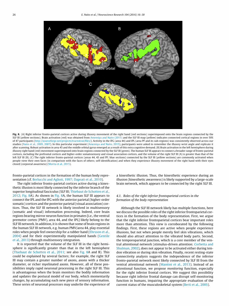
26 E. Naito et al. / Neuroscience Research 104 (2016) 16–30
Fig. 8. (A) Right inferior fronto-parietal cortices active during illusory movement of the right hand (red sections) superimposed onto the brain regions connected by theSLF III (yellow sections). Brain activation (red) was obtained from Amemiya and Naito (2015) and the SLF III map (yellow) indicates connected cortical regions in over 50%of 47 participants (http://sourceforge.net/projects/tractotron/files/). Activity in the IFG (area 44) and IPL (area PF and its sub-regions) was consistently observed across ourstudies (Naito et al., 2005, 2007). In this particular experiment (Amemiya and Naito, 2015), participants were asked to remember the illusory wrist angle and replicate itafter scanning. Robust activation in area 45 and the middle orbital gyrus emerged as a result of this extra cognitive demand. (B) Brain activation in the left hemisphere duringillusory right hand (red) movement superimposed onto brain regions connected by the SLF III (green). The human SLF III appears to connect a broader range of fronto-parietalcortices, including the prefrontal cortices and higher-order somatosensory and visual association cortices, and the volume of the right SLF III (A) is greater than that of theleft SLF III (B). (C) The right inferior fronto-parietal cortices (areas 44, 45 and PF; blue sections) connected by the SLF III (yellow sections) are commonly activated whenp cationc
fs
ts2cst(rpStr2e
s(cIdsiacT
eople view their own faces (in comparison with the faces of others; self-identifilosed (corporeal awareness) (Morita et al., 2015).
ronto-parietal cortices in the formation of the human body repre-entation (cf. Berlucchi and Aglioti, 1997; Daprati et al., 2010).
The right inferior fronto-parietal cortices active during a kines-hetic illusion is most likely connected by the inferior branch of theuperior longitudinal fasciculus (SLF III; Thiebaut de Schotten et al.,012; Fig. 8A). As shown in Fig. 8A, the human SLF III appears toonnect the IPL and the IFG with the anterior parietal (higher-orderomatic) cortices and the posterior parietal (visual association) cor-ices. Thus, the SLF III network is likely involved in multisensorysomatic and visual) information processing. Indeed, core brainegions bearing mirror neuron function in primates [i.e., the ventralremotor cortex (PMV), area 44, and the IPL] likely belong to theLF III network. In addition, it is shown that some of brain regions inhe human SLF III network, e.g. human PMV/area 44, play essentialoles when people feel ownership for a rubber hand (Ehrsson et al.,004) and for their experimentally manipulated hands (Gentilet al., 2013) through multisensory integration.
It is reported that the volume of the SLF III in the right hemi-phere is significantly greater than that in the left hemisphereThiebaut de Schotten et al., 2011; see also Fig. 8A and B). Thisould be explained by several factors; for example, the right SLFII may contain a greater number of axons, axons with a thickeriameter, or richer myelinated axons. However, all of these pos-ibilities imply rapid neuronal processing in the right SLF III. This
s advantageous when the brain monitors the bodily informationnd updates the postural model of our body, which momentarilyhanges, by accumulating each new piece of sensory information.hese series of neuronal processes may underlie the experience of) and when they experience illusory movement of the right hand with their eyes
a kinesthetic illusion. Thus, the kinesthetic experience during anillusion (kinesthetic awareness) is likely supported by a large-scalebrain network, which appears to be connected by the right SLF III.
4.1. Roles of the right inferior frontoparietal cortices in theformation of the body representation
Although the SLF III network likely has multiple functions, herewe discuss the possible roles of the right inferior frontoparietal cor-tices in the formation of the body representation. First, we arguethat the right inferior frontoparietal cortices bear important rolesmore than attention. This view is corroborated by the followingfindings. First, these regions are active when people experienceillusions, but not when people merely feel skin vibrations, whichshould also attract attention to the vibrated body parts. Second,the temporoparietal junction, which is a core member of the ven-tral attentional network (stimulus-driven attention: Corbetta andShulman, 2002), does not appear to be activated either during ten-don vibration or during skin vibration. Finally, recent resting-stateconnectivity analysis suggests the independence of the inferiorfronto-parietal network most likely connected by SLF III from theventral attentional network (Mantini et al., 2013). Instead of anattentional function, we propose monitoring function, especially
for the right inferior frontal cortices. We suggest this possibilitybecause right inferior frontal damage can disrupt self-monitoringfunction in humans, impairing the appropriate evaluation of thecurrent status of the musculoskeletal system (Berti et al., 2005).
E. Naito et al. / Neuroscience Research 104 (2016) 16–30 27
F tal syh
fwmcrtsptl2ab2awiuihrn1mms
ig. 9. Contribution of multiple brain systems (the motor network, specialized parieuman body representation.
In the non-human primate literature, cells in the inferiorrontal and inferior parietal cortices are characterized according tohether they process body-centered or body-part-centered infor-ation (Graziano et al., 1997; Ishida et al., 2010). This is also the
ase for cells in the putamen (Graziano and Gross, 1993). These cellsepresent sensory stimuli in a coordinate system that is anchoredo a particular body part, and thus can strongly contribute to multi-ensory (somatic and visual) integration associated with the bodyart and the adjacent space. In favor of this view, we have shownhat the right inferior fronto-parietal cortices, in concert with theeft cerebellar hemisphere (Lobule VI and Crus I; Kipping et al.,013), engage in visuokinesthetic processing when participants aresked to identify the spatial location of their moving right handsy combining visual and kinesthetic information (Hagura et al.,009). Given that the right inferior fronto-parietal cortices are alsoctivated when individuals experience illusory limb movementsith their eyes closed (Fig. 7A–C), it is conceivable that the right
nferior fronto-parietal network may also function to build andpdate the postural model of the body. These actions likely occur
n concert with the right putamen (Fig. 7B) and the left cerebellaremisphere (Lobule VI and Crus I; see above). Indeed, damage toight SLF III fibers and also to the right putamen may cause a super-umerary phantom limb (third arm) experience (Halligan et al.,
993). This could be attributed to a miscomputation in the for-ation (building-up and updating) of the body representation, asediated by the right SLF III network and its associated subcorticaltructures.
stems, and the right inferior fronto-parietal SLF III network) in the formation of the
An important aspect of supernumerary phantom limb syndromeis that the patient feels the subjective reality of this “third arm”(i.e., a conscious experience of owning the arm). We assume thatcorporeal awareness, including kinesthetic illusory awareness, isan attribute of the neuronal activities in the right SLF III networkthat occurs while the brain constructs and updates the posturalmodel of our body (cf. Kinsbourne, 2006). This claim is supportedby several findings. First, we have recently shown that activity inthe right inferior fronto-parietal (areas 45 and PF) cortices changesaccording to the amount of illusory right hand movement reportedby blindfolded participants (Amemiya and Naito, 2015). This is inaccordance with another recent finding that a certain amount ofbrain activity in the right fronto-parietal network is required toexperience illusory foot movement (Cignetti et al., 2014). Second,high-intensity electrical stimulation of the human right IPL causesthe illusory sensation of limb movement (Desmurget et al., 2009).This is direct evidence that right IPL activity is capable of elicitingcorporeal awareness of limb movement.
4.2. The right SLF III network, self-identification, andself-consciousness
In general, corporeal awareness allows us to conceive of our-
selves as the source of incoming sensations and as an independentfunctional entity separate from other agents and the externalworld. Thus, we speculate that corporeal awareness could be abasis for self-identification and self-consciousness. As with the
2 ce Re
dtias
rSoarMcf(ctttmcr
seh(polft
bttcoopbdr
4
ab(tocmwNkwwsbdata
8 E. Naito et al. / Neuroscien
ominance of the right-hemisphere in corporeal awareness (kines-hetic illusory awareness), the right hemisphere has also beenmplicated as playing a stronger role in self-face recognition (Devuend Brédart, 2011) and in the sense of the physical and emotionalelf (Devinsky, 2000).
This concept is upheld by our recent finding that the right infe-ior fronto-parietal cortices (areas 44, 45, and the PF), connected byLF III, were commonly activated when participants viewed theirwn faces compared with the faces of others (self-identification)nd when they experienced kinesthetic illusory movement of theight hand with their eyes closed (corporeal awareness; Fig. 8C:orita et al., 2015). The importance of the right fronto-parietal
ortices (ventrolateral prefrontal cortices and IPL) in self-otherace discrimination has also been reported by previous studiesUddin et al., 2005, 2006), and the right inferior fronto-parietalortices have been found to be predominantly activated when par-icipants visually discriminate their own faces and bodies fromhose of others (Sugiura et al., 2006). These lines of evidence seemo indicate that the right inferior fronto-parietal SLF III network
ay bear higher-order functions for self-identification and self-onsciousness, presumably by extending the basic role that thisegion plays in corporeal awareness.
A recent resting-state connectivity analysis revealed human-pecific fronto-parietal networks that represent our greatestvolutionary divergence from monkeys, as these regions do notave topological or functional correspondents in monkey brainsMantini et al., 2013). These brain regions seem to include the intra-arietal cortex, frontal operculum, and the middle frontal gyrus, allf which are likely core constituents of the SLF III network in theeft and right hemispheres. Thus, we speculate that right inferiorronto-parietal SLF III activity in the human brain could subservehe human-specific conscious experience of the self.
In this paper, we could not cover the roles of much higher-orderrain regions (e.g. insula: Craig, 2009; Tsakiris, 2010). However,he right ventrolateral prefrontal cortices seem to be involved inhe processing of both physical and metaphysical concepts asso-iated with the self, such as the evaluation of the appearance ofne’s own face (Morita et al., 2008), the discrimination of one’swn voice (Nakamura et al., 2001), the appraisal of one’s ownersonal traits (Ochsner et al., 2005) and the retrieval of auto-iographical memories (Fink et al., 1996). Further studies areefinitely needed to ascertain the roles of much higher-order brainegions.
.3. Brief summary
The right inferior fronto-parietal SLF III network might bear series of functions including monitoring and accumulating theodily information and updating the postural model of our bodybody representation). It is likely able to perform these func-ions due to its high capacity for rapid multisensory processingf neuronal information represented in body-centered/body-parts-entered coordinate systems. To monitor the current status of theusculoskeletal system, the network most likely communicatesith the motor network (e.g., through the frontal aslant tract; cf.aito et al., 2005) that processes the fundamental elements ofinesthesia (e.g., which limb is moving toward which direction tohat extent). The anatomical connection features of the SLF III net-ork allow good access to specialized parietal systems, which can
ubserve the adaptability and flexibility of the body representationy transforming and integrating the information in different coor-
inate systems (see Section 3). Finally, we presume that corporealwareness might emerge from the series of neuronal processes inhe right SLF III network, which could underlie self-identificationnd self-consciousness.search 104 (2016) 16–30
5. Conclusions
We have described how the human brain represents the bodyrepresentation using examples about a variety of kinesthetic illu-sions, such as those where people experience changes of limbposition or body configuration. It appears that multiple brain sys-tems, namely, the motor network, specialized parietal systems, andthe right inferior fronto-parietal SLF III network, work togetherto form the body representation (Fig. 9). Thus, the series of ourstudies have elucidated more specified sets of brain systems thanpreviously proposed (Melzack, 1990; Berlucchi and Aglioti, 1997;Daprati et al., 2010).
The direct involvement of the M1-centred motor network inthe formation of the body representation indicates tight couplingbetween the body representation (body schema) and motor con-trol. Activity in the motor control network is a strong gauge ofthe current status of the musculoskeletal system, enabling fastonline feedback control. The involvement of specialized parietalsystems demonstrates the importance of goal-directed transforma-tion/integration of information across different coordinate systems,which may subserve the adaptability and flexibility of the bodyrepresentation. Finally, the right inferior fronto-parietal SLF III net-work might play essential roles in the monitoring of bodily statesand the updating of body representation, which leads to corporealawareness, probably one of the origins of self-consciousness.
In this review, we described the neuronal representation ofthe human body representation, which is highly adaptive, flexibleand plastic. However, many questions remain about, for exam-ple, how a relatively constant self-body image is formed from thischangeable body representation, and how mental and emotionalself-consciousness develop with corporeal self-consciousness.Despite these questions, the present findings strongly indicate thatthe neuronal representation of the body representation is funda-mental for motor control and corporeal awareness, which form thebasis of human life.
Acknowledgements
This work was supported by Scientific Research on InnovativeAreas “Understanding brain plasticity on body representations topromote their adaptive functions” (JSPS KAKENHI No. 26120003),and partially supported by a Grant-in-Aid for Specially PromotedResearch (No. 24000012). We would like to thank Dr Thiebaut deSchotten for providing the diffusion tensor imaging data. Finally,we appreciate invaluable comments from Dr. H. Henrik Ehrsson.
References
Amemiya, K., Naito, E., 2015. Right inferior superior longitudinal fasciculus involvesawareness of kinesthetic experiences. Paper presented at the Annual Meetingof the Organization for Human Brain Mapping, Hawaii (2390).
Averbeck, B.B., Battaglia-Mayer, A., Guglielmo, C., Caminiti, R., 2009. Statisticalanalysis of parieto-frontal cognitive-motor networks. J. Neurophysiol. 102,1911–1920.
Berlucchi, G., Aglioti, S., 1997. The body in the brain: neural bases of corporealawareness. Trends Neurosci. 20, 560–564.
Berti, A., Bottini, G., Gandola, M., Pia, L., Smania, N., Stracciari, A., Castiglioni, I., Vallar,G., Paulesu, E., 2005. Shared cortical anatomy for motor awareness and motorcontrol. Science 309, 488–491.
Binkofski, F., Buccino, G., Stephan, K.M., Rizzolatti, G., Seitz, R.J., Freund, H.J., 1999. Aparieto-premotor network for object manipulation: evidence from neuroimag-ing. Exp. Brain Res. 128, 210–213.
Bodegard, A., Geyer, S., Grefkes, C., Zilles, K., Roland, P.E., 2001. Hierarchicalprocessing of tactile shape in the human brain. Neuron 31, 317–328.
Burke, D., Gandevia, S.C., Macefield, G., 1988. Responses to passive movement ofreceptors in joint, skin and muscle of the human hand. J. Physiol. 402, 347–361.
Burke, D., Hagbarth, K., Lofstedt, L., Wallin, G., 1976. The responses of human musclespindle endings to vibration of non-contracting muscles. J. Physiol. (Lond.) 261,673–693.
Cadoret, G., Smith, A.M., 1995. Input–output properties of hand-related cells in theventral cingulate cortex in the monkey. J. Neurophysiol. 73, 2584–2590.

ce Re
C
C
C
C
C
C
C
C
C
C
D
d
D
D
D
D
E
E
E
E
F
F
F
G
G
G
G
G
G
G
G
H
E. Naito et al. / Neuroscien
aspers, S., Schleicher, A., Bacha-Trams, M., Palomero-Gallagher, N., Amunts, K.,Zilles, K., 2013. Organization of the human inferior parietal lobule based onreceptor architectonics. Cereb. Cortex 23, 615–628.
atani, M., Allin, M.P., Husain, M., Pugliese, L., Mesulam, M.M., Murray, R.M., Jones,D.K., 2007. Symmetries in human brain language pathways correlate with verbalrecall. Proc. Natl. Acad. Sci. U. S. A. 104, 17163–17168.
heney, P.D., Fetz, E.E., 1984. Corticomotoneuronal cells contribute tolong-latency stretch reflexes in the rhesus monkey. J. Physiol. 349,249–272.
hristensen, M.S., Lundbye-Jensen, J., Grey, M.J., Vejlby, A.D., Belhage, B., Nielsen,J.S., 2010. Illusory sensation of movement induced by repetitive transcranialmagnetic stimulation. PLoS ONE 5, e13301, http://dx.doi.org/10.1371/journal.pone.0013301.
ignetti, F., Vaugoyeau, M., Nazarian, B., Roth, M., Anton, J.L., Assaiante, C., 2014.Boosted activation of right inferior frontoparietal network: a basis for illusorymovement awareness. Hum. Brain Mapp. 35, 5166–5178.
ohen, L.G., Bandinelli, S., Findley, T.W., Hallett, M., 1991. Motor reorganization afterupper limb amputation in man. A study with focal magnetic stimulation. Brain114, 615–627.
olebatch, J.G., Sayer, R.J., Porter, R., White, O.B., 1990. Responses of monkey pre-central neurons to passive movements and phasic muscle stretch: relevance toman. Electroencephalogr. Clin. Neurophysiol. 75, 44–55.
orbetta, M., Shulman, G.L., 2002. Control of goal-directed and stimulus-drivenattention in the brain. Nat. Rev. Neurosci. 3, 201–215.
raig, A.D., 2009. How do you feel now? The anterior insula and human awareness.Nat. Rev. Neurosci. 10, 59–70.
ulham, J.C., Danckert, S.L., DeSouza, J.F., Gati, J.S., Menon, R.S., Goodale, M.A., 2003.Visually guided grasping produces fMRI activation in dorsal but not ventralstream brain areas. Exp. Brain Res. 153, 180–189.
aprati, E., Sirigu, A., Nico, D., 2010. Body and movement: consciousness in theparietal lobes. Neuropsychologia 48, 756–762.
e Vignemont, F., Ehrsson, H.H., Haggard, P., 2005. Bodily illusions modulate tactileperception. Curr. Biol. 15, 1286–1290.
esmurget, M., Reilly, K.T., Richard, N., Szathmari, A., Mottolese, C., Sirigu, A., 2009.Movement intention after parietal cortex stimulation in humans. Science 324,811–813.
evinsky, O., 2000. Right cerebral hemisphere dominance for a sense of corporealand emotional self. Epilepsy Behav. 1, 60–73.
evue, C., Brédart, S., 2011. The neural correlates of visual self-recognition. Con-scious. Cogn. 20, 40–51.
uffy, F.H., Burchfiel, J.L., 1971. Somatosensory system: organizational hierarchyfrom single units in monkey area 5. Science 172, 273–275.
din, B.B., Vallbo, A.B., 1988. Stretch sensitization of human muscle spindles. J.Physiol. 400, 101–111.
din, B.B., Vallbo, A.B., 1990. Dynamic response of human muscle spindle afferentsto stretch. J. Neurophysiol. 63, 1297–1306.
hrsson, H.H., Kito, T., Sadato, N., Passingham, R.E., Naito, E., 2005. Neural sub-strate of body size: illusory feeling of shrinking of the waist. PLoS Biol. 3,e412.
hrsson, H.H., Spence, C., Passingham, R.E., 2004. That’s my hand! Activity in pre-motor cortex reflects feeling of ownership of a Limb. Science 305, http://dx.doi.org/10.1126/science.1097011.
etz, E.E., Finocchio, D.V., Baker, M.A., Soso, M.J., 1980. Sensory and motor responsesof precentral cortex cells during comparable passive and active joint move-ments. J. Neurophysiol. 43, 1070–1089.
ink, G.R., Markowitsch, H.J., Reinkemeier, M., Bruckbauer, T., Kessler, J., Heiss, W.D.,1996. Cerebral representation of one’s own past: neural networks involved inautobiographical memory. J. Neurosci. 16, 4275–4282.
ried, I., Katz, A., McCarthy, G., Sass, K.J., Williamson, P., Spencer, S.S., Spencer, D.D.,1991. Functional organization of human supplementary motor cortex studiedby electrical stimulation. J. Neurosci. 11, 3656–3666.
anesh, G., Yoshioka, T., Osu, R., Ikegami, T., 2014. Immediate tool incorporationprocesses determine human motor planning with tools. Nat. Commun. 5, 4524,http://dx.doi.org/10.1038/ncomms5524.
entile, G., Guterstam, A., Brozzoli, C., Ehrsson, H.H., 2013. Disintegration of multi-sensory signals from the real hand reduces default limb self-attribution: an fMRIstudy. J. Neurosci. 33, 13350–13366.
hez, C., Gordon, J., Ghilardi, M.F., 1995. Impairments of reaching movements inpatients without proprioception. II. Effects of visual information on accuracy. J.Neurophysiol. 73, 361–372.
oldenberg, G., Karnath, H.O., 2006. The neural basis of imitation is body part spe-cific. J. Neurosci. 26, 6282–6287.
oldenberg, G., Randerath, J., 2015. Shared neural substrates of apraxia and aphasia.Neuropsychologia 75, 40–49.
oodwin, G.M., McCloskey, D.I., Matthews, P.B., 1972. Proprioceptive illusionsinduced by muscle vibration: contribution by muscle spindles to perception?Science 175, 1382–1384.
raziano, M.S., Gross, C.G., 1993. A bimodal map of space: somatosensory receptivefields in the macaque putamen with corresponding visual receptive fields. Exp.Brain Res. 97, 96–109.
raziano, M.S.A., Hu, X.T., Gross, C.G., 1997. Visuospatial properties of ventral pre-
motor cortex. J. Neurophysiol. 77, 2268–2292.agura, N., Oouchida, Y., Aramaki, Y., Okada, T., Matsumura, M., Sadato, N., Naito, E.,2009. Visuokinesthetic perception of hand movement is mediated by cerebro-cerebellar interaction between the left cerebellum and right parietal cortex.Cereb. Cortex 19, 176–186.
search 104 (2016) 16–30 29
Hagura, N., Takei, T., Hirose, S., Aramaki, Y., Matsumura, M., Sadato, N., Naito, E.,2007. Activity in the posterior parietal cortex mediates visual dominance overkinesthesia. J. Neurosci. 27, 7047–7053.
Halligan, P.W., Marshall, J.C., Wade, D.T., 1993. Three arms: a case study of super-numerary phantom limb after right hemisphere stroke. J. Neurol. Neurosurg.Psychiatry 56, 159–166.
He, S.Q., Dum, R.P., Strick, P.L., 1995. Topographic organization of corticospinalprojections from the frontal lobe: motor areas on the medial surface of thehemisphere. J. Neurosci. 15, 3284–3306.
Head, H., Holmes, G., 1911. Sensory disturbances from cerebral lesions. Brain 34,102–254.
Huerta, M.F., Pons, T.P., 1990. Primary motor cortex receives input from area 3a inmacaque. Brain Res. 537, 367–371.
Iriki, A., Tanaka, M., Iwamura, Y., 1996. Coding of modified body representationduring tool use by macaque postcentral neurones. Neuroreport 7, 2325–2330.
Ishida, H., Nakajima, K., Inase, M., Murata, A., 2010. Shared mapping of own andothers’ bodies in visuotactile bimodal area of monkey parietal cortex. J. Cogn.Neurosci. 22, 83–96.
Iwamura, Y., 1998. Hierarchical somatosensory processing. Curr. Opin. Neurobiol. 8,522–528.
Iwamura, Y., Iriki, A., Tanaka, M., 1994. Bilateral hand representation in the postcen-tral somatosensory cortex. Nature 369, 554–556.
Johnson-Frey, S.H., 2004. The neural bases of complex tool use in humans. TrendsCogn. Sci. 8, 71–78.
Kaneko, F., Blanchard, C., Lebar, N., Nazarian, B., Kavounoudias, A., Romaiguère, P.,2015. Brain regions associated to a kinesthetic illusion evoked by watching avideo of one’s own moving hand. PLoS ONE 10, e0131970, http://dx.doi.org/10.1371/journal.pone.0131970.
Kinsbourne, M., 2006. From unilateral neglect to the brain basis of consciousness.Cortex 42, 869–874.
Kipping, J.A., Grodd, W., Kumar, V., Taubert, M., Villringer, A., Margulies, D.S., 2013.Overlapping and parallel cerebello-cerebral networks contributing to sensor-imotor control: an intrinsic functional connectivity study. Neuroimage 83,837–848.
Kito, T., Hashimoto, T., Yoneda, T., Katamoto, S., Naito, E., 2006. Sensory processingduring kinesthetic aftereffect following illusory hand movement elicited by ten-don vibration. Brain Res. 1114, 75–84.
Lackner, J.R., 1988. Some proprioceptive influences on the perceptual representationof body shape and orientation. Brain 111, 281–297.
Lehéricy, S., Ducros, M., Van de Moortele, P.F., Franc ois, C., Thivard, L., Poupon, C.,Swindale, N., Ugurbil, K., Kim, D.S., 2004. Diffusion tensor fiber tracking showsdistinct corticostriatal circuits in humans. Ann. Neurol. 55, 522–529.
Liepert, J., Tegenthoff, M., Malin, J.P., 1995. Changes of cortical motor area size duringimmobilization. Electroencephalogr. Clin. Neurophysiol. 97, 382–386.
Lim, S.H., Dinner, D.S., Pillay, P.K., Luders, H., Morris, H.H., Klem, G., Wyllie, E., Awad,I.A., 1994. Functional anatomy of the human supplementary sensorimotor area:results of extraoperative electrical stimulation. Electroencephalogr. Clin. Neu-rophysiol. 91, 179–193.
Mantini, D., Corbetta, M., Romani, G.L., Orban, G.A., Vanduffel, W., 2013. Evolution-arily novel functional networks in the human brain? J. Neurosci. 33, 3259–3275.
Maravita, A., Iriki, A., 2004. Tools for the body (representation). Trends Cogn. Sci. 8,79–86.
Matsumoto, R., Nair, D.R., Ikeda, A., Fumuro, T., Lapresto, E., Mikuni, N., Bingaman, W.,Miyamoto, S., Fukuyama, H., Takahashi, R., Najm, I., Shibasaki, H., Lüders, H.O.,2012. Parieto-frontal network in humans studied by cortico-cortical evokedpotential. Hum. Brain Mapp. 33, 2856–2872.
Melzack, R., 1990. Phantom limbs and the concept of a neuromatrix. Trends Neurosci.13, 88–92.
Mercier, C., Reilly, K.T., Vargas, C.D., Aballea, A., Sirigu, A., 2006. Mapping phan-tom movement representations in the motor cortex of amputees. Brain 129,2202–2210.
Morita, T., Saito, D., Ban, M., Shimada, K., Okamoto, Y., Kosaka, H., Okazawa, H., Asada,M., Naito, E., 2015. Shared right inferior fronto-parietal substrates for corporealawareness and self-identification. Paper presented at the Annual Meeting of theOrganization for Human Brain Mapping, Hawai (4195).
Morita, T., Itakura, S., Saito, D.N., Nakashita, S., Harada, T., Kochiyama, T., Sadato,N., 2008. The role of the right prefrontal cortex in self-evaluation of the face: afunctional magnetic resonance imaging study. J. Cogn. Neurosci. 20, 342–355.
Murata, A., Fadiga, L., Fogassi, L., Gallese, V., Raos, V., Rizzolatti, G., 1997. Objectrepresentation in the ventral premotor cortex (area F5) of the monkey. J. Neu-rophysiol. 78, 2226–2230.
Murata, A., Gallese, V., Luppino, G., Kaseda, M., Sakata, H., 2000. Selectivity for theshape, size, and orientation of objects for grasping in neurons of monkey parietalarea AIP. J. Neurophysiol. 83, 2580–2601.
Nachev, P., Kennard, C., Husain, M., 2008. Functional role of the supplementary andpre-supplementary motor areas. Nat. Rev. Neurosci. 9, 856–869.
Naito, E., 2004a. Imaging human somatosensory systems. Shinkei Kenkyu No Sinpo(Adv. Neurol. Sci.) 48, 249–260.
Naito, E., 2004b. Sensing limb movements in the motor cortex: how humans senselimb movement. Neuroscientist 10, 73–82.
Naito, E., Ehrsson, H.H., 2006. Somatic sensation of hand-object interactive move-
ment is associated with activity in the left inferior parietal cortex. J. Neurosci.26, 3783–3790.Naito, E., Ehrsson, H.H., Geyer, S., Zilles, K., Roland, P.E., 1999. Illusory arm move-ments activate cortical motor areas: a positron emission tomography study. J.Neurosci. 19, 6134–6144.

3 ce Re
N
N
N
N
N
N
N
N
N
O
O
P
P
P
P
P
recognition activates a frontoparietal mirror network in the right hemisphere:
0 E. Naito et al. / Neuroscien
aito, E., Kochiyama, T., Kitada, R., Nakamura, S., Matsumura, M., Yonekura, Y.,Sadato, N., 2002a. Internally simulated movement sensations during motorimagery activate the cortical motor areas and the cerebellum. J. Neurosci. 22,3683–3691.
aito, E., Matsumoto, R., Hagura, N., Oouchida, Y., Tomimoto, H., Hanakawa, T., 2010.Importance of precentral motor regions in human kinesthesia: a single casestudy. Neurocase 17, 133–147.
aito, E., Mizuguchi, N., Uehara, S., Amemiya, K., 2013. Brain mechanisms in motorimagery. Aiming for rehabilitation applications. J. Rehab. Neurosci. 13, 1–10.
aito, E., Nakashima, T., Kito, T., Aramaki, Y., Okada, T., Sadato, N., 2007. Human limb-specific and non-limb-specific brain representations during kinesthetic illusorymovements of the upper and lower extremities. Eur. J. Neurosci 25, 3476–3487.
aito, E., Roland, P.E., Ehrsson, H.H., 2002b. I feel my hand moving: a new role ofthe primary motor cortex in somatic perception of limb movement. Neuron 36,979–988.
aito, E., Roland, P.E., Grefkes, C., Choi, H.J., Eickhoff, S., Geyer, S., Ziles, K., Ehrsson,H.H., 2005. Dominance of the right hemisphere and role of area 2 in humankinesthesia. J. Neurophysiol. 93, 1020–1034.
aito, E., Scheperjans, F., Eickhoff, S.B., Amunts, K., Roland, P.E., Zilles, K., Ehrsson,H.H., 2008. Human superior parietal lobule is involved in somatic perception ofbimanual interaction with an external object. J. Neurophysiol. 99, 695–703.
akamura, K., Kawashima, R., Sugiura, M., Kato, T., Nakamura, A., Hatano, K.,Nagumo, S., Kubota, K., Fukuda, H., Ito, K., Kojima, S., 2001. Neural substrates forrecognition of familiar voices: a PET study. Neuropsychologia 39, 1047–1054.
ishitani, N., Schurmann, M., Amunts, K., Hari, R., 2005. Broca’s region: from actionto language. Physiology 20, 60–69.
chsner, K.N., Beer, J.S., Robertson, E.R., Cooper, J.C., Gabrieli, J.D., Kihsltrom,J.F., D’Esposito, M., 2005. The neural correlates of direct and reflected self-knowledge. Neuroimage 28, 797–814.
ouchida, Y., Okada, T., Nakashima, T., Matsumura, M., Sadato, N., Naito, E., 2004.Your hand movements in my somatosensory cortex: a visuo-kinesthetic func-tion in human area 2. Neuroreport 15, 2019–2023.
ascual-Leone, A., Peris, M., Tormos, J.M., Pascual, A.P., Catala, M.D., 1996. Reor-ganization of human cortical motor output maps following traumatic forearmamputation. Neuroreport 7, 2068–2070.
avlides, C., Miyashita, E., Asanuma, H., 1993. Projection from the sensory to themotor cortex is important in learning motor skills in the monkey. J. Neuro-physiol. 70, 733–741.
eeters, R., Simone, L., Nelissen, K., Fabbri-Destro, M., Vanduffel, W., Rizzolatti, G.,Orban, G.A., 2009. The representation of tool use in humans and monkeys: com-mon and uniquely human features. J. Neurosci. 29, 11523–11539.
hillips, C.G., Powell, T.P.S., Wiesendanger, M., 1971. Projection from low threshold
muscle afferents of hand and forearm to area 3a of baboon’s cortex. J. Physiol.217, 419–446.orter, R., Lemon, R., 1993. Inputs from peripheral receptors to motor cortex neurons.In: Porter, R., Lemon, R. (Eds.), Corticospinal Function and Voluntary Movement.Oxford University Press, New York, pp. 247–272.
search 104 (2016) 16–30
Pruszynski, J.A., Kurtzer, I., Nashed, J.Y., Omrani, M., Brouwer, B., Scott, S.H., 2011.Primary motor cortex underlies multi-joint integration for fast feedback control.Nature 478, 387–390.
Ribot-Ciscar, E., Roll, J.P., 1988. Ago-antagonist muscle spindle inputs contributetogether to joint movement coding in man. Brain Res. 791, 167–176.
Roll, R., Kavounoudias, A., Albert, F., Legré, R., Gay, A., Fabre, B., Roll, J.P., 2012. Illusorymovements prevent cortical disruption caused by immobilization. Neuroimage62, 510–519.
Roll, J.P., Vedel, J.P., 1982. Kinaesthetic role of muscle afferents in man, studied bytendon vibration and microneurography. Exp. Brain Res. 47, 177–190.
Roll, J.P., Vedel, J.P., Ribot, E., 1989. Alteration of proprioceptive messages inducedby tendon vibration in man: a microneurographic study. Exp. Brain Res. 76,213–222.
Romaiguere, P., Antonb, J.L., Roth, M., Casinia, L., Roll, J.P., 2003. Motor and parietalcortical areas both underlie kinaesthesia. Brain Res. Cogn. Brain Res. 16, 74–82.
Rothwell, J.C., Traub, M.M., Day, B.L., Obeso, J.A., Thomas, P.K., Marsden, C.D., 1982.Manual motor performance in a deafferented man. Brain 105, 515–542.
Sakata, H., Takaoka, Y., Kawarasaki, A., Shibutani, H., 1973. Somatosensory propertiesof neurons in the superior parietal cortex (area 5) of the rhesus monkey. BrainRes. 64, 85–102.
Scott, S.H., 2004. Optimal feedback control and the neural basis of volitional motorcontrol. Nat. Rev. Neurosci. 5, 532–546.
Strick, P.L., Dum, R.P., Fiez, J.A., 2009. Cerebellum and nonmotor function. Annu. Rev.Neurosci. 32, 413–434.
Sugiura, M., Sassa, Y., Jeong, H., Miura, N., Akitsuki, Y., Horie, K., Sato, S., Kawashima,R., 2006. Multiple brain networks for visual self-recognition with different sen-sitivity for motion and body part. Neuroimage 32, 1905–1917.
Taoka, M., Toda, T., Iwamura, Y., 1998. Representation of the midline trunk, bilateralarms, and shoulders in the monkey postcentral somatosensory cortex. Exp. BrainRes. 123, 315–322.
Thiebaut de Schotten, M., Dell’Acqua, F., Forkel, S.J., Simmons, A., Vergani, F., Murphy,D.G., Catani, M., 2011. A lateralized brain network for visuospatial attention. Nat.Neurosci. 14, 1245–1246.
Thiebaut de Schotten, M., Dell’Acqua, F., Valabregue, R., Catani, M., 2012. Monkey tohuman comparative anatomy of the frontal lobe association tracts. Cortex 48,82–96.
Thyrion, C., Roll, J.P., 2009. Perceptual integration of illusory and imagined kines-thetic images. J. Neurosci. 29, 8483–8492.
Tsakiris, M., 2010. My body in the brain: a neurocognitive model of body ownership.Neuropsychologia 48, 703–712.
Uddin, L.Q., Kaplan, J.T., Molnar-Szakacs, I., Zaidel, E., Iacoboni, M., 2005. Self-face
an event-related fMRI study. Neuroimage 25, 926–935.Uddin, L.Q., Molnar-Szakacs, I., Zaidel, E., Iacoboni, M., 2006. rTMS to the right inferior
parietal lobule disrupts self-other discrimination. Soc. Cogn. Affect. Neurosci. 1,65–71.