bodyworks - ge healthcare systems | ge healthcare (united
TRANSCRIPT

SIGNA™Works Fueling the future of MR
BodyWorks
GE Healthcare Tomorrow Today

GE HEALTHCARE2 STANDARD APPS ELECTIVE APPS INNOVATIVE APPSBODYWORKSSIGNA™WORKS
Standard Applications
Energize your clinical capabilities with all the tools you need to complete an exam. Imaging solutions cover a variety of contrasts, 2D and 3D volumetric data and motion correction capabilities.
Innovative Applications
Expand your expertise to the next level, to deliver improved image quality, higher efficiency and a more streamlined workflow, so you perform better than ever before.
Our new SIGNA™Works productivity platform redefines productivity across the breadth of our core imaging techniques. It takes full advantage of Total Digital Imaging (TDI), further advancing diagnostics and quickening throughput, while improving patient outcomes and your ROI. It is upgradeable and customizable with additional applications to suit your growing practice.
find out more find out more
Not all Elective Applications come standard on every system. Please contact your GE Representative for the most current information.
SIGNA™WorksThe new standard is extraordinary

GE HEALTHCARE3 STANDARD APPS ELECTIVE APPS INNOVATIVE APPSBODYWORKSSIGNA™WORKS
Standard Applications
Innovative Applications
BodyWorks
One of the fastest growing areas in MR, BodyWorks allows you to image abdominal and pelvic anatomy with user flexibility to adapt to different patient types.
CVWorks
Gain crucial insights into vascular structure and flow dynamics and access morphology, flow, function and tissue viability with CVWorks.
NeuroWorks
This one-stop solution enables you to image brain, spine, vascular and peripheral nerve anatomy with exceptional tissue contrast.
OncoWorks
Delivers robust tissue contrast, motion-insensitive, high temporal and spatial resolution imaging techniques that capture anatomical and morphological data for oncological assessment.
OrthoWorks
This extensive library of musculoskeletal imaging techniques enables you to image bone, joint and soft tissue with remarkable tissue contrast.
PaedWorks
Delivers distinctive child-centered imaging techniques that provide ease of use for the user and clinical excellence for your smallest, most fragile patients.
SIGNA™WorksThe new standard is extraordinary

GE HEALTHCARE4 STANDARD APPS ELECTIVE APPS INNOVATIVE APPSSIGNA™WORKS BODYWORKS
Standard Applications
Innovative Applications
HyperWorks
HyperWorks means hyper scanning with astonishing imaging and impressive speed. It includes HyperSense, which can deliver higher spatial resolution images or reduced scan times.
ImageWorks
ImageWorks boosts your overall MR performance. READYView visualization and MAGiC one-and-done scanning help ensure consistent and clear results.
SilentWorks
SilentWorks is GE’s most advanced noise reducing technology. Traditional exams can be extremely loud. SilentWorks brings the sound level down to ambient noise.
ViosWorks
ViosWorks reduces the complexity and cost of cardiac imaging. For the first time, all 7 dimensions of information can be captured in a cardiovascular scan in 10 minutes or less.
SIGNA™WORKS
SIGNA™WorksThe new standard is extraordinary

GE HEALTHCARE5 STANDARD APPS ELECTIVE APPS INNOVATIVE APPSSIGNA™WORKS BODYWORKS
BodyWorks
BodyWorks is GE’s solution for fast body scanning.
With more patients being scanned in the pelvis, abdomen, breast and prostate areas than ever before, body scanning is one of the fastest growing areas in MR. Our all-inclusive BodyWorks library allows you to image abdominal and pelvic anatomy with user flexibility that adapts to different patient types.
BodyWorks includes a range of applications designed to advance your body imaging capabilities.
Standard Applications
Elective Applications
Innovative Applications
Not all Elective Applications come standard on every system. Please contact your GE Representative for the most current information.
Percentage of sites performing specific MR procedures:
36% Breast
21% Prostate
88% Abdomen and Pelvis
Source: IMV 2016 MR Market Outlook Report

GE HEALTHCARE6 ELECTIVE APPS INNOVATIVE APPSSIGNA™WORKS BODYWORKS STANDARD APPS
Liver Acquisition with Volume Acceleration (LAVA) is a rapidly accelerated, 3D T1 dynamic (DCE) body imaging technique that uses a unique PSD waveform to allow for a reduction of scan time needed for dynamic imaging.
Clinical benefits:
• Produces 2D ARC parallel imaging for short scans
• Turbo mode further reduces scan time up to 50%
• Achieves higher spatial resolution in the same scan time as conventional LAVA/LAVA Flex imaging
• Provides adiabatic special fat suppression for robust imaging
• Compatible with Navigator for free- breathing acquisition
LAVA
BodyWorks Standard Applications
PROPELLERLAVA NavigatoreDWI
Free-breathing, enterography exam demonstrating extraordinary detail of the small bowel vascular structure

GE HEALTHCARE7 ELECTIVE APPS INNOVATIVE APPSSIGNA™WORKS BODYWORKS STANDARD APPS
LAVA
BodyWorks Standard Applications
PROPELLERLAVA NavigatoreDWI
Progressive contrast uptake of the tail of the pancreas and communication between nodular formation and dilation of pancreatic duct with comparison to CT (upper left) and FOCUS diffusion (upper middle)
Arterial phase Intermediate phase Venous phase

GE HEALTHCARE8 ELECTIVE APPS INNOVATIVE APPSSIGNA™WORKS BODYWORKS STANDARD APPS
BodyWorks Standard Applications
PROPELLERLAVA NavigatoreDWI
T2 PROPELLER MB 320 x 320 19 FOV/4mm 3:40 min
PROPELLER Multi-Blade (MB) is a multi-shot approach that preserves tissue contrast regardless of weighting while also reducing motion artifacts and providing a more signal-rich image. Additionally, this technique allows for all contrasts for 2D FSE: T1, T2, STIR and PD weightings.
Clinical benefits:
• Delivers motion-artifact-free diagnostic images (respiration and peristalsis)
• Increases productivity and decreases the number of repeated scans
• Enables sedation-free scanning and increases patient tolerance
PROPELLER

GE HEALTHCARE9 STANDARD APPS ELECTIVE APPS INNOVATIVE APPSSIGNA™WORKS BODYWORKS
Diffusion Weighted Imaging (DWI) is used to image diffusivity of water molecules (Brownian motion). This enhanced Diffusion Weighted Imaging (eDWI) technique is designed to provide high signal-to-noise-ratio (SNR) diffusion images, with short-acquisition time and shortest possible echo time (TE). Its multi-b feature is designed to provide measurement of apparent diffusion coefficient (ADC) map with reduced effect of perfusion.
Clinical benefits:
• Helps to improve patient tolerance with shortened breath-hold time or free-breathing Navigator
• Increases sensitivity and specificity of lesions
• Decreases overall exam sequences and time
BodyWorks Standard Applications
eDWI
STANDARD APPS
PROPELLERLAVA NavigatoreDWI
eDWI b800

GE HEALTHCARE10 STANDARD APPS ELECTIVE APPS INNOVATIVE APPSSIGNA™WORKS BODYWORKS
Navigator uses a tracker to detect the motion of the diaphragm which enables free-breathing body imaging acquisition. The navigator tracker is automatically placed over the right hemidiaphragm, the acquisition synchronizes the patient's breathing pattern and minimizes ghosting artifacts.
Clinical benefits:
• Enhances workflow
• Eliminates need to use respiratory bellows
• Allows for adjustment of threshold and acceptance window in real time as the patient's respiration changes
BodyWorks Standard Applications
Navigator
STANDARD APPS
PROPELLERLAVA NavigatoreDWI
LAVA ASPIR with Navigator
T2 PROPELLER FatSat Diffusion EPI
LAVA Flex with Navigator

GE HEALTHCARE11 STANDARD APPS ELECTIVE APPS INNOVATIVE APPSSIGNA™WORKS BODYWORKS
LAVA Flex is a T1-weighted 3D volume for body imaging that uses a Dixon-based fat separation technique that provides an unparalleled exclusion of fat and the ability to acquire 4 data sets from one acquisition.
Clinical benefits:
• Compatible with free-breathing Navigator
• Produces homogenous image quality in a large, full-coverage FOV
• Shortens exam time due to fewer rescans
• More comfortable for patients undergoing abdominal exams
LAVA Flex
MR Touch IDEAL IQ DISCO Inhance SuiteFOCUS DWI
ELECTIVE APPS
BodyWorks Elective Applications
Not all Elective Applications come standard on every system. Please contact your GE Representative for the most current information.
LAVA Flex
case study
LAVA Flex Coronal

GE HEALTHCARE12 STANDARD APPS ELECTIVE APPS INNOVATIVE APPSSIGNA™WORKS BODYWORKS
back to app
BodyWorks Elective Applications
Case Study: Free-breathing Pediatric Kidney Exam with Turbo LAVAClinical solutions
System: DiscoveryTM MR750 3.0T Coil: 32 channel Cardiac
Protocols used
Axial DWI b50/1000 with RT, Coronal LAVA post IV arterial, venous and late phases with Navigator, Coronal LAVA Flex High Resolution with Navigator
Patient history
Ultrasound at 22 weeks of pregnancy showed an intra-uterine cyst, diagnosed as multicystic renal dysplasia (both kidneys). MR was performed on 9-month-old infant to characterize the inter renal-splenic lesion.
Procedure
Complete free-breathing examination with Body Navigator and Respiratory Trigger is useful for pediatric patients. Turbo LAVA Flex with Navigator allows for correct timing without breath-hold. This is crucial to detect the arterial phase for lesion characterization. DWI with high b-value shows clear delineation between the cyst and the lesion.
MR findings
The MR exam confirmed the presence of bilateral renal dysplasia and showed a lesion at the level of the left adrenal gland which was not seen on the previous ultrasound exam. Visual was enhanced after gadolinium injection at arterial phase; suspicious of a neuroblastoma.
Turbo LAVA Flex with Nav 3.6mm FOV30cm matrix 204x204 4 phases 1:22 min
Turbo LAVA Flex high res with Nav 2.8mm FOV30cm matrix 224x300 1:58 min
Not all Elective Applications come standard on every system. Please contact your GE Representative for the most current information.
MR Touch IDEAL IQ DISCO Inhance SuiteFOCUS DWI
ELECTIVE APPS
LAVA Flex

GE HEALTHCARE13 STANDARD APPS ELECTIVE APPS INNOVATIVE APPSSIGNA™WORKS BODYWORKS
FOV Optimized & Constrained Undistorted Single-shot (FOCUS), a 2D Spatially Selective RF Excitation method for DW-EPI and DTI, reduces geometric distortion, eliminates phase wrap artifacts and increases image sharpness. Available for both 1.5T and 3.0T, it provides high resolution DWI scans, especially useful when the region of interest is small in the phase encode direction.
Clinical benefits:
• Reduces distortion at air/tissue interfaces, e.g., bowel
• Provides a higher spatial resolution diffusion via smaller FOV
• Helps detect and evaluate small lesions that maybe obscured by distortion
• Reduces motion contamination outside the region of interest
FOCUS DWI
ELECTIVE APPS
BodyWorks Elective Applications
Not all Elective Applications come standard on every system. Please contact your GE Representative for the most current information.
case study
MR Touch IDEAL IQ DISCOFOCUS DWILAVA Flex
FOCUS DWI helped to more clearly define the structures within the prostate
Routine DWI b1000 T2 PROPELLERFOCUS DWI b1000
PROPELLER T2 FOCUS DWI
Inhance Suite

GE HEALTHCARE14 STANDARD APPS ELECTIVE APPS INNOVATIVE APPSSIGNA™WORKS BODYWORKS ELECTIVE APPS
BodyWorks Elective Applications
Not all Elective Applications come standard on every system. Please contact your GE Representative for the most current information.
back to app
Case Study: High Resolution Pancreas Imaging with FOCUS DWIClinical solutions
System: Discovery™ MR750 3.0T Coil used: 32 channel Torso
Patient history
41-year-old patient presented with severe abdominal pain. CT exam revealed an enlarged corporeo-caudal portion of the pancreas. Patient was referred to MR to check for pancreatitis.
Procedure
The eDWI image showed an increased signal in the tail of the pancreas. The FOCUS eDWI b500 image did not exhibit a hypersignal of the nodular lesion. FOCUS with increased spatial resolution helped clearly depict the pancreas from the nodular lesion. Progressive contrast uptake of the tail of the pancreas served as a fibrosis indicator and suggested autoimmune pancreatitis with no risk factor for a 41 year old.
MR findings
The hyposignal of the pancreas tail on the LAVA Flex series suggests pancreatitis and MRCP revealed a discrete dilatation of the Wirsung canal upstream the lesion. The clinician's final diagnosis was suspicion of a pancreatitis autoimmune type II, with a focal lesion in the tail of the pancreas.
DWI FOCUS b500
eDWI b500
MR Touch IDEAL IQ DISCOFOCUS DWILAVA Flex Inhance Suite

GE HEALTHCARE15 STANDARD APPS ELECTIVE APPS INNOVATIVE APPSSIGNA™WORKS BODYWORKS
MR Touch is a combination of hardware and software that uses sound waves to identify variations in liver stiffness. It uses low frequency mechanical waves produced by the Active Driver that are transferred to the patient via the Passive Driver and pneumatic pulses. These pulses probe the elastic properties of the tissue.
Clinical benefits:
• Can assess the stiffness of the patient’s liver, especially valuable in evaluating and treating cirrhosis
• Enables patient-friendly exam with short breath-hold scan
• Provides non-contrast, non-invasive evaluation of liver disease
• Automatic inline task generates quantitative parametric maps directly on console
MR Touch
ELECTIVE APPS
BodyWorks Elective Applications
Not all Elective Applications come standard on every system. Please contact your GE Representative for the most current information.
Shear Stiffness
0 2 4 6 8
Shear Stiffness
0 2 4 6 8
Healthy Liver Diseased Liver
MR Touch IDEAL IQ DISCOFOCUS DWILAVA Flex Inhance Suite

GE HEALTHCARE16 STANDARD APPS ELECTIVE APPS INNOVATIVE APPSSIGNA™WORKS BODYWORKS
IDEAL IQ provides volumetric whole-liver coverage in a single breath-hold and generates estimated T2* and triglyceride fat fraction maps in a non-invasive manner. It is intended for breath-held abdominal imaging to evaluate diffuse liver diseases such as hepatic steatosis of the liver.
Clinical benefits:
• Corrects for confounding factors from short T2* pathologies
• Delivers liver fat percentage with short breath-holds
• Generates 6 contrasts with a single scan: fat-only, water-only, in-phase, out-of-phase, fat fraction and T2* (iron)
• Provides non-contrast, non-invasive evaluation of liver disease
• Delivers automatic quantitative, fat fraction results
• Compatible with parallel imaging (ARC)
IDEAL IQ
ELECTIVE APPS
BodyWorks Elective Applications
Not all Elective Applications come standard on every system. Please contact your GE Representative for the most current information.
3 months post treatmentBaseline
MR Touch IDEAL IQ DISCOFOCUS DWILAVA Flex
51%
31%
35%
55%
Inhance Suite

GE HEALTHCARE17 STANDARD APPS ELECTIVE APPS INNOVATIVE APPSSIGNA™WORKS BODYWORKS
DynamIc Scan Optimization (DISCO) combines 3D DCEMRI + 2 pt Dixon + Parallel Imaging + Temporal Acceleration to drastically reduce scan time. DISCO uses novel techniques to disperse motion and view sharing to improve temporal resolution.
Clinical benefits:
• Generates extreme, high resolution for 3D dynamic imaging
• Detects smaller lesions with higher spatial resolution and less blurring
• Shortens patient breath-holds
• Compatible with Navigator for free- breathing dynamic scans
• Has the potential to eliminate bolus timing
DISCO
ELECTIVE APPS
BodyWorks Elective Applications
Not all Elective Applications come standard on every system. Please contact your GE Representative for the most current information.
T2 PROPELLER
Positive enhancement map
Ktrans Fused
Positive enhancement map fused to T2w images using Integrated Registration on READYView
MR Touch IDEAL IQ DISCOFOCUS DWILAVA Flex
case study
Inhance Suite

GE HEALTHCARE18 STANDARD APPS ELECTIVE APPS INNOVATIVE APPSSIGNA™WORKS BODYWORKS
DISCO
ELECTIVE APPS
BodyWorks Elective Applications
Not all Elective Applications come standard on every system. Please contact your GE Representative for the most current information.
Contrast enhanced DISCO with high temporal resolution demonstrating perihepatitis 4s/phase
MR Touch IDEAL IQ DISCOFOCUS DWILAVA Flex
case study
Inhance Suite

GE HEALTHCARE19 STANDARD APPS ELECTIVE APPS INNOVATIVE APPSSIGNA™WORKS BODYWORKS ELECTIVE APPS
BodyWorks Elective Applications
Not all Elective Applications come standard on every system. Please contact your GE Representative for the most current information.
Clinical solutions
System: Optima™ MR450w GEM 1.5T
Protocols used
T2w PROPELLER, DISCO Flex, DISCO Single Echo, 3D MIP of DISCO
Patient history
An elderly patient was referred for an MR exam of a left breast lesion after mammography and ultrasound. The lesion appeared to be markedly hypoechoic on the ultrasound exam.
Procedure
The DISCO sequence improved spatial resolution compared to standard DCE imaging. The DISCO Flex or Single Echo options provided tailored CNR.
MR findings
A 9mm spiculated lesion was noticed in the infero external quadrant of the left breast, corresponding to the lesion seen at mammography and ultrasound. Clinician suspects a high grade ductal carcinoma. Also noticed two additional small lesions, located in the upper breast, at the junction of external quadrants.
Confirmatory imaging results
Ultrasound-guided biopsy confirmed three locations of invasive ductal carcinoma.
Case Study: High Resolution Breast Imaging with DISCO
back to app
DISCO 512x512 1.2mm/-0.6mm 1:12 min/phase
MIP of DISCO phase 7
MR Touch IDEAL IQ DISCOFOCUS DWILAVA Flex Inhance Suite

GE HEALTHCARE20 STANDARD APPS ELECTIVE APPS INNOVATIVE APPSSIGNA™WORKS BODYWORKS
The Inhance Suite improves your workflow with easy setup by allowing visualization of blood flow in diverse anatomies with an advanced array of powerful pulse sequences – with no need for gadolinium.
The Suite includes:
• 3D IFIR • 3D Velocity • 2D InFlow • 3D DeltaFlow
Clinical benefits:
• No injection needed, which eliminates potential contrast reaction
• Eliminates bolus timing
• Enhances evaluation of renal conditions and lower extremities
• Uses peripheral gating instead of full cardiac gating
BodyWorks Elective Applications
Inhance Suite
case study
Inhance Inflow IR
ELECTIVE APPS
MR Touch IDEAL IQ DISCOFOCUS DWILAVA Flex Inhance Suite
Not all Elective Applications come standard on every system. Please contact your GE Representative for the most current information.

GE HEALTHCARE21 STANDARD APPS ELECTIVE APPS INNOVATIVE APPSSIGNA™WORKS BODYWORKS
BodyWorks Elective Applications
Case Study: Accessing Renal Arteries with Inhance Suite
back to app
Clinical solutions
Applications: CE MRA System: SIGNA™ Explorer 1.5T
Patient history
A 27-year-old patient complained of hypertension. MR was advised for renal artery evaluation due to lowered eGFR of 47 mL/min/1.73m2. Patient underwent renal ultrasonography examination which revealed small-sized right kidney.
Procedure
Non-contrast Inhance InFlow Inversion Recovery (IFIR) and contrast enhanced (CE) magnetic resonance angiography (MRA) were used to assess renal artery patency.
MR findings
Renal artery stenosis confirmed with both non-contrast IFIR and CE MRA.
IFIR
CE MRA
ELECTIVE APPS
MR Touch IDEAL IQ DISCOFOCUS DWILAVA Flex Inhance Suite
Not all Elective Applications come standard on every system. Please contact your GE Representative for the most current information.
GE HEALTHCARE22 STANDARD APPS ELECTIVE APPS INNOVATIVE APPSSIGNA™WORKS BODYWORKS
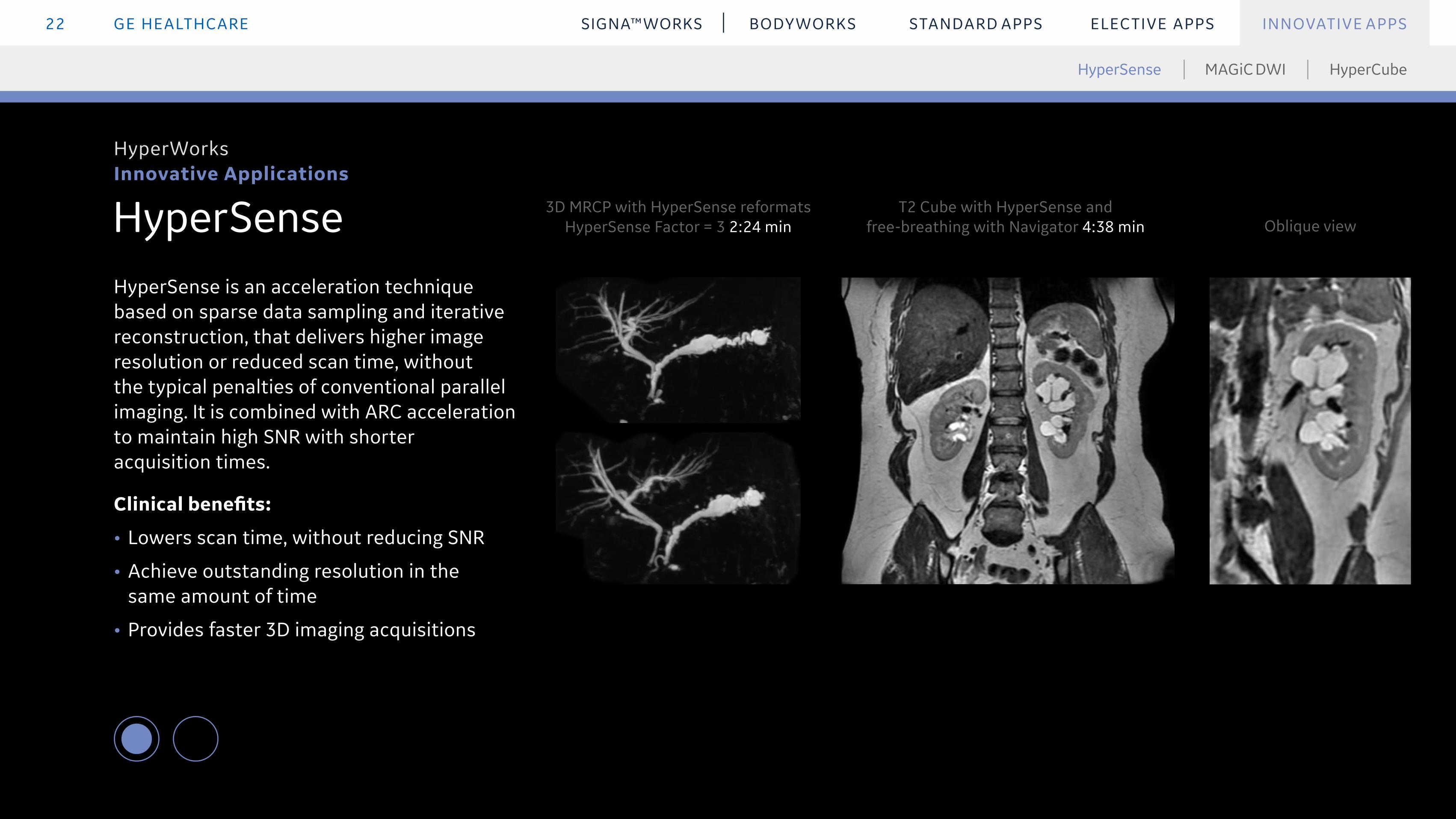
HyperSense is an acceleration technique based on sparse data sampling and iterative reconstruction, that delivers higher image resolution or reduced scan time, without the typical penalties of conventional parallel imaging. It is combined with ARC acceleration to maintain high SNR with shorter acquisition times.
Clinical benefits:
• Lowers scan time, without reducing SNR
• Achieve outstanding resolution in the same amount of time
• Provides faster 3D imaging acquisitions
HyperSense
HyperCubeMAGiC DWIHyperSense
INNOVATIVE APPS
HyperWorks Innovative Applications
T2 Cube with HyperSense and free-breathing with Navigator 4:38 min
3D MRCP with HyperSense reformats HyperSense Factor = 3 2:24 min Oblique view

GE HEALTHCARE23 STANDARD APPS ELECTIVE APPS INNOVATIVE APPSSIGNA™WORKS BODYWORKS
HyperSense
HyperCubeMAGiC DWIHyperSense
INNOVATIVE APPS
HyperWorks Innovative Applications
HyperCube with HyperSense female pelvis
Sagittal reformat 4mm Axial reformat 4mm Coronal reformat 4mm

GE HEALTHCARE24 STANDARD APPS ELECTIVE APPS INNOVATIVE APPSSIGNA™WORKS BODYWORKS
MAGiC DWIMAGnetic resonance image Compilation Diffusion Weighted Imaging (MAGiC DWI) generates multiple synthetic b-values from one DWI scanned series so you can view diffusion contrast changes in real time after acquisition. It delivers high b-values without stressing protocol parameters, and shorter scan times without sacrificing contrast or anatomy coverage. It also allows shorter TE, improving SNR and sharpness.
Clinical benefits:
• Multiple synthetic b-values from a single DWI scan
• High b-values in shorter scan times
• Compatible with FOCUS diffusion
• Calculates high b-value as recommended by PIRADS for prostate
INNOVATIVE APPS
ImageWorks Innovative Applications
HyperSense HyperCubeMAGiC DWI
3.4mm FOV 40cm, matrix 128x128 3:52 min
DWI b1000
MAGIC DWI b2000
MAGIC DWI b1000
MAGIC DWI b2500

GE HEALTHCARE25 STANDARD APPS ELECTIVE APPS INNOVATIVE APPSSIGNA™WORKS BODYWORKS
HyperCubeHyperCube reduces scan time and limits artifacts such as motion and aliasing by reducing the phase FOV. It can be applied with or without fat suppression and significantly lowers imaging time without sacrificing contrast quality. It focuses on the area of interest, can be used on the entire body and is compatible with HyperSense.
Clinical benefits:
• Lowers scan time without SNR loss, reducing the potential for patient motion and repeats
• Eliminates time-consuming parameters
• Provides high-resolution small FOV imaging
• Helps with large FOV robust fat suppression when combined with FSE Flex
INNOVATIVE APPS
HyperWorks Innovative Applications
HyperSense HyperCubeMAGiC DWI
case study
T2 Cube Coronal, HyperCube, 320 x 256, 2mm slice 4:38 min Oblique T2 Cube Coronal MIP

GE HEALTHCARE26 STANDARD APPS ELECTIVE APPS INNOVATIVE APPSSIGNA™WORKS BODYWORKS
Case Study: Detecting Nodules in Prostate Imaging with HyperWorks
INNOVATIVE APPS
HyperWorks Innovative Applications
Clinical solutions
System: SIGNA™ Architect
Protocols used
Axial HyperCube T2 with HyperSense, Axial T2 FSE High resolution (only for comparison with the cube), Axial MAGiC FOCUS DWI, Axial DISCO dynamic series
Patient history
58-year-old patient with elevated Prostate-specific antigen (PSA) (10ng/mL). MR exam 6 years prior was normal. Patient was referred for follow-up and search of suspect target.
Procedure
HyperCube T2 with HyperSense improved the spatial resolution to study the lesion in 3D compared to standard FSE T2 planes. 3D T2 in 5:32 min instead of 3 x 2D planes in 10 minutes. With MAGiC FOCUS DWI, we increased the b2000 DWI resolution (3mm instead of 4mm slice thickness) in lower scan time. The improvements in resolution and SNR facilitate diagnosis on the b2000 with shorter scan times.
MR findings
Prostate of 68 cc. Nodule in the transitional area with hyposignal T2, restricted diffusion, low ADC of 0.9x10⁻³ mm²/s. The vascularization of the nodule is slightly higher than the rest of the prostate.
back to app
HyperCube T2 Axial 0.8mm slice
HyperSense HyperCubeMAGiC DWI

© 2020 General Electric Company – All rights reserved. GE Healthcare reserves the right to make changes in specifications and features shown herein, or discontinue the product described at any time without notice or obligation. Contact your GE Healthcare representative for the most current information. SIGNA, GE and the GE Monogram, are trademarks of General Electric Company. GE Healthcare, a division of General Electric Company. GE Medical Systems, Inc., doing business as GE Healthcare.
JB52418XX(1)
Images courtesy of: Centre Cardiologique du Nord, Paris; St. Joseph’s Hospital, Paris, France; GIE IRM, Creil, France; University Hull, UK; Hopital Tenon, France; Seirei Hamamatsu Hospital, Japan; University of Wisconsin School of Medicine and Public Health, WI, US; BRMI Dyker Heights, Brookyn, NY, US; Children’s National Medical Center, US; Memorial Sloan Kettering Cancer Center, NY, US; Spectrum Medical Imaging, Sydney, Australia; Mansoura Advanced Medical Imaging Center, Mansoura, Egypt; Necker Hospital, Paris, France; Addenbrooke's Hospital, Cambridge, UK