bollettino della società italiana di biologia sperimentale pag... · bollettino della società...
TRANSCRIPT
Journal ofBiologicalResearchBollettino della Società Italiana di Biologia Sperimentale
www.jbiolres.org
Journal ofal ofJournBiological
al ofJournBiological
al ofBiologicalResearchBollettino della Società Italiana di Biologia Sperimentale
ResearchBollettino della Società Italiana di Biologia Sperimentale
ResearchBollettino della Società Italiana di Biologia Sperimentale
jbrjbrjbrwww.jbiolres.orgwww.jbiolres.org
jjjbjbr
Volume 88(1) - 2015
86th SIBS National Congress
Palermo, Italy, 24-25 October 2013Botanical Garden, Lanza Hall

[page II] [Journal of Biological Research 2015; 88(1)]
Editorial StaffLucia Zoppi, Managing EditorClaudia Castellano, Production EditorTiziano Taccini, Technical Support
PublisherPAGEPress Publicationsvia Giuseppe Belli 727100 Pavia, ItalyTel. +39.0382.1751762 – Fax. [email protected] – www.pagepress.org
Journal of Biological Research 2015; volume 88
Journal of Biological ResearchBollettino della Società Italiana di Biologia Sperimentale
eISSN 2284-0230
EDITOR IN CHIEFMarco Giammanco (University of Palermo, Italy)
ASSOCIATE EDITORSRenzo Antolini (University of Trento, Italy)Massimo Cocchi (Alma Mater Studiorum-University of Bologna, Italy)Proto Gavino Pippia (University of Sassari, Italy)Luigi Pane (University of Genoa, Italy)Emma Rabino Massa (University of Turin, Italy)
EDITORIAL BOARD James Anthony (Michigan State University, USA)Maria Grazia Bridelli (University of Parma, Italy)Dario Cantino (University of Turin, Italy)David Caramelli (University of Florence, Italy)Giuseppe Caramia (G. Salesi Ancona Hospital, Italy)Emilio Carbone (University of Turin, Italy)Brunetto Chiarelli (University of Florence, Italy)Amelia De Lucia (University of Bari, Italy)Andrea Drusini (University of Padua, Italy)Luciano Fadiga (University of Ferrara, Italy)Vittorio Farina (University of Sassari, Italy)William Galanter (University of Illinois, USA)Millie Hughes-Fulford (University of San Francisco, USA)Gaetano Leto (University of Palermo, Italy)Gianni Losano (University of Turin, Italy)Mansoor A. Malik (Howard University Hospital, USA)Gian Luigi Mariottini (University of Genoa, Italy)Neville A. Marsh (Queensland University of Technology, Australia)Bruno Masala (University of Sassari, Italy)Alejandro M.S. Mayer (Midwestern University, USA)Vincenzo Mitolo (University of Bari, Italy)Werner E.G. Muller (Johannes Gutenberg University, Germany)Kary B. Mullis (Oakland Research Institute, USA)Giuseppe Murdaca (University of Genoa, Italy)Giuseppe Palumbo (University of Naples Federico II, Italy)Gian Luigi Panattoni (University of Turin, Italy)Giovanni Pizzuti (University of Naples Federico II, Italy)Massimo Pregnolato (University of Pavia, Italy)Mark R. Rasenick (University of Illinois, USA)Angela Maria Rizzo (University of Milan, Italy)Giacomo Rizzolatti (University of Parma, Italy)Aldo Rustioni (University of North Carolina, USA)Salvatore Sapienza (University of Catania, Italy)Pietro Scotto Di Vettimo (University of Naples, Italy)Vinicio Serino (University of Siena, Italy)Lynne Christine Weaver (University of Western Ontario, Canada)Mario Wiesendanger (University of Friburg, Germany)
[Journal of Biological Research 2015; 88(1)] [page III]
Journal of Biological Research 2015; volume 88
PRESIDENT
Marco Giammanco (University of Palermo, Italy)
SCIENTIFIC COMMITTEE
Emma Rabino Massa (University of Turin, Italy)
Proto Pippia (University of Sassari, Italy)
Giovanni Pizzuti (University of Naples, Italy)
Renzo Antolini (University of Trento, Italy)
Maria Bellomo (University of Enna, Italy)
Mariagrazia Bridelli (University of Parma, Italy)
David Caramelli (University of Florence, Italy)
Massimo Cocchi (University of Bologna, Italy)
Amelia De Lucia (University of Bari, Italy)
Maria Assunta Dessì (University of Cagliari, Italy)
Andrea Drusini (University of Padua, Italy)
Caterina Faggio (University of Messina, Italy)
Marco Giammanco (University of Palermo, Italy)
Sandra Imbrogno (University of Calabria, Italy)
Gian Luigi Mariottini (University of Genoa, Italy)
Chiara Motta (University of Naples, Italy)
Luigi Pane (University of Genoa, Italy)
Agostino Palmeri (University of Catania, Italy)
Angela Maria Rizzo (University of Milan, Italy)

[page IV] [Journal of Biological Research 2015; 88(1)]
Journal of Biological Research 2015; volume 88
86th SIBS National CongressPalermo, Italy, 24-25 October 2013
Botanical Garden, Lanza Hall
PRESIDENT
Marco Giammanco (University of Palermo, Italy)
ORGANIZING COMMITTEE
Stefania Aiello (University of Palermo, Italy)
Antonella Cascio (University of Palermo, Italy)
Marilena Crescimanno (University of Palermo, Italy)
Danila Di Majo (University of Palermo, Italy)
Carla Flandina (University of Palermo, Italy)
Marco Giammanco (University of Palermo, Italy)
Maurizio La Guardia (University of Palermo, Italy)
Gaetano Leto (University of Palermo, Italy)
Antonio Palma (University of Palermo, Italy)
Diego Planeta (University of Palermo, Italy)
Giovanni Tomasello (University of Palermo, Italy)
Marcello Traina (University of Palermo, Italy)
Francesca Maria Tumminello (University of Palermo, Italy)
[Journal of Biological Research 2015; 88(1)] [page V]
Journal of Biological Research 2015; volume 88
In memory of
Prof. SANTO GIAMMANCOFull Professor of General Physiology, University of Palermo, Italy
Immunosuppression during spaceflight is a major barrier to safelong-term human space habitation and travel. Remarkable findings inspace have shown that gravity changes affect important cellular mech-anisms like proliferation, differentiation, genetic expression,cytoskeletal architecture and motility in lymphocytes, monocytes andother mammalian cells. In particular, several experiments performedin space demonstrated that human T lymphocytes have remarkablyreduced mitogenic activation (80-90%), thus implicating gravity as anecessary factor in normal immune function.1,2 Subsequent spacestudies using sounding rockets, shuttles and International SpaceStation (ISS) demonstrated that T cell activation requires tight con-tacts between each other as well between T cell and monocytes as anti-gen-presenting cells. We were able to see that cells displayautonomous movements and interactions in space.3 Moreover, weinvestigated the structure of the cytoskeleton and in particular of tubu-lin and intermediate filaments of vimentin in Jurkat cells byimmunofluorescence technique on the sounding rocket MAXUS 1B.We observed, already 30 min after exposure to microgravity, a signifi-cant higher formation of large bundles of filaments, showing that thecytoskeleton undergoes important and immediate changes in micro-gravity.4 Again important differences between the actin patternbetween 1xg and 0xg in J-111 cells were observed in an experiment onboard ISS.5 Such experiments were accompanied by extensive investi-gations performed in the ground laboratory by the three-dimensionalclinostat, called Random Positioning Machine (RPM). This machinehas proven, in the last 15 years, to be a useful tool to simulate low g inthe ground laboratory and to prepare space investigations. Next exper-iments conducted in space and in RPM indicate that there are directgravitational effects on the genetic expression of IL-2 and its receptorin human T lumphocytes. In our investigation on the IL-2R, we focusedour attention on the alfa and beta-chains only, because the gamma-chain is not constitutively expressed. Surprisingly, the expression ofthe alfa-chain was significantly inhibited whereas the expression ofthe beta-chain was not influenced by microgravity.6
Moreover, experiments in RPM using gene arrays and quantitativeRT-PCR demonstrated that induction of 91 genes was altered in simu-
lated microgravity conditions. Promoter region analysis found that themajority of genes downregulated in microgravity were controlled bytranscription factors NFkB, CREB, ELK, AP-1 and STAT. The fact thatphosphorylation of the linker of activation in T cells (LAT) is notdown-regulated in simulated microgravity indicating that cholesterol-rich lipid rafts are not involved in the down-regulation of thetranscription factors.7
Our LEUKIN spaceflight experiment on board the ISS allowedthe evaluation of the global gene expression pattern of human T cellsafter 1.5 hours of stimulation by ConA and anti- CD28 in order to iden-tify the immediate early genes whose transcription may be inhibitedin microgravity conditions. Importantly, an onboard centrifuge wasused to generate a 1xg simultaneous control to isolate the effects ofmicrogravity from other variables of spaceflight. Microarray expres-sion analysis after 1.5 hours of activation demonstrated that 0xg and1xg- activated T cells had distinct patterns of global gene expressionand identified 47 genes that were significantly differentially down-reg-ulated by at least 2 fold in microgravity. Expression of many genesinvolved in mitogenesis, cytokine production, apoptosis, and signaltransduction and several key immediate early genes were inhibited inmicrogravity. In particular, transactivation of Rel/NFkB, CREB, andSRF gene targets were down-regulated. Expression of cREL gene tar-gets were significantly inhibited and transcription of cREL itself wassignificantly reduced in microgravity. Analysis of gene connectivityindicated that the tumor necrosis factor (TNF) pathway is likely amajor early downstream effector pathway inhibited in microgravityand may lead to ineffective pro-inflammatory host defenses againstinfectious pathogens during spaceflight.8
Recently, we studied the influence of altered gravity on expres-sion and function of cytoskeletal proteins, chemokines, cytokines andtheir rceptors by the experiment STIM (Signal Transduction InMicrogravity) on board the sounding rocket Maser 12. The launch tookplace the 13th of feruary 2012 at Esrange Space Center and the micro-gravity lasted 390 sec. During the flight, one automed plunger activa-tion mechanism initiated the confluente between the activators(Concanavalin A, anti-CD28, anti-CD3) and the cells (human T lym-phocytes), while a second plunger initiated that between fixative (for-malin) and activated cells in a subsequent phase. T h e hypergravityphase during the launch resulted in a down regulation of the IL-2 andCD3 receptor and reduction of tyrosine phosphorylation, p44/42-MAPKphosphorylation and histone H3 acetylation, whereas LAT phosphory-lation was increased. Compared to the baseline situation at the pointof entry into the microgravity phase, CD3 and IL-2 receptor expressionat the surface of non-activated T cells were reduced after 6 min. ofmicrogravity. Importantly, p44/42-MAPK- phosphorylation was alsoreduced in low gravity. In activated T cells, the reduced CD3 and IL-2receptor expression recovered significantly during in-flight 1xg condi-tions, but not during microgravity conditions. Beta-tubulin increasedsignificantly after onset of microgravity until the end of the micrograv-ity phase, but not in the in-flight 1xg condition. The results of STIMexperiment suggest that key proteins of T cell signal modules are notseverely altered in microgravity conditions. Instead, it can be sup-
Correspondence: Proto Pippia, Department of Biomedical Sciences,University of Sassari, via Muroni 25, 07100 Sassari, Italy.Tel: +39.329.1710218. E-mail: [email protected]
©Copyright C. Secchi et al., 2015Licensee PAGEPress, ItalyJournal of Biological Research 2015; 88:5161
This article is distributed under the terms of the Creative CommonsAttribution Noncommercial License (by-nc 3.0) which permits any noncom-mercial use, distribution, and reproduction in any medium, provided the orig-inal author(s) and source are credited.
Recent advances in human T lymphocyte biology in spaceC. Secchi, C. Crescio, A. Pantaleo, P. PippiaDepartment of Biomedical Sciences, University of Sassari, Italy
[Journal of Biological Research 2015; 88:5161] [page 1]
Journal of Biological Research 2015; volume 88:5161
[page 2] [Journal of Biological Research 2015; 88:5161]
posed that the strong T cell inhibiting signal occurs downstreamfrom membrane proximal signaling, such as at the transcriptionallevel. However, this study could identify signal molecules, which aresensitive to altered gravity, and indicates that gravity is obviously notonly a requirement for transcriptional processes as described before,but also for specific phosphorylation/dephosphorylation of signalmolecules and surface receptor dynamics.9
Future researches in space and in simulated microgravity condi-tions should focus on delineating the specific mechanisms of howmicrogravity causes dysregulation of these signal transduction path-ways in order to further clarify the molecular basis of spaceflightimmunosuppression. Moreover, these findings suggest that the alter-ations of single cell behaviour observed in the absence of gravity maybe exploited for biotechnological and biomedical applications.
References
1. Cogoli A, Tschopp A, Fuchs-Bislin P. Cell sensitivity to gravity.Science 1984;225:228-30.
2. Cogoli A, Bechler B, Cogoli-Greuter M, et al. Mitogenic signal
transduction in T lymphocytes in microgravity. J Leukoc Biol 1993;55:569-75.
3. Cogoli-Greuter M, Meloni MA, Spano A, et al. Movements andinteractions of leukocytes in microgravity. J Biotechnol 1996;47:279-87.
4. Cogoli-Greuter M, Spano A, Sciola L, et al. Influence of micrograv-ity on mitogen binding, motility and cytoskeleton patterns of T lym-phocytes and Jurkat cells. Experiments on sounding rockets. JAerospace Env Med 1998;35:27-39.
5. Meloni MA, Galleri G, Pani G, et al. Space flight affects motilitàand cytoskeletal structures in human monocyte cell line J-111.Cytoskeleton 2011;68:125-37.
6. Walther I, Cogoli A, Pippia P, et al. Human immune cells as spacetravelers. Eur J Med Res 1999;4:361-3.
7. Boonyaratanakomkit JB, Cogoli A, Li CF, et al. Key gravity-sensi-tive signaling pathways drive T-cell activation. FASEB J 2005;19:2020-42.
8. Chang TT, Walther I, Li CF, et al. The Rel/NFkB pathway and tran-scription of immediate early genes in T cell activation are inhibit-ed by microgravity. J Leuk Biol 2012;92:1133-45.
9. Haushild S, Tauber S, Crescio C, et al. Signal transduction in primaryhuman T lymphocytes – results of the MASER-12 suborbital spaceflight mission. Cell Communication and Signaling 2013;11:32.
Article
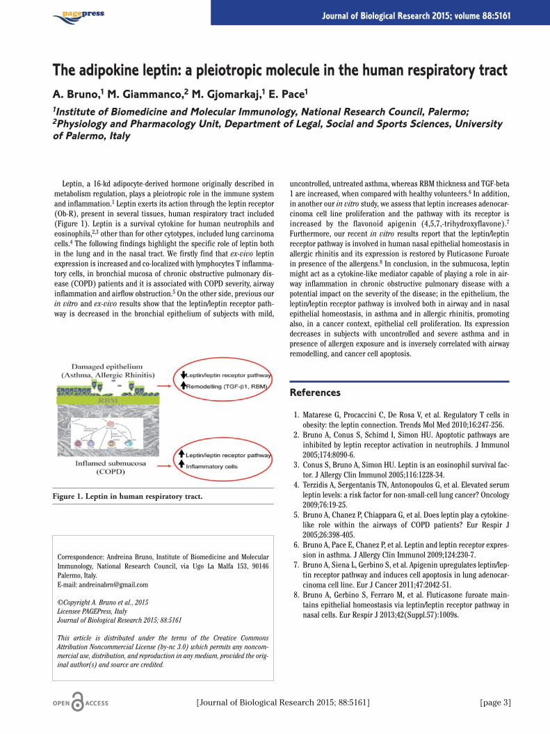
Leptin, a 16-kd adipocyte-derived hormone originally described inmetabolism regulation, plays a pleiotropic role in the immune systemand inflammation.1 Leptin exerts its action through the leptin receptor(Ob-R), present in several tissues, human respiratory tract included(Figure 1). Leptin is a survival cytokine for human neutrophils andeosinophils,2,3 other than for other cytotypes, included lung carcinomacells.4 The following findings highlight the specific role of leptin bothin the lung and in the nasal tract. We firstly find that ex-vivo leptinexpression is increased and co-localized with lymphocytes T inflamma-tory cells, in bronchial mucosa of chronic obstructive pulmonary dis-ease (COPD) patients and it is associated with COPD severity, airwayinflammation and airflow obstruction.5 On the other side, previous ourin vitro and ex-vivo results show that the leptin/leptin receptor path-way is decreased in the bronchial epithelium of subjects with mild,
uncontrolled, untreated asthma, whereas RBM thickness and TGF-beta1 are increased, when compared with healthy volunteers.6 In addition,in another our in vitro study, we assess that leptin increases adenocar-cinoma cell line proliferation and the pathway with its receptor isincreased by the flavonoid apigenin (4,5,7,-trihydroxyflavone).7
Furthermore, our recent in vitro results report that the leptin/leptinreceptor pathway is involved in human nasal epithelial homeostasis inallergic rhinitis and its expression is restored by Fluticasone Furoatein presence of the allergens.8 In conclusion, in the submucosa, leptinmight act as a cytokine-like mediator capable of playing a role in air-way inflammation in chronic obstructive pulmonary disease with apotential impact on the severity of the disease; in the epithelium, theleptin/leptin receptor pathway is involved both in airway and in nasalepithelial homeostasis, in asthma and in allergic rhinitis, promotingalso, in a cancer context, epithelial cell proliferation. Its expressiondecreases in subjects with uncontrolled and severe asthma and inpresence of allergen exposure and is inversely correlated with airwayremodelling, and cancer cell apoptosis.
References
1. Matarese G, Procaccini C, De Rosa V, et al. Regulatory T cells inobesity: the leptin connection. Trends Mol Med 2010;16:247-256.
2. Bruno A, Conus S, Schimd I, Simon HU. Apoptotic pathways areinhibited by leptin receptor activation in neutrophils. J Immunol2005;174:8090-6.
3. Conus S, Bruno A, Simon HU. Leptin is an eosinophil survival fac-tor. J Allergy Clin Immunol 2005;116:1228-34.
4. Terzidis A, Sergentanis TN, Antonopoulos G, et al. Elevated serumleptin levels: a risk factor for non-small-cell lung cancer? Oncology2009;76:19-25.
5. Bruno A, Chanez P, Chiappara G, et al. Does leptin play a cytokine-like role within the airways of COPD patients? Eur Respir J2005;26:398-405.
6. Bruno A, Pace E, Chanez P, et al. Leptin and leptin receptor expres-sion in asthma. J Allergy Clin Immunol 2009;124:230-7.
7. Bruno A, Siena L, Gerbino S, et al. Apigenin upregulates leptin/lep-tin receptor pathway and induces cell apoptosis in lung adenocar-cinoma cell line. Eur J Cancer 2011;47:2042-51.
8. Bruno A, Gerbino S, Ferraro M, et al. Fluticasone furoate main-tains epithelial homeostasis via leptin/leptin receptor pathway innasal cells. Eur Respir J 2013;42(Suppl.57):1009s.
Correspondence: Andreina Bruno, Institute of Biomedicine and MolecularImmunology, National Research Council, via Ugo La Malfa 153, 90146Palermo, Italy.E-mail: [email protected]
©Copyright A. Bruno et al., 2015Licensee PAGEPress, ItalyJournal of Biological Research 2015; 88:5161
This article is distributed under the terms of the Creative CommonsAttribution Noncommercial License (by-nc 3.0) which permits any noncom-mercial use, distribution, and reproduction in any medium, provided the orig-inal author(s) and source are credited.
The adipokine leptin: a pleiotropic molecule in the human respiratory tractA. Bruno,1 M. Giammanco,2 M. Gjomarkaj,1 E. Pace11Institute of Biomedicine and Molecular Immunology, National Research Council, Palermo;2Physiology and Pharmacology Unit, Department of Legal, Social and Sports Sciences, Universityof Palermo, Italy
[Journal of Biological Research 2015; 88:5161] [page 3]
Journal of Biological Research 2015; volume 88:5161
Figure 1. Leptin in human respiratory tract.
This work describes different effects of K:D-Rib solution treatment:from one side the slow down of cell proliferation and the reduction ofchemoinvasive potential of human breast cancer cell line (HTB-126)and from the other the maintenance of normal proliferation and nor-mal morphology in mammary human not cancer epithelial cell line(HTB-125). K:D-Rib is a water solution of D-ribose and KHCO3. Therole of D-ribose on the energetic metabolism and its involvement intoglycogen synthesis,1,2 as well as the importance of K+ into the cellphysiology, are well known.3,4 It has been found that K+ is essential tofold and to stabilize G-quadruplex5 with a strong relevance for telomer-ic structures2,6 and for oncogenic promoter regions.Our results showed that K:D-Rib has a cytostatic effect on canine
carcinoma cell line (A72), slows the colony formation ability of theHTB-126 cell line and has an antioxidant behaviour reducing MTT saltto formazan in absence of cells.1 These results are confirmed by ourmost recent work, demonstrating that 5mM K:D-Rib increase the cellcycle time of HTB-126 cell line treated with K:D-Rib at the concentra-tion of 5mM, from 44h to 59h. Here it will be show how K:D-Rib inter-feres both on HTB-126 cell line proliferation and cell morphology.Results on cell morphology using Atomic Force Microscopy (AFM) arepresented. K:D-Rib is tested also on human mammary epithelial cellline (HTB-125). HTB-125 cells treated with 5 mM K:D-Rib do not dis-play toxicity or notable cell proliferation decreasing rate compared tothe control one. HTB-125 cell morphology is analyzed by AFM. As men-tioned before a key point of cancer cells is the capability to invade tis-sues nearby or far from cancer formation site. Tumour motility is animportant step in the intricate process leading to the formation ofmetastasis. It has been shown that metastatic cells are more motilethan non-metastatic tumour cells and most motile of normal cells.Metastatic cells lose growth-inhibitory responses, undergo alterationsin adhesiveness and demonstrate enhanced production of enzymesthat can degrade extracellular matrix components. Since the develop-ment of metastatic disease in breast cancer is one of main responsible
of cancer mortality, the stopping and the understanding of the mecha-nisms that facilitate metastatic tumour progression is of prime impor-tance.7 We have investigated if K:D-Rib solution within 9 days canmodify the migration and the invasion ability. The experiments showHTB-126 cells are able to migrate across the coverslip toward the FBS– agar spot and to invade it within 48, but the relative cell numberinside the AGAR-FBS decrease already after five days of treatment.After nine days of treatment with K:D-Rib the relative cell number,inside the AGAR-FBS spot is reduced to 25%, demonstrating thattumorigenic potential is highly decreased with K:D-Rib treatment.These results show that 5mM K:D-Rib causes the change of someaspects like migration, invasion and proliferation of HTB-126 cell line.Despite these evidences K:D-Rib does not interfere neither with theproliferation of HTB-125 cell line nor with cell morphology.
References
1. Croci S, Bruni L, Bussolati S, et al. Potassium bicarbonate and D-ribose effects on A72 canine and HTB-126 human cancer cell lineproliferation in vitro. Cancer Cell Int 2011;11:30.
2. Heiden MGV, Cantley LC, Thompson CB. Understanding the war-burg effect: the metabolic requirements of cell proliferation.Science 2009;324:1029-33.
3. Dai JX CM, Yang DZ. Polymorphism of human telomeric quadru-plex structures. Biochimie 2008;90:1172-83.
4. Zhou FS, Tian Y, Youngbull C, et al. A new highly selective fluores-cent K+ sensor. Journal of the American Chemical Society2011:18530-3.
5. Parkinson GN, Lee MPH, Neidle S. Crystal structure of parallelquadruplexes from human telomeric DNA. Nature 2002;417:876-80.
6. Lipps HJ, Rhodes D. G-quadruplex structures: in vivo evidence andfunction. Trends in Cell Biology 2009;19:414-22.
7. Fernandis AZ, Prasad A, Band H, et al. Regulation of CXCR4-medi-ated chemotaxis and chemoinvasion of breast cancer cells.Oncogene 2004;23:157-67.
Correspondence: Luca Bruni, Unit of Biophysics and Medical Physics,Department of Neuroscience, University of Parma, via Volturno 39/E, 43125Parma, Italy.E-mail: [email protected]
©Copyright L. Bruni and S. Croci, 2015Licensee PAGEPress, ItalyJournal of Biological Research 2015; 88:5161
This article is distributed under the terms of the Creative CommonsAttribution Noncommercial License (by-nc 3.0) which permits any noncom-mercial use, distribution, and reproduction in any medium, provided the orig-inal author(s) and source are credited.
K:D-Rib on biology of human cancer and not cancer cell lineL. Bruni,1-3 S. Croci1,21Unit of Biophysics and Medical Physics, Department of Neuroscience, University of Parma;2National Institute of Biostructures and Biosystems, Rome, Italy; 3Valsè Pantellini Foundation,Oviedo, Spain
[page 4] [Journal of Biological Research 2015; 88:5161]
Journal of Biological Research 2015; volume 88:5161
Introduction
It is known that factors such as exercise intensity,1 length2 and type3
can modify the plasmatic oxidant/anti-oxidant production. In rhythmicgymnastics, adolescent female athletes showed a higher lipid peroxi-dation4 and an altered antioxidant enzyme profile compared with theiruntrained peers.5 The aim of this study was to assess whether the plas-matic H2O2 levels and antioxidant capacity were affected by the exer-cise training intensity in rhythmic gymnastics coaches.
Materials and Methods
Ten women (age: 23.8±3.42 y; weight: 52.58±4.57 kg; height:158.42±2.20 cm; body mass index: 20.88±1.23), with 13.14±5.40 yearsof practice in rhythmic gymnastics and coaches from at least 4 years ata competitive level, voluntarily participated into this study. One weekbefore training, trainers performed a laboratory graded exercise teston the treadmill in order to determine their maximal heart rate(HRmax), maximal oxygen consumption (VO2max) and anaerobic thresh-old (AT). Two interval-training sessions, separated by 48 hours ofrecovery, were performed with different intensities. The first was car-ried out at a low-moderate intensity; while the second at a high inten-sity. Both lasted 45 minutes and consisted of exercises aimed to devel-op anaerobic power, strength, flexibility and body balance. Before cooldown, two performances of competition technical skills coordinated tothe music were also executed. During training, HR was continuouslymonitored with ‘Polar team’ system. Immediately before and after thetraining session, blood samples were taken from fingertip’s coaches
and H2O2 levels and antioxidant capacity were measured through reac-tive oxygen metabolites (dROMs) and biological antioxidant potential(BAP) test, respectively. Newman-Keuls multiple comparison test wasused for evaluating the significant differences. Alpha level for signifi-cance was set to P<0.05.
Results
Coaches executed the first training session at an average intensity of66% HRmax, mainly in aerobic condition and only 5.5% of total time inanaerobic condition; while in the second session they spent 42% of totaltime at an intensity ranging from 80 to 100% HRmax and for 25% above theanaerobic threshold. After low-moderate intensity training, H2O2 levelswere significantly lower than baseline and they came back to baseline fol-lowing 48 h of recovery. After high intensity training, H2O2 amount slight-ly decreased compared with baseline (P>0.05); while it was significantlyhigher than after low-moderate intensity training. All these values corre-sponded to a middle oxidative stress when compared with a standardrange.6 Antioxidant capacity did not change following low-moderateintensity training, while it significantly increased after 48 h of recovery.In contrast, it significantly decreased in response to high intensity train-ing reaching the values obtained after low-moderate intensity training.
Conclusions
These results show that training intensity has different effects onROS production and antioxidant capacity in rhythmic gymnastics. Indetail, a low-moderate intensity session induces H2O2 production; whilea high intensity session negatively affects the antioxidant defences.Therefore, it would be appropriate to introduce a anti-oxidant diet or
supplementation for protecting rhythmic gymnastics trainers by oxida-tive stress.
References
1. Bloomer RJ, Goldfarb AH, Wideman L, et al. Effects of acute aero-bic and anaerobic exercise on blood markers of oxidative stress. JStrength Cond Res 2005;19:276-85.
2. Bogdanis GC, Stavrinou P, Fatouros IG, et al. Short-term high-intensity interval exercise training attenuates oxidative stressresponses and improves antioxidant status in healthy humans.Food Chem Toxicol 2013;61:171-7.
Correspondence: Marianna Bellafiore, Department of Legal, Social and SportsSciences, University of Palermo, via Maqueda 172, 90133 Palermo, Italy.Tel: +39.091.23892001 - Fax: +39.091.329355.E-mail: [email protected]
©Copyright M. Bellafiore et al., 2015Licensee PAGEPress, ItalyJournal of Biological Research 2015; 88:5161
This article is distributed under the terms of the Creative CommonsAttribution Noncommercial License (by-nc 3.0) which permits any noncom-mercial use, distribution, and reproduction in any medium, provided the orig-inal author(s) and source are credited.
Does the exercise training intensity affect plasmatic redox statusin rhythmic gymnastics?M. Bellafiore,1,2 G. Battaglia,1,2 G. Caramazza,1,2 M. Petrucci,1,2 M. Giaccone,1,2 A. Bianco,1,2A. Palma1,21Department of Legal, Social and Sports Sciences, University of Palermo; 2Regional Sport Schoolof CONI Sicily, Palermo, Italy
[Journal of Biological Research 2015; 88:5161] [page 5]
Journal of Biological Research 2015; volume 88:5161

[page 6] [Journal of Biological Research 2015; 88:5161]
3. Cubrilo D, Djordjevic D, Zivkovic V, et al. Oxidative stress andnitrite dynamics under maximal load in elite athletes: relation tosport type. Mol Cell Biochem 2011;355:273-9.
4. Guerra A, Rego C, Laires MJ, et al. Lipid profile and redox status inhigh performance rhythmic female teenagers gymnasts. J SportsMed Phys Fitness 2001;41:505-12.
5. Alshammari E, Shafi S, Nurmi-Lawton J, et al. Altered antioxidant
and trace-element status in adolescent female gymnasts. Int JSport Nutr Exerc Metab 2010;20:291-8.
6. Alberti A, Bolognini L, Macciantelli D, Carratelli M. The radicalcation of N,N-diethyl- para-phenylendiamine: a possible indicatorof oxidative stress in biological samples. Res Chem Intermed2000;26:253-67.
Article

Aims
Heart rate recovery after exercise (HRR) is an estimate of autonomicmodulation of the heart, and has been shown to be inversely associatedwith insulin resistance, metabolic syndrome, and type 2 diabetes.1 Type 2diabetes is associated with poor exercise tolerance and maximal aerobiccapacity (VO2max).2 Aim of our study was to assess the relationshipbetween HRR and VO2max in sedentary patients with type 2 diabetes.
Methods
Maximal treadmill exercise testing using standard or modified Bruceprotocol was performed in 16 (8 males and 8 females) sedentary patientswith type 2 diabetes (T2D), and in 16 (9 males and 7 females) age-matched sedentary non-diabetic controls (ND). HRR (bpm) was definedas the difference between maximum heart rate during the exercise testand heart rate 2 minutes after cessation of the exercise (Figure 1). Therecovery protocol consisted of walking on treadmill at 2.0 km/h of speedand 0% of grade. Oxygen uptake was recorded and VO2max (mL/kg/min)was defined as the highest 30 seconds average achieved during the test.For the statistical analysis of the data, Student’s t-test for independentsamples and linear regression analysis were used.
Results
The characteristics of subjects are shown in Table 1. The two groupswere similar in age and body weight. BMI was higher in T2D (30.1±3.6vs 26.9±4.2, P=0.029). VO2max was significantly lower in T2D comparedto ND (20.6±8.4 vs 28.2±8.1 mL/kg/min, P=0.002) and, according to
Normative Table by age and gender from ACMS, the aerobic capacity wasclassified very poor in all T2D and in 11/16 of ND. HRR was significantlylower in T2D (28±8.4 vs 37±8.9 bpm, P=0.008). A significant correlationbetween HRR and VO2max has been found in both T2D (Figure 2) andND (r=0.672, P=0.004 and r=0.620, P=0.010 respectively).
Correspondence: Angelo Cataldo, Department of Sports Sciences, via E. Duse 2,Palermo, Italy.Tel: +39.091.23865500.E-mail: [email protected]
©Copyright A. Cataldo et al., 2015Licensee PAGEPress, ItalyJournal of Biological Research 2015; 88:5161
This article is distributed under the terms of the Creative CommonsAttribution Noncommercial License (by-nc 3.0) which permits any noncom-mercial use, distribution, and reproduction in any medium, provided the orig-inal author(s) and source are credited.
Heart rate recovery after exercise and maximal oxygen uptakein sedentary patients with type 2 diabetesA. Cataldo, D. Cerasola, D. Zangla, P. Proia, G. Russo, R. Lo Presti, M. TrainaDepartment of Sports Sciences, University of Palermo, Italy
[Journal of Biological Research 2015; 88:5161] [page 7]
Journal of Biological Research 2015; volume 88:5161
Figure 1. Example of heart rate recovery phase analysis after amaximum reached heart rate.
Table 1. Characteristics of subjects.
T2D (n=16) Control (n=16) PMean±SD Mean±SD
Age (years) 57±7.5 53±6.9 0.100Height (cm) 165±7.7 166±9.8 0.826Weight (kg) 82±10.7 74±15.5 0.119BMI 30.1±3.6 26.9±4.2 0.029HRmax (bpm) 130±16.1 143±13.5 0.024VO2max (mL/kg/min) 20.6±4.3 28.2±8.1 0.002HRR (bpm) 28±8.4 37±8.9 0.008T2D, type 2 diabetes; HRR, heart rate recovery.
Figure 2. Correlation between heart rate recovery and VO2max intype 2 diabetes patients.

[page 8] [Journal of Biological Research 2015; 88:5161]
Conclusions
The results of our study showed that both HRR and VO2max were sig-nificantly reduced in T2D versus ND. The positive linear correlationbetween HRR and VO2max suggests that in T2D the heart rate recoveryafter exercise, index of autonomic modulation, might improve inresponse to a training aimed to increase aerobic capacity.
References
1. Sacre JW, Jellis CL, Coombes JS, Marwick TH. Diagnostic accuracyof heart-rate recovery after exercise in the assessment of diabeticcardiac autonomic neuropathy. Diabet Med 2012;29:e312-20.
2. Colberg SR, Sigal RJ, Fernhall B, et al. Exercise and type 2 diabetes:ACSM and ADA joint position statement. Diabet Care 2010;33:e147-67.
Article

Il fegato è la centrale metabolica del nostro organismo. I farmaci, inseguito alle reazioni di fase I e II vengono trasformati in metabolitiattivi meno tossici, ma idrofili pronti per essere eliminati. Il sistemaresponsabile di questa trasformazione è il citocromo P450 presente nelreticolo endoplasmatico liscio dell’epatocita. In seguito, intervengonole glucoronosil-tranferasi, le sulfotransferasi e le GSH-transferasi cheidrolizzano definitivamente il composto. Il trasporto del metabolita delfarmaco fuori dall’epatocita avviene attraverso il polo biliare ed èmediato da un sistema di trasportatori di membrana, l’ATP BindingCassette (ABC) Transporter Superfamily. L’alterazione dell’attività deitrasportatori, a diversi livelli, molecolare e trascrizionale, è uno deimeccanismi responsabile di epatotossicità.I polimorfismi genetici e/o i fattori ambientali, ad esempio l’alcol e/o i
farmaci concomitanti, contribuiscono alla suscettibilità individuale neldeterminare il danno epatico da farmaci. L’HLA si è dimostrato essereuno dei più importanti predittore di suscettibilità individuale anche perquei farmaci per i quali questa non era stata mai sospettata. A questo pro-posito, è stato dimostrato che c’è una forte associazione tra il danno epa-tico indotto da flucloxacillina e l’allele HLA B* 5701, e tra aplotipi HLA diclasse II e danno epatico da amoxicillina-clavulanico e ximelagatran.I farmaci, in generale, possono avere un effetto diretto sugli epatociti o
suscitare una reazione immune che può essere di due tipi: innata o adat-tativa. Nella maggior parte dei casi, la bioattivazione di un farmaco portaad un metabolita reattivo che determina una disfunzione mitocondrialecon conseguente riduzione dei livelli di ATP, disaggregazione del citosche-letro e quindi rottura della membrana cellulare epatocitaria. I metabolitiattivi influenzano il trasporto delle proteine (MDR-3) attraverso il polobiliare della membrana eritrocitaria determinando l’interruzione del flus-so biliare, il blocco di escrezione della bilirubina e infine la colestasi.In alternativa ad una azione diretta sulla membrana cellulare, lo stress
epatocitario determina l’attivazione del sistema immune innato attraver-so le cellule natural killer (NK) del fegato che secernono interferone-
gamma (IFNg) ed interleuchina (IL)-4, e sono in grado di uccidere diret-tamente le cellule tramite il sistema Fas/FasLigand. Cellule di Kupffer edNK contribuiscono alla progressione del danno epatico producendomediatori pro-infiammatori (citochine, chemochine, ROS); questi pos-sono avere azione citotossica diretta (perossido d’idrogeno, ossido nitri-co) degradando la matrice extracellulare, oppure promuovendo l’adesio-ne e l’infiltrazione cellulare dei leucociti polimorfonucleati.Nella patogenesi del danno epatico è coinvolto anche il sistema
immune adattativo. Il metabolita reattivo può infatti legarsi in modocovalente ed alterare le proteine del fegato, promuovendo l’attivazionedelle cellule T citotossiche e la produzione di citochine (reazioneimmuno- mediato). Il meccanismo del danno da farmaci immunomediato non è ben chiaro, e comporta un’azione apten-like.Generalmente infatti le sostanze chimiche a basso PM non sonoimmunogeni ma possono diventare tali quando sono legati ad unamacromolecola, come una proteina. Se un metabolita attivo di un far-maco prodotto dal citocromo P450 è in grado di agire come un aptene,e si lega covalentemente ad una proteine del fegato, il sistema immu-nitario la percepirà come non-self causando una reazione autoimmu-ne. Il risultato di questi eventi, sia attraverso una reazione diretta sullamembrana cellulare, sia attraverso l’induzione di una risposta immu-nitaria, è la morte cellulare: necrosi o apoptosi.L’induzione dell’apoptosi piuttosto che la necrosi dipende da diversi
fattori, tra cui lo stato energetico (ATP). Una lesione grave per i mito-condri determina deplezione energetica della cellula, che perde laregolazione osmotica e va in necrosi. Una lesione meno grave senzaimportante deplezione di ATP è in grado di mantenere la regolazioneosmotica e porta all’apoptosi. La necrosi epatocellulare è l’evento prin-cipale di cui è responsabile il danno epatico da farmaci; ne possonoessere bersaglio sia cellule endoteliali che quelle dei dotti biliari. Infatti distinguiamo il danno epatico da farmaci di tipo epatocellulare(nimesulide), di tipo colestatico (amoxicillina clavulanico) e misto.
Correspondence: Anna Licata, Gastroenterology Unit, Department ofInternal and Specialized Medicine, University of Palermo, Piazza delleCliniche 2, 90127 Palermo, Italy.Tel: +39.091.23867516.E-mail: [email protected]
©Copyright A. Licata et al., 2015Licensee PAGEPress, ItalyJournal of Biological Research 2015; 88:5161
This article is distributed under the terms of the Creative CommonsAttribution Noncommercial License (by-nc 3.0) which permits any noncom-mercial use, distribution, and reproduction in any medium, provided the orig-inal author(s) and source are credited.
Meccanismi immunologici e molecolari del danno epatico da farmaciA. Licata,1 S. Aiello,2 V. Calvaruso,1 P.L. Almasio11Gastroenterology Unit, Department of Internal and Specialized Medicine, University of Palermo;2Physiology and Pharmacology Unit, Department of Legal, Social and Sports Sciences, Universityof Palermo, Italy
[Journal of Biological Research 2015; 88:5161] [page 9]
Journal of Biological Research 2015; volume 88:5161
In Mediterranean and in southeast Europe the activities of a significantpart of the population are traditionally linked with agriculture, forestry andanimal husbandry. However, many rural communities are experiencingserious difficulties associated with low income per person and pooremployment prospects combined with increased demographic decline.Alternative activities such as the collection and trading of wild ediblemushrooms as well as the cultivation of choice species could contribute atproviding valuable solutions both in financial and environmental terms.The total number of fungal species which are considered having edi-
ble and/or medicinal value is over 2300.1 Most of them form large con-spicuous sporophores (i.e. mushrooms) during their life-cycle, whichare either harvested from the wild or cultivated on a wide range of plantand agro-industrial residues and by-products. Foraying and picking ofwild edible mushrooms has a long tradition in most European countries;therefore it constitutes a significant socioeconomic activity, while at thesame time reflects local knowledge and social practices that are worthpreserving. Recent food market tendencies reveal a high demand poten-tial for wild edible mushrooms among urban consumers. In those casesthat wild fungi are not well-known because pertinent knowledge was notspread within families or local communities, people avoid their harvest;instead they are oriented at consuming cultivated mushrooms which
become increasingly popular. This latter type of activity is tightly associ-ated with environmental protection through recycling and valorizationof low-value substrates together with the conservation of some highlysought-after mushroom species.2,3
The Mycoticon project (EU, LdV-ToI) involves Universities,Technological, and Research Institutions as well as local stakeholdersand associated end-users from four European countries, i.e. Bulgaria,Cyprus, Greece and Italy. These partners combine their experience andexpertise at developing an integrated educational and training packagetogether with its respective tools to meet the demands of suitable target-groups willing to create collective entrepreneurship schemes for exploit-ing the economic potential of wild mushrooms in rural areas. Ultimately,the objective is to facilitate the generation of a new source of non-sub-sidized income and create new jobs in areas desperately in need of both.In parallel, local people are expected to be presented with incentives toadopt sustainable management and harvesting practices for wild ediblemushrooms together with basic knowledge on mushroom cultivation.Among other anticipated deliverables, national reports were compiled
for each participating country as regards the current knowledge/situa-tion on diversity, harvest and trade of wild edibl mushrooms as well ason commercial mushroom production. In addition, a voluminous text-book was prepared4 which provided a detailed description of 22 choiceedible and 11 selected poisonous mushrooms (together with many otherrelated taxa) of significance in all four countries. Moreover, it includedgeneral information about biology and ecology of mushroom fungi, theircommon habitats/ecosystems, proper harvest practices and suitable foodpreservation methods, relevant legislation and conservation issues, andbasic guidelines for the cultivation of the most popular species togetherwith prospects for developing tourism activities associated with mush-rooms. All of them formed the basis for the development of an innovativetraining material established both on paper and online by creating amoodle web-page (http://moodle.teilar.gr/). This electronic tool wasassembled in four languages (English, Italian, Greek and Bulgarian) andit now provides a user-friendly and flexible modular training coursethrough which e-self-assessment and e-accreditation could be alsoaccomplished. The training package complies with EQF rules and it willbe further structured according to EC-VET provisions. Its content isanticipated to enhance the development of pertinent skills and subse-quently increase employment of qualified people in rural areas.Furthermore, it provides the prerequisites for combining local assetsand resources into mushroom products that meet consumers’ expecta-
Correspondence: Georgios I. Zervakis, Laboratory of General andAgricultural Microbiology, Agricultural University of Athens, Iera Odos 75,11855 Athens, Greece.Tel: +30.210.5294341.E-mail: [email protected]
Acknowledgements: this work was performed in the frame of theIdentification and sustainable exploitation of wild edible mushrooms in ruralareas (Mycoticon) project, and it was funded by the European Union(Leonardo da Vinci – Transfer of Innovation).
©Copyright G.I. Zervakis et al., 2015Licensee PAGEPress, ItalyJournal of Biological Research 2015; 88:5161
This article is distributed under the terms of the Creative CommonsAttribution Noncommercial License (by-nc 3.0) which permits any noncom-mercial use, distribution, and reproduction in any medium, provided the orig-inal author(s) and source are credited.
Identification and sustainable exploitation of wild edible mushroomsin rural areas (Mycoticon, LdV-ToI project): development of an innovativetraining package to meet educational and income-generating demandsin South Europe and to improve the use of mushrooms as high-value foodG.I. Zervakis,1 G. Venturella,2 C.M. Denchev,3 P.T. Fitsilis,4 V.C. Gerogiannis,4S. Papaefthimiou,5 J. Georgi,6 A. Saitta,2 E. Polemis,1 T.T. Denchev,5 B. Assyov,5 M.L. Gargano21Laboratory of General and Agricultural Microbiology, Agricultural University of Athens, Greece;2Department of Agricultural and Forest Sciences, University of Palermo, Italy;3Institute of Biodiversity and Ecosystem Research, Bulgarian Academy of Sciences, Sofia, Bulgaria;4Department of Project Management, Technological Education Institute of Thessalia, Larissa;5KEK Aegeas, Larissa, Greece; 6Neapolis University of Pafos, Cyprus
[page 10] [Journal of Biological Research 2015; 88:5161]
Journal of Biological Research 2015; volume 88:5161

tions. Such activities constitute a highly recommended approach inEurope since rural income could derive from integrated direct and indi-rect recourses (by also supporting conservation and environmental sus-tainability) and not only by the primary agricultural production.
References
1. Marshall E, Nair NG. Make money by growing mushrooms, p. 64.Rome, Italy: Food and Agriculture Organization of the UnitedNations; 2009.
2. Gargano ML, Zervakis GI, Venturella G. Pleurotus nebrodensis, avery special mushroom, p. 150 (e-book). The Netherlands:Bentham Science Publishers; 2013.
3. Zervakis G, Venturella G. Mushroom breeding and cultivationfavors ex situ conservation of Mediterranean Pleurotus taxa. In:Engels JMM, Ramanantha Rao V, Brown AHD, Jackson MT, eds.Managing plant genetic diversity. UK: IPGRI-CABI Publishing;2002. pp. 351-358.
4. Denchev C, Venturella G, Zervakis G. Identification and sustainableexploitation of wild edible mushrooms in rural areas. Thessalia,Greece: Technological Education Institute of Thessalia; 2013.
[Journal of Biological Research 2015; 88:5161] [page 11]
Article
The Acute Phase Response (APR) is a core part of the innateimmune response and is present in all animal species including mam-mals, fish and invertebrates. APR is a reaction of the body to injury,trauma or infection and the basic aim is to restore homeostasis. TheAPR can appear as a local reaction but mainly as systemic reactionsincluding increased secretion of some hormones, activation of thecomplement system and increased synthesis and secretion of AcutePhase Proteins (APP).1 Approximately 40 plasma proteins have beendefined as APP.2 Those proteins can have various functions and changein their plasma levels. APP’s are classified as positive when their plas-ma concentration increases and negative when their plasma concen-tration decreases following the APR. Studies suggest that fish have anAPR response that is delayed compared to mammals.3,4 APR has beenstudied in several fish species and a large number of APPs have beenidentified in fish. In this study, APR was induced in Atlantic cod (Gadusmorhua L.) by intramuscular injection of Aeromonas salmonicidasubsp. achromogenes (Asa), a common bacterial pathogen in cod andother fish species, causing atypical furunculosis.5 Asa is endemic inIceland and caused serious losses of farmed salmonids before vaccina-tion became a common practice.6 Commercial Asa vaccines are as yetnot available for cod and experimental vaccines show variable andoften poor protection.7 The aim of the current study was to examinethe acute phase induced by Asa by mesuring the gene expression incod’s kidney and spleen as well as cortisol levels in serum. Cod, meanweight 97.3 g, were divided into three groups: two groups receivedintramuscular injections of two different concentrations of Asa and thethird, a control group, was injected intramuscularly with PBS. Kidneyand spleen were sampled from seven fish from each group at time 0before treatment and at 1 hour, 24 hours, 72 hours and 168 hours after
the injection. Blood was collected from the caudal vessel. An ELISAassay (Neogen Corp KY,USA) was used to measure cortisol. For thegene expression analysis of IL-1β, hepcidin and transferrin, total RNAwas isolated from tissue samples from kidney and spleen with aNucleoSpin® RNA II kit following the manufacturer’s instructions(Macherey-Nagel). Complementary DNA (cDNA) was prepared withthe Revertaid™ First Strand cDNA Synthesis Kit, according to themanufacturer’s instructions (Thermo). Quantitative real time PCR(qPCR) analyses were performed on a StepOne Plus™ real time PCRinstrument (Applied Biosystems). The gene expression data were nor-malized to the expression of the reference genes ubiquitin or RPL4with identical results. The expression of the reference genes in the tis-sue samples used was relatively stable. The results for gene expressionin the controls and Asa injected fish were compared at each time point.Overall, the gene expression results showed a stronger response in thespleen than in the kidney. In Asa injected fish there was a significantincrease in IL-1β and hepcidin gene expression at 24h, compared tocontrols, in both organs. Transferrin gene expression was also signifi-cantly elevated in both organs, reached a maximum peak at 72h in thekidney and at 168h in the spleen (Table 1). The results of cortisolanalysis showed a statistically significant increase of cortisol levelswith a peak at 72h after injection (Figure 1). At the end of the experi-ment the cortisol levels were significantly elevated compared to thecontrol fish. IL-1β is one of the earliest pro-inflammatory cytokines torespond to infection and induces a cascade of reactions leading toinflammation.8 The observed early increase in IL-1β gene expressionfollowing an acute phase induction with Asa was in agreement withother studies in fish. The serum cortisol levels observed in this studyreached a maximum concentration at 72h when the IL-1β gene expres-sion had started to decrease. This could mean that cortisol had a rolein the suppression of the IL-1β gene expression as described by other.9
Transferrin is the major iron binding protein and hepcidin is anCorrespondence: Bergljot Magnadottir, Institute for Experimental Pathology,University of Iceland, Keldur, Keldnavegur 3, 112-Reykjavik, Iceland.E-mail: [email protected]
Acknowledgements: this research was supported by AVS R&D Found ofMinistry of Fisheries and Agriculture in Iceland.
©Copyright A. Fazio et al., 2015Licensee PAGEPress, ItalyJournal of Biological Research 2015; 88:5161
This article is distributed under the terms of the Creative CommonsAttribution Noncommercial License (by-nc 3.0) which permits any noncom-mercial use, distribution, and reproduction in any medium, provided the orig-inal author(s) and source are credited.
The acute phase response of Atlantic cod (Gadus Morhua L.):expression of immune response genes after infection withAeromonas Salmonicida subsp. AchromogenesA. Fazio,1 B.T. Bragason,2 B. Magnadottir,2 C. Faggio,1 S. Gudmundsdottir21Department of Biological and Environmental Sciences, University of Messina, Italy;2Institute for Experimental Pathology, University of Iceland, Reykjavik, Iceland
[page 12] [Journal of Biological Research 2015; 88:5161]
Journal of Biological Research 2015; volume 88:5161
Table 1. Expression patterns of the genes IL-1β, hepcidin andtransferrin of both organs at main time points. The max (maxi-mum) indicates the time point where each gene reached the max-imum peak.
Gene Organ 24 h 72 h 168 h
IL-1β Spleen ⇑ (max) ⇓ ⇓Kidney ⇑ (max) ⇓ ⇓
Hepcidin Spleen ⇑ (max) ⇓ ⇓Kidney ⇑ (max) ⇓ ⇓
Transferrin Spleen ⇑ ⇑ ⇑ (max)Kidney ⇑ (max) ⇑ ⇓
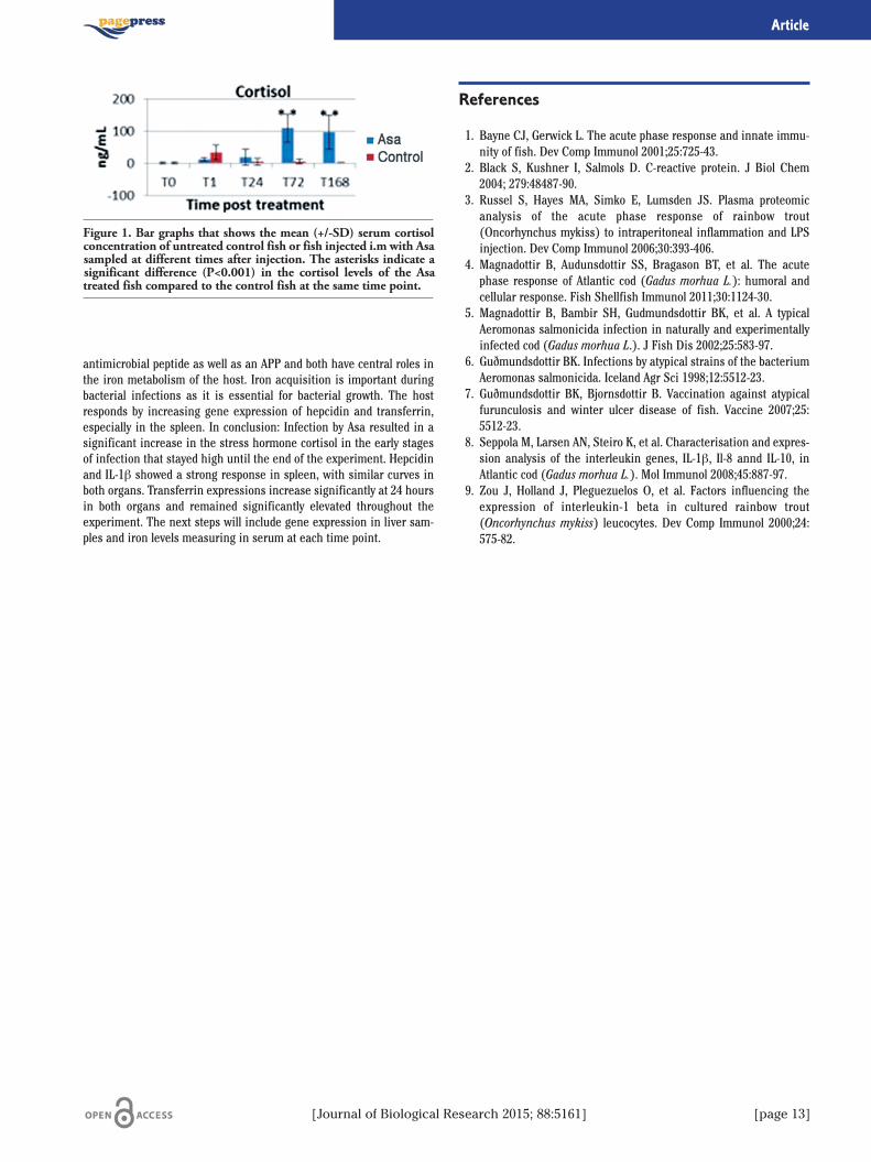
antimicrobial peptide as well as an APP and both have central roles inthe iron metabolism of the host. Iron acquisition is important duringbacterial infections as it is essential for bacterial growth. The hostresponds by increasing gene expression of hepcidin and transferrin,especially in the spleen. In conclusion: Infection by Asa resulted in asignificant increase in the stress hormone cortisol in the early stagesof infection that stayed high until the end of the experiment. Hepcidinand IL-1β showed a strong response in spleen, with similar curves inboth organs. Transferrin expressions increase significantly at 24 hoursin both organs and remained significantly elevated throughout theexperiment. The next steps will include gene expression in liver sam-ples and iron levels measuring in serum at each time point.
References
1. Bayne CJ, Gerwick L. The acute phase response and innate immu-nity of fish. Dev Comp Immunol 2001;25:725-43.
2. Black S, Kushner I, Salmols D. C-reactive protein. J Biol Chem2004; 279:48487-90.
3. Russel S, Hayes MA, Simko E, Lumsden JS. Plasma proteomicanalysis of the acute phase response of rainbow trout(Oncorhynchus mykiss) to intraperitoneal inflammation and LPSinjection. Dev Comp Immunol 2006;30:393-406.
4. Magnadottir B, Audunsdottir SS, Bragason BT, et al. The acutephase response of Atlantic cod (Gadus morhua L.): humoral andcellular response. Fish Shellfish Immunol 2011;30:1124-30.
5. Magnadottir B, Bambir SH, Gudmundsdottir BK, et al. A typicalAeromonas salmonicida infection in naturally and experimentallyinfected cod (Gadus morhua L.). J Fish Dis 2002;25:583-97.
6. Guðmundsdottir BK. Infections by atypical strains of the bacteriumAeromonas salmonicida. Iceland Agr Sci 1998;12:5512-23.
7. Guðmundsdottir BK, Bjornsdottir B. Vaccination against atypicalfurunculosis and winter ulcer disease of fish. Vaccine 2007;25:5512-23.
8. Seppola M, Larsen AN, Steiro K, et al. Characterisation and expres-sion analysis of the interleukin genes, IL-1β, Il-8 annd IL-10, inAtlantic cod (Gadus morhua L.). Mol Immunol 2008;45:887-97.
9. Zou J, Holland J, Pleguezuelos O, et al. Factors influencing theexpression of interleukin-1 beta in cultured rainbow trout(Oncorhynchus mykiss) leucocytes. Dev Comp Immunol 2000;24:575-82.
[Journal of Biological Research 2015; 88:5161] [page 13]
Article
Figure 1. Bar graphs that shows the mean (+/-SD) serum cortisolconcentration of untreated control fish or fish injected i.m with Asasampled at different times after injection. The asterisks indicate asignificant difference (P<0.001) in the cortisol levels of the Asatreated fish compared to the control fish at the same time point.
Among Cnidaria, Pelagia noctiluca, is one of the most dangerous jel-lyfish in the Mediterranean Sea, where its blooming has been veryabundant for many years. Toxicology of crude venom from P. noctilucastinging cells is reported in this presentation. Both in vivo and in vitrobiological assays have been performed to verify and, possibly, measurethe toxicity of P. noctiluca crude venom, whose composition is still notcompletely defined.At first we tested the hemolytic activity of crude venom from single
nematocysts discharged by a chemical non enzymatic method. Thedelivered venom induced a powerful and rapid hemolytic activity. As asecond step, crude venom extracted from a population of isolatednematocysts, provoked a dose-dependent hemolysis in erythrocytesfrom different sources, including eel, rabbit, chicken and human.Moreover, P. noctiluca crude venom directly induced mitochondrialtrans-membrane potential (ΔΨm) collapse and Reactive OxygenSpecies (ROS) generation in SH-SY5Y cells derived from human neu-roblastoma.In order to better characterize the biological effects of the crude
venom, in vivo assays were also performed. Injection of crude venominto the rat paw evoked an inflammatory reaction in a dose-dependentmanner. Immunohistochemical analysis showed a marked acute inflam-matory response in the tissues, with accumulation of polymorphonu-clear neutrophils. Treatment with melatonin as antioxidant significantlyreduced the inflammatory response, thereby confirming that oxidativestress plays a major role in inducing the observed pathological changes.In addition to hemolytic and cytolytic assays, a test on cell volume
regulation capability was also chosen to describe the biological activityof P. noctiluca crude venom. As already demonstrated, isolated nema-tocytes of the sea anemone A. mutabilis exhibit Regulatory VolumeDecrease (RVD) when stimulated with a 35% hypotonic solution. Innematocytes exposed to different concentrations of crude venom (cor-responding to the amount contained in 10, 25 and 50 nematocysts/μL)RVD was partially inhibited 25 nematocysts/μL crude venom concen-tration and fully blocked at 50 nematocysts/μl completely recovered,therefore indicating that K+ channels inhibition may account for thevenom-induced RVD impairment. RVD tests were also performed onHEK293 Phoenix cells, a human embryonic kidney cell line. In controlconditions, the cells stimulated by hypotonicity showed an initialswelling followed by RVD, whereas in 0.025 μg/μL crude venom-con-taining extracellular hypotonic solution, RVD was dramaticallyimpaired. Furthermore, pre-incubation of cells in a crude venom-con-taining extracellular isotonic solution prevented RVD after hypotonicstress. Surprisingly, the presence of toxin in the extracellular isotonicsolution led to cell swelling even in the absence of an osmotic gradient.This phenomenon was not observed in control conditions and the pre-cise mechanism needs to be further elucidated.We conclude that P. noctiluca crude venom extract has hemolytic
activity, pro-inflammatory action, induces mitochondrial potential col-lapse and ROS production. In addition, crude venom inhibits RVD inboth cnidarians and mammalian cells after hypotonic stress and leads tocell swelling in isotonic conditions. Our experiments add novel informa-tion to understand the mechanism of action of P. noctiluca venom.
Correspondence: Angela Marino, Department of Human and Social Sciences,University of Messina, viale Ferdinando Stagno D’Alcontres 31, 98166Messina, Italy.Tel: +39.090.6765214 - Fax: +39.090.394030.E-mail: [email protected]
©Copyright R. Morabito et al., 2015Licensee PAGEPress, ItalyJournal of Biological Research 2015; 88:5161
This article is distributed under the terms of the Creative CommonsAttribution Noncommercial License (by-nc 3.0) which permits any noncom-mercial use, distribution, and reproduction in any medium, provided the orig-inal author(s) and source are credited.
Effects of Pelagia Noctiluca crude venom on cell viabilityand volume regulationR. Morabito,1 A. Marino,2 S. Dossena,3 M. Paulmichl,3 G. La Spada21Department of Human and Social Sciences, University of Messina; 2Department of Biological andEnvironmental Sciences, University of Messina, Italy; 3Institute of Pharmacology and Toxicology,Paracelsus Medizinische Privatuniversitat, Salzburg, Austria
[page 14] [Journal of Biological Research 2015; 88:5161]
Journal of Biological Research 2015; volume 88:5161
Energy metabolism offers a valuable gauge to monitor the geneticalterations that promote cellular dysfunction and hence, is a usefulbiomarker in human Rare Diseases.Genetic alterations that result in cellular dysfunction and thus in an
overt phenotypic presentation are usually accompanied by alterationsin the proteome of energy metabolism. Cancer provides one example.1
The development of high-throughput OMIC techniques allows thesimultaneous interrogation of a large number of genes, proteins andmetabolites in the same assay. Reverse phase protein microarrays(RPPmA) is a high-throughput proteomic technique that allows thequantification (fentomolar range) of a given marker in minuteamounts of protein from biological specimens. The application of thistechnique in oncology has been largely documented as it is most usefulfor the identification and quantification of biomarkers of survival andof the response to chemotherapy. Herein, we have studied the expres-sion of twenty proteins of energy metabolism which include membersof the TCA cycle, β-oxidation, electron transport, oxidative phosphory-lation, glycolysis and oxidative stress using highly specific antibodiesin a cohort of seventy three muscle biopsies of control donors andpatients affected of neuromuscular diseases. The cohort includedDuchenne (DMD), Becker (DMB), symptomatic forms of DMD and
DMB in female carriers (Xp21 Carriers) and Limb Girdle MuscularDystrophy Type 2C (LGMD2C) biopsies as well as of patients affectedof glycogenesis type V (Mc Ardle disease), complex I mitochondrialmyopathies, various intensive care unit myopathies (ICU) and neu-ronal ceroid lipofuscinosis (NCL) also known as Batten disease, a neu-rodegenerative disease. The samples were obtained with informedconsent following the Declaration of Helsinki and coded for anonymity.The final aim of the study was to verify the potential applicability ofRPPmA technique in the field of Rare Diseases for the identification ofnew molecular markers of diagnosis to contribute to the improvementof the clinical handling of these patients. The results indicate that thephenotype of energy metabolism offers relevant diagnostic markers inRare Diseases.
Reference
1. Aldea M, Clofent J, Núñez de Arenas C, et al. Reverse phase pro-tein microarrays quantify and validate the bioenergetic signatureas biomarker in colorectal cancer. Cancer Letters 2011;311:210-8.
Correspondence: Fulvio Santacatterina, Department of Molecular Biology,Center of Molecular Biology Severo Ochoa CSIC- UAM, AutonomousUniversity of Madrid, calle Caracas 8, 28010 Madrid, Spain.E-mail: [email protected]
©Copyright F. Santacatterina et al., 2015Licensee PAGEPress, ItalyJournal of Biological Research 2015; 88:5161
This article is distributed under the terms of the Creative CommonsAttribution Noncommercial License (by-nc 3.0) which permits any noncom-mercial use, distribution, and reproduction in any medium, provided the orig-inal author(s) and source are credited.
Reverse phase protein microarray technology to provide new diagnosticmarkers of metabolism in rare diseasesF. Santacatterina,1-3 C. Navarro,2,4 M. Sánchez-Arago,1-3 M.A. Martin,2,3 J.M. Cuezva1-31Department of Molecular Biology, Center of Molecular Biology Severo Ochoa CSIC-UAM,Autonomous University of Madrid; 2CIBERER-ISCIII, Madrid; 3Institute of Hospital Investigation,Madrid; 4Department of Pathology and Neuropathology, University Hospital of Vigo, Spain
[Journal of Biological Research 2015; 88:5161] [page 15]
Journal of Biological Research 2015; volume 88:5161
Inflammatory bowel disease (IBD) is a chronic disorder character-ized by a relapsing-remitting course, which alternates between activeand quiescent states, ultimately impairing a patients’ quality of life.The two main types of IBD are Crohn’s disease (CD) and ulcerative
colitis (UC). CD shows a transmural granulomatous inflammation thatcan involve any segment of the intestine affecting all layers of theintestinal wall while UC is limited to the mucosa and superficial sub-mucosa of the colon. In physiological conditions the gut is costantlyexposed to various antigens, commensal microflora and pathogens andthe inflammatory response is finely balanced. Anyhow in some individ-uals with genetic susceptibility an anomalous inflammatory responsecan arise due to the deregulation of the negative feedback mecha-nisms implicated in its self-regulation. It is thought that a vast numberof environmental risk factors may be implicated in the development ofIBD, including smoking, dietary factors, psychological stress, use ofnon- steroidal anti-inflammatory drugs and oral contraceptives, appen-dectomy, breastfeeding, as well as infections. Nutritional support as aprimary therapy has a crucial role in the management of patients withIBD since it can control the inflammatory process, treat malnutritionand its consequences, and avoid the use of immune-modulating drugsand their side effects. The gut microbiota is clearly manipulated bydietary components such as n-3 PUFA and coniugated linoleic acid(CLA) which favorably reduce endotoxin load via shifts in the compo-sition and metabolic activity of the microbial community.In particular, the beneficial effect of n-3 polyunsaturated fatty acids
(PUFAs) and fermentable fiber, during the remission/quiescent phaseof both CD and UC is highlighted. In fact, PUFAs are associated with aless grade of inflammation since they are metabolized to 3-seriesprostaglandins and thromboxanes and 5-series leukotrienes and, inaddition, exert antiinflammatory effects when compared with their n-6 PUFA counterparts. In similar action to dietary n-3 PUFA, coniugatedlinoleic acid (CLA) have been reported to ameliorate intestinal inflam-mation in animal models of IBD. In contrast to corticosteroids, CLAsuppresses gut inflammatory responses while enhancing antigen spe-cific responsiveness of T cells against viral and bacterial pathogens.Available data about nutritional interventions do not always match
due to the incomplete knowledge of pathogenic mechanisms underly-ing IBD development. Further studies are therefore needed to improvenutritional therapeutic approach. In particular, is still unclear the roleof the fiber in helping the remission of the disease. There are mainlytwo theories. On one hand, dietary fibers can act as effective prebioticsby altering the intestinal microbial composition and promoting thegrowth of beneficial bacterial communities within the large intestine.Some authors reported a positive effect associated with the produc-
tion by colonic microflora of short chain fatty acids (SCFA), able todown-regulate the production of pro-inflammatory cytokines, to pro-mote the restoration of intracellular Reactive Oxygen Specie (ROS)balance, and the activation of NF-kB.On the other hand, fibers can promote diarrhea, pain and gas aggra-
vating the clinical state. We suggest that the consumption of fer-mentable fibers may have a good impact on patients’ health. Now iswell known that various SNPs are linked to the risk of IBD develop-ment and therefore there is the possibility of predict if an individual ispredisposed to the disease. The identification of some polymorphismshas an essential role because it allows the modification of diet in thehope of controlling symptoms or preventing relapse. As a consequence,foods that can potentially exacerbate symptoms are eliminated andsubstituted with those that promote a well-being state.
Correspondence: Giovanni Tomasello, Department of Surgical, Oncologicaland Stomatological Sciences, University of Palermo, Via Liborio Giuffrè 5,90127 Palermo, Italy.E-mail: [email protected]
©Copyright A. Abruzzo et al., 2015Licensee PAGEPress, ItalyJournal of Biological Research 2015; 88:5161
This article is distributed under the terms of the Creative CommonsAttribution Noncommercial License (by-nc 3.0) which permits any noncom-mercial use, distribution, and reproduction in any medium, provided the orig-inal author(s) and source are credited.
Nutrition in inflammatory bowel disease patientsA. Abruzzo,1 G. Tomasello,2,3 E. Sinagra,2,4 F. Cappello,3,5 P. Damiani,3,4 F. Damiani,1M. Bellavia,3 C. Campanella,3,6 F. Rappa,3,6 A. Marino Gammazza,3,7 L. Cicero,8A.I. Lo Monte1,21School in Surgical Biotechnology and Regenerative Medicine, Faculty of Medicine, University ofPalermo; 2Department of Surgical, Oncological and Stomatological Sciences, University ofPalermo; 3S. Raffaele-G. Giglio Institute Foundation, Cefalù; 4Department of ExperimentalBiomedicine and Clinical Neuroscience, Section of Human Anatomy, University of Palermo;5School of Medicine Specialization in Geriatrics, University of Palermo; 6Euro-MediterraneanInstitute of Science and Technology, Palermo; 7Department of Biological, Chemical andPharmaceutical Sciences and Technologies, University of Palermo; 8Institute for ExperimentalVeterinary Medicine of Sicily A. Mirri, Palermo, Italy
[page 16] [Journal of Biological Research 2015; 88:5161]
Journal of Biological Research 2015; volume 88:5161
Introduzione
Studi condotti sui vini rossi, hanno dimostrato come un loro mode-rato consumo quotidiano possa ridurre l’incidenza delle malattie car-diovascolari1 perché in essi sono contenute sostanze antiossidantied in particolare il transresveratrolo e il piceatannolo.2 I meccanismiprotettivi sarebbero indipendenti dall’effetto di quote equivalenti dialcool contenute nel vino ma riconducibili alle caratteristiche delvitigno e al contenuto di polifenoli, le cui concentrazioni dipendonomolto dalla tecnica di vinificazione (flavonoidi, trans-resveratrolo etannini polimerici).3 Con l’obiettivo di migliorare la quantità di poli-fenoli in una cultivar di Nero d’Avola, e stata effettuata su alcuni fila-ri di un Vigneto sito presso Castelbuono (PA), la tecnica di defoglia-zione eseguita alla fase fenologica di acino pisello su un campione di500 piante con l’eliminazione manuale delle foglie fino al primo nodosopra il grappolo. I confronti sono stati eseguiti con un testimonenon defogliato (controllo).
Materiali e Metodi
Il contenuto in solidi solubili (°Brix) è stato determinato con unrifrattometro di Abbe. Il pH è stato misurato con piaccametro. Per ladeterminazione degli antociani totali, le bucce degli acini sono stateposte in un tampone tartarico a pH 3,2 al fine di estrarre tutti i polife-noli, l’acidificazione del campione è stata eseguita con HCl conc., ladiluizione con etanolo cloridrico, l’analisi spettrofotometrica (UV)misurando l’assorbanza a 540 nm utilizzando cuvette in quarzo concammino ottico di 1 cm. Per la determinazione dei flavonoidi totali si
misura la densità ottica a 280 nm e l’assorbanza dipendendo dalla con-centrazione. La determinazione delle proantocianidine viene fatta perdifferenza fra l’assorbanza a 540 nm, misurata prima e dopo l’idrolisie confronto del Δ di assorbanza con una curva di calibrazione determi-nata con soluzioni standard di proantocianidine. Lo studio della com-ponente polifenolica è stato eseguito su campioni di bucce e polpa,acino e raspo, rachide e vino. Le analisi sono state condotte con tecni-ca HPLC con cromatografo liquido Agilent 1100, rilevatore a serie didiodi (DAD) posizionato a 305 e 325 nm, autoiniettore con volume diiniezione 50 μl, colonna Phenomenex Luna C18, eluenti: acetonitrile etampone fosfato (KH2PO4+H3PO4) 0,02 M a pH=3,0, flusso 1,0ml/min., per calcolare i tempi di ritenzione sono stati preventivamenteiniettati i rispettivi standard. Buccia e polpa, acino intero e raspo,rachide hanno subito lo stesso trattamento: macerazione con CH3OHal 95%, omogeneizzazione e agitazione tramite agitatore orbitale, fil-trazione con filtro in microfibra di vetro, evaporazione in rotovapor,prima estrazione con NaHCO3 al 5% seguita da tre estrazioni con ace-tato di etile, evaporazione dell’estratto e risospensione con CH3OH80%. Sul vino sono state effettuate le analisi in HLPC senza trattamen-ti preliminari.
Risultati e Discussione
Gli obiettivi dello studio sono stati, analizzare l’andamento dei com-ponenti dell’uva nelle differenti tesi e valutare l’influenza dei fattoristudiati sul biochimismo dei polifenoli.Dalla determinazione nel vino del contenuto di antociani totali
(controllo=201 mg/L, defogliato=267), flavonoidi totali (control-lo=1832 mg/L, defogliato=2122) e proantocianidine (controllo=1761mg/L, defogliato=1767), si deduce che l’aumento di esposizione degliacini nelle prime fasi di sviluppo ha prodotto un incremento degliantociani e degli altri flavonoidi. La defogliazione ha però causatouno squilibrio nelle dinamiche di maturazione, maturità fenolicaraggiunta prima di quella tecnologica. La tesi controllo si è compor-tata in maniera più equilibrata presentando dinamiche di maturazio-ne più regolari.Dai parametri di altre analisi riportati in Tabella 1 si evince che la
tecnica della defogliazione non ha comportato un aumento di antocia-ni, flavonoidi e prontocianidine nelle bucce.Dall’analisi della componente di alcuni polifenoli (Tabella 2) si
può notare che il trattamento di defogliazione effettuato ha compor-tato un aumento delle concentrazioni dei composti piceatannolo gli-cosilato, trans-resveratrolo glicosilato e piceatannolo negli acini digrappoli provenienti da viti defogliate mentre nel vino i valori relativiagli stessi composti nelle le due tesi sono comparabili. Per quantoriguarda il trans-resverratrolo, la sua concentrazione è maggiorenella tesi controllo.
Correspondence: Pasquale Agozzino, Department of Biological, Chemicaland Pharmaceutical Sciences and Technologies, University of Palermo, Vialedelle Scienze 16, 90128 Palermo, Italy.Tel: +39.091.23891201.E-mail: [email protected]
©Copyright P. Agozzino et al., 2015Licensee PAGEPress, ItalyJournal of Biological Research 2015; 88:5161
This article is distributed under the terms of the Creative CommonsAttribution Noncommercial License (by-nc 3.0) which permits any noncom-mercial use, distribution, and reproduction in any medium, provided the orig-inal author(s) and source are credited.
Tecniche di defogliazione applicate a vitigni e vini di Nero d’Avolaper la determinazione delle sostanze nutraceuticheP. Agozzino,1 G. Avellone,1 F. Filizzola21Department of Biological, Chemical and Pharmaceutical Sciences and Technologies, University ofPalermo; 2Research and Scientific Development Area, University of Palermo, Italy
[Journal of Biological Research 2015; 88:5161] [page 17]
Journal of Biological Research 2015; volume 88:5161

[page 18] [Journal of Biological Research 2015; 88:5161]
Conclusioni
Le analisi condotte per la determinazione dei flavonoidi hanno evi-denziato che il controllo ha presentato dinamiche di maturazione piùregolari. Ciononostante, il vino della tesi defogliata ha presentato unmaggior contenuto di antociani. Le analisi condotte sugli stilbenihanno evidenziato che i valori del piceatannolo glic., trans-resveratrologlic. e piceatannolo sono confrontabili nel vino (defogliato e controllo),mentre si trovano in maggiore concentrazione nel grappolo defogliato.Il trans-resveratrolo è invece presente in maggiore quantità nel control-lo. La defogliazione influisce più sull’uva rispetto al vino. In base ai datiottenuti, considerando le condizioni pedo-climatiche del luogo dove èstata eseguita la sperimentazione, la defogliazione risulta essere unaoperazione non necessaria, non influenzando la componente polifeno-lica del prodotto finale.
Bibliografia
1. Renaud S, de Lorgeril M. Wine, alcohol, platelets, and the Frenchparadox for coronary heart disease. Lancet 1992;339:1523-6.
2. Careri M, Corradini C, Elviri L, et al. Liquid chromatography-elec-trospray-tandem mass spectrometry of cis-resveratrol and trans-resveratrol: development, validation and application of the methodto red wine, grape and winemaking by products. J Agric Food Chem2004;52:6868-74.
3. Kondo K, Matsumoto A, Kurata H, et al. Inhibition of oxidation oflow-density lipoprotein with red wine. (letter) Lancet 1994;344.
Article
Tabella 1. La scala in gradi Brix mostra la concentrazione percentuale di tutte le sostanze disciolte nell’acqua. L’indice degli antocianitotali (mg/acino) esprime il contenuto di antociani monomeri e polimeri. L’indice dei flavonoidi totali (mg/acino) esprime il contenutodi antociani e tannini.
16 Agosto 24 agosto 19 settembreTesi Controllo Defogliato Controllo Defogliato Controllo Defogliato
Solidi solubili (°Brix) 21,00 21,60 24,35 24,51 24,10 24,51pH 2,89 2,95 2,98 2,94 3,43 3,37Acidità titolabile (g/L ac. tartarico) 13,30 10,70 11,70 10,00 8,10 8,40Alcool (% V) 12,20 12,65 14,55 14,70 14,40 14,70Antociani totali nelle bucce (mg/acino) 0,93 1,44 1,47 1,82 1,62 1,51Flavonoidi totali nelle bucce (mg/acino) 2,75 3,54 3,48 4,10 4,10 3,98Prontocianidine totali nelle bucce (mg/acino) 2,23 2,64 2,89 2,39 2,06 2,66
Tabella 2. Analisi della componente polifenolica.
Composto Vino Vino Polpa e Polpa e Acini e Acini e Rachide Rachidecontrollo defogliato buccia buccia raspo raspo controllo defogliato
controllo defogliato controllo defogliato
Piceatannolo glicosilato 0,44 0,45 78,39 93,58 0,21 0,34 13,72 10,74Trans-resveratrolo glicosilato 0,16 0,15 39,37 55,92 46,60 51,52 57,13 40,51Piceatannolo 0,17 0,15 3,58 5,48 3,045 4,19 91,32 12,53Trans-resveratrolo 0,20 0,13 0,130 0,03 0,04 nd 0,13 0,15Concentrazione espressa in ng/μL.

Introduction
Hiatal hernia, defined as transitory or stable dislocation of a part ofthe stomach in mediastinum through the diaphragmatic crura delimit-ing esophageal hiatus. Its appearance presupposes anatomic anom-alies or weakening of structures and mechanisms able to maintainesophago- gastric junction and stomach in the abdominal cavity. 1
Classically hiatal hernia was classified in four types using Hill’s clas-sification: Type 1 hiatal hernia is associated with GERD in 50-90% ofcases, in facts its presence gradually compromises esophago-gastricjunction’s continence favouriting the backwater of acid secretion andits reflux in contact with esophageal mucosa during transient relax-ations of the LES and also reducing clearing systems overall for largehiatal hernias. 2,3 Several randomized controlled trials with long-termfollow-up comparing surgical with medical therapy for the treatmentof GERD, strongly support surgery as an effective alternative to med-ical therapy.4 Fundoplication has also been demonstrated to lead toimproved or at least comparable quality of life to that of medicallytreated patients and it is associated with high patients satisfactionsrate.5 A laparoscopic total fundoplication is considered today the pro-cedure of choice increasing the resting pressure and length of thelower esophageal sphincter, decreasing the number of transient LESrelaxations and improving quality of esophageal peristalsis and fol-low-up demonstrates complete symptoms control in 80-90% ofpatients 10 years later. 6 However primary laparoscopic hiatal herniarepair is associated with up 42% recurrence rate.7 Several level datasuggest that mesh reinforcement of the crural closure for hiatal her-nia repair decreases the recurrence of hernia, but can lead toesophageal erosion and stenosis or disphagya, above all non-absorbable mesh.8,9 For this clinical case, we experiment a new totallyabsorbable Gore Bio-A® mesh.10
Clinical Case
Female patient; 65-year old; 6-year classic history of GERD (regurgi-tation, belching, bloating, acid in the throat treated for several years bymultiple proton pump inhibitors); BMI 22. An EGDS revealed a >3 cmhiatal hernia, grade B Los Angeles esophagitis. 24-hour pH study waspositive for acid reflux and esophageal manometry revealed LESintrathoracic dislocation. With laparoscopic 5-trocars approach, thehiatal hernia defect was identified and primarily repaired, by cruralclosure, with size 0 permanent suture (ETHIBOND). GORE BIO-A®
Tissue Reinforcement was trimmed to fit the defect with a U shapecutout to accommodate the esophagus. It was secured using twoabsorbable sutures (VICYRL). At least A Nissen fundoplication wasperformed without incident. Result: Gore BIO-A® mesh was easilyplaced through a 10-12 mm trocar. It had good handling characteristicslaparoscopically, and no pre- operative preparation was required of theprosthetic. It can be cut and tailored intraoperatively to an optimaladaptation. There were no short-term complications from the mesh.The patient had not significant post-operative sequelae.
Conclusions
Crural closure reinforcement during hiatal hernia repair can bedone readily with this new totally absorbable Gore Bio A TissueReinforcement: it is a 3D web of completely absorbable synthetic poly-mers replaced by soft tissue over six months; it is a mix of glycolic acidand trimethylene carbonate and its function consistes in stimulatingcollagens deposition and ingrowth of new connective soft tissue.11 Itwas demonstrated that Gore Bio-A increases cellular in-growth in 7-30days more and more previously than biologics mesh; it also increasesnew blood vessels formation in 7-14 days reaching the greatest vascu-lar in-growth. Instead the biologic meshes gore BIO-A seems to inducethe least inflammatory infiltrate. Gore BIO-A tissue reinforcementseems to have all the best characteristics to hernia hiatal laparoscopicrepair reducing both recurrence rates and post-operative mesh-relatedcomplications, even if several other cases and studies are necessary.However further data and studies are needed to evaluate long-termefficacy and complications associated with its use.
References
1. Schmidt E, Shaligram A, Reynoso Jf, et al. Hiatal hernia repairwith biologic mesh reinforcement reduces recurrence rate insmall hiatal hernias. Dis Esophagus 2014;27:13-7.
Correspondence: Giorgio Romano, Department of General and EmergencySurgery, University of Palermo, Via L. Giuffrè 5, 90128 Palermo, Italy.E-mail: [email protected]
©Copyright A. Agrusa et al., 2015Licensee PAGEPress, ItalyJournal of Biological Research 2015; 88:5161
This article is distributed under the terms of the Creative CommonsAttribution Noncommercial License (by-nc 3.0) which permits any noncom-mercial use, distribution, and reproduction in any medium, provided the orig-inal author(s) and source are credited.
A 3D totally absorbable synthetic mesh in antireflux surgery:Gore Bio-A tissue reinforcement for hiatal hernia repairingA. Agrusa, G. Romano, G. Frazzetta, A. Inviati, G. De Vita, S. Di Giovanni, D. Chianetta,G. Di Buono, S. Bonventre, G. GulottaDepartment of General and Emergency Surgery, University of Palermo, Italy
[Journal of Biological Research 2015; 88:5161] [page 19]
Journal of Biological Research 2015; volume 88:5161

[page 20] [Journal of Biological Research 2015; 88:5161]
2. Obeid N, Velanovich V. The choice of primary repair or mesh repairfor paraesophageal hernia: a decision analysis based on utilityscores. Ann Surg 2013;257:655-64.
3. Braghetto MI, Korn BO, Cardemil HG, et al. Laparoscopic surgeryfor benign esophageal diseases. Rev Med Chil 2012;140:703-12.
4. Grubnik V, Malinovskiĭ A Analysis of long-term results of laparo-scopic repair of hiatal hernias and Nissen fundoplication. Klin Khir2012;10:34-6.
5. Hermann J, Kościński T, Malinger S, et al. Strangulation of the stom-ach and the transverse colon following laparoscopic esophageal hiatalhernia repair. Wideochir Inne Tech Malo Inwazyjne 2012;7:311-4.
6. Brandalise A, Aranha N, Brandalise N. The polypropylene mesh inthe laparoscopic repair of large hiatal hernias: technical aspects.Arq Bras Cir Dig 2012;25:224-8.
7. Porziella V, Cesario A, Lococo F, et al. Complete transmural gastricmigration of ptfe mesh after surgery for a recurrent hiatal hernia.Eur Rev Med Pharmacol Sci 2012;16:42-3.
8. Carlson M, Richards C, Frantzides C. Laparoscopic prosthetic rein-forcement of hiatal herniorrhaphy. Dig Surg 1999;16:407-10.
9. Frantzides C, Carlson M. Prosthetic reinforcement of posteriorcruroplasty during laparoscopic hiatal herniorrhaphy. Surg Endosc1997;11:769-71.
10. Masullo JM, Singh TP, Dunnican WJ, Binetti BR. Preliminary studyof hiatal hernia repair using polyglicolic acid: trimetilene carbon-ate mesh. Jlss 2012; 16:55-9.
11. Sutton P, Evans J, Uzair S, Varghese J. The use of gore bio-a inthe management of the open abdomen. Bmj Case Rep 2013;25:2013.
Article
Many ubiquitous yeast are primarily pathogens for immunocompro-mised patients, individuals with AIDS and organ transplanted are athigh risk of cryptococcosis and candidiasis. In this setting, fungalinfections are particularly difficult to treat because antifungal therapyusually does not eradicate the infection and require lifelong treatmentwith antifungal drugs. Consequently, the need for novel antifungalagents for opportunistic infections is apparent in light of significantproblems associated with current drugs and makes the development ofnew drug entities all more urgent.We have reported that some 3-(3-alkyl-4-nitroso-1H-5-pyrazolyl)-5-
R-isoxazoles1 and the isomeric 5-(1-alkyl-4-nitroso-1H-3-pyrazolyl)-3-R-isoxazoles2 showed in vitro potent antifungal activity at non cytotox-ic concentrations. This antifungal activity was correlated to: i) theinteraction of the isoxazolic nitrogen with the alkyl group bound to thepyrazolyl nitrogen; ii) the cis or trans configuration adopted by thenitroso group with respect to the alkyl chain bound to the pyrazolicnitrogen and perpendicularly folded to the molecular plane.To verify this hypothesis, we synthesized compounds in which the isox-
azole was substituted by a pyrazole moiety, leading to the new isomericseries I and II (Figure 1). The title compounds tested in vitro for antifun-gal activity against C. Neoformans and C. Krusei, displayed an interestingantifungal activity, in particular compound Ib was 2 and 32 fold morepotent than Amphotericin B and Fluconazole, respectively, against C. kru-sei, fungus with intrinsic resistance to many of antifungal azoles.These results suggest that, depending on the heterocyclic molecule
bound to the 5 position of 1H-1-R-4-nitrosopyrazoles, it is possible to mod-ulate the antifungal activity of 4- nitrosopyrazoles. In vitro metabolismstudies and in vivo assay are in progress for all described compounds.
References
1. Aiello E, Aiello S, Mingoia F, et al. Synthesis and antimicrobialactivity of new 3-(1-R-3- methyl-4-nitroso-1H-5-pyrazolyl)-5-methylisoxazole derivatives. Bioorg Med Chem 2000;8:2719-28.
2. Aiello S, Aiello E, Orioli M, Carini M. 3-(1-R-3-methyl-4-nitroso-1H-5-pyrazolyl)-5-methylisoxazoles: a new class of antifungal com-pounds. In vitro metabolism by rat liver:LC and LC-MS studies.Convegno Nazionale SCI, Sorrento 18-22 Settembre 2002.
Correspondence: Stefania Aiello, Physiology and Pharmacology Unit,Department of Legal, Social and Sports Sciences, University of Palermo, viaCaduti senza Croce 3, 90127 Palermo, Italy.E-mail: [email protected]
©Copyright S. Aiello et al., 2015Licensee PAGEPress, ItalyJournal of Biological Research 2015; 88:5161
This article is distributed under the terms of the Creative CommonsAttribution Noncommercial License (by-nc 3.0) which permits any noncom-mercial use, distribution, and reproduction in any medium, provided the orig-inal author(s) and source are credited.
Synthesis and structure-activity relationship studies of new 3-methyl-5-(5-propyl-1H-1-R’-3- pyrazolyl)-1H-1-R-4-nitrosopyrazolesas antimicotic agentsS. Aiello,1 L.C. López-Cara,2 F. Venturella,3 A. Licata41Physiology and Pharmacology Unit, Department of Legal, Social and Sports Sciences, Universityof Palermo, Italy; 2Department of Pharmaceutical and Organic Chemistry, University of Granada,Spain; 3Department of Biological, Chemical and Pharmaceutical Sciences and Technologies,University of Palermo; 4Gastroenterology Unit, Department of Internal and Specialized Medicine,University of Palermo, Italy
[Journal of Biological Research 2015; 88:5161] [page 21]
Journal of Biological Research 2015; volume 88:5161
Figure 1. Isomeric series I and II.

RAS are small membrane-bound GTPase proteins involved in sig-nalling pathways that regulate proliferation, differentiation and apop-tosis in all cell types, and whose activating mutations have oncogeniceffects. The three major isoforms of p21 RAS (H-, K- and N-RAS) havea high degree of homology, especially in the regions involved in inter-actions with GDP/GTP and with regulatory proteins and effectors, butdiffer significantly in the C-terminal25 amino acids region. This hypervariable domain is the site of post-
translational modifications specific for each isoform, which result indistinct intracellular trafficking routes and final subcellular localiza-tions, where the type and concentration of regulators and effectorsmay differ.1 This may explain the observed non-overlapping functionsof these proteins, the different biological effects of their physiologicalactivation, and their differential involvment, when mutated, in specifictumor types. In almost all cases, the genetic alterations detected intumoral cells are missense point mutations in codons 12 or 13, morerarely in codon 61, and they always result in a constitutively active pro-tein by inactivating its GTPase activity. Mutations in the K- RAS iso-form are a frequent, early event in colorectal tumorigenesis and theiroccurrence is considered to be a resistance factor to therapies basedon anti-EGFR monoclonal antibodies.2
However, molecular epidemiological studies in different primaryand metastatic tumors suggest that mutations in different codons ordifferent mutations in the same codon of Ras may have diverse biolog-ical consequences3 and may lead to a different response to drug treat-ments. In particular, one of the new drugs developed for the treatmentof colorectal carcinoma is Cetuximab, a monoclonal chimerichuman/mouse antibody IgG1, which acts against the extracellulardomain of EGFR. The binding of this antibody to the receptor causes adirect inhibition of its tyrosine kinase activity resulting in the inhibi-tion of several pathways of signal transduction mediated by RAS, suchas those of PI3K/AKT, and RAF/MAPKs. This stimulates pro-apoptoticmechanisms and the inhibition of cell proliferation. Several clinical
trials conducted in recent years have shown that patients with CRCwho have mutations in K-ras codon 12 or 13 respond heterogeneouslyto Cetuximab treatment and for this reason are currently excludedfrom treatment with this drug. However, it has recently been reportedthat tumors bearing the K-RASG13D mutation may show someresponse to the therapy.4 It is also currently unclear whether muta-tions in BRAF (an effector of RAS) affect the response to Cetuximab.2
To shed more light on the molecular mechanisms responsible forthe different effects of Ras mutations, we established an experimentalsystem by isolating stable clones of HT-29 cells (a human colorectaladenocarcinoma cell line characterized by mutations in the BRAF andPIK3CA genes and in which the endogenous Ras genes are wild type)transfected with cDNAs codifying K-RASG12V (clone K12) and K-RASG13D (clone K13) under the control of a Mifepristone-induciblepromoter. Cell proliferation assays and cytofluorimetric analysis revealthat activation of the expression of K-RASG12V and of K-RASG13Dhave distinct biological effects on the cells. We have also analysed theresponse of the induced and not induced cells to treatment withinhibitors of the two main RAS effectors (MEK and PI3K) and with theanti-EGFR monoclonal antibody Cetuximab.
References
1. Priora IA, Hancockb JF. Ras trafficking, localization and compart-mentalized signalling. Seminars in Cell & Developmental Biology2012;23:145-53.
2. Grossmann AH, Samowitz WS. Epidermal growth factor receptorpathway mutations and colorectal cancer therapy. Arch Pathol LabMed 2011;135:1278-88.
3. Andreyev HJN, Norman AR, Cunningham D, et al. Kirsten rasmutations in patients with colorectal cancer: the multicenter“RASCAL” study. Journal of the National Cancer Institute1998;90:675-84.
4. De Roock W, Jonker DJ, Di Nicolantonio F, et al. Association ofKRAS p.G13D mutation with outcome in patients with chemother-apy-refractory metastatic colorectal cancer treated with cetux-imab. JAMA 2010;304:1812-20.
Correspondence: Ida Albanese, Department of Biological, Chemical andPharmaceutical Sciences and Technologies, University of Palermo, vialedelle Scienze 16, 90128 Palermo, Italy.E-mail: [email protected]
©Copyright M.M. Barreca et al., 2015Licensee PAGEPress, ItalyJournal of Biological Research 2015; 88:5161
This article is distributed under the terms of the Creative CommonsAttribution Noncommercial License (by-nc 3.0) which permits any noncom-mercial use, distribution, and reproduction in any medium, provided the orig-inal author(s) and source are credited.
Distinct biological effects are observed in HT-29 colorectal carcinoma cellsinduced to express K-RASG12V or K-RASG13DM.M. Barreca, M.R. Saladino, I. AlbaneseDepartment of Biological, Chemical and Pharmaceutical Sciences and Technologies, University ofPalermo, Italy
[page 22] [Journal of Biological Research 2015; 88:5161]
Journal of Biological Research 2015; volume 88:5161
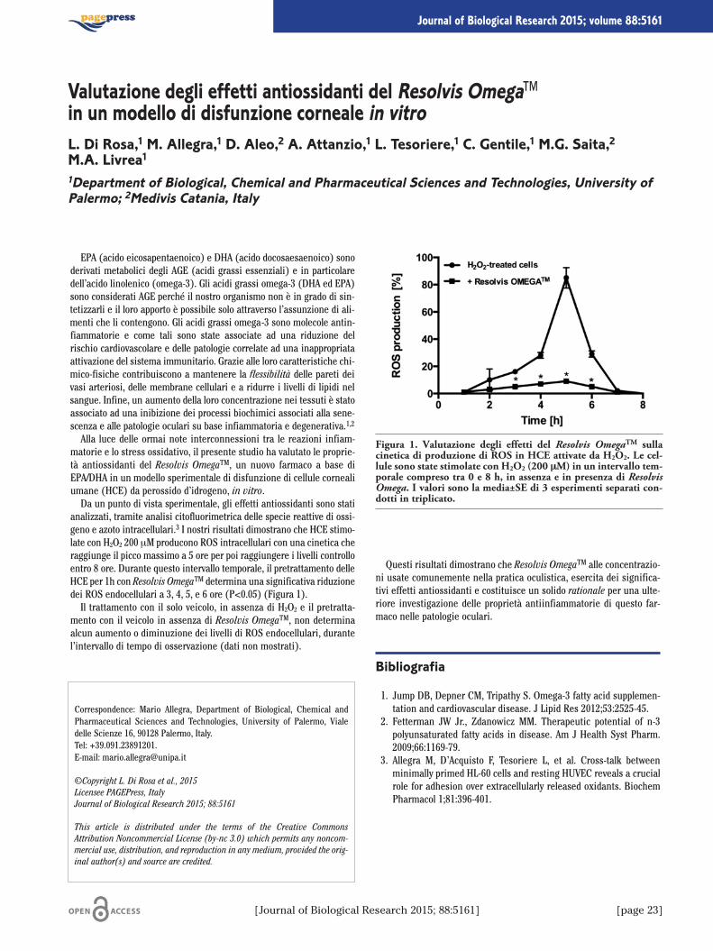
EPA (acido eicosapentaenoico) e DHA (acido docosaesaenoico) sonoderivati metabolici degli AGE (acidi grassi essenziali) e in particolaredell’acido linolenico (omega-3). Gli acidi grassi omega-3 (DHA ed EPA)sono considerati AGE perché il nostro organismo non è in grado di sin-tetizzarli e il loro apporto è possibile solo attraverso l’assunzione di ali-menti che li contengono. Gli acidi grassi omega-3 sono molecole antin-fiammatorie e come tali sono state associate ad una riduzione delrischio cardiovascolare e delle patologie correlate ad una inappropriataattivazione del sistema immunitario. Grazie alle loro caratteristiche chi-mico-fisiche contribuiscono a mantenere la flessibilità delle pareti deivasi arteriosi, delle membrane cellulari e a ridurre i livelli di lipidi nelsangue. Infine, un aumento della loro concentrazione nei tessuti è statoassociato ad una inibizione dei processi biochimici associati alla sene-scenza e alle patologie oculari su base infiammatoria e degenerativa.1,2
Alla luce delle ormai note interconnessioni tra le reazioni infiam-matorie e lo stress ossidativo, il presente studio ha valutato le proprie-tà antiossidanti del Resolvis OmegaTM, un nuovo farmaco a base diEPA/DHA in un modello sperimentale di disfunzione di cellule cornealiumane (HCE) da perossido d’idrogeno, in vitro.Da un punto di vista sperimentale, gli effetti antiossidanti sono stati
analizzati, tramite analisi citofluorimetrica delle specie reattive di ossi-geno e azoto intracellulari.3 I nostri risultati dimostrano che HCE stimo-late con H2O2 200 μM producono ROS intracellulari con una cinetica cheraggiunge il picco massimo a 5 ore per poi raggiungere i livelli controlloentro 8 ore. Durante questo intervallo temporale, il pretrattamento delleHCE per 1h con Resolvis OmegaTM determina una significativa riduzionedei ROS endocellulari a 3, 4, 5, e 6 ore (P<0.05) (Figura 1).Il trattamento con il solo veicolo, in assenza di H2O2 e il pretratta-
mento con il veicolo in assenza di Resolvis OmegaTM, non determinaalcun aumento o diminuzione dei livelli di ROS endocellulari, durantel’intervallo di tempo di osservazione (dati non mostrati).
Questi risultati dimostrano che Resolvis OmegaTM alle concentrazio-ni usate comunemente nella pratica oculistica, esercita dei significa-tivi effetti antiossidanti e costituisce un solido rationale per una ulte-riore investigazione delle proprietà antiinfiammatorie di questo far-maco nelle patologie oculari.
Bibliografia
1. Jump DB, Depner CM, Tripathy S. Omega-3 fatty acid supplemen-tation and cardiovascular disease. J Lipid Res 2012;53:2525-45.
2. Fetterman JW Jr., Zdanowicz MM. Therapeutic potential of n-3polyunsaturated fatty acids in disease. Am J Health Syst Pharm.2009;66:1169-79.
3. Allegra M, D’Acquisto F, Tesoriere L, et al. Cross-talk betweenminimally primed HL-60 cells and resting HUVEC reveals a crucialrole for adhesion over extracellularly released oxidants. BiochemPharmacol 1;81:396-401.
Correspondence: Mario Allegra, Department of Biological, Chemical andPharmaceutical Sciences and Technologies, University of Palermo, Vialedelle Scienze 16, 90128 Palermo, Italy.Tel: +39.091.23891201.E-mail: [email protected]
©Copyright L. Di Rosa et al., 2015Licensee PAGEPress, ItalyJournal of Biological Research 2015; 88:5161
This article is distributed under the terms of the Creative CommonsAttribution Noncommercial License (by-nc 3.0) which permits any noncom-mercial use, distribution, and reproduction in any medium, provided the orig-inal author(s) and source are credited.
Valutazione degli effetti antiossidanti del Resolvis Omega™in un modello di disfunzione corneale in vitroL. Di Rosa,1 M. Allegra,1 D. Aleo,2 A. Attanzio,1 L. Tesoriere,1 C. Gentile,1 M.G. Saita,2M.A. Livrea11Department of Biological, Chemical and Pharmaceutical Sciences and Technologies, University ofPalermo; 2Medivis Catania, Italy
[Journal of Biological Research 2015; 88:5161] [page 23]
Journal of Biological Research 2015; volume 88:5161
Figura 1. Valutazione degli effetti del Resolvis OmegaTM sullacinetica di produzione di ROS in HCE attivate da H2O2. Le cel-lule sono state stimolate con H2O2 (200 μM) in un intervallo tem-porale compreso tra 0 e 8 h, in assenza e in presenza di ResolvisOmega. I valori sono la media±SE di 3 esperimenti separati con-dotti in triplicato.

The 1997 FAO/WHO Expert Consultation suggested that theGlycaemic Index (GI) might provide an useful help to select the mostappropriate carbohydrate-containing foods in order to reduce severaldiseases. The glycaemic response to food, which in turn affects theinsulin response, depends on the rate of gastric emptying, digestionand absorption of carbohydrate from the small intestine, as well as onthe effects of the other food factors to potentiate non-glucose mediatedinsulin secretion. A range of food factors has been identified as impor-tant determinant of the glycaemic response to carbohydrate foods.Therefore, different food products or composition of meals with thesame amount and type of carbohydrates show differences in glycaemicand insulinemic responses.In this view, the aim of this research was to identify the technolog-
ical factors which influence the glycaemic index (GI) in bread making
of reground semolina and wholemeal flour. Five ancient accessions(Russello, Timilia, Bidì, Biancolilla and Senatore Cappelli) were com-pared to five modern commercial blends and an ancient mix betweenTimilia and Russello accessions under technological and nutritionalprofiles. The parameters under study were: moisture, protein, totalstarch, ash, farinographic profile, wet and dry gluten, gluten index, �-amylase activity, particle size and damaged starch.Our results showed the best attitude of wholemeal flour by old acces-
sions in a health point of view, due to the higher gluten content, poor qual-ity of gluten and greater absorption of water. On the contrary, modern vari-eties of reground semolina are more suitable for breadmaking, owing tothe higher technological attitude and the greater alpha-amylase activity.This work is considered the first step of a PhD project conducted
within the University of Foggia.
Correspondence: Nathascia Amato, Department of Agricultural, Food andEnvironment Sciences, University of Foggia, via Napoli 25, 71100 Foggia,Italy. E-mail: [email protected]
©Copyright N. Amato et al., 2015Licensee PAGEPress, ItalyJournal of Biological Research 2015; 88:5161
This article is distributed under the terms of the Creative CommonsAttribution Noncommercial License (by-nc 3.0) which permits any noncom-mercial use, distribution, and reproduction in any medium, provided the orig-inal author(s) and source are credited.
Carbohydrate digestibility on wheat durum bread:preliminary hypotheses on raw materials roleN. Amato,1 S. Lombardo,2 G. Pandino,2 G. Russo31Department of Agricultural, Food and Environment Sciences, University of Foggia; 2Departmentof Agricultural and Food Production Sciences, University of Catania; 3Reserach Consortium G.P.Ballatore, Palermo, Italy
[page 24] [Journal of Biological Research 2015; 88:5161]
Journal of Biological Research 2015; volume 88:5161
Photodynamic therapy (PDT) is a promising therapeutic option inthe management of cancer and other diseases. The mechanism of pho-todynamic therapy is based on the selective combination of non-toxicdyes or photosensitizers with harmless visible light which, in the pres-ence of oxygen, leads to the generation of cytotoxic species and conse-quently to cell apoptotic or necrotic death.1 Since in clinical PDT it iscommon practice to irradiate wider fields that include healthy tissueswhich might contain microscopic malignant foci, the photosensitizerselectivity (target versus healthy tissue) become crucial. Otherwise ahigh concentration of photosensitizer is required to ensure enoughconcentration at the tumor site, with consequent incidental toxicity inhealthy tissue and increased treatment cost. Both side effects andtreatment costs could be significantly reduced by targeted delivery ofthe photosensitizer to the region of interest at a well-defined time.In this work we use Photosens (a mixture of sulfonated aluminum
phthalocyanines AlPcSn, with n=2, 3 or 4) as photosensitizing drug2
and calcium carbonate (CaCO3) as delivery system. CaCO3 exists inthree different anhydrous crystalline polymorphs: calcite, aragonite,and vaterite. Vaterite particles have a large porosity, large surfacearea, and can decompose fast under relatively mild conditions. Vateriteis the least stable phase of CaCO3 since it slowly dissolves and recrys-tallizes to form calcite in contact with water. Previous studiesdescribed the possibility of synthesizing spherical mono-dispersedvaterite particles in the size range from 2 to 10 μm3 and could veryrecently be downscaled to 400 nm4 which will strongly improve the cel-lular uptake efficiency. A controllable release mechanism based on acrystal phase transition has recently been demonstrated.5,6
Preliminary tests have been performed to assess the particles cell tox-icity, cell uptake and drug release.5 Also, as a proof of principle, the pHsensitivity of the delivery system has been demonstrated,7 as well asthe possibility to be used as a sensor platform.8
The release mechanism depending on the surrounding pH has beenstudied, showing a fast degradation of the carriers in buffers below pH
7. These results hold out the prospect of a novel photodynamic therapydrug delivery system. Variations of particle size or additional coatingsallow to custom-design workload release curves. An intrinsic cancer-sensitivity can be expected from the pH-dependent release in theacidic microenvironment of cancer tissue.During our work, we performed a detailed study of the cumulative
release and recrystallization process in different media and in the pHrange from 3 to 7. Modification of particle size and pH will allow cus-tomizing the workload release curves. A drug delivery system with pH-controlled release promises intracellular delivery with a high selectiv-ity to cancer cells. In combination with an otherwise poorly selectivephotosensitizer, this could become a strong cancer-therapeutic tool,where the carrier degradability could be tuned to control the rate ofdrug release. We investigated the encapsulation efficiency for the pho-tosensitizer in micrometer- and sub- micrometer-sized carriers.Release mechanism dependent on the surrounding pH was studied,showing a fast degradation of the carriers in buffers below pH 7. These results hold out the prospect of a novel drug delivery system.
Cancer-sensitivity can be achieved due to the enhanced uptake andfast release in the low pH endocytic vesicles of viable cancer cells.
References
1. DeRosa M, Crutchley R. Photosensitized singlet oxygen and itsapplications. Coord Chem Rev 2002;233-234:351-71.
2. EA Lukyanets. Phthalocyanines as photosensitizers in the photody-namic therapy of cancer J Porphyr Phthalocyanines 1999;3:424-32.
3. Schmidt S, Volodkin DV. Microparticulate biomolecules by mildCaCO3 templating. J Mater Chem B 2013;1:1210.
4. Parakhonskiy BV, Haase A, Antolini R. Sub-micrometer vateritecontainers: synthesis, substance loading, and release. AngewChem Int Ed Engl 2012;51:1195-7.
5. Parakhonskiy BV, Foss C, Carletti E, et al. Tailored intracellulardelivery via a crystal phase transition in 400 nm vaterite particles.Biomater Sci 2013;1:1273-81
6. Parakhonskiy BV, Tessarolo F, Haase A, Antolini R. Dependence ofsub-micron vaterite container release properties on pH and ionicstrength of the surrounding solution. Adv Sci Technol 2012;86:81-5.
7. Svenskaya Y, Parakhonskiy BV, Haase A, et al. Anticancer drugdelivery system based on calcium carbonate particles loaded witha photosensitizer. Biophys Chem 2013;182:11-5.
8. Yashchenok AM, Borisova D, Parakhonskiy BV, et al. Nanoplasmonicsmooth silica versus porous calcium carbonate bead biosensors fordetection of biomarkers. Ann Phys 2012;524:723-32.
Correspondence: Renzo Antolini, Department of Physics, University ofTrento, via Sommarive 14, 38123 Trento, Italy.E-mail: [email protected]
©Copyright B. Parakhonskiy et al., 2015Licensee PAGEPress, ItalyJournal of Biological Research 2015; 88:5161
This article is distributed under the terms of the Creative CommonsAttribution Noncommercial License (by-nc 3.0) which permits any noncom-mercial use, distribution, and reproduction in any medium, provided the orig-inal author(s) and source are credited.
Anticancer drug delivery system based on vaterite particlesB. Parakhonskiy,1,2 Y. Svenskaya,1,3 A. Haase,4 E. Lukyanets,5 R. Antolini41BIOtech Center Department of Industrial Engineering, University of Trento, Mattarello, Italy;2AV Shubnikov Institute of Crystallography, Russian Academy of Science, Moscow, Russia;3Saratov State University, Russia; 4Department of Physics, University of Trento, Italy;5Organic Intermediates and Dyes Institute, Moscow, Russia
[Journal of Biological Research 2015; 88:5161] [page 25]
Journal of Biological Research 2015; volume 88:5161
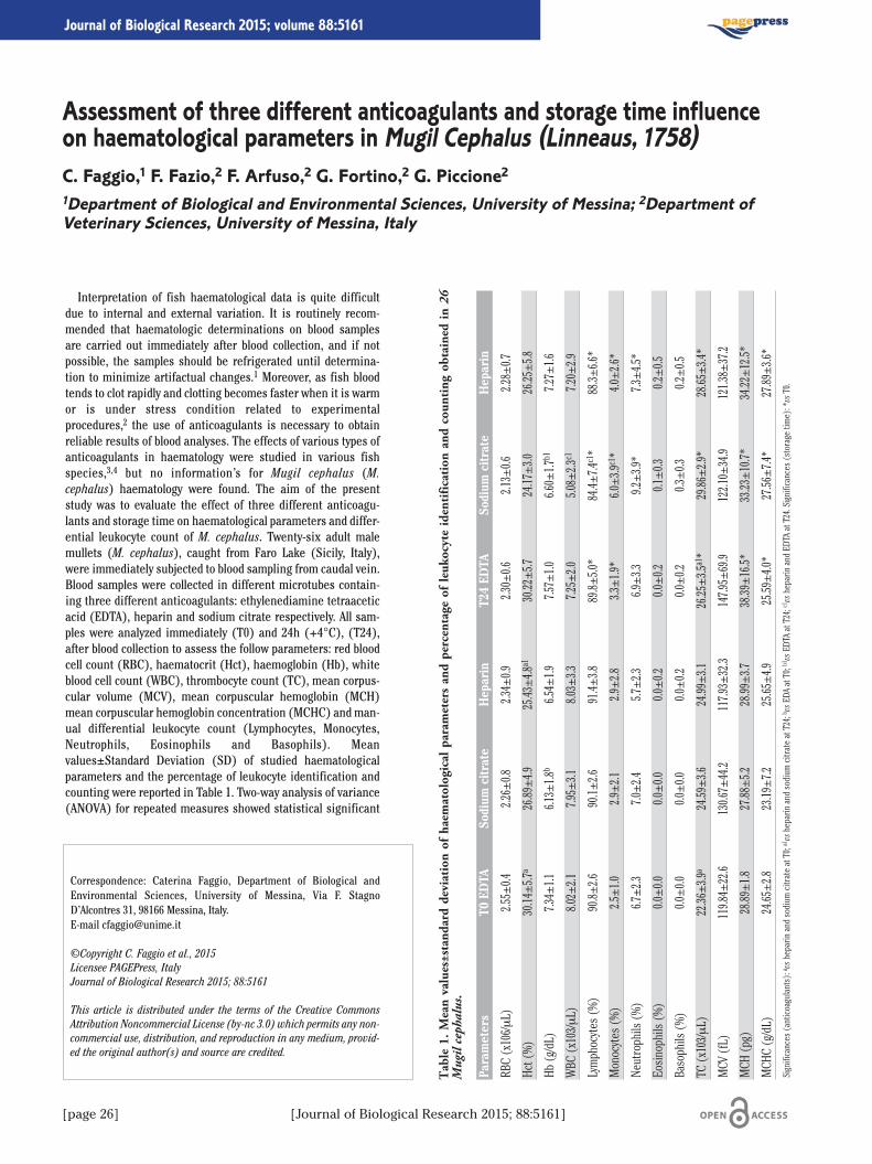
Interpretation of fish haematological data is quite difficultdue to internal and external variation. It is routinely recom-mended that haematologic determinations on blood samplesare carried out immediately after blood collection, and if notpossible, the samples should be refrigerated until determina-tion to minimize artifactual changes.1 Moreover, as fish bloodtends to clot rapidly and clotting becomes faster when it is warmor is under stress condition related to experimentalprocedures,2 the use of anticoagulants is necessary to obtainreliable results of blood analyses. The effects of various types ofanticoagulants in haematology were studied in various fishspecies,3,4 but no information’s for Mugil cephalus (M.cephalus) haematology were found. The aim of the presentstudy was to evaluate the effect of three different anticoagu-lants and storage time on haematological parameters and differ-ential leukocyte count of M. cephalus. Twenty-six adult malemullets (M. cephalus), caught from Faro Lake (Sicily, Italy),were immediately subjected to blood sampling from caudal vein.Blood samples were collected in different microtubes contain-ing three different anticoagulants: ethylenediamine tetraaceticacid (EDTA), heparin and sodium citrate respectively. All sam-ples were analyzed immediately (T0) and 24h (+4°C), (T24),after blood collection to assess the follow parameters: red bloodcell count (RBC), haematocrit (Hct), haemoglobin (Hb), whiteblood cell count (WBC), thrombocyte count (TC), mean corpus-cular volume (MCV), mean corpuscular hemoglobin (MCH)mean corpuscular hemoglobin concentration (MCHC) and man-ual differential leukocyte count (Lymphocytes, Monocytes,Neutrophils, Eosinophils and Basophils). Meanvalues±Standard Deviation (SD) of studied haematologicalparameters and the percentage of leukocyte identification andcounting were reported in Table 1. Two-way analysis of variance(ANOVA) for repeated measures showed statistical significant
Correspondence: Caterina Faggio, Department of Biological andEnvironmental Sciences, University of Messina, Via F. StagnoD’Alcontres 31, 98166 Messina, Italy. E-mail [email protected]
©Copyright C. Faggio et al., 2015Licensee PAGEPress, ItalyJournal of Biological Research 2015; 88:5161
This article is distributed under the terms of the Creative CommonsAttribution Noncommercial License (by-nc 3.0) which permits any non-commercial use, distribution, and reproduction in any medium, provid-ed the original author(s) and source are credited.
Assessment of three different anticoagulants and storage time influenceon haematological parameters in Mugil Cephalus (Linneaus, 1758)C. Faggio,1 F. Fazio,2 F. Arfuso,2 G. Fortino,2 G. Piccione21Department of Biological and Environmental Sciences, University of Messina; 2Department ofVeterinary Sciences, University of Messina, Italy
[page 26] [Journal of Biological Research 2015; 88:5161]
Journal of Biological Research 2015; volume 88:5161
Table 1. M
ean values±standard deviation of haematological parameters and percentage of leukocyte identification and counting obtained in 26
Mugil ceph
alus.
Parameters
T0 EDTA
Sodium
citrate
Heparin
T24 ED
TASo
dium
citrate
Heparin
RBC (x106/μL)
2.55±0.4
2.26±0.8
2.34±0.9
2.30±0.6
2.13±0.6
2.28±0.7
Hct (%)
30.14±5.7
a26.89±4.9
25.43±4.8
a130.22±5.7
24.17±3.0
26.25±5.8
Hb (g/dL)
7.34±1.1
6.13±1.8
b6.54±1.9
7.57±1.0
6.60±1.7
b17.27±1.6
WBC (x103/μL)
8.02±2.1
7.95±3.1
8.03±3.3
7.25±2.0
5.08±2.3
c17.20±2.9
Lymphocytes (%)
90.8±
2.690.1±
2.691.4±
3.889.8±
5.0*
84.4±
7.4c1*
88.3±
6.6*
Monocytes (%)
2.5±1.0
2.9±2.1
2.9±2.8
3.3±1.9*
6.0±3.9
c1*
4.0±2.6*
Neutrophils (%)
6.7±2.3
7.0±2.4
5.7±2.3
6.9±3.3
9.2±3.9*
7.3±4.5*
Eosinophils (%)
0.0±0.0
0.0±0.0
0.0±0.2
0.0±0.2
0.1±0.3
0.2±0.5
Basophils (%)
0.0±0.0
0.0±0.0
0.0±0.2
0.0±0.2
0.3±0.3
0.2±0.5
TC (x103/μL)
22.36±3.9
a24.59±3.6
24.99±3.1
26.25±3.5
a1*
29.86±2.9*
28.65±3.4*
MCV (fL)
119.84±22.6
130.67±44.2
117.93±32.3
147.95±69.9
122.10±34.9
121.38±37.2
MCH (pg)
28.89±1.8
27.88±5.2
28.99±3.7
38.39±16.5*
33.23±10.7*
34.22±12.5*
MCHC (g/dL)
24.65±2.8
23.19±7.2
25.65±4.9
25.59±4.0*
27.56±7.4*
27.89±3.6*
Significances (anticoagulants): avsheparin and sodium citrate at T0; a1vsheparin and sodium citrate at T24; bvsEDA at T0; b1
vsEDTA at T24; c1 vsheparin and EDTA at T24. Significances (storage time): *
vsT0.
effects of different anticoagulants and storage time for some parame-ters studied. In particular, statistical analysis showed significant effectof anticoagulants on Hct, Hb, TC (P<0.001), WBC (P<0.05),Lymphocytes and Monocytes (P<0.005), and significant effects of stor-age time (T0 and T24) on MCH, MCHC, TC, WBC, Lymphocytes,Monocytes and Neutrophils (P=0.005). By the results of this study, itwas found that haematological parameters and differential leukocytecount of M. cephalus were influenced by anticoagulants used and that,according to our previous study,5 haematologic determinations shouldbe assessed within 24 hours after collection, because long-term storagemodifies the results of the analysis. All haematological parameters val-ues obtained in EDTA, heparin and sodium citrate treated samples atT0 fall within values obtained in our previous study on M. cephalus5 andleukocytes differential count of the samples treated with the three anti-coagulants were within fish reference range.6 Thus, haematologicdetermination and leukocyte differential count showed no cellularalteration depending on used anticoagulants. However studied param-eters showed a higher reliability using EDTA as anticoagulant.Comparison of literature data indicates that modifications of bloodparameters induced by internal or/and external factors, such as antico-agulants and storage time, could represent a species-specific respons-es. Further studies designed specifically to investigate the impact ofdifferent anticoagulants and storage times on these parameters couldbe still needed in various fish species to validate an appropriate methodfor haematological analysis useful for the evaluation of the health sta-tus of animal living in captivity and in aquaculture.
References
1. Wood BL, Andrews J, Miller S, Sabbath DE. Refrigerated storageimproves the stability of complete blood count and automated dif-ferential. Am J Clin Pathol 1999;112:687-95.
2. Korcock DE, Houston AH, Gray JD. Effects of sampling conditionson selected blood variables of rainbow trout, Salmo gairdneriRichardson. J Fish Biol 1988;33:319-330.
3. Walencik J, Witeska M. The effects of anticoagulants on hematolog-ical indices and blood cell morphology of common carp (Cyprinuscarpio L.). Comp Biochem Physiol Part C 2007;146:331-5.
4. Ekanem AP, Udoh AJ, Inyang-Etoh AP. Effect of different anticoagu-lants on hematological parameters of Oreochromis Niloticus. Int JSci Advan Technol 2012;2:17-20.
5. Faggio C, Casella S, Arfuso F, et al. Effect of storage time on haema-tological parameters in mullet, Mugil cephalus. Cell Biochem Funct2013;31:412-6.
6. Smith C. Hematology and cell type differentiation. In: Mumford S,Heidel J, Smith C, Morrison J, MacConnell B, Blazer V, eds. Fishhistology and histopathology. USFWS-NCTC; 2007. pp 1-12.
[Journal of Biological Research 2015; 88:5161] [page 27]
Article
Macroalgae show a broad range of applications and their importancein several sectors is steadily increasing worldwide. Aquaculture pro-duced 15.8 million tonnes of aquatic plants, with a total estimated valueof US$ 7.4 billion.1 About 47% of the total production is used for humanconsumption, 43% in the extraction of colloids, 7% in the production ofmaërl and the remaining 3% in the fields of pharmacology, cosmetics,agriculture and waste water treatments from aquaculture, sewage, agri-cultural and industrial run-off. Algal biomass are also employed in thefield of renewable energies for the production of biogas. Among alterna-tive energies, the solar photovoltaic technology is one such source thatcan looked up to as vast research is being carried out and a significantimprovement in performance has been achieved. However, macroalgaeare an underexploited resource in the photovoltaic technologies.2,3
Dye-sensitized nanocrystalline solar cells (DSSCs) has become animportant topic in solar cell research. DSSCs was inspired by the ener-gy and electron transfer mechanisms in natural photosynthesis, andtheir are based on the photosensitization of nanocrystalline TiO2semiconductor electrodes by dyes.4 In nature, some fruits, flowers andleaves contain several pigments that can be easily extracted andemployed in DSSCs. Many reports have showed that chlorophyll, whichacts as an effective photosensitizer in photosynthesis, has the poten-tial to be an environment friendly dye sources.5-7 Presently, macroalgaeare an underexploited resource in comparison to crop plants.In the present study, we utilized chlorophylls from samples of the
brown alga Undaria pinnatifida as sensitizer in DSSCs to investigatethe light to electron conversion efficiency. Samples of U. pinnatifidawere collected in Venice Lagoon (45°26’ N; 12°20’ E); chlorophyllswere extracted by treatment with acetone, according to the protocol ofWang et al.3 The dye,extracted by frozen seaweeds and used without any chemical
purification, showed a very good fill factor (0.69). Even the photelec-trochemical parameters if compared with the existent literature arevery interesting.
U. pinnatifida is a highly invasive species and has caused concern allover the world because it has invaded coastal environments, has thepotential to displace native species, significantly alters habitat for asso-ciated fauna, and disturbs navigation. An exploitation of U. pinnatifidawould result in the conversion of a waste into a valuable biomass.
References
1. FAO. The state of world fisheries and aquaculture 2012. FAO:Rome; 2012.
2. Kuo C-G, Sheen B-J. Seaweed chlorophyll on the light-electronefficiency of DSSC. Journal of the Chinese Chemical Society2011;58:186-90.
3. Wang X-F, Zhan C-H, Maoka T, et al. Fabrication of dye-sensitizedsolar cells using chlorophylls c1 and c2 and their oxidized formsc’1 and c’2 from Undaria pinnatifida (Wakame). Chemical PhysicsLetters 2007;447:79-85.
4. Grätzel M. Dye-sensitized solar cells. Journal of Photochemistryand Photobiology C: Photochemistry Reviews 2003;4:145-53.
5. Calogero G, Di Marco G, Caramori S, et al. Natural dye senstizersfor photoelectrochemical cells. Energy & Environmental Science2009;2:1162-72.
6. Calogero G, Di Marco G, Cazzanti S, et al. Efficient dye-sensitizedsolar cells using red turnip and purple wild Sicilian prickly pearfruits. International Journal of Molecular Sciences 2010;11:254-67.
7. Narayan MR. Review: dye sensitized solar cells based on naturalphotosensitizers. Renewable and Sustainable Energy Reviews2012;16:208-15.
Correspondence: Simona Armeli Minicante, Department of EnvironmentalSciences, Informatics and Statistics, University Ca’ Foscari, Dorsoduro 2137,30123 Venice, Italy. E-mail: [email protected]
©Copyright S. Armeli Minicante et al., 2015Licensee PAGEPress, ItalyJournal of Biological Research 2015; 88:5161
This article is distributed under the terms of the Creative CommonsAttribution Noncommercial License (by-nc 3.0) which permits any noncom-mercial use, distribution, and reproduction in any medium, provided the orig-inal author(s) and source are credited.
Algal pigments as dye sources in the solar photovoltaic technologyS. Armeli Minicante,1 I. Citro,2 M. Morabito,3 G. Genovese,3 G. Calogero21Department of Environmental Sciences, Informatics and Statistics, University Ca’ Foscari, Venice;2CNR-IPCF Institute for Chemical-Physical Processes, Messina; 3Department of Biological andEnvironmental Sciences, University of Messina, Italy
[page 28] [Journal of Biological Research 2015; 88:5161]
Journal of Biological Research 2015; volume 88:5161
Circadian rhythms represent an inherent property of living organ-isms that seem to guarantee an optimal functioning of the biologicalsystem, with maximum efficiency, performance and welfare.1 In mam-mals, a master clock located in the suprachiasmatic nuclei (SCN) ofthe hypothalamus adjusts the timing of other self-sustained oscillatorsin the brain and peripheral organs.2 In most species, the daily light-dark (L/D) cycle is the primary environmental stimulus (Zeitgeber) forthe entrainment of the SCN pacemaker. The SCN receives light infor-mation from the retina and regulates several physiological processesby synchronizing molecular clockwork mechanisms consisted by a coregroup of clock genes in each cell.3 Among all physiological processes,the total locomotor activity (TLA) is one of the most susceptible to theL/D cycle. Light acutely suppresses locomotor activity in nocturnal(night active) animals such as rats and owls but promotes activity indiurnal (day active) animals like dogs and eagles.4 Since animals havea species-typical organization of activity patterns,5 the aim of thisstudy was to compare the TLA in small and large domestic animals likerabbits, cats, dogs, goats, sheep, cows, donkeys and horses. Five clini-cally healthy female subjects from eight different species: rabbits(body weight 2.5±0.2 kg), cats (body weight 4.5±0.3 kg), dogs (bodyweight 13.5±1 kg), goats (body weight 40±2 kg), sheep (body weight45±2 kg), cows (body weight 390±10 kg), donkeys (body weight395±20) and horses (body weight 565±42) were enrolled in the studywith owners consent. Animals were housed under natural photoperiod(March) 12:12 hours L/D cycle (5.30 am sunrise, 5.30 pm sunset)according to specific farm management, except for cats and dogs thatlived outdoors. Water was available ad libitum and feeding was suit-able for each species. Total activity pattern was recorded for 10 daysusing actigraphy-based data loggers Actiwatch-Mini (CambridgeNeurotechnology Ltd, UK) placed on each animal through collars orhalters according to the species. Activity was monitored with a sam-pling interval of 5 minutes. Total daily amount of activity, mount ofactivity during the photophase and the scotophase were calculated
using Actiwatch Activity Analysis 5.06 (Cambridge NeurotechnologyLtd, UK). The Cosine peak of a rhythm (the time of the daily peak) wascomputed by cosinor rhythmometry6 as implement in the ActiwatchActivity Analysis 5.06 program. The temporal resolution of the locomo-tor activity data was reduced to t h bins by the averaging of all 15 datapoints within each 1 h bin to apply the statistical analysis. To analyzethe locomotor activity a trigonometric statistical model was applied toeach time series to statistically describe the periodic phenomenon, bycharacterizing the main rhythmic parameters according to the singlecosinor procedure.6 Four rhythmic parameters were determined: meanlevel, amplitude, acrophase (the time at which the peak of a rhythmoccurs), and robustness (strength of rhythmicity). For each animal,the mean level of the rhythm was computed as the arithmetic mean ofall values in the data set (24 data points). The amplitude of the rhythmwas calculated as half the maximum-minimum range of the oscilla-tion, which was computed as the difference between peak and trough.Robustness was computed as the percentage of the maximal scoreattained by the chi-square periodogram statistic for ideal data sets ofcomparable size and 24-h periodicity.7 Two-way analysis of variance(ANOVA) was used for the assessment of effects due to species anddays on the daily amount of activity per 24 h. Statistical analysisshowed significant differences among domestic species. The highestdaily amount of activity was observed during the photophase(p<0.0001) in dogs, sheep, goats, cows, donkeys and horses, and dur-ing the scotophase (p<0.0001) in rabbits and cats. Our results showdifferent pattern of locomotor activity in every domestic species(Figure 1), underlining a diurnal pattern of locomotor activity in dogs,goats, sheep, cows, donkeys and horses while rabbits and cats have amain nocturnal pattern. As previously observed by several authors,8-10
our study confirms that locomotor activity exhibits a robust daily rhyth-
Correspondence: Giuseppe Piccione, Department of Veterinary Sciences,University of Messina, Polo Universitario dell'Annunziata, Viale Annunziata,98168 Messina, Italy.E-mail: [email protected]
©Copyright M. Bazzano et al., 2015Licensee PAGEPress, ItalyJournal of Biological Research 2015; 88:5161
This article is distributed under the terms of the Creative CommonsAttribution Noncommercial License (by-nc 3.0) which permits any noncom-mercial use, distribution, and reproduction in any medium, provided the orig-inal author(s) and source are credited.
Daily rhythm of total activity/rest patternin small and large domestic animalsM. Bazzano,1 C. Faggio,2 M. Panzera,1 A. Assenza,1 G. Piccione11Department of Veterinary Sciences, University of Messina; 2Department of Biological andEnvironmental Sciences, University of Messina, Italy
[Journal of Biological Research 2015; 88:5161] [page 29]
Journal of Biological Research 2015; volume 88:5161
Figure 1. Analysis of rhythm parameters in domestic species: a. TotalLocomotor Activity, b. Amplitude, c. Acrophase, d. Robustness.
[page 30] [Journal of Biological Research 2015; 88:5161]
micity during the photophase in dogs, cows and horses, therefore inthese species the rhythm can be poorly affected by external stimuli. Onthe contrary, other domestic species can spontaneously shift from diur-nal to nocturnal activity pattern. Sheep with restricted night time feed-ing can shift the main bout of activity during the night8 or cats, that areconsidered mainly nocturnal, use to loose their rhythm when they livein symbiosis with humans.11 Therefore, the daily pattern of TLA doesnot depend only on L/D cycle but it can be affected by several environ-mental variables including different activities such as feeding, drink-ing, walking, grooming, playing as well as all conscious and uncon-scious movements.
References
1. Weinert D, Waterhouse J. The circadian rhythm of core tempera-ture: Effects of physical activity and aging. Physiol Behav2007;90:246-56.
2. Challet E. Interactions between light, mealtime and calorie restric-tion to control daily timing in mammals. J Comp Physiol B2010;180:631-44.
3. Murphy BA, Martin AM, Elliot JA. Light: dark, circadian, and ultra-
dian regulation of motor activity and skeletal muscle gene expres-sion in the horse. J Equine Vet Sci 2009;29:313-4.
4. Refinetti R. The diversity of temporal niches in mammals. BiolRhythms Res 2008;39,173-92.
5. Giannetto C, Casella S, Caola G, Piccione G. Photic and non-photicentrainment on daily rhythm of locomotor activity in goats. AnimSci J 2010;8:122-8.
6. Nelson W, Tong U, Lee J, Halberg F. Methods for cosinor rhythmom-etry. Chronobiol 1979;6:305-23.
7. Refinetti R. Non-stationary time series and the robustness of circa-dian rhythms. J Theor Biol. 2004; 227, 571-581.
8. Piccione G, Giannetto C, Casella S, Caola G. Daily locomotor activ-ity in five domestic animals. Anim Biol 2010;60:15-24.
9. Siwak CT, Tapp PD, Zicker SC, et al. Locomotor activity rhythms indogs vary with age and cognitive status. Behav Neurosci.2003;117:813-24.
10. Zanghi MB, Kerr W, de Rivera C, et al. Effect of age and feedingschedule on diurnal rest/activity rhythms in dogs. J Vet Behav2013;8:195-203.
11. Piccione G, Marafioti S, Giannetto C, et al. Daily rhythm of totalactivity pattern in domestic cats (Felis silvestris catus) maintainedin two different housing conditions. J Vet Behav 2013;8:189-94.
Article
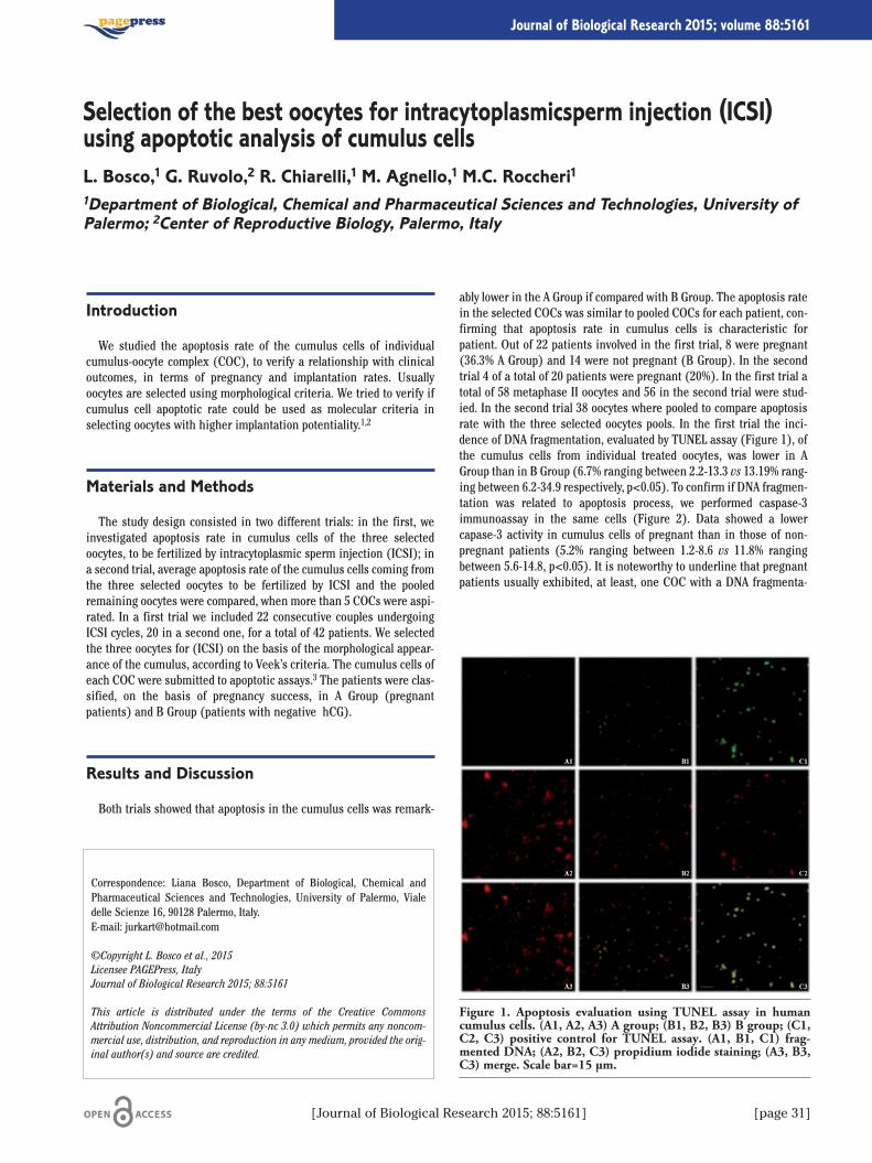
Introduction
We studied the apoptosis rate of the cumulus cells of individualcumulus-oocyte complex (COC), to verify a relationship with clinicaloutcomes, in terms of pregnancy and implantation rates. Usuallyoocytes are selected using morphological criteria. We tried to verify ifcumulus cell apoptotic rate could be used as molecular criteria inselecting oocytes with higher implantation potentiality.1,2
Materials and Methods
The study design consisted in two different trials: in the first, weinvestigated apoptosis rate in cumulus cells of the three selectedoocytes, to be fertilized by intracytoplasmic sperm injection (ICSI); ina second trial, average apoptosis rate of the cumulus cells coming fromthe three selected oocytes to be fertilized by ICSI and the pooledremaining oocytes were compared, when more than 5 COCs were aspi-rated. In a first trial we included 22 consecutive couples undergoingICSI cycles, 20 in a second one, for a total of 42 patients. We selectedthe three oocytes for (ICSI) on the basis of the morphological appear-ance of the cumulus, according to Veek’s criteria. The cumulus cells ofeach COC were submitted to apoptotic assays.3 The patients were clas-sified, on the basis of pregnancy success, in A Group (pregnantpatients) and B Group (patients with negative �hCG).
Results and Discussion
Both trials showed that apoptosis in the cumulus cells was remark-
ably lower in the A Group if compared with B Group. The apoptosis ratein the selected COCs was similar to pooled COCs for each patient, con-firming that apoptosis rate in cumulus cells is characteristic forpatient. Out of 22 patients involved in the first trial, 8 were pregnant(36.3% A Group) and 14 were not pregnant (B Group). In the secondtrial 4 of a total of 20 patients were pregnant (20%). In the first trial atotal of 58 metaphase II oocytes and 56 in the second trial were stud-ied. In the second trial 38 oocytes where pooled to compare apoptosisrate with the three selected oocytes pools. In the first trial the inci-dence of DNA fragmentation, evaluated by TUNEL assay (Figure 1), ofthe cumulus cells from individual treated oocytes, was lower in AGroup than in B Group (6.7% ranging between 2.2-13.3 vs 13.19% rang-ing between 6.2-34.9 respectively, p<0.05). To confirm if DNA fragmen-tation was related to apoptosis process, we performed caspase-3immunoassay in the same cells (Figure 2). Data showed a lowercapase-3 activity in cumulus cells of pregnant than in those of non-pregnant patients (5.2% ranging between 1.2-8.6 vs 11.8% rangingbetween 5.6-14.8, p<0.05). It is noteworthy to underline that pregnantpatients usually exhibited, at least, one COC with a DNA fragmenta-
Correspondence: Liana Bosco, Department of Biological, Chemical andPharmaceutical Sciences and Technologies, University of Palermo, Vialedelle Scienze 16, 90128 Palermo, Italy.E-mail: [email protected]
©Copyright L. Bosco et al., 2015Licensee PAGEPress, ItalyJournal of Biological Research 2015; 88:5161
This article is distributed under the terms of the Creative CommonsAttribution Noncommercial License (by-nc 3.0) which permits any noncom-mercial use, distribution, and reproduction in any medium, provided the orig-inal author(s) and source are credited.
Selection of the best oocytes for intracytoplasmicsperm injection (ICSI)using apoptotic analysis of cumulus cellsL. Bosco,1 G. Ruvolo,2 R. Chiarelli,1 M. Agnello,1 M.C. Roccheri11Department of Biological, Chemical and Pharmaceutical Sciences and Technologies, University ofPalermo; 2Center of Reproductive Biology, Palermo, Italy
[Journal of Biological Research 2015; 88:5161] [page 31]
Journal of Biological Research 2015; volume 88:5161
Figure 1. Apoptosis evaluation using TUNEL assay in humancumulus cells. (A1, A2, A3) A group; (B1, B2, B3) B group; (C1,C2, C3) positive control for TUNEL assay. (A1, B1, C1) frag-mented DNA; (A2, B2, C3) propidium iodide staining; (A3, B3,C3) merge. Scale bar=15 µm.
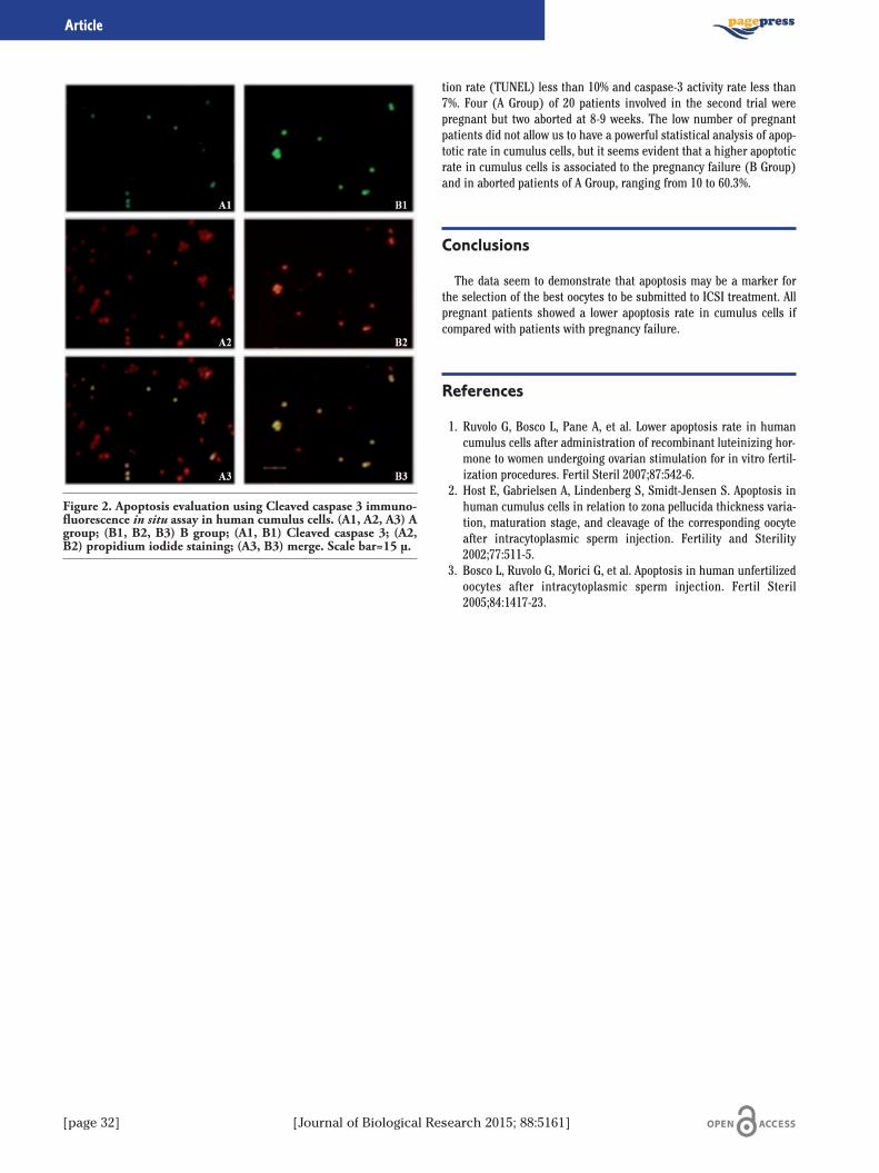
[page 32] [Journal of Biological Research 2015; 88:5161]
tion rate (TUNEL) less than 10% and caspase-3 activity rate less than7%. Four (A Group) of 20 patients involved in the second trial werepregnant but two aborted at 8-9 weeks. The low number of pregnantpatients did not allow us to have a powerful statistical analysis of apop-totic rate in cumulus cells, but it seems evident that a higher apoptoticrate in cumulus cells is associated to the pregnancy failure (B Group)and in aborted patients of A Group, ranging from 10 to 60.3%.
Conclusions
The data seem to demonstrate that apoptosis may be a marker forthe selection of the best oocytes to be submitted to ICSI treatment. Allpregnant patients showed a lower apoptosis rate in cumulus cells ifcompared with patients with pregnancy failure.
References
1. Ruvolo G, Bosco L, Pane A, et al. Lower apoptosis rate in humancumulus cells after administration of recombinant luteinizing hor-mone to women undergoing ovarian stimulation for in vitro fertil-ization procedures. Fertil Steril 2007;87:542-6.
2. Host E, Gabrielsen A, Lindenberg S, Smidt-Jensen S. Apoptosis inhuman cumulus cells in relation to zona pellucida thickness varia-tion, maturation stage, and cleavage of the corresponding oocyteafter intracytoplasmic sperm injection. Fertility and Sterility2002;77:511-5.
3. Bosco L, Ruvolo G, Morici G, et al. Apoptosis in human unfertilizedoocytes after intracytoplasmic sperm injection. Fertil Steril2005;84:1417-23.
Article
Figure 2. Apoptosis evaluation using Cleaved caspase 3 immuno-fluorescence in situ assay in human cumulus cells. (A1, A2, A3) Agroup; (B1, B2, B3) B group; (A1, B1) Cleaved caspase 3; (A2,B2) propidium iodide staining; (A3, B3) merge. Scale bar=15 µ.
Introduction
The social interaction test, introduced by File and Hyde in 1978,1 isa well known behavioral model used to investigate anxiety-relatedbehaviors in rodents. Basic premise of the test is that the behavior ofa rodent influences the one of the other.An increased interaction between the two subjects is indicative of a
reduced anxiety condition, in contrast, a reduced interaction indicatesa higher anxiety. Albeit the test has been largely used to study anxiolyt-ic and neuroleptic drugs, no data are available on the temporal struc-ture of the behavioral interactions. In addition, interesting questionsstill remain unanswered.For instance, is it possible to identify recurring temporal sequences
from the two interacting rodents? If so, do such sequences encompassspecific behavioral events? Could such sequences have an ethologicalmeaning?
Materials and Methods
In this preliminary study, four male Wistar rats (220-250 gr) wereused. Each subject was housed in a thermo-regulated room. The dayof the experiment, pairs of rats, randomly taken from housing room,were placed in an open field apparatus (50 x 50 cm) and observedfor 15 min. The behavior of the animals was recorded with a digital
video camera and video files stored in a personal computer. Using anethogram, obtained on the basis of previous observations,2-5 videofiles were coded by means of a software coder and the obtainedevent log files used for following analyses. Both quantitative andmultivariate t-pattern analyses were carried out. The latter is a mul-tivariate approach based on the utilization of a specific softwarethat, by means of an advanced search algorithm, processes event logfiles evaluating possible significant relationships among the eventsin the course of time.6 Theories, concepts and procedures concern-ing such a multivariate analysis of behavior can be found in our pre-vious articles.6-8
Results
The ethogram is presented in Table 1. The behavioral activities areclassified taking into consideration their characteristics: non social(produced in absence of interactions), and social (produced duringinteractions).Preliminary results, obtained from the analysis of two pairs of
rats, are presented. Per cent distribution, evaluated both for socialand non social activities are illustrated in Figure 1. ISn, Wa, Cl, Re,FPL, Imm and Sh represent 95.50% of the non social behavioralrepertoire; on the other hand, SoS, App, Wit, LeO, SoG and GeS rep-resent 84.50% of the social one. T-pattern analysis demonstrated, inboth pairs of rats, the presence of significant constraints amongnumerous events in the course of time. Figure 2 illustrates a t-pat-tern detected in one pair of rats.
Discussion
The current research represents the first effort to study the tempo-ral structure of social interaction in rats by means of multivariate t-pattern analysis. Per cent distribution (Figure 1) shows that sniffingrelated (ISn, Cl, Re, SoS, GeS) and walking related (Wa, App, Wit)activities (Table 1) are the most represented both in non social andsocial behavior.Coherently, the t-pattern presented in Figure 2, encompassing only
sniffing- and walking-related activities, well depicts the role of thesebehavioral events in moulding the temporal structure of the behavior.Interestingly, some kinds of behavioral symmetries were observed,where a behavior, carried out by one of the rats, was followed by thesame behavior carried out by the other animal (Figure 2).
Correspondence: Giuseppe Crescimanno, Human Physiology Unit,Department of Experimental Biology and Chemical Neurosciences,University of Palermo, Via del Vespro 129, 90127 Palermo, Italy.Tel: +39.091.6555847.E-mail: [email protected]
©Copyright M. Casarrubea et al., 2015Licensee PAGEPress, ItalyJournal of Biological Research 2015; 88:5161
This article is distributed under the terms of the Creative CommonsAttribution Noncommercial License (by-nc 3.0) which permits any noncom-mercial use, distribution, and reproduction in any medium, provided the orig-inal author(s) and source are credited.
Application of multivariate T-pattern analysis in the studyof social interaction in ratsM. Casarrubea,1,2 D. Cancemi,3 A. Cudia,3 M. Cardaci,2,3 F. Sorbera,1 F. Faulisi,1M.S. Magnusson,4 G. Crescimanno1,21Human Physiology Unit, Department of Experimental Biology and Chemical Neurosciences,University of Palermo; 2Department of Psychology, University of Palermo, Italy; 3Human BehaviorLaboratory, University of Iceland, Reykjavik, Iceland; 4Interdepartmental Center of Technologies ofKnowledge, University of Palermo, Italy
[Journal of Biological Research 2015; 88:5161] [page 33]
Journal of Biological Research 2015; volume 88:5161
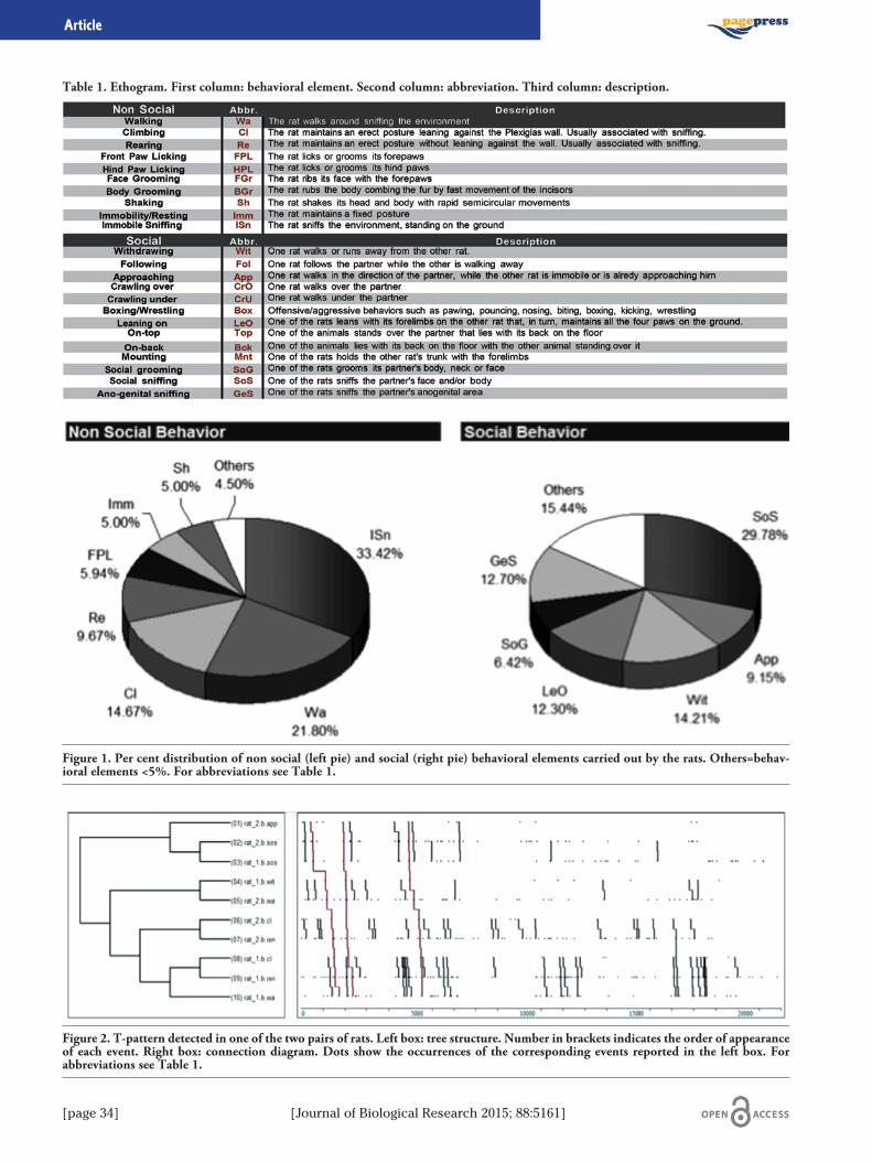
[page 34] [Journal of Biological Research 2015; 88:5161]
Article
Table 1. Ethogram. First column: behavioral element. Second column: abbreviation. Third column: description.
Figure 1. Per cent distribution of non social (left pie) and social (right pie) behavioral elements carried out by the rats. Others=behav-ioral elements <5%. For abbreviations see Table 1.
Figure 2. T-pattern detected in one of the two pairs of rats. Left box: tree structure. Number in brackets indicates the order of appearanceof each event. Right box: connection diagram. Dots show the occurrences of the corresponding events reported in the left box. Forabbreviations see Table 1.
Conclusions
To sum up, preliminary results obtained from t-pattern analysisdemonstrate that the behavior of rats in the social interaction test isextremely complex and structured on the basis of numerous, recurringand statistically significant sequences of events.
References
1. File SE, Hyde JR. Can social interaction be used to measure anxiety? Br J Pharmacol 1978;62:19-24.
2. Spruijt BM. Progressive decline in social attention in aging rats: aninformation-statistical method. Neurobiol Aging 1992;13:145-51.
3. Vanderschuren LJ, Spruijt BM, Hol T, et al. Sequential analysis of
social play behavior in juvenile rats: effects of morphine. BehavBrain Res 1995;72:89-95.
4. Van Den Berg CL, Van Ree JM, Spruijt BM. Sequential analysis ofjuvenile isolation- induced decreased social behavior in the adultrat. Physiol Behav 1999;67:483-8.
5. Yamada-Haga Y. Characteristics of social interaction betweenunfamiliar male rats (Rattus norvegicus): comparison of juvenileand adult stages. J Ethol 2002;20:55-62.
6. Magnusson MS. Discovering hidden time patterns in behavior: t-patterns and their detection. Behav Res Methods Instrum Comput2000;32:93-110.
7. Casarrubea M, Roy V, Sorbera F, et al. Temporal structure of therat’s behavior in elevated plus maze test. Behav Brain Res2013;237:290-9.
8. Casarrubea M, Roy V, Sorbera F, et al. Significant divergencesbetween the temporal structure of the behavior in Wistar and inthe spontaneously more anxious DA/Han strain of rats tested inelevated plus maze. Behav Brain Res 2013;250:166-73.
[Journal of Biological Research 2015; 88:5161] [page 35]
Article
Inflammation is a complex physiological response that requires theactivity of a sophisticated regulatory network involving the activationof specific genes for defense, tissue repair and remodeling.Although transcriptional activation has been shown to be critical
in the regulation of inflammatory genes1 the role of epigenetic phe-nomena in the modulation of the inflammatory response is nowemerging.2 Specifically, it has been recently reported that proinflam-matory stimuli induce DNA demethylation in the interleukin IL-1βpromoter of human articular chondrocytes.3 IL-1β cytokine, amongseveral proinflammatory agents, represents an essential player in theinflammatory conditions of the gut:4 functioning as the strongest sig-nal transduction to NF-kB, IL-1β increases in intestinal paracellularpermeability and over- expression of proinflammatory genes.5 In thistissue, moreover, inflammatory response is crucial to maintain itsstructural integrity and function, thus, alteration and deregulation ofinflammatory pathways contribute to tissue damage and ulceration,and are pivotal factors in the pathogenesis of several inflammatorygut diseases.In the present study we evaluate both wide-ranging and gene-specif-
ic epigenetic changes in the inflammatory response of Caco-2 cells dif-ferentiated into intestinal epithelial cells and exposedto the inflammatory actions of IL-1β. Our results clearly show that
IL-1β induces changes in the DNA methylation either at genome andgene level and that the local methylation changes are induced in twopro-inflammatory genes that are IL-1β-regulated. In particular, weshow that a cell exposure to IL-1β for 24 h induces, in a dose-depen-dent manner, hypomethylation of genomic DNA in respect to untreatedcells. We also observe a reduced DNA methyltransferases activity ofcell lysates obtained from IL-1β that IL-1β treated cells . Finally our
data shows that IL-1β is able to induce hypomethylation of specificCpG sites in IL-6 and IL-8 genes.These preliminary results suggest that IL-1β in intestinal epithelial
cells is able to act as an epigenetic modulator towards the entiregenome and specific genes.Modulation of epigenetic changes in inflammation may provide a
new reading frame of the inflammatory diseases molecular basis.Since epigenetic modifications are potentially reversible, a thoroughunderstanding of these changes during inflammatory response opensopportunities to develop efficient agents for specific targets.
References
1. Medzhitov R, Horng T. Transcriptional control of the inflammatoryresponse. Nat Rev Immunol 2009;9:692-703.
2. Bayarsaihan D. Epigenetic mechanisms in inflammation. J DentRes 2011;90:9-17.
3. Hashimoto K, Oreffo RO, Gibson MB, et al. DNA demethylation atspecific CpG sites in the IL1B promoter in response to inflamma-tory cytokines in human articular chondrocytes. Arthritis Rheum2009;60:3303-13.
4. Ligumsky M, Simon PL, Karmeli F, Rachmilewitz D. Role of inter-leukin 1 in inflammatory bowel disease-enhanced production dur-ing active disease. Gut 1990;31:686-9.
5. Van De Walle J, Hendrickx A, Romier B, et al. Inflammatory param-eters in Caco-2 cells: effect of stimuli nature, concentration, com-bination and cell differentiation. Toxicol In Vitro 2010;24:1441-9.
Correspondence: Fabio Caradonna, Department of Biological, Chemical andPharmaceutical Sciences and Technologies, University of Palermo, Vialedelle Scienze 16, 90128 Palermo, Italy.E-mail: [email protected]
©Copyright I. Schifano et al., 2015Licensee PAGEPress, ItalyJournal of Biological Research 2015; 88:5161
This article is distributed under the terms of the Creative CommonsAttribution Noncommercial License (by-nc 3.0) which permits any noncom-mercial use, distribution, and reproduction in any medium, provided the orig-inal author(s) and source are credited.
IL-1β induces DNA demethylation, at genome level and in specific CpGsites of IL-6 and IL-8 genes in human intestinal epithelial cellsI. Schifano, C. Gentile, F. Naselli, L. Sposito, M. Allegra, L. Tesoriere, M.A. Livrea,F. CaradonnaDepartment of Biological, Chemical and Pharmaceutical Sciences and Technologies, University ofPalermo, Italy
[page 36] [Journal of Biological Research 2015; 88:5161]
Journal of Biological Research 2015; volume 88:5161
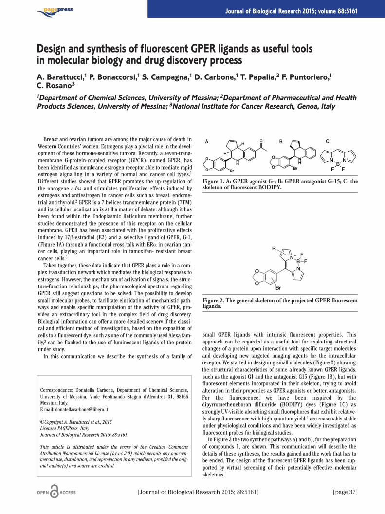
Breast and ovarian tumors are among the major cause of death inWestern Countries’ women. Estrogens play a pivotal role in the devel-opment of these hormone-sensitive tumors. Recently, a seven-trans-membrane G-protein-coupled receptor (GPCR), named GPER, hasbeen identified as membrane estrogen receptor able to mediate rapidestrogen signalling in a variety of normal and cancer cell types.1
Different studies showed that GPER promotes the up-regulation ofthe oncogene c-fos and stimulates proliferative effects induced byestrogens and antiestrogen in cancer cells such as breast, endome-trial and thyroid.2 GPER is a 7 helices transmembrane protein (7TM)and its cellular localization is still a matter of debate: although it hasbeen found within the Endoplasmic Reticulum membrane, furtherstudies demonstrated the presence of this receptor on the cellularmembrane. GPER has been associated with the proliferative effectsinduced by 17β-estradiol (E2) and a selective ligand of GPER, G-1,(Figure 1A) through a functional cross-talk with ERα in ovarian can-cer cells, playing an important role in tamoxifen- resistant breastcancer cells.3
Taken together, these data indicate that GPER plays a role in a com-plex transduction network which mediates the biological responses toestrogens. However, the mechanism of activation of signals, the struc-ture-function relationships, the pharmacological spectrum regardingGPER still suggest questions to be solved. The possibility to developsmall molecular probes, to facilitate elucidation of mechanistic path-ways and enable specific manipulation of the activity of GPER, pro-vides an extraordinary tool in the complex field of drug discovery.Biological information can offer a more detailed scenery if the classi-cal and efficient method of investigation, based on the exposition ofcells to a fluorescent dye, such as one of the commonly used Alexa fam-ily,3 can be flanked to the use of luminescent ligands of the proteinunder study.In this communication we describe the synthesis of a family of
small GPER ligands with intrinsic fluorescent properties. Thisapproach can be regarded as a useful tool for exploiting structuralchanges of a protein upon interaction with specific target moleculesand developing new targeted imaging agents for the intracellularreceptor. We started in designing small molecules (Figure 2) showingthe structural characteristics of some a lready known GPER ligands,such as the agonist G1 and the antagonist G15 (Figure 1B), but withfluorescent elements incorporated in their skeleton, trying to avoidalteration in their properties as GPER agonists or, better, antagonists.For the fluorescence, we have been inspired by thedipyrrometheneboron difluoride (BODIPY) dyes (Figure 1C) asstrongly UV-visible absorbing small fluorophores that exhibit relative-ly sharp fluorescence with high quantum yield,4 are reasonably stableunder physiological conditions and have been widely investigated asfluorescent probes for biological studies.In Figure 3 the two synthetic pathways a) and b), for the preparation
of compounds 1, are shown. This communication will describe thedetails of these syntheses, the results gained and the work that has tobe ended. The design of the fluorescent GPER ligands has been sup-ported by virtual screening of their potentially effective molecularskeletons.
Correspondence: Donatella Carbone, Department of Chemical Sciences,University of Messina, Viale Ferdinando Stagno d'Alcontres 31, 98166Messina, Italy.E-mail: [email protected]
©Copyright A. Barattucci et al., 2015Licensee PAGEPress, ItalyJournal of Biological Research 2015; 88:5161
This article is distributed under the terms of the Creative CommonsAttribution Noncommercial License (by-nc 3.0) which permits any noncom-mercial use, distribution, and reproduction in any medium, provided the orig-inal author(s) and source are credited.
Design and synthesis of fluorescent GPER ligands as useful toolsin molecular biology and drug discovery processA. Barattucci,1 P. Bonaccorsi,1 S. Campagna,1 D. Carbone,1 T. Papalia,2 F. Puntoriero,1C. Rosano31Department of Chemical Sciences, University of Messina; 2Department of Pharmaceutical and HealthProducts Sciences, University of Messina; 3National Institute for Cancer Research, Genoa, Italy
[Journal of Biological Research 2015; 88:5161] [page 37]
Journal of Biological Research 2015; volume 88:5161
Figure 1. A: GPER agonist G-; B: GPER antagonist G-15; C: theskeleton of fluorescent BODIPY.
Figure 2. The general skeleton of the projected GPER fluorescentligands.
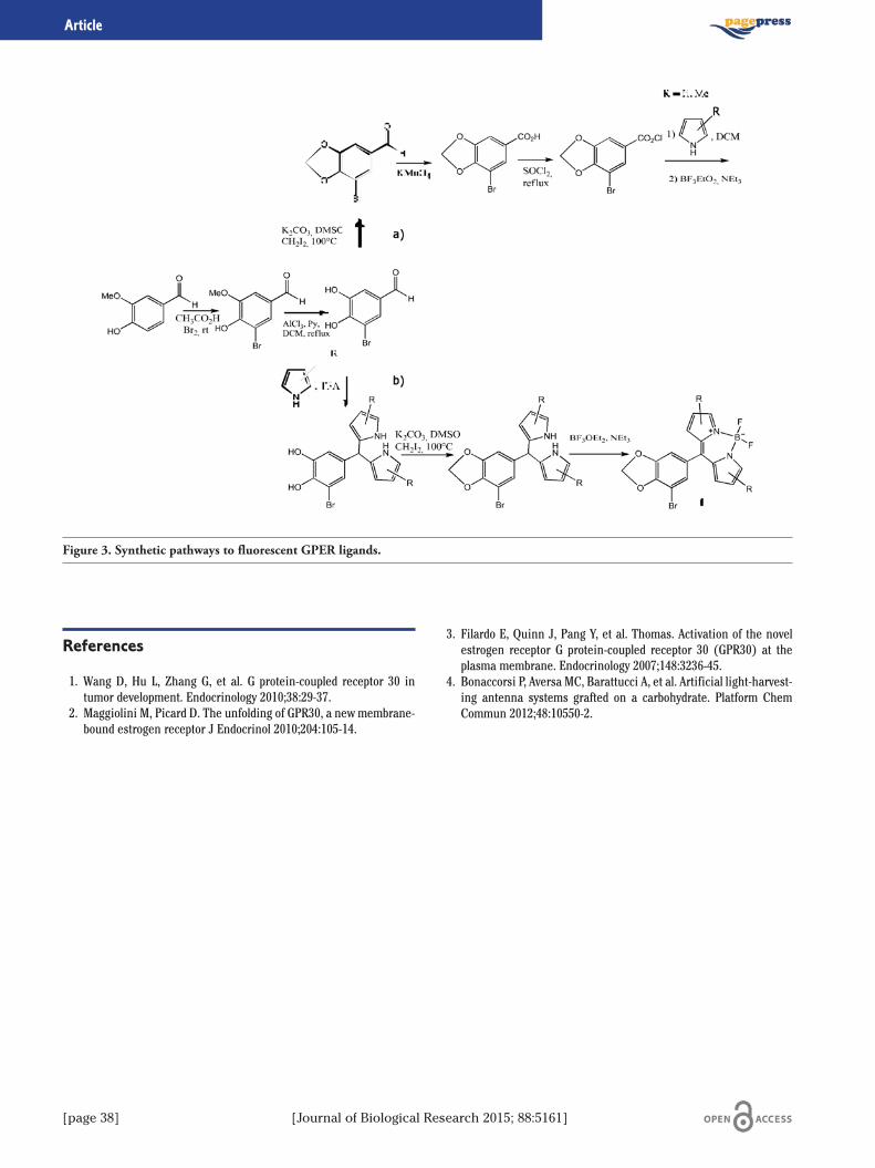
[page 38] [Journal of Biological Research 2015; 88:5161]
References
1. Wang D, Hu L, Zhang G, et al. G protein-coupled receptor 30 intumor development. Endocrinology 2010;38:29-37.
2. Maggiolini M, Picard D. The unfolding of GPR30, a new membrane-bound estrogen receptor J Endocrinol 2010;204:105-14.
3. Filardo E, Quinn J, Pang Y, et al. Thomas. Activation of the novelestrogen receptor G protein-coupled receptor 30 (GPR30) at theplasma membrane. Endocrinology 2007;148:3236-45.
4. Bonaccorsi P, Aversa MC, Barattucci A, et al. Artificial light-harvest-ing antenna systems grafted on a carbohydrate. Platform ChemCommun 2012;48:10550-2.
Article
Figure 3. Synthetic pathways to fluorescent GPER ligands.
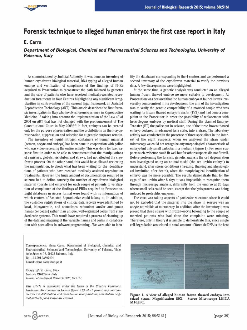
As commissioned by Judicial Authority, it was done an inventory ofhuman cryo-frozen biological material, DNA typing of alleged humanembryo and verification of compliance of the findings of PHRsacquired to Prosecution to reconstruct the path followed by gametesand the care of patients who have received medically-assisted repro-duction treatments in four Centres highlighting any significant irreg-ularities in contravention of the current legal framework on AssistedReproduction Technology (ART). This article describes the first foren-sic investigation in Italy who involves forensic science in ReproductiveMedicine,1,2 taking into account the implementation of the Law 40 of2004 on ART that has not changed with the pronouncement of TheConstitutional Court in May 2009.3-5 In fact, embryos can be createdonly for the purpose of procreation and the prohibitions on their cryop-reservation, suppression and selection for eugenetic purposes remain.The inventory of liquid nitrogen containers of human material
(semen, oocyte and embryo) has been done in cooperation with policewho was video-recording the entire activity. This was done for two rea-sons: first, in order to be able to demonstrate that the manipulationsof canisters, globets, visiotubes and straws, had not affected the cryo-frozen process. On the other hand, this would have allowed reviewingthe manipulation, to check what has been writing for the identifica-tions of patients who have received medically assisted reproductiontreatments. However, the huge amount of documentation required inseizure had to define correctly the number of cryo-frozen biologicalmaterial (oocyte and embryo) for each couple of patients to verifica-tion of compliance of the findings of PHRs acquired to Prosecution.Eight databases in Access format were found with no information ofwhich centres of Assisted Reproductive could belong to. In addition,the customer registrations of clinical data records were identified bylocal, idiosyncratic, and sometimes redundant and/or ambiguousnames (or codes) rather than unique, well-organized codes from stan-dard code systems. This would have required a process of cleaning upof the data and mapping of the variable names and codes in collabora-tion with specialists in software programming . We were able to iden-
tify the databases corresponding to the 4 centers and we performed asecond inventory of the cryo-frozen material to verify the previousdata. A few discrepancies were highlighted.At the same time, a genetic analysis was conducted on an alleged
human frozen thawed embryo no more suitable to development. AtProsecution was declared that the human embryo at four cells was irre-versibly compromised in its development: the aim of the investigationwas to verify the genetic compatibility of a married couple who waswaiting for frozen thawed embryo transfer (FET) and had done a com-plaint to the Prosecutor in order the possibility of replacement withheterologous embryos by medical staff. During the planned Embryo-Transfer (ET) the police put in seizure, one of the three frozen thawedembryo declared in advanced lysis state, into a straw. The laboratoryactivity was conducted in the presence of three specialists in the inter-est of the eight Suspects: when we analyzed the straw undermicroscopy we could not recognize any morphological characteristic ofembryo but only small particles in a medium (Figure 1). For some sus-pects such evidence could fit well but for other suspects did not fit well.Before performing the forensic genetic analysis the cell degenerationwas investigated using an animal model (the sea urchin embryo) tounderstand, after stress conditions (freezing, thawing and physiologi-cal involution after death), when the morphological identification ofembryo was no more possible. The results demonstrate that for theeggs of sea urchin after 8 days it was impossible to recognize themthrough microscopy analysis, differently from the embryo at 20 dayswhere small cells could be seen, except that the lysis process was beinginduced by proteolitic enzymes.The case was taking aspects of particular relevance since it could
not be excluded that the material into the straw in seizure was anoocyte not visible at microscopy. In addition, after the inventory it wasproved that three straws with frozen-oocyte belonging to the couple ofmarried patients who had done the complaint were missing.Therefore, only in theory it is simple to demonstrate this, since singlecell degradation associated to small amount of forensic DNA is the best
Correspondence: Elena Carra, Department of Biological, Chemical andPharmaceutical Sciences and Technologies, University of Palermo, Vialedelle Scienze 16, 90128 Palermo, Italy.Tel: +39.091.23897404. E-mail: [email protected]
©Copyright E. Carra, 2015Licensee PAGEPress, ItalyJournal of Biological Research 2015; 88:5161
This article is distributed under the terms of the Creative CommonsAttribution Noncommercial License (by-nc 3.0) which permits any noncom-mercial use, distribution, and reproduction in any medium, provided the orig-inal author(s) and source are credited.
Forensic technique to alleged human embryo: the first case report in ItalyE. CarraDepartment of Biological, Chemical and Pharmaceutical Sciences and Technologies, University ofPalermo, Italy
[Journal of Biological Research 2015; 88:5161] [page 39]
Journal of Biological Research 2015; volume 88:5161
Figure 1. A view of alleged human frozen thawed embryo intoseized straw. Magnification 80X - Stereo Microscope LEICAM165FC.
[page 40] [Journal of Biological Research 2015; 88:5161]
situation to obtain complex DNA profiles. A method of collecting thealleged embryo residues from the straw has been developed conform-ing to forensic procedure to ensure the repetition of analysis. The sam-ple was collected using Whatman 3MM filter strip paper and was divid-ed in 7 parts under absolute sterile conditions; each strip was extractedusing a performed low copy number (LCN) extraction procedure. Lowcopy number (LCN) typing, particularly for current short tandem repeat(STR) typing, refers to the analysis of any sample that contains lessthan 200 pg of template DNA. Generally, LCN typing simply can bedefined as the analysis of any DNA sample where the results are belowthe stochastic threshold for reliable interpretation.6,7 The extractedDNA was amplified using reduced amplification volume and higherPCR. cycle numbers. Autosomal DNA profiles were obtained from mostof the 3MM strips. These profiles were concordant with the profilesobtained from the couple of married patients proving the presence ofhuman lysis embryo into the seized straw.Random analysis on IVF-ET sheets, Clinical Records, IVF Registers,
etc. combined with the computerization for each type of PHRs of suchrecords: date of pick up, women, man, oocyte, IVF technique, MIIoocyte, semen, embryo, discarded oocyte, discarded embryo, frozen-oocyte, frozen-embryo, embryo-transfer ET, MD, Biologist, clinicalanalysis, note, has permitted to reconstruct the path followed bygametes of patients who had received medically-assisted reproduction
treatments highlighting significant irregularities in contravention ofthe artt. 13 and 14 of the Law 40 of 2004.
References
1. Trounson AO, Wood C. IVF and related technology. The present andthe future. Med J Aust 1993;158:853-7.
2. Shufaro Y, Laufer N. Epigenetic concerns in assisted reproduction:update and critical review of the current literature. Fertil Steril2013;99:605-6.
3. Law 40/2004 - Gazzetta Ufficiale n. 45 del 24 febbraio 2004.4. Fineschi V, Neri M, Turillazzi E. The new Italian law on assistedreproduction technology (Law 40/2004). J Med Ethics 2005;31:536-9.
5. Robertson JA. Protecting embryos and burdening women: assistedreproduction in Italy. Hum Reprod 2004;19:1693-6.
6. Budowle B, Eisenberg AJ, van Daal A. Validity of low copy numbertyping and applications to forensic science. Croat Med J 2009;50:207-17.
7. Gill P. Application of low copy number DNA profiling. Croat Med J2001;42:229-32.
Article
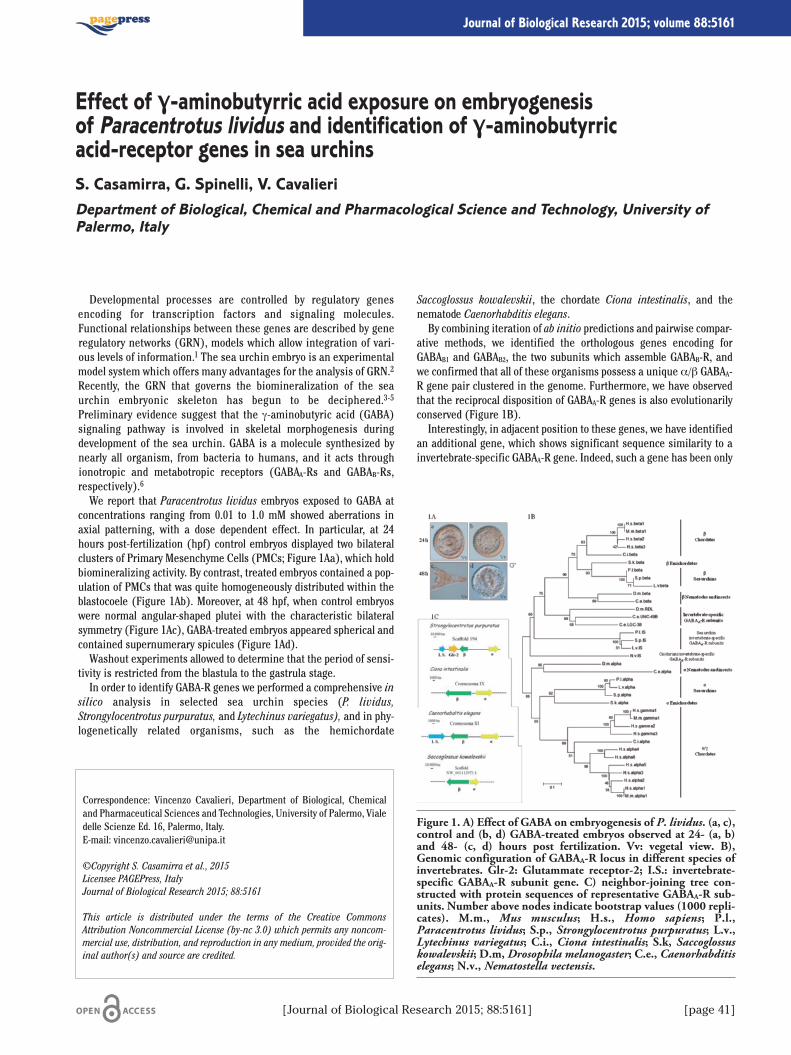
Developmental processes are controlled by regulatory genesencoding for transcription factors and signaling molecules.Functional relationships between these genes are described by generegulatory networks (GRN), models which allow integration of vari-ous levels of information.1 The sea urchin embryo is an experimentalmodel system which offers many advantages for the analysis of GRN.2
Recently, the GRN that governs the biomineralization of the seaurchin embryonic skeleton has begun to be deciphered.3-5
Preliminary evidence suggest that the g-aminobutyric acid (GABA)signaling pathway is involved in skeletal morphogenesis duringdevelopment of the sea urchin. GABA is a molecule synthesized bynearly all organism, from bacteria to humans, and it acts throughionotropic and metabotropic receptors (GABAA-Rs and GABAB-Rs,respectively).6
We report that Paracentrotus lividus embryos exposed to GABA atconcentrations ranging from 0.01 to 1.0 mM showed aberrations inaxial patterning, with a dose dependent effect. In particular, at 24hours post-fertilization (hpf) control embryos displayed two bilateralclusters of Primary Mesenchyme Cells (PMCs; Figure 1Aa), which holdbiomineralizing activity. By contrast, treated embryos contained a pop-ulation of PMCs that was quite homogeneously distributed within theblastocoele (Figure 1Ab). Moreover, at 48 hpf, when control embryoswere normal angular-shaped plutei with the characteristic bilateralsymmetry (Figure 1Ac), GABA-treated embryos appeared spherical andcontained supernumerary spicules (Figure 1Ad).Washout experiments allowed to determine that the period of sensi-
tivity is restricted from the blastula to the gastrula stage.In order to identify GABA-R genes we performed a comprehensive in
silico analysis in selected sea urchin species (P. lividus,Strongylocentrotus purpuratus, and Lytechinus variegatus), and in phy-logenetically related organisms, such as the hemichordate
Saccoglossus kowalevskii, the chordate Ciona intestinalis, and thenematode Caenorhabditis elegans.By combining iteration of ab initio predictions and pairwise compar-
ative methods, we identified the orthologous genes encoding forGABAB1 and GABAB2, the two subunits which assemble GABAB-R, andwe confirmed that all of these organisms possess a unique α/β GABAA-R gene pair clustered in the genome. Furthermore, we have observedthat the reciprocal disposition of GABAA-R genes is also evolutionarilyconserved (Figure 1B).Interestingly, in adjacent position to these genes, we have identified
an additional gene, which shows significant sequence similarity to ainvertebrate-specific GABAA-R gene. Indeed, such a gene has been only
Correspondence: Vincenzo Cavalieri, Department of Biological, Chemicaland Pharmaceutical Sciences and Technologies, University of Palermo, Vialedelle Scienze Ed. 16, Palermo, Italy.E-mail: [email protected]
©Copyright S. Casamirra et al., 2015Licensee PAGEPress, ItalyJournal of Biological Research 2015; 88:5161
This article is distributed under the terms of the Creative CommonsAttribution Noncommercial License (by-nc 3.0) which permits any noncom-mercial use, distribution, and reproduction in any medium, provided the orig-inal author(s) and source are credited.
[Journal of Biological Research 2015; 88:5161] [page 41]
Journal of Biological Research 2015; volume 88:5161
Effect of γ-aminobutyrric acid exposure on embryogenesisof Paracentrotus lividus and identification of γ-aminobutyrricacid-receptor genes in sea urchinsS. Casamirra, G. Spinelli, V. CavalieriDepartment of Biological, Chemical and Pharmacological Science and Technology, University ofPalermo, Italy
Figure 1. A) Effect of GABA on embryogenesis of P. lividus. (a, c),control and (b, d) GABA-treated embryos observed at 24- (a, b)and 48- (c, d) hours post fertilization. Vv: vegetal view. B),Genomic configuration of GABAA-R locus in different species ofinvertebrates. Glr-2: Glutammate receptor-2; I.S.: invertebrate-specific GABAA-R subunit gene. C) neighbor-joining tree con-structed with protein sequences of representative GABAA-R sub-units. Number above nodes indicate bootstrap values (1000 repli-cates). M.m., Mus musculus; H.s., Homo sapiens; P.l.,Paracentrotus lividus; S.p., Strongylocentrotus purpuratus; L.v.,Lytechinus variegatus; C.i., Ciona intestinalis; S.k, Saccoglossuskowalevskii; D.m, Drosophila melanogaster; C.e., Caenorhabditiselegans; N.v., Nematostella vectensis.

[page 42] [Journal of Biological Research 2015; 88:5161]
identified in C. elegans, Drosophila melanogaster, and Nematostellavectensis.7,8
We also retrieved several cDNA sequences from staged EST databas-es of the three sea urchin species inspected, indicating that thesegenes are actively transcribed during development. Some selectedcDNA plasmids were also isolated from P. lividus total RNA samples andfully sequenced.Hypothetical proteins were deduced and used for phylogenetic analy-
sis, including a selection of vertebrate and invertebrate GABAA-R sub-unit sequences. The resulting phylogenetic tree (Figure 1C) stronglysupport the hypothesis that the sea urchins contain genes encoding forboth canonical and invertebrate-specific GABAA-R subunits. Such a col-lection of data should provide a support to better understand theinvolvement of GABA-signalling pathway in the skeletal GRN.
References
1. Levine M, Davidson EH. Gene regulatory networks for develop-ment. Annu Rev Biophys Biomol Struct 2005;102:4936-42.
2. McClay DR. Evolutionary crossroads in developmental biology: seaurchins. Development 2011;138:2639-48.
3. Duloquin L, Lhomond G, Gache C. Localized VEGF signaling fromectoderm to mesenchyme cells controls morphogenesis of the seaurchin embryo skeleton. Development 2007;134:2293-302.
4. Röttinger E, Saudemont A, Duboc V, et al. FGF signals guide migra-tion of mesenchymal cells, control skeletal morphogenesis and reg-ulate gastrulation during sea urchin development. Development2008;135:353-36.
5. Cavalieri V, Guarcello R, Spinelli G (2011). Specific expression of aTRIM-containing factor in ectoderm cells affects the skeletal mor-phogenetic program of the sea urchin embryo. Development138:4279-90.
6. Watanabe M, Maemura K, Kanbara K, et al. GABA and GABA recep-tors in the central nervous system and other organs. Int Rev Cytol2002;213:1-47.
7. Tsang SY, Ng SK, Xu Z, Xue H. The evolution of GABAA receptor-likegenes. Mol Bio Evol 2007;24:599-610.
8. Anctil M. Chemical transmission in the sea anemone Nematostellavectensis: a genomic perspective. Comp Biochem Physiol Part DGenomics Proteomics 2009;4:268-89.
Article
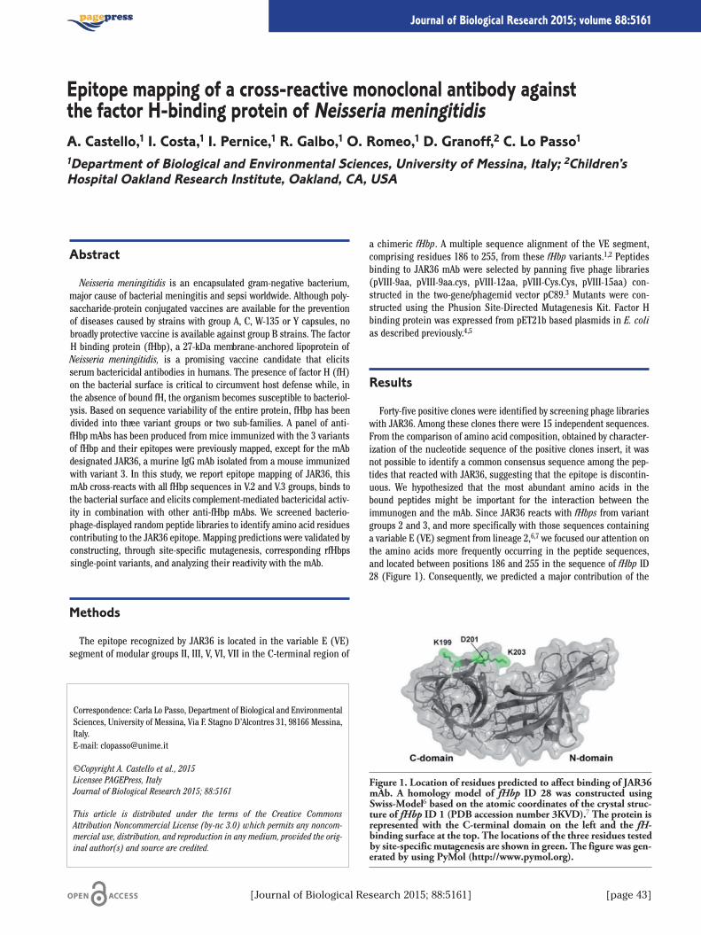
[Journal of Biological Research 2015; 88:5161] [page 43]
Journal of Biological Research 2015; volume 88:5161
Abstract
Neisseria meningitidis is an encapsulated gram-negative bacterium,major cause of bacterial meningitis and sepsi worldwide. Although poly-saccharide-protein conjugated vaccines are available for the preventionof diseases caused by strains with group A, C, W-135 or Y capsules, nobroadly protective vaccine is available against group B strains. The factorH binding protein (fHbp), a 27-kDa membrane-anchored lipoprotein ofNeisseria meningitidis, is a promising vaccine candidate that elicitsserum bactericidal antibodies in humans. The presence of factor H (fH)on the bacterial surface is critical to circumvent host defense while, inthe absence of bound fH, the organism becomes susceptible to bacteriol-ysis. Based on sequence variability of the entire protein, fHbp has beendivided into three variant groups or two sub-families. A panel of anti-fHbp mAbs has been produced from mice immunized with the 3 variantsof fHbp and their epitopes were previously mapped, except for the mAbdesignated JAR36, a murine IgG mAb isolated from a mouse immunizedwith variant 3. In this study, we report epitope mapping of JAR36, thismAb cross-reacts with all fHbp sequences in V.2 and V.3 groups, binds tothe bacterial surface and elicits complement-mediated bactericidal activ-ity in combination with other anti-fHbp mAbs. We screened bacterio-phage-displayed random peptide libraries to identify amino acid residuescontributing to the JAR36 epitope. Mapping predictions were validated byconstructing, through site-specific mutagenesis, corresponding rfHbpssingle-point variants, and analyzing their reactivity with the mAb.
Methods
The epitope recognized by JAR36 is located in the variable E (VE)segment of modular groups II, III, V, VI, VII in the C-terminal region of
a chimeric fHbp. A multiple sequence alignment of the VE segment,comprising residues 186 to 255, from these fHbp variants.1,2 Peptidesbinding to JAR36 mAb were selected by panning five phage libraries(pVIII-9aa, pVIII-9aa.cys, pVIII-12aa, pVIII-Cys.Cys, pVIII-15aa) con-structed in the two-gene/phagemid vector pC89.3 Mutants were con-structed using the Phusion Site-Directed Mutagenesis Kit. Factor Hbinding protein was expressed from pET21b based plasmids in E. colias described previously.4,5
Results
Forty-five positive clones were identified by screening phage librarieswith JAR36. Among these clones there were 15 independent sequences.From the comparison of amino acid composition, obtained by character-ization of the nucleotide sequence of the positive clones insert, it wasnot possible to identify a common consensus sequence among the pep-tides that reacted with JAR36, suggesting that the epitope is discontin-uous. We hypothesized that the most abundant amino acids in thebound peptides might be important for the interaction between theimmunogen and the mAb. Since JAR36 reacts with fHbps from variantgroups 2 and 3, and more specifically with those sequences containinga variable E (VE) segment from lineage 2,6,7 we focused our attention onthe amino acids more frequently occurring in the peptide sequences,and located between positions 186 and 255 in the sequence of fHbp ID28 (Figure 1). Consequently, we predicted a major contribution of the
Correspondence: Carla Lo Passo, Department of Biological and EnvironmentalSciences, University of Messina, Via F. Stagno D’Alcontres 31, 98166 Messina,Italy.E-mail: [email protected]
©Copyright A. Castello et al., 2015Licensee PAGEPress, ItalyJournal of Biological Research 2015; 88:5161
This article is distributed under the terms of the Creative CommonsAttribution Noncommercial License (by-nc 3.0) which permits any noncom-mercial use, distribution, and reproduction in any medium, provided the orig-inal author(s) and source are credited.
Epitope mapping of a cross-reactive monoclonal antibody againstthe factor H-binding protein of Neisseria meningitidisA. Castello,1 I. Costa,1 I. Pernice,1 R. Galbo,1 O. Romeo,1 D. Granoff,2 C. Lo Passo11Department of Biological and Environmental Sciences, University of Messina, Italy; 2Children’sHospital Oakland Research Institute, Oakland, CA, USA
Figure 1. Location of residues predicted to affect binding of JAR36mAb. A homology model of fHbp ID 28 was constructed usingSwiss-Model6 based on the atomic coordinates of the crystal struc-ture of fHbp ID 1 (PDB accession number 3KVD).7 The protein isrepresented with the C-terminal domain on the left and the fH-binding surface at the top. The locations of the three residues testedby site-specific mutagenesis are shown in green. The figure was gen-erated by using PyMol (http://www.pymol.org).

[page 44] [Journal of Biological Research 2015; 88:5161]
electrostatically charged residues aspartate and/or lysine to the JAR36epitope. We examined the positions of relevant aspartate and lysineresidues in a homology model of the fHbp ID 28 protein. We then substi-tuted the residues K199, D201 and K203 with alanine and expressed andpurified the fHbp mutants. Binding of JAR36 or human fH to site-specificmutants of fHbp ID 28 was measured by ELISA.
Conclusions
Three single amino acids positions composing the JAR36 epitopewere predicted and the corresponding fHbp mutants were prepared.The K199A and K203A point mutants show decreased binding to humanfH, but not to JAR36. The amino acid residue substitution, D201A infHbp affects the binding of JAR36, but not that of human fH, thusAspartate 201 is a necessary component of the JAR36 epitope.
References
1. Beernink PT, Granoff DM. The modular architecture of meningo-coccal factor H-binding protein. Microbiology 2009;155:2873-83.
2. Pajon R, Beernink PT, Harrison LH, Granoff DM. Frequency of fac-tor H-binding protein modular groups and susceptibility to cross-reactive bactericidal activity in invasive meningococcal isolates.Vaccine 2010;28:2122-9.
3. Felici F, Castagnoli L, Musacchio A, et al. Selection of antibody lig-ands froma large library of oligopeptides expressed on a multiva-lent exposition vector. J Mol Biol 1991;222:301-10.
4. Masignani V, Comanducci M, Giuliani MM, et al. Vaccinationagainst Neisseria meningitidis using three variants of the lipopro-tein GNA1870. J Exp Med 2003;197:789-99.
5. Beernink PT, Granoff DM Bactericidal antibody responses inducedby meningococcal recombinant chimeric factor H-binding proteinvaccines. Infect Immun 2008;76:2568-75.
6. Schwede T, Kopp J, Guex N, Peitsch MC. SWISS-MODEL: an auto-mated protein homology- modeling server. Nucleic Acids Res2003;31:3381-5.
7. Cendron L, Veggi D, Girardi E, Zanotti G Structure of the uncom-plexed Neisseria meningitidis factor H-binding protein fHbp(rLP2086). Acta Crystallogr Sect F Struct Biol Cryst Commun2011;67:531-5.
Article

Increased use of chemical pesticides results in the excess inflow oftoxic chemicals into the aquatic ecosystem and may affect aquatic floraand fauna. Recent emphasis is on the use of natural pesticides, whichare usually of plant origin. Use of plant based pesticides is generally lessharmful and more ecofriendly. Plant based pesticides contain easilybiodegradable molecules which are more target specific than the highlypersistent broad-spectrum synthetic chemical moieties.1 Azadirachtin isan active compound extracted from the neem tree (Azadirachta indica A.Juss) grown widely in Africa and Asia. Its anti-insecticidal, antiviral,antibacterial, and antifungal properties have been known for long time.2
Azadirachtin is the active substance of insecticide formulationNeemAzal T/S (concentration of active ingredients, 10 g/L), which isused to control whitefly, leaf miners, and other pests including pear psyl-la. In foreign countries, it has been generally distributed and widely usedfor a long period of time. Nevertheless, NeemAzal T/S has been regis-tered in the Czech Republic since 2010. NeemAzal T/S is classified ashighly toxic to aquatic animals.The objective of our study was to evaluate changes in antioxidant
defense enzymes and detoxifying system in early life stages of com-mon carp after exposure to NeemAzal T/S. Subchronic toxic effects onembryos and larvae were investigated during a 31-day embryolarvaltoxicity test, which was carried out using a modified protocol accord-ing to the OECD guideline 210 (Fish, Early-Life Stage Toxicity test).Testing solutions were prepared from NeemAzal T/S with the nominalconcentration of azadirachtin: 30, 100, 300 and 600 µg/L. Twenty-fourhours after fertilization, one hundred fertilized eggs were separatedand randomly distributed into crystallization dishes containing one ofthe four concentrations of azadirachtin solution, or a control dishes
(azadirachtin-free tap water). The experiment was conducted in trip-licate (a total of 300 fertilized eggs for each concentration and con-trol). A semistatic method was used and the testing solution wasreplaced twice daily. During the test, larvae were fed freshly hatchedArtemia salina ad libitium twice a day. The temperature, pH, and oxy-gen saturation were recorded daily. At the end of the experiment, thefish were killed, immediately frozen, and stored at -85°C until analysesof biomarkers. Whole body samples were weighed and homogenisedusing phosphate buffer. Supernatant fraction of homogenate was usedfor determination of activity of glutathione S-transferase (GST),3 glu-tathione reductase (GR),4 glutathione peroxidase (GPx)5 and catalase(CAT).6 Protein concentration was determined by Bicinchoninic AcidProtein Essay Kit (Sigma-Aldrich). Statistical analysis was performedusing Statistica 8.0 for Windows software. Biomarkers were tested fornormal distribution and after testing of homogeneity of varianceacross groups, an analysis of variance (one-way ANOVA) was used.The differences among groups were assessed with the Tukey-HSD test.Effects of subchronic exposure to NeemAzal T/S on selected oxida-
tive stress indices and activity of detoxifying enzyme are presented inTable 1. In all experimental groups, increases of the GR, GPx and GSTactivities were observed compared to the control group but only in thegroups exposed to azadirachtin at 300 and 600 �g/L the level reachedsignificance (P<0.05). In case of CAT activity we did not obtain differ-ences among groups.In our experiment, we found higher activities in almost all enzymes
(GR, GPx and GST) in all experimental groups compared to the controlgroup. An increase in antioxidant enzymes contributes to the elimina-tion of reactive oxygen species, which are induced by pesticide expo-sure. The increase in GST activity is connected with activation ofdetoxifying system after NeemAzal T/S exposure. Although botanicalpesticides are being considered as less toxic and safe, our results indi-cate that azadirachtin may affect antioxidant defence system anddetoxifying ability of fish organism. Negative effects of azadirachtinwere also reported by other toxicology studies. Kumar and colleagues7
observed morphological changes in vital organs such as gill ofHeteropneustes fossilis after short- and long-term exposure to purifiedneem extract. Winkaler and colleagues8 reported effects of acute lethaland sublethal exposure to neem leaf extract on the neotropical fresh-water fish Prochilodus lineaus. Plasma glucose levels were higher infish exposed to neem extract relative to controls, indicating a typicalstress response. Neem extract did not interfere with osmoregulationcapacities of the fish, as plasma sodium, chloride, total protein, andosmolarity did not change. It was shown to affect the antioxidantdefence system of P. lineatus, as there was a decrease in liver CATactivity at all neem concentrations, and the detoxifying enzyme GSTwas activated in fish exposed to the highest concentration. At all con-centrations, exposed fish exhibited damaged gill and kidney tissue.
Correspondence: Caterina Faggio, Department of Biological andEnvironmental Sciences, University of Messina, viale F. Stagno D’Alcontres 31,98166 Messina, Italy.E-mail: [email protected]
Acknowledgements: this research was supported by GACR P502/12/P163.
©Copyright F. Casuscelli di Tocco et al., 2015Licensee PAGEPress, ItalyJournal of Biological Research 2015; 88:5161
This article is distributed under the terms of the Creative CommonsAttribution Noncommercial License (by-nc 3.0) which permits any noncom-mercial use, distribution, and reproduction in any medium, provided the orig-inal author(s) and source are credited.
Oxidative stress responses in early life stages of common carp (Cyprinuscarpio) after subchronic exposure to NeeMazal T/S (azadirachtin 10 g/L)F. Casuscelli di Tocco,1 J. Blahova,2 L. Plhalova,2 D. Zivna,2 M. Sevcikova,2 L. Chromcova,2L. Divisova,2 C. Faggio,1 Z. Svobodova21Department of Biological and Environmental Sciences, University of Messina, Italy; 2Departmentof Veterinary Public Health and Animal Welfare, University of Veterinary and PharmaceuticalSciences, Brno, Czech Republic
[Journal of Biological Research 2015; 88:5161] [page 45]
Journal of Biological Research 2015; volume 88:5161
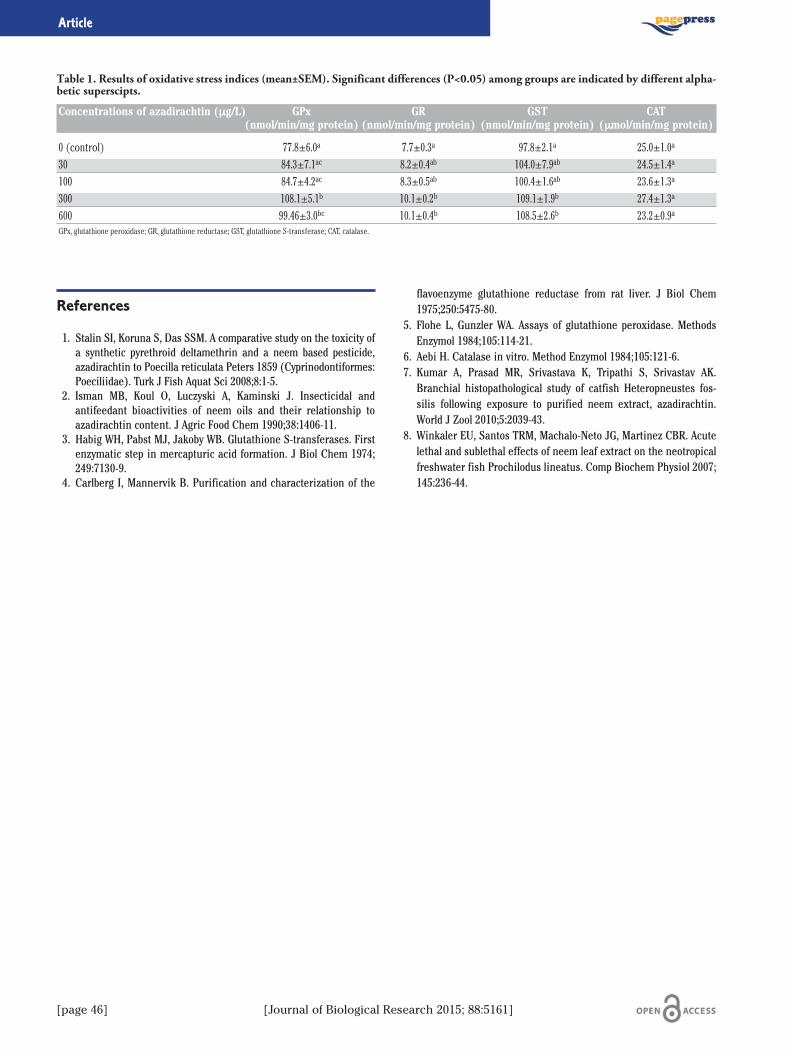
[page 46] [Journal of Biological Research 2015; 88:5161]
References
1. Stalin SI, Koruna S, Das SSM. A comparative study on the toxicity ofa synthetic pyrethroid deltamethrin and a neem based pesticide,azadirachtin to Poecilla reticulata Peters 1859 (Cyprinodontiformes:Poeciliidae). Turk J Fish Aquat Sci 2008;8:1-5.
2. Isman MB, Koul O, Luczyski A, Kaminski J. Insecticidal andantifeedant bioactivities of neem oils and their relationship toazadirachtin content. J Agric Food Chem 1990;38:1406-11.
3. Habig WH, Pabst MJ, Jakoby WB. Glutathione S-transferases. Firstenzymatic step in mercapturic acid formation. J Biol Chem 1974;249:7130-9.
4. Carlberg I, Mannervik B. Purification and characterization of the
flavoenzyme glutathione reductase from rat liver. J Biol Chem1975;250:5475-80.
5. Flohe L, Gunzler WA. Assays of glutathione peroxidase. MethodsEnzymol 1984;105:114-21.
6. Aebi H. Catalase in vitro. Method Enzymol 1984;105:121-6.7. Kumar A, Prasad MR, Srivastava K, Tripathi S, Srivastav AK.Branchial histopathological study of catfish Heteropneustes fos-silis following exposure to purified neem extract, azadirachtin.World J Zool 2010;5:2039-43.
8. Winkaler EU, Santos TRM, Machalo-Neto JG, Martinez CBR. Acutelethal and sublethal effects of neem leaf extract on the neotropicalfreshwater fish Prochilodus lineatus. Comp Biochem Physiol 2007;145:236-44.
Article
Table 1. Results of oxidative stress indices (mean±SEM). Significant differences (P<0.05) among groups are indicated by different alpha-betic superscipts.
Concentrations of azadirachtin (μg/L) GPx GR GST CAT(nmol/min/mg protein) (nmol/min/mg protein) (nmol/min/mg protein) (μmol/min/mg protein)
0 (control) 77.8±6.0a 7.7±0.3a 97.8±2.1a 25.0±1.0a
30 84.3±7.1ac 8.2±0.4ab 104.0±7.9ab 24.5±1.4a
100 84.7±4.2ac 8.3±0.5ab 100.4±1.6ab 23.6±1.3a
300 108.1±5.1b 10.1±0.2b 109.1±1.9b 27.4±1.3a
600 99.46±3.0bc 10.1±0.4b 108.5±2.6b 23.2±0.9a
GPx, glutathione peroxidase; GR, glutathione reductase; GST, glutathione S-transferase; CAT, catalase.
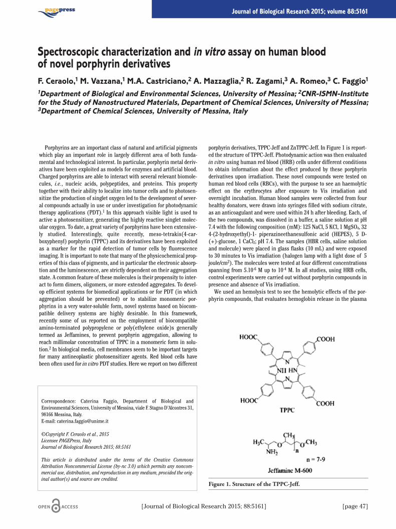
Porphyrins are an important class of natural and artificial pigmentswhich play an important role in largely different area of both funda-mental and technological interest. In particular, porphyrin metal deriv-atives have been exploited as models for enzymes and artificial blood.Charged porphyrins are able to interact with several relevant biomole-cules, i.e., nucleic acids, polypeptides, and proteins. This propertytogether with their ability to localize into tumor cells and to photosen-sitize the production of singlet oxygen led to the development of sever-al compounds actually in use or under investigation for photodynamictherapy applications (PDT).1 In this approach visible light is used toactive a photosensitizer, generating the highly reactive singlet molec-ular oxygen. To date, a great variety of porphyrins have been extensive-ly studied. Interestingly, quite recently, meso-tetrakis(4-car-boxyphenyl) porphyrin (TPPC) and its derivatives have been exploitedas a marker for the rapid detection of tumor cells by fluorescenceimaging. It is important to note that many of the physicochemical prop-erties of this class of pigments, and in particular the electronic absorp-tion and the luminescence, are strictly dependent on their aggregationstate. A common feature of these molecules is their propensity to inter-act to form dimers, oligomers, or more extended aggregates. To devel-op efficient systems for biomedical applications or for PDT (in whichaggregation should be prevented) or to stabilize monomeric por-phyrins in a very water-soluble form, novel systems based on biocom-patible delivery systems are highly desirable. In this framework,recently some of us reported on the employment of biocompatibleamino-terminated polypropylene or poly(ethylene oxide)s generallytermed as Jeffamines, to prevent porphyrin aggregation, allowing toreach millimolar concentration of TPPC in a monomeric form in solu-tion.2 In biological media, cell membranes seem to be important targetsfor many antineoplastic photosensitizer agents. Red blood cells havebeen often used for in vitro PDT studies. Here we report on two different
porphyrin derivatives, TPPC-Jeff and ZnTPPC-Jeff. In Figure 1 is report-ed the structure of TPPC-Jeff. Photodynamic action was then evaluatedin vitro using human red blood (HRB) cells under different conditionsto obtain information about the effect produced by these porphyrinderivatives upon irradiation. These novel compounds were tested onhuman red blood cells (RBCs), with the purpose to see an haemolyticeffect on the erythrocytes after exposure to Vis irradiation andovernight incubation. Human blood samples were collected from fourhealthy donators, were drawn into syringes filled with sodium citrate,as an anticoagulant and were used within 24 h after bleeding. Each, ofthe two compounds, was dissolved in a buffer, a saline solution at pH7.4 with the following composition (mM): 125 NaCl, 5 KCl, 1 MgSO4, 324-(2-hydroxyethyl)-1- piperazineethanesulfonic acid (HEPES), 5 D-(+)-glucose, 1 CaCl2; pH 7.4. The samples (HBR cells, saline solutionand molecule) were placed in glass flasks (10 mL) and were exposedto 30 minutes to Vis irradiation (halogen lamp with a light dose of �5joule/cm2). The molecules were tested at four different concentrationsspanning from 5.10-6 M up to 10-4 M. In all studies, using HRB cells,control experiments were carried out without porphyrin compounds inpresence and absence of Vis irradiation.We used an hemolysis test to see the hemolytic effects of the por-
phyrin compounds, that evaluates hemoglobin release in the plasma
Correspondence: Caterina Faggio, Department of Biological andEnvironmental Sciences, University of Messina, viale F. Stagno D’Alcontres 31,98166 Messina, Italy.E-mail: [email protected]
©Copyright F. Ceraolo et al., 2015Licensee PAGEPress, ItalyJournal of Biological Research 2015; 88:5161
This article is distributed under the terms of the Creative CommonsAttribution Noncommercial License (by-nc 3.0) which permits any noncom-mercial use, distribution, and reproduction in any medium, provided the orig-inal author(s) and source are credited.
Spectroscopic characterization and in vitro assay on human bloodof novel porphyrin derivativesF. Ceraolo,1 M. Vazzana,1 M.A. Castriciano,2 A. Mazzaglia,2 R. Zagami,3 A. Romeo,3 C. Faggio11Department of Biological and Environmental Sciences, University of Messina; 2CNR-ISMN-Institutefor the Study of Nanostructured Materials, Department of Chemical Sciences, University of Messina;3Department of Chemical Sciences, University of Messina, Italy
[Journal of Biological Research 2015; 88:5161] [page 47]
Journal of Biological Research 2015; volume 88:5161
Figure 1. Structure of the TPPC-Jeff.

[page 48] [Journal of Biological Research 2015; 88:5161]
following molecule and irradiation exposure.3 After overnight incuba-tion at 37°C, the tubes were centrifuged (10 minutes at 1500 rpm at4°C) and the supernatants were determined photometrically with acolorimeter and after with a spectrophotometer at the absorption ofhemoglobin (540 nm). The spectroscopic measurements of haemo-globin released are much easier and faster to carry out than cellstaining and counting.4 The absorption of the supernatant of erythro-cytes lysed in distilled water was defined as 100% haemolysis. Themolecules under study induce hemolysis in human erythrocytes atthe concentrations tested and then overnight incubation, as show inTable 1. The concentrations 5.10-6 M and 10-4 M are respectively onetoo low and the other too high to see a reliable hemolytic effect. Whilewe observed that TPPC-Jeff induce an hemolytic effect at the concen-tration of 5.10-5 M, and the Zn TPPC-Jeff induce the release of hemo-globin at the concentration of 10-5 M. We have seen that Vis irradia-tion does not increase the efficacy of the compounds in inducinghemolysis.
Then we assayed the morphology of erythrocytes by an optical micro-scope after incubation with porphyrin compounds, 30 minutes of irra-diation and overnight incubation and the result was that they lost theirnormal biconcave profile and presented a spiny configuration withblebs in their surfaces. A normal profile was observed in the control.Representative results are show in Figure 2. HRB cells obtained fromfour subjects were used to assess an hemolytic activity of porphyrins.Results suggest that all the molecules under study show an hemolyticeffect. Further studies are required to optimize potential therapeuticdosing strategies to inform and encourage clinical trial design.
References
1. Pandey RK, Zheng G. Porphyrins as photosensitizers in photody-namic therapy. In: Kadish KM, Smith KM, Guilard R (eds.), The por-phyrin handbook. Academic Press: New York; 2000. p. 157.
2. Castriciano MA, Romeo A, Angelini N, et al. Structural features ofmeso-tetrakis(4-carboxyphenyl) porphyrin interacting with amino-terminated poly (propylene oxide), Macromolecules 2006;39:5489-96.
3. Bonaccorsi P, Aversa MC, Barattucci A, et al. Sulfenic acid-derivedglycoconjugated dusulfides and sulfoxides: a biological evaluation onhuman red blood cells. Journal of Sulfur Chemistry 2013 (in press).
4. Ochoa A.L., Tempesti T.C., Spesia M.B., Milanesio E., DurantiniE.N. Synthesis and photodynamic properties of adamantylethoxyZn (II) phthalocyanine derivatives in different media and inhuman red blood cells. European Journal of Medicinal Chemistry2012;20:280-7.
Article
Figure 2. Photograph showing morphology of HRB cells (A) controlwithout tretracycline and irradiated for 30 min; (B) TPPC-Jeff andirradiated for 30 min; (C) Zn TPPC-Jeff and irradiated for 30 min.
Table 1. Percentage of hemolysis by compounds TPPC-Jeff (5.10-5M), Zn TPPC-Jeff (10-5 M) osmolarity (mOsm/kg).
Compounds 300 200 150 100 0
Control 0 1 2 18 100TPPC-Jeff 12 11 12 30 100ZnTPPC-Jeff 17 16 23 100 100

The sea urchin egg is a favourite model for studies of the molecularbiology and physiology of fertilization and early development, yet weknow sparingly little of its oocytes and of mitochondria behaviour dur-ing oogenesis. The process of oogenesis in most echinoderms is asyn-chronous so each ovary lobe has hundreds of oocytes at all stages ofdevelopment. At the beginning of oogenesis, the oocyte is about 10 μmin diameter. During the vitellogenic phase of oogenesis, the oocyteaccumulates yolk proteins and grows to ten times their original size to80 to 100 μm in sea urchins. The oocyte, arrested at the prophase ofthe first meiotic division, is apparent with its large nucleus, the germi-nal vesicle (GV), containing a prominent nucleolus. Echinoid (such assea urchin) and Holothurian oocytes complete meiotic maturationprior to fertilization, distinct from other echinoderms and almost allothers animals. As maturation progresses, it occurs the GV breaksdown (GVBD). These eggs may then be stored for weeks to monthswithin the female before they are spawned, and the proportion of eggsin the ovary increases from early to late season, as the numbers ofoocytes decline.1
Mitochondria, generally known as the powerhouses of eukaryotic cells,play a primary role in cellular energetic metabolism, homeostasis anddeath. These organelles, with their multicopy genome maternally inher-ited, are directly involved at several levels in the reproductive processsince their functional status influences the quality of oocytes and con-tributes to the process of fertilization and embryonic development.It has been demonstrated that the number of maternal mitochondria
is sufficient to support development until late stages without new syn-thesis of mitochondrial DNA or production of new organelles.2 Duringembryogenesis mitochondrial mass does not change, whereas mito-chondrial respiration increases.3 The behaviour of these organellesduring oogenesis remains at moment unclear.In the present paper we studied, by Confocal Laser Scanning
Microscopy tecnologies (CLSM), the mass and distribution, the activityand the DNA content of sea urchin Paracentrotus lividus mitochondriaduring oogenesis, by in vivo incubating oocytes of different size with cell-
permeant probes specific for mitochondria and for DNA and by immun-odetection of hsp60 chaperonine, a well known mitochondrial marker.In particular the oocytes were grouped in six classes: <10, 20/30,
40/50, 60/70, 80/90 μm, and 90 μm ovulated egg, on the base of diam-eters. Microscopic observations were performed capturing 2 μm thicklayers of oocytes. Of the several thousands oocytes we observed, 20 foreach different oogenesis stage were analyzed and processed. In orderto interpret results and to draw unequivocal conclusions, we measuredby IMAGE J software analysis the intensity values of fluorescent sig-nals, as suggested in Agnello and colleagues 2008.4
The mitochondria of oocytes with a diameter between 20 and 70 μm,appeared to give rise to clusters that disappear in that of 80 μm. In theoocytes between 60 and 90 μm the red fluorescence seems to be moreevident around the germinal vesicle (the merge tends to red), suggest-ing an increasing oxidative phosphorylation activity.In the ovulated eggs, red and green fluorescence are uniformly dis-
tributed suggesting that mitochondria are dispersed in the cytoplasm.In addition the merge of green and red colours shows that the wholemitochondrial population is consuming oxygen at the same level (theresulting colours tends to yellow; Figure 1).In order to calculate the total mitochondrial mass and activity we
integrated the values of pixel intensities for all captured sections andused the arithmetic means to draw a statistical analysis. Results sug-gest a parallel rise of mitochondrial mass and activity, suggesting thatthe amount and activity of organelles change remarkably during ooge-nesis. Results also suggest that mitochondria are actively duplicatingand that mitochondrial DNA is replicating during the different oogen-esis phases. It is noteworthy that around the germinal vesicle, espe-cially in the larger oocytes, next to the germinal vesicle breakdown, theorganelles are more active in oxygen consumption, probably due to themajor energetic needing in this key moment of gametogenesis.
Correspondence: Maria Carmela Roccheri, Department of Biological,Chemical and Pharmaceutical Sciences and Technologies, University ofPalermo, viale delle Scienze, Ed. 16, 90127 Palermo, Italy.E-mail: [email protected]
©Copyright M. Agnello et al., 2015Licensee PAGEPress, ItalyJournal of Biological Research 2015; 88:5161
This article is distributed under the terms of the Creative CommonsAttribution Noncommercial License (by-nc 3.0) which permits any noncom-mercial use, distribution, and reproduction in any medium, provided the orig-inal author(s) and source are credited.
Mitochondrial mass, distribution and activity during sea urchin oogenesisM. Agnello, A.M. Rinaldi, R. Chiarelli, L. Bosco, G. Morici, M.C. RoccheriDepartment of Biological, Chemical and Pharmaceutical Sciences and Technologies, University ofPalermo, Italy
[Journal of Biological Research 2015; 88:5161] [page 49]
Journal of Biological Research 2015; volume 88:5161
Figure 1. Distribution of hsp60 protein detected by immunoflu-orescence analysis (A), the mitochondrial and genomic DNA,after in vivo incubation with PicoGreen probe (B) and the mergeof green and red fluorescence signal, respectively due to mito-chondrial mass and activity, after in vivo incubation withMitotraker Green and Orange (C). The size of the oocytes report-ed is 80 μm.

[page 50] [Journal of Biological Research 2015; 88:5161]
References
1. Wessel GM, Voronina E, Brooks JM. Obtaining and handling echin-oderm oocytes. In: Methods in cell biology. Amterdam: Elsevier;2004. pp. 87-114.
2. Matsumoto L, Kasamatsu H, Piko L, Vinograd J. Mitochondrial DNA
replication in sea urchin oocytes. J Cell Biol 1974;63:146-59.
3. Morici G, Agnello M, Spagnolo F, et al. Confocal microscopy study of
the distribution, content and activity of mitochondria during
Paracentrotus lividus development. J Microscopy 2007;228:165-73.
4. Agnello M, Morici G, Rinaldi AM. A method for measuring mito-
chondrial mass and activity. Cytotechnology 2008;56:145-9.
Article
Stem cells have a key role in regenerative medicine and tissue engi-neering. Although not immortal, they are able to expand manyfold inculture retaining at the same time their growth and multilineagepotential. They also show a migratory capacity when transplanted sys-temically in animal model with injuries.Thanks to their properties and their plasticity stem cells are of great
importance since they can be used as a tool for repair damaged tissuesand organs.Mesenchymal stem cells, in particular, have the ability to differenti-
ate into lineages of mesodermal tissues, such as skeletal muscle, bone,tendons, cartilage, and fat under appropriate culturing conditions.Recent evidence suggest that the adipose tissue is a promising
source of mesenchymal stem cells attracting the interest ofresearchers and clinicians. It is rich of pluripotent stromal cells , avail-able in large amounts and more readily accessible than bone marrow.Furthermore, comparative analysis of mesenchymal stem cells of bonemarrow and adipose tissue show that cells are not different regardingmorphology, immune phenotype, success rate of isolating and differen-tiation ability.
Our experience at the Experimental Zooprophylactic Institute ofSicily A. Mirri allows us to define a protocol for stem cells isolation ofmurine origin.We used 6 Winstar breed male rats whose average weight was 350g.All animals were sedated with an intramuscular injection of midazo-
lam and anesthesia was maintained with isoflurane and oxygen gasmixture administered with a mask.The adipose tissue has been taken from the root of the animal’s
thigh with a small incision. Different steps are needed for processingand digesting the tissue. First it is washed several times in a solutionenriched of antibiotics and then is mechanically fragmented. Thehomogenate is therefore digested enzymatically under permanentshaking.We obtain an heterogeneous population of cells that were subse-
quently selected through the plastic adhesion. These cells are able togrow and proliferate and show all the characteristics typical of stemcells. In conclusion, we report a multistep and reproducible techniquefor providing a substantial number of mesenchymal stem cells and formaintaining them in culture.
Correspondence: Roberta Altomare, School in Surgical Biotechnology andRegenerative Medicine, University of Palermo, via del Vespro 131, 90127Palermo, Italy.E-mail: [email protected]
©Copyright R. Altomare et al., 2015Licensee PAGEPress, ItalyJournal of Biological Research 2015; 88:5161
This article is distributed under the terms of the Creative CommonsAttribution Noncommercial License (by-nc 3.0) which permits any noncom-mercial use, distribution, and reproduction in any medium, provided the orig-inal author(s) and source are credited.
Obtaining mesenchymal stem cells from adipose tissue of murine origin:experimental studyR. Altomare,1 A. Abruzzo,1 V. Palumbo,1,2 G. Damiano,2 G. Spinelli,2 A. Bruno,2 L. Cicero,3G. Cassata,3 G. Tomasello,2 F. Cacciabaudo,1 M.C. Gioviale,4 F. Damiani,1,4 A.I. Lo Monte1,2,41School in Surgical Biotechnology and Regenerative Medicine, University of Palermo; 2Departmentof Surgical, Oncological and Stomatological Sciences, University of Palermo; 3The Institute forExperimental Veterinary Medicine of Sicily A. Mirri, Palermo, Italy; 4P. Giaccone Hospital,University of Palermo, Italy
[Journal of Biological Research 2015; 88:5161] [page 51]
Journal of Biological Research 2015; volume 88:5161
Introduction and ObjectivesHerpes simplex virus type 1 (HSV-1) is a double stranded DNA
(dsDNA) virus that causes a variety of infections in humans.1 HSV-1, asmany DNA viruses, has developed different strategies during the evolu-tion to modify the cellular environment in favor of its replication.Different aspects of HSV-1 biology render this virus a good model tostudy the complexity of virus-host cell interactions. The eukaryotic cells,indeed, respond to external stimuli through the activation of differentsignal pathways, as the Ras/Raf/MEK/ERK signal pathway. Among theseprotein kinases, the extracellular-signal-regulated-kinases (ERK) haveproven critical in the control of the progression G1/S that involved spe-cific regulator proteins, such as cyclins and cyclin-dependent kinases(CDKs).2,3 That HSV-1 infection requires involvement of ERK1/2 andmitogen activated proteins kinases (MAPKs) signal pathway and con-trols cell cycle proteins is already known.4-9 In fact, the activity of CDKinvolved in the transition from late G1 to early S phase seems to berequired for viral DNA transcription and replication. However, the evi-dence for the overall understanding of networks and gene productsinvolved in these interaction requires further investigations. Based onthese knowledge the current work was focused to study the activity ofERK1/2 protein during viral replication and the correlation between ERKprotein recruitment and G1/S phases regulation by HSV-1 infection.
Materials and Methods
Western Blot analysis was used to evaluate nuclear and cytoplasmicprotein accumulations. The MAPK/MEK-ERK pathway was modulated
using the inhibitor U0126 to study replication in HEp-2 (human larynxepidermoid carcinoma cell line). A stably transfected cell line wasderived from wild type (wt) HEp-2, by transfection of plasmid codingfor dominant negative form of ERK protein (HEp-dnERK). StandardPlaque Assay was done on VERO cells. Immunofluorescence assay andquantitative Real Time PCR were used to evaluate the levels of viraland cellular gene transcription and viral DNA synthesis.
Results and Discussion
We have analyzed the activation of ERK1/2 protein during wt HSV-1infection in HEp-2 wt cell line. HSV-1 leads to the activation of ERK1/2protein during the first phases of infection, and subsequent decreaseduring late phases compared to uninfected cells, suggesting involve-ment of ERK1/2 activity during infection. HSV-1 replication was studiedin wt HEp-2 cells where ERK1/2 activity was chemically inhibited. Thedata showed a defect in viral progeny production in treated and infectedcells as compared with non-treated and infected cells. These data wereconfirmed by the differences in the accumulation of ICP0 (immediateearly) and Us11 (late) viral proteins. Moreover, we evaluated the phos-phorylated forms of key regulators of G1/S progression, such as cyclin Eand CDK2 proteins in presence or in absence of U0126. The resultsdemonstrated that the treatment inhibits the accumulation of cyclin Eand CDK2 proteins. These results were further confirmed by using HEp-dnERK cell lines. In this cell system HSV-1 replication was compromizedcompared with parental cell lines. Indeed, using q-PCR, viral DNA thecellular genes CDK2 and cyclin E, and the viral immediately early gene(ICP0) and the late gene, (gB) were evaluated in HEp-dnERK infected bywt HSV-1 compared to control. A decrease was observed in viral DNA syn-thesis, as well as in cellular gene transcripts, in cells where ERK1/2activity was compromised, demonstrating that MAPK-ER proteins playsa fundamental role during HSV-1 replication. However, further investi-gations are necessary.
Conclusions
The new information obtained could be contribute to developmentof new pro-host tools that would be useful to set up effective preven-tion strategies, such as therapeudical approaches for severe HSV-asso-ciated infections. Because the Raf/Ras/MEK/ERK pathway is modifiedin 60% of solid tumors, understanding the interactions between HSV-1 and this pathway could contribute to the design of HSV-1-basedoncolytic vectors.
Correspondence: Ivana Colao, Department of Biological and EnvironmentalSciences, University of Messina, viale F. Stagno d’Alcontres, 98166 Messina,Italy.Tel: +39.349.7844545.E-mail: [email protected]
©Copyright I. Colao et al., 2015Licensee PAGEPress, ItalyJournal of Biological Research 2015; 88:5161
This article is distributed under the terms of the Creative CommonsAttribution Noncommercial License (by-nc 3.0) which permits any noncom-mercial use, distribution, and reproduction in any medium, provided the orig-inal author(s) and source are credited.
Role of ERK1/2 protein in the regulation of Herpes simplex virus type Ireplicative cycleI. Colao,1 G. Siracusano,1 M.G. Ceraolo,1 A.Venuti,2 H. Veijo,3 A. Mastino,1 M.T. Sciortino11Department of Biological and Environmental Sciences, University of Messina; 2Division ofImmunology, Transplantation and Infectious Diseases, San Raffaele Scientific Institute, Milan, Italy;3Department of Virology, University of Turku, Finland
[page 52] [Journal of Biological Research 2015; 88:5161]
Journal of Biological Research 2015; volume 88:5161
References
1. Whitley RJ, Roizman B. Herpes simplex virus infections. Lancet2001;357:1513-8.
2. Chambard JC, Lefloch R, Pouysseguer J, Lenormand P. ERK impli-cation in cell cycle regulation. Biochim Biophys Acta 2007;1773:1299-1310.
3. Villanueva J, Yung Y, Walker JL, et al. ERK activity and G1 phaseprogression: identifying dispensable versus essential activities andprimary versus secondary targets. Mol Biol Cell 2007;18:1457-63.
4. Farassati F, Lee PW. Ras signalling pathway: a gateway for HSV-1infection. Sci World J 2003;3:533-5.
5. Torres NI, Castilla V, Bruttomesso AC, et al. In vitro antiviral activityof dehydroepiandrosterone, 17 synthetic analogs and ERK modula-
tors against herpes simplex virus type 1. Antiviral Res 2012;95:37-48.6. Chuluunbaatar U, Roller R, Mohr I. Suppression of extracellular
signal-regulated kinase activity in HSV-1 infected cells by the Us3protein kinase. J Virol 2012;86:7771-6.
7. Kalamvoki M, Roizman B. Interwoven roles of cyclin D3 and cdk4recruited by ICP0 and ICP4 in the expression of herpes simplexvirus genes. J Virol 2010;84:9709-17.
8. Ehmann GL, McLean TI, Bachenheimer SL. Herpes simplex virustype 1 infection imposes a G(1)/S block in asynchronously growingcells and prevents G(1) entry in quiescent cells. Virology 2000;267:335-49.
9. Orlando JS, Astor TL, Rundle SA, Schaffer PA. The products of theherpes simplex virus type 1 immediate-early US1/US1.5 genesdownregulate levels of S-phase-specific cyclins and facilitate virusreplication in S-phase Vero cells. J Virol 2006;80:4005-16.
[Journal of Biological Research 2015; 88:5161] [page 53]
Article
It is know that mesenchymal stem cells (MSCs) actively secretemultiple biologically-active factors during their process of differentia-tion which gives rise to a variey of cytotypes including bone and fatcells. It is also acknowledged that the chemokines secreted throughoutMSC differentiation may play an important role in the developmentand growth of tumor cells, although literature data appear somewhatindeterminate due to the contradictory evidence often found.1
The purpose of this study was to evaluate the effect of conditionedmedia (CMs) from MSCs, cultured for 7, 14, 21 and 28 days in osteo-,adipo-differentiating and undifferentiated conditions, on MDA-MB231breast cancer cells, an in vitromodel system derived from a triple-neg-ative breast cancer (TNBC). MTT assay showed that the CMs collectedafter 28 days of both osteo- and adipo-differentiation induced growthinhibition on MDA-MB231 cells after 24 h of incubation. In light ofsuch evidence, these CMs were used to treat cells and perform cytoflu-orimetric assays to better evaluate their biological effects onviability/proliferation, cell cycle progression, apoptosis/autophagyinduction and mitochondrial activity/reactive oxygen species (ROS)accumulation of MDA-MB231 cells.The most interesting results regard the ability of CMs from osteo-
differentiating MSC to induce an alteration of cell proliferation with anarrest in the G2/M transition phase of the cycle coupled to both apop-totic and autophagic promotion. No accumulation of ROS and impair-ment of mitochondrial respiration was observed at the end of treat-ment. On the other hand, preliminary indications suggest that the CMsisolated from adipo-differentiating MSCs have different effect fromthose obtained by osteo-differentiating cultures, being the lethalityunlinked to apoptosis and autophagy, and thereby prompting to getmore insight into the anti-TNBC activity shown by the different CMs atthe molecular level.
References
1. Alan PM, Fiachra TM, Roisin MD, et al. Mesenchymal stem cellsecretion of chemokines during differentiation into osteoblasts,and their potential role in mediating interaction with breast can-cer cells. Int J Cancer 2009;124:326-32.
Correspondence: Claudio Luparello, Department of Biological, Chemical andPharmaceutical Sciences and Technologies, University of Palermo, vialedelle Scienze Ed.16, 90127 Palermo, Italy.E-mail: [email protected]
Funding: this work was supported by University of Palermo (R.S. ex60% andFFR 2013) and Progetto Vigoni 2011 for Claudio Luparello; BMBF, AdiPaD,1720X06, FHprofUnt, FKZ: 03FH012PB2; FH-Extra, FKZ: z1112fh012; DAAD,PPP Vigoni, FKZ: 54669218; BMBF-AIF, FKZ: 1720X06; and Geräteprogramm,Fachhochschulen NRW 2008 for Edda Tobiasch.
©Copyright M. Librizzi et al., 2015Licensee PAGEPress, ItalyJournal of Biological Research 2015; 88:5161
This article is distributed under the terms of the Creative CommonsAttribution Noncommercial License (by-nc 3.0) which permits any noncom-mercial use, distribution, and reproduction in any medium, provided the orig-inal author(s) and source are credited.
Effect of conditioned media from osteo- and adipo-differentiatingmesenchymal stem cells on triple negative MDA-MB231 breast cancer cellsM. Librizzi,1 A. Longo,1 O. Consiglio,1 E. Tobiasch,2 C. Luparello11Department of Biological, Chemical and Pharmaceutical Sciences and Technologies, University ofPalermo, Italy; 2Department of Natural Sciences, University of Applied Sciences, Bonn-Rhein-Sieg,Germany
[page 54] [Journal of Biological Research 2015; 88:5161]
Journal of Biological Research 2015; volume 88:5161
Hepcidin, a peptide hormone produced mainly by the liver,1 has anessential role in iron homeostasis. It is produced as prepropeptide of84 amino acids and, after two consecutive cleavage, a form of 25 aminoacids is secreted in the bloodstreem.2-4 Hepcidin is able to control thebody iron concentration by the binding to the only known protein inv-oled in iron export from the cells, ferroportin, that is present on thebasolateral membrane of enterocytes, on macrophages, hepatocytesand other cells types.5,6 After the binding, ferroportin is degraded andthe iron uptake by the diet and iron recycling from senescent red bloodcells, is inhibited.7 View the important role of hepcidin in iron metab-olism, its expression is strictly regulated; in fact, dysfunctions or muta-tions that modify its expression, lead to different pathologies, such ashemochromatosis and anemia.8-10 Some crystallographic studies havedemonstrated that an other important metal for the life of all theorganisms, copper, could be important for hepcidin funtion; in fact, thepresence of copper in solution can influence the aggregation state ofhepcidin and studies in which a cell line was used as model system,have highlighted the importance of this element in the capacity of hep-cidin to induce ferroprtin degradation.11,12 The link between copperand iron is demonstarated also by an other important plasma protein,ceruloplasmin, a ferroxidase enzyme that contain copper in the cat-alytic site and is responsable of the transport of 90% of plasma copper13
Different studies have demonstrated that animals fed with a copperdeficient diet, have low levels of plasma ceruloplasmin and, its charac-teristic oxidase activity, is strongly reduced;14,15 this kind of situationis normally present in Wilson disease patients, in which a defect incopper transport in the Golgi, is cause of the lack of plasma ceruloplas-min.16 To better understand if the copper has a role on hepcidinexpression, we have used a human hepatoma cell line, HepG2, toinvestigate the influence of copper chelation and inflammation, by theuse of the citokine interleukin-6,17 on the level of hepcidin expression.To corroborate our results, we have also studied ceruloplasmin expres-sion. Our results have demonstarted that the use of the chelation sub-strate, Bathocuproinedisulfonic acid (BCS) has a slight effect on ceru-
loplasmin mRNA levels, both in normal and stimulated IL-6 cells, whilethe intracellular level of protein is markedly reduced when BCS isadded in both conditions. The inhibitory effects is more evident if thesecreted form of the protein is considered; in fact, western blot and ingel oxidase activity analysis, have shown a disappearence of the pro-tein and of its activity in the growth cellular media, results coherentwith bibliographic data. The effect on hepcidin is also evident. Even ifno statistical significative differences were found at mRNA level, theeffect on the concentration of the intracellular prepropeptide is moreevident, especially when the citokine and BCS are used togheter. Thepresence of both substrates leads to a decrease of the protein levelwhen it is compared to the use of interleukin-6 alone and this effectwas more evident when the concentration of the peptide in the cellulargrowth media was assayed. Our results demonstrate that a copper defi-ciency could negatively influence hepcidin expression even if otherstudies are necessary to better understand this mechanism.
References
1. Krause A, Neitz S, Mägert H, et al. LEAP-1, a novel highly disul-fide-bonded human peptide, exhibits antimicrobial activity. FEBSLett 2000;480:147-50.
2. Pigeon C, Ilyin G, Courselaud B, et al. A new mouse liver- specificgene, encoding a protein homologous to human antimicrobial pep-tide hepcidin, is overexpressed during iron overload. J Biol Chem2001;276:7811-9.
3. Park CH, Valore EV, Waring AJ, Ganz T. Hepcidin, a urinary antimi-crobial peptide synthesized in the liver. J Biol Chem 2001;276:7806.
4. Valore EV, Ganz T. Posttranslational processing of hepcidin inhuman hepatocytes is mediated by the prohormone convertasefurin. Blood Cells Mol Dis 2008;40:132-8.
5. Donovan A, Lima CA, Pinkus JL, et al. The iron exporter ferro-portin/Slc40a1 is essential for iron homeostasis. Cell Metab2005;1:191-200.
6. Hentze MW, Muckenthaler MU, Galy B, Camaschella C. Two totango: regulation of mammalian iron metabolism. Cell2010;142:24-38.
7. Nemeth E, Tuttle MS, Powelson J, et al. Hepcidin regulates cellulariron efflux by binding to ferroportin and inducing its internaliza-tion. Science 2004;306:2090-3.
8. Lanzara C, Roetto A, Daraio F, et al. Spectrum of hemojuvelin genemutations in 1q-linked juvenile hemochromatosis. Blood2004;103:4317-21.
9. Lee P, Peng H, Gelbart T, Beutler E. The IL-6- and lipopolysaccha-ride-induced transcription of hepcidin in HFE-, transferrin recep-tor 2-, and beta 2-microglobulin-deficient hepatocytes. P Natl AcadSci USA 2004;101:9263-5.
Correspondence: Maria Rosa Felice, Department of Biological andEnvironmental Sciences, University of Messina, via Stagno D’Alcontres 31,98166 Messina, Italy.E-mail: [email protected]
©Copyright L.M. Di Bella and M.R. Felice, 2015Licensee PAGEPress, ItalyJournal of Biological Research 2015; 88:5161
This article is distributed under the terms of the Creative CommonsAttribution Noncommercial License (by-nc 3.0) which permits any noncom-mercial use, distribution, and reproduction in any medium, provided the orig-inal author(s) and source are credited.
Effect of copper chelation on hepcidin expression in the human hepatomacell line, HepG2L.M. Di Bella, M.R. FeliceDepartment of Biological and Environmental Sciences, University of Messina, Italy
[Journal of Biological Research 2015; 88:5161] [page 55]
Journal of Biological Research 2015; volume 88:5161

[page 56] [Journal of Biological Research 2015; 88:5161]
10. Kearney SL, Nemeth E, Neufeld EJ, et al. Urinary hepcidin in con-genital chronic anemias. Pediatr Blood Cancer 2007;48:57-63.
11. Hunter HN, Fulton DB, Ganz T, Vogel HJ. The solution structure ofhuman hepcidin, a peptide hormone with antimicrobial activitythat is involved in iron uptake and hereditary hemochro matosis JBiol Chem 2002;277:37597-603.
12. Melino S, Garlando L, Patamia M, et al. Metal-binding site is pres-ent in the amino terminal region of the bioactive iron regulatorhepcidin-25. J Pept Res 2005;66(Suppl.1):65-71.
13. Meyer LA, Durley AP, Prohaska JR, Harris ZL. Copper transport andmetabolism are normal in aceruloplasminemic mice. J Biol Chem2001;276:36857-61.
14. Pyatskowit JW, Prohaska JR. Copper deficient rats and mice both
develop anemia but only rats have lower plasma and brain iron lev-els. Comp Biochem Physiol C Toxicol Pharmacol. 2008;147:316-23.
15. Broderius M, Mostad E, Wendroth K, Prohaska JR. Levels of plasmaceruloplasmin protein are markedly lower following dietary copperdeficiency in rodents. Comp Biochem Physiol C Toxicol Pharmacol2010;151:473-9.
16. Matsuda I, Pearson T, Holtzman NA. Determination of apocerulo-plasmin by radioimmunoassay in nutritional copper deficiency,Menkes’ kinky hair syndrome, Wilson’s disease, and umbilical cordblood. Pediatr Res 1974;8:821-4.
17. Nemeth E, Rivera S, Gabayan V, et al. IL-6 mediates hypoferremiaof inflammation by inducing the synthesis of the iron regulatoryhormone hepcidin. J Clin Invest 2004;113:1271-6.
Article
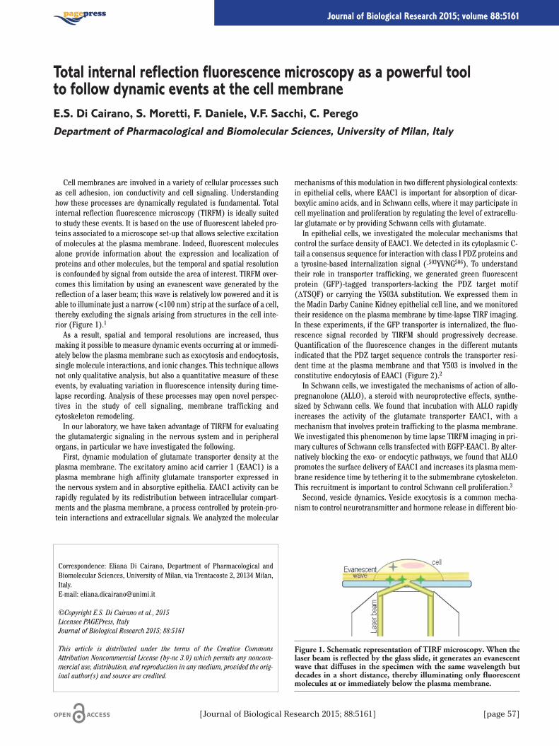
Cell membranes are involved in a variety of cellular processes suchas cell adhesion, ion conductivity and cell signaling. Understandinghow these processes are dynamically regulated is fundamental. Totalinternal reflection fluorescence microscopy (TIRFM) is ideally suitedto study these events. It is based on the use of fluorescent labeled pro-teins associated to a microscope set-up that allows selective excitationof molecules at the plasma membrane. Indeed, fluorescent moleculesalone provide information about the expression and localization ofproteins and other molecules, but the temporal and spatial resolutionis confounded by signal from outside the area of interest. TIRFM over-comes this limitation by using an evanescent wave generated by thereflection of a laser beam; this wave is relatively low powered and it isable to illuminate just a narrow (<100 nm) strip at the surface of a cell,thereby excluding the signals arising from structures in the cell inte-rior (Figure 1).1
As a result, spatial and temporal resolutions are increased, thusmaking it possible to measure dynamic events occurring at or immedi-ately below the plasma membrane such as exocytosis and endocytosis,single molecule interactions, and ionic changes. This technique allowsnot only qualitative analysis, but also a quantitative measure of theseevents, by evaluating variation in fluorescence intensity during time-lapse recording. Analysis of these processes may open novel perspec-tives in the study of cell signaling, membrane trafficking andcytoskeleton remodeling.In our laboratory, we have taken advantage of TIRFM for evaluating
the glutamatergic signaling in the nervous system and in peripheralorgans, in particular we have investigated the following.First, dynamic modulation of glutamate transporter density at the
plasma membrane. The excitatory amino acid carrier 1 (EAAC1) is aplasma membrane high affinity glutamate transporter expressed inthe nervous system and in absorptive epithelia. EAAC1 activity can berapidly regulated by its redistribution between intracellular compart-ments and the plasma membrane, a process controlled by protein-pro-tein interactions and extracellular signals. We analyzed the molecular
mechanisms of this modulation in two different physiological contexts:in epithelial cells, where EAAC1 is important for absorption of dicar-boxylic amino acids, and in Schwann cells, where it may participate incell myelination and proliferation by regulating the level of extracellu-lar glutamate or by providing Schwann cells with glutamate.In epithelial cells, we investigated the molecular mechanisms that
control the surface density of EAAC1. We detected in its cytoplasmic C-tail a consensus sequence for interaction with class I PDZ proteins anda tyrosine-based internalization signal (-503YVNG506). To understandtheir role in transporter trafficking, we generated green fluorescentprotein (GFP)-tagged transporters-lacking the PDZ target motif(ΔTSQF) or carrying the Y503A substitution. We expressed them inthe Madin Darby Canine Kidney epithelial cell line, and we monitoredtheir residence on the plasma membrane by time-lapse TIRF imaging.In these experiments, if the GFP transporter is internalized, the fluo-rescence signal recorded by TIRFM should progressively decrease.Quantification of the fluorescence changes in the different mutantsindicated that the PDZ target sequence controls the transporter resi-dent time at the plasma membrane and that Y503 is involved in theconstitutive endocytosis of EAAC1 (Figure 2).2
In Schwann cells, we investigated the mechanisms of action of allo-pregnanolone (ALLO), a steroid with neuroprotective effects, synthe-sized by Schwann cells. We found that incubation with ALLO rapidlyincreases the activity of the glutamate transporter EAAC1, with amechanism that involves protein trafficking to the plasma membrane.We investigated this phenomenon by time lapse TIRFM imaging in pri-mary cultures of Schwann cells transfected with EGFP-EAAC1. By alter-natively blocking the exo- or endocytic pathways, we found that ALLOpromotes the surface delivery of EAAC1 and increases its plasma mem-brane residence time by tethering it to the submembrane cytoskeleton.This recruitment is important to control Schwann cell proliferation.3
Second, vesicle dynamics. Vesicle exocytosis is a common mecha-nism to control neurotransmitter and hormone release in different bio-
Correspondence: Eliana Di Cairano, Department of Pharmacological andBiomolecular Sciences, University of Milan, via Trentacoste 2, 20134 Milan,Italy.E-mail: [email protected]
©Copyright E.S. Di Cairano et al., 2015Licensee PAGEPress, ItalyJournal of Biological Research 2015; 88:5161
This article is distributed under the terms of the Creative CommonsAttribution Noncommercial License (by-nc 3.0) which permits any noncom-mercial use, distribution, and reproduction in any medium, provided the orig-inal author(s) and source are credited.
Total internal reflection fluorescence microscopy as a powerful toolto follow dynamic events at the cell membraneE.S. Di Cairano, S. Moretti, F. Daniele, V.F. Sacchi, C. PeregoDepartment of Pharmacological and Biomolecular Sciences, University of Milan, Italy
[Journal of Biological Research 2015; 88:5161] [page 57]
Journal of Biological Research 2015; volume 88:5161
Figure 1. Schematic representation of TIRF microscopy. When thelaser beam is reflected by the glass slide, it generates an evanescentwave that diffuses in the specimen with the same wavelength butdecades in a short distance, thereby illuminating only fluorescentmolecules at or immediately below the plasma membrane.
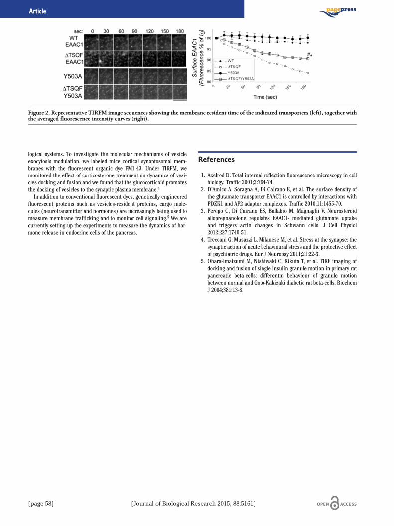
[page 58] [Journal of Biological Research 2015; 88:5161]
logical systems. To investigate the molecular mechanisms of vesicleexocytosis modulation, we labeled mice cortical synaptosomal mem-branes with the fluorescent organic dye FM1-43. Under TIRFM, wemonitored the effect of corticosterone treatment on dynamics of vesi-cles docking and fusion and we found that the glucocorticoid promotesthe docking of vesicles to the synaptic plasma membrane.4
In addition to conventional fluorescent dyes, genetically engineeredfluorescent proteins such as vesicles-resident proteins, cargo mole-cules (neurotransmitter and hormones) are increasingly being used tomeasure membrane trafficking and to monitor cell signaling.5 We arecurrently setting up the experiments to measure the dynamics of hor-mone release in endocrine cells of the pancreas.
References
1. Axelrod D. Total internal reflection fluorescence microscopy in cellbiology. Traffic 2001;2:764-74.
2. D’Amico A, Soragna A, Di Cairano E, et al. The surface density ofthe glutamate transporter EAAC1 is controlled by interactions withPDZK1 and AP2 adaptor complexes. Traffic 2010;11:1455-70.
3. Perego C, Di Cairano ES, Ballabio M, Magnaghi V. Neurosteroidallopregnanolone regulates EAAC1- mediated glutamate uptakeand triggers actin changes in Schwann cells. J Cell Physiol2012;227:1740-51.
4. Treccani G, Musazzi L, Milanese M, et al. Stress at the synapse: thesynaptic action of acute behavioural stress and the protective effectof psychiatric drugs. Eur J Neuropsy 2011;21:22-3.
5. Ohara-Imaizumi M, Nishiwaki C, Kikuta T, et al. TIRF imaging ofdocking and fusion of single insulin granule motion in primary ratpancreatic beta-cells: differentm behaviour of granule motionbetween normal and Goto-Kakizaki diabetic rat beta-cells. BiochemJ 2004;381:13-8.
Article
Figure 2. Representative TIRFM image sequences showing the membrane resident time of the indicated transporters (left), together withthe averaged fluorescence intensity curves (right).

Introduction
The oxidative stress results from a change in the physiological bal-ance between oxidant and antioxidant species. This condition inducesan chemical change in the redox state of cells. Ever growing evidencehas demonstrated that the trans- Plasma Membrane ElectronTransport (t-PMET) occurs in all types of organisms, including bacte-ria, yeast plants and animals.1 The function of t-PMET is to cooperatewith intracellular redox pairs, like piruvate and lactate, to maintain thecytoplasmatic NAD+/NADH balance. The t-PMET is correlated with themodulation of internal pH and redox homeostasis, as it is able to acti-vate proton release.1 Thus, t-PMET causes external and internal pHmodification, as well as development of an inside negative membranepotential. These changes are responsible of cellular functions alter-ations, including cell-volume sensing and regulation; mitogenic/apop-togenic signalling and transport across membranes1 The malfunctionof the tPMET is relates to some diseases such as cancer, cardiovasculardiseases, aging, obesity, neurodegenerative diseases, pulmonary fibro-sis, asthma.1 The activity of t-PMET is critical to redox homeostasis inblood. In hypoxic condition the activation of t-PMET may serve to com-pensate the impaired pentose phosphate pathway, thus ensuring afunctional reducing capacity; in this conditions t-PMET may use ascor-bic acid or polyphenols as electron donator, since NADH derived fromenhanced glycolysis is preferentially utilized by meta-hemoglobinreductase.2 The aim of this work was evaluate the erythrocytes redoxstatus in a group of health volunteers and then to study whether someof flavonoids, enclosed in sub-class of flavonols (Quercetin andKaempferol), are able to modify the erythrocytes redox homeostasis.Our attention has been focused on red blood cells (RBC) because aclose link between t-PMET and metabolic status of erythrocytes hasbeen reported.3 The RBCs act as antioxidants to themselves and, in
addition, their mobility carries their antioxidant capabilities to all theplasma accessible parts of the body.
Methods
The subjects that participating at this work attested no supplementintake or some other substance that could interfere with our tests. Theprotocol of the study was in conformity with the guidelines of theInstitutional Ethical Committee. Human venous blood from twentyhealthy volunteers of both sexes between the ages of 25-50 years wereobtained by venipuncture in heparin after an over-night fast and cen-trifuged. The antioxidant capacity of plasma was analyzed by crocinbleaching assay4 and FRAP.5 On the other hand, the reducing activityin erythrocytes that represents the body redox state of the last 120 dayswas evaluated by FRAP method.5 After centrifugation at 3000 rpm for10 minutes at 4°C, plasma was separate from red blood cells. Theresultant plasma was transferred to microcentrifuge tubes and used atleast in part for ferric-reducing activity power (FRAP) and for CrocinBleaching Assay (CBA) and the others aliquots was stored -80°Cbefore to use for further analysis. Red blood cells, after removal ofbuffy coat and upper 15% of the packed red blood cells, were washedtwo times with cold PBS according to Fiorani et al. method (2005). Astock solution (20mM) of each flavonoid was prepared in dimethyl sul-foxide and then diluted 1:2 with PBS. Packed RBC (10%v/v) were incu-bated in PBS containing 5mM glucose at 37°C for 10. minutes with a50 μM concentration of each flavonoids. After this time the suspen-sions was centrifuged, the RBC were washed and then analyzed. Theresulting supernatant fractions were assayed for their FOC contentusing 1,10-phenanthroline as an indicator and measuring absorptionat 510 nm. The results are expressed in �mol ferrocyanide/mL RBC/30min, through Lamber-Beer law, e=10500 M-1cm-1. The percentage ofhemolysis was evaluated in the same sample by measuring the haemo-globin contents. The Drabking test, purchased from Sigma-Aldrich, hasbeen used to evaluate the haemoglobin contents. The extent of lysiswas not different than the controls and never higher than 0.5%.
Results
Table 1 shows the values of plasma and erythrocyte antioxidant sta-tus of health volunteers. All the values confirm the data of literature6,7
about the antioxidant status of human being in physiological condi-tion. All compounds were taken up by the erythrocytes and displayingsignificant FIC-reducing activity. Figure 1 reports the values of reduc-ing capability of the control (erythrocytes without flavonoids) towards
Correspondence: Danila Di Majo, Physiology and Pharmacology Unit,Department of Legal, Social and Sports Sciences, University of Palermo,viale delle Scienze, Ed. 16, 90127 Palermo, Italy.E-mail: [email protected]
©Copyright D. Di Majo et al., 2015Licensee PAGEPress, ItalyJournal of Biological Research 2015; 88:5161
This article is distributed under the terms of the Creative CommonsAttribution Noncommercial License (by-nc 3.0) which permits any noncom-mercial use, distribution, and reproduction in any medium, provided the orig-inal author(s) and source are credited.
Influence of flavonoids on the transmembrane electron transport:study ex-vivoD. Di Majo,1 M. La Guardia,2 M. Crescimanno,1 C. Flandina,1 G. Leto,1 M. Giammanco11Physiology and Pharmacology Unit, Department of Legal, Social and Sports Sciences, Universityof Palermo; 2Division of Cell Biology, Department of Biological, Chemical and PharmaceuticalSciences and Technologies, University of Palermo, Italy
[Journal of Biological Research 2015; 88:5161] [page 59]
Journal of Biological Research 2015; volume 88:5161
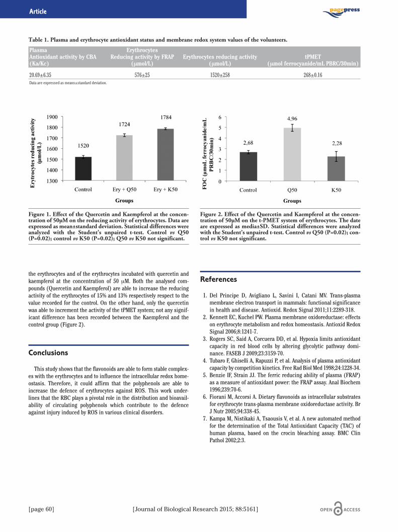
[page 60] [Journal of Biological Research 2015; 88:5161]
the erythrocytes and of the erythrocytes incubated with quercetin andkaempferol at the concentration of 50 μM. Both the analysed com-pounds (Quercetin and Kaempferol) are able to increase the reducingactivity of the erythrocytes of 15% and 13% respectively respect to thevalue recorded for the control. On the other hand, only the quercetinwas able to increment the activity of the tPMET system; not any signif-icant difference has been recorded between the Kaempferol and thecontrol group (Figure 2).
Conclusions
This study shows that the flavonoids are able to form stable complex-es with the erythrocytes and to influence the intracellular redox home-ostasis. Therefore, it could affirm that the polyphenols are able toincrease the defence of erythrocytes against ROS. This work under-lines that the RBC plays a pivotal role in the distribution and bioavail-ability of circulating polyphenols which contribute to the defenceagainst injury induced by ROS in various clinical disorders.
References
1. Del Principe D, Avigliano L, Savini I, Catani MV. Trans-plasmamembrane electron transport in mammals: functional significancein health and disease. Antioxid. Redox Signal 2011;11:2289-318.
2. Kennett EC, Kuchel PW. Plasma membrane oxidoreductase: effectson erythrocyte metabolism and redox homeostasis. Antioxid RedoxSignal 2006;8:1241-7.
3. Rogers SC, Said A, Corcuera DD, et al. Hypoxia limits antioxidantcapacity in red blood cells by altering glycolytic pathway domi-nance. FASEB J 2009;23:3159-70.
4. Tubaro F, Ghiselli A, Rapuzzi P, et al. Analysis of plasma antioxidantcapacity by competition kinetics. Free Rad Biol Med 1998;24:1228-34.
5. Benzie IF, Strain JJ. The ferric reducing ability of plasma (FRAP)as a measure of antioxidant power: the FRAP assay. Anal Biochem1996;239:70-6.
6. Fiorani M, Accorsi A. Dietary flavonoids as intracellular substratesfor erythrocyte trans-plasma membrane oxidoreductase activity. BrJ Nutr 2005;94:338-45.
7. Kampa M, Nistikaki A, Tsaousis V, et al. A new automated methodfor the determination of the Total Antioxidant Capacity (TAC) ofhuman plasma, based on the crocin bleaching assay. BMC ClinPathol 2002;2:3.
Article
Table 1. Plasma and erythrocyte antioxidant status and membrane redox system values of the volunteers.
Plasma ErythrocytesAntioxidant activity by CBA Reducing activity by FRAP Erythrocytes reducing activity tPMET(Ka/Kc) (μmol/L) (μmol/L) (μmol ferrocyanide/mL PBRC/30min)
20.69±6.35 576±25 1520±258 268±0.16Data are expressed as means±standard deviation.
Figure 1. Effect of the Quercetin and Kaempferol at the concen-tration of 50µM on the reducing activity of erythrocytes. Data areexpressed as mean±standard deviation. Statistical differences wereanalyzed with the Student’s unpaired t-test. Control vs Q50(P=0.02); control vs K50 (P=0.02); Q50 vs K50 not significant.
Figure 2. Effect of the Quercetin and Kaempferol at the concen-tration of 50µM on the t-PMET system of erythrocytes. The dateare expressed as media±SD. Statistical differences were analyzedwith the Student’s unpaired t-test. Control vsQ50 (P=0.02); con-trol vs K50 not significant.
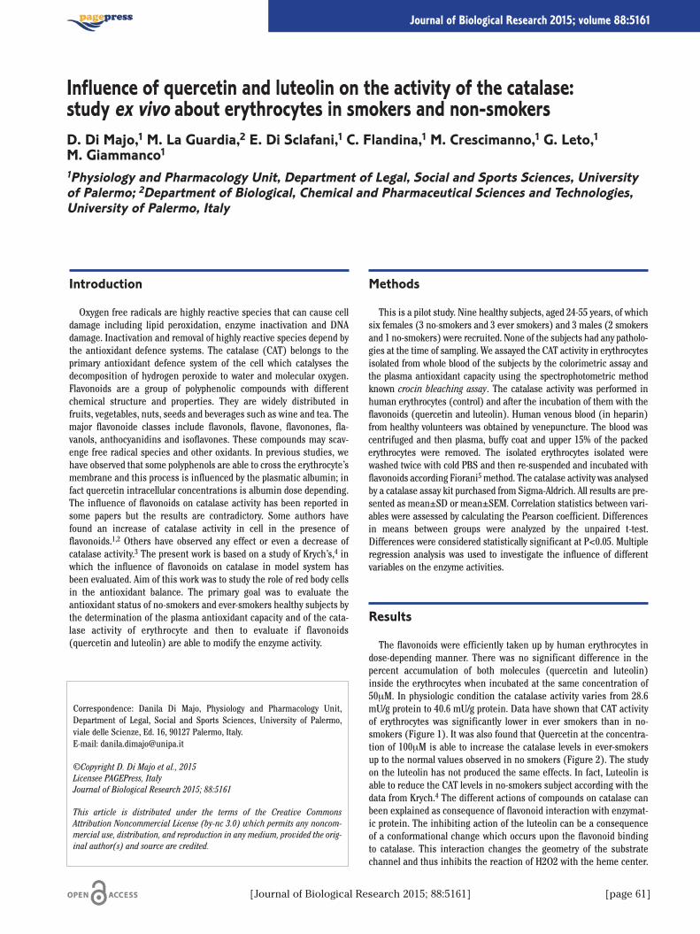
Introduction
Oxygen free radicals are highly reactive species that can cause celldamage including lipid peroxidation, enzyme inactivation and DNAdamage. Inactivation and removal of highly reactive species depend bythe antioxidant defence systems. The catalase (CAT) belongs to theprimary antioxidant defence system of the cell which catalyses thedecomposition of hydrogen peroxide to water and molecular oxygen.Flavonoids are a group of polyphenolic compounds with differentchemical structure and properties. They are widely distributed infruits, vegetables, nuts, seeds and beverages such as wine and tea. Themajor flavonoide classes include flavonols, flavone, flavonones, fla-vanols, anthocyanidins and isoflavones. These compounds may scav-enge free radical species and other oxidants. In previous studies, wehave observed that some polyphenols are able to cross the erythrocyte’smembrane and this process is influenced by the plasmatic albumin; infact quercetin intracellular concentrations is albumin dose depending.The influence of flavonoids on catalase activity has been reported insome papers but the results are contradictory. Some authors havefound an increase of catalase activity in cell in the presence offlavonoids.1,2 Others have observed any effect or even a decrease ofcatalase activity.3 The present work is based on a study of Krych’s,4 inwhich the influence of flavonoids on catalase in model system hasbeen evaluated. Aim of this work was to study the role of red body cellsin the antioxidant balance. The primary goal was to evaluate theantioxidant status of no-smokers and ever-smokers healthy subjects bythe determination of the plasma antioxidant capacity and of the cata-lase activity of erythrocyte and then to evaluate if flavonoids(quercetin and luteolin) are able to modify the enzyme activity.
Methods
This is a pilot study. Nine healthy subjects, aged 24-55 years, of whichsix females (3 no-smokers and 3 ever smokers) and 3 males (2 smokersand 1 no-smokers) were recruited. None of the subjects had any patholo-gies at the time of sampling. We assayed the CAT activity in erythrocytesisolated from whole blood of the subjects by the colorimetric assay andthe plasma antioxidant capacity using the spectrophotometric methodknown crocin bleaching assay. The catalase activity was performed inhuman erythrocytes (control) and after the incubation of them with theflavonoids (quercetin and luteolin). Human venous blood (in heparin)from healthy volunteers was obtained by venepuncture. The blood wascentrifuged and then plasma, buffy coat and upper 15% of the packederythrocytes were removed. The isolated erythrocytes isolated werewashed twice with cold PBS and then re-suspended and incubated withflavonoids according Fiorani5 method. The catalase activity was analysedby a catalase assay kit purchased from Sigma-Aldrich. All results are pre-sented as mean±SD or mean±SEM. Correlation statistics between vari-ables were assessed by calculating the Pearson coefficient. Differencesin means between groups were analyzed by the unpaired t-test.Differences were considered statistically significant at P<0.05. Multipleregression analysis was used to investigate the influence of differentvariables on the enzyme activities.
Results
The flavonoids were efficiently taken up by human erythrocytes indose-depending manner. There was no significant difference in thepercent accumulation of both molecules (quercetin and luteolin)inside the erythrocytes when incubated at the same concentration of50μM. In physiologic condition the catalase activity varies from 28.6mU/g protein to 40.6 mU/g protein. Data have shown that CAT activityof erythrocytes was significantly lower in ever smokers than in no-smokers (Figure 1). It was also found that Quercetin at the concentra-tion of 100μM is able to increase the catalase levels in ever-smokersup to the normal values observed in no smokers (Figure 2). The studyon the luteolin has not produced the same effects. In fact, Luteolin isable to reduce the CAT levels in no-smokers subject according with thedata from Krych.4 The different actions of compounds on catalase canbeen explained as consequence of flavonoid interaction with enzymat-ic protein. The inhibiting action of the luteolin can be a consequenceof a conformational change which occurs upon the flavonoid bindingto catalase. This interaction changes the geometry of the substratechannel and thus inhibits the reaction of H2O2 with the heme center.
Correspondence: Danila Di Majo, Physiology and Pharmacology Unit,Department of Legal, Social and Sports Sciences, University of Palermo,viale delle Scienze, Ed. 16, 90127 Palermo, Italy.E-mail: [email protected]
©Copyright D. Di Majo et al., 2015Licensee PAGEPress, ItalyJournal of Biological Research 2015; 88:5161
This article is distributed under the terms of the Creative CommonsAttribution Noncommercial License (by-nc 3.0) which permits any noncom-mercial use, distribution, and reproduction in any medium, provided the orig-inal author(s) and source are credited.
Influence of quercetin and luteolin on the activity of the catalase:study ex vivo about erythrocytes in smokers and non-smokersD. Di Majo,1 M. La Guardia,2 E. Di Sclafani,1 C. Flandina,1 M. Crescimanno,1 G. Leto,1M. Giammanco11Physiology and Pharmacology Unit, Department of Legal, Social and Sports Sciences, Universityof Palermo; 2Department of Biological, Chemical and Pharmaceutical Sciences and Technologies,University of Palermo, Italy
[Journal of Biological Research 2015; 88:5161] [page 61]
Journal of Biological Research 2015; volume 88:5161
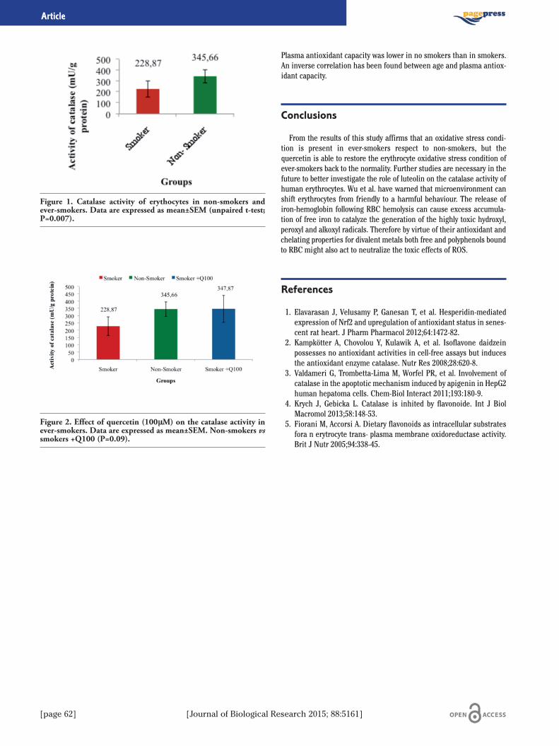
[page 62] [Journal of Biological Research 2015; 88:5161]
Plasma antioxidant capacity was lower in no smokers than in smokers.An inverse correlation has been found between age and plasma antiox-idant capacity.
Conclusions
From the results of this study affirms that an oxidative stress condi-tion is present in ever-smokers respect to non-smokers, but thequercetin is able to restore the erythrocyte oxidative stress condition ofever-smokers back to the normality. Further studies are necessary in thefuture to better investigate the role of luteolin on the catalase activity ofhuman erythrocytes. Wu et al. have warned that microenvironment canshift erythrocytes from friendly to a harmful behaviour. The release ofiron-hemoglobin following RBC hemolysis can cause excess accumula-tion of free iron to catalyze the generation of the highly toxic hydroxyl,peroxyl and alkoxyl radicals. Therefore by virtue of their antioxidant andchelating properties for divalent metals both free and polyphenols boundto RBC might also act to neutralize the toxic effects of ROS.
References
1. Elavarasan J, Velusamy P, Ganesan T, et al. Hesperidin-mediatedexpression of Nrf2 and upregulation of antioxidant status in senes-cent rat heart. J Pharm Pharmacol 2012;64:1472-82.
2. Kampkötter A, Chovolou Y, Kulawik A, et al. Isoflavone daidzeinpossesses no antioxidant activities in cell-free assays but inducesthe antioxidant enzyme catalase. Nutr Res 2008;28:620-8.
3. Valdameri G, Trombetta-Lima M, Worfel PR, et al. Involvement ofcatalase in the apoptotic mechanism induced by apigenin in HepG2human hepatoma cells. Chem-Biol Interact 2011;193:180-9.
4. Krych J, Gebicka L. Catalase is inhited by flavonoide. Int J BiolMacromol 2013;58:148-53.
5. Fiorani M, Accorsi A. Dietary flavonoids as intracellular substratesfora n erytrocyte trans- plasma membrane oxidoreductase activity.Brit J Nutr 2005;94:338-45.
Article
Figure 1. Catalase activity of erythocytes in non-smokers andever-smokers. Data are expressed as mean±SEM (unpaired t-test;P=0.007).
Figure 2. Effect of quercetin (100μM) on the catalase activity inever-smokers. Data are expressed as mean±SEM. Non-smokers vssmokers +Q100 (P=0.09).
Introduction
Blood transfusion (BT) as blood doping practice is banned by theWorld Anti-Doping Agency (WADA) and can be abused by cheatingathletes to increase the rate of oxygen transport to tissues with theaim to improve sport performance. At present, a method for the detec-tion of Homologous Blood Transfusion (HBT) has been implementedby the WADA accredited antidoping laboratories worldwide, while nointernationally recognized method has been finalized so far for thedirect detection of autologous blood transfusions (ABT), which can atpresent be only detected indirectly by targeting longitudinal profilingof key hematological parameters. In this perspective, several research-es approaching different fields are underway to find reliable biomark-ers to be suitable in the development of a method to directly detectautologous blood transfusion. In this work we have experimented thepossible role of microRNA (miRNAs) as biomarkers related to the exe-cution of an autologous blood transfusion practice. MiRNAs are a classof 18-24 nucleotides long non coding RNAs acting as post-trancription-al modulators of mammalian gene expression. Normally they areinvolved in the regulation of many physiological processes (such aserythropoiesis), and recent evidences shown that they have a role asuseful biomarkers in certain diseases such as cancer and heart malig-nancies. This study aims to experiment their possible role as biomark-ers of blood doping.
Materials and Methods
A total of eight miRNAs (mi923, mi150, mi144, mi96, mi196a,mi30b, mi197, mi451) have been extracted and quantified from sixwhole blood samples from healthy athletes at three different times
(T=0 within 24h from sample taking, T=1 after 15 days, T=2 after30 days). Another blood sample was withdrawn fresh from a donor,then stored as erythrocyte concentrated and, after 30 days, used to getan ex-vivo autologous blood transfusion with new fresh blood fromthe same donor. All miRNAs were extracted with a specific kit(miRneasy, Qiagen), then quantified with a specific ChipElectrophoresis System (Bioanalyzer 2100, Agilent), then retro-tran-scribed to cDNA and analyzed to quantitative PCR (qPCR) usingPCR7500 Fast system (Applied biosystems). Analysis of expression wasmade with the technique of relative quantification that was choosenas the more suitable for the aims of this experiments. In data analysis,a key step was the identification of the most appropriate housekeep-ing gene who resulted mi150 as its variability was found to be themost stable among all miRNAs studied. The study of the variation ofthe expression was evaluated estimating the relative quantities (RQ)of the miRNAs expressed by the samples compared to a calibrationsample used as reference.
Results and Discussion
We observed a gradual tendency of the miRNAs analyzed toincrease their expression in samples at T=2 compared to T=0 wheremi144 and mi923 shown the most consistent differences however withgreat variability among the samples. A marked differences has beenobserved for the erythrocytes concentrate sample (analyzed after 30days of storage) where expression levels of mi923, mi30b, mi197,mi96 and mi451 resulted higher compared to the fresh samples atthe time of the withdrawal. Moreover the most important result relieson the observation that the expression of some miRNAs (such asmi197, mi30b, mi451, mi96 and mi923) is very high in the erythro-cytes concentrated sample and it is detectable also in the ex- vivotransfused sample with levels higher compared to the fresh non-trans-fused sample (even though lower compared to the concentrated ery-throcytes sample because of the post-transfusion dilution effect).Moreover, two miRNAs (mi144 and mi923) show the most significantincreasing in the transfused sample compared to the fresh one. Asresult, it is very important to note that, from the data obtained, thepossibility to use miRNAs expression both as biomarkers of storageand biomarkers of effect in blood doping detetction emerges. Resultswe achieved in this work seem to be significant for several reasons.Firstly, at methodological level, the development of a specific protocolfor the extraction and the quantitation of miRNA and a proper strategyin data analysis (including a correct strategy in the choice of the mostsuitable housekeeping gene ) have allowed for the first time our labo-ratory to experiment and apply molecular biology techniques in thefield of doping control. More in detail, an accurate and sensitive quan-tification of the extracted miRNAs represented a first key step to sub-
Correspondence: Francesco Donati, Dope Testing Laboratory, Italian SportMedicine Federation, largo Giulio Onesti 1, 00197 Rome, Italy. E-mail: [email protected]
©Copyright F. Donati et al., 2015Licensee PAGEPress, ItalyJournal of Biological Research 2015; 88:5161
This article is distributed under the terms of the Creative CommonsAttribution Noncommercial License (by-nc 3.0) which permits any noncom-mercial use, distribution, and reproduction in any medium, provided the orig-inal author(s) and source are credited.
The approach of microRNA expression analysis in the detectionof autologous blood transfusion in doping controlF. Donati,1 F. Boccia,1 A. Stampella,1 X. de la Torre,1 F. Botrè1,21Dope Testing Laboratory, Italian Sport Medicine Federation, Rome; 2Management Department,University La Sapienza, Rome, Italy
[Journal of Biological Research 2015; 88:5161] [page 63]
Journal of Biological Research 2015; volume 88:5161

[page 64] [Journal of Biological Research 2015; 88:5161]
sequently achieve accurate and reproducibile genetic expression databy quantitative real time PCR.
Conclusions
Finally, the methodological approach here followed and resultsobtained in this work can be considered as preparatory to the openingof new research perspectives in the field of doping control. The nextstep we aim for the future is first of all to extend both the panel of
miRNAs to quantitate and the number of samples to be analyzed alsoconsidering the important issue of ethnical diversity between individ-uals. Moreover, the strategies and studies we intend to apply in thenear future also include the extention of this approach to differenthematological matrixes considering for example circulating miRNAsin serum and plasma and, in a further step, considering miRNAsextracted from urine samples also to the final scope to experiment andapply miRNAs expression data as biomarkers for the detection of otherbanned doping practices and drugs as for example the abuse of syn-thetic erythropoietins, insulin and analogs, growth hormone and relat-ed growth factors.
Article

Le scimmie del genere Aotus (Platyrrhine, Cebidae) presentanocaratteristiche peculiari essendo notturne e monogame. Hannoun’ampia distribuzione geografica che si estende dallo stretto diPanama al nord dell’Argentina; questo determina una notevole com-plessità nella distribuzione e di conseguenza nell’interpretazionesistematica e filogenetica. Lo studio del genere Aotus mediantel’analisi di caratte ri morfologici e genetici ha prodotto una tassono-mia controversa. Inizialmente era riconosciuta solo la specie Aotustrivirgatus, successivamente in base alla colorazione del pelo delcollo, alla diversa suscettibilità alla malaria e ai dati citogeneticisono state identificate fino a nove specie, oltre a due sibling species,per un totale di 11. 1,2 Il numero diploide di cromosomi in Aotus variada 46 a 56; nel cariotipo sono presenti molti polimorfismi dovutiall’ibridazione che si verifica in specie simpatiche; in alcune specieè presente una traslocazione Y-autosoma. Le undici specie sonostate suddivise in due gruppi monofiletici: il gruppo grey-black neckdistribuito a nord e il gruppo red neck distribuito a sud del Rio delleAmazzoni. Le specie del gruppo red neck sono omogenee da unpunto di v ista del cariotipo con un numero diplolide 2n=49(maschio)/50 (femmina) e una traslocazione Y- autosoma. Le speciedel gruppo grey-black neck presentano numero diploide di cromoso-mi variabile, con il più basso 2n=46 in A. vociferans e il più alto2n=56 in A. lemurinus. Solo due specie (Aotus nancymae e Aotuslemurinus) sono state analizzate mediante la citogenetica molecola-re (painting cromosomico). Quest’analisi ha permesso di dimostra-re che le specie del genere Aotus posseggono un cariotipo piuttostoderivato se confrontato con quello ipotetico ancestrale dellePlatyrrhinae, da cui si è originato attraverso fusioni, fissioni, traslo-cazioni ed inversioni.3-5
L’identificazione tassonomica di taxa di Aotus mediante l’analisicromosomica rappresenta, in cattività, il prerequisito per programmidi breeding in quanto la ricostruzione del cariotipo bandeggiato èl’unico approccio per identificare la maggior parte delle specie delgenere Aotus. Mediante le tecniche citogenetiche vengono identifica-
ti individui compatibili da un punto di vista cromosomico che posso-no essere incrociati tra loro al fine di evitare ibridazioni interspecie6
e favorire la conservazione delle diverse specie.L’obiettivo del presente lavoro riguarda l’identificazione mediante
bandeggio cromosomico di individui di una colonia di Aotus, al finedi avviare un programma di conservazione delle specie mediantebreeding. Il gruppo di scimmie, originario della Bolivia, è presente inGiappone dal 1977 presso The Primate Research Institute di Tokyo;tra gli individui della colonia si è inavvertitamente verificata la pro-duzione di ibridi prima che fossero riconosciute le diverse specie delgenere. Si riportano dati preliminari sull’identificazione di individuiidonei da incrociare, in particolare il cariotipo bandeggiato di unmaschio di A.l griseimbra (2n=54) (Figura 1). Inoltre si sono revisio-nati dati citogenetici presenti in letteratura su Aotus al fine di sotto-lineare l’importanza della citogenetica classica e molecolare neglistudi filogenetici e in quelli riguardanti la conservazione delle specie(Tabella 1).
Bibliografia
1. Hershkovitz P. Two new species of night monkeys, genus Aotus(Cebidae, Platyrrhini): a preliminary report on Aotus taxonomy.Am J Primatol 1983;4:209-43.
2. Ma NS, Jones TC, Miller AC, et al. Chromosome polymorphism andbanding patterns in the owl monkey (Aotus). Laboratory AnimalScience 1976;26:1022-36.
3. Stanyon R, Bigoni F, Slaby T, et al. Multi-directional chromosomepainting maps homologies between species belonging to threegenera of New World monkeys and humans. Chromosoma2004;113:305-15.
Correspondence: Francesca Dumas, Department of Biological, Chemical andPharmaceutical Sciences and Technologies, University of Palermo, viaArchirafi 18, 90127 Palermo, Italy.E-mail: [email protected]
©Copyright F. Dumas et al., 2015Licensee PAGEPress, ItalyJournal of Biological Research 2015; 88:5161
This article is distributed under the terms of the Creative CommonsAttribution Noncommercial License (by-nc 3.0) which permits any noncom-mercial use, distribution, and reproduction in any medium, provided the orig-inal author(s) and source are credited.
Identificazione tassonomica di Aotus (Platyrrhinae) mediante la citogeneticaF. Dumas,1 L. Sineo,1 T. Ishida21Department of Biological, Chemical and Pharmaceutical Sciences and Technologies, University ofPalermo, Italy; 2Graduate School of Science, University of Tokyo, Japan
[Journal of Biological Research 2015; 88:5161] [page 65]
Journal of Biological Research 2015; volume 88:5161
Figura 1. Cariotipo ricostruito mediante bandeggio g di un indi-viduo di Aotus l. griseimembra (2n=54) proveniente dal centroGiapponese; un maschio con un grande cromosoma submetacen-trico risultato di una fissione (prima riga, primo cromosoma) edue cromosomi non omologhi (seconda riga ultimi due cromoso-mi); ricostruzione in accordo con Ma e colleghi.2
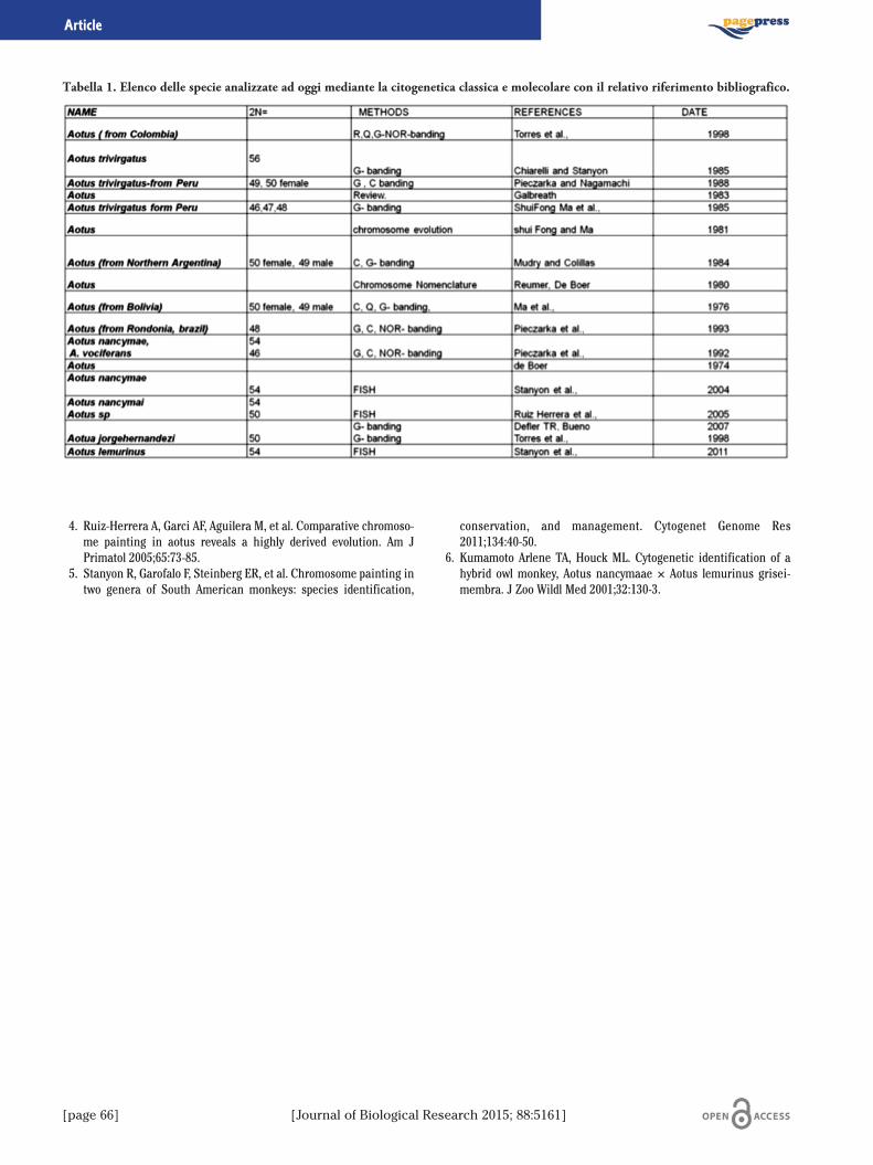
[page 66] [Journal of Biological Research 2015; 88:5161]
4. Ruiz-Herrera A, Garci�AF, Aguilera M, et al. Comparative chromoso-me painting in aotus reveals a highly derived evolution. Am JPrimatol 2005;65:73-85.
5. Stanyon R, Garofalo F, Steinberg ER, et al. Chromosome painting intwo genera of South American monkeys: species identification,
conservation, and management. Cytogenet Genome Res2011;134:40-50.
6. Kumamoto Arlene TA, Houck ML. Cytogenetic identification of ahybrid owl monkey, Aotus nancymaae × Aotus lemurinus grisei-membra. J Zoo Wildl Med 2001;32:130-3.
Article
Tabella 1. Elenco delle specie analizzate ad oggi mediante la citogenetica classica e molecolare con il relativo riferimento bibliografico.
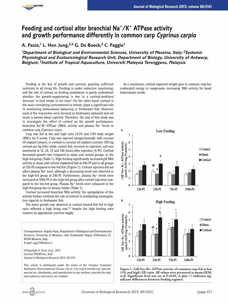
Feeding is the key of growth and survival, granting sufficientnutrients in all living life. Feeding is under endocrine monitoring,and the role of cortisol on feeding modulation is poorly understood:whether the growth-suppressing is due to a cortisol-mediateddecrease in food intake is not clear.1 On the other hand, cortisol isthe main circulating corticosteroid in teleost, plays a significant rolein monitoring homeostasis balancing in freshwater fish. However,most of the researches were focused on freshwater salmonid and notmuch is known about cyprinid. Therefore, the aim of this study wasto investigate the effect of cortisol on the growth performance,branchial Na+/K+-ATPase (NKA) activity and plasma Na+ levels incommon carp Cyprinus carpio.Carp was fed at low and high ratio (0.5% and 3.0% body weight
(BW)) for 6 weeks. Carp was injected intraperitoneally with coconutoil implant (sham), or cortisol in coconut oil implant (cortisol–250 mgcortisol per kg fish) while control fish received no injection, and wasmonitored at 12, 24, 72 and 168 hours after injection (h-PI). Cortisoldecreased growth rate compared to sham and control groups, in thehigh-fed group (Table 1). High feeding significantly increased gill NKAactivity in sham and cortisol implanted fish at 24h-PI and in all groupsat 72h-PI compared to low-fed fish (Figure 1). Cortisol injection did notaffect plasma Na+ level, although a decreasing trend was observed inthe high-fed group at 24h-PI. Furthermore, plasma Na+ levels wereincreased at 168h-PI in the high-fed group after cortisol injection com-pared to the low-fed group. Plasma Na+ levels were enhanced in thehigh-fed group due to dietary intake (Table 1).Cortisol increased branchial NKA activity: the upregulation of this
activity further confirms the role of cortisol in modulating ionoregula-tion capacity in freshwater fish.The lower growth rate observed in cortisol treated-fish fed to high
ratio reflected a high living cost,2-4 despite the high feeding ratioensures an appropriate nutrient supply.
As a conclusion, cortisol impaired weight gain in common carp butreallocated energy to compensate increasing NKA activity for basalhomeostasis needs.
Correspondence: Angela Fazio, Department of Biological and EnvironmentalSciences, University of Messina, viale Ferdinando Stagno D’Alcontres 31,98166 Messina, Italy.E-mail: [email protected]
©Copyright A. Fazio et al., 2015Licensee PAGEPress, ItalyJournal of Biological Research 2015; 88:5161
This article is distributed under the terms of the Creative CommonsAttribution Noncommercial License (by-nc 3.0) which permits any noncom-mercial use, distribution, and reproduction in any medium, provided the orig-inal author(s) and source are credited.
Feeding and cortisol alter branchial Na+/K+ ATPase activityand growth performance differently in common carp Cyprinus carpioA. Fazio,1 L. Hon Jung,2,3 G. De Boeck,2 C. Faggio11Department of Biological and Environmental Sciences, University of Messina, Italy; 2SystemicPhysiological and Ecotoxicological Research Unit, Department of Biology, University of Antwerp,Belgium; 3Institute of Tropical Aquaculture, Universiti Malaysia Terengganu, Malaysia
[Journal of Biological Research 2015; 88:5161] [page 67]
Journal of Biological Research 2015; volume 88:5161
Figure 1. Gill Na+/K+ ATPase activity of common carp fed at low(1A) and high (1B) ratio. All values were presented as mean±SEM,n=8. Significant level was set at P<0.05. A plus (+) indicates sig-nificant differences between feeding regimes.

[page 68] [Journal of Biological Research 2015; 88:5161]
References
1. Bernier NJ, Peter RE. Appetite-suppressing effects of urotensin Iand corticotropin-releasing hormone in goldfish (Carassius aura-tus). Neuroendocrinology 2001;73:248-60.
2. Bernier NJ, Bedard N, Peter RE. Effects of cortisol on food intake,growth, and forebrain neuropeptide Y and corticotropin-releasing
factor gene expression in goldfish. Gen Comp Endocrinol 2004;135:230-40.
3. De Boeck G, Alsop D, Wood CM. Cortisol effects on aerobic andanaerobic metabolism, nitrogen excretion, and whole-body composi-tion in juvenile rainbow trout. Phys Biochem Zool 2001;74:858-68.
4. Gregory TR, Wood CM. The effects of chronic plasma cortisol eleva-tion on the feeding behaviour, growth, competitive ability, andswimming performance of juvenile rainbow trout. Phys BiochemZool 1999;72:286-95.
Article
Table 1. Growth rate (%) and Plasma Na+ (mmol/L) of common carp fed at different ratio without (control) and with implantation(sham and cortisol) over 168h-PI. All values were presented as mean±SEM, n=8. Significant level was set at P<0.05. A plus (+) indicatessignificant differences between feeding regimes.
Low feeding High feedingControl Sham Cortisol Control Sham Cortisol
Growth rate (%) 12 h-PI 3.2±0.9 0.7±0.4 1.0±0.4 9.6±0.4+ 2.2±0.8 1.5±0.924 h-PI 4.2±0.6 1.6±1.0 1.8±0.5 6.2±0.7 2.2±0.7 2.4±0.872 h-PI 2.3±1.0 1.1±0.3 0.8±0.5 5.3±0.4+ 2.0±0.4 2.2±0.6168 h-PI 2.2±0.7 0.8±0.1 0.8±0.1 7.0±1.0+ 4.1±0.9+ 2.5±0.7
Plasma Na+ (mmol/L) 12 h-PI 119±4.6 113±4.9 107±2.9 131±5.8 126±5.3 118±4.624 h-PI 122±1.5 114±5.5 110±2.7 136±1.8+ 123±5.1 115±7.272 h-PI 121±2.2 116±4.3 111±3.7 133±4.1+ 127±6.9 119±6.3168 h-PI 118±7.0 113±6.1 109±2.9 130±1.8 125±4.6 124±4.9+

Introduzione
Tutte le specie di Moringa sono originarie delle regioni Sub-Himalaiane e la più diffusa risulta essere la Moringa oleifera v.Lamarck attualmente coltivata in diversi paesi dell’Africa, dell’Asia eSud-America. Dalla fine degli anni ottanta si è registrato un crescendodi lavori centrati sulla possibile applicazione in campo alimentare, far-maceutico, cosmetico1 nonché agricolo delle radici, delle foglie, deisemi oppure dell’olio di moringa. Sono apparse una miriade di pubbli-cazioni che, spesso attingendo dalla tradizione e/o medicina popolare,evocano per i vari derivati di moringa proprietà iperboliche, miracoli-stiche se non magiche.2 Di fatto pochi lavori esaminano rigorosamen-te la chimica dei costituenti e rari sono gli studi clinici2 che ne docu-mentano l’efficacia nutrizionale in modo puntuale.2-4 D’altra parte, nelsahel senegalese, zona da cui provengono le foglie e i semi oggetto diquesto studio, l’albero di moringa è considerato una risorsa polivalen-te. Il legno è impiegato in falegnameria o come legna per ardere, lefoglie fresche vengono utilizzate per integrare l’alimentazione se sec-cate e ridotte in polvere vengono impiegate per combattere il diabete.Dell’olio ne fanno un uso culinario nonché cosmetico. Scopo di questostudio preliminare e quello di monitorare e le capacità antiossidanti eil profilo degli acidi grassi introducendo un semplice, ma efficacemodo per calcolare la relazione tra saturi ed insaturi, che aiuta a chia-rire la notevole resistenza dell’olio di moringa all’ossidazione.
Materiali e Metodi
Il profilo degli acidi grassi è stato effettuato mediante trans metila-zione5 e analizzati in gas-cromatografia con rivelatore di massa. Laconcentrazione dei polifenoli totali liberi è stata eseguita mediante ilFolin-Ciocolteau.6 La capacitaà antiossidante è stata misurata median-te lo sbiancamento della crocina.7
Risultati e Discussione
Come si può osservare dalla Tabella 1 l’olio di moringa presenta unprofilo in acidi grassi molto simile a quello dell’olio di oliva, special-mente nel C18:1, in più nell’olio di moringa è presente in modo cospi-cuo il C22:0. In Tabella 2 sono riportati i valori dei polifenoli e dellacapacità antiossidante dell’estratto acqua:metanolo delle foglie dimoringa, comparati con quelli di altri prodotti alimentari. I valori pervini e per il the bianco sono riportati in Di Majo e Giammanco.8 Comeè possibile osservare dalla tabella l’estratto alcolico presenta eccellentivalori sia di capacità antiossidante che di concentrazione di polifenolitotali liberi. In questo studio abbiamo voluto introdurre un diversoapproccio matematico rispetto ai calcoli che comunemente si fannoper quanto riguarda il rapporto Sat/Ins. A nostro avviso questo tipo dicalcolo è fuorviante, in quanto non tiene conto del grado d’insaturazio-ne dei diversi acidi grassi insaturi. Però anche il solo grado d’insatu-razione non è esaustivo, perché non considera assolutamente gli acidigrassi saturi, perciò abbiamo introdotto un altro tipo di rapporto e pre-cisamente il rapporto Saturi/Grado d’insaturazione.
Conclusioni
In conclusione, mediante questo nuovo rapporto si evidenzia comel’olio di moringa si discosta nettamente dall’olio extravergine di oliva,cosa non molto evidente con gli altri due indici (Sat/Ins, e Grado d’in-saturazione), collocandolo tra l’olio extravergine di oliva e lo strutto. Siè inoltre verificato il buon corredo antiossidante custodito dalle fogliedi questa pianta.
Bibliografia
1. Anwar F, Latif S, Ashraf M, Gilani AH. Moringa oleifera: a food plantwith multiple medicinal uses. Phytother Res 2007;21:17-25.
2. Fahey JW. Moringa oleifera: a review of the medical evidence for itsnutritional, therapeutic, and prophylactic properties. Trees Life J2005;1:5.
3. Abdulkarim SM, Lai OM, Muhammad SKS, et al. Frying quality andstability of high-oleic Moringa oleifera seed oil in comparison withother vegetable oils. Food Chem 2007;105:1382-9.
4. Tsaknis J, Lalas S. Stability during frying of Moringa oleifera seedoil variety Periyakulam 1. J Food Comp Anal 2002;15:79-101.
5. Mingrone G, Greco AV, Finotti E, Passi S. Free fatty acid: a stimulusfor mucin hypersecretion in cholesterol gallstone biles. BiochBioph Acta 1988;958:52-9.
6. Coseteng MY, Lee CY. Changes in apple polyphenolxidase and poly-
Correspondence: Enrica Finotti, CRA-NUT-Food and Nutrition ResearchCenter, via Ardeatina 546, 00178 Rome, Italy.E-mail: [email protected]
©Copyright E. Finotti et al., 2015Licensee PAGEPress, ItalyJournal of Biological Research 2015; 88:5161
This article is distributed under the terms of the Creative CommonsAttribution Noncommercial License (by-nc 3.0) which permits any noncom-mercial use, distribution, and reproduction in any medium, provided the orig-inal author(s) and source are credited.
Caratteristiche nutrizionali dell’olio di Moringa oleiferaE. Finotti,1 G. Rossoni,1 I. Garaguso,1 M. Giammanco,2 D. Di Majo21CRA-NUT-Food and Nutrition Research Center, Rome; 2Physiology and Pharmacology Unit,Department of Legal, Social and Sports Sciences, University of Palermo, Italy
[Journal of Biological Research 2015; 88:5161] [page 69]
Journal of Biological Research 2015; volume 88:5161
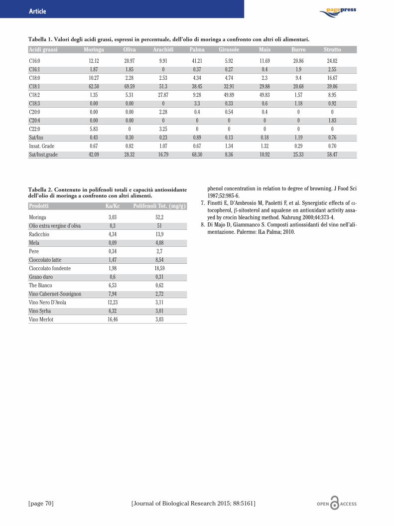
[page 70] [Journal of Biological Research 2015; 88:5161]
phenol concentration in relation to degree of browning. J Food Sci1987;52:985-6.
7. Finotti E, D’Ambrosio M, Paoletti F, et al. Synergistic effects of α-tocopherol, β-sitosterol and squalene on antioxidant activity assa-yed by crocin bleaching method. Nahrung 2000;44:373-4.
8. Di Majo D, Giammanco S. Composti antiossidanti del vino nell’ali-mentazione. Palermo: ILa Palma; 2010.
Article
Tabella 1. Valori degli acidi grassi, espressi in percentuale, dell’olio di moringa a confronto con altri oli alimentari.
Acidi grassi Moringa Oliva Arachidi Palma Girasole Mais Burro Strutto
C16:0 12.12 20.97 9.91 41.21 5.92 11.69 20.86 24.02C16:1 1.87 1.85 0 0.37 0.27 0.4 1.9 2.55C18:0 10.27 2.28 2.53 4.34 4.74 2.3 9.4 16.67C18:1 62.50 69.59 51.3 38.45 32.91 29.88 20.68 39.06C18:2 1.35 5.31 27.87 9.28 49.89 49.83 1.57 8.95C18:3 0.00 0.00 0 3.3 0.33 0.6 1.18 0.92C20:0 0.00 0.00 2.28 0.4 0.54 0.4 0 0C20:4 0.00 0.00 0 0 0 0 0 1.83C22:0 5.83 0 3.25 0 0 0 0 0Sat/Ins 0.43 0.30 0.23 0.89 0.13 0.18 1.19 0.76Insat. Grade 0.67 0.82 1.07 0.67 1.34 1.32 0.29 0.70Sat/Inst.grade 42.09 28.32 16.79 68.30 8.36 10.92 25.33 58.47
Tabella 2. Contenuto in polifenoli totali e capacità antiossidantedell’olio di moringa a confronto con altri alimenti.
Prodotti Ka/Kc Polifenoli Tot. (mg/g)
Moringa 3,03 52,2Olio extra vergine d’oliva 0,3 51Radicchio 4,34 13,9Mela 0,09 4,08Pere 0,34 2,7Cioccolato latte 1,47 8,54Cioccolato fondente 1,98 18,59Grano duro 0,6 0,31The Bianco 6,53 0,62Vino Cabernet-Souvignon 7,94 2,72Vino Nero D’Avola 12,23 3,11Vino Syrha 6,32 3,01Vino Merlot 16,46 3,03
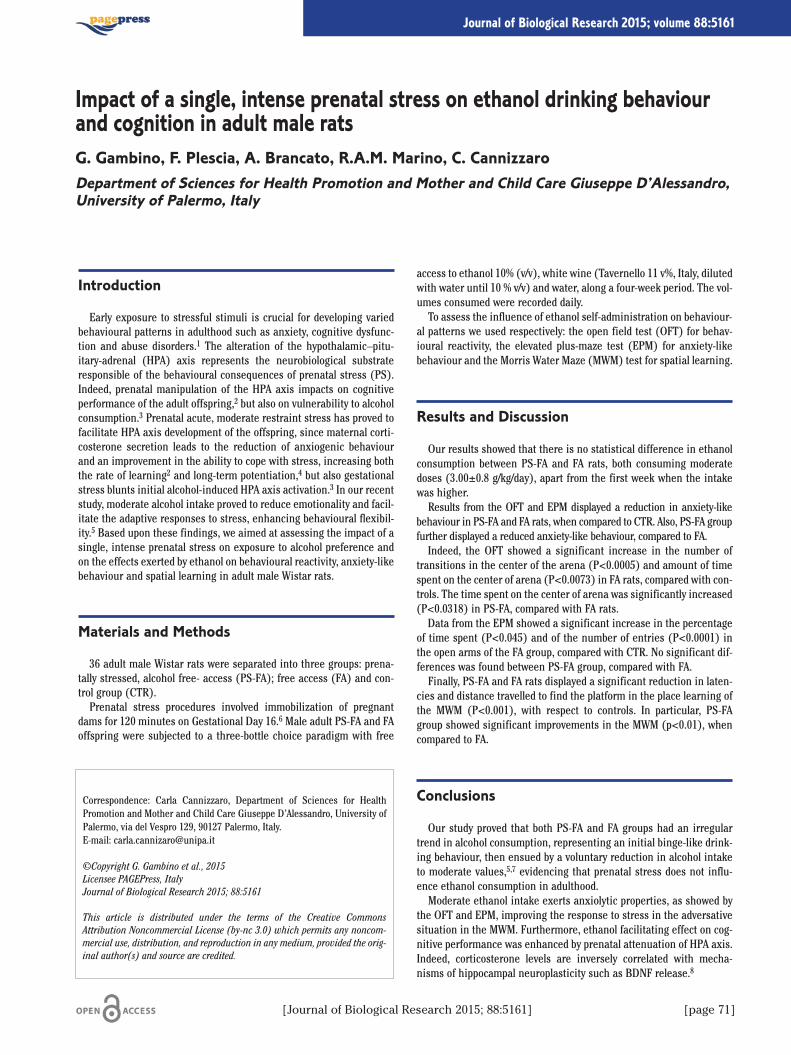
Introduction
Early exposure to stressful stimuli is crucial for developing variedbehavioural patterns in adulthood such as anxiety, cognitive dysfunc-tion and abuse disorders.1 The alteration of the hypothalamic–pitu-itary-adrenal (HPA) axis represents the neurobiological substrateresponsible of the behavioural consequences of prenatal stress (PS).Indeed, prenatal manipulation of the HPA axis impacts on cognitiveperformance of the adult offspring,2 but also on vulnerability to alcoholconsumption.3 Prenatal acute, moderate restraint stress has proved tofacilitate HPA axis development of the offspring, since maternal corti-costerone secretion leads to the reduction of anxiogenic behaviourand an improvement in the ability to cope with stress, increasing boththe rate of learning2 and long-term potentiation,4 but also gestationalstress blunts initial alcohol-induced HPA axis activation.3 In our recentstudy, moderate alcohol intake proved to reduce emotionality and facil-itate the adaptive responses to stress, enhancing behavioural flexibil-ity.5 Based upon these findings, we aimed at assessing the impact of asingle, intense prenatal stress on exposure to alcohol preference andon the effects exerted by ethanol on behavioural reactivity, anxiety-likebehaviour and spatial learning in adult male Wistar rats.
Materials and Methods
36 adult male Wistar rats were separated into three groups: prena-tally stressed, alcohol free- access (PS-FA); free access (FA) and con-trol group (CTR).Prenatal stress procedures involved immobilization of pregnant
dams for 120 minutes on Gestational Day 16.6 Male adult PS-FA and FAoffspring were subjected to a three-bottle choice paradigm with free
access to ethanol 10% (v/v), white wine (Tavernello 11 v%, Italy, dilutedwith water until 10 % v/v) and water, along a four-week period. The vol-umes consumed were recorded daily.To assess the influence of ethanol self-administration on behaviour-
al patterns we used respectively: the open field test (OFT) for behav-ioural reactivity, the elevated plus-maze test (EPM) for anxiety-likebehaviour and the Morris Water Maze (MWM) test for spatial learning.
Results and Discussion
Our results showed that there is no statistical difference in ethanolconsumption between PS-FA and FA rats, both consuming moderatedoses (3.00±0.8 g/kg/day), apart from the first week when the intakewas higher.Results from the OFT and EPM displayed a reduction in anxiety-like
behaviour in PS-FA and FA rats, when compared to CTR. Also, PS-FA groupfurther displayed a reduced anxiety-like behaviour, compared to FA.Indeed, the OFT showed a significant increase in the number of
transitions in the center of the arena (P<0.0005) and amount of timespent on the center of arena (P<0.0073) in FA rats, compared with con-trols. The time spent on the center of arena was significantly increased(P<0.0318) in PS-FA, compared with FA rats.Data from the EPM showed a significant increase in the percentage
of time spent (P<0.045) and of the number of entries (P<0.0001) inthe open arms of the FA group, compared with CTR. No significant dif-ferences was found between PS-FA group, compared with FA.Finally, PS-FA and FA rats displayed a significant reduction in laten-
cies and distance travelled to find the platform in the place learning ofthe MWM (P<0.001), with respect to controls. In particular, PS-FAgroup showed significant improvements in the MWM (p<0.01), whencompared to FA.
Conclusions
Our study proved that both PS-FA and FA groups had an irregulartrend in alcohol consumption, representing an initial binge-like drink-ing behaviour, then ensued by a voluntary reduction in alcohol intaketo moderate values,5,7 evidencing that prenatal stress does not influ-ence ethanol consumption in adulthood.Moderate ethanol intake exerts anxiolytic properties, as showed by
the OFT and EPM, improving the response to stress in the adversativesituation in the MWM. Furthermore, ethanol facilitating effect on cog-nitive performance was enhanced by prenatal attenuation of HPA axis.Indeed, corticosterone levels are inversely correlated with mecha-nisms of hippocampal neuroplasticity such as BDNF release.8
Correspondence: Carla Cannizzaro, Department of Sciences for HealthPromotion and Mother and Child Care Giuseppe D’Alessandro, University ofPalermo, via del Vespro 129, 90127 Palermo, Italy. E-mail: [email protected]
©Copyright G. Gambino et al., 2015Licensee PAGEPress, ItalyJournal of Biological Research 2015; 88:5161
This article is distributed under the terms of the Creative CommonsAttribution Noncommercial License (by-nc 3.0) which permits any noncom-mercial use, distribution, and reproduction in any medium, provided the orig-inal author(s) and source are credited.
Impact of a single, intense prenatal stress on ethanol drinking behaviourand cognition in adult male ratsG. Gambino, F. Plescia, A. Brancato, R.A.M. Marino, C. CannizzaroDepartment of Sciences for Health Promotion and Mother and Child Care Giuseppe D’Alessandro,University of Palermo, Italy
[Journal of Biological Research 2015; 88:5161] [page 71]
Journal of Biological Research 2015; volume 88:5161

[page 72] [Journal of Biological Research 2015; 88:5161]
In conclusion, our findings further highlight the role of prenatalexperiences on ethanol-induced mechanisms of neuroadaptation, indi-cating that a single, intense stress during early gestational periodinteracts with alcohol effects in adulthood.
References
1. Weinstock M. The potential influence of maternal stress hormoneson development and mental health of the offspring. Brain BehavImmunol 2005;19:296-308.
2. Cannizzaro C. Single, intense prenatal stress decreases emotional-ity and enhances learning performance in the adolescent rat off-spring: interaction with a brief, daily maternal separation. BehavBrain Res 2006;169:128-36.
3. Van Waes V. Hypo-response of the hypothalamic-pituitary-adreno-
cortical axis after an ethanol challenge in prenatally stressed ado-lescent male rats. Eur J Neurosci 2006;24:1193-200.
4. Fujioka A. Differential effects of prenatal stress on the morpholog-ical maturation of hippocampal neurons. Neuroscience2006;141:907-15.
5. Cacace S. Evaluation of chronic alcohol self-administration by a 3-bottle choice paradigm in adult male rats. Effects on behaviouralreactivity, spatial learning and reference memory. Behav Brain Res2011;219:213-20.
6. Pacak K. Adrenalectomy augments in vivo release of norepineph-rine in the paraventricular nucleus during immobilization stress.Endocrinology 1993;133:1404-10.
7. Juárez J, Barrios de Tomasi E. Sex differences in alcohol drinkingpatterns during forced and voluntary consumption in rats. Alcohol1999;19:15-22.
8. Radecki DT. BDNF protects against stress-induced impairments inspatial learning and memory and LTP. Hippocampus 2005;15:246-53.
Article
Intestinal epithelial cells play an important role in the mucosalinflammatory response. These cells synthesize and secrete inflamma-tory mediators, and selectively modulate the permeability of theepithelial monolayer thus exposing immune cells to antigens.Although intestinal inflammatory response is crucial to maintain gutstructural integrity and function, alteration and dysregulation ofinflammatory pathways contribute to tissue damage and ulceration,and are thought to be pivotal factors in the pathogenesis of differentinflammatory gut diseases.1 The limited efficacy of conventional phar-macological therapy in the intestinal inflammatory conditions has fos-tered research on alternatives and, at the same time, stresses theimportance of prevention. In this context, the influence of dietary com-ponents, becoming in a physiological close proximity to intestinal cellsand then to inflammatory processes within the intestinal mucosa,appears of nutritional and clinical interest.2 Among natural preventiveand complementary approaches to improve inflammatory symptoms,dietary polyphenols represent potential candidates and proanthocyani-dins are particularly interesting.3 For their relatively high concentra-tion in a number of edible plants and their high digestive stability andlimited intestinal adsorption,4,5 proanthocyanidins reach the colon atrelatively high concentrations and may have direct effects on the intes-tinal mucosa through their interaction with the intestinal epithelialcell membranes.6,7
The edible pistachio nut has been ranked among the first 50 foodproducts highest in antioxidant potential.8 A number of data show thatthe pistachio nut consumption has positive effects in human serumlipid profile and cardiovascular disease (CVD) risk factors9,10 and sig-nificantly improves oxidative status and reduces circulating inflamma-tory biomarkers.11 Our previous research provided evidence that ahydrophilic extract from Sicilian pistachio nuts (HPE) contains sub-stantial amounts of polyphenols, including proanthocyanidins, andpossesses radical scavenging and antioxidative properties in in vitromodels of lipid oxidation.12 Moreover we also demonstrated that HPE
has anti-inflammatory activities in lipopolysaccharide (LPS)-activatedmacrophages interfering with the NF-kB activation, and that the highmolecular weight proanthocyanidin fraction (PF) can play a major roleas the bioactive component of HPE.13
In the present study we investigated the activity of HPE, and of itspolymeric proanthocyanidin fraction as well, in an in vitro model ofintestinal inflammation, consisting of Caco-2 cells differentiated intoepithelial intestinal cells and exposed to the inflammatory actions ofinterleukin (IL-1β). Our results clearly show that HPE effectivelyinhibits the inflammatory response in intestinal epithelial cells, and thathighly polymeric proanthocyanidin components exhibit qualitative andquantitative effects substantially comparable to those of whole extractswhen tested at the same concentration found in the extracts. The pro-tective effects are expressed through a marked decrease in release andexpression of inflammatory mediators and occur in parallel with areduced activation of the nuclear factor-1β. Moreover, our results clearlyshow that HPE can partially prevent the IL-1β-induced gap formationwith perturbation of the monolayer integrity, as shown by a limited IL-1β-induced increase of paracellular permeability. Finally we provide evi-dence that HPE treatment increases transepithelial electrical resistenceof Caco-2 cells monolayer, demonstrating that protective effects of HPEunder our conditions occur in parallel with molecular interaction of nutcomponents with the epithelial cells membranes.To assess the physiological relevance of the tested concentration it
is worth noting that a single serving of pistachio nut (28.34 g) (USDANational Nutrient Database for Standard Reference) contains around62,34 mg polymeric proanthocyanidins (cyanidin equivalents). Oncediluted in a gastrointestinal volume of 600 mL, this result in a 3.5 µMconcentration (104 µg/mL) (as cyanidin equivalents) of polymericproanthocyanidins which represent a plausible concentration in thehuman gut.14 This concentration is one order higher than the concen-trations selected in our cell model and this suggests that our resultsmight be physiologically relevant in the gastrointestinal tract.Data here presented may further remark the potentially beneficial
health effects that may arise by daily intake of small quantity of pista-chio nut. Widely available, inexpensive and frequently consumed, thisnut for its favorable fatty acid profile and high content in bioactiveantioxidant compounds, positively influences the plasma lipid param-eters and oxidative status, and elicits antinflammatory properties. Inthis respect high content in large proanthocyanidins consumption ofpistachio nut can exerts locally significant beneficial effects to physi-ology of gastrointestinal tract.
References
1. Mayer L. Evolving paradigms in the pathogenesis of IBD. JGastroenterol. 2010;45:9-16.
Correspondence: Carla Gentile, Department of Biological, Chemical andPharmaceutical Sciences and Technologies, University of Palermo, vialedelle Scienze, Ed. 16, 90127 Palermo, Italy.E-mail: [email protected]
©Copyright C. Gentile et al., 2015Licensee PAGEPress, ItalyJournal of Biological Research 2015; 88:5161
This article is distributed under the terms of the Creative CommonsAttribution Noncommercial License (by-nc 3.0) which permits any noncom-mercial use, distribution, and reproduction in any medium, provided the orig-inal author(s) and source are credited.
Anti-inflammatory effects of Sicilian pistachio (Pistacia vera L.) nutin an in vitro model of human intestinal epitheliumC. Gentile, A. Perrone, A. Attanzio, A.M. Pintaudi, L. Tesoriere, M.A. LivreaDepartment of Biological, Chemical and Pharmaceutical Sciences and Technologies, University ofPalermo, Italy
[Journal of Biological Research 2015; 88:5161] [page 73]
Journal of Biological Research 2015; volume 88:5161

[page 74] [Journal of Biological Research 2015; 88:5161]
2. Netsch MI, Gutmann H, Aydogan C, Drewe J. Green tea extractinduces interleukin-8 (IL-8) mRNA and protein expression butspecifically inhibits IL-8 secretion in caco-2 cells. Planta Med2006;72:697-702.
3. Shapiro H, Singer P, Halpern Z, Bruck R. Polyphenols in the treat-ment of inflammatory bowel disease and acute pancreatitis. Gut2007;56:426-35.
4. Deprez S, Mila I, Huneau JF, et al. Transport of proanthocyanidindimer, trimer, and polymer across monolayers of human intestinalepithelial Caco-2 cells. Antioxid Redox Signal 2001;3:957-67.
5. Rios LY, Bennett RN, Lazarus SA, et al. Cocoa procyanidins are stableduring gastric transit in humans. Am J Clin Nutr 2002;76:1106-10.
6. Erlejman AG, Jaggers G, Fraga CG, Oteiza PI. TNFalpha-inducedNF-kappaB activation and cell oxidant production are modulated byhexameric procyanidins in Caco-2 cells. Arch Biochem Biophys2008;476:186-95.
7. Oteiza PI, Erlejman AG, Verstraeten SV, et al. Flavonoid-membraneinteractions: a protective role of flavonoids at the membrane sur-face? Clin Dev Immunol 2006;12:19-25.
8. Halvorsen BL, Carlsen MH, Phillips KM, et al. Content of redox-active compounds (ie, antioxidants) in foods consumed in the
United States. Am J Clin Nutr 2006;84:95-135.9. Edwards K, Kwaw I, Matud J, Kurtz I. Effect of pistachio nuts onserum lipid levels in patients with moderate hypercholesterolemia.J Am Coll Nutr 1999;18:229-32.
10. Kocyigit A, Koylu AA, Keles H. Effects of pistachio nuts consump-tion on plasma lipid profile and oxidative status in healthy volun-teers. Nutr Metab Cardiovasc Dis 2006;16:202-9.
11. Sari I, Baltaci Y, Bagci C, et al. Effect of pistachio diet on lipidparameters, endothelial function, inflammation, and oxidative sta-tus: a prospective study. Nutrition. 2010;26:399-404.
12. Gentile C, Allegra M, Angileri F, et al. Polymeric proanthocyanidinsfrom Sicilian pistachio (Pistacia vera L.) nut extract inhibitlipopolysaccharide-induced inflammatory response in RAW 264.7cells. Eur J Nutr 2012;51:353-63.
13. Gentile C, Tesoriere L, Butera D, et al. Antioxidant activity ofSicilian pistachio (Pistacia vera L. var. Bronte) nut extract and itsbioactive components. J Agric Food Chem 2007;55:643-8.
14. Fraga CG, Galleano M, Verstraeten SV, Oteiza PI. Basic biochemicalmechanisms behind the health benefits of polyphenols. MolAspects Med. 2010;31:435-45.
Article
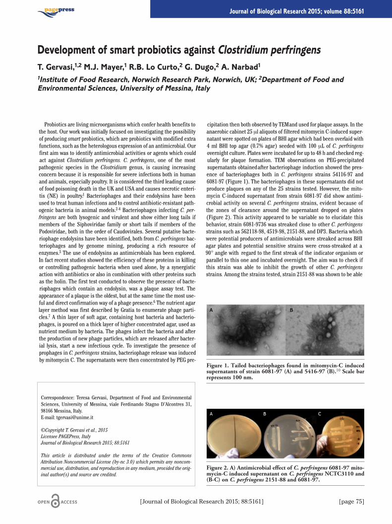
Probiotics are living microorganisms which confer health benefits tothe host. Our work was initially focused on investigating the possibilityof producing smart probiotics, which are probiotics with modified extrafunctions, such as the heterologous expression of an antimicrobial. Ourfirst aim was to identify antimicrobial activities or agents which couldact against Clostridium perfringens. C. perfringens, one of the mostpathogenic species in the Clostridium genus, is causing increasingconcern because it is responsible for severe infections both in humanand animals, especially poultry. It is considered the third leading causeof food poisoning death in the UK and USA and causes necrotic enteri-tis (NE) in poultry.1 Bacteriophages and their endolysins have beenused to treat human infections and to control antibiotic-resistant path-ogenic bacteria in animal models.2-4 Bacteriophages infecting C. per-fringens are both lysogenic and virulent and show either long tails ifmembers of the Siphoviridae family or short tails if members of thePodoviridae, both in the order of Caudovirales. Several putative bacte-riophage endolysins have been identified, both from C. perfringens bac-teriophages and by genome mining, producing a rich resource ofenzymes.5 The use of endolysins as antimicrobials has been explored.In fact recent studies showed the efficiency of these proteins in killingor controlling pathogenic bacteria when used alone, by a synergisticaction with antibiotics or also in combination with other proteins suchas the holin. The first test conducted to observe the presence of bacte-riophages which contain an endolysin, was a plaque assay test. Theappearance of a plaque is the oldest, but at the same time the most use-ful and direct confirmation way of a phage presence.6 The nutrient agarlayer method was first described by Gratia to enumerate phage parti-cles.7 A thin layer of soft agar, containing host bacteria and bacterio-phages, is poured on a thick layer of higher concentrated agar, used asnutrient medium by bacteria. The phages infect the bacteria and afterthe production of new phage particles, which are released after bacter-ial lysis, start a new infectious cycle. To investigate the presence ofprophages in C. perfringens strains, bacteriophage release was inducedby mitomycin C. The supernatants were then concentrated by PEG pre-
cipitation then both observed by TEM and used for plaque assays. In theanaerobic cabinet 25 μl aliquots of filtered mitomycin C-induced super-natant were spotted on plates of BHI agar which had been overlaid with4 ml BHI top agar (0.7% agar) seeded with 100 μL of C. perfringensovernight culture. Plates were incubated for up to 48 h and checked reg-ularly for plaque formation. TEM observations on PEG-precipitatedsupernatants obtained after bacteriophage induction showed the pres-ence of bacteriophages both in C. perfringens strains 54116-97 and6081-97 (Figure 1). The bacteriophages in these supernatants did notproduce plaques on any of the 25 strains tested. However, the mito-mycin C-induced supernatant from strain 6081-97 did show antimi-crobial activity on several C. perfringens strains, evident because ofthe zones of clearance around the supernatant dropped on plates(Figure 2). This activity appeared to be variable so to elucidate thisbehavior, strain 6081-9736 was streaked close to other C. perfringensstrains such as 562118-98, 4519-98, 2151-88, and DP3. Bacteria whichwere potential producers of antimicrobials were streaked across BHIagar plates and potential sensitive strains were cross-streaked at a90° angle with regard to the first streak of the indicator organism orparallel to this one and incubated overnight. The aim was to check ifthis strain was able to inhibit the growth of other C. perfringensstrains. Among the strains tested, strain 2151-88 was shown to be able
Correspondence: Teresa Gervasi, Department of Food and EnvironmentalSciences, University of Messina, viale Ferdinando Stagno D’Alcontres 31,98166 Messina, Italy. E-mail: [email protected]
©Copyright T. Gervasi et al., 2015Licensee PAGEPress, ItalyJournal of Biological Research 2015; 88:5161
This article is distributed under the terms of the Creative CommonsAttribution Noncommercial License (by-nc 3.0) which permits any noncom-mercial use, distribution, and reproduction in any medium, provided the orig-inal author(s) and source are credited.
Development of smart probiotics against Clostridium perfringensT. Gervasi,1,2 M.J. Mayer,1 R.B. Lo Curto,2 G. Dugo,2 A. Narbad11Institute of Food Research, Norwich Research Park, Norwich, UK; 2Department of Food andEnvironmental Sciences, University of Messina, Italy
[Journal of Biological Research 2015; 88:5161] [page 75]
Journal of Biological Research 2015; volume 88:5161
Figure 1. Tailed bacteriophages found in mitomycin-C inducedsupernatants of strain 6081-97 (A) and 5416-97 (B).10 Scale barrepresents 100 nm.
Figure 2. A) Antimicrobial effect of C. perfringens 6081-97 mito-mycin-C induced supernatant on C. perfringens NCTC3110 and(B-C) on C. perfringens 2151-88 and 6081-97.
[page 76] [Journal of Biological Research 2015; 88:5161]
to inhibit the growth of strain 6081-97 (Figure 2B,C), but its antimi-crobial activity was not constant in repeat tests. In the same way strain5416-97 showed an antimicrobial activity against other C. perfringensstrains, but again with non-constant responses. It has been previouslyreported that mitomycin C can induce the production of bacteri ocinsfrom C. perfringens.8 The genomes of both C. perfringens strains havebeen sequenced using Illumina technology and are currently beingmined for genes associated with bacteriocin production. Further stud-ies are in progress to investigate bacteriocin production and to assessthe meaning of the observed variability in the antimicrobial activity ofC. perfringens strains tested. The genome sequencin g also allowed theidentification of an active endolysin against C. perfringens from strain5146-979 and we will investigate the possibility of a further lysin fromstrain 6081-97.
References
1. Keyburn AL. NetB, a new toxin that is associated with avian necrot-ic enteritis caused by Clostridium perfringens. PLoS Pathog2008;4:26.
2. Fischetti VA. Bacteriophage lytic enzymes: novel anti-infectives.Trends Microbiol 2005;13:491-6.
3. Fischetti VA. Bacteriophage lysins as effective antibacterials. CurrOpin Microbiol 2008;11:393-400.
4. Fischetti VA. Bacteriophage endolysins: a novel anti-infective tocontrol Gram-positive pathogens. Int J Med Microbiol 2010;300:357-62.
5. Schmitz JE. Lytic enzyme discovery through multigenomicsequence analysis in Clostridium perfringens. Appl MicrobiolBiotechnol 2011;89:1783-95.
6. Gallet R, Kannoly S, Wang IN, Effects of bacteriophage traits onplaque formation. BMC Microbiol 2011;11:181.
7. Gratia A. Numerical relations between lysogenic bacteria and par-ticles of bacteriophage. Ann Inst Pasteur 1936;57:652-76.
8. Mahony DE. Induction of bacteriocins from Clostridium perfrin-gens by treatment with mitomycin C. Antimicrob AgentsChemother 1977;11:1067-8.
9. Gervasi T, Horn N, Wegmann U, et al. Expression and delivery of anendolysin to combat Clostridium perfringens. Appl MicrobiolBiotechnol 2014;98:2495-505.
10. Gervasi T, Lo Curto RB, Narbad A, Mayer M. Complete genomesequence of �CP51, a temperate bacteriophage Of Clostridium per-fringens. Arch Virol 2013;158:2015-7.
Article
Emotional factors have great influence on eating habits andincrease the risk of obesity.1 Few studies demonstrate that patientsaffected by binge eating disorder felt negative emotions beforeovereatings; the main emotions reported were anxiety, then sadnessand tiredness. Emotional eating is related to the stress,2,3 so when sub-jects feel anxiety and depression, is more probable that they use foodas modulator of their emotions, in this way the weight develops fastwith all risks of obesity. The obesity is often attended by a specific syn-drome called metabolic syndrome; the main symptoms are insulinresistence, hyperinsulinemia, not-insulin-dependent diabetes melli-tus, dyslipidemia, central obesity, hyperuricemia, hypertension; thisclinic condition predisposes to cardiovascular diseases.4 So the aim ofour study is to observe how CBT can help to manage negative emotionsas anxiety e depression5 and in this way modulate the emotional eat-ing. We began to select a sample of obese subjects, and gave them fewquestionnaires to evaluate the degree of anxiety and depression(STAI/BDI), the level of self-esteem (BASIC SE), and if there was adiagnosis of Binge Eating Disorder (BED) by a Binge Eating Scale(BES). The first results show a significant relation between anxietyand depression, low level of self-esteem, and their relation with BED.We analyzed the results after an year of treatment with CBT and
dietotherapy and we found that patients without BED had a more sig-nificant loss of weight. In the subjects with BED the binges disap-peared and the self-esteem increased.
References
1. Dallman MF. Stress-induced obesity and the emotional nervoussystem. Trends Endocrinol Metab 2010;21:159-65.
2. Masheb RM, Grilo CM. Emotional overeating and its associationswith eating disorder psychopatology among overweight patientswith Binge Eating Disorder. Int J Eating Dis 200;39:141-6.
3. Gluck ME. Stress response and binge eating disorder. Appetite2006;46:26-30.
4. Reaven GM. Role of insulin resistance in human disease. Diabetes1988;37:1595-607.
5. Baumesteir RF, Campbell GD, Krueger JD, Vohs KD. Does highself-esteem cause better performance, interpersonal success, hap-piness, or healthier lifestyle? Psyc Sci Pub Int 2003;4:1-44.
Correspondence: Giulia Giammanco, Physiology and Pharmacology Unit,Department of Legal, Social and Sports Sciences, University of Palermo, viadelle Scienze ed. 16, 90127 Palermo, Italy.E-mail: [email protected]
©Copyright G. Giammanco et al., 2015Licensee PAGEPress, ItalyJournal of Biological Research 2015; 88:5161
This article is distributed under the terms of the Creative CommonsAttribution Noncommercial License (by-nc 3.0) which permits any noncom-mercial use, distribution, and reproduction in any medium, provided the orig-inal author(s) and source are credited.
How cognitive behavioural therapy can modulate negative emotionalfactors as anxiety or depression in obese patientsG. Giammanco, M. La Guardia, S. Martorana, D. Di Majo, M. GiammancoPhysiology and Pharmacology Unit, Department of Legal, Social and Sports Sciences, University ofPalermo
[Journal of Biological Research 2015; 88:5161] [page 77]
Journal of Biological Research 2015; volume 88:5161
Abstract
Several studies report that the plasma antioxidant capacity (PAC)increases after a single ingestion of red wine. However, data on thedifferent behaviour of PAC after food intake in subjects of the samesex are still lacking. On the basis of these observations some inves-tigations were undertaken in order to evaluate the effects of red wineintake on PAC in a homogeneous groups of healthy female volunteers(n=18). Additionally, the possible correlation between increase ofPAC values and increase in uric acid levels was also assessed. PACand uric acid concentrations were determined before wine intakeand 50, 120 and 240 minutes thereafter. The results obtained follow-ing these studied highlighted two different patterns of variation ofplasma AC values after red wine intake in women. Group A exhibitsa significant increase in PAC at 120 minutes after wine consumptionwhile Group B showed a peak level of AC 50 minutes after wineintake. However, no significant correlation was highlighted betweenincreased levels of uric acid and PAC. These results provide a strongargument for the hypothesis that sampling procedures may be one ofthe confounding factor in studies on the plasma antioxidant statusafter food or beverage consumption. These preliminary observationsindicate that sex-based selection of volunteers should be consideredin further investigations.
Introduction
Experimental, clinical and epidemiologic, observations report thatthe consumption of flavonoid-rich food is associated with a lower inci-dence of heart disease, ischemic stroke, cancer and other chronic dis-eases.1 Red wine is known to be a source of polyphenolic compoundswith antioxidant properties.2 Plasma antioxidant capacity (PAC) is a
good parameter of the antioxidant status.3 Total antioxidant capacity(TAC) represents the non-enzymatic antioxidants network. TAC com-prises the cumulative effects of the whole antioxidants moleculespresent in plasma matrix.
Materials and Methods
Eighteen healthy female volunteers, aged 24-32 years wererecruited among the laboratory staff. These subjects were non-smok-ers and no habitual alcohol consumers. All these subjects were nor-molipidemic and with a regular ovulatory cycle. None of these volun-teers had previous cardiovascular, hepatic, gastrointestinal or renaldiseases. Blood samples were obtained before the wine intake (base-line value) and then 50, 120, 240 minutes after wine consumptionrespectively. Analysis of Plasma Antioxidant Capacity by the CrocinBleaching Assay (CBA) is a competition kinetics assay method formeasuring the antioxidant capacity of individual compounds, plantextracts, or plasma.4-6
Results
The results identified two groups according to the trend of the PACvalues observed after red wine intake. Group A included ten subjectswhile Group B consisted of eight subjects. The statistic analysis ofthe data show that, in Group A, PAC levels, determined before wineintake were significantly different (P<0.01) from those measured120 min after wine ingestion. The rate of increase was 69% after 120min from red wine intake. However, in this group no significant dif-ference (P>0.05) was observed in the value determined after 50 min-utes as compared to baseline value. PAC remained significantly ele-vated after 240 minutes of red wine intake. On the other hand, ingroup B the highest values were observed after 50 minutes from redwine intake (P<0.01). PAC levels increased of 77.6% at 50 min ascompared to the baseline value. These levels significantly decreased120 min after wine consumption (P=0.01) and then returned to base-line values after 240 min.
Discussion
The present study shows that the intake of red wine induce a time-dependent increase of the PAC in healthy female subjects.Interestingly, our results highlight that, in these subjects, two maindifferent time-dependent pattern of variations of plasma antioxidantvalues can be observed following a red wine intake. In fact, in a group
Correspondence: Marco Giammanco, Physiology and Pharmacology Unit,Department of Legal, Social and Sports Sciences, University of Palermo, viaElia 3, 90127 Palermo, Italy. E-mail: [email protected]
©Copyright M. Giammanco et al., 2015Licensee PAGEPress, ItalyJournal of Biological Research 2015; 88:5161
This article is distributed under the terms of the Creative CommonsAttribution Noncommercial License (by-nc 3.0) which permits any noncom-mercial use, distribution, and reproduction in any medium, provided the orig-inal author(s) and source are credited.
Evaluation of plasma antioxidant status after red wine intakeM. Giammanco, M. La Guardia, S. Martorana, D. Di MajoPhysiology and Pharmacology Unit, Department of Legal, Social and Sports Sciences, University ofPalermo
[page 78] [Journal of Biological Research 2015; 88:5161]
Journal of Biological Research 2015; volume 88:5161
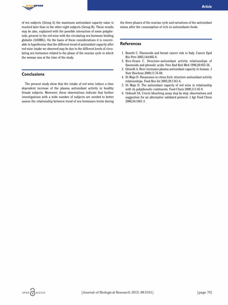
of ten subjects (Group A) the maximum antioxidant capacity value isreached later than in the other eight subjects (Group B). These resultsmay be also, explained with the possible interaction of some polyphe-nols, present in the red wine with the circulating sex-hormone bindingglobulin (hSHBG). On the basis of these considerations it is conceiv-able to hypothesize that the different trend of antioxidant capacity afterred wine intake we observed may be due to the different levels of circu-lating sex hormones related to the phase of the ovarian cycle in whichthe woman was at the time of the study.
Conclusions
The present study show that the intake of red wine induce a timedependent increase of the plasma antioxidant activity in healthyfemale subjects. Moreover, these observations indicate that furtherinvestigations with a wide number of subjects are needed to betterassess the relationship between trend of sex hormones levels during
the three phases of the ovarian cycle and variations of the antioxidantstatus after the consumption of rich-in-antioxidants foods.
References
1. Bosetti C. Flavonoids and breast cancer risk in Italy. Cancer EpidBio Prev 2005;144:805-8.
2. Rice-Evans C. Structure-antioxidant activity relationships offlavonoids and phenolic acids. Free Rad Biol Med 1996;20:933-56.
3. Ghiselli A. Beer increases plasma antioxidant capacity in human. JNutr Biochem 2000;11:76-80.
4. Di Majo D. Flavanones in citrus fruit: structure-antioxidant activityrelationships. Food Res Int 2005;38:1161-6.
5. Di Majo D. The antioxidant capacity of red wine in relationshipwith its polyphenolic costituents. Food Chem 2008;111:45-9.
6. Ordoudi SA. Crocin bleaching assay step by step: observations andsuggestion for an alternative validated protocol. J Agr Food Chem2006;54:1661-3.
[Journal of Biological Research 2015; 88:5161] [page 79]
Article
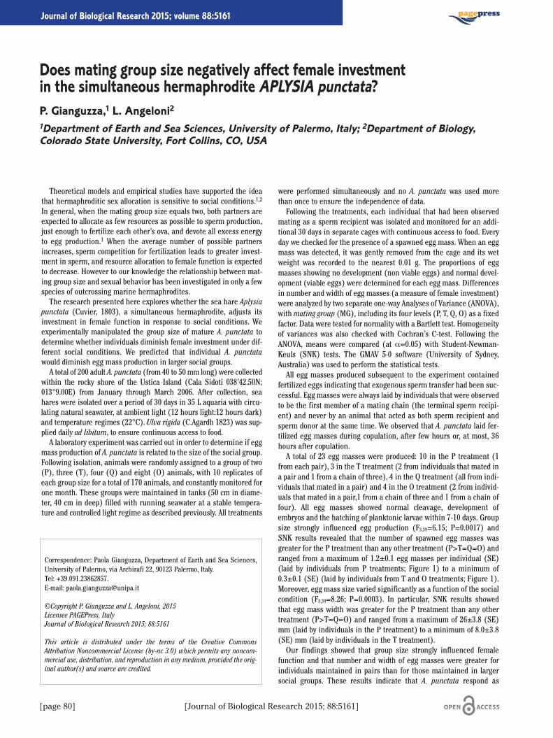
[page 80] [Journal of Biological Research 2015; 88:5161]
Journal of Biological Research 2015; volume 88:5161
Theoretical models and empirical studies have supported the ideathat hermaphroditic sex allocation is sensitive to social conditions.1,2
In general, when the mating group size equals two, both partners areexpected to allocate as few resources as possible to sperm production,just enough to fertilize each other’s ova, and devote all excess energyto egg production.1 When the average number of possible partnersincreases, sperm competition for fertilization leads to greater invest-ment in sperm, and resource allocation to female function is expectedto decrease. However to our knowledge the relationship between mat-ing group size and sexual behavior has been investigated in only a fewspecies of outcrossing marine hermaphrodites.The research presented here explores whether the sea hare Aplysia
punctata (Cuvier, 1803), a simultaneous hermaphrodite, adjusts itsinvestment in female function in response to social conditions. Weexperimentally manipulated the group size of mature A. punctata todetermine whether individuals diminish female investment under dif-ferent social conditions. We predicted that individual A. punctatawould diminish egg mass production in larger social groups.A total of 200 adult A. punctata (from 40 to 50 mm long) were collected
within the rocky shore of the Ustica Island (Cala Sidoti 038’42.50N;013°9.00E) from January through March 2006. After collection, seahares were isolated over a period of 30 days in 35 L aquaria with circu-lating natural seawater, at ambient light (12 hours light:12 hours dark)and temperature regimes (22°C). Ulva rigida (C.Agardh 1823) was sup-plied daily ad libitum, to ensure continuous access to food.A laboratory experiment was carried out in order to determine if egg
mass production of A. punctata is related to the size of the social group.Following isolation, animals were randomly assigned to a group of two(P), three (T), four (Q) and eight (O) animals, with 10 replicates ofeach group size for a total of 170 animals, and constantly monitored forone month. These groups were maintained in tanks (50 cm in diame-ter, 40 cm in deep) filled with running seawater at a stable tempera-ture and controlled light regime as described previously. All treatments
were performed simultaneously and no A. punctata was used morethan once to ensure the independence of data.Following the treatments, each individual that had been observed
mating as a sperm recipient was isolated and monitored for an addi-tional 30 days in separate cages with continuous access to food. Everyday we checked for the presence of a spawned egg mass. When an eggmass was detected, it was gently removed from the cage and its wetweight was recorded to the nearest 0.01 g. The proportions of eggmasses showing no development (non viable eggs) and normal devel-opment (viable eggs) were determined for each egg mass. Differencesin number and width of egg masses (a measure of female investment)were analyzed by two separate one-way Analyses of Variance (ANOVA),with mating group (MG), including its four levels (P, T, Q, O) as a fixedfactor. Data were tested for normality with a Bartlett test. Homogeneityof variances was also checked with Cochran’s C-test. Following theANOVA, means were compared (at α=0.05) with Student-Newman-Keuls (SNK) tests. The GMAV 5·0 software (University of Sydney,Australia) was used to perform the statistical tests.All egg masses produced subsequent to the experiment contained
fertilized eggs indicating that exogenous sperm transfer had been suc-cessful. Egg masses were always laid by individuals that were observedto be the first member of a mating chain (the terminal sperm recipi-ent) and never by an animal that acted as both sperm recipient andsperm donor at the same time. We observed that A. punctata laid fer-tilized egg masses during copulation, after few hours or, at most, 36hours after copulation.A total of 23 egg masses were produced: 10 in the P treatment (1
from each pair), 3 in the T treatment (2 from individuals that mated ina pair and 1 from a chain of three), 4 in the Q treatment (all from indi-viduals that mated in a pair) and 4 in the O treatment (2 from individ-uals that mated in a pair,1 from a chain of three and 1 from a chain offour). All egg masses showed normal cleavage, development ofembryos and the hatching of planktonic larvae within 7-10 days. Groupsize strongly influenced egg production (F3,39=6.15; P=0.0017) andSNK results revealed that the number of spawned egg masses wasgreater for the P treatment than any other treatment (P>T=Q=O) andranged from a maximum of 1.2±0.1 egg masses per individual (SE)(laid by individuals from P treatments; Figure 1) to a minimum of0.3±0.1 (SE) (laid by individuals from T and O treatments; Figure 1).Moreover, egg mass size varied significantly as a function of the socialcondition (F3,39=8.26; P=0.0003). In particular, SNK results showedthat egg mass width was greater for the P treatment than any othertreatment (P>T=Q=O) and ranged from a maximum of 26±3.8 (SE)mm (laid by individuals in the P treatment) to a minimum of 8.0±3.8(SE) mm (laid by individuals in the T treatment).Our findings showed that group size strongly influenced female
function and that number and width of egg masses were greater forindividuals maintained in pairs than for those maintained in largersocial groups. These results indicate that A. punctata respond as
Correspondence: Paola Gianguzza, Department of Earth and Sea Sciences,University of Palermo, via Archirafi 22, 90123 Palermo, Italy. Tel: +39.091.23862857.E-mail: [email protected]
©Copyright P. Gianguzza and L. Angeloni, 2015Licensee PAGEPress, ItalyJournal of Biological Research 2015; 88:5161
This article is distributed under the terms of the Creative CommonsAttribution Noncommercial License (by-nc 3.0) which permits any noncom-mercial use, distribution, and reproduction in any medium, provided the orig-inal author(s) and source are credited.
Does mating group size negatively affect female investmentin the simultaneous hermaphrodite APLYSIA punctata?P. Gianguzza,1 L. Angeloni21Department of Earth and Sea Sciences, University of Palermo, Italy; 2Department of Biology,Colorado State University, Fort Collins, CO, USA
[Journal of Biological Research 2015; 88:5161] [page 81]
Article
expected according to sex allocation theory for simultaneous hermaph-rodites.3 Furthermore, our experiments reveal that female allocation inthis species is phenotypically flexible, as fully mature individuals wereable to adjust their investment in the number and size of egg masseswith the size of the social group.
References
1. Charnov EL. Simultaneous hermaphroditism and sexual selection.P Natl Acad Sci 1979;76:2480-4.
2. Charnov EL. The theory of sex allocation. Princeton, NJ: PrincetonUniversity Press; 1982.
3. Michiels NK. Mating conflicts and sperm competition in simultane-ous hermaphrodites. In: A.P. Møller (ed.), Sperm competition andsexual selection. London: Academic Press; 1998. pp. 219-54.
Figure 1. Egg masses laid by individuals from groups of two (P),three (T), four (Q) and eight (O) animals.

[page 82] [Journal of Biological Research 2015; 88:5161]
Journal of Biological Research 2015; volume 88:5161
Cornu aspersum (Muller, 1774), specie che in passato era attribuitaal genere Helix (Helix aspersa), è un mollusco gasteropode terrestreappartenente alla famiglia degli Helicidae è diffuso nel bacino delMediterraneo e nell’Europa nord-occidentale. In Italia è presente intutta la penisola, isole comprese. Il diametro della sua conchiglia puòarrivare a 35 mm., con un’altezza di 27-31mm; il suo peso è di circa 13-15 g. La colorazione di fondo va dal verdastro al giallastro; può essereuniforme, ma più spesso sono presenti da 1 a 5 bande spiralate marro-ni, con screziatura gialla o bianca. Come molte chiocciole, C. aspersumè ermafrodita, cioè ogni individuo possiede sia organi riproduttivimaschili che femminili; non è tuttavia in grado di autofecondarsi(ermafroditismo insufficiente).L’azienda elicicola La Lumaca del Belice sita nel territorio di
Sambuca di Sicilia (AG) su una superficie di circa 10.000 mq, usa unsistema di allevamento a ciclo naturale biologico in campo aperto chedà un aumento notevole della produzione per metro quadrato masoprattutto dà la possibilità di portare le chiocciole alle caratteristicheottimali nel più breve tempo possibile. Con questo sistema di alleva-mento le chiocciole nascono e crescono dentro lo stesso recinto, evi-tandosi i lavori di spostamento dei piccoli causa di mortalità elevatadovuta allo stress provocato dal cambio di habitat ed allo schiaccia-mento durante il trasferimento a causa della fragilità del tenero guscioche le protegge. Il ciclo di allevamento inizia con la coltivazione di bie-tola e cavolfiore per l’alimentazione e di trifoglio per ricreare l’habitatnaturale nei mesi di marzo/aprile, fino ad arrivare al mese di novem-bre quando la raccolta delle chiocciole sarà completata ed il terrenoverrà preparato per il successivo ciclo. Durante questo periodo gliortaggi non verranno trattati né con concimi chimici né con pesticidi.Le prime chiocciole riproduttrici verranno immesse nei recinti nelprimo periodo primaverile, quando la vegetazione avrà raggiunto lecaratteristiche ottimali di vegetazione. Qui le chiocciole si accoppie-ranno e concepiranno le nuove chiocciole che, già nei primi mesi disettembre-ottobre, avranno raggiunto le caratteristiche ottimali di pez-zatura e peso e saranno pronte per essere commercializzate. Diversistudi hanno contributo ad approfondire le conoscenze utili ai fini della
valorizzazione del prodotto alimentare lumaca, non solo in termini ali-mentari ma anche economico-sociali in virtù della crescente richiestadel prodotto da parte del mercato interno e della prospettiva di poterintegrare, attraverso l’attività di allevamento del mollusco, l’attivitàagricola. L’indagine analitica finalizzata all’approfondimento conosci-tivo delle proprietà dietetico-nutrizionali di cinque diverse specie dimolluschi polmonati del genere Helix di interesse alimentare, condot-ta da Novelli1 permette di trarre alcune considerazioni. Il contenuto insostanza secca della parte edibile è prossimo al 20%, i due-terzi delquale sono rappresentati da proteine. Il contenuto lipidico è decisa-mente ridotto (inferiore all’1%) e ciò non è sorprendente in ragionedella predisposizione di tali molluschi a prediligere riserve energeti-che tissutali in forma di polisaccaridi piuttosto che di lipidi. La quotaparte costituita da acidi grassi è per quasi il 75% rappresentata daacidi grassi insaturi, tre-quarti dei quali sono polinsaturi. Da un puntodi vista strettamente nutrizionale gli acidi grassi della serie n6 e dellaserie n3 sono fra loro in rapporto sbilanciato a favore dei primi, in cuisono significativamente rappresentati l’acido linoleico e l’acido ara-chidonico. Ugualmente elevata è risultata la componente in sali mine-rali (più del doppio del normale contenuto in ceneri delle carni deiVertebrati terrestri). Differenze interspecifiche sono state rilevatesoprattutto a carico del contenuto in proteine e ceneri; il confrontointra-specifico fra prodotto d’allevamento e prodotto raccolto in naturanon ha evidenziato sostanziali differenze. Le caratteristiche alimenta-ri della lumaca si distinguono per un ridotto contenuto calorico conse-guenza della limitata quantità di grasso di deposito nelle massemuscolari. L’apporto in acidi grassi di elevato valore nutrizionale daparte della massa muscolare del piede è ragguardevole.Le lumache rispondono ai danni cutanei provocati dai predatori o
dagli incidenti rigenerando le cellule danneggiate. Non sviluppanoun’eccessiva reazione infiammatoria e inoltre le ferite sono riparatevelocemente senza la formazione di cicatrici evidenti. I ricercatorihanno visto che la pelle delle lumache ha la stessa composizione diquella umana, con i medesimi elementi strutturali come il collagene el’elastina. Quando la pelle del corpo umano è danneggiata o attaccatada microrganismi, la reazione infiammatoria che si scatena è moltodifferente e più forte di quella che si verifica nelle lumache. La lumacareagisce ai danni cutanei producendo una grande quantità di mucoche, attraverso la formazione di numerose bolle, bagna e aderisce com-pletamente alla superficie della cute. Il fluido naturale idrata efficace-mente la pelle e nello stesso tempo la protegge con i suoi peptidi anti-microbici, le sostanze antiossidanti e le molecole che incentivano l’or-dinario processo rigenerativo che ristruttura e rinnova le cellule deitessuti danneggiati.2 La bava di lumaca ha peculiari caratteristicheadesive.per cui grazie a una particolare componente proteica, anche inconcentrazioni minime, aderisce in maniera efficace, in ambientiumidi anche a superfici irregolari.3 La bava di lumaca di Cornu asper-sum (Helix aspersa) ha una composizione complessa di sostanze attiveche la rendono un ingrediente cosmetico unico e non replicabile inlaboratorio con un prodotto di sintesi o una miscela di essi. L’analisi
Correspondence: Francesca Grisafi, Dipartimento di Scienze Agrarie eForestali, Università di Palermo, viale delle Scienze 11, 90127 Palermo, Italy.E-mail: [email protected]
©Copyright G. Russo and F. Grisafi 2015Licensee PAGEPress, ItalyJournal of Biological Research 2015; 88:5161
This article is distributed under the terms of the Creative CommonsAttribution Noncommercial License (by-nc 3.0) which permits any noncom-mercial use, distribution, and reproduction in any medium, provided the orig-inal author(s) and source are credited.
Allevamento a ciclo biologico naturale di Cornu aspersum (Müller, 1774):proprietà alimentari e usi del secreto o bava di lumacaG. Russo, F. GrisafiDipartimento di Scienze Agrarie e Forestali, Università di Palermo, Italy
[Journal of Biological Research 2015; 88:5161] [page 83]
Article
chimica quali-quantitativa ha evidenziato la presenza in particolare diallantoina 0.3-0.5%, collagene 0.1-0.3%, acido glicolico 0.05-0.1%, acidolattico 0.05-0.1%, anti-proteasi 1.3-1.8%, vitamine e minerali in tracce.In un recente lavoro4 è stato messo in evidenza che il secreto diCrymptophalus aspersa, utilizzato per applicazioni cutanee, possiedeproprietà rigenerative per la pelle umana per le quali sono state fatteinteressanti ipotesi di meccanismo d’azione Studi clinici hanno dimo-strato che prodotti cosmetici a base di secreto di Cornu aspersum (Helix aspersa) favoriscono la cicatrizzazione delle ustioni dei bambiniriducendo la formazione del cheloide, l’iperpigmentazione e miglioran-do complessivamente l’aspetto estetico della cicatrice. In altri studi èstata confermata l’efficacia del secreto di Cornu aspersum (Helix asper-sa) nella cicatrizzazione delle ustioni facciali di soggetti adulti. Sembrache la secrezione di Cornu aspersum (Helix aspersa) contribuisca contutti i suoi componenti a promuovere la cicatrizzazione della ferita e lariduzione della formazione del cheloide. In particolare l’allantoina inessa contenuta ha spiccate proprietà cicatrizzanti già note anche perfavorire la riparazione di ferite suppuranti, ulcere resistenti, emorroidie varie infezioni dermatologiche. L’allantoina stimola la formazionetessutale e rende più rapida la cicatrizzazione delle ferite. Diverse pre-parazioni medicamentose topiche per la cicatrizzazione dei tessutisono formulate con allantoina e si sono dimostrate particolarmenteefficaci nel velocizzare la riparazione dei tessuti e nel ridurre la forma-
zione delle cicatrici e dei cheloidi. L’acido glicolico e l’acido lattico,naturalmente contenuti nella bava di Cornu aspersum (Helix aspersa),contribuiscono a idratare e levigare la pelle riducendo l’iperpigmenta-zione e prevenendo la formazione delle smagliature anche durante lagravidanza. Migliorano l’estetica delle cicatrici anche di vecchia data.L’acido glicolico promuove il turnover epidermico e favorisce la prolife-razione dei cheratinociti.
Bibliografia
1. Novelli E, Giaccone V, Balzan S, et al. Indagine sul valore dietetico-nutrizionale della lumaca. Confronto fra specie e fra soggetti rac-colti in natura ed allevati. Ann. Fac. Medic. Vet. di Parma2002;22:49-56.
2. Penazzi G. Potenziali proprietà ed usi in cosmesi del secreto diHelix aspersa o bava di lumaca. Cosmetic Technol 2010;13:25-8.
3. Pawlicki JM, Pease LB, Pierce CM, et al. The effect of molluscanglue proteins on gel mechanics. J Exp Biol 2004;207:1127-35.
4. Brieva A, Philips N, Tajedor R, et al. Molecular basis for the rege-nerative properties of a secretion of the mollusk Crymptophalusaspersa. Skin Pharmacol Physiol 2008;21:15-22.
[page 84] [Journal of Biological Research 2015; 88:5161]
Journal of Biological Research 2015; volume 88:5161
It is well confirmed that a strong relationship exists between Down’ssyndrome (DS) and Alzheimer’s disease (AD). Neurochemical investi-gations reported that many central neurotransmitter systems are simi-larly affected in aging Down and in Alzheimer patients, respectively.Airaksinem and colleagues1 found numerous neurofibrillary tangles inthe tuberomammillary area of the hypothalamus, where cell bodies ofhistaminergic neurons are located. While Mazurkiewicz-Kwilecki andcolleagues2 found deficits of the endogenous diamine, Cacabelos andcolleagues3 reported an increase of central histamine levels. In the pres-ent study, in order to test whether AD-like neuropathological changesinvolve the central histaminergic system, we measured the concentra-tion of histamine, histidine as well as the activity of histidine decarboxy-lase (HDC) and histamine-N-methyltransferase (HMT) in temporal cor-tex (TC) of aging Down, Alzheimer and control patients.Post-mortem samples (temporal cortex, TC; grey matter) of AD neu-
ropathologically confirmed cases (72.1±7.6 years old), of karyotypedpatients with DS (56.1±7.1 years old), and control adults (72,7±9.7 yearsold) were obtained from the MRC London Brain Bank forNeurodegenerative Diseases, Department of Neuropathology, Institute ofpsychiatry, London, U.K.Each block of brain tissue from AD, DS and controls were thawed on
ice and homogenized in ice-cold HDC-solution of 0.1 M sodium phos-phate buffer (pH 6.8) containing dithiothreitol and antipain proteaseinhibitor. Homogenates of brain specimens were centrifuged at 12,000 xg for 20 min at 4°C. The supernatants were poured into CENTRIPEP-3concentrators (Amicon), and centrifuged at 2,000 x g for two 10 minperiods at 4°C. The clear extracts were stored in small quantities inEppendorf tubes at -80°C until analysis. HDC activity has been meas-ured with the procedure described by Gueli and colleagues4 and brieflysummarized. Extract aliquots were pre- incubated for 10 min with HDCassay-solution (0.1 M PBS, 0.2 mM DTT, 0.01 mM PLP, 0.1 mMAminoguanidine), then incubation was started by adding 0.5 mM L-his-tidine for 0-3 h at 37°C. At the established times, the reactions werestopped with 60% ice-cold PCA, and stored overnight. Finally, the reac-
tion mixture was centrifuged at 19,000 x g for 30 min at 4°C. The super-natants were withdrawn and filtered (0.45 �m Millipore filter). The HPLCsystem consisted of a 600E Waters pump with a Waters 474 scanning flu-orescence detector (ex 350 nm, em 450 nm). Chromatograms and calcu-lations were performed by Empower TM2 Data Software. Histamine wasseparated and quantified after pre-column derivatization with Shore’s o-phthalaldehyde reaction,5 using a Spherisorb ODS2 analytical column,particle size 3 �m (20 x 0.46 cm; Waters, Milano), a 10 μL injection vol-ume, and a mobile phase of methanol, 20 mmol/L sodium acetate inwater, acetic acid (55:43:2 v/v) and 0.33 mmol/L 1-octanesulfonic acidsodium salt. The flow rate was 1.0 ml/min. In order to measure HMTactivity brain tissue was disperged with a glass Teflon homogenizer in0.1 M PBS (pH 7.2). After centrifugation the supernatant was used forthe radioenzymatic assay.6 Histidine contents were measured using theprocedure described by Borum.7
We observed a increase of histamine levels in temporal cortex of AD(+15%) patients. Down brains also showed a mild increase of theendogenous diamine concentration (+8%). HDC activity in both groupsof diseased brains was significantly increased compared with controls(+59% for DS and +21% for AD, respectively). In accord to HDC actvity,HMT activity run in parallel in both pathological groups. In contrast tohistamine, histidine levels were markedly decreased in temporal cortexof both pathological groups. These results put together leads us to thinkof a similar high turnover rate in the metabolic happenings of the hista-minergic system in the temporal cortex of the patients with Alzheimer’sdisease and Down’s syndrome. The fast histaminergic changes may con-tribute to the clinical manifestation of dementia in both disorders.
References
1. Airaksinen MS, Paetau A, Paljarvi L, et al. Histamine neurons inhuman hypothalamus: anatomy in normal and Alzheimer diseasedbrains. Neuroscience 1991;9:465-81.
2. Mazurkiewicz-Kwilecki IM, Nsonwah S. Changes in the regionalbrain histamine and histidine levels in postmortem brains ofAlzheimer patients. Can J Physiol Pharmacol 1989;67:75-8.
3. Cacabelos R, Yamatodani A, Niigawa H, et al. Brain histamine inAlzheimer’s disease. Meth Find Exp Clin Pharmacol 1989;11:353-60.
4. Gueli MC. The mental disorders in the elderly: new therapeuticapproaches. Rome: Brunello Ed.; 1997.
5. Shore PA, Burkhalter A, Cohn VH. A method for the fluorometricassay of histamine in tissues. J Pharmacol Exp Ther 1959;127:182-6.
6. Prast H, Gujrati V, Walser S, Philippu A. Histamine, histidinedecarboxylase and histamine-N-methyltransferase in brain areasof spontaneously hypertensive rats, Naunyn-Schmiedeberg’s Arch.Pharmacol 1988;338:573-83.
7. Borum PR. Manual for amino acid analysis of physiological samples.Atlanta, GA: American Association for Clinical Chemistry Publ.; 1997.
Correspondence: Department of Experimental Biomedicine and ClinicalNeurosciences, University of Palermo, via Del Vespro 129, 90127, Palermo,Italy.E-mail: [email protected]
©Copyright M.C. Gueli, 2015Licensee PAGEPress, ItalyJournal of Biological Research 2015; 88:5161
This article is distributed under the terms of the Creative CommonsAttribution Noncommercial License (by-nc 3.0) which permits any noncom-mercial use, distribution, and reproduction in any medium, provided the orig-inal author(s) and source are credited.
High turnover rate of central histaminergic system in patientswith Down syndrome and Alzheimer diseaseM.C. GueliDepartment of Experimental Biomedicine and Clinical Neurosciences, University of Palermo, Italy
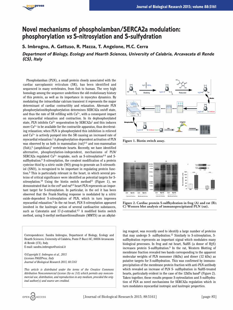
[Journal of Biological Research 2015; 88:5161] [page 85]
Journal of Biological Research 2015; volume 88:5161
Phospholamban (PLN), a small protein closely associated with thecardiac sarcoplasmic reticulum (SR), has been identified andsequenced in many vertebrates, from fish to human. The very highhomology among the sequence underlines the old evolutionary historyof this protein, as well as its importance in myocytes dynamics. Bymodulating the intracellular calcium transient it represents the majordeterminant of cardiac contractility and relaxation. Alternate PLNphosphorylation/dephosphorylation determines SERCA2a on/off state,and thus the rate of SR refilling with Ca2+, with a consequent impacton myocardial relaxation and contraction. In its dephosphorylatedstate, PLN inhibits Ca2+ sequestration by SERCA2a1 and this inducesmore Ca2+ to be available for the contractile apparatus, thus decelerat-ing relaxation; when PLN is phosphorylated this inhibition is relievedand Ca2+ is actively pumped into the SR causing an increased rate ofmyocardial relaxation.2 A phosphorylation-dependent activation of PLNwas observed by us both in mammalian (rat)3-5 and non-mammalian(fish),6 (amphibian)7 vertebrate hearts. Recently, we have identifiedalternative, phosphorylation-independent, mechanisms of PLN/SERCA2a regulated Ca2+ reuptake, such as S-nitrosylation4-6 and S-sulfhydration.8 S-nitrosylation, the covalent modification of a proteincysteine thiol by a nitric oxide (NO) group to generate an S-nitrosoth-iol (SNO), is recognized to be important in regulating protein func-tion.9 This is particularly relevant in the heart, in which several pro-teins of critical significance were identified as potential targets for S-nitrosylation.10 Using the biotin switch method11 (Figure 1), wedemonstrated that in the eel6 and rat4,5 heart PLN represents an impor-tant target for S-nitrosylation. In particular, in the eel it has beenobserved that the Frank-Starling response is modulated by a nitricoxide-dependent S-nitrosylation of PLN, which in turn improvesmyocardial relaxation.6 In the rat heart, PLN S-nitrosylation appearedinvolved in the lusitropic action of several cardioactive substances,such as Catestatin and 17-β-estradiol.4,5 A modified biotin switchmethod, using S-methyl methanethiosulfonate (MMTS) as an alkylat-
ing reagent, was recently used to identify a large number of proteinsthat may undergo S- sulfhydration.12 Similarly to S-nitrosylation, S-sulfhydration represents an important signal which modulates manybiological processes. In frog and rat heart, NaHS (a donor of H2S)increases protein S-sulfhydration.8 In the rat, Western Blotting ofmembrane fraction revealed two bands corresponding to the apparentmolecular weights of PLN monomer (6kDa) and dimer (12 kDa) asputative targets for S-sulfhydration. This was confirmed by immuno-precipitation of the membrane protein fraction with anti-PLN antibodywhich revealed an increase of PLN S- sulfhydration in NaHS-treatedhearts, particularly evident in the case of the 12kDa band8 (Figure 2).Taken together, these results propose S-nytrosilation and S-sulfhydra-tion of PLN as novel mechanisms for SERCA2a regulation which inturn modulates myocardial inotropic and lusitropic properties.
Correspondence: Sandra Imbrogno, Department of Biology, Ecology andHearth Sciences, University of Calabria, Ponte P. Bucci 6C, 84036 Arcavacatadi Rende (CS), Italy.E-mail: [email protected]
©Copyright S. Imbrogno et al., 2015Licensee PAGEPress, ItalyJournal of Biological Research 2015; 88:5161
This article is distributed under the terms of the Creative CommonsAttribution Noncommercial License (by-nc 3.0) which permits any noncom-mercial use, distribution, and reproduction in any medium, provided the orig-inal author(s) and source are credited.
Novel mechanisms of phospholamban/SERCA2a modulation:phosphorylation vs S-nitrosylation and S-sulfydrationS. Imbrogno, A. Gattuso, R. Mazza, T. Angelone, M.C. CerraDepartment of Biology, Ecology and Hearth Sciences, University of Calabria, Arcavacata di Rende(CS), Italy
Figure 1. Biotin switch assay.
Figure 2. Cardiac protein S-sulfhydration in frog (A) and rat (B);C) Western blot analysis of immunoprecipitated PLN (rat).
[page 86] [Journal of Biological Research 2015; 88:5161]
Article
References
1. Kimura Y, Kurzydlowski K, Tada M, MacLennan DH.Phospholamban inhibitory function is activated by depolymeriza-tion. J Biol Chem 1997;272:15061-4.
2. Schmidt AG, Edes I, Kranias EG. Phospholamban: a promising thera-peutic target in heart failure? Cardiovasc Drugs Ther 2001;15:387-96.
3. Angelone T, Quintieri AM, Brar BK, et al. The antihypertensivechromogranin a peptide catestatin acts as a novel endocrine/paracrine modulator of cardiac inotropism and lusitropism.Endocrinology 2008;149:4780-93.
4. Angelone T, Quintieri AM, Pasqua T, et al. Phosphodiesterase type-2 and NO-dependent S-nitrosylation mediate the cardioinhibitionof the anti-hypertensive catestatin. Am J Physiol Heart Circ Physiol2012;302:431-42.
5. Filice E, Angelone T, De Francesco EM, et al. Crucial role of phos-pholamban phosphorylation and S-nitrosylation in the negativelusitropism induced by 17β-estradiol in the male rat heart. CellPhysiol Biochem 2011;28:41-52.
6. Garofalo F, Parisella ML, Amelio D, et al. Phospholamban S-nitrosy-lation modulates starling response in fish heart. Proc Biol Sci2009;276:4043-52.
7. Mazza R, Gattuso A, Mannarino C, et al. Catestatin (chromograninA344-364) is a novel cardiosuppressive agent: inhibition of isopro-terenol and endothelin signaling in the frog heart. Am J PhysiolHeart Circ Physiol 2008;295:113-22.
8. Mazza R, Pasqua T, Cerra MC, et al. Akt/eNOS signaling and PLN S-sulfhydration are involved in H2S-dependent cardiac effects in frogand rat. Am J Physiol Regul Integr Comp Physiol 2013; 305:443-51.
9. Hess DT, Matsumoto A, Kim SO, et al. Protein S-nitrosylation:purview and parameters. Nat Rev Mol Cell Biol 2005;6:150-66.
10. Lima B, Forrester MT, Hess DT, Stamler JS. S-nitrosylation in car-diovascular signaling. Circ Res 2010;106:633-46.
11. Jaffrey SR, Snyder SH. The biotin switch method for the detectionof S-nitrosylated proteins. Sci STKE 2001:86:11.
12. Mustafa AK, Gadalla MM, Sen N, et al. H2S signals through proteinS-Sulfhydration. Sci Signal 2009;2:72.

[Journal of Biological Research 2015; 88:5161] [page 87]
Journal of Biological Research 2015; volume 88:5161
Mesenchymal stem cells (MSCs) are an attractive cell source forRegenerative Dentistry in particular due to their ability to differentiatetowards osteoblasts, among other lineages. Tooth and jaw bone lossare frequent sequelae of traumatic and pathological conditions in boththe young and the elderly and must be met by appropriate prostheticreplacements. For successful osseointegration of the dental implant asufficient bone level is necessary. Besides the utilization of bone auto-grafts or synthetic biomaterials, medical research is more and morefocused on the utilization of MSCs. Compared to cells obtained fromliposuction material, ectomesenchymal stem cells derived from thehead area e.g. out of dental follicles or particulate, non-vascularizedbone chips show a higher differentiation potential towards osteoblasts.This implies that due to their different origin, ectomesenchymal stemcells are stronger committed towards hard tissues and are thereforeinteresting candidate cells for bone regeneration.1,2
Parathyroid hormone-related protein (PTHrP) is known to beinvolved in tooth eruption. It acts as a signaling molecule that stimu-lates local bone resorption.3 Recently, PTHrP was found to affect theMSC differentiation process. The differential expression level of spe-cific PTHrP isoforms might be considered as a molecular signatureassociated with the respective differentiation state during osteogene-sis.4 Moreover, we could show that in addition to the role of purinergic2 (P2) receptors in cellular processes such as proliferation, migrationand apoptosis, they are also involved in stem cell differentiation.Several P2 receptor subtypes play a role in key steps during osteogeniclineage commitment. Further development of MSCs into progenitorcells, pre-osteoblasts and osteoblasts seemed to be triggered via thealteration of their P2 receptor expression patterns.5
Human mesenchymal stem cells were isolated from bone chip mate-rial harvested during oral surgery intervention. Their stem cell charac-ter was demonstrated by plastic-adherence and expression of the sur-face markers CD73, CD90, and CD105 following differentiation along
the osteogenic lineage.6 The mineralization process was monitored byAlizarin Red S staining of extracellular matrix components. Among theexamined P2 receptor subtypes, down-regulation of P2Y14 appeared tobe involved in the onset of this differentiation process. Today severalartificial P2 receptor ligands are present in the market. The adminis-tration of a stimulating P2Y14 receptor ligand had a direct influence onthe osteogenic differentiation potential. More precisely, the applica-tion of the potent and selective P2Y14 agonist MRS 2690 led to a dose-dependent reduction of the extracellular mineralization.Taken together, bone chip-derived mesenchymal stem cells are
promising candidate cells for bone replacement. Here we show thatthey are capable of differentiating into osteoblasts. The application ofan artificial receptor agonist confirmed the functional role of P2Y14during osteogenesis. Therefore, it is of major interest to develop selec-tive and potent antagonists directed against this recently discoveredmember of the purinergic receptor family. Controlling the P2Y14 recep-tor signaling might improve future bone tissue engineering approach-es in regenerative dentistry.
References
1. Haddouti EM, Skroch M, Zippel N, et al. Human dental follicle pre-cursor cells of wisdom teeth: isolation and differentiation towardsosteoblasts for implants with and without scaffolds. Mat-WissWerkstofftech 2009;40:732-7.
2. Kaebisch C, Winter M, Khan D, et al. Examination of the quality ofparticulate bone chip material. (in press).
3. Philbrick WM, Dreyer BE, Nakchbandi IA, Karaplis AC. Parathyroidhormone-related protein is required for tooth eruption. P NatlAcad Sci USA 1998;95:11846-51.
4. Longo A, Librizzi M, Naselli F, et al. PTHrP in differentiatinghuman mesenchymal stem cells: Transcript isoform expression,promoter methylation, and protein accumulation. Biochimie2013;95:1888-96.
5. Zippel N, Limbach CA, Ratajski N, et al. Purinergic receptors influ-ence the differentiation of human mesenchymal stem cells. StemCells Dev 2012;21:884-900.
6. Dominici M, Le Blanc K, Mueller I, et al. Minimal criteria for defin-ing multipotent mesenchymal stromal cells. Cytotherapy 2006;8:315-7.
Correspondence: Edda Tobiasch, Department of Natural Sciences, Universityof Applied Sciences, Bonn-Rhine-Sieg, Rheinbach, Germany.E-mail: [email protected]
©Copyright C. Kaebisch et al., 2015Licensee PAGEPress, ItalyJournal of Biological Research 2015; 88:5161
This article is distributed under the terms of the Creative CommonsAttribution Noncommercial License (by-nc 3.0) which permits any noncom-mercial use, distribution, and reproduction in any medium, provided the orig-inal author(s) and source are credited.
The osteogenic differentiation of bone chip-derived mesenchymal stem cellsis controlled via specific receptor signalingC. Kaebisch,1 J.K. Gorski,2 Y.A. Issa,3 M.Winter,4 E. Tobiasch11Department of Natural Sciences, University of Applied Sciences, Rheinbach, Germany; 2SwansonSchool of Engineering, University of Pittsburgh, PA, USA; 3Bethlehem University, Palestine; 4OralSurgery Practice, Rheinbach, Germany

[page 88] [Journal of Biological Research 2015; 88:5161]
Journal of Biological Research 2015; volume 88:5161
We designed and developed a simple and reproducible screeningassay to explore the ability of a number of small compounds, interfer-ing with evolutionary conserved signalling pathways, to inhibit epithe-lial mesenchimal transition (EMT) in vivo. EMT is an importantprocess during development by which fully polarized epithelial cellsacquire mesenchymal, fibroblast-like properties and show reducedintercellular adhesion and increased motility. EMT mechanism is re-activated in tumor progression, tissue invasion, metastasis and theacquisition of resistance to therapy.1,2 Targeting EMT, therefore, repre-sents an important strategy for cancer treatment.Sea urchin embryos provide a useful system to study EMT progres-
sion, just few hours after fertilization. The process is well character-ized. Prior to ingression, the future primary mesenchyme cells (PMC)are indistinguishable from the neighbouring epithelial blastomeres,but their regulatory apparatus is actively preparing new molecular pro-grams by which cells are going to turn on the expression of mes-enchyme-specific molecular markers. Thus, at the right time PMCs,previously adherent to the adjacent epithelial cells via cadherin andadherens junctions, loose cell adhesion, enter the basal lamina andmove through the blastocoele. Later, after migration to the proper loci,the specified PMCs will give rise to the embryonic skeleton.3
So far, potential EMT inhibitor compounds have been identifiedusing a carcinoma cell line specifically induced to undergo EMT by theactivation of growth factor signalling pathways.4 Nevertheless, thisapproach restricts the research to bio-molecules only affecting theselected growth factor induced signals and in some cases the reportercell lines are not responsive to all exogenous growth factors known tobe EMT inducers.Here, we propose an in vivo screening assay, using sea urchin
embryos (Figure 1); we picked out a selection of pharmacologicallyactive compounds from a commercial library (LOPAC1280TM, Sigma-Aldrich) and tested their ability to inhibit EMT in embryos. These mol-ecules are known to interfere with some evolutionarily conserved sig-nalling pathways: P38 mitogen-activated protein kinase (MAPK),platelet-derived growth factor receptors (PDGF-R) epidermal growthfactor receptor (EGFR) tyrosine-protein kinase (Src), Glycogen syn-
thase kinase 3 (GSK-3). We set up the experiments as follows: two dif-ferent batches of Paracentrotus lividus embryos at zygote (just post-fertilization) or hatching blastula (12h post-fertilization and 4h priorPMC ingression) stages were incubated in multiwell plates in the pres-ence of different concentrations of the selected drug. Treated or con-trol embryos were then monitored under an inverted microscope andscored for timely precise PMC formation, number and migration capa-bility. Embryos were then photographed, phenotipically classified andin some experiments assayed for the expression of specific antigens.We obtained evidence that some of these compounds inhibit EMT andprevent the expression of mesenchymal specific molecular markers.We propose this low-, medium-throughput Sea Urchin embryonic EMTAssay (SU-EMTA) as an affordable and useful method to screen a highnumber of compounds, with potential anti-metastatic activity.
Correspondence: Daniele Romancino, Institute of Biomedicine andMolecular Immunology, National Research Center, via Ugo La Malfa 153,90146 Palermo, Italy.E-mail: [email protected]
Fundung: the present study has been partially supported by a grant from theItalian Ministry of Economy and Finance to the CNR for the Project FaReBiodi Qualità.
©Copyright R. La Tona et al., 2015Licensee PAGEPress, ItalyJournal of Biological Research 2015; 88:5161
This article is distributed under the terms of the Creative CommonsAttribution Noncommercial License (by-nc 3.0) which permits any noncom-mercial use, distribution, and reproduction in any medium, provided the orig-inal author(s) and source are credited.
In vivo assay for the identification of potential inhibitorsof epithelial-mesenchymal transition R. La Tona,1 A. Lavanco,2 L. Anello,2 A. Bongiovanni,2 D.P. Romancino,2 M. Di Bernardo21Department of Biological, Chemical and Pharmaceutical Sciences and Technologies, Palermo;2Institute of Biomedicine and Molecular Immunology, National Research Center, Palermo, Italy
Figure 1. A) Set up of a standard epithelial mesenchimal transi-tion assay on sea urchin embryos; B) drug addition at fertilization(zygote) for 16 h or at hatching blastula stage (HB) for 4 h.Phenotype analysis at mesenchyme blastula stage (MB) and later(36 h after fertilization).

[Journal of Biological Research 2015; 88:5161] [page 89]
Article
References
1. Kalluri R, Weinberg RA. The basics of epithelial-mesenchymaltransition. J Clin Invest 2009;119:1420-8.
2. Lim J, Thiery JP. Epithelial-mesenchymal transitions: insights fromdevelopment. Development 2012;139:3471-86.
3. Wu S-Y, Ferkowicz M, McClay DR. Ingression of primary mes-enchyme cells of the sea urchin embryo: a precisely timedepithelial mesenchymal transition. Birth Defects Research 2007;81:241-52.
4. Chua KN, Sim W-J, Racine V, et al. A cell-based small moleculescreening method for identifying inhibitors of epithelial-mes-enchymal transition in carcinoma. PLoS ONE 2012;7:e33183.
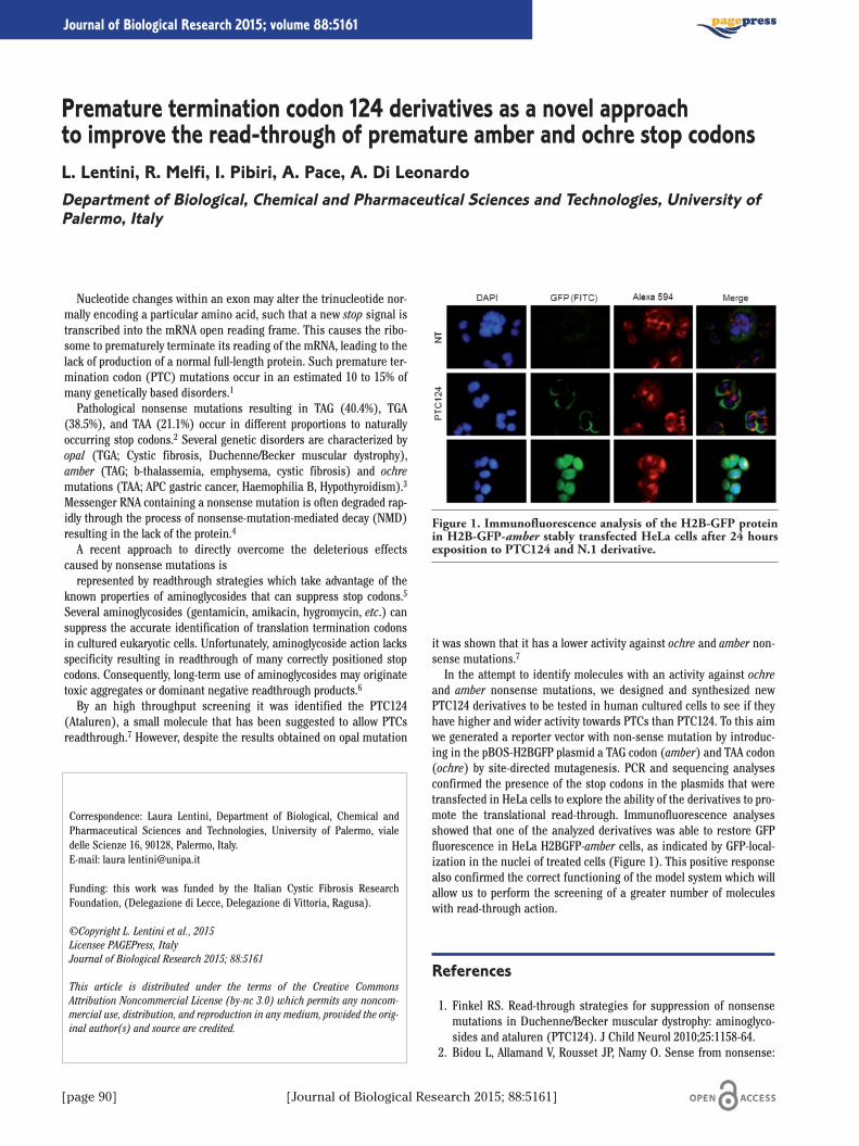
[page 90] [Journal of Biological Research 2015; 88:5161]
Journal of Biological Research 2015; volume 88:5161
Nucleotide changes within an exon may alter the trinucleotide nor-mally encoding a particular amino acid, such that a new stop signal istranscribed into the mRNA open reading frame. This causes the ribo-some to prematurely terminate its reading of the mRNA, leading to thelack of production of a normal full-length protein. Such premature ter-mination codon (PTC) mutations occur in an estimated 10 to 15% ofmany genetically based disorders.1
Pathological nonsense mutations resulting in TAG (40.4%), TGA(38.5%), and TAA (21.1%) occur in different proportions to naturallyoccurring stop codons.2 Several genetic disorders are characterized byopal (TGA; Cystic fibrosis, Duchenne/Becker muscular dystrophy),amber (TAG; b-thalassemia, emphysema, cystic fibrosis) and ochremutations (TAA; APC gastric cancer, Haemophilia B, Hypothyroidism).3
Messenger RNA containing a nonsense mutation is often degraded rap-idly through the process of nonsense-mutation-mediated decay (NMD)resulting in the lack of the protein.4
A recent approach to directly overcome the deleterious effectscaused by nonsense mutations isrepresented by readthrough strategies which take advantage of the
known properties of aminoglycosides that can suppress stop codons.5
Several aminoglycosides (gentamicin, amikacin, hygromycin, etc.) cansuppress the accurate identification of translation termination codonsin cultured eukaryotic cells. Unfortunately, aminoglycoside action lacksspecificity resulting in readthrough of many correctly positioned stopcodons. Consequently, long-term use of aminoglycosides may originatetoxic aggregates or dominant negative readthrough products.6
By an high throughput screening it was identified the PTC124(Ataluren), a small molecule that has been suggested to allow PTCsreadthrough.7 However, despite the results obtained on opal mutation
it was shown that it has a lower activity against ochre and amber non-sense mutations.7
In the attempt to identify molecules with an activity against ochreand amber nonsense mutations, we designed and synthesized newPTC124 derivatives to be tested in human cultured cells to see if theyhave higher and wider activity towards PTCs than PTC124. To this aimwe generated a reporter vector with non-sense mutation by introduc-ing in the pBOS-H2BGFP plasmid a TAG codon (amber) and TAA codon(ochre) by site-directed mutagenesis. PCR and sequencing analysesconfirmed the presence of the stop codons in the plasmids that weretransfected in HeLa cells to explore the ability of the derivatives to pro-mote the translational read-through. Immunofluorescence analysesshowed that one of the analyzed derivatives was able to restore GFPfluorescence in HeLa H2BGFP-amber cells, as indicated by GFP-local-ization in the nuclei of treated cells (Figure 1). This positive responsealso confirmed the correct functioning of the model system which willallow us to perform the screening of a greater number of moleculeswith read-through action.
References
1. Finkel RS. Read-through strategies for suppression of nonsensemutations in Duchenne/Becker muscular dystrophy: aminoglyco-sides and ataluren (PTC124). J Child Neurol 2010;25:1158-64.
2. Bidou L, Allamand V, Rousset JP, Namy O. Sense from nonsense:
Correspondence: Laura Lentini, Department of Biological, Chemical andPharmaceutical Sciences and Technologies, University of Palermo, vialedelle Scienze 16, 90128, Palermo, Italy. E-mail: laura [email protected]
Funding: this work was funded by the Italian Cystic Fibrosis ResearchFoundation, (Delegazione di Lecce, Delegazione di Vittoria, Ragusa).
©Copyright L. Lentini et al., 2015Licensee PAGEPress, ItalyJournal of Biological Research 2015; 88:5161
This article is distributed under the terms of the Creative CommonsAttribution Noncommercial License (by-nc 3.0) which permits any noncom-mercial use, distribution, and reproduction in any medium, provided the orig-inal author(s) and source are credited.
Premature termination codon 124 derivatives as a novel approachto improve the read-through of premature amber and ochre stop codonsL. Lentini, R. Melfi, I. Pibiri, A. Pace, A. Di LeonardoDepartment of Biological, Chemical and Pharmaceutical Sciences and Technologies, University ofPalermo, Italy
Figure 1. Immunofluorescence analysis of the H2B-GFP proteinin H2B-GFP-amber stably transfected HeLa cells after 24 hoursexposition to PTC124 and N.1 derivative.

[Journal of Biological Research 2015; 88:5161] [page 91]
Article
therapies for premature stop codon diseases. Trends Mol Med2012;18:679-88.
3. Mort M, Ivanov D, Cooper DN, Chuzhanova NA. A meta-analysis ofnonsense mutations causing human genetic disease. Hum Mutat2008;29:1037-47.
4. Mendell JT, Dietz HC. When the message goes awry: disease-pro-ducing mutations that influence mRNA content and performance.Cell 2001;107:411-4.
5. Hermann T. Aminoglycoside antibiotics: old drugs and new thera-peutic approaches. Cell Mol Life Sci 2007;64:1841-52.
6. Diop D, Chauvin C, Jean-Jean O. Aminoglycosides and other fac-tors promoting stop codon readthrough in human cells. C R Biol2007;330:71-9.
7. Welch EM. PTC124 targets genetic disorders caused by nonsensemutations. Nature 2007;447:87-91.
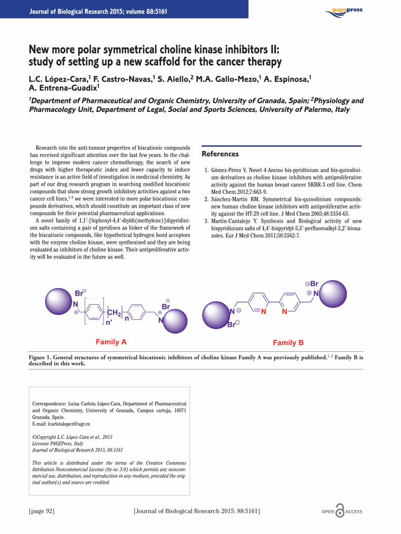
[page 92] [Journal of Biological Research 2015; 88:5161]
Journal of Biological Research 2015; volume 88:5161
Research into the anti-tumour properties of biscationic compoundshas received significant attention over the last few years. In the chal-lenge to improve modern cancer chemotherapy, the search of newdrugs with higher therapeutic index and lower capacity to induceresistance is an active field of investigation in medicinal chemistry. Aspart of our drug research program in searching modified biscationiccompounds that show strong growth inhibitory activities against a twocancer cell lines,1-3 we were interested in more polar biscationic com-pounds derivatives, which should constitute an important class of newcompounds for their potential pharmaceutical applications.A novel family of 1,1’-[biphenyl-4,4’-diyldi(methylene)]dipyridini-
um salts containing a pair of pyridines as linker of the framework ofthe biscationic compounds, like hypothetical hydrogen bond acceptorswith the enzyme choline kinase, were synthesized and they are beingevaluated as inhibitors of choline kinase. Their antiproliferative activ-ity will be evaluated in the future as well.
References
1. Gómez-Pérez V. Novel 4-Amino bis-pyridinium and bis-quinolini-um derivatives as choline kinase inhibitors with antiproliferativeactivity against the human breast cancer SKBR-3 cell line. ChemMed Chem 2012;7:663-9.
2. Sánchez-Martín RM. Symmetrical bis-quinolinium compounds:new human choline kinase inhibitors with antiproliferative activ-ity against the HT-29 cell line. J Med Chem 2005;48:3354-63.
3. Martín-Cantalejo Y. Synthesis and Biological activity of newbispyridinium salts of 4,4’-bispyridyl-5,5’-perfluoroalkyl-2,2’-bioxa-zoles. Eur J Med Chem 2011;50:5562-7.
Correspondence: Luisa Carlota López-Cara, Department of Pharmaceuticaland Organic Chemistry, University of Granada, Campus cartuja, 18071Granada, Spain. E-mail: [email protected]
©Copyright L.C. López-Cara et al., 2015Licensee PAGEPress, ItalyJournal of Biological Research 2015; 88:5161
This article is distributed under the terms of the Creative CommonsAttribution Noncommercial License (by-nc 3.0) which permits any noncom-mercial use, distribution, and reproduction in any medium, provided the orig-inal author(s) and source are credited.
New more polar symmetrical choline kinase inhibitors II:study of setting up a new scaffold for the cancer therapyL.C. López-Cara,1 F. Castro-Navas,1 S. Aiello,2 M.A. Gallo-Mezo,1 A. Espinosa,1A. Entrena-Guadix11Department of Pharmaceutical and Organic Chemistry, University of Granada, Spain; 2Physiology andPharmacology Unit, Department of Legal, Social and Sports Sciences, University of Palermo, Italy
Figure 1. General structures of symmetrical biscationic inhibitors of choline kinase Family A was previously published.1-3 Family B isdescribed in this work.
[Journal of Biological Research 2015; 88:5161] [page 93]
Journal of Biological Research 2015; volume 88:5161
Acid and base transport through cell membranes is essential for allcells in order to maintain acid-base balance. In some cells this trans-port is used for peculiar purposes. Two examples are the pancreaticduct cells and the enterocytes of marine teleosts. The former secrete aHCO3
– -rich fluid necessary to increase the chime pH and to activatethe intestinal enzymes. Marine teleost enterocytes actively secreteHCO3
– in order to precipitate the large amounts of Ca2+(and Mg2+)introduced by swallowing seawater. Divalent cation precipitation hasthe role to reduce the osmotic pressure of the intestinal fluid, thusallowing water absorption, necessary to compensate water loss intothe hyperosmotic external medium. In addition it inhibits Ca2+ over-absorption and may prevent renal stones formation.1,2
The mechanisms of base secretion and their regulation in seawaterteleosts, including Sparus aurata, have been extensively studied.3-6 Inthis paper the involvement of proton pumps in bicarbonate secretion(BCS), already suggested in mammal pancreatic duct cells,7 was inves-tigated in isolated tissues mounted in Ussing chambers, where thetransepithelial electrical parameters (short circuit current, Isc, and tis-sue resistance, Rt) were also measured. We found that the apical addi-tion of 10 μM omeprazole, a specific H+-K+-ATPase inhibitor produceda significant decrease of BCS and a parallel increase in Isc positivity inboth the anterior intestine and the rectum, irrespective of the pres-ence of serosal bicarbonate. Experiments performed with the aim oftesting the reversibility of omeprazole effect on BCS led to an intrigu-ing result: a large and transient increase of BCS following omeprazoleremoval from the apical solution. This response was strongly reducedin tissues pre-incubated with 100 μM colchicine, a known inhibitor ofmicrotubule polymerization.Acetazolamide, a carbonic anhydrase inhibitor, which strongly
inhibited BSC, was not able to modify Isc in the anterior intestine butproduced a small increase in Isc positivity in the rectum. Apical addi-tion of the selective inhibitor of the V-type H+-ATPase, Bafilomycin A1(0.1 μM), was not able to reduce BCS in the rectum while exhibited a
significant inhibitory effect in the anterior intestine. The addition ofomeprazole after Bafilomycin A1 produced a further BCS inhibition. Incontrast Bafilomycin A1 was ineffective when tested in tissues inwhich omeprazole had produced its maximal inhibition of BCS.The results of our experiments lead us to assume that two proton
pumps are necessary for the regulation of bicarbonate secretion in theintestine of Sparus aurata.Omeprazole experiments suggest that H+-K+-ATPase plays a role in
BCS both in the anterior intestine and in the rectum, this role seemsindependent from the source of secreted bicarbonate (endogenouslyformed or extracellular). The possibility that omeprazole can inhibit car-bonic anhydrase8 and hence the intracellular bicarbonate generation,seems ruled out by the observation that acetazolamide, that inhibitedBCS, produced effects on Isc that are different from those produced byomeprazole both in the anterior intestine and in the rectum. However aneffect of omeprazole on Cl– channel9 cannot be excluded.The finding that the large and transient increase of BCS, observed
when omeprazole was removed from the luminal solution, was reducedin tissues pre-incubated with colchicine could suggest that omeprazoleremoval stimulates the H+-K+-ATPase activity, due to an insertion ofproton pumps into plasma membrane by a mechanism microtubuledependent, already demonstrated in the gastric parietal cells.10
Bafilomycin experiments suggest that the V-type H+-ATPase is alsoinvolved in the regulation of bicarbonate secretion in the anteriorintestine but has a minor role in the rectum. However further studiesare necessary to confirm our conclusions.The sensitivity of intestinal BCS to omeprazole and bafilomycin points
the functional involvement of two proton pumps in the simultaneoussecretion of H+ and HCO3
– in the intestine that could be explained by twohypotheses: i) the titration of luminal HCO3
– with protons near the lumi-nal surface of the enterocyte could reduce the net HCO3
– gradient acrossthe luminal membrane to sustain the action of a Cl–/ HCO3
– exchanger;ii) apical H+ secretion could defend cytosolic pH in the apical regionwhere carbonic anhydrase is abundant and catalyzes the hydration of CO2
to form H+ and HCO3–, necessary for Cl–/ HCO3
– exchange. Protonremoval could prevent reversal of the hydration reaction.3
References
1. Wilson RW, Wilson JM, Grosell M. Intestinal bicarbonate secretionby marine teleost fish - why and how? Biochim Biophys Acta 2002;1566:182-93.
2. Wilson J, Grosell M. Intestinal bicarbonate secretion in marineteleost fish-source of bicarbonate, pH sensitivity, and conse-quences for whole animal acid-base and calcium homeostasis.Biochim Biophys Acta 2003;1618:163-74.
3. Grosell M, Mager EM,Williams C, Taylor JR. High rates of HCO3–
Correspondence: Francesca Trischitta, Department of Biological andEnvironmental Sciences, University of Messina, viale F. Stagno d’Alcontres 31,98166 Messina, Italy.E-mail: [email protected]
©Copyright M. Lupica Infirri et al., 2015Licensee PAGEPress, ItalyJournal of Biological Research 2015; 88:5161
This article is distributed under the terms of the Creative CommonsAttribution Noncommercial License (by-nc 3.0) which permits any noncom-mercial use, distribution, and reproduction in any medium, provided the orig-inal author(s) and source are credited.
Two proton pumps are required for regulation of bicarbonate secretionin the intestine of Sparus aurataM. Lupica Infirri,1 E.S.M. Carvalho,2 F. Trischitta,1 J. Fuentes21Department of Biological and Environmental Sciences, University of Messina, Italy; 2Center ofMarine Sciences, University of Algarve, Faro, Portugal

[page 94] [Journal of Biological Research 2015; 88:5161]
Article
secretion and Cl-absorption against adverse gradiewnts in theteleost intestine: the involvement of an electrogenic anionexchanger and H+-pump metabolon? J Exp Biol 2009;212:1684-96.
4. Fuentes J, Power DM, Canario AVM. Parathyroid hormone-relatedprotein- stanniocalcin antagonism in regulation of bicarbonatesecretion and calcium precipitation in a marine fish intestine. AmJ Physiol-Reg Int Comp Phys 2010;299:150-8.
5. Faggio C, Torre A, Lando G, et al. Carbonate precipitates and bicar-bonate secretion in the intestine of sea bass, Dicentrarchus labrax.J Comp Physiol B 2011;181:517-25.
6. Ferlazzo A, Carvalho ESM, Gregorio SF, et al. Prolactin regulates
luminal bicarbonate secretion in the intestine of the sea bream(Sparus aurata L.). J Exp Biol 2012;215:3836-44.
7. Novak I, Wang J, Henriksen KL, et al. Pancreatic bicarbonate secre-tion involves two proton pumps. J Biol Chem 2011;286:280-9.
8. Puscas I, Coltau M, Baican M, Domuta G. Omeprazole has a dualmechanism of action: it inhibits both H+ K+ ATPase and gastricmucosa carbonic anhydrase enzyme in humans (in vitro and invivo experiments). J Pharm Exp Ther 1999;290:530-4.
9. Schmarda A, Dinkhauser P, Gschwentner M, et al. The gastric H,K-ATPase blocker lansoprazole is an inhibitor of chloride channels.Brit J Pharmacol 2000;129:598-604.
10. Yao X, Forte JG. Cell biology of acid secretion by parietal cell. AnnRev Physiol 2003;65:103-31.

[Journal of Biological Research 2015; 88:5161] [page 95]
Journal of Biological Research 2015; volume 88:5161
Ecosystem ecology is today one of the most important scientific toolsthat have direct applications for management and conservation ofmarine enviroment. I had possibility to work as an Erasmus Placementstudent at Barcelona in the ICM-CSIC (Institute of Marine Sciences) ina project named ECOTRANS leadered by Dr. Isabel Palomera. The projectwill last for three years (2012-2014). It’s a scientific project funded bythe Spanish Ministry of Economy and Competitivness and the team ofresearchers which I was part of, are studying the trophic relationsbetween different aspects of the pelagic ecosystem in the WesternMediterranean Sea, with a special focus on the dynamics and ecologicalrole of small pelagic fish,in particular European anchovy (Engraulisencrasicolus), European sardine (Sardina pilchardus), and sardinella(Sardinella aurita) such as dominant and key species in terms of bio-mass and production, and their transfer of energy as trophic levelsamong the trophic chain. The area of interest and study includes thecontinental shelf in front of the Ebro river delta from Tarragona north-wards until Castellón de la Plana. The total study area is about 1800squared miles (Figure 1). This project is articulated into two fundamen-tal, parts, necessary to recollect all the informations and datas,the bigone is the sampling by two oceanographic-fishing cruises, that will beconducted to cover the following objectives: i) sampling micro and meso-zooplankton; ii) sampling larvae of selected pelagic; and iii) trawl sam-pling (pelagic and benthonic fish) to get the species and functionalgroups biomass in the ecosystem that will allow to actualize the existingtrophic models. I participate at the second cruise that was conductedduring the spawning of anchovy as one of the objectives was to get theirlarvae, and also larva of gilt sardine, horse-mackerel and mackerel. Theplankton methods conducted at wich I mostly collaborate were the sam-pling with plankton nets aimed at obtaining the microplankton andmesozooplankton biomass, and the fish larvae. We used various kind ofnets everyone with a different technique of sampling, depending if weneed to catch microplancton or mesozooplancton, like CALVET net andWP2; also Bongo net sampling for catching fish larvae in the same sta-tion and finally samples with RMT-1m network to capture larvae and
crustaceans in depth. There will be also catches with PATIN neuston forcapturing larger larvae, which are in the surface layers during the night.The plankton samples collected for the study of micro and mesozoo-plankton are being used to calculate the biomass (dry weight) of eachgroup and another part for the corresponding biochemical analysis(calories, fat and stable isotopes) of the functional groups of planktonand the larvae. At same time the larvae samples recollected will be alsoconserved in cryovials at -80°C. The rest of each fish larvae will be con-served in 5% formalin for subsequent separation and determination ofthe size structure. Another important part of this study was developedconsequently at the laboratory to follow the protocols for plankton prepa-ration and analysis divided into: i) calculate and obtain the data of bio-mass abundance and distribution of micro and mesoplankton; ii) sortingfunctional group of zooplankton as Copepods, larvae of Decapods,Eufasiacea and Mysidiacea, Cladocers and Appendicularians that will beprepared for isotopic and calorimetric analysis; iii) identification andsorting of eggs and larvae of sardine and anchovy for each station. Fromall these data recollected we are attending a partial spatial-distributionon the area of study, like the use of SURFER software for plankton dis-tribution analysis and know approximately the percentage of energy/bio-mass from intermediate trophic levels available to upper trophic levels.
Correspondence: Francesca Trischitta, Department of Biological andEnvironmental Sciences, University of Messina, viale F. Stagno d’Alcontres31, 98166 Messina, Italy.E-mail: [email protected]
©Copyright E. Macaluso et al., 2015Licensee PAGEPress, ItalyJournal of Biological Research 2015; 88:5161
This article is distributed under the terms of the Creative CommonsAttribution Noncommercial License (by-nc 3.0) which permits any noncom-mercial use, distribution, and reproduction in any medium, provided the orig-inal author(s) and source are credited.
Ecology and energy transfer from planktonic organisms to small pelagic fishesin the North West Mediterranean SeaE. Macaluso,1 M.P. Olivar,2 F. Trischitta,1 I. Palomera21Department of Biological and Environmental Sciences, University of Messina, Italy; 2Institute ofMarine Sciences, Barcelona, Spain
Figure 1. Sampling map of the study area indicating the grid ofplanktonic station.
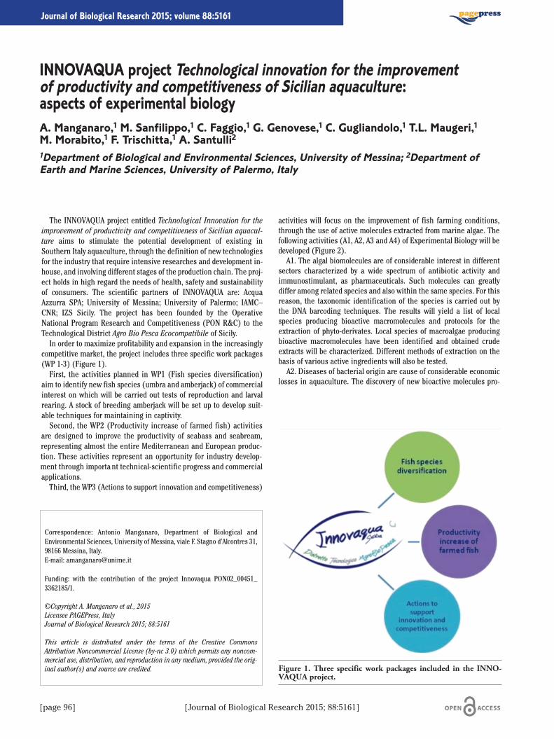
[page 96] [Journal of Biological Research 2015; 88:5161]
Journal of Biological Research 2015; volume 88:5161
The INNOVAQUA project entitled Technological Innovation for theimprovement of productivity and competitiveness of Sicilian aquacul-ture aims to stimulate the potential development of existing inSouthern Italy aquaculture, through the definition of new technologiesfor the industry that require intensive researches and development in-house, and involving different stages of the production chain. The proj-ect holds in high regard the needs of health, safety and sustainabilityof consumers. The scientific partners of INNOVAQUA are: AcquaAzzurra SPA; University of Messina; University of Palermo; IAMC–CNR; IZS Sicily. The project has been founded by the OperativeNational Program Research and Competitiveness (PON R&C) to theTechnological District Agro Bio Pesca Ecocompatibile of Sicily.In order to maximize profitability and expansion in the increasingly
competitive market, the project includes three specific work packages(WP 1-3) (Figure 1).First, the activities planned in WP1 (Fish species diversification)
aim to identify new fish species (umbra and amberjack) of commercialinterest on which will be carried out tests of reproduction and larvalrearing. A stock of breeding amberjack will be set up to develop suit-able techniques for maintaining in captivity.Second, the WP2 (Productivity increase of farmed fish) activities
are designed to improve the productivity of seabass and seabream,representing almost the entire Mediterranean and European produc-tion. These activities represent an opportunity for industry develop-ment through important technical-scientific progress and commercialapplications.Third, the WP3 (Actions to support innovation and competitiveness)
activities will focus on the improvement of fish farming conditions,through the use of active molecules extracted from marine algae. Thefollowing activities (A1, A2, A3 and A4) of Experimental Biology will bedeveloped (Figure 2).A1. The algal biomolecules are of considerable interest in different
sectors characterized by a wide spectrum of antibiotic activity andimmunostimulant, as pharmaceuticals. Such molecules can greatlydiffer among related species and also within the same species. For thisreason, the taxonomic identification of the species is carried out bythe DNA barcoding techniques. The results will yield a list of localspecies producing bioactive macromolecules and protocols for theextraction of phyto-derivates. Local species of macroalgae producingbioactive macromolecules have been identified and obtained crudeextracts will be characterized. Different methods of extraction on thebasis of various active ingredients will also be tested.A2. Diseases of bacterial origin are cause of considerable economic
losses in aquaculture. The discovery of new bioactive molecules pro-
Correspondence: Antonio Manganaro, Department of Biological andEnvironmental Sciences, University of Messina, viale F. Stagno d’Alcontres 31,98166 Messina, Italy.E-mail: [email protected]
Funding: with the contribution of the project Innovaqua PON02_00451_3362185/1.
©Copyright A. Manganaro et al., 2015Licensee PAGEPress, ItalyJournal of Biological Research 2015; 88:5161
This article is distributed under the terms of the Creative CommonsAttribution Noncommercial License (by-nc 3.0) which permits any noncom-mercial use, distribution, and reproduction in any medium, provided the orig-inal author(s) and source are credited.
INNOVAQUA project Technological innovation for the improvementof productivity and competitiveness of Sicilian aquaculture:aspects of experimental biologyA. Manganaro,1 M. Sanfilippo,1 C. Faggio,1 G. Genovese,1 C. Gugliandolo,1 T.L. Maugeri,1M. Morabito,1 F. Trischitta,1 A. Santulli21Department of Biological and Environmental Sciences, University of Messina; 2Department ofEarth and Marine Sciences, University of Palermo, Italy
Figure 1. Three specific work packages included in the INNO-VAQUA project.
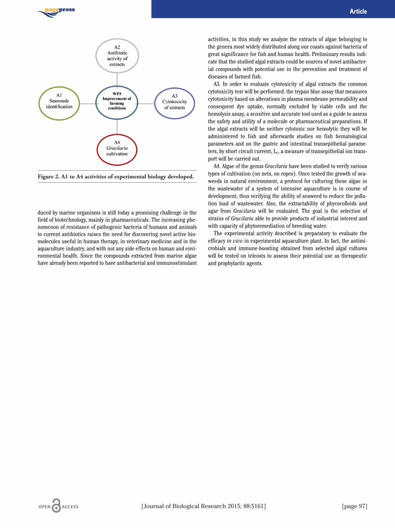
[Journal of Biological Research 2015; 88:5161] [page 97]
Article
duced by marine organisms is still today a promising challenge in thefield of biotechnology, mainly in pharmaceuticals. The increasing phe-nomenon of resistance of pathogenic bacteria of humans and animalsto current antibiotics raises the need for discovering novel active bio-molecules useful in human therapy, in veterinary medicine and in theaquaculture industry, and with not any side effects on human and envi-ronmental health. Since the compounds extracted from marine algaehave already been reported to have antibacterial and immunostimulant
activities, in this study we analyze the extracts of algae belonging tothe genera most widely distributed along our coasts against bacteria ofgreat significance for fish and human health. Preliminary results indi-cate that the studied algal extracts could be sources of novel antibacter-ial compounds with potential use in the prevention and treatment ofdiseases of farmed fish.A3. In order to evaluate cytotoxicity of algal extracts the common
cytotoxicity test will be performed: the trypan blue assay that measurescytotoxicity based on alterations in plasma membrane permeability andconsequent dye uptake, normally excluded by viable cells and thehemolysis assay, a sensitive and accurate tool used as a guide to assessthe safety and utility of a molecule or pharmaceutical preparations. Ifthe algal extracts will be neither cytotoxic nor hemolytic they will beadministered to fish and afterwards studies on fish hematologicalparameters and on the gastric and intestinal transepithelial parame-ters, by short circuit current, Isc, a measure of transepithelial ion trans-port will be carried out.A4. Algae of the genus Gracilaria have been studied to verify various
types of cultivation (on nets, on ropes). Once tested the growth of sea-weeds in natural environment, a protocol for culturing these algae inthe wastewater of a system of intensive aquaculture is in course ofdevelopment, thus verifying the ability of seaweed to reduce the pollu-tion load of wastewater. Also, the extractability of phycocolloids andagar from Gracilaria will be evaluated. The goal is the selection ofstrains of Gracilaria able to provide products of industrial interest andwith capacity of phytoremediation of breeding water.The experimental activity described is preparatory to evaluate the
efficacy in vivo in experimental aquaculture plant. In fact, the antimi-crobials and immune-boosting obtained from selected algal cultureswill be tested on teleosts to assess their potential use as therapeuticand prophylactic agents.
Figure 2. A1 to A4 activities of experimental biology developed.
[page 98] [Journal of Biological Research 2015; 88:5161]
Journal of Biological Research 2015; volume 88:5161
Introduction
Adverse life events during the neonatal period result in long-termeffects on physiology and behavior.1 Early postnatal experiences, such asa modification of the mother–infant interaction, may influence thedevelopment of neural systems that underlie the expression of neuroen-docrine and behavioural responses to environmental challenges, involv-ing changes in the hypothalamic-pituitary-adrenal (HPA) axis2 togetherwith decreased levels of brain-derived neurotrophic factor (BDNF) andnerve growth factor (NGF).3 Disturbances in mother-infant interactionrepresent a natural stressor which may lead to maladaptive develop-ment;4 indeed protracted Maternal Separation (MS) reduces maternalcare thus induce in abnormal HPA axis responses, hippocampal BDNFdown-regulation5 impaired memory formation.6 The opposite isobserved when maternal care is increased by a daily brief, maternal sep-aration. The polymorphism of neuroendocrine processes and behaviour-al responses following brief MS procedure 7 includes gender-related dif-ferences; however very few studies exist on sex-specific behaviours.7
Based upon these findings, the present study was carried out to investi-gate the consequences of a brief, maternal separation on declarative andspatial memory, focusing on sex related alteration due to the discreteeffects that hormones may play on the brain circuits.
Materials and Methods
24 adult male and female Wistar rats were divided in the followingexperimental groups: maternally separated males (MS-m) and females(MS-f), and non-separated males (NS-m) and females (NS-f).
Maternal manipulation involved a 15- minutes daily separation of lit-ters from the mothers from postnatal day 2 (PND 2) to 21 (PND 21).Non separated rats were left undisturbed in their home cages untilweaning. To assess the influence of maternal separation on cognitivefunction we used respectively: the Object Recognition (OR) Test fordeclarative memory and the Morris Water Maze (MWM) for spatiallearning and reference memory, performing respectively place learn-ing and probe sessions.
Results
Our results indicate that a brief, daily maternal separation was ableto induce in MS-f group an increase in the time spent exploring thenovel object in the OR in both after 1h (P<0.001), and24h (P<0.05) retention intervals compared to NS-f controls, while
no significant differences were observed in MS-m group when com-pared to respective controls.In the MWM, during the Place learning paradigm (day 1) MS-m rats
showed a reduction in escape latency (P<0.001) and, during the probephase, an increase in time spent in the target quadrant after platformremoval (P<0.05), compared to NS-m group. On the contrary, MS-fgroup showed non-significant differences in escape latency in theplace learning compared to NS-f controls. MS-f rats spent more time(P<0.05) in the target quadrant in probe phase, compared to respec-tive controls.
Conclusions
The present study was designed to examine the effects of brief dailymother–offspring separation on learning and memory performance,focusing on sex-related differences in declarative and spatial memory.In detail, our study showed that exposure to a brief, maternal separa-tion results in sexually-dimorphic cognitive alterations that depend onthe nature of the behavioral task: Iindeed we found that MS-f groupoutperformed MS-m group on Object Recognition Test, a working, non-spatial memory dependent task that utilizes both cortical and hip-pocampal input.8 This result could be due to a potential improvementof the perirhinal cortex and dorsal hippocampus activity in femaleswhen compared to MS-m group and to controls.9 In addition, in theMorris Water Maze, maternal separation affected learning in adultmale and female rats in a task-specific manner. In particular, it wasobserved that this procedure enhanced the ability of processing spatialinformation only in male rats during the first day of place learning
Correspondence: Carla Cannizzaro, Department of Sciences for HealthPromotion and Mother and Child Care Giuseppe D’Alessandro, University ofPalermo, via del Vespro 129, 90127 Palermo, Italy. E-mail: [email protected]
©Copyright R.A.M. Marino et al., 2015Licensee PAGEPress, ItalyJournal of Biological Research 2015; 88:5161
This article is distributed under the terms of the Creative CommonsAttribution Noncommercial License (by-nc 3.0) which permits any noncom-mercial use, distribution, and reproduction in any medium, provided the orig-inal author(s) and source are credited.
Brief maternal separation procedures occurring early in lifeaffect learning and memory in adult Wistar rats: sex-related differencesin cognitive behaviourR.A.M. Marino, F. Plescia, A. Brancato, G. Gambino, C. CannizzaroDepartment of Sciences for Health Promotion and Mother and Child Care Giuseppe D’Alessandro,University of Palermo, Italy
[Journal of Biological Research 2015; 88:5161] [page 99]
Article
whereas, both groups showed improved performances during probe,confirming the discrete effects of maternal separation on mechanismsof memory storage. In conclusion, a brief, maternal separation affectslearning and memory in adult rats not only in a sex-related, manner,but also in a task- specific way.10 This study provides us with differentoutcomes useful to clarify how early life events can influence thebehavioural adaptive mechanisms in adulthood in a sex specific man-ner probably due to differences in the modulation of hippocampal func-tion and HPA axis response11,12 highliting, overall, the main role playedby a high mother-infant relationship in the correct development ofphysiology and behaviour in adulthood.
References
1. Gareau MG, Jury J, Perdue MH. Neonatal maternal separation of ratpups results in abnormal cholinergic regulation of epithelial perme-ability. Am J Physiol Gastrointest Liver Physiol 2007;293:198-203.
2. Mesquita AR, Pego JM, Summaveille T, et al. Neurodevelopment-milestone abnormalities in rats exposed to stress in early life.Neuroscience 2007;147:1022-33.
3. Griesbach GS, Hovda DA, Molteni R, et al. Voluntary exercise follow-ing traumatic brain injury: Brain-derived neurotrophic factor upreg-ulation and recovery of function. Neuroscience 2004;125:129-39.
4. Daniels WMU, Pietersen CY, Carstens ME, Stein DJ. Maternal sep-aration in rats leads to anxiety-like behavior and a blunted ACTHresponse and altered neurotransmitter levels in response to a sub-sequent stressor. Metab Brain Dis 2004;19:1-2.
5. Marais L, van Rensburg SJ, van Zyl JM, et al. Maternal separationof rat pups increases the risk of developing depressive-like behav-ior after subsequent chronic stress by altering corticosterone andneurotrophin levels in the hippocampus. Neurosci Res 2008;61:106-12.
6. Knuth ED, Etgen AM. Long-term behavioral consequences of brief,repeated neonatal isolation. Brain Res 2007;1128:139-47.
7. Korosi A, Naninck EF, Oomen CA, et al. Early-life stress mediatedmodulation of adult neurogenesis and behavior. Behav Brain Res2012;227:400-9.
8. Steckler T, Drinkenburg WHIM, Sahgal A, Aggleton JP. Recognitionmemory in rats. Concepts and classification. Prog Neurobiol 1998;54:289-311.
9. Baxter MG. I’ve seen it all before: explaining age-related impair-ments in object recognition. Theoretical comment on Burke et al.Behav Neurosci 2010;124:706-9.
10. Noschang C, Krolow R, Mar Arcego D, et al. Neonatal handlingaffects learning, reversal learning and antioxidant enzymes activi-ties in a sex-specific manner in rats. Int J Dev Neurosci 2012;30:285-91.
11. Cannizzaro E, Martire M, Gagliano M, et al. Reversal of prenataldiazepam-induced deficit in a spatial-object learning task by brief,periodic maternal separation in adult rats. Behav Brain Res2005;161:320-30.
12. Cannizzaro C, Plescia F, Martire M, et al. Single, intense prenatalstress decreases emotionality and enhances learning performancein the adolescent rat offspring: interaction with a brief, dailymaternal separation. Behav Brain Res 2006;169:128-36.
[page 100] [Journal of Biological Research 2015; 88:5161]
Journal of Biological Research 2015; volume 88:5161
The term of biofouling or biological fouling means the accumulationof microorganisms, plants, algae, or animals on wetted surfaces withconsequent loss of the intrinsic characteristic of the material.Biofouling occurs worldwide in various submerged substrates ofindustrial interest as well as on materials that have an archeologicaland historical importance such as wood, metals and stones.1 Theprocess begins with the formation of a biofilm produced by bacteria orother microorganisms2 and once it starts archeological materials aresubjected to an increase of deterioration due to the grazing action ofepilithic and endolithic marine micro- and macroorganisms. To avoidand/or to stop either micro or macro-biofouling processes, in marineenvironment conventional procedures include anti-fouling paints toprevent the attachment of sealife such as algae and molluscs.3
However, these compounds slowly leach into the sea water, killing seal-ife, harming the environment and possibly entering in the food chainand in any case they cannot be directly applied on submerged archeo-logical materials.4 Hence, new products that are i) antifouling agents,ii) harmless to the valuable materials and iii) eco-friendly, are wel-comed.3,5 In the last ten years, Titanium oxides are proven to haveantibacterial activity, if exposed to certain light wave lengths (photo-catalytic activation). In particular, TiO2 is activated at UV wavelength,while Ag-TiO2 is photoactivated by visible spectrum and thus beingeffective also to a certain depth.5 The aim of this study was to comparethe antimicrobial activity of Titanium oxide (TiO2) and a Titaniumoxide doped with Ag (Ag-TiO2) as described by Ruffolo and colleagues5
against pure cultures of Gram positive and Gram negative bacteria aswell as to evaluate the ability to interfere with the adhesion of bacteriaon submerged treated marble slabs simulating a marine environmentin laboratory conditions. For this purpose we carried out different setsof experiment in agarized medium and in UP distilled water to evalu-ate the antibacterial activity of the titanium oxides suspensions andthe preventive effect against microbial adhesion on submerged mate-rial after 24-72 hrs. Laboratory experiments carried out on UP distilledwater suspension showed that Ag-TiO2 activity was slightly higherthan TiO2 (tested at concentration of 0.1 and 0.01%), perhaps due tothe intrinsic antibacterial activity of the Ag alone. On marble slabs,both treatment with TiO2 and AgTiO2 were efficient to prevent themicrobial colonization of the surfaces as shown by the SEM analysis.Untreated marble showed a diffuse presence of Extracellular PolymericSubstances (EPS) while on marbles slabs treated with TiO2 or TiO2-Agor Ag no EPS production and no microbial colonization was evidenced.In conclusion, our preliminary results demonstrate that Titaniumoxides could be successfully applied on the surface of submergedarcheological items to prevent the initial microbial biofouling.
References
1. Stanczak M. Biofouling: it’s not just barnacles anymore. Availablefrom: http://www.csa.com/discoveryguides/biofoul/overview.php
2. Gehrke T, Sand W. Interactions between microorganisms andphysicochemical factors cause MIC of steel pilings in harbours.Houston, TX: NACE International; 2003.
3. Gomez de Saravia SG, Guiamet PS, Videla HA. Preventing biocor-rosion without damaging the environment. Four innovative strate-gies. Northampton: Institute of Corrosion; 2001.
4. Ricci S, Pietrini AM, Bartolini M, Sacco Perasso C. Role of themicroboring marine organisms in the deterioration of archaeolog-ical submerged lapideous artefacts (Baia, Naples, Italy).International Biodeterioration and Biodegradation (in press).
5. Ruffolo SA, Macchia A, La Russa MF, et al. Marine antifouling forunderwater archaeological sites: TiO2 and Ag-doped TiO2. Int JPhotoenergy 2013;2013:251647.
Correspondence: Clara Enza Urzì, Department of Biological andEnvironmental Sciences, University of Messina, viale F. Stagno d’Alcontres 31,98166 Messina, Italy.E-mail: [email protected]
Acknoweldgements: this research is supported by Italian national researchproject COMAS (Planned COnservation, in situ, of underwater archaeologi-cal artefacts).
©Copyright L. Mazza et al., 2015Licensee PAGEPress, ItalyJournal of Biological Research 2015; 88:5161
This article is distributed under the terms of the Creative CommonsAttribution Noncommercial License (by-nc 3.0) which permits any noncom-mercial use, distribution, and reproduction in any medium, provided the orig-inal author(s) and source are credited.
Evaluation of antifouling activity of TiO2 and Ag-doped TiO2in laboratory conditions to be applied on submerged archeological materialL. Mazza,1 S.A. Ruffolo,2 A. Macchia,3 M.F. La Russa,2 M. Barberio,4 G.M. Crisci,2F. De Leo,1 C.E. Urzì11Department of Biological and Environmental Sciences, University of Messina; 2Department ofBiology, Ecology and Earth Sciences, University of Calabria, Arcavacata di Rende (CS); 3SyremontS.p.A., Arcavacata di Rende (CS); 4Department of Physics, University of Calabria, Arcavacata diRende (CS), Italy
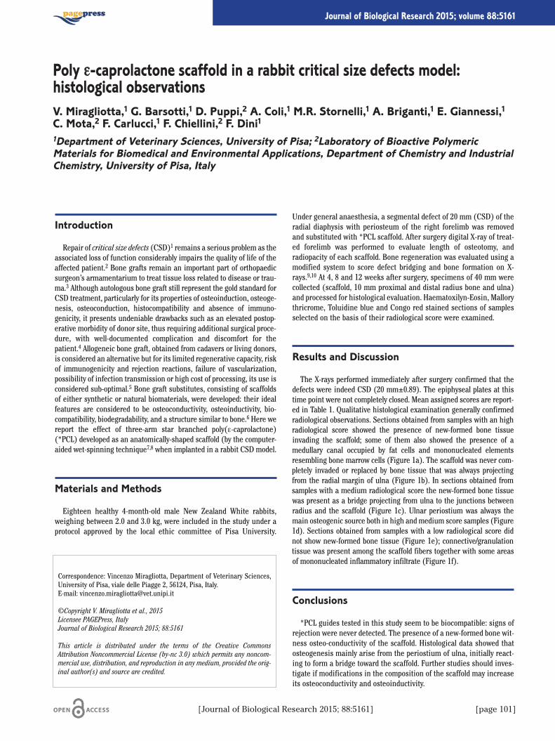
[Journal of Biological Research 2015; 88:5161] [page 101]
Journal of Biological Research 2015; volume 88:5161
Introduction
Repair of critical size defects (CSD)1 remains a serious problem as theassociated loss of function considerably impairs the quality of life of theaffected patient.2 Bone grafts remain an important part of orthopaedicsurgeon’s armamentarium to treat tissue loss related to disease or trau-ma.3 Although autologous bone graft still represent the gold standard forCSD treatment, particularly for its properties of osteoinduction, osteoge-nesis, osteoconduction, histocompatibility and absence of immuno-genicity, it presents undeniable drawbacks such as an elevated postop-erative morbidity of donor site, thus requiring additional surgical proce-dure, with well-documented complication and discomfort for thepatient.4 Allogeneic bone graft, obtained from cadavers or living donors,is considered an alternative but for its limited regenerative capacity, riskof immunogenicity and rejection reactions, failure of vascularization,possibility of infection transmission or high cost of processing, its use isconsidered sub-optimal.5 Bone graft substitutes, consisting of scaffoldsof either synthetic or natural biomaterials, were developed: their idealfeatures are considered to be osteoconductivity, osteoinductivity, bio-compatibility, biodegradability, and a structure similar to bone.6 Here wereport the effect of three-arm star branched poly(e-caprolactone)(*PCL) developed as an anatomically-shaped scaffold (by the computer-aided wet-spinning technique7,8 when implanted in a rabbit CSD model.
Materials and Methods
Eighteen healthy 4-month-old male New Zealand White rabbits,weighing between 2.0 and 3.0 kg, were included in the study under aprotocol approved by the local ethic committee of Pisa University.
Under general anaesthesia, a segmental defect of 20 mm (CSD) of theradial diaphysis with periosteum of the right forelimb was removedand substituted with *PCL scaffold. After surgery digital X-ray of treat-ed forelimb was performed to evaluate length of osteotomy, andradiopacity of each scaffold. Bone regeneration was evaluated using amodified system to score defect bridging and bone formation on X-rays.9,10 At 4, 8 and 12 weeks after surgery, specimens of 40 mm werecollected (scaffold, 10 mm proximal and distal radius bone and ulna)and processed for histological evaluation. Haematoxilyn-Eosin, Mallorythricrome, Toluidine blue and Congo red stained sections of samplesselected on the basis of their radiological score were examined.
Results and Discussion
The X-rays performed immediately after surgery confirmed that thedefects were indeed CSD (20 mm±0.89). The epiphyseal plates at thistime point were not completely closed. Mean assigned scores are report-ed in Table 1. Qualitative histological examination generally confirmedradiological observations. Sections obtained from samples with an highradiological score showed the presence of new-formed bone tissueinvading the scaffold; some of them also showed the presence of amedullary canal occupied by fat cells and mononucleated elementsresembling bone marrow cells (Figure 1a). The scaffold was never com-pletely invaded or replaced by bone tissue that was always projectingfrom the radial margin of ulna (Figure 1b). In sections obtained fromsamples with a medium radiological score the new-formed bone tissuewas present as a bridge projecting from ulna to the junctions betweenradius and the scaffold (Figure 1c). Ulnar periostium was always themain osteogenic source both in high and medium score samples (Figure1d). Sections obtained from samples with a low radiological score didnot show new-formed bone tissue (Figure 1e); connective/granulationtissue was present among the scaffold fibers together with some areasof mononucleated inflammatory infiltrate (Figure 1f).
Conclusions
*PCL guides tested in this study seem to be biocompatible: signs ofrejection were never detected. The presence of a new-formed bone wit-ness osteo-conductivity of the scaffold. Histological data showed thatosteogenesis mainly arise from the periostium of ulna, initially react-ing to form a bridge toward the scaffold. Further studies should inves-tigate if modifications in the composition of the scaffold may increaseits osteoconductivity and osteoinductivity.
Correspondence: Vincenzo Miragliotta, Department of Veterinary Sciences,University of Pisa, viale delle Piagge 2, 56124, Pisa, Italy. E-mail: [email protected]
©Copyright V. Miragliotta et al., 2015Licensee PAGEPress, ItalyJournal of Biological Research 2015; 88:5161
This article is distributed under the terms of the Creative CommonsAttribution Noncommercial License (by-nc 3.0) which permits any noncom-mercial use, distribution, and reproduction in any medium, provided the orig-inal author(s) and source are credited.
Poly e-caprolactone scaffold in a rabbit critical size defects model:histological observationsV. Miragliotta,1 G. Barsotti,1 D. Puppi,2 A. Coli,1 M.R. Stornelli,1 A. Briganti,1 E. Giannessi,1C. Mota,2 F. Carlucci,1 F. Chiellini,2 F. Dini11Department of Veterinary Sciences, University of Pisa; 2Laboratory of Bioactive PolymericMaterials for Biomedical and Environmental Applications, Department of Chemistry and IndustrialChemistry, University of Pisa, Italy

[page 102] [Journal of Biological Research 2015; 88:5161]
Article
References
1. Hollinger JO, Kleinschmidt JC. The critical size defect as an exper-imental model to test bone repair materials. J Craniofacial Surg1990;1:60-8.
2. Schieker M, Mutschler W. Bridging post-traumatic bony defects.Established and new methods. Der Unfallchirurg 2006;109:715-32.
3. Soucacos PN, Johnson EO, Babis G. An update on recent advancesin bone regeneration. Injury 2008;39(Suppl.2):1-4.
4. Dimitriou R, Jones E, McGonagle D, Giannoudis PV. Bone regener-ation: current concepts and future directions. BMC Med 2011;9:66.
5. Finkemeier CG. Bone-grafting and bone-graft substitutes. J BoneJoint Surg Am 2002;84:454-64.
6. Puppi D, Chiellini F, Piras AM, Chiellini E. Polymeric materials forbone and cartilage repair. Prog Polymer Sci 2010;35:403-40.
7. Puppi D, Dinucci D, Bartoli C, et al. Development of 3D wet-spunpolymeric scaffolds loaded with antimicrobial agents for bone engi-neering. J Bioact Compat Pol 2011;26:478-92.
8. Mota C, Puppi D, Dinucci D, et al. Additive manufacturing of starpoly(e-caprolactone) wet-spun scaffolds for bone tissue engineer-ing applications. J Bioact Compat Pol 2013;28:320-40.
9. Hedberg EL, Kroese-Deutman HC, Shih CK, et al. Methods: a com-parative analysis of radiography, microcomputed tomography, andhistology for bone tissue engineering. Tissue Eng 2005;11:1356-67.
10. Bodde EW, Spauwen PH, Mikos AG, Jansen JA. Closing capacity ofsegmental radius defects in rabbits. J Biomed Mat A 2008;85:206-17.
Table 1. Mean assigned scores and standard deviation values.
Time after Mean assigned Standardsurgery (weeks) score deviation
4 7.75 3.88 9.33 2.7112 7.66 4.72
Figure 1. Representative photomicrographs of high-score (a, b)medium score (c, d) and low score (e, f) samples. a) Asterisksindicate the implanted scaffolds, while the arrowhead indicatesthe fat cells populating the new-formed medullary canal; scalebar=500 μm. b) The bone tissue (left) is growing into theimplanted scaffold; scale bar 800 μm. c) The arrow indicates thenew-formed bone as a bridge projecting from ulna to the junc-tions between radius and the scaffold; scale bar=2 μm. d) Theulnar periosteum is the main source of new-formed bone invad-ing the scaffold (arrowhead); scale bar=800 μm. e) The scaffold(up-left) is not invaded by new-formed bone; scale bar=2 mm. f)The asterisk indicates the presence of mononucleated inflamma-tory cells; scale bar=200 μm.
[Journal of Biological Research 2015; 88:5161] [page 103]
Journal of Biological Research 2015; volume 88:5161
Thioridazine, a phenothiazine drug with antipsychotic efficacy1,2
and anti-microbial activity,2 is particularly useful for the treatment ofmultidrug resistant tuberculosis.3,4 This drug is known to trigger ane-mia.5 At least in theory, the anemia could result from stimulation ofsuicidal erythrocyte death or eryptosis,6 which is characterized by cellshrinkage, phospholipid scrambling of the cell membrane with phos-phatidylserine exposure at the erythrocyte surface, increase of cytoso-lic Ca2+-activity ([Ca2+]i) and activation of p38 kinase, a kinaseexpressed in human erythrocytes and activated by hyperosmotic shock,a known trigger of eryptosis.7 The present study explored whetherthioridazine triggers eryptosis. The concentrations required to triggereryptosis are within the range of concentrations (6 μg/mL≈15 μM)encountered in vivo.8 Since different erythrocyte specimens used indistinct experiments are differently susceptible to triggers of eryptosis,the same erythrocyte specimens have been used for control and exper-imental conditions. A 48 h exposure to thioridazine was followed by asignificant: i) increase of [Ca2+]i (30 μM), estimated from Fluo3-flu-orescence in flow cytometry (Fluo-3/AM); ii) decrease cell volume,estimated from forward scatter (30 μM) in flow cytometry; iii)increase of the percentage of phosphatidylserine exposure, estimatedfrom annexin-V-binding (≥12 μM) in flow cytometry (Annexin-V-FITC,1:200 dilution); iv) increase of the percentage of hemolysed erytro-cytes, estimated from the hemoglobin concentration in the super-natant This in turn was determined photometrically at 405 nm. Theabsorption of the supernatant of erythrocytes lysed in distilled waterwas defined as 100% hemolysis. As illustrated in Figure 1A, thiori-dazine treatment significantly increased the hemoglobin concentra-tion in the supernatant. The percentage of hemolytic erythrocytes was,however, clearly smaller than the percentage of annexin V binding ery-throcytes. Both, cell membrane scrambling and cell shrinkage couldhave resulted from increase of cytosolic Ca2+ activity ([Ca2+]i).However, even in the absence of extracellular Ca2+, thioridazine stillsignificantly increased the percentage of annexin-V-binding erythro-cytes pointing to additional mechanisms involved. In order to explorewhether the additional mechanisms could include p38 kinase, erythro-
Correspondence: Paola Modicano, Department of Physiology, Eberhard-Karls-University of Tuebingen, Gmelinstrasse 5, 72076 Tuebingen, Germany.E-mail: [email protected]
©Copyright P. Modicano et al., 2015Licensee PAGEPress, ItalyJournal of Biological Research 2015; 88:5161
This article is distributed under the terms of the Creative CommonsAttribution Noncommercial License (by-nc 3.0) which permits any noncom-mercial use, distribution, and reproduction in any medium, provided the orig-inal author(s) and source are credited.
Effect of thioridazine on erythrocytesP. Modicano,1,2 E. Lang,2 M. Arnold,2 R. Bissinger,2 C. Faggio,1 M. Abed,2 F. Lang21Department of Biological and Environmental Sciences, University of Messina, Italy; 2Departmentof Physiology, Eberhard-Karls-University of Tuebingen, Germany
Figure 1. A) Effect of thioridazine on phosphatidylserine expo-sure and hemolysis. Arithmetic means±SEM (n=6) of erythrocytesannexin-V-binding following incubation for 48 hours to Ringersolution without (white bar) or with (black bars) presence ofthioridazine (6-30 μM). For comparison, arithmetic means±SEM(n=5) of the percentage of hemolysis is shown as grey bars.***(P<0.001) indicates significant differences from the absence ofthioridazine (ANOVA). B) Effect of thioridazine on phos-phatidylserine exposure in the presence or absence of p38 kinaseinhibitor SB203580. Arithmetic means±SEM (n=6) of erythro-cytes annexin-V-binding following incubation for 48 h to Ringersolution without or with presence of thioridazine (6-30 μM) inthe absence (white bars) or presence (black bars) of 2 μMSB203580. **(P<0.01); ***(P<0.001) indicates significant differ-ences from the absence of thioridazine (ANOVA); #(P<0.05);###(P<0.001) indicates significant differences from the absenceof SB203580 (ANOVA).
[page 104] [Journal of Biological Research 2015; 88:5161]
Article
cytes were exposed in further experiments to 6-30 μM thioridazine for48 h in either the presence or absence of the p38 kinase inhibitorSB203580 (2 μM) (Figure 1B). In conclusion, thioridazine stimulateseryptosis and is partially effective by activation of p38 kinase and byincrease of cytocolic Ca2+ activity. Nominal absence of extracellularCa2+ and p38 kinase inhibitor SB203580 significantly blunted but didnot abolish annexin-V-binding following thioridazine exposure.
References
1. Purhonen M, Koponen H, Tiihonen J, Tanskanen A. Outcome ofpatients after market withdrawal of thioridazine: a retrospectiveanalysis in a nationwide cohort. Pharmacoepidem Dr S 2012;21:1227-31.
2. Thanacoody RH. Thioridazine: the good and the bad. Recent PatAnti-Inf 2011;6:92-8.
3. Amaral L. Thioridazine: an old neuroleptic effective against totallydrug resistant tuberculosis. Acta Mèdica Portuguesa 2012;25:118-21.
4. Amaral L, Viveiros M. Why thioridazine in combination with antibi-otics cures extensively drug-resistant Mycobacterium tuberculosisinfections. Int J Antimicrob Ag 2012;39:376-80.
5. King DJ, Wager E. Haematological safety of antipsychotic drugs. JPsychopharmacol 1998;12:283-8.
6. Lang F, Gulbins E, Lerche H, et al. Eryptosis, a window to systemicdisease. Cell Physiol Biochem 2008;22:373-80.
7. Gatidis S, Zelenak C, Fajol A, et al. p38 MAPK activation and func-tion following osmotic shock of erythrocytes. Cell Physiol Biochem2011;28:1279-86.
8. Gottschalk LA, Biener R, Noble EP, et al. Thioridazine plasma levelsand clinical response. Compr Psyc 1975;16:323-37.

[Journal of Biological Research 2015; 88:5161] [page 105]
Journal of Biological Research 2015; volume 88:5161
Introduzione
Questo studio è stato svolto nell’ambito del Progetto ALISAL:Miglioramento delle proprietà igienico-sanitarie, salutistiche e funzionalidi materie prime per l’alimentazione dell’uomo e/o degli animali, promos-so dal Ministero delle Politiche Agricole, Alimentari e Forestali italiano(MIPAAF). Lo studio in oggetto è parte del progetto sopra indicato il cuiscopo era quello di selezionare nuovi genotipi di cereali, in particolare difrumento duro, al fine di migliorare le caratteristiche nutrizionali e tec-nologiche dell’ intera filiera produttiva. La fase preliminare dello sviluppoprogettuale è stata quella di sviluppare un metodo per la caratterizzazio-ne morfologica e ultrastrutturale delle granelle d’interesse allo scopo difornire indicazioni qualitative e quantitative per la precisa caratterizza-zione dei campioni. I risultati ottenuti sono di seguito riportati.
Materiali e Metodi
Al fine di evidenziare eventuali differenze morfologiche e ultrastrut-turali tra i campioni, le cariossidi di frumento duro sono state analiz-zate tal quali mediante microscopia elettronica a scansione in ambien-tale e a pressione variabile utilizzando il microscopio SEM EVO LS10ZEISS. Questo microscopio è dotato di una sorgente di elettroni LaB6in grado di fornire una luminosità del fascio elettronico uniforme, sta-bile e di conseguenza un alto contrasto; tali caratteristiche lo rendonoparticolarmente adattato all’osservazione di campioni sia disidratatiche umidi. Sulle cariossidi di grano duro si è eseguito un adeguatotaglio sagittale e le sezioni tal quali, senza alcuna preparativa sonostate fissate su gli appositi stub, successivamente si è passati all’osser-vazione predisponendo il microscopio a pressione variabile ed utiliz-
zando il detector CZBSD come rivelatore primario e il detector SE1come rivelatore secondario. Il mix dei due ci ha fornito una precisamappa ultrastrutturale di ogni cariosside testata. Secondo le specifi-che progettuali abbiamo preso in considerazione tre cultivar di fru-mento duro coltivate nel Sud Italia (Sicilia) negli anni 2010 e 2011. Icampioni sono stati classificati come SA1(cultivar Sant’Agata),DU1(cultivar Duilio), SI1(cultivar Simeto) per le produzioni dell’anno2010 e SA2, DU2, SI2 per le produzioni dell’anno 2011.
Risultati e Discussione
I campioni di grano duro classificati, come detto in precedenza, sonostati sottoposti all’analisi in scansione elettronica che è stata eseguita indue fasi. Una prima fase è stata finalizzata allo studio della superficiedelle cariossidi a basso ingrandimento per rilevare le caratteristiche mor-fologiche più grezze quali la lunghezza e la larghezza media (Figura 1).La seconda serie di osservazioni è stata realizzata ad alto ingrandi-
mento in modo da evidenziare le componenti ultrastrutturali dellecariossidi, quali lo strato aleuronico, la camera embrionale, e la morfo-logia dell’endosperma, cercando per ogni campione di fare le osservazio-ni in posizioni di coordinate analoghe e quindi confrontabili. Non è statarilevata alcuna variazione morfo-ultrastrutturale significativa per lamaggior parte dei campioni: tuttavia nei campioni DU2 (Figura 2B) cul-tivar Duilio produzione del 2011 e nel campione SA1 (Figura 2A) cultivarSant’Agata produzione del 2010, abbiamo osservato una serie di modifi-cazioni di notevole entità. Nello specifico il campione DU2 presenta unaforte discrasia fra cariossidi dello stesso campione e, comunque, le
Correspondence: Fabio Nobili, Council Research and Experimental Center inAgriculture-Research Center for Food and Nutrition, via Nazionale 82, 00184Rome, Italy.E-mail [email protected]
©Copyright I. Garaguso et al., 2015Licensee PAGEPress, ItalyJournal of Biological Research 2015; 88:5161
This article is distributed under the terms of the Creative CommonsAttribution Noncommercial License (by-nc 3.0) which permits any noncom-mercial use, distribution, and reproduction in any medium, provided the orig-inal author(s) and source are credited.
Microscopia elettronica a scansione per l’analisi morfologicae ultrastrutturale di cariossidi di frumento duroI. Garaguso,1 M. Giammanco,2 R. Acquistucci,1 D. Di Majo,2 F. Nobili11Council Research and Experimental Center in Agriculture-Research Center for Food and Nutrition,Rome; 2Physiology and Pharmacology Unit, Department of Legal, Social and Sports Sciences,University of Palermo, Italy
Figura 1. Misura della larghezza (A) e della lunghezza (B) (in mm)delle cariossidi dei frumenti Sant’Agata (giallo), Simeto (arancio),Duilio (verde).
[page 106] [Journal of Biological Research 2015; 88:5161]
Article
dimensioni dei parametri macroscopici, come si evidenzia nella Figura 1,sono inferiori a tutti gli altri campioni testati. Le dimensioni volumetricheridotte e coartate dell’endosperma (Figura 2B) fanno presupporre che il
suddetto campione abbia subito uno stress da temperatura in fine stagio-ne o una conservazione anomala. Il campione SA1 (Figura 2A) invece pre-senta le cariossidi più omogenee fra loro, mentre presenta lo strato aleu-ronico iperplasico (Figura 2A) in contrasto con quello della normale mor-fologia descritto in letteratura. Un’ipotesi è che tale campione abbia subi-to uno stress da temperatura in un momento precoce di crescita e cheabbia poi innescato successivamente un parziale recupero realizzandouna iperplasia delle cellule aleuroniche in maniera di aumentare la pro-duzione di amido primaria e compensare il gap di crescita iniziale.
Conclusioni
Questo studio preliminare ci ha permesso di caratterizzare morfolo-gicamente i campioni di grano duro fornitoci e ci ha consentito di indi-viduare il campione più performante dal punto di vista morfologico eultrastrutturale che è risultato essere il grano duro della cultivarSant’Agata. La tecnica utilizzata in questo lavoro (microscopia elettro-nica a scansione) può rappresentare un valido supporto per caratteriz-zare campioni biologici con struttura definita e parametrizzabile, inquesto caso grano duro, da destinare a ulteriori indagini di natura ana-litico- funzionale.
Figura 2. Campioni SA1 (A) cultivar Sant’Agata produzione del2010 e DU2 (B) cultivar Duilio produzione del 2011.
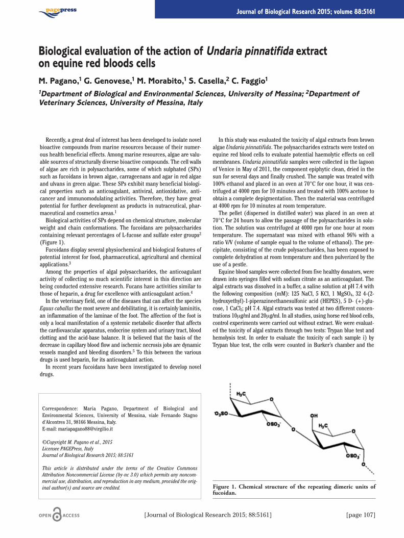
[Journal of Biological Research 2015; 88:5161] [page 107]
Journal of Biological Research 2015; volume 88:5161
Recently, a great deal of interest has been developed to isolate novelbioactive compounds from marine resources because of their numer-ous health beneficial effects. Among marine resources, algae are valu-able sources of structurally diverse bioactive compounds. The cell wallsof algae are rich in polysaccharides, some of which sulphated (SPs)such as fucoidans in brown algae, carrageenans and agar in red algaeand ulvans in green algae. These SPs exhibit many beneficial biologi-cal properties such as anticoagulant, antiviral, antioxidative, anti-cancer and immunomodulating activities. Therefore, they have greatpotential for further development as products in nutraceutical, phar-maceutical and cosmetics areas.1
Biological activities of SPs depend on chemical structure, molecularweight and chain conformations. The fucoidans are polysaccharidescontaining relevant percentages of L-fucose and sulfate ester groups2
(Figure 1).Fucoidans display several physiochemical and biological features of
potential interest for food, pharmaceutical, agricultural and chemicalapplications.3
Among the properties of algal polysaccharides, the anticoagulantactivity of collecting so much scientific interest in this direction arebeing conducted extensive research. Fucans have activities similar tothose of heparin, a drug for excellence with anticoagulant action.4
In the veterinary field, one of the diseases that can affect the speciesEquus caballus the most severe and debilitating, it is certainly laminitis,an inflammation of the laminae of the foot. The affection of the foot isonly a local manifestation of a systemic metabolic disorder that affectsthe cardiovascular apparatus, endocrine system and urinary tract, bloodclotting and the acid-base balance. It is believed that the basis of thedecrease in capillary blood flow and ischemic necrosis jobs are dynamicvessels mangled and bleeding disorders.5 To this between the variousdrugs is used heparin, for its anticoagulant action.In recent years fucoidans have been investigated to develop novel
drugs.
In this study was evaluated the toxicity of algal extracts from brownalgae Undaria pinnatifida. The polysaccharides extracts were tested onequine red blood cells to evaluate potential haemolytic effects on cellmembranes. Undaria pinnatifida samples were collected in the lagoonof Venice in May of 2011, the component epiphytic clean, dried in thesun for several days and finally crushed. The sample was treated with100% ethanol and placed in an oven at 70°C for one hour, it was cen-trifuged at 4000 rpm for 10 minutes and treated with 100% acetone toobtain a complete depigmentation. Then the material was centrifugedat 4000 rpm for 10 minutes at room temperature.The pellet (dispersed in distilled water) was placed in an oven at
70°C for 24 hours to allow the passage of the polysaccharides in solu-tion. The solution was centrifuged at 4000 rpm for one hour at roomtemperature. The supernatant was mixed with ethanol 96% with aratio V/V (volume of sample equal to the volume of ethanol). The pre-cipitate, consisting of the crude polysaccharides, has been exposed tocomplete dehydration at room temperature and then pulverized by theuse of a pestle.Equine blood samples were collected from five healthy donators, were
drawn into syringes filled with sodium citrate as an anticoagulant. Thealgal extracts was dissolved in a buffer, a saline solution at pH 7.4 withthe following composition (mM): 125 NaCl, 5 KCl, 1 MgSO4, 32 4-(2-hydroxyethyl)-1-piperazineethanesulfonic acid (HEPES), 5 D- (+)-glu-cose, 1 CaCl2; pH 7.4. Algal extracts was tested at two different concen-trations 10μg/ml and 20μg/ml. In all studies, using horse red blood cells,control experiments were carried out without extract. We were evaluat-ed the toxicity of algal extracts through two tests: Trypan blue test andhemolysis test. In order to evaluate the toxicity of each sample i) byTrypan blue test, the cells were counted in Burker’s chamber and the
Correspondence: Maria Pagano, Department of Biological andEnvironmental Sciences, University of Messina, viale Fernando Stagnod'Alcontres 31, 98166 Messina, Italy.E-mail: [email protected]
©Copyright M. Pagano et al., 2015Licensee PAGEPress, ItalyJournal of Biological Research 2015; 88:5161
This article is distributed under the terms of the Creative CommonsAttribution Noncommercial License (by-nc 3.0) which permits any noncom-mercial use, distribution, and reproduction in any medium, provided the orig-inal author(s) and source are credited.
Biological evaluation of the action of Undaria pinnatifida extracton equine red bloods cellsM. Pagano,1 G. Genovese,1 M. Morabito,1 S. Casella,2 C. Faggio11Department of Biological and Environmental Sciences, University of Messina; 2Department ofVeterinary Sciences, University of Messina, Italy
Figure 1. Chemical structure of the repeating dimeric units offucoidan.
[page 108] [Journal of Biological Research 2015; 88:5161]
Article
cells were expressed as percentages of viable cells; ii) the hemolysis testto see the hemolytic effects of the algal extracts, that evaluates hemoglo-bin release in the plasma following molecule. After incubation at roomtemperature, the tubes were centrifuged (5 minutes at 1500 rpm) andthe supernatants were determined photometrically with a spectropho-tometer at the absorption of hemoglobin (540 nm). As a measure ofhemolysis, Hb concentration of the supernatants was determined photo-metrically. The absorption of the supernatant of erythrocytes lysed in dis-tilled water was defined as 100% haemolysis. In the treatment of RBCswith polysaccharides extracted from Undaria pinnatifida, hemolysisoccurence at identical concentrations of the control conditions suggestthat the compounds does not shows toxicity effects in equine red bloodcells at the concentration tested (Table 1).Currently in the lagoon of Venice Undaria pinnatifida is removed and
treates as waste stored in landfills and incinerated. This study shows apossible exploitation of Undaria pinnatifida, not toxic to equine redblood cells, as a source of anticoagulant drug with the aim of trasform-ing a waste into a valuable biomass.
References
1. Wijesekara I, Pangestuti R, Kim S-K. Biological activities andpotential health benefits of sulfated polysaccharides derived frommarine algae. Carbohydrate Polymers 2011;84:14-21.
2. Li B, Lu F, Wei X, Zhao R. Fucoidan: structure and bioactivity.Molecules 2008;13:1671-95.
3. Lahaye M, Robic A. Structure and functional properties of ulvan, apolysaccharide from green seaweeds. Biomacromolecules 2007;8:1765-74.
4. Costa L, Fidelis GP, Cordeiro SL, et al. Biological activities of sulfat-ed polysaccharides from tropical seaweeds. Biomed Pharmacother2010;64:21-8.
5. Van Eps AW. Studies on the pathogenesis and prevention of equinelaminitis. PhD Diss. St. Lucia: School of Veterinary Science, TheUniversity of Queensland ed.; 2007.
Table 1. Percentage of hemolysis in control and after the additional of Undaria pinnatifida polisaccaride extract.
300mOsm 200mOsm 150mOsm 100mOsm 0mOsm
Control 0 0 9 85 10010 μg/1mL 0 0 10 84 10020 μg/1mL 0 0 9 81 100
[Journal of Biological Research 2015; 88:5161] [page 109]
Journal of Biological Research 2015; volume 88:5161
Introduction
The extra virgin olive oil is very important in the Mediterraneandiet. From the nutritional point of view, the high energy content andan optimal ratio between saturated and unsaturated fatty acids, togeth-er with the presence of minor compounds such as phenols and toco-pherols and the related antioxidant capacity, confer to this product ahigh healthy value.1 The extra-virgin oil’s nutritional propertiesdepend on the pedoclimatic conditions, cultivar and the productionprocess. In the last years many studies have been performed to remarkand describe the safety and nutritional quality of the extra-virgin oliveoils but it is difficult to compare different parameters representative ofoil’s nutritional quality and to establish which of them is more impor-tant to define the nutritional value of an oil. In 2007, Finotti and col-leagues2 developed a Functional Mathematic Index (FMI) in order toquantify the nutritional quality of extra virgin olive oil and then thesame authors applied it at other foods, such as tea,3 potatoes.4 Thisindex is important because it is able to convert a concept as the qualityinto a numeric quantity. The FMI takes into account different nutri-tional, antioxidant and chemical parameters representative of oil prop-erties. These parameters, suitably processed with the FMI Workbenchfree software, provide a unique value indicated as global quality index(IGQ). We have calculated both the global FMI and the local FMIs. Aimof this study is to compare the nutritional properties of extra virginolive oils from different olive varieties, Biancolilla and Oglialora grownin Sicily, by the functional mathematic index.5
Materials and Methods
Samples of extra-virgin olive oils from two different cultivar(Biancolilla, Oglialora) and processed by different extraction methods(continuous cycle and hydraulic press) have been analyzed. The drupesare grown in the same climatic conditions. The parameters analyzed tocalculate the FMI have been divided in three groups which are related tochemical, nutritional, technologic properties of oil. The three groupsare: i) nutritional parameters: fatty acids (numbers from 1 to 4; Table 1);ii) chemical parameters: acidity and peroxide number (numbers 5 and6; Table 1); iii) technological process parameters: lipidic antioxidantcapacity, tocopherols and total phenols (numbers 7-9; Table 1). Theresults of the analyses were processed by the software and the IGQ andLQ of each oil sample have been compared in order to define which ofthem had the better nutritional properties. The FMI varies from zero toone (good to poor). The global index has been expressed as goodnesspercentage (%FMI). The upper and the lower bounds for each group areestablished and they are reported in Table 1.
Results and Discussion
The analyses allow to classify all oil samples as extra-virgin oliveoils. All parameters are within the limits of the Italian (E.U.) law andthose established in Table 1. No difference among two cultivars aboutacidity, peroxide number and fatty acids profile was found. The lipofil-ic antioxidant capacity was higher in Biancolilla variety than inOglialora with both extractive methods (P=0.0001); sample oilsextracted with continuous method have shown a lower of 16% contentof tocopherols for both variety.The FMI of all oil samples varies from 0.27 to 0.34, therefore they sat-
isfy the necessary conditions to be considered high quality extra-virginoil; Nonetheless, the cultivar Biancolilla has shown a global index betterthan the Oglialora. From the study of local FMI we observed that thenutritional quality of oils extracted by continuous cycle is penalized inthe total polyphenols and lipofilic antioxidant capacity parameters.
Conclusions
The FMI allows to detect that the Biancolilla variety is better thanOglialora and then to underline which parameter can be modified inorder to increase the nutritional quality of the oil analyzed.
Correspondence: Danila Di Majo, Physiology and Pharmacology Unit,Department of Legal, Social and Sports Sciences, University of Palermo,viale delle Scienze, Ed. 16, 90127 Palermo, Italy.E-mail: [email protected]
©Copyright D. Di Majo et al., 2015Licensee PAGEPress, ItalyJournal of Biological Research 2015; 88:5161
This article is distributed under the terms of the Creative CommonsAttribution Noncommercial License (by-nc 3.0) which permits any noncom-mercial use, distribution, and reproduction in any medium, provided the orig-inal author(s) and source are credited.
Nutritional quality of extra virgin olive oil determinedby the application of a new functional matematic indexD. Di Majo,1 E. Finotti,2 M. La Guardia,3 L. La Neve,1 S. Palumbo,1 M. Giammanco11Physiology and Pharmacology Unit, Department of Legal, Social and Sports Sciences, Universityof Palermo; 2Council Research and Experimental Center in Agriculture-Research Center for Foodand Nutrition, Rome; 3Department of Biological, Chemical and Pharmaceutical Sciences andTechnologies, University of Palermo
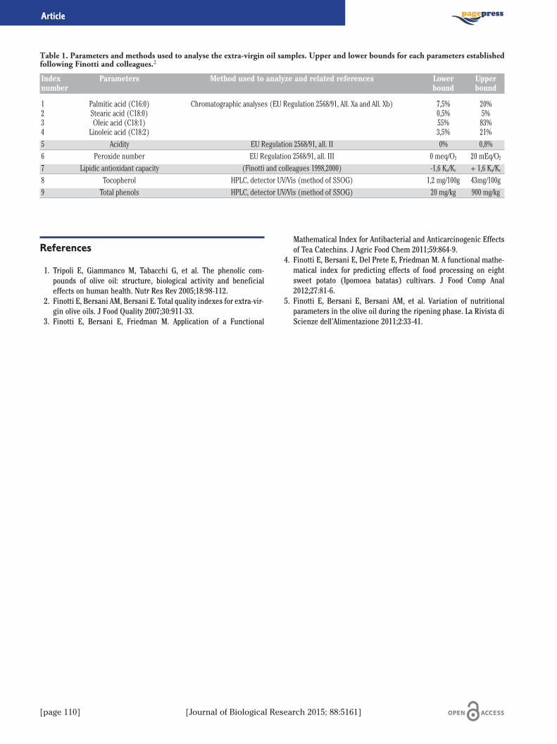
[page 110] [Journal of Biological Research 2015; 88:5161]
Article
References
1. Tripoli E, Giammanco M, Tabacchi G, et al. The phenolic com-pounds of olive oil: structure, biological activity and beneficialeffects on human health. Nutr Res Rev 2005;18:98-112.
2. Finotti E, Bersani AM, Bersani E. Total quality indexes for extra-vir-gin olive oils. J Food Quality 2007;30:911-33.
3. Finotti E, Bersani E, Friedman M. Application of a Functional
Mathematical Index for Antibacterial and Anticarcinogenic Effectsof Tea Catechins. J Agric Food Chem 2011;59:864-9.
4. Finotti E, Bersani E, Del Prete E, Friedman M. A functional mathe-matical index for predicting effects of food processing on eightsweet potato (Ipomoea batatas) cultivars. J Food Comp Anal2012;27:81-6.
5. Finotti E, Bersani E, Bersani AM, et al. Variation of nutritionalparameters in the olive oil during the ripening phase. La Rivista diScienze dell’Alimentazione 2011;2:33-41.
Table 1. Parameters and methods used to analyse the extra-virgin oil samples. Upper and lower bounds for each parameters establishedfollowing Finotti and colleagues.2
Index Parameters Method used to analyze and related references Lower Uppernumber bound bound
1 Palmitic acid (C16:0) Chromatographic analyses (EU Regulation 2568/91, All. Xa and All. Xb) 7,5% 20%2 Stearic acid (C18:0) 0,5% 5%3 Oleic acid (C18:1) 55% 83%4 Linoleic acid (C18:2) 3,5% 21%5 Acidity EU Regulation 2568/91, all. II 0% 0,8%6 Peroxide number EU Regulation 2568/91, all. III 0 meq/O2 20 mEq/O2
7 Lipidic antioxidant capacity (Finotti and colleagues 1998,2000) -1,6 Ka/Kc + 1,6 Ka/Kc8 Tocopherol HPLC, detector UV/Vis (method of SSOG) 1,2 mg/100g 43mg/100g9 Total phenols HPLC, detector UV/Vis (method of SSOG) 20 mg/kg 900 mg/kg
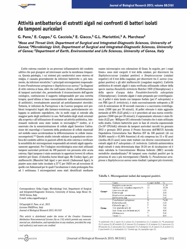
[Journal of Biological Research 2015; 88:5161] [page 111]
Journal of Biological Research 2015; volume 88:5161
L’otite esterna consiste in un processo infiammatorio del condottouditivo che può giungere ad interessare anche la membrana timpani-ca. Questa patologia, i cui sintomi più caratteristici sono otorrea edotalgia, è causata generalmente da infezioni batteriche e, più rara-mente, da infezioni micotiche;1 i principali microrganismi responsabi-li sono Pseudomonas aeruginosa e Staphylococcus aureus.2 La diagnosidi otite esterna si basa, oltre che sull’esame clinico, sull’effettuazionedi tamponi auricolari che, permettendo il riconoscimento dell’agenteeziologico, costituiscono il supporto indispensabile per una correttaterapia; quest’ultima si basa essenzialmente sulla somministrazionedi antibiotici, eventualmente associati ad antinfiammatori steroidei.Tuttavia, le infezioni da P.aeruginosa e da S.aureus pongono seri pro-blemi terapeutici legati alla farmaco-resistenza, particolarmente svi-luppata in ambiente ospedaliero, che in molti ceppi si estende allamaggior parte degli antibiotici in uso. Nell’ambito degli studi orientatialla scoperta e all’utilizzazione di sostanze ad attività antibiotica, inte-ressanti molecole sono state isolate in microalghe;3,4 inoltre, si èosservato che la somministrazione di cellule algali favorisce l’attiva-zione dei macrofagi e l’aumento della produzione di cellule staminalinel midollo osseo accelerandone la differenziazione in cellule immu-nocompetenti.4,5 Questo studio intende valutare la popolazione micro-bica del condotto uditivo in pazienti affetti da otite esterna e verificarela sensibilità dei microrganismi responsabili ad estratti algali opportu-namente approntati. Per l’indagine microbiologica sono stati utilizzatitamponi auricolari prelevati da 100 pazienti con presunta otite acutaesterna. Ogni tampone è stato seminato in opportuni terreni di colturaselettivi per Gram- (Columbia horse blood agar, Mc Conkey Agar), perstafilococchi (Mannitol Salt Agar) e per miceti (Sabouraud Agar); lepiastre sono state tutte incubate a 37°C per 18/24 ore ad eccezione diquelle con Sabouraud per le quali l’incubazione è stata prolungata finoad 1 settimana. I microrganismi sono stati identificati mediante
esame microscopico con colorazione di Gram. In seguito, per i ceppiGram+ sono stati eseguiti il test della catalasi, per discernere traStaphylococcaceae (catalasi positive) e Streptococcaceae (catalasinegative) ed il test della coagulasi, per discernere tra S. aureus (coa-gulasi positivo) e gli altri stafilococchi (coagulasi negativi). L’attivitàantibiotica di estratti algali è stata verificata utilizzando colture dellaspecie marina Dunaliella tertiolecta Butcher 1959 (Chlorophyceae) edella specie d’acqua dolce Pseudokirchneriella subcapitata(Chlorophyceae). L’estratto algale è stato preparato per centrifugazio-ne; il pellet è stato lavato con tampone fosfato (per P. subcapitata) ocon PBS (per D. tertiolecta), è stato successivamente sottoposto a 20cicli di sonicazione di 30 secondi ciascuno e a successiva centrifuga-zione (3500 rpm per 30 minuti). Al pellet ottenuto è stato aggiuntometanolo al 60% (0,25 g/mL) e si è proceduto ad una nuova centrifu-gazione (3500 rpm per 20 minuti); il sopranatante ottenuto è stato fil-trato (0,22 µm - Millipore GV) ottenendo l’estratto che è stato utilizzatonello studio. Colture batteriche pure in fase di crescita esponenziale(5×10⁵ CFU/mlL) ottenute da tamponi auricolari raccolti tra gennaio2012 e gennaio 2013 presso il Pronto Soccorso dell’IRCCS AziendaOspedaliera Universitaria San Martino IST da 100 pazienti (di cui58,06% maschi e 41,94% femmine) di età compresa tra 15 e 92 anni(media=48,15 anni) sono state trattate con diverse concentrazioni diestratti algali di P. subcapitata e D. tertiolecta. L’attività antimicrobicadegli estratti è stata determinata dopo 18-24 ore di incubazione ed èstata calcolata la Concentrazione Minima Inibente (MIC) secondometodiche standardizzate;6 84 tamponi sono risultati positivi per lapresenza di uno o più microrganismi (Tabella 1). Pseudomonas aeru-ginosa e Staphylococcus aureus sono risultati i patogeni più ricorrenti;
Correspondence: Erika Coppo, Microbiology Unit, Department of Surgicaland Integrated-Diagnostic Sciences, University of Genoa, largo Benzi 10,16132 Genoa, Italy. E-mail: [email protected]
©Copyright G. Pane, et al., 2015Licensee PAGEPress, ItalyJournal of Biological Research 2015; 88:5161
This article is distributed under the terms of the Creative CommonsAttribution Noncommercial License (by-nc 3.0) which permits any noncom-mercial use, distribution, and reproduction in any medium, provided the orig-inal author(s) and source are credited.
Attività antibatterica di estratti algali nei confronti di batteri isolatida tamponi auricolariG. Pane,1 E. Coppo,2 G. Cacciola,2 E. Giacco,3 G.L. Mariottini,3 A. Marchese21Nose and Throat Unit, Department of Surgical and Integrated-Diagnostic Sciences, University ofGenoa; 2Microbiology Unit, Department of Surgical and Integrated-Diagnostic Sciences, Universityof Genoa; 3Department of Earth, Environmental and Life Sciences, University of Genoa, Italy
Tabella 1. Microrganismi isolati dai tamponi positivi.
*S. epidermidis (14), S. capitis (4), S. xylosus (2), S. haemolyticus (1), S. symulans (1); ^K. pneumoniae(5), K. oxytoca (2); §P. mirabilis (2), E. cloaceae (2), E. aerogenes (1), R. terrigena (1), S. marcescens (1);#W. paucula (2), A. xylosoxydans (2), A. baumannii (1), P. alcaligenes (1), B. cepacia (1), R. picketti (1),S. paucimobilis (1), A. hydrophylia (1); °Candida spp. (6), Aspergillus niger (5).
[page 112] [Journal of Biological Research 2015; 88:5161]
Article
le micosi hanno avuto un’incidenza molto inferiore (<9 %). I valori diMIC ottenuti trattando ceppi di P.aeruginosa e di S. aureus con estrattoda cellule di D. tertiolecta sono risultati compresi rispettivamente tra1,4×109 e 5,6×10⁹ cell./mL e tra 2,8×10⁹ e 1,1×10¹⁰ cell./mL. I valori diMIC dei ceppi sottoposti all’estratto da cellule di P. subcapitata sonorisultati compresi tra 6,2×10⁹ e 1,2×10¹⁰ cell./mL per P. aeruginosa e tra1,6×109 e 1,2×10¹⁰ cell./mL per S. aureus. Questo studio ha confermato che P. aeruginosa e S. aureus sono gli
agenti principali responsabili delle otiti esterne. Nel complesso, P. aeru-ginosa è prevalente e presenta una maggiore incidenza nei mesi estivi.Gli estratti provenienti da D. tertiolecta e P. subcapitata hanno mostratoattività antibatterica, in particolare nei confronti di P. aeruginosa,(MIC90=5,6×10⁹ e 6,2×10⁹ cell./mL rispettivamente) ed un minoreeffetto nei confronti di S. aureus (MIC90=1,12×10¹⁰ e 1,25×10¹⁰cell./mL rispettivamente). Considerate le difficoltà derivanti dalla pre-senza di ceppi resistenti, l’individuazione di nuove molecole bioattivedi origine naturale può costituire una prospettiva terapeutica di note-vole interesse. In questo ambito, i risultati ottenuti indicano che estrat-ti da specie algali fitoplanctoniche possono risultare adatti per otteneresostanze ad attività antibiotica utili per il trattamento di patologie oto-rinolaringoiatriche causate da agenti microbici.
Bibliografia
1. Sander R. Otitis externa: a practical guide to treatment and preven-tion. Am Fam Physician 2001;63:927-36.
2. Schaefer P, Baugh RF. Acute otitis externa: an update. Am FamPhysician 2012;86:1055-61.
3. Reichelt JL, Borowiyzka MA. Antimicrobial activity from marinealgae: results of a large scale screening program. Hydrobiology1984;116/117:158-67.
4. Al-Haj NA, Mashan NI, Shamsudin MN, et al. Antibacterial activityin marine algae Eucheuma denticulatum against Staphylococcusaureus and Streptococcus pyogenes. Research Journal of BiologicalSciences 2009;4:519-24.
5. Guzman S, Gato A, Lamela M, et al. Anti-infiammatory and immuno-modulatory activities of polysaccharide from Chlorella stigmatopho-ra and Phaeodactylum tricornutum. Phytother Res 2003;17:665-70.
6. CLSI. Performance Standards for antimicrobial susceptibilitytesting; Sixteenth Informational Supplement. Approved StandardM100-S18. Clinical and Laboratory Standards Institute, Wayne, PA,USA; 2008.
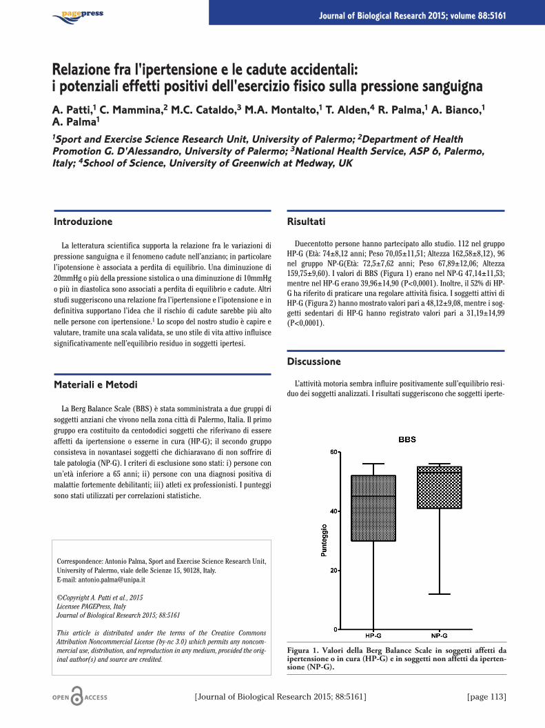
[Journal of Biological Research 2015; 88:5161] [page 113]
Journal of Biological Research 2015; volume 88:5161
Introduzione
La letteratura scientifica supporta la relazione fra le variazioni dipressione sanguigna e il fenomeno cadute nell’anziano; in particolarel’ipotensione è associata a perdita di equilibrio. Una diminuzione di20mmHg o più della pressione sistolica o una diminuzione di 10mmHgo più in diastolica sono associati a perdita di equilibrio e cadute. Altristudi suggeriscono una relazione fra l’ipertensione e l’ipotensione e indefinitiva supportano l’idea che il rischio di cadute sarebbe più altonelle persone con ipertensione.1 Lo scopo del nostro studio è capire evalutare, tramite una scala validata, se uno stile di vita attivo influiscesignificativamente nell’equilibrio residuo in soggetti ipertesi.
Materiali e Metodi
La Berg Balance Scale (BBS) è stata somministrata a due gruppi disoggetti anziani che vivono nella zona città di Palermo, Italia. Il primogruppo era costituito da centododici soggetti che riferivano di essereaffetti da ipertensione o esserne in cura (HP-G); il secondo gruppoconsisteva in novantasei soggetti che dichiaravano di non soffrire ditale patologia (NP-G). I criteri di esclusione sono stati: i) persone conun’età inferiore a 65 anni; ii) persone con una diagnosi positiva dimalattie fortemente debilitanti; iii) atleti ex professionisti. I punteggisono stati utilizzati per correlazioni statistiche.
Risultati
Duecentotto persone hanno partecipato allo studio. 112 nel gruppoHP-G (Età: 74±8,12 anni; Peso 70,05±11,51; Altezza 162,58±8,12), 96nel gruppo NP-G(Età: 72,5±7,62 anni; Peso 67,89±12,06; Altezza159,75±9,60). I valori di BBS (Figura 1) erano nel NP-G 47,14±11,53;mentre nel HP-G erano 39,96±14,90 (P<0,0001). Inoltre, il 52% di HP-G ha riferito di praticare una regolare attività fisica. I soggetti attivi diHP-G (Figura 2) hanno mostrato valori pari a 48,12±9,08, mentre i sog-getti sedentari di HP-G hanno registrato valori pari a 31,19±14,99(P<0,0001).
Discussione
L’attività motoria sembra influire positivamente sull’equilibrio resi-duo dei soggetti analizzati. I risultati suggeriscono che soggetti iperte-
Correspondence: Antonio Palma, Sport and Exercise Science Research Unit,University of Palermo, viale delle Scienze 15, 90128, Italy.E-mail: [email protected]
©Copyright A. Patti et al., 2015Licensee PAGEPress, ItalyJournal of Biological Research 2015; 88:5161
This article is distributed under the terms of the Creative CommonsAttribution Noncommercial License (by-nc 3.0) which permits any noncom-mercial use, distribution, and reproduction in any medium, provided the orig-inal author(s) and source are credited.
Relazione fra l'ipertensione e le cadute accidentali:i potenziali effetti positivi dell'esercizio fisico sulla pressione sanguignaA. Patti,1 C. Mammina,2 M.C. Cataldo,3 M.A. Montalto,1 T. Alden,4 R. Palma,1 A. Bianco,1A. Palma11Sport and Exercise Science Research Unit, University of Palermo; 2Department of HealthPromotion G. D'Alessandro, University of Palermo; 3National Health Service, ASP 6, Palermo,Italy; 4School of Science, University of Greenwich at Medway, UK
Figura 1. Valori della Berg Balance Scale in soggetti affetti daipertensione o in cura (HP-G) e in soggetti non affetti da iperten-sione (NP-G).
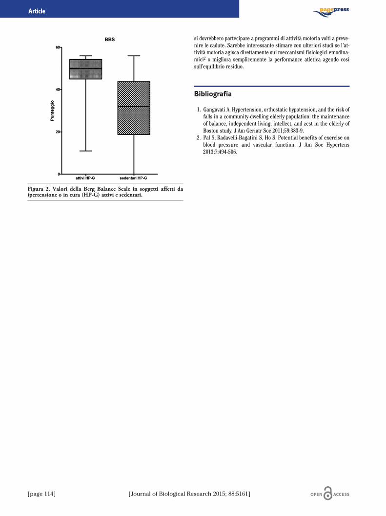
[page 114] [Journal of Biological Research 2015; 88:5161]
Article
si dovrebbero partecipare a programmi di attività motoria volti a preve-nire le cadute. Sarebbe interessante stimare con ulteriori studi se l’at-tività motoria agisca direttamente sui meccanismi fisiologici emodina-mici2 o migliora semplicemente la performance atletica agendo cosìsull’equilibrio residuo.
Bibliografia
1. Gangavati A. Hypertension, orthostatic hypotension, and the risk offalls in a community-dwelling elderly population: the maintenanceof balance, independent living, intellect, and zest in the elderly ofBoston study. J Am Geriatr Soc 2011;59:383-9.
2. Pal S, Radavelli-Bagatini S, Ho S. Potential benefits of exercise onblood pressure and vascular function. J Am Soc Hypertens2013;7:494-506.
Figura 2. Valori della Berg Balance Scale in soggetti affetti daipertensione o in cura (HP-G) attivi e sedentari.
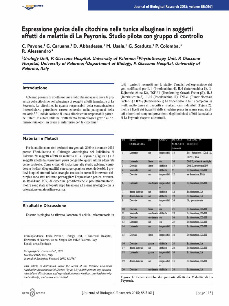
[Journal of Biological Research 2015; 88:5161] [page 115]
Journal of Biological Research 2015; volume 88:5161
Introduzione
Abbiamo pensato di effettuare uno studio che indagasse circa la pre-senza delle citochine nell’albuginea di soggetti affetti da malattia di LaPeyronie. Le citochine, in quanto responsabili della comunicazioneintercellulare, potrebbero essere coinvolte nella patogenesi dellamalattia.1,2 L’individuazione di una o più citochine responsabili potreb-be, infatti, risultare utile nel trattamento farmacologico grazie ai c.d.farmaci biologici, in grado di interferire con le citochine.3
Materiali e Metodi
Per lo studio sono stati reclutati tra gennaio 2009 e dicembre 2010presso l’Ambulatorio di Chirurgia Andrologica del Policlinico diPalermo 20 soggetti affetti da malattia di La Peyronie (Figura 1) e 8soggetti affetti da recurvatum penis congenito, questi ultimi adoperaticome controllo. Come criteri di inclusione allo studio abbiamo consi-derato i criteri di operabilità con corporoplastica secondo Nesbit. I pre-lievi bioptici ottenuti dalle losanghe escisse in corso di intervento chi-rurgico sono stati utilizzati per saggiare l’espressione genica, attraver-so Real-Time PCR, di citochine pro-fibrotiche e pro-infiammatorie.Inoltre sono stati sottoposti dopo fissazione ad esame istologico con lacolorazione ematossilina-eosina.
Risultati e Discussione
L’esame istologico ha rilevato l’assenza di cellule infiammatorie in
tutti i pazienti recensiti per lo studio. L’analisi dell’espressione deigeni codificanti per IL-4 (Interleuchina-4), IL-6 (Interleuchina-6), IL-13(Interleuchina-13), TGF-β1 (Trasforming Growth Factor-β1), IL-2(Interleuchina-2), IL-10 (Interleuchina-10), TNF-α (Tumor NecrosisFactor-α) e IFN-g (Interferone- g) ha evidenziato in tutti i campioni unlivello molto basso di trascritti e in alcuni casi indosabili (Figura 2).Inoltre i livelli dei trascritti delle citochine prese in esame sono risul-tati minori nei campioni provenienti dagli individui affetti da malaltiadi La Peyronie rispetto ai controlli.
Correspondence: Carlo Pavone, Urology Unit, P. Giaccone Hospital,University of Palermo, via del Vespro 129, 90127 Palermo, Italy.E-mail: [email protected]
©Copyright C. Pavone et al., 2015Licensee PAGEPress, ItalyJournal of Biological Research 2015; 88:5161
This article is distributed under the terms of the Creative CommonsAttribution Noncommercial License (by-nc 3.0) which permits any noncom-mercial use, distribution, and reproduction in any medium, provided the orig-inal author(s) and source are credited.
Espressione genica delle citochine nella tunica albuginea in soggettiaffetti da malattia di La Peyronie. Studio pilota con gruppo di controlloC. Pavone,1 G. Caruana,1 D. Abbadessa,1 M. Usala,2 G. Scaduto,1 P. Colomba,3R. Alessandro31Urology Unit, P. Giaccone Hospital, University of Palermo; 2Physiotherapy Unit, P. GiacconeHospital, University of Palermo; 3Department of Biology, P. Giaccone Hospital, University ofPalermo, Italy
Figura 1. Caratteristiche dei pazienti affetti da Malattia di LaPeyronie.
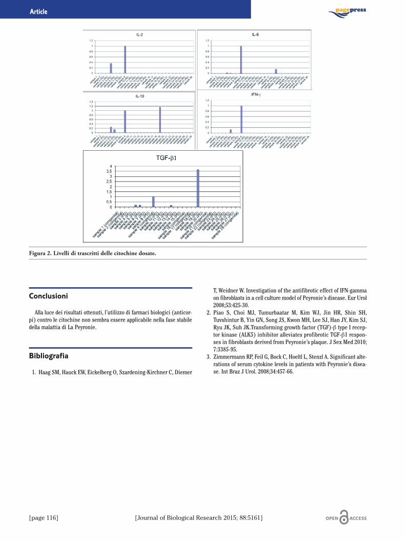
[page 116] [Journal of Biological Research 2015; 88:5161]
Article
Conclusioni
Alla luce dei risultati ottenuti, l’utilizzo di farmaci biologici (anticor-pi) contro le citochine non sembra essere applicabile nella fase stabiledella malattia di La Peyronie.
Bibliografia
1. Haag SM, Hauck EW, Eickelberg O, Szardening-Kirchner C, Diemer
T, Weidner W. Investigation of the antifibrotic effect of IFN-gammaon fibroblasts in a cell culture model of Peyronie’s disease. Eur Urol2008;53:425-30.
2. Piao S, Choi MJ, Tumurbaatar M, Kim WJ, Jin HR, Shin SH,Tuvshintur B, Yin GN, Song JS, Kwon MH, Lee SJ, Han JY, Kim SJ,Ryu JK, Suh JK.Transforming growth factor (TGF)-β type I recep-tor kinase (ALK5) inhibitor alleviates profibrotic TGF-β1 respon-ses in fibroblasts derived from Peyronie’s plaque. J Sex Med 2010;7:3385-95.
3. Zimmermann RP, Feil G, Bock C, Hoeltl L, Stenzl A. Significant alte-rations of serum cytokine levels in patients with Peyronie’s disea-se. Int Braz J Urol. 2008;34:457-66.
Figura 2. Livelli di trascritti delle citochine dosate.

[Journal of Biological Research 2015; 88:5161] [page 117]
Journal of Biological Research 2015; volume 88:5161
Introduzione
Lo stress psicologico è da un lato una condizione che potenzia le capa-cità reattive dell’individuo di fronte a condizioni di allarme e dall’altroun fattore di rischio per lo sviluppo e la progressione di numerose malat-tie.1 È opinione condivisa dalla comunità scientifica che l’attività fisicasia in grado di indurre cambiamenti fisiologici a largo spettro, influendopositivamente anche suli livelli di stress.2 Scopo del presente lavoro èstato quello di valutare se ed in che modo una particolare tecnica dirilassamento, nota come tecnica di respirazione profonda (deep breath-ing), fosse in grado di produrre miglioramenti fisiologici sui valori dicortisolo, su un campione di soggetti volontari.
Metodi
Hanno preso parte allo studio 38 soggetti volontari sani; 19 hannocostituito il gruppo di soggetti sui quali studiare gli effetti della respi-razione profonda (deep breathing) e 19 il gruppo di controllo. Tutti isoggetti hanno firmato il consenso informato preparato sulla basedelle indicazioni del Comitato Etico della nostra Università. I livelli distress psicologico sono stati misurati sulla base dei livelli di cortisolosalivare.3-5 Il protocollo consisteva in 10 sedute di deep breating6 delladurata di un’ora e trenta minuti. Sono state eseguite le misurazionidei livelli di cortisolo in tre tempi: la prima è stata all’inizio durante laprima sessione (I), la seconda durante la quinta sessione (II), la terzadurante l’ultima sessione (III).
Risultati
I valori di cortisolo ottenuti durante la I sessione mostrano valori
simili fra il gruppo di controllo ed il deep breathing group. La Figura 1mostra a sinistra i valori di cortisolo misurati nei soggetti facenti partedel deep breathing group; è possibile osservare un miglioramento sta-tisticamente significativo fra la I e la III sessione. La stessa figuramostra a destra i valori di cortisolo del gruppo di controllo che nonmostrano modificazioni significative nelle diverse sedute.
Discussione e Conclusioni
I presenti risultati confermano quelli di Martarelli e collaboratori6
che hanno osservato come il rilassamento indotto da respirazione dia-frammatica aumenti lo stato di difesa antiossidante negli atleti dopoesercizio esaustivo, e che questi effetti si correlano con la concomitan-te diminuzione del cortisolo. I risultati ottenuti dal presente lavoromostrano un miglioramento significativo dei valori di cortisolo nelgruppo che ha praticato deep breathing. È possibile ipotizzare che pra-ticare con regolarità e costanza la tecnica di respirazione profondapossa essere un facile e utile strumento per una buona qualità dellavita, agendo indirettamente sulla gestione delle tensioni- stress che lavita di tutti i giorni costringe a seguire.
Bibliografia
1. Muscatell KA, Eisenberger NI. a social neuroscience perspective onstress and health. Soc Personal Psychol Compass 2012;6:890-904.
2. Best John R. Effects of physical activity on children’s executivefunction: Contributions of experimental research on aerobic exer-cise. Devl Rev 2010;30:331-51.
Correspondence: Valentina Perciavalle, Department of Formative Process,University of Catania, via Ofelia, 95124 Catania, Italy.E-mail: [email protected]
©Copyright M. Coco et al., 2015Licensee PAGEPress, ItalyJournal of Biological Research 2015; 88:5161
This article is distributed under the terms of the Creative CommonsAttribution Noncommercial License (by-nc 3.0) which permits any noncom-mercial use, distribution, and reproduction in any medium, provided the orig-inal author(s) and source are credited.
Qualità della vita e livelli di cortisoloM. Coco,1 M. Blandini,1 T. Maci,2 M.C. Petralia,1 S. Massimino,1 V. Perciavalle,1 V. Perciavalle31Physiology Unit, Department of Bio-Medical Sciences, University of Catania; 2Department G.F.Ingrassia, University of Catania; 3Department of Formative Process, University of Catania, Italy
Figura 1. Livelli di cortisolo salivare misurato nelle diverse ses-sioni (I, II, e III) nei soggetti che praticavano deep breathing (adestra) e nei controlli.

[page 118] [Journal of Biological Research 2015; 88:5161]
Article
3. Hellhammer DH, Wust S, Kudielka BM. Salivary cortisol as a bio-marker in stress research. Psychoneuroendocrinology 2009;34:163-71.
4. Cacioppo JT. Somatic responses to psychological stress: the reactivi-ty hypothesis. In: Adair JG, Belanger D, Dion KL, eds. Advances inpsychological science. Hove, UK: Psychology Press; 1998. pp 87-109.
5. Kirschbaum C, Hellhammer DH. Salivary cortisol in psycho-biological research: an overview. Neuropsychobiology 1989;22:150-69.
6. Martarelli D, Cocchioni M, Scuri S, Pompei P. Diaphragmaticbreathing reduces exercise-induced oxidative stress. Evid BasedComplement Altern Med 2011;2011:932430.
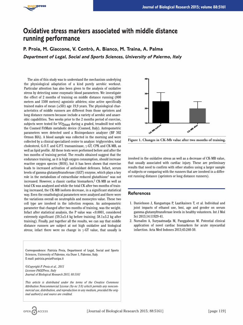
[Journal of Biological Research 2015; 88:5161] [page 119]
Journal of Biological Research 2015; volume 88:5161
The aim of this study was to understand the mechanism underlyingthe physiological adaptation of a kind purely aerobic workout.Particular attention has also been given to the analysis of oxidativestress by detecting some enzymatic blood parameters. We investigatethe effect of 2 months of training on middle distance running (800meters and 1500 meters) agonistic athletes; nine active specificallytrained males of mean (±SD) age 19,9 years. The physiological char-acteristics of middle runners are different from those sprinters andlong distance runners because include a variety of aerobic and anaer-obic capabilities. Two weeks prior to the 2 months period of exercise,subjects were tested for VO2max during a graded, treadmill test withthe Cosmed FitMate metabolic device (Cosmed, Italy). Antropometricparameters were detected used a Bioimpedance analyzer (BF 302Ormon BIA). A blood sample was collected in the morning and werecollected by a clinical specialized center to analyze: triglycerides, totalcholesterol, G.O.T. and G.P.T. transaminase, g-GT, CPK and CK-MB, aswell as lipid profile. All these tests were performed before and after thetwo months of training period. The results obtained suggest that theendurance training, as it is high oxygen consumption, should increasereactive oxygen species (ROS), but it has been shown that exerciseleads to increased activation of antioxidant defenses. Infact, serumlevels of gamma-glutamyltransferase (GGT) enzyme, which plays a keyrole in the metabolism of extracellular reduced glutathione1 was notincreased. However, a classic cardiac biomarkers,2 CK-MB as well astotal CK was analyzed and while the total CK after two months of train-ing increased, the CK-MB isoform decrease, in a significant statisticalway. Even the emathological parameters were analyzed and there werethe variations overall on neutrophils and monocytes value. These twocell type are involved in the infection respons. An antropometricparameter that changed after two months of training, was the weight.Infact after statistical analysis, the P value was <0.0001, consideredextremely significant (59.3±5.4 kg before training; 58.1±5.2 kg aftertraining). Finally, put together all the results, we can say that middledistance runners are subject at not high oxidative and biologicalstress; infact there were no change in g-GT value, that usually is
involved in the oxidative stress as well as a decrease of CK-MB value,that usually associated with cardiac injury. These are preliminaryresults that need to confirm with other studies using a larger sampleof subjects or comparing with the runners that are involved in a differ-ent running distance (sprinters or long distance runners).
References
1. Danielsson J, Kangastupa P, Laatikainen T, et al. Individual andjoint impacts of ethanol use, bmi, age and gender on serumgamma-glutamyltransferase levels in healthy volunteers. Int J MolSci 2013;14:11929-41.
2. Nursalim A, Suryaatmadja M, Panggabean M. Potential clinicalapplication of novel cardiac biomarkers for acute myocardialinfarction. Acta Med Indones 2013;45:240-50.
Correspondence: Patrizia Proia, Department of Legal, Social and SportsSciences, University of Palermo, via Duse 1, Palermo, Italy.E-mail: [email protected]
©Copyright P. Proia et al., 2015Licensee PAGEPress, ItalyJournal of Biological Research 2015; 88:5161
This article is distributed under the terms of the Creative CommonsAttribution Noncommercial License (by-nc 3.0) which permits any noncom-mercial use, distribution, and reproduction in any medium, provided the orig-inal author(s) and source are credited.
Oxidative stress markers associated with middle distancerunning performanceP. Proia, M. Giaccone, V. Contrò, A. Bianco, M. Traina, A. PalmaDepartment of Legal, Social and Sports Sciences, University of Palermo, Italy
Figure 1. Changes in CK-Mb value after two months of training.
[page 120] [Journal of Biological Research 2015; 88:5161]
Journal of Biological Research 2015; volume 88:5161
Negli anni 1971-1972 nell’ambito dell’International BiologicalProgramme: Human Adaptability Project, l’UNESCO promosse un pro-getto di ricerca sull’insediamento esquimese di Igloolik (Canada). Ilprogramma di ricerca, coordinato dal Prof. Brunetto Chiarelli, già ordi-nario di Antropologia presso l’Università degli Studi di Firenze, avevacome obiettivo lo studio delle caratteristiche genetiche della popolazio-ne eschimese a confronto con le caratteristiche delle popolazioni urba-nizzate. Lo spunto per questo studio nasceva dall’ipotesi che le popola-zioni isolate, non esposte ai vari agenti mutageni del mondo industria-lizzato, avrebbero dovuto presentare una percentuale molto bassa diaberrazioni cromosomiche rispetto alle altre popolazioni. Gli Eschimesio meglio gli Inuit, per le loro caratteristiche di popolazione adattata adun ambiente estremo e isolata biologicamente, si rivelarono un cam-pione di studio adatto per sperimentare le tecniche della moderna cito-
genetica. Agli inizi degli anni Settanta la disciplina aveva già conosciu-to la grande rivoluzione metodologica rappresentata dalla tecnica delbandeggio che consente di produrre bande orizzontali di differenteintensità di colorazione su tutti i cromosomi del corredo rendendo pos-sibile così una facile identificazione e un preciso accoppiamento. Lostudio condotto negli anni Settanta sugli abitanti di Igloolik rappresen-tò la prima applicazione delle tecniche di bandeggiamento dei cromo-somi su popolazioni artiche. Il nostro contributo ha lo scopo di ripercor-rere la storia di questo studio pionieristico e del contesto storico-geo-grafico che lo ispirò alla luce anche delle nuove metodologie di indagi-ne, tuttora in continuo perfezionamento. Vogliamo, inoltre, sottolineareche gli strumenti e le tecniche del passato possono rappresentare unpatrimonio di conoscenze con valenze museali quando sono associatialle testimonianze materiali del popolo studiato.
Correspondence: Donatella Minaldi, Department of Life Sciences andBiology of Systems, University of Torino, via Accademia Albertina 13, 10123Torino, Italy.E-mail: [email protected]
©Copyright D. Minaldi et al., 2015Licensee PAGEPress, ItalyJournal of Biological Research 2015; 88:5161
This article is distributed under the terms of the Creative CommonsAttribution Noncommercial License (by-nc 3.0) which permits any noncom-mercial use, distribution, and reproduction in any medium, provided the orig-inal author(s) and source are credited.
La comunità Inuit di Igloolik (Canada) nella seconda metà del Novecento:una ricerca pionieristica di citogenetica per raccontare la storia di un popoloD. Minaldi,1 B. Chiarelli,2 R. Boano,1 E. Rabino Massa11Department of Life Sciences and Biology of Systems, University of Torino ; 2Department ofEvolutionary Biology, University of Florence, Italy
[Journal of Biological Research 2015; 88:5161] [page 121]
Journal of Biological Research 2015; volume 88:5161
I polifenoli estratti da diversi frutti, come mele, pompelmi, arance,limoni o melograni, sono noti per le loro azioni benefiche sulla salutedell’uomo, per la loro attività antiossidante,1 antitumorale2 e anti-infiammatoria.3 È stato, inoltre, riscontrato un loro effetto neuropro-tettivo e anti-amiloidogenico in vitro.4,5 Studi recenti hanno infattidimostrato che alcuni polifenoli, come l’acido gallico, l’epicatechinagallate e l’epigallocatechina gallate esercitano un’azione neuroprotet-tiva nei confronti della tossicità indotta dalla proteina β- amiloide(peptide Aβ), coinvolta nella patologia dell’Alzheimer.4 In particolare èstato dimostrato come polifenoli estratti dalle mele, con alto contenutodi procianidine, siano in grado di inibire in vitro l’aggregazione delpeptide Aβ.6 La malattia di Alzheimer (AD) è la forma più comune didemenza neurodegenerativa invalidante ad esordio prevalentementesenile. Colpisce le più alte funzioni cerebrali come la memoria e lefunzioni cognitive e ciò si ripercuote sulle capacità intersociali deimalati di tale patologia causando stati confusionali, cambiamenti diumore e disorientamento spazio-temporale. Osservazioni eseguite tra-mite impiego di tecniche di immagine, indicano che i pazienti affettida AD mostrano, a livello neurologico macroscopico, una diminuzionedel peso e del volume cerebrale per atrofia corticale, con allargamentodei solchi e corrispondente appiattimento delle circonvoluzioni. Neipazienti con AD si osservano deposizioni a livello dell’ippocampo edella corteccia cerebrale chiamate placche senili, costituite da aggre-gati proteici di un peptide di 40-42 amminoacidi, il peptide β-amiloide.Il peptide Aβ deriva da uno specifico pathway proteolitico di una glico-proteina di transmembrana ubiquitariamente espressa, nota comeproteina precursore dell’amiloide le cui funzioni non sono ancoraconosciute, anche se recenti studi suggeriscono una sua implicazionenella formazione delle sinapsi, nella plasticità neuronale e nell’esportodi ferro. L’accumulo del peptide Aβ è ipotizzato come inizio di unacascata patogenetica che eventualmente porta alla malattia.7 In condi-zioni fisiologiche il rapporto tra Aβ-42 e Aβ-40 è circa 1:10. In lettera-
tura, l’aggregazione del peptide amiloide è descritta secondo una tipi-ca cinetica di nucleazione-polimerizzazione, in cui ognuna delle fasi ècaratterizzata da specifici intermedi strutturali che presentano diversedimensioni, morfologie e potenziale citotossico.8
Il presente lavoro è finalizzato allo studio dell’effetto di alcuni com-posti naturali, quali le procianidine, sull’inibizione del processo di for-mazione di fibre amiloidi in vitro.Mediante cromatografia liquida (HPLC-UV/Vis) si è provveduto ini-
zialmente all’identificazione ed alla quantificazione di polifenoli in meledella cultivar Gala, coltivate in Sicilia in regime di agricoltura biologica.Studi bibliografici individuano cinque principali gruppi polifenolici indiverse varietà di mele: acidi idrossicinnamici, procianidine, antociani-ne, flavonoli e diidrocalconi. Le procianidine sono i maggiori componen-ti del profilo polifenolico, presenti per il 59,7% nella buccia e per il 55,7%nella polpa.9 Dopo aver verificato la presenza di Procianidina B2 e delmonomero (–)-Epicatechina tra le procianidine presenti in queste mele,è stato valutato mediante spettroscopia di fluorescenza (Test dellaTioflavina T) il loro effetto sulle proprietà di aggregazione di una protei-na modello, la k-caseina, che, analogamente al peptide Aβ, forma fibreamiloidi a partire da un monomero intrinsecamente disordinato checontribuisce al core fibrillare con un double strand β. Le misure di fluo-rescenza mostrano che sia il dimero che il monomero, se pur in misuradiversa, inibiscono in maniera dose-dipendente l’aggregazione della k-caseina. Misure di dicroismo circolare (CD) hanno anche permesso divalutare la variazione della struttura secondaria di aggregati formati inassenza ed in presenza di Procianidina B2 ed (–)-Epicatechina. Daglispettri CD risulta che l’(–)-Epicatechina (65 µg/mL) non influenza laconversione strutturale della proteina a foglietti β ordinati, caratteristicidelle specie amiloidi. Infatti dopo incubazione a 37°C per 44 ore si assi-ste allo shift del minimo CD da 205nm (valore di pre-incubazione) versolunghezze d’onda maggiori, analogamente a quanto riscontrato nel cam-pione controllo (k-caseina nativa). I risultati ottenuti costituiscono labase metodologica-sperimentale per la successiva analisi dell’effetto ditali polifenoli sull’aggregazione e tossicità del peptide Aβ coinvolto nelmorbo di Alzheimer e per l’indagine sul loro meccanismo di azione aifini di un potenziale impiego terapeutico.
Bibliografia
1. Leontowicz H., Gorinstein S., Lojek A., Leontowicz M., Ciz M., Soliva-Fortuny R.C., Park Y-S., Jung S-T., Trakhtenberg S. and Martin-Belloso O. Comparative content of some bioactive compounds inapples, peaches and pears and their influence on lipids and antioxi-dant capacity in rats. Journal of Nutritional Biochemistry, 2002, vol.13, pp. 603-10.
Correspondence: Valeria Guarrasi, Institute of Biophysics, NationalResearch Council, via Ugo La Malfa 153, Palermo, Italy.E-mail: [email protected]
©Copyright G.C. Rappa et al., 2015Licensee PAGEPress, ItalyJournal of Biological Research 2015; 88:5161
This article is distributed under the terms of the Creative CommonsAttribution Noncommercial License (by-nc 3.0) which permits any noncom-mercial use, distribution, and reproduction in any medium, provided the orig-inal author(s) and source are credited.
Procianidina b2 ed (–)-Epicatechina presenti nelle mele:studio preliminare sul loro effetto sui meccanismi di aggregazionee di inibizione di proteine amiloidiG.C. Rappa,1,2 V. Guarrasi,1 S. Vilasi,1 M.A. Germanà,2 P.L. San Biagio11Institute of Biophysics, National Research Council, Palermo; 2Department of Agricultural andForestry Sciences, University of Palermo, Italy

[page 122] [Journal of Biological Research 2015; 88:5161]
Article
2. Femia A.P., Caderni G., Vignali F., Salvadori M., Giannini A., BiggeriA., Gee J., Przybylska K., Cheynier V., Dolara P. Effect of polypheno-lic extracts from red wine and 4-OH- coumaric acid on 1,2-dime-thylhydrazine-induced colon carcinogenesis in rats. EuropeanJournal of Nutrition, 2005, vol. 44, pp. 79-84.
3. Terra X., Palozza P., Fernandez-Larrea J., Ardevol A., Blade C.,Pujadas G., Salvado J., Arola L. and Blay M.T. Procyanidin dimer B1and trimer C1 impair inflammatory response signalling in humanmonocytes. Free Radical Research, 2011, vol. 45, pp. 611-9.
4. Bastianetto S., Yao Z.X., Papadopoulos V. and Quirion R.Neuroprotective effects of green and black teas and their catechingallate esters against β-amyloid-induced toxicity. EuropeanJournal of Nutrition, 2006, vol. 23, pp 55-64.
5. Ono K., Yoshiike Y., Takashima A., Hasegawa K., Naiki H. andYamada M. Potent anti- amyloidogenic and fibril-destabilizing
effects of polyphenols in vitro: implications for the prevention andtherapeutics of Alzheimer’s disease. Journal of Neurochemistry,2003, vol. 87, pp 172-8.
6. Toda T., Sunagawa T., Kanda T., Tagashira M., Shirasawa T. andShimizu T.. Apple Procyanidins Suppress Amyloid β-proteinAggregation. Biochemistry Research International, 2011, pp 1-8.
7. Hardy J.A., Higgins G.A. Alzheimer’s disease: the amyloid cascadehypothesys. Science, 1992, vol. 256, pp 184-5.
8. Carrotta R., Canale C., Diaspro A., Trapani A., San Biagio P.L.,Bulone D. Inhibiting effect of �s1-casein on Aβ1-40 fibrillogenesis.Biochimica et Biophysica Acta, 2012, vol. 1820, pp 124-32.
9. Tsao R., Yang R., Yang J.C. and Zhu H. Polyphenolic profiles ineight apple cultivars using high-performance liquid chromatogra-phy (HPLC). Journal of Agriculture Food Chemistry, 2003, vol. 51,pp 6347-53.

[Journal of Biological Research 2015; 88:5161] [page 123]
Journal of Biological Research 2015; volume 88:5161
Introduction
Heat shock proteins (Hsps) are an important class of molecules withvarious functions. Their classic role is to assist other proteins in fold-ing and re-folding and, when proteins are defective or irreversibly mis-folded, to drive their degradation. For this reason, some Hsps are alsonamed molecular chaperones. During evolution, this class of proteinshas also acquired extrachaperoning roles such as participation inimmune system regulation, cell differentiation, programmed cell deathand carcinogenesis. Hsp10 is a partner of Hsp60 in the Hsp60/10 fold-ing machine, but numerous scientific studies have shown that Hsp10may also play other roles. In fact, Hsp10 seems to have animmunomodulatory activity and a role in tumor progression. Hsp90regulates late-stage maturation, activation and stability of a range ofclient proteins, such as HER2, EGFR and BRAF, some of which areinvolved in signal transduction and other key pathways important formalignancy in several cancers, including large bowel carcinomas. Theaim of the present study was to evaluate levels and expression ofHsp10 and Hsp90 in a series of samples of large bowel mucosaobtained from healthy controls and patients with adenocarcinomas.
Materials an Methods
Twenty samples of large bowel human mucosa from healthy controlsubjects and twenty samples of large bowel adenocarcinomas withmoderate grade of differentiation, were obtained from the DICHIRONSDepartment of the University of Palermo, Italy. RT-PCR and WesternBlotting analyses were performed on these samples in order to studygene and protein expression of Hsp10 and Hsp90 (both Hsp90� andHsp90� isoforms). Moreover, an immunohistochemical study for Hsp10and Hsp90 was performed to evaluate the localization of these proteinsin both the epithelium and the lamina propria.
Results
RT-PCR analysis showed a higher gene expression of Hsp10 andHsp90 in adenocarcinoma samples compared to healthy mucosa. TheWestern Blotting analysis confirmed a greater amount of Hsp10 andHsp90 proteins in the samples of adenocarcinoma of large bowel com-pared to healthy mucosa. Finally, levels of Hsp10 were higher in ade-nocarcinoma compared to normal mucosa in both the epithelium andin the lamina propria, as revealed by immunohistochemistry. By con-trast, Hsp90 levels were not significantly different in the epithelium,while they were higher in the lamina propria of adenocarcinoma sam-ples compared to normal mucosa.
Conclusions
These data suggest that Hsp10 and Hsp90 may be involved in thecarcinogenesis of the large bowel by different molecular mechanisms.
Correspondence: Francesca Rappa, Department of ExperimentalBiomedicine and Clinical Neuroscience, Section of Human Anatomy,University of Palermo, via del Vespro 129, 90127 Palermo, Italy.E-mail: [email protected]
©Copyright F. Rappa et al., 2015Licensee PAGEPress, ItalyJournal of Biological Research 2015; 88:5161
This article is distributed under the terms of the Creative CommonsAttribution Noncommercial License (by-nc 3.0) which permits any noncom-mercial use, distribution, and reproduction in any medium, provided the orig-inal author(s) and source are credited.
Comparative analysis of Hsp10 and Hsp90 in large bowel healthy mucosaand adenocarcinomasF. Rappa,1,2 M. Lo Bello,1 C. Caruso Bavisotto,1 A. Marino Gammazza,1,2 C. Campanella,1,2S. David,1 C. Sciumè,3 F. Zarcone,3 G. Geraci,3 G. Modica,3 F. Carini,1 G. Tomasello,2,3M. Bellavia,2 M.L. Uzzo,4 G. Spatola,4 G. Bonaventura,4 A. Leone,4 P. Damiani,5F. Buccheri,1,2 A. Gerbino,4 F. Farina,1 F. Cappello1,21Department of Experimental Biomedicine and Clinical Neuroscience, Section of Human Anatomy,University of Palermo; 2Euro-Mediterranean Institute of Science and Technology, Palermo;3Department of Surgical, Oncological and Stomatological Sciences, University of Palermo;4Department of Experimental Biomedicine and Clinical Neuroscience, Section of Histology andEmbryology, University of Palermo; 5Department of Internal and Specialty Medicine, University ofPalermo, Italy

[page 124] [Journal of Biological Research 2015; 88:5161]
Journal of Biological Research 2015; volume 88:5161
In un campione di 189 persone con più di 60 anni appartenenti allecomunità della Val Cenischia (Piemonte) e originari della valle daalmeno tre generazioni è stata valutata la prevalenza di sindromemetabolica secondo i criteri del NCEP ATPIII.1 A questo fine ad ognipartecipante è stato effettuato un prelievo venoso a digiuno per ladeterminazione della glicemia, della trigliceridemia e della colestero-lemia totale e HDL, è stata misurata la pressione arteriosa, sono staterilevate alcune misure antropometriche (peso, altezza e circonferenzaaddominale) ed è stata somministrata un’intervista standardizzatavolta ad indagare le abitudini alimentari, il livello di attività fisica edaltri aspetti dello stile di vita.Nel campione, composto da 104 donne (età media: 73,6±8,4 anni) e
85 uomini (età media: 73,0±7,4 anni) la sindrome metabolica ha unaprevalenza del 42,1% (F=43,2%; M=40,8%). Lo stato di sovrappeso(definito da un BMI compreso tra 24,9 e 29,9 kg/mq) ha una prevalenzadel 39,7% (F=40,2%; M=39,1%), mentre l’obesità (definita da unBMI≥30,0 kg/mq) del 23,8% (F=22,5%; M=25,3%).Dall’analisi dei dati clinici e dei dati nutrizionali è emersa una asso-
ciazione significativa tra l’apporto nutrizionale della prima colazione euno stato di obesità/sovrappeso. In particolare è stata riscontrata lapresenza di una correlazione negativa tra il valore energetico dellaprima colazione e il BMI (R=-0,227, P=0,002). Le donne che consuma-no una colazione non adeguata (cioè inferiore al 15,0% dell’apportoenergetico quotidiano) hanno un BMI medio significativamente supe-riore rispetto a coloro che consumano una colazione adeguata (27,6 vs
25,6; T=2,669, DF=102, P=0,009) e lo stesso avviene per gli uomini(31,0 vs 26,7; T=3,298, DF=83, P=0,001).Inoltre, è emersa un’associazione significativa anche tra assunzione
di quantità eccessive di alcool (>20 g/dì per le donne e >40 g/dì per gliuomini) e obesità addominale (definita da una circonferenza addomina-le >88 cm per le donne e >102 cm per gli uomini) in entrambi i sessi(donne: χ2=4,989; DF=1; p=0,025; O.R.=2,706 [IC95%: 1,115 - 6,569];uomini: χ2=7,167; DF=1; P=0,007; O.R.=3,525 [IC95%: 1,363-9,119]).Questo lavoro fa parte di un progetto di ricerca più ampio svolto in
collaborazione con il dipartimento di Neuroscienze dell’Università diTorino e con il Dipartimento di Scienze Biologiche, Geologiche eAmbientali dell’Università di Bologna volto a studiare il decadimentocognitivo legato all’invecchiamento in relazione a fattori di rischio car-diovascolari di tipo clinico, genetico e comportamentale e ha ricevutol’approvazione del Comitato di Bioetica dell’Università di Torino.
Bibliografia
1. Expert Panel on Detection, Evaluation, and Treatment of High BloodCholesterol in Adults. Executive summary of the third report of thenational cholesterol education program (NCEP) expert panel ondetection, evaluation, and treatment of high blood cholesterol inadults (adult treatment panel III). JAMA 2001;285:2486-97.
Correspondence: Marianna Rinaldi, Laboratory of Anthropology, Departmentof Life Sciences and Biology of Systems, University of Turin, via AccademiaAlbertina 13, 10123 Torino. E-mail: [email protected]
©Copyright M. Rinaldi et al., 2015Licensee PAGEPress, ItalyJournal of Biological Research 2015; 88:5161
This article is distributed under the terms of the Creative CommonsAttribution Noncommercial License (by-nc 3.0) which permits any noncom-mercial use, distribution, and reproduction in any medium, provided the orig-inal author(s) and source are credited.
Nutrizione, obesità e sindrome metabolica in un campione di anzianidella Val CenischiaM. Rinaldi,1 G. Graffi,2 E. Rabino Massa11Laboratory of Anthropology, Department of Life Sciences and Biology of Systems, University ofTurin; 2Local Health Service ASL TO3, Susa (TO)
[Journal of Biological Research 2015; 88:5161] [page 125]
Journal of Biological Research 2015; volume 88:5161
Recent reports of new and important roles for serotonin in theperiphery have served to increase interest in circulating serotonin (5-HT). The much smaller pool of free (extraplatelet) plasma 5- HT isaccessible to sites of action and receptors, and may be important inmany processes. Assessing this extraplatelet plasma pool could be verydifficult also because many factors could influence 5-HT levels.1
Horses kept in stalls are deprived of opportunities for social interac-tions and the performance of natural behaviors is limited. Thehypothesis of this study was that stalling horses results in a neg-ative effect on their welfare. As marker of poor welfare we evalu-ate plasma 5-HT and its precursor tryptophan (TRP) in 14 adult horsesheterogeneous for sex, breed and age (13±7 years). In previous stud-ies lower levels of plasma 5-HT were found in horses with cribbingbehaviour2 and in subjects feed with high levels in concentrates.3
Horses of this study were divided in two groups: Stall (S) Group andPasture (P) Group. Group S (n.6 horses) was maintained in individualbox under a natural photoperiod (sunrise at 06:06, sunset at 18:49)and natural indoor temperature (19-21°C) from the day before theexperiment to the afternoon of follow day. Horses were fastedovernights (12-14 hours) and then feed with hay that was provided at08:30 and 12:30, water was available ad libitum. Group P (n.8horses) was maintained at the same condition of Group S until 8:30then it was transferred from box stalls to pasture. Blood samples wereobtained from the jugular vein at 08:00, 12:00 and 16:00 and collectedinto EDTA-containing tubes. Within 30 minutes from the venipunc-ture, sample tubes were centrifuged at 1350 x g for 10 minutes toobtain the fraction defined as platelet poor plasma. One hundred µLof plasma were then supplemented with an equal volume of an inter-nal standard represented by N-methylserotonin and treated with 100µL of a precipitating reagent to ensure protein removal. Samples werethe vortex-mixed for 30 seconds, allow to stand for 10 minutes at 4°Cand centrifuged in a top-bench centrifuge at the maximal speed.The resulting clear supernatants were stored at -20°C and analysedwithin one week for the HPLC quantification of 5-HT and TRP accord-
ing to protocols earlier described.4 All the results obtained wereexpressed as mean values±standard deviation (SD). One-way repeat-ed measure analysis of variance (ANOVA) was performed to deter-mine the statistically significance and Bonferroni’s test was applied aspost hoc comparison test. Mann Whitney test was used to compare dif-ferences between groups. The data were analysed using the softwareSTATISTICA 8 (Stat Soft Inc.). Results for 5-HT and TRP are shown onTable 1 and Figure 1 respectively. The influence of time evaluate byANOVA was significant in both Groups (P<0.001) with levels signifi-cantly higher at 12:00 and at 16:00 compared to levels at 08:00. 5-HTlevels were significantly higher in Group P compared to Group S at12:00 (P<0.01) and 16:00 (P<0.001). Also TRP levels were signifi-cantly influenced by time in both Groups (P<0.001) with higher levelsat 12:00 and at 16:00 compared to levels at 08:00. No differencebetween Groups were found for TRP concentrations. The pattern of 5-HT and TRP levels confirmed previous results on equine dailyrhythms for these parameters.5 The lower levels of 5-HT measured at12:00 and at 16:00 in Group S could indicate that factors asabsence of exercise and isolation could influence 5-HT levels.Regardless of this, we recognize that, in addition to differences inexercise and social interaction there are confounding factors betweentreatment groups including nutrition rate and exposure to sunlight.However, these same confounding factors would be present in anyoperation where a decision has to be made as to whether to stall hors-es or provide access to pasture. In conclusion obtained results in thepresent study showed the modulation of plasma 5-HT by two differentmanagement conditions. Our suggestion is to improve the knowledgeabout factors that can increase plasma equine 5-HT levels in order toguarantee the animal welfare.
Correspondence: Giuseppe Piccione, Department of Veterinary Sciences,University of Messina, Polo Universitario dell’Annunziata, 98168 Messina,Italy. E-mail [email protected]
©Copyright D. Alberghina et al., 2015Licensee PAGEPress, ItalyJournal of Biological Research 2015; 88:5161
This article is distributed under the terms of the Creative CommonsAttribution Noncommercial License (by-nc 3.0) which permits any noncom-mercial use, distribution, and reproduction in any medium, provided the orig-inal author(s) and source are credited.
Plasma serotonin in horses: comparison betweentwo different management conditionsD. Alberghina, C. Giannetto, M. Rizzo, M. Panzera, G. PiccioneDepartment of Veterinary Sciences, University of Messina, Italy
Figure 1. Patterns of mean values (±standard deviation) of plasmatryptophan in Group S (grey bar) and in Group P (dark bar) from08:00 to 16:00.
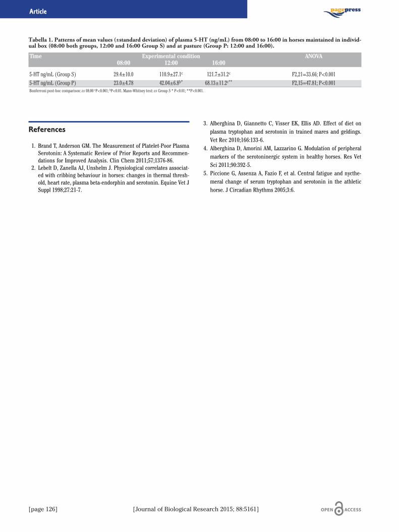
[page 126] [Journal of Biological Research 2015; 88:5161]
Article
References
1. Brand T, Anderson GM. The Measurement of Platelet-Poor PlasmaSerotonin: A Systematic Review of Prior Reports and Recommen-dations for Improved Analysis. Clin Chem 2011;57;1376-86.
2. Lebelt D, Zanella AJ, Unshelm J. Physiological correlates associat-ed with cribbing behaviour in horses: changes in thermal thresh-old, heart rate, plasma beta-endorphin and serotonin. Equine Vet JSuppl 1998;27:21-7.
3. Alberghina D, Giannetto C, Visser EK, Ellis AD. Effect of diet onplasma tryptophan and serotonin in trained mares and geldings.Vet Rec 2010;166:133-6.
4. Alberghina D, Amorini AM, Lazzarino G. Modulation of peripheralmarkers of the serotoninergic system in healthy horses. Res VetSci 2011;90:392-5.
5. Piccione G, Assenza A, Fazio F, et al. Central fatigue and nycthe-meral change of serum tryptophan and serotonin in the athletichorse. J Circadian Rhythms 2005;3:6.
Tabella 1. Patterns of mean values (±standard deviation) of plasma 5-HT (ng/mL) from 08:00 to 16:00 in horses maintained in individ-ual box (08:00 both groups, 12:00 and 16:00 Group S) and at pasture (Group P: 12:00 and 16:00).
Time Experimental condition ANOVA08:00 12:00 16:00
5-HT ng/mL (Group S) 29.4±10.0 110.9±27.1c 121.7±31.2c F2,21=33.66; P<0.0015-HT ng/mL (Group P) 23.0±4.78 42.04±6.8b* 68.13±11.2c** F2,15=47.81; P<0.001Bonferroni post-hoc comparison: vs 08:00 cP<0.001; bP<0.01. Mann-Whitney test: vs Group S * P<0.01; **P<0.001.

[Journal of Biological Research 2015; 88:5161] [page 127]
Journal of Biological Research 2015; volume 88:5161
Oxidative stress can be regarded as an imbalance between prooxi-dant/free radical production and opposing antioxidant defenses. Thereis growing evidence that oxidative stress (OS) significantly impairsorganic function and plays a major role in the aetiology and pathogen-esis of several metabolic diseases in veterinary medicine.1 In view ofthis, we evaluate some oxidative stress markers in ten Comisana ewes(3 years old, clinically healthy) at different experimental conditions:shearing, road transportation, different reproductive status. Moreoverwe evaluate daily rhythms of oxidative stress markers. On each sub-ject, blood samples were collected by jugular venipuncture to assessoxidative stress markers during: shearing (before, after shearing and8h, 1d, 2d, 3d, 4d, 5d post- shearing); road transportation for 6 h overa distance about 490 km with an average speed of 80 km/h (before,after transportation and 8, 24, 48 h post-transport); different reproduc-tive status (days 1, 40 and 200 of lactation and during dry period); andcircadian rhythm under natural photoperiod -sunrise 5:10, sunset20:45- (every 3 h over a 24 h period, starting at 8:00 on day 1 and fin-ishing at 8:00 on day 2). All blood samples were collected into vacutain-er tubes with no additive and were centrifuged at 3000×g for 20 min.The sera obtained were immediately analyzed by means of a UV spec-trophotometer (Slim SEAC, Firenze, Italy) for the assessment ofdROMs, Oxy-adsorbent and SHp. These techniques are based on thespin traps system, molecules which react with free radicals, creatingcomplexes revealed by spectrophotometry. During milking and dryperiod was also evaluate daily rhythm of oxidative stress markers(mean level, amplitude, acrophase and robustness) applying a trigono-metric statistical model on mean values. All the results obtained wereexpressed as mean values±standard deviation (SD). Data were nor-mally distributed (P<0.05, Kolmogorov-Smirnov test). One-way repeat-ed measure analysis of variance (ANOVA) was performed to determinethe statistically significance and Bonferroni’s test was applied as posthoc comparison test. The data were analyzed using the software STA-TISTICA 8 (Stat Soft Inc.).
The lowest values of dROMs observed before shearing and a theirsubsequent increase, could be due to the energy deficiency thatoccurs in ewes after shearing. Ambient temperature, relative humid-ity and shearing can influence thermoregulatory mechanisms and theproductive performance and welfare of ewes. The high values of oxy-adsorbent and SHp values further demonstrate the compensatoryresponse of the organism to the increase of free radicals (dROMs)inducing oxidative stress after shearing (Table 1). Our results indi-cate that shearing cause s a change in the ewes homeostatic balancethat leads to oxidative stress. The road transportation in ewes appearsto have an influence on the increase of catabolic reactions that leadsto the onset of oxidative stress. The results of our study (Table 1)showed an increase of dROMs, Oxy-adsorbent and SHp values in ewesafter the road transportation in comparison with basal level (P<0.05).The pattern of dROMs, characterized by a significant increase duringpost transport respect to, is in agreement with other researchers.2
The increase of dROMs after 48 h rest time corresponds to approxi-mately 95.85% and represents the energetic deficiency that occurs inewes during transport period. The high values of Oxy-adsorbent andSHp during rest time further demonstrated the compensatoryresponse of the organism to the increase of dROMs inducing oxida-tive stress successively to road transportation. During the differentreproductive periods, lowest values of dROMs at the start of experi-mental period could be due to the energetic deficiency which occursin ewes during the last period of pregnancy. The significant increaseof dROMs on days 40 and 200 of lactation and during dry period com-pared to values obtained on day 1 of lactation shows high oxidativeprocesses which occur during lactation in ewes. The pattern ofdROMs characterized by low values at the beginning of lactation andby a significant increase at the mid-point of lactation was previouslyobserved in lactating ewes. 3,4 The high values of Oxy-adsorbent andSHp at the end of lactation document the compensative response ofthe organism to oxidative stress. Oxidative processes increased(P<0.05) at the end of milking period (Figure 1), together with a com-pensative response of the organism to this stress and suggest theimportant role of oxidative status in dairy ewes. Our result showedthat the high energetic requirements of milking period in ewes aredirectly proportional to free radicals formation and oxidative stress.The trigonometric statistical model of the single cosinor procedureindicated the existence of daily rhythm of dROMs, Oxy-ads and SHp inewes with a nocturnal acrophase (dROMs=21:00; Oxy-ads.=21:30;SHp=21:15). All three markers showed a rhythm robustness abovesignificance line (dROMs=81.5%; Oxy-ads.=79.1%; SHp=81.70%). Inconclusion we can claim that there is a synergism between oxidativestress markers and the circad ian rhythm of anti-oxidant power inewes. The right zootechnics management and the formulation ofration that totally satisfy the requirements of the lactating subjects,seem o be essential in order to guarantee the productive status andthe animal welfare.
Correspondence: Francesco Fazio, Department of Veterinary Sciences,University of Messina, Polo Universitario dell'Annunziata, 98168 Messina,Italy. E-mail [email protected]
©Copyright V. Monteverde et al., 2015Licensee PAGEPress, ItalyJournal of Biological Research 2015; 88:5161
This article is distributed under the terms of the Creative CommonsAttribution Noncommercial License (by-nc 3.0) which permits any noncom-mercial use, distribution, and reproduction in any medium, provided the orig-inal author(s) and source are credited.
Evaluation of some oxidative stress markers in ovis ariesduring different experimental conditionsV. Monteverde,1 M. Rizzo,2 D. Alberghina,2 F. Fazio,2 E. Giudice,2 G. Piccione21Institute for Experimental Veterinary Medicine of Sicily, Palermo; 2Department of VeterinarySciences, University of Messina, Italy
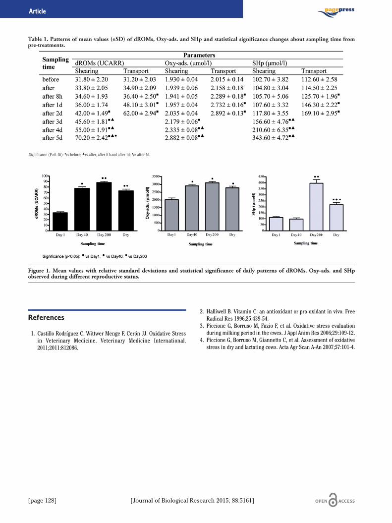
[page 128] [Journal of Biological Research 2015; 88:5161]
Article
References
1. Castillo Rodríguez C, Wittwer Menge F, Cerón JJ. Oxidative Stressin Veterinary Medicine. Veterinary Medicine International.2011;2011:812086.
2. Halliwell B. Vitamin C: an antioxidant or pro-oxidant in vivo. FreeRadical Res 1996;25:439-54.
3. Piccione G, Borruso M, Fazio F, et al. Oxidative stress evaluationduring milking period in the ewes. J Appl Anim Res 2006;29:109-12.
4. Piccione G, Borruso M, Giannetto C, et al. Assessment of oxidativestress in dry and lactating cows. Acta Agr Scan A-An 2007;57:101-4.
Table 1. Patterns of mean values (±SD) of dROMs, Oxy-ads. and SHp and statistical significance changes about sampling time frompre-treatments.
Significance (P<0. 05): ■vs before; ▲vs after, after 8 h and after 1d; ●vs after 4d.
Figure 1. Mean values with relative standard deviations and statistical significance of daily patterns of dROMs, Oxy-ads. and SHpobserved during different reproductive status.

[Journal of Biological Research 2015; 88:5161] [page 129]
Journal of Biological Research 2015; volume 88:5161
The sensory system of rodent vibrissae has been extensively analysedto understand how these animals may successfully explore the nearbyenvironment, detect objects and orient their behaviour using themacrovibrissae. Recent data1 showed that besides the Gasser’s ganglianeurons also the trigeminal mesencephalic nucleus (Me5) appears sig-nificantly involved in the sensory perception of the whisker pad struc-tures. In fact: i) Tracer injection into mystacial pad of the rats signifi-cantly labelled the Me5 neurons and ii) electrophysiological experi-ments showed that Me5 neurons are responsive to spontaneous move-ment of the macrovibrissae. It has been proposed that Me5 neurons maybe involved in relaying kinetic information to the CNS.The present study was performed to better clarify the functional role
of the Me5 sensory innervation of the whisker pad structures byanalysing the electrophysiological responses of Me5 neurons tomechanical deflection of the macrovibrissae.The spontaneous electrical activity of the Me5 neurons, identified
by their responses to the masseter muscle stretch, was extracellularlyrecorded using tungsten-in-glass microelectrodes (impedance 700-900KΩ) carefully advanced into the Me5 by an electronic microdrive(David Kopf). The electrical signals were relayed to conventional pre-amplifiers connected to oscilloscopes and then to computers for thespecific analysis (Tecfen computerscope analysis ISC-16 software, andPowerLab 4/30 Chart 5, V 5.4.2 software). The spontaneous electricalactivity of the Me5 neurons multiunit activity (MUA) was continuouslymonitored and recorded under resting conditions (i.e. vibrissaemotionless), during the masseter muscle stretch, and during/after themechanical deflection of the vibrissae bundle, which was performed infour principal directions (forward/backward, backward/forward,up/down and down/up) using a delicate glass rod connected to a craft-made electronic drive.The electrophysiological results demonstrated that mechanical
deflection of the macrovibrissae induced a significant increase in the
spontaneous electrical activity of the Me5 neurons as well as signifi-cant changes in the spontaneous firing pattern. It appeared that Me5neurons could be activated in response to macrovibrissae deflection inspecific directions.The results of the present and previous experiments, allow us to
conclude that in addition to the neurons connected to the masticatorymuscles2 and those connected to the periodontal ligament,3 the Me5enclose primary neurons specifically dedicated to encoding kineticinformation related to vibrissae movements.It is known that the central terminals of Me5 neurons join the brain
Correspondence: Antonella Russo, Physiology Section, Department ofBiomedical Sciences, University of Catania, viale A. Doria 6, 95125 Catania,Italy. E-mail: [email protected]
©Copyright A. Russo et al., 2015Licensee PAGEPress, ItalyJournal of Biological Research 2015; 88:5161
This article is distributed under the terms of the Creative CommonsAttribution Noncommercial License (by-nc 3.0) which permits any noncom-mercial use, distribution, and reproduction in any medium, provided the orig-inal author(s) and source are credited.
Mechanical deflection of macrovibrissae induces in rats activationof trigeminal mesencephalic nucleus neuronsA. Russo,1 S. Stanzani,1 R. Pellitteri,2 M.A. Caria,3 O. Mameli31Physiology Section, Department of Biomedical Sciences, Catania University; 2Institute ofNeurological Sciences-National Research Council, Catania; 3Human Physiology Division,Department of Clinical and Experimental Medicine, University of Sassari, Italy
Figure 1. Multiunit electrical activity (MUA) of trigeminal mes-encephalic neurons (Me5) recorded in the medial-caudal part ofthe Me5 during mechanical deflection of the macrovibrissae. A)schematic drawings of the experimental set up; B) specimensshowing the Me5 neurons response to mechanical stretch of themasseter muscle to functionally identify the Me5; C) diagramsresuming the four principal directions used during mechanicaldeflection of the vibrissae bundle together with the schematic pat-tern of the correspondent Me5 MUA response; D) tonic increaseof Me5 MUA, although with adaptation, to vibrissae deflection inback/forward direction; E) tonic increase of the same Me5 MUAto vibrissae deflection in down/up direction, the increased activi-ty lasted throughout the stimulus application as well as severalseconds after its end. ON and OFF indicate the beginning and theend of vibrissae displacement. Horizontal calibration: 1 sec; ver-tical calibration: 1 mV.

[page 130] [Journal of Biological Research 2015; 88:5161]
Article
stem trigeminal sensory nuclei, which in turn receive, by the primaryGasser’s ganglia neurons, tactile information from macrovibrissaereceptors. If so, it is possible to hypothesize that the brain stem trigem-inal nuclei may deduce higher-order information by combining touchinformation from macrovibrissae receptors, and carried out by affer-ents from the Gasser’s neurons, with kinetic information arising fromthe Me5 neurons. The integrated information can be then relayed tomore rostral SNC structures to allow a central reconstruction and rep-resentation of the spatial displacement of the macrovibrissae.
References
1 Mameli O, Stanzani S, Mulliri G, et al. Role of the trigeminal mes-encephalic nucleus in rat whisker pad proprioception. Behav BrainFunctions 2010;6:69-79.
2. Darian-Smith I. The trigeminal system. In: Iggo A. (ed.), Handbookof sensory physiology (vol. II). Somatosensory system. Berlin:Springer Verlag; 1973. pp. 271-314.
3. Linden RWA, and Scott BJJ. Distribution of mesencephalic nucleusand trigeminal ganglion mechanoreceptors in the periodontal liga-ment of the cat. J Physiol 1989;410:35-44.
Figure 2. Multiunit electrical activity (MUA) of trigeminal mes-encephalic neurons (Me5) recorded in the medial-caudal part ofthe Me5 during mechanical deflection of the macrovibrissae. A)schematic drawings of the experimental set up; B) specimensshowing the Me5 neurons response to mechanical stretch of themasseter muscle to functionally identify the Me5; C) diagramsresuming the four principal directions used during mechanicaldeflection of the vibrissae bundle together with the schematic pat-tern of the correspondent Me5 MUA response; D) bursts of Me5MUA, in response to vibrissae deflection in forward/backwarddirection; E) the same Me5 MUA responded with bursts, but atlonger latency and rapid adaptation, to vibrissae deflection indown/up direction. ON and OFF indicate the beginning and theend of vibrissae displacement. Horizontal calibration: 1 sec; ver-tical calibration: 1 mV.
[Journal of Biological Research 2015; 88:5161] [page 131]
Journal of Biological Research 2015; volume 88:5161
Cereals are an important source of carbohydrates in the human con-tributing to a certain extent also to the need of protein. They constitutea rich source of both nonstarch polysaccharides (dietary fiber) andstarch, which together comprise 70-77% of kernel.The Guidelines for a Healthy Diet in Italy1 define the opportunity to
take, on a daily basis, at least 60% of energy from carbohydrates, andparticularly 45% from complex carbohydrates. In Italy, especially inSicily, these nutrients are contained in traditional food as pasta andbread made starting from durum wheat as grow material. Recently,especially in the bigger towns, there is also widespread consumptionof bread made with soft wheat.2,3
In the last decade, consumer behavior is changing in Italy4 and it isclear a trend of decline in consumption of pasta and bread, insteadthere is an opposite trend for the consumption of products consideredas a substitute for bread (brioches, crackers, biscuits, and other bakedgoods). It is evident how the nutritional profile of the latter productsdeviates from the bread due to the fat content, that ranges from 5 to20%. It is also known as the derivatives of durum wheat, preferablywhole grain products, are also characterized by a lower glycemic indexthan products of soft wheat.So cereals are a key component to set a proper diet. Some studies
also report that influence of aregular and correct consumption of cereals may contribute to reach
and to maintain normal weight.5
In 2011 was carried out investigations with the aim to understandbetter the relationship and the presence of cereals in the diet of pre-teen. Moreover, it is known as in Sicily, as well as other areas of south-ern Europe, there is a serious problem of childhood obesity,6,7 mainlyrelated to lifestyles considered incorrect and a lower level of educationof the population than other European areas that do not registers thisphenomenon.The investigation involved a sample of 1335 subjects, aged between
10 and 13 years, identified through the involvement of 62 schools dis-tributed throughout the region. The sample involved in the investiga-tion was extrapolated according to a stratification that has taken intoaccount the size of the population under study, age ,sex, and distribu-
tion by province. For each individual was recorded during the first halfof December 2011, thanks to personnel appropriately trained to collectinformation in a standardized way, a 24-h recall questionnaire (notingthe foods eaten in the last 24 hours from the individual). In addition tothe parents were asked to complete a questionnaire on the frequencyto record information on lifestyle, frequency of intake of cereals. Foreach individual was recorded weight (kg), height (cm) and body massindex (BMI, Body Mass Index), according to the standards proposed bythe International Obesity Task Force.8 About frequency of the condi-tions of the weight of the individuals in the sample, the results haveconfirmed what was already mentioned by other authors for Sicily.6,7
Results of the study shows as the population sample, in the variousdaily meals, have not a correct relationship with cereals, considering themodels known for the Mediterranean population. In particular, it is clearthe high frequency of subjects who have not took any kind of cereals atbreakfast (n=699, 52%) or at morning-snack (n=396, 30%). Moreover17% of individuals (n=228) has neither taken any kind of breakfastcereal nor at morning-snack; these individuals have access to the pri-mary source of complex carbohydrates, needed daily, only with lunch.The individuals who regularly eat pasta are characterized by a lower
Body Mass Index than those who do not consume habitually; the sameis for those who regularly take breakfast, according as noted by otherauthors in specific studies conducted in the Mediterranean basin,9 andfor individuals who have eaten at least one type of cereal for breakfast.Regarding this survey it can be stated that further studies are need-
ed to define the extent to which an erroneous presence and distribu-tion in diets of cereals can affect overweight and obesity, particularlyamong younger age groups of the population. It is evident, however,that it is more necessary than ever to support an effective educationcampaign that fill the gaps on nutritional knowledge on cereals; con-tribute to implement pathways on enhancement of cereals mainly onnutritional point of view, with the aim to promoting correct lifestylesand diet patterns better oriented to limit the risks of occurrence in thepopulation of chronic degenerative diseases related to obesity.
References
1. INRAN. Linee guida per una sana alimentazione italiana. Rome:INRAN; 2003.
2. Russo G, Messina B. Indagini conoscitive per la valorizzazione deiprodotti da forno nel comprensorio del GAL Terre del Gattopardo(Sicilia). Atti dell’8° Convegno AISTEC-Aci Castello (CT). 2011.250-254 pp.
3. Costanzo E, Liberto M, Russo G. Schede dei pani siciliani. In:Consorzio Gian Pietro Ballatore per la ricerca su specifici settoridella filiera cerealicola, 2001. Atlante del Pane di Sicilia. Palermo:Grafiche Renna; 2001.
4. Casini L, Contini C, Marone E, Romano C, 2013. Food habits.Changes among young Italians in the last 10 years. Appetite2013;68:21-9.
Correspondence: Giuseppe Russo, Research Constortium Gian PietroBallatore, via dei Pini 21, 90040 Isola delle Femmine (PA), Italy. E-mail: [email protected]
©Copyright G. Russo et al., 2015Licensee PAGEPress, ItalyJournal of Biological Research 2015; 88:5161
This article is distributed under the terms of the Creative CommonsAttribution Noncommercial License (by-nc 3.0) which permits any noncom-mercial use, distribution, and reproduction in any medium, provided the orig-inal author(s) and source are credited.
Could a wrong consumption of cereals influence preteens obesity in Sicily?G. Russo,1 D. Di Majo,2 M. Giammanco21Research Constortium Gian Pietro Ballatore; 2Physiology and Pharmacology Unit, Department ofLegal, Social and Sports Sciences, University of Palermo

[page 132] [Journal of Biological Research 2015; 88:5161]
Article
5. Marquart L, Jacobs DR Jr, Slavin JL. Whole grains and health: anoverview. J Am Coll Nutr 2000;19:289-90.
6. Baratta R, Degano C, Leonardi D, et al. High prevalence of over-weight and obesity in 11e15-year-old children from Sicily. NutrMetab Cardio Dis 2006;16:249-55.
7. Spinelli A, Lamberti A, Baglio G, et al. OKkio alla SALUTE: sistema disorveglianza su alimentazione e attività fisica nei bambini della scuo-la primaria. Risultati 2008. Roma: Istituto Superiore di Sanità; 2009.
8. Cole TJ, Bellizzi MC, Flegal KM, Dietz WH. Establishing a standarddefinition for child overweight and obesity worldwide: internation-al survey. Brit Med J 2000;320:1-6.
9. Demosthenes BP, Antonogeorgos G, Papadimitriou A, et al.Breakfast cereal is associated with a lower prevalence of obesityamong 10-12 year-old children: the PANACEA study. Nutr MetabCardio Dis 2008;18:606-12.

[Journal of Biological Research 2015; 88:5161] [page 133]
Journal of Biological Research 2015; volume 88:5161
The major use of durum wheat is for pasta products, particularly inthe European and North American countries, whereas in other areas(Middle East and North Africa) also it is used as couscous and for vari-ous types of bread. The durum wheat is therefore a staple food in theMediterranean and is one of the commodities that characterize thedietary pattern of the Mediterranean diet. During the year 2012 weremade in Ciminna (Palermo) in Sicily, four fields of experimental culti-vation using four different varieties of durum wheat cultivated in Sicily:Simeto, Iride, Duilio and Saragolla. For each variety were set up two dif-ferent kinds of cultivation tecniques, in order to verify the influence ofthe environment on the final results. In particular, the technique Aemployed the same seeding density for the 4 varieties (2.4 quintals ofseed per hectare), using the pre-sowing and coverage fertilization withrespectively 30.6 and 47.25 units of nitrogen, distributed by the use ofdiammonium phosphate (18/46) in pre-sowing and ammonium nitrate(27%) in coverage; technique B employed a seeding density lower for 4varieties (variable between 1.8 and 2.2 quintals of seed per hectare);fertilizing in pre-sowing was carried out with sodium superphosphate(that means zero units of nitrogen) and in coverage with 101.79 unitsof total nitrogen, distributed through the use of ammonium nitrate(27%) used in two different moments after the crop raised. The resultsshow that the protein content of the different batches analyzed stoodbetween 12.9 and 16.1% D.M., higher than the average recorded for theSicilian crops.1 Investigations on the amino acid profile showed somedifferences between the different varieties and also between the differ-ent cultivation techniques (Table 1). The most represented amino acidwas glutamic acid which is also the one who showed greater variabilityamong the four varieties. Even proline showed a variability in the con-text of the four varieties evaluated. The other amino acids showedslight or no differences among the four varieties evaluated. The surveyresults are partially comparable with the data available in the literaturefor other varieties grown in Italy.2-4
Table 1. Average values and standard deviation in amino acidscontent in four varieties of durum wheat grown with two differ-ent cultivation techniques in Sicily.
Amino acid Agronomic Agronomictechnique A technique Bg/100 g g/100 g
of protein SD of protein SD
Alanine 3,4 0,06 3,3 0,07Arginine 4,5 0,14 4,4 0,11Aspartic acid 4,6 0,12 4,6 0,14Glutamic acid 27,1 0,75 27,4 0,81Glycine 3,6 0,04 3,5 0,04Histidine 2,3 0,02 2,3 0,03Isoleucine 3,4 0,05 3,3 0,04Leucine 6,6 0,08 6,6 0,16Lysine 2,7 0,08 2,6 0,09Phenylalanine 4,4 0,13 4,4 0,10Proline 9,4 0,30 9,5 0,28Serine 4,5 0,12 4,5 0,05Threonine 2,7 0,07 2,7 0,07Thyrosine 2,7 0,07 2,7 0,07Valine 4,3 0,12 4,2 0,11Thryptophan (basic hydrolysis) 1,3 0,04 1,2 0,07Cystine+cysteine 2,1 0,07 2,1 0,09Methionine 1,7 0,17 1,8 0,04For the determination of tryptophan was used the method EN ISO 13904:2005 (hydrolysis in a basicenvironment). For all other amino acids was used the method ISO 13903:2005. The values are reportedin g/100 g of protein.
References
1. Russo G, Messina B, Campisi D, et al. Monitoraggio della qualitàdel grano duro prodotto in Sicilia: risultati delle campagne di rac-colto 2007 e 2008. Osservatorio della Filiera Cerealicola Siciliana.2009. pp. 119-35.
2. Abdel-Hamid Youssef Abdel-Rahman. Amino acid composition oftwo Italian durum wheat varieties and their semolina. Food Chem1983;10:155-8.
3. Jiang XL, Tian JC, Hao Z, Zhang WD. Protein content and aminoacid composition in grains of wheat-related species. Agr Sci China2008;7:272-9.
4. Konzak CF. Genetic control of content, amino acid composition,and processing properties of proteins in wheat. Adv Genet1977;19:407-582.
Correspondence: Giuseppe Russo, Research Constortium Gian PietroBallatore, via dei Pini 21, 90040 Isola delle Femmine (PA), Italy. E-mail: [email protected]
©Copyright G. Russo et al., 2015Licensee PAGEPress, ItalyJournal of Biological Research 2015; 88:5161
This article is distributed under the terms of the Creative CommonsAttribution Noncommercial License (by-nc 3.0) which permits any noncom-mercial use, distribution, and reproduction in any medium, provided the orig-inal author(s) and source are credited.
Amino acid profile of four varieties of durum wheat grown in SicilyG. Russo,1 M. Giammanco,2 D. Di Majo,2 B. Messina11Research Constortium Gian Pietro Ballatore; 2Physiology and Pharmacology Unit, Department ofLegal, Social and Sports Sciences, University of Palermo
[page 134] [Journal of Biological Research 2015; 88:5161]
Journal of Biological Research 2015; volume 88:5161
Both tumor and normal brain cells release membrane vesicles(MVs) into the extracellular space.1-3 MVs shed by astrocytes and neu-rons contain FGF2 and VEGF, which could be responsible for inducingendothelial cells to form the blood-brain barrier.2,3 MVs shed fromG26/24 oligodendroglioma cells, when added to primary cultures of ratcortical neurons, inhibit neurite outgrowth, and induce apoptosis inabout 75% of the cells.1 The same amount of shed MVs induce apopto-sis in about 40% of cultured astrocytes.4 The analysis of G26/24 vesi-cles demonstrated the presence of Fas Ligand and TRAIL, which couldcooperate in inducing brain cell death.1,4 The horizontal transfer oflabeled proteins from oligodendroglioma cells to astrocytes in culturewas also demonstrated.4 Since MVs were shown to transfer RNA, and,on the other hand, some RBPs are involved in tumorigenesis, wesearched for RBPs in MVs. A preliminary analysis in A375 melanomacells revealed the presence of at least three RBPs, with apparent MWof about 64, 50 and 36 kDa, respectively. These proteins are able tobind H1° mRNA. In developing rat brain, the amount of histone H1°increases during neuronal differentiation, while the level of the corre-sponding mRNA decreases, suggesting that H1° gene expression ismainly regulated at the post-transcriptional level.5 Similar results wereobtained with cultured astrocytes, while G2624 maintain high levels ofboth H1° protein and mRNA. Moreover, oligodendroglioma cells, butnot astrocytes, release H1° protein into the culture medium by shed-ding MVs.6 These findings suggest that oligodendroglioma cells canescape antiproliferative cues by discharging into the extracellular
environment molecules expressed concomitant with differentiation,such as H1° histone.
References
1. D’Agostino S, Salamone M, Di Liegro I, Vittorelli ML. Membranevesicles shed by oligodendroglioma cells induce neuronal apopto-sis. Int J Oncol 2006;29:1075-85.
2. Schiera G, Proia P, Alberti C, et al. Neurons produce FGF2 andVEGF and secrete them at least in part by shedding extracellularvesicles. J Cell Mol Med 2007;11:1384-94.
3. Proia P, Schiera G, Mineo M, et al. Astrocytes shed extracellularvesicles that contain fibroblast growth factor-2 and vascularendothelial growth factor. Int J Mol Med 2008;21:63-7.
4. Lo Cicero A, Schiera G, Proia P, et al. Oligodendroglioma cells shedmicrovesicles which contain TRAIL as well as molecular chaperonesand induce cell death in astrocytes. Int J Oncol 2011;39:1353-7.
5. Castiglia D, Cestelli A, Scaturro M, et al. H1(0) and H3.3B mRNAlevels in developing rat brain. Neurochem Res 1994;19:1531-7.
6. Schiera G, Di Liegro CM, Saladino P, et al. Oligodendroglioma cellssynthesize the differentiation-specific linker histone H1° andrelease it into the extracellular environment through shed vesi-cles. Int J Onc 2013;43:1771-6.
Correspondence: Patrizia Saladino, Department of Biological, Chemical andPharmaceutical Sciences and Technologies, University of Palermo, vialedelle Scienze 16, 90128 Palermo, Italy.E-mail: [email protected]
©Copyright P. Saladino et al., 2015Licensee PAGEPress, ItalyJournal of Biological Research 2015; 88:5161
This article is distributed under the terms of the Creative CommonsAttribution Noncommercial License (by-nc 3.0) which permits any noncom-mercial use, distribution, and reproduction in any medium, provided the orig-inal author(s) and source are credited.
Extracellular vesicles can shuttle molecules among brain cellsP. Saladino,1 G. Schiera,1 C.M. Di Liegro,1 P. Proia,2 I. Di Liegro31Department of Biological, Chemical and Pharmaceutical Sciences and Technologies, University ofPalermo; 2Department of Law, Sports, Society and Sciences, University of Palermo; 3Departmentof Experimental Biomedicine and Clinical Neurosciences, University of Palermo, Italy

[Journal of Biological Research 2015; 88:5161] [page 135]
Journal of Biological Research 2015; volume 88:5161
Introduzione
La termoporazione dinamica irreversibile (DIT) consiste in unnuovo tipo di tecnologia di pastorizzazione ideato da Koulik e colleghi1
basato su un processo termico dinamico, caratterizzato da riscalda-menti molto rapidi (shock termici) a temperature finali non superioria 70°C. L’obiettivo del nostro studio è stato la valutazione dell’influen-za di parametri di processo quali la temperatura di partenza (T1) delcampione da trattare, la temperatura finale in corso di trattamento(T2), la differenza tra temperatura iniziale e temperatura finale (ΔT),la velocità di riscaldamento (θ) e la durata del trattamento sull’abbat-timento della carica batterica nella matrice alimentare.
Materiali e Metodi
I test sono stati effettuati su ceppi di riferimento di microrganismidi possibile interesse alimentare: Escherichia coli, Pseudomonas aeru-ginosa, Staphylococcus aureus, Enterococcus hirae, Listeria innocua. Lasospensione test è stata preparata poco prima dell’esecuzione dei testcon le seguenti composizioni: 10% sospensione microbica (108CFU/mL) in acqua peptonata, da una coltura fresca (18-24 ore); 10%sostanza interferente (albumina bovina 0,3 g/L o saccarosio 10 g/L);80% acqua dura (calcio e magnesio).2 I test DIT sono stati effettuatifissando per i parametri velocità di riscaldamento, temperatura inizia-le e temperatura finale i seguenti valori: DIT 1. θ 20°C/s ; T1 30°C; T260°C (ΔT 30°C); DIT 2. θ 20°C/s; T1 30°C; T2 65°C (ΔT 35°C); DIT 3. θ30°C/s; T1 30°C; T2 60°C (ΔT 30°C); DIT 4. θ 30°C/s; T1 30°C; T2 65°C(ΔT 35°C). È stato anche saggiato l’effetto di un prolungato manteni-mento della temperatura T2. A fine trattamento, aliquote di ciascun
campione sono state seminate in piastre di terreno solido. La lettura èstata effettuata dopo 24 e 48 ore di incubazione a 37°C. L’attività bat-tericida è stata definita come la dimostrazione di una riduzione dialmeno 5 log (logR>5) rispetto alla carica batterica iniziale.
Risultati
Le Tabelle 1 e 2 riportano l’efficacia dei trattamenti DIT su ciascunmicrorganismo. I trattamenti con T2=60°C e ΔT=30°C (DIT1 e DIT3)hanno dimostrato efficacia solo quando seguiti dal mantenimentodella temperatura T2. Questo risultato è stato osservato su P. aerugino-sa e meno costantemente su E. coli. I trattamenti con T2=65°C eΔT=35°C (DIT2 e DIT4) sono risultati efficaci su tutti i microrgani-smi. Su E. coli, L. innocua, P. aeruginosa e S. aureus logR>5 è stataottenuta anche senza mantenimento della temperatura T2, e indipen-dentemente dalla sostanza interferente (Tabella 1). Diversamente, perE. hirae logR>5 è stata ottenuta solamente sui campioni trattati conDIT2 o DIT4 seguiti da un prolungato mantenimento di T2 (Tabella 2).L’efficacia dei trattamenti è stata più marcata e costante per la sospen-sione contenente saccarosio.
Tabella 1. Trattamenti di termoporazione dinamica irreversibileefficaci su 4 ceppi batterici di interesse alimentare. Numero ditest efficaci/numero di test effettuati.
Microrganismo DIT1+ DIT3+ DIT2 DIT4testato mant. mant.
A S A S A S A S
E. coli 1/4 1/4 1/4 0/4 4/4 4/4 4/4 4/4
L. innocua - - - - 4/4 4/4 4/4 4/4
P. aeruginosa 2/4 4/4 4/4 3/3 4/4 4/4 3/4 4/4
S. aureus 0/4 0/4 0/4 0/4 5/5 5/5 5/5 5/5A, albumina; S, saccarosio.
Tabella 2. Trattamenti di termoporazione dinamica irreversibileefficaci su E. hirae. Numero di test efficaci/numero di test effet-tuati.
Microrganismo DIT2+mant. DIT+mant.testato A S A S
E. hirae 0/4 3/4 1/4 4/4A, albumina; S, saccarosio.
Correspondence: Laura Saporito, Department of Sciences for HealthPromotion, University of Palermo, via del Vespro 129, 90127 Palermo, Italy.E-mail: [email protected]
©Copyright L. Saporito et al., 2015Licensee PAGEPress, ItalyJournal of Biological Research 2015; 88:5161
This article is distributed under the terms of the Creative CommonsAttribution Noncommercial License (by-nc 3.0) which permits any noncom-mercial use, distribution, and reproduction in any medium, provided the orig-inal author(s) and source are credited.
Valutazione della termoporazione dinamica irreversibile come strumentoper l’abbattimento della carica batterica in matrici alimentariL. Saporito,1 F. Bonura,1 D. Geraci,1 T. Rubino,2 E. Pipitone,2 M. Cammalleri,2G.M. Giammanco,1 C. Mammina11Department of Sciences for Health Promotion, University of Palermo; 2Department of Chemical,Management, Informatic, Mechanical Engineering, University of Palermo, Italy

[page 136] [Journal of Biological Research 2015; 88:5161]
Article
Discussione
Il trattamento DIT è risultato efficace nel ridurre la carica battericainiziale di almeno 5 log su tutti i ceppi batterici esaminati con almenouno dei protocolli testati. Tra i parametri di processo valutati, la tempe-ratura T2 sembra avere influenzato notevolmente l’efficacia del tratta-mento, unitamente alla differenza tra temperatura iniziale e tempera-tura finale (ΔT) del processo DIT. Infatti, gli obiettivi DIT con T2=60°Ce ΔT=30°C (DIT1 e DIT3) non si sono dimostrati efficaci su nessunadelle specie batteriche testate; al contrario, gli obiettivi DIT conT2=65°C e ΔT=35°C (DIT2 e DIT4), hanno determinato un abbattimen-to della carica batterica > 5 log per tutte le specie, ad eccezione di E.hirae, la cui resistenza ai trattamenti termici è nota in letteratura.3,4 E.hirae è risultato sensibile ai trattamenti DIT 2 e DIT4 solo se seguiti dalmantenimento della temperatura T2 e in presenza di saccarosio. Perquesto microrganismo, in particolare, sarebbe opportuno ottimizzare iltrattamento DIT 2 e 4 (ΔT=35°C) cercando di ridurre al massimo iltempo di mantenimento della T2. La velocità di riscaldamento (θ) e ledue sostanze interferenti utilizzate nella preparazione della sospensio-ne test non hanno generalmente influenzato la suscettibilità delle spe-cie batteriche ai trattamenti DIT.
Conclusione
In conclusione, questi test preliminari suggeriscono che il processoDIT può essere efficace nell’ottenere l’abbattimento della carica batte-rica in un substrato liquido. I parametri che influenzano maggiormentel’efficacia del processo sembrano essere quelli puramente termici: ΔTe T2. Ulteriori sperimentazioni tenderanno ad adattare i parametri diprocesso alle esigenze della produzione a livello industriale.
Bibliografia
1. Sterilisation of liquids in hermetically closed vessels; PCT PatentNo. 2008/114136; China Patent No. 200880014495; United StatesPatent Application No. 20110123690
2. UNI EN 1276:2009 Quantitative suspension test for the evaluationof bactericidal activity of chimica disinfectants and antisepticsused in food, industrial, domestic and institutional areas.
3. Spinks AT, Dunstan RH, Harrison T, et al. Thermal inactivation ofwater-borne pathogenic and indicator bacteria at sub-boiling tem-peratures. Water Res 2006;40:1326-32.
4. Aguirre JS, Pin C, Rodríguez MR, García de Fernando GD. Analysisof the variability in the number of viable bacteria after mild heattreatment of food. Appl Environ Microbiol 2009;75:6992-7.
[Journal of Biological Research 2015; 88:5161] [page 137]
Journal of Biological Research 2015; volume 88:5161
Epilepsy, affecting at least 50 million persons worldwide, is one of themost common neurological disorders. Despite the significant advancesin understanding epileptogenic mechanisms and in counteracting theirpathological consequences, this clinical condition still has to be faced oftreating more effectively the symptoms (epileptic seizures) and of pre-venting their unfavourable evolution. So far, research has been unsuc-cessful involved in developing effective antiepileptic drugs (AEDs) capa-ble of preventing the development of the pathogenic process, set inmotion by different etiological factors, that leads ultimately to chronicepilepsies.1,2 So, a substantial need remains to develop new AEDs withbetter safety, less toxicity, and higher efficacy.3,4 Valproic acid, VPA, isone of the four most widely prescribed AEDs. Besides its wide use inboth generalized and partial epilepsies, VPA has also gained widespreaduse in recent years for the treatment of bipolar disorders, neuropathicpain and for prophylactic treatment of migraine.5,6 However the use ofVPA is limited by two rare but potentially life-threatening side effects,hepatotoxicity, induced from the formation of metabolite(s) with a ter-minal double bond, specifically 4-ene-VPA,7 and teratogenicity, associat-ed with the parent compound itself.8 In a previous work we reported thesynthesis of aminoacidic ester derivatives of VPA as resulted of chemicalconjugation of VPA with esters of essential neutral aminoacids, with theaim of modifying the physicochemical properties relevant to bioavail-ability, such as solubility or lipophilicity, improving the efficacy andreducing unwanted side or toxic effects of VPA.9 We had reported alsothe synthesis of N-valproyl-L-tryptophan, that has shown adequatephysicochemical characteristics to permeate biological membranes andantiepileptic activity at lower concentration than VPA.10,11 In this paper,we focused our research on synthesis and characterization of newaminoacidic compounds with potential antiepileptic activity: N-Valproyl-L-Leucine (ValLeu), N-Valproyl-L-methionine (ValMet) and N-Valproyl-L-Histidine (ValHist). The conjugation could consent to obtain VPAderivatives, lacking of structural characteristics usually implicated onVPA teratogenicity, and avoiding formation of possible hepatotoxic
metabolites. The aminoacidic derivatives of VPA was successfullyobtained covalent linking carboxyl group of drug with aminic group of L-aminoacids, by synthesis involving two main steps. The first step,described in our previous work9 was modified by adding DMAP as fur-ther coupling agent together with DCC. The structures of obtained com-pounds were assigned on the basis of respective analytical data-sets, FT-IR, MS and 1H and 13C-NMR spectral data. Since the drug lipophilicity isan important factor conditioning brain uptake, the apparent partitioncoefficient (Papp) could be used as simple descriptor of ability to cross theBBB: values of log Papp within -0.2 to 1.3 have been described as optimalfor cerebral transport; on the other hand higher values than these couldreduce the rate of transport inside the membrane.12,13 Apparent partitioncoefficient (Papp) of ValLeu, ValMet and ValHist were determined in n-octanol/phosphate buffer pH 7.4 solution and expressed as Log Papp.The determined Log Papp resulted -0.11,- 1,02 and -1,61 respectively. TheLog DpH7.4 values indicate that ValLeu, ValMet are adequate to cross bio-logical membranes and in particular BBB barrier while ValHist value istoo low, probably due to the fact that was obtained as hydrochloride.Compared to others drug administration routes, the oral one remains
the most preferred as it implies ease of administration as well as highpatient compliance. However, the transit through the gastrointestinaltract could constitute a limiting step to bioavailability as a consequenceof degradation correlated to the environmental pH. In view of a possibleadministration of ValLeu, ValMet and ValHist by oral route, studies ontheir chemical stability were performed in simulated gastro-intestinalbuffer (37°C, pH 1.2 to 8.0) and monitored by HPLC analysis. The exper-iments demonstrated that ValLeu, ValMet and ValHist remainedunchanged up to 24 h, and did not produce degradation products orpotential metabolites. This behaviour indicates high stability at pH con-ditions of gastro-intestinal tract.Since compounds containing amide functional group could be suscep-
tible of hydrolysis by plasma and/or cerebral enzymes, our experimentswere focused on the evaluation of stability of ValLeu, ValMet and ValHistin these biological environments. Otherwise, plasma stability of drugcandidates plays an important role in drug discovery and development; itis essential for maintaining acceptable drug concentration and half-lifein order to achieve desirable pharmacological effects.14
Experimental data highlighted that ValLeu, ValMet and ValHistremained unmodified up to 24 h in plasma environment. In rat brainhomogenate ValLeu, ValMet and ValHist did not undergo cleavage after24 h, indicating that synthetized compounds have also good stability tocerebral enzymes.
References
1. Arroyo S, Brodie MJ, Avanzini G, et al. Is refractory epilepsy pre-ventable? Epilepsia 2002;43:437-44.
Correspondence: Anna Lisa Scaturro, Department of Biological, Chemicaland Pharmaceutical Sciences and Technologies, University of Palermo, viaArchirafi 32, 90123, Palermo, Italy. E-mail: [email protected]
©Copyright V. De Caro et al., 2015Licensee PAGEPress, ItalyJournal of Biological Research 2015; 88:5161
This article is distributed under the terms of the Creative CommonsAttribution Noncommercial License (by-nc 3.0) which permits any noncom-mercial use, distribution, and reproduction in any medium, provided the orig-inal author(s) and source are credited.
N-Valproyl-aminoacids as new potential antiepileptic drugs:synthesis, characterization and in vitro studies on stabilityV. De Caro, A.L. Scaturro, F.M. Sutera, L.I. GiannolaDepartment of Biological, Chemical and Pharmaceutical Sciences and Technologies, University ofPalermo, Italy
[page 138] [Journal of Biological Research 2015; 88:5161]
Article
2. Walker MC, White HS, Sander JWAS. Disease modification in par-tial epilepsy. Brain 2002;125:1937-50.
3. Bialer M, Yagen B. Valproic acid: second generation.Neurotherapeutics 2007;4:130-7.
4. Shimshoni JA, Bialer M, Yagen B. Synthesis and anticonvulsantactivity of aromatictetramethylcyclopropanecarboxamide deriva-tives. Bioorg Med Chem 2008;16:6297-305.
5. Lennkh C, Simhandl C. Current aspects of valproate in bipolar dis-order. Int Clin Psychopharmacol 2000;15:1-11.
6. Guidotti A, Dong E, Kundakovic M, et al. Characterization of theaction of antipsychotic subtypes on valproate-induced chromatinremodelling. Trends Pharmacol Sci 2008;30:55-60.
7. Silva MFB, Aires CC, Luis PB, et al. Valproic acid metabolism andits effects on mitochondrial fatty acid oxidation: a review. J InheritMetab Dis 2008;31:205-16.
8. Bojic U, Ehlers K, Ellerbeck U, et al. Studies on the teratogen phar-macophore of Valproic acid analogues: Evidence of interactions ata hydrophobic centre. Eur J Pharmacol 1998;354:289-99.
9. Giannola LI, Lamartina L, De Caro V. Synthesis and characteriza-
tion of aminoacidic pro-drugs of valproic acid. Pharmazie 1998;53:829-34.
10. De Caro V, Giandalia G, Siragusa MG, et al. N-Valproyl-L-Tryptophan for CNS-targeting: synthesis, characterization and effi-cacy in vitro studies of a new potential antiepileptic drug. MedChem 2011;7:9-17.
11. Sardo P, Rizzo V, Friscia S, et al. Inhibitory effects of N-valproyl-L-tryptophan on high potassium, low calcium and low magnesium-induced CA1 hippocampal epileptiform bursting activity in ratbrain slices. J Neural Transm 2012;119:1249-59.
12. Trojnar MK, Weirzchowska-Cioch E, Krzyzanowski M, et al. Newgeneration of valproic acid. Pol J Pharmacol 2004;56:283-8.
13. Gimenez F, Fernandez C, Mabondzo A. Transport of HIV proteaseinhibitors through the bloodbrain barrier and interactions with theefflux proteins, P-glycoprotein and multidrug resistance proteins. JAcquir Immune Defic Syndr 2004;36:649-58.
14. Di L, Kerns EH, HongY, Chen H. Development and application ofhigh throughput plasma stability assay for drug discovery. Int JPharm 2005;297:110-9.
[Journal of Biological Research 2015; 88:5161] [page 139]
Journal of Biological Research 2015; volume 88:5161
Introduction
Recent molecular studies showed that the dimorphic fungusSporothrix schenckii is no longer the only species able to causesporotrichosis, a cutaneous lymphatic or systemic mycosis particular-ly frequent in certain geographical areas such as Mexico, Brazil, Peru,and India. 1 In fact, S. schenckii can now be recognized as a speciescomplex comprising at least six sibling species: Sporothrix brasilien-sis, Sporothrix globosa, Sporothrix luriei, Sporothrix mexicana,Sporothrix pallida (formerly Sporothrix albicans) and S. schenckiisensu stricto.2,3 Like S. schenckii, all these new species have beenreported to cause diseases in humans and in other animals1,4,5
although the extent of their impact on human infections is not yetcompletely known. However, infections due to S. schenckii have alsobeen reported from other parts of the world, including Europe, wheresporotrichosis is considered a rare disease.6 Nevertheless, in recentyears, several clinical autochthonous cases have been described inpatients and animals that live in European countries, showing thatthis pathogenic fungus is more widespread than is now believed.1,6 Atpresent, there are relatively few works that have evaluated the sus-ceptibility of S. schenckii sensu lato to antifungal agents and thedrugs tested so far have shown, in general, poor activity especiallyagainst S. pallida, S. globosa, and S. mexicana. Therefore, in this studywe decided to evaluate the activities of a panel of antifungal drugsagainst all members of the S. schenckii complex with particular refer-ence to Italian isolates. To our knowledge this is the first study thatevaluates in vitro activities of antifungal agents against a number ofSporothrix spp. isolates recovered from clinical and environmentalsamples in Italy.
Materials and Methods
Fourteen Sporothrix spp. were examined in this study (Table 1).Seven of them were environmental S. pallida isolates that have alreadybeen well characterized in our previous study.6 The identity of each iso-late was determined by partial amplification and sequencing of thecalmodulin-encoding gene according to recent studies.2,6 Antifungalactivity of seven drugs (Table 1) was evaluated by disk diffusion methodaccording to the procedures reported in the National Committee forClinical Laboratory Standards (NCCLS) document M44-A.In this study, a total of 14 clinical and environmental Sporothrix spp.
were examined to evaluate their susceptibility to a panel of antifungalagents. The resulting values of the in vitro susceptibility of S. schenckiisensu lato isolates are shown in Table 1.All fungal species were resistant to fluconazole, flucytosine and
metronidazole whereas were susceptible to nystatin. An excellentbroad-spectrum antifungal activity of miconazole was observed againstall examined strains. Regarding ketoconazole, different degree of sus-ceptibility were observed. In particular this drug was active against S.schenckii, S. brasiliensis and S. mexicana but for S. pallida, S. globosaand S. luriei was not possible to measure the diameter of the zone ofinhibition due to the presence of a high number of resistant colonies.
Discussion and Conclusions
The discovery of genetically different species within the S.schenckii population has generated considerable interest on differ-ent aspects of their biology including epidemiology, virulence andantifungal susceptibilities. Previous studies have clearly shown thatthe geographic distribution of members of the S. schenckii complexas well as their trends in antifungal susceptibilities are variable1,2,7
and therefore more attention should be paid in the diagnosis andtherapeutic treatment of infections caused by these species.Throughout this work, miconazole, and to a lesser extent nystatin,showed a good activity against all Sporothrix species tested while allisolates were resistant to fluconazole, flucytosine and metronidazolewhich is in agreement with other previous studies.7 The broad invitro resistance to fluconazole in clinical isolates of the S. schenckiicomplex suggests an intrinsic resistance to this drug. This is aninteresting topic for further study, because this drug is consideredthe second-line treatment for sporotrichosis. One important result ofthis study is the excellent broad-spectrum antifungal activity dis-played by miconazole, an synthetic imidazole antifungal agent whichhas never been tested against all members of the S. schenckii com-plex so far. Thus, based on our in vitro data, we believe that this drugmay represent a very promising antifungal agent in the treatment ofhuman and animal sporotrichosis.
Correspondence: Orazio Romeo, Department of Biological and EnvironmentalSciences, University of Messina, viale F. Stagno d’Alcontres 31, 98166Messina, Italy. E-mail: [email protected]
©Copyright F. Scordino et al., 2015Licensee PAGEPress, ItalyJournal of Biological Research 2015; 88:5161
This article is distributed under the terms of the Creative CommonsAttribution Noncommercial License (by-nc 3.0) which permits any noncom-mercial use, distribution, and reproduction in any medium, provided the orig-inal author(s) and source are credited.
Antifungal susceptibilities of species of the Sporothrix Schenckii complexisolated in ItalyF. Scordino, I. Pernice, C. Lo Passo, R. Galbo, M.A. Medici, G. Criseo, O. RomeoDepartment of Biological and Environmental Sciences, University of Messina, Italy
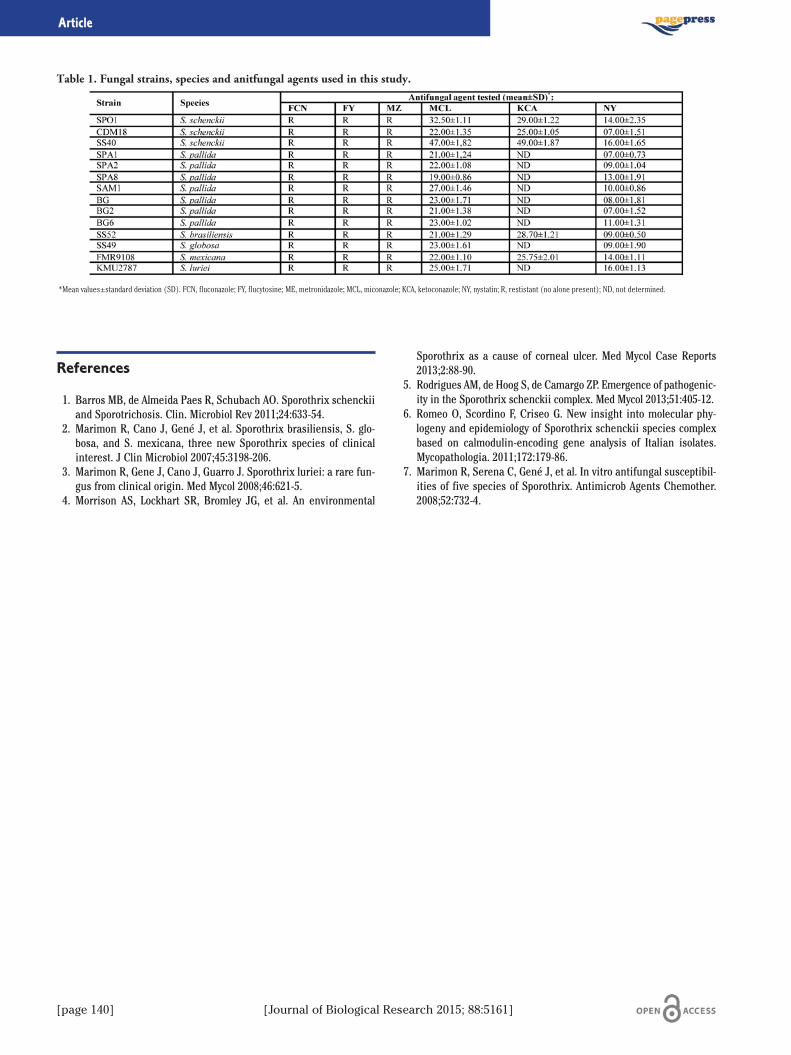
[page 140] [Journal of Biological Research 2015; 88:5161]
Article
References
1. Barros MB, de Almeida Paes R, Schubach AO. Sporothrix schenckiiand Sporotrichosis. Clin. Microbiol Rev 2011;24:633-54.
2. Marimon R, Cano J, Gené J, et al. Sporothrix brasiliensis, S. glo-bosa, and S. mexicana, three new Sporothrix species of clinicalinterest. J Clin Microbiol 2007;45:3198-206.
3. Marimon R, Gene J, Cano J, Guarro J. Sporothrix luriei: a rare fun-gus from clinical origin. Med Mycol 2008;46:621-5.
4. Morrison AS, Lockhart SR, Bromley JG, et al. An environmental
Sporothrix as a cause of corneal ulcer. Med Mycol Case Reports2013;2:88-90.
5. Rodrigues AM, de Hoog S, de Camargo ZP. Emergence of pathogenic-ity in the Sporothrix schenckii complex. Med Mycol 2013;51:405-12.
6. Romeo O, Scordino F, Criseo G. New insight into molecular phy-logeny and epidemiology of Sporothrix schenckii species complexbased on calmodulin-encoding gene analysis of Italian isolates.Mycopathologia. 2011;172:179-86.
7. Marimon R, Serena C, Gené J, et al. In vitro antifungal susceptibil-ities of five species of Sporothrix. Antimicrob Agents Chemother.2008;52:732-4.
Table 1. Fungal strains, species and anitfungal agents used in this study.
*Mean values±standard deviation (SD). FCN, fluconazole; FY, flucytosine; ME, metronidazole; MCL, miconazole; KCA, ketoconazole; NY, nystatin; R, restistant (no alone present); ND, not determined.
[Journal of Biological Research 2015; 88:5161] [page 141]
Journal of Biological Research 2015; volume 88:5161
Il lievito Saccharomyces cerevisiae è ampiamente utilizzato per lacomprensione di molti meccanismi cellulari di base ed il suo impiego,come organismo modello nello studio dei processi che controllano l’in-vecchiamento, si è rivelato uno strumento di grande validità per l’iden-tificazione delle due più importanti vie che modulano l’invecchiamen-to negli eucarioti. La prima è la via Ras-dipendente, che vede l’attiva-zione della proteina chinasi A e la conseguente inibizione dei fattoriMsn2 e Msn4 che regolano la trascrizione di alcune heat shock pro-teins, della catalasi citoplasmatica e delle due superossidodismutasi(Sods). La delezione di RAS è infatti in grado di aumentare la soprav-vivenza e tale fenotipo viene revertito dalla successiva delezione deigeni che codificano per Msn2, Msn4 e Sod2, mentre alleli attenuati del-l’adenilato ciclasi, effettore positivo della chinasi A, aumentano la lon-gevità delle cellule di lievito confermando il ruolo della chinasi A nel-l’invecchiamento e nella resistenza agli stress.1-3 Nella seconda viapro-aging gioca un ruolo chiave la serina treonina chinasi Sch9, orto-loga di Akt e S6K dei mammiferi, e attivata principalmente tramiteTOR; ambedue le vie metaboliche convergono sulla proteina chinasiRim15, quest’ultima quando è attiva impedisce l’espressione di genicoinvolti nella sopravvivenza e nella risposta agli stress.4,5 È moltointeressante il fatto che i mediatori che fanno parte di queste vie cel-lulari pro-aging trovino i loro ortologhi funzionali o strutturali neglieucarioti superiori, ed è stato confermato che i meccanismi fin oradescritti sono sostanzialmente conservati dal lievito fino ai mammife-ri.6 La via di Ras e quella di Sch9 hanno molti elementi in comune, adesempio stimolano la crescita e la glicolisi e rispondono entrambe ainutrienti. Sch9 è stato peraltro isolato come soppressore multi copia diun allele termosensibile di Cdc25 il fattore di scambio del nucleotidelegato a Ras.7 Inoltre il carbossiterminale di questa proteina somigliaa una chinasi cAMP-dipendente. Infine, è stato osservato che l’aumen-to della durata della fase G1 di ceppi di lievito con la delezione di Sch9può essere compensato da un’iperattivazione della chinasi A. Questiesperimenti suggeriscano una sovrapposizione funzionale delle duevie, tuttavia la contemporanea delezione di Ras2 e Sch9 ha un effettomolto più pronunciato rispetto alle rispettive singole delezioni e ipunti di comunicazione o divergenza tra le due vie metaboliche non
sono ancora affatto chiari. È in questo contesto che si inserisce ilnostro studio. È stato scoperto che l’unità trascrizionale di Sch9 con-tiene due ORFs, quella più grande che codifica per Sch9, e una a monteall’interno della regione 5’ che può codificare per un peptide di 54 ami-noacidi di cui non si conosce la funzione e che è stata chiamata Sch1.8
Nello studio è stato analizzato il ruolo della regione 5’UTR di Sch9 e ilsuo coinvolgimento potenziale nel processo d’invecchiamento.Abbiamo dimostrato che l’overespressione di Sch1 aumenta la soprav-vivenza e la resistenza agli stress nei ceppi deleti su Sch9, e che que-sto aumento di resistenza è revertito dalla parziale inattivazione dellaPKA, ciò fa supporre che Sch9 inibisca Sch1 che a sua volta inibisce lachinasi A rappresentando così un link tra la via di Sch9 e quella di Rasfin ora sconosciuto.
Bibliografia
1. Kaeberlein M. Molecular basis of ageing. EMBO 2007;8:907.2. Fabrizio P, Longo VD. The chronological life span ofSaccharomyces cerevisiae. Aging Cell 2003;2:73.
3. Fabrizio P. SOD2 Functions Downstream of Sch9 to ExtendLongevity in Yeast. Genetics 2003;163:35.
4. Fabrizio P. Regulation of longevity and stress resistance by Sch9 inyeast. Science 2001;292:288.
5. Wei M. Life span extension by calorie restriction depends onRim15 and transcription factors downstream of Ras/PKA, Tor, andSch9. PLoS Genetics 2008;4:e13.
6. Fontana L. Extending healthy life span from yeast to humans.Science 2010;328:321.
7. Toda T. SCH9, a gene of Saccharomyces cerevisiae that encodes aprotein distinct from, but functionally and structurally related tocAMP-dependent protein kinase catalytic subunits. Genes Dev1988;2:517-27.
8. Di Blasi F. The SCH9 protein kinase mRNA contains a long 5’ lea-der with a small open reading frame. Yeast 1993;9:21-32.
Correspondence: Department of Medical and Forensic Biotechnologies,University of Palermo, via Divisi, 83, 90133 Palermo, Italy.E-mail: [email protected]
©Copyright G. Taormina et al., 2015Licensee PAGEPress, ItalyJournal of Biological Research 2015; 88:5161
This article is distributed under the terms of the Creative CommonsAttribution Noncommercial License (by-nc 3.0) which permits any noncom-mercial use, distribution, and reproduction in any medium, provided the orig-inal author(s) and source are credited.
Possibile ruolo di Sch1 nel crosstalk tra i due maggiori proaging pathwaysG. Taormina, A. Gallo, M.G. MirisolaDepartment of Medical and Forensic Biotechnologies, University of Palermo, Italy
[page 142] [Journal of Biological Research 2015; 88:5161]
Journal of Biological Research 2015; volume 88:5161
Currently, bioethanol is produced at industrial scale from sugar andstarch; however, bioethanol production systems evidence several con-cerns about competition with food and feed supplies.1,2 Alternatively,lignocellulosic biomasses such as agricultural wastes, woody biomass-es, and lignocellulosic energy crops, today are expected to be the newsecond generation feedstock for bioethanol production because do notcompete with food sources.3,4 Nowadays, industrial bioethanol produc-tion is mainly focused on corn, wheat and sugarcane, as well as onhighly abundant agricultural wastes. Lignocellulose-containing bio-mass is mainly composed of hemicellulose (five carbon polymers), cel-lulose (six carbon polymers) and lignin (phenol polymers) and there-fore has to be pre-treated prior of its use in ethanol production byyeast. The use of residual biomass for bioethanol productions has theadded advantage of transforming a waste material into a high-valueproduct.5 The hydrolysis of celluloses and hemicelluloses to hexosesand pentoses is generally achieved by the addition of several differentenzymes such as cellulases and hemicellulases.6 In this study pineap-ple wastes, a material rich in sugars and lignocellulosic components,were assayed with the purpose of obtaining a valuable product fromthe residues of the juice and canning industries. Pineapple wastes,enclosing fruit skin and core, were homogenized in a fruit blender. Theresulting homogenate, with a dry matter content of 14% (w/w), wasdiluted with water to a 9% dry matter in a working volume of 1.5 L, andimmediately treated at 100°C for 10 min under continuous mixing toinactivate endogenous hydrolytic enzymes and reduce in the sametime any microbial spoilage. No further sterilization procedure wasadopted. Simultaneous saccharification and fermentation (SSF) wascarried out adding together a commercially-available cocktails of cell-wall degrading enzymes and active Saccharomyces cerevisiae NCYC2826 inoculum (approximately 107 cells per mL) to the substrate.
Fermentation parameters were: 30°C, pH 4.5 and constant stirring at200 rpm. CO2 evolution was measured during the fermentation andrepresentative samples of the fermenting substrate were taken at reg-ular intervals. For each sample, ethanol, glycerol, soluble and insolublesugars were evaluated using GC and HPLC methods. Moreover, totalprotein determination by Kijeldahal method and Klason lignin werecarried out. Substrate initial fibers and soluble sugars were 23.9 and42.2% respectively (Table 1); at the beginning of the fermentationGlucose and Xylose were the most abundant neutral monosaccharidesfollowed by Galacturonic acid, Arabinose, Galactose and Mannose, withsmaller proportions of Rhamnose and Fucose. The main sugars in thesoluble fraction were Glucose and Mannose; only small amounts ofGalacturonic acid and Galactose was detected. By 21 hours soluble sug-ars and fiber utilization by the yeast, as well as ethanol production,stopped (Figure 1). The highest ethanol production was 3.7%, reach-
Correspondence: Alessia Tropea, Department of Environmental, Safety,Territory, Food and Health Sciences, University of Messina, viale F. Stagnod’Alcontres 31, 98166, Messina, Italy. E-mail: [email protected]
©Copyright A. Tropea et al., 2015Licensee PAGEPress, ItalyJournal of Biological Research 2015; 88:5161
This article is distributed under the terms of the Creative CommonsAttribution Noncommercial License (by-nc 3.0) which permits any noncom-mercial use, distribution, and reproduction in any medium, provided the orig-inal author(s) and source are credited.
Simultaneous saccharification and fermentation of lignocellulosic wastematerial for second generation ethanol productionA. Tropea,1,2 D. Wilson,1 R.B. Lo Curto,2 G. Dugo,2 P. Saugman,3 P. Troy-Davies,4K.W. Waldron11Institute of Food Research, Norwich, UK; 2Department of Environmental, Safety, Territory, Foodand Health Sciences, University of Messina, Italy; 3PBI S.A., San José, Costa Rica; 4The OldLibrary, Fleetwood, UK
Figure 1. Fiber % (square), soluble sugar % (triangle) calculated oninitial dry matter, EtOH % (diamond) and glycerol % (circle) inpineapple waste fermented by Saccharomyces cerevisiae NCYC2826 during simultaneous saccharification and fermentation (SSF).
Table 1. Dry matter (DM), fiber and soluble sugars on dry mat-ter, pH, EtOH amount and theoretical yield (TY*), glycerol, pro-tein lignin and ash for SSF process.
[Journal of Biological Research 2015; 88:5161] [page 143]
Article
ing a 96% of the theoretical yield (TY). Data about fiber, soluble sugars,ethanol, glycerol, protein, lignin and ash are reported in Table 1.Though the ethanol yield obtained appears rather low, due of course tothe low sugar content in the starting material, SSF of pineapple wastescould be attractive since TY, calculated on dry matter loss, reached up96%, making these wastes an excellent raw material for ethanol pro-duction by S. cerevisiae NCYC 2826. Moreover substrate resulting fromthe fermentation process is enriched in protein and lignin, suitable,after separation, for feed and further fuel production respectively.
References
1. Hahn-Hägerdal B, Galbe M, Gorwa-Grauslund MF, et al. Bio-
ethanol: the fuel of tomorrow from the residues of today. TrendsBiotechnol 2006;24:549-56.
2. Field CB, Campbell JE, Lobell DB. Biomass energy: the scale of thepotential resource. Trends Ecol Evol 2008;23:65-72.
3. Galbe M, Zacchi G. Pretreatment of lignocellulosic materials forefficient bioethanol production. Adv Biochem Engin/Biotechnol2007;108:41-65.
4. Hu G, Heitmann JA, Rojas OJ. Feedstock pretreatment strategiesfor producing ethanol from wood, bark, and forest residues.BioResources 2008;3:270-94.
5. Goh CS, Tan KT, Lee KT, Bhatia S. Bio-ethanol from lignocellulose:status perspectives and challenges in Malaysia. Biores Tech2010;101:4834-41.
6. Merino ST, Cherry J. Progress and challenges in enzyme develop-ment for biomass utilization. Adv Biochem Engin/Biotechnol2007;108:95-120.
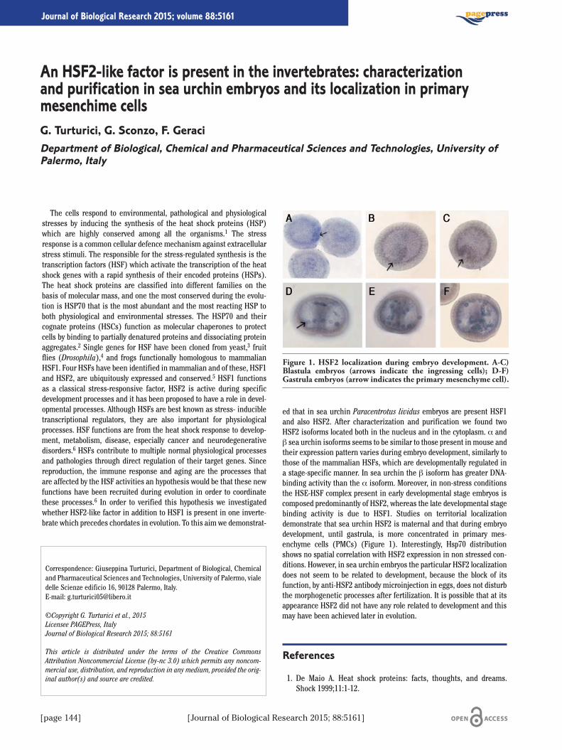
[page 144] [Journal of Biological Research 2015; 88:5161]
Journal of Biological Research 2015; volume 88:5161
The cells respond to environmental, pathological and physiologicalstresses by inducing the synthesis of the heat shock proteins (HSP)which are highly conserved among all the organisms.1 The stressresponse is a common cellular defence mechanism against extracellularstress stimuli. The responsible for the stress-regulated synthesis is thetranscription factors (HSF) which activate the transcription of the heatshock genes with a rapid synthesis of their encoded proteins (HSPs).The heat shock proteins are classified into different families on thebasis of molecular mass, and one the most conserved during the evolu-tion is HSP70 that is the most abundant and the most reacting HSP toboth physiological and environmental stresses. The HSP70 and theircognate proteins (HSCs) function as molecular chaperones to protectcells by binding to partially denatured proteins and dissociating proteinaggregates.2 Single genes for HSF have been cloned from yeast,3 fruitflies (Drosophila),4 and frogs functionally homologous to mammalianHSF1. Four HSFs have been identified in mammalian and of these, HSF1and HSF2, are ubiquitously expressed and conserved.5 HSF1 functionsas a classical stress-responsive factor, HSF2 is active during specificdevelopment processes and it has been proposed to have a role in devel-opmental processes. Although HSFs are best known as stress- inducibletranscriptional regulators, they are also important for physiologicalprocesses. HSF functions are from the heat shock response to develop-ment, metabolism, disease, especially cancer and neurodegenerativedisorders.6 HSFs contribute to multiple normal physiological processesand pathologies through direct regulation of their target genes. Sincereproduction, the immune response and aging are the processes thatare affected by the HSF activities an hypothesis would be that these newfunctions have been recruited during evolution in order to coordinatethese processes.6 In order to verified this hypothesis we investigatedwhether HSF2-like factor in addition to HSF1 is present in one inverte-brate which precedes chordates in evolution. To this aim we demonstrat-
ed that in sea urchin Paracentrotus lividus embryos are present HSF1and also HSF2. After characterization and purification we found twoHSF2 isoforms located both in the nucleus and in the cytoplasm. α andβ sea urchin isoforms seems to be similar to those present in mouse andtheir expression pattern varies during embryo development, similarly tothose of the mammalian HSFs, which are developmentally regulated ina stage-specific manner. In sea urchin the β isoform has greater DNA-binding activity than the α isoform. Moreover, in non-stress conditionsthe HSE-HSF complex present in early developmental stage embryos iscomposed predominantly of HSF2, whereas the late developmental stagebinding activity is due to HSF1. Studies on territorial localizationdemonstrate that sea urchin HSF2 is maternal and that during embryodevelopment, until gastrula, is more concentrated in primary mes-enchyme cells (PMCs) (Figure 1). Interestingly, Hsp70 distributionshows no spatial correlation with HSF2 expression in non stressed con-ditions. However, in sea urchin embryos the particular HSF2 localizationdoes not seem to be related to development, because the block of itsfunction, by anti-HSF2 antibody microinjection in eggs, does not disturbthe morphogenetic processes after fertilization. It is possible that at itsappearance HSF2 did not have any role related to development and thismay have been achieved later in evolution.
References
1. De Maio A. Heat shock proteins: facts, thoughts, and dreams.Shock 1999;11:1-12.
Correspondence: Giuseppina Turturici, Department of Biological, Chemicaland Pharmaceutical Sciences and Technologies, University of Palermo, vialedelle Scienze edificio 16, 90128 Palermo, Italy.E-mail: [email protected]
©Copyright G. Turturici et al., 2015Licensee PAGEPress, ItalyJournal of Biological Research 2015; 88:5161
This article is distributed under the terms of the Creative CommonsAttribution Noncommercial License (by-nc 3.0) which permits any noncom-mercial use, distribution, and reproduction in any medium, provided the orig-inal author(s) and source are credited.
An HSF2-like factor is present in the invertebrates: characterizationand purification in sea urchin embryos and its localization in primarymesenchime cellsG. Turturici, G. Sconzo, F. GeraciDepartment of Biological, Chemical and Pharmaceutical Sciences and Technologies, University ofPalermo, Italy
Figure 1. HSF2 localization during embryo development. A-C)Blastula embryos (arrows indicate the ingressing cells); D-F)Gastrula embryos (arrow indicates the primary mesenchyme cell).

[Journal of Biological Research 2015; 88:5161] [page 145]
Article
2. Bukau B, Horwich AL. The Hsp70 and Hsp60 chaperone machines.Cell 1998;92:351-66.
3. Wiederrecht G, Seto D, Parker CS. Isolation of the gene encoding theS. cerevisiae heat shock transcription factor. Cell 1988;54:841-53.
4. Clos J, Westwood JT, Becher PB, et al. Molecular cloning andexpression of a hexameric Drosophila heat shock factor subject to
negative regulation. Cell 1990;63:1085-97.5. Morano KA, Thiele DJ. Heat shock factor function and regulation inresponse to cellular stress, growth, and differentiation signals.
6. Akerfelt M, Morimoto RI, Sistonen L. Heat shock factors: integra-tors of cell stress, development and lifespan. Nat Rev Mol Cell Biol2010;11:545-55.
[page 146] [Journal of Biological Research 2015; 88:5161]
Journal of Biological Research 2015; volume 88:5161
Nuclear Factor-κB (NF-κB) are ubiquitous transcription factors thatin mammals regulate many biological process including inflammation,immunoregulation, apoptosis, cell growth and cell proliferation.1 Manystudies show that NF-κB is overexpressed in solid and haematologicaltumours. These data suggest that a deregulated expression of NF-κBpathway is closely related with oncogenic phenotype.2 NF-κB familymembers include RelA (p65), c-Rel, RelB, p50 and p52. These proteinsexert their functions by binding as homodimers or heterodimers tospecific DNA target sites (κB consensus). The p65/p50 heterodimer isthe most abundant and investigated form of NF-κB3 A new isoform ofp65, named p65(-1), have been discovered in human and mouse. Thisisoform contains an unknown exon (named exon-1) located upstreamto the first known exon of RelA, coding for p65. Transcription of theexon-1 leads to an alternative splicing between exon -1 and exon 1,thus skipping exon 0. By consequence p65(-1) has a smaller RHD thanp65. Previous evidences show that p65(-1), compared to p65, has dif-ferent biochemical properties in some cellular mechanism like tran-scriptional activity on κB consensus, apoptosis and regulation of theglucocorticoid receptor (GR) activation. In this study we investigatedthe function of p65(-1) by an in vitro, ex vivo and an in vivo approach.In order to test the transcriptional role of p65(-1) we have analysed thetransactivation of p65(-1) using both artificial and natural promoterregions, linked with the pathway of NF-κB. We have performedluciferase assays with: NF-κB-Luc (nuclear factor κB) CRE-Luc (cAMPresponse element), AP1-Luc (AP1 response element), SRE-Luc (serumresponse element), HSE-Luc (heat shock protein response element),pANXA-1-Luc (annexina 1) e pIL-6-Luc (interleukin 6) to study the
activity of p65(-1) and we have also analyzed p65(-1) activity with p65or p50 under the same conditions. Our data suggest that p65(-1) has acentral role during the regulation of pro and anti-inflammatoryresponses through a specific transcriptional activation using differentpartners according to the cellular requirements. We have also studiedp65(-1) expression on human peripheral blood mononuclear cells(PBMC); it is shown that the expression of mRNA is always present inthe analyzed samples. Further, our data demonstrate different expres-sion profiles between individuals considered. The detection of addi-tional factors belonging to NF-κB complex enhances the hypothesis oftuning responses. According with central role of NF-κB in the biologi-cal responses we propose to investigate the function of p65(-1) and itsnetwork of interaction with other transcriptional factors, in many oth-ers cellular processes like apoptosis and cell proliferation.
References
1. Li Q, Verma IM. NF-kappaB regulation in the immune system. NatRev Immunol 2002;2:725-34.
2. Kim HJ, Hawke N, Baldwin AS. NF-κB and IKK as therapeutic tar-get in cancer. Cell Death Differ 2006;13:738-47.
3. Verma IM, Stevenson JK, Schwarz EM, et al. REl/NF-κB/IkB family:intimate tales is association and dissociation. Genes Dev1995;9:2723-35.
Correspondence: Francesca Valentino, Department of Biological, Chemicaland Pharmaceutical Sciences and Technologies, University of Palermo, vialedelle Scienze 4, 90121 Palermo, Italy.E-mail: [email protected]
©Copyright F. Valentino et al., 2015Licensee PAGEPress, ItalyJournal of Biological Research 2015; 88:5161
This article is distributed under the terms of the Creative CommonsAttribution Noncommercial License (by-nc 3.0) which permits any noncom-mercial use, distribution, and reproduction in any medium, provided the orig-inal author(s) and source are credited.
Functional characterization of p65(-1), a new isoform of p65from NF-κB complexF. Valentino,1,2 A. Artale,1 G. Spinelli,1 F. Gianguzza,2 F. Di Blasi11Institute of Biomedicine and Molecular Immunology A. Monroy, National Research Council,Palermo; 2Department of Biological, Chemical and Pharmaceutical Sciences and Technologies,University of Palermo, Italy
[Journal of Biological Research 2015; 88:5161] [page 147]
Journal of Biological Research 2015; volume 88:5161
Tra i fattori verosimilmente più implicati nel processo d’invecchia-mento (in particolare nell’invecchiamento senza successo, caratteriz-zato dalla comparsa delle malattie correlate all’età e da disabilità),emerge il ruolo chiave dei radicali liberi, molecole o frammenti mole-colari contenenti uno o più elettroni spaiati, e di conseguenza dotati direattività elevata, e dello stress ossidativo (inteso come condizioneossidante, derivante da uno squilibrio tra fattori ossidanti ed anti-ossi-danti a favore dei primi), capaci di indurre infiammazione e dannotessutale.1 Lo stress ossidativo induce alterazione del bilancio redoxdelle cellule, con la conseguente attivazione di vie di trasduzione delsegnale e di fattori di trascrizioni redox sensibili, capaci di evocareuno stato infiammatorio. Si ha, di conseguenza, un’attivazione conti-nua e prolungata, che determina il rilascio di notevoli quantità dimediatori infiammatori, responsabili della conseguente evocazione diuno stato infiammatorio cronico. L’accumulo con l’età di molecole dan-neggiate a tutti i livelli (lipidi, proteine e DNA) è indubbiamente unodegli attori principali nei processi d’invecchiamento e di malattie, inquanto può essere responsabile di senescenza cellulare, infiammazio-ne e cancerogenesi.2 I radicali dell’ossigeno (ROS) e dell’azoto (RNS)appaiono quindi importanti mediatori della risposta infiammatoria dauna parte ma responsabili pure del danneggiamento delle cellule dal-l’altra.3 Attualmente nei paesi occidentali l’uso di cibi addizionati daintegratori antiossidanti sta ricevendo una crescente attenzione e staper essere sempre più adottato. Un migliore approccio terapeutico euna migliore informazione potrebbe derivare dalla corretta valutazio-ne globale dello stress ossidativo mediante test diagnostici ove le duecomponenti contrapposte, quella pro- ed anti-ossidante possono esserevalutate distintamente.4 Lo studio si è proposto di valutare in uno stu-dio longitudinale l’effetto dell’assunzione dell’integratore alimentaresu 50 soggetti, ugualmente suddivisi per genere in un range di età 50-
70 anni. L’integratore contiene tra l’altro polifenoli come; acido Gallico,catechine/epicatechine (monomeri), procianidine dimeri (B1-B4)epolifenoli identificati min. 35%. I biomarcatori valutati nell’ambitodello stress lipidico sono stati gli isoprostani nelle urine e le LDL ossi-date nel siero. Ulteriormente, solo nella popolazione maschile è statovalutato il livello di testosterone. In questo studio di coorte prospetticol’integratore alimentare a base di estratti di acido Gallico,catechine/epicatechine (monomeri), procianidine dimeri (B1-B4)edepolifenoli si è dimostrato capace in esperimenti ex vivo di:Influenzare positivamente la concentrazione di LDL ossidate in manie-ra statisticamente significativa, di avere potenzialità come stimolantenella produzione di testosterone nel genere e di decrementare inmaniera statisticamente significativa la concentrazioni di isoprostaninelle urine.
Bibliografia
1. Jacob KD, Noren Hooten N, Trzeciak AR, Evans MK. Markers ofoxidant stress that are clinically relevant in aging and age-relateddisease. Mech Ageing Dev 2013;134:139-57.
2. Mohsenzadegan M, Mirshafiey A. The immunopathogenic role ofreactive oxygen species in Alzheimer disease. Iran J AllergyAsthma Immunol 2012;11:203-16.
3. Jenny NS. Inflammation in aging: cause, effect, or both? DiscovMed 2012;13:451-60.
4. Gaman L, Stoian I, Atanasiu V. Can ageing be slowed? Hormeticand redox perspectives. J Med Life 2011;4:346-5.
Correspondence: Sonya Vasto, Department of Biological, Chemical andPharmaceutical Sciences and Technologies, University of Palermo, viaArchirafi, 36 90136 Palermo, Italy.E-mail: [email protected]
©Copyright S. Vasto et al., 2015Licensee PAGEPress, ItalyJournal of Biological Research 2015; 88:5161
This article is distributed under the terms of the Creative CommonsAttribution Noncommercial License (by-nc 3.0) which permits any noncom-mercial use, distribution, and reproduction in any medium, provided the orig-inal author(s) and source are credited.
La perossidazione lipidica come esempio di stress ossidativoS. Vasto,1 A. Barera,2 S. Indelicato,1 D. Nuzzo,2 M. Di Carlo,2 C. Caruso31Department of Biological, Chemical and Pharmaceutical Sciences and Technologies, University ofPalermo; 2IBIM-CNR, Palermo; 3Department of Biopathology and Medical Biotechnologies,University of Palermo, Italy
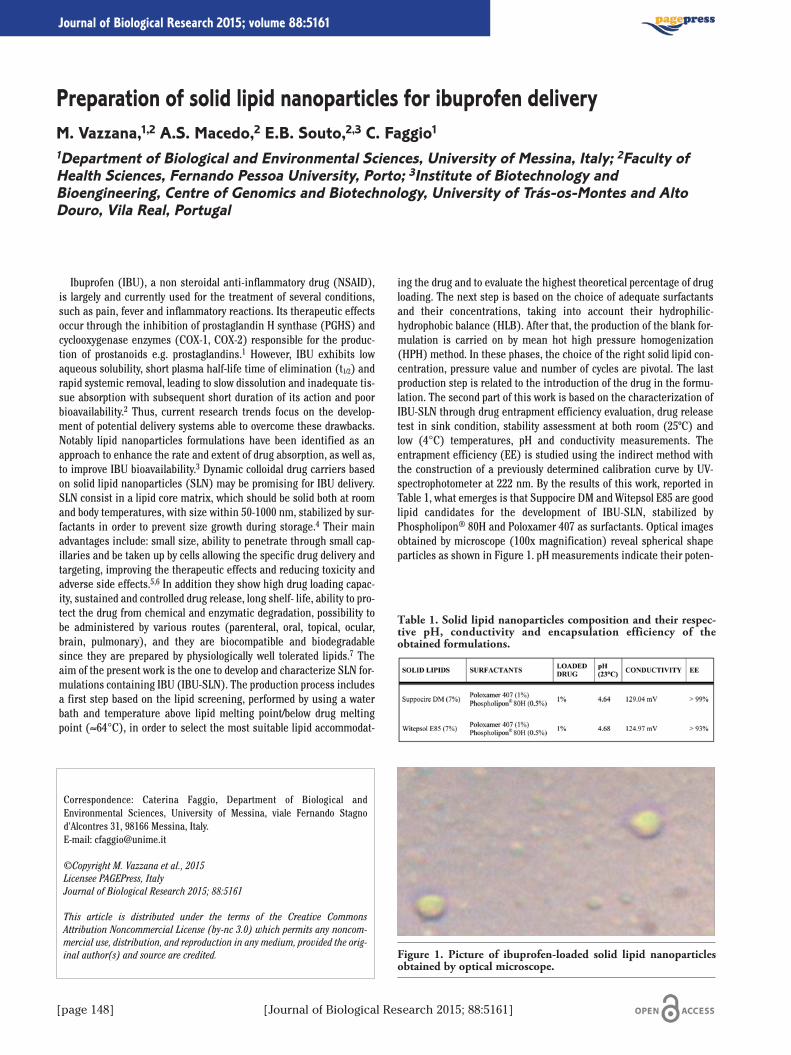
[page 148] [Journal of Biological Research 2015; 88:5161]
Journal of Biological Research 2015; volume 88:5161
Ibuprofen (IBU), a non steroidal anti-inflammatory drug (NSAID),is largely and currently used for the treatment of several conditions,such as pain, fever and inflammatory reactions. Its therapeutic effectsoccur through the inhibition of prostaglandin H synthase (PGHS) andcyclooxygenase enzymes (COX-1, COX-2) responsible for the produc-tion of prostanoids e.g. prostaglandins.1 However, IBU exhibits lowaqueous solubility, short plasma half-life time of elimination (t1/2) andrapid systemic removal, leading to slow dissolution and inadequate tis-sue absorption with subsequent short duration of its action and poorbioavailability.2 Thus, current research trends focus on the develop-ment of potential delivery systems able to overcome these drawbacks.Notably lipid nanoparticles formulations have been identified as anapproach to enhance the rate and extent of drug absorption, as well as,to improve IBU bioavailability.3 Dynamic colloidal drug carriers basedon solid lipid nanoparticles (SLN) may be promising for IBU delivery.SLN consist in a lipid core matrix, which should be solid both at roomand body temperatures, with size within 50-1000 nm, stabilized by sur-factants in order to prevent size growth during storage.4 Their mainadvantages include: small size, ability to penetrate through small cap-illaries and be taken up by cells allowing the specific drug delivery andtargeting, improving the therapeutic effects and reducing toxicity andadverse side effects.5,6 In addition they show high drug loading capac-ity, sustained and controlled drug release, long shelf- life, ability to pro-tect the drug from chemical and enzymatic degradation, possibility tobe administered by various routes (parenteral, oral, topical, ocular,brain, pulmonary), and they are biocompatible and biodegradablesince they are prepared by physiologically well tolerated lipids.7 Theaim of the present work is the one to develop and characterize SLN for-mulations containing IBU (IBU-SLN). The production process includesa first step based on the lipid screening, performed by using a waterbath and temperature above lipid melting point/below drug meltingpoint (≈64°C), in order to select the most suitable lipid accommodat-
ing the drug and to evaluate the highest theoretical percentage of drugloading. The next step is based on the choice of adequate surfactantsand their concentrations, taking into account their hydrophilic-hydrophobic balance (HLB). After that, the production of the blank for-mulation is carried on by mean hot high pressure homogenization(HPH) method. In these phases, the choice of the right solid lipid con-centration, pressure value and number of cycles are pivotal. The lastproduction step is related to the introduction of the drug in the formu-lation. The second part of this work is based on the characterization ofIBU-SLN through drug entrapment efficiency evaluation, drug releasetest in sink condition, stability assessment at both room (25ºC) andlow (4°C) temperatures, pH and conductivity measurements. Theentrapment efficiency (EE) is studied using the indirect method withthe construction of a previously determined calibration curve by UV-spectrophotometer at 222 nm. By the results of this work, reported inTable 1, what emerges is that Suppocire DM and Witepsol E85 are goodlipid candidates for the development of IBU-SLN, stabilized byPhospholipon® 80H and Poloxamer 407 as surfactants. Optical imagesobtained by microscope (100x magnification) reveal spherical shapeparticles as shown in Figure 1. pH measurements indicate their poten-
Correspondence: Caterina Faggio, Department of Biological andEnvironmental Sciences, University of Messina, viale Fernando Stagnod'Alcontres 31, 98166 Messina, Italy.E-mail: [email protected]
©Copyright M. Vazzana et al., 2015Licensee PAGEPress, ItalyJournal of Biological Research 2015; 88:5161
This article is distributed under the terms of the Creative CommonsAttribution Noncommercial License (by-nc 3.0) which permits any noncom-mercial use, distribution, and reproduction in any medium, provided the orig-inal author(s) and source are credited.
Preparation of solid lipid nanoparticles for ibuprofen deliveryM. Vazzana,1,2 A.S. Macedo,2 E.B. Souto,2,3 C. Faggio11Department of Biological and Environmental Sciences, University of Messina, Italy; 2Faculty ofHealth Sciences, Fernando Pessoa University, Porto; 3Institute of Biotechnology andBioengineering, Centre of Genomics and Biotechnology, University of Trás-os-Montes and AltoDouro, Vila Real, Portugal
Table 1. Solid lipid nanoparticles composition and their respec-tive pH, conductivity and encapsulation efficiency of theobtained formulations.
Figure 1. Picture of ibuprofen-loaded solid lipid nanoparticlesobtained by optical microscope.
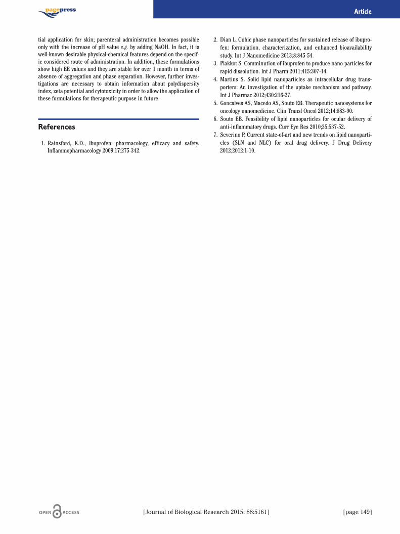
[Journal of Biological Research 2015; 88:5161] [page 149]
Article
tial application for skin; parenteral administration becomes possibleonly with the increase of pH value e.g. by adding NaOH. In fact, it iswell-known desirable physical-chemical features depend on the specif-ic considered route of administration. In addition, these formulationsshow high EE values and they are stable for over 1 month in terms ofabsence of aggregation and phase separation. However, further inves-tigations are necessary to obtain information about polydispersityindex, zeta potential and cytotoxicity in order to allow the application ofthese formulations for therapeutic purpose in future.
References
1. Rainsford, K.D., Ibuprofen: pharmacology, efficacy and safety.Inflammopharmacology 2009;17:275-342.
2. Dian L. Cubic phase nanoparticles for sustained release of ibupro-fen: formulation, characterization, and enhanced bioavailabilitystudy. Int J Nanomedicine 2013;8:845-54.
3. Plakkot S. Comminution of ibuprofen to produce nano-particles forrapid dissolution. Int J Pharm 2011;415:307-14.
4. Martins S. Solid lipid nanoparticles as intracellular drug trans-porters: An investigation of the uptake mechanism and pathway.Int J Pharmac 2012;430:216-27.
5. Goncalves AS, Macedo AS, Souto EB. Therapeutic nanosystems foroncology nanomedicine. Clin Transl Oncol 2012;14:883-90.
6. Souto EB. Feasibility of lipid nanoparticles for ocular delivery ofanti-inflammatory drugs. Curr Eye Res 2010;35:537-52.
7. Severino P. Current state-of-art and new trends on lipid nanoparti-cles (SLN and NLC) for oral drug delivery. J Drug Delivery2012;2012:1-10.
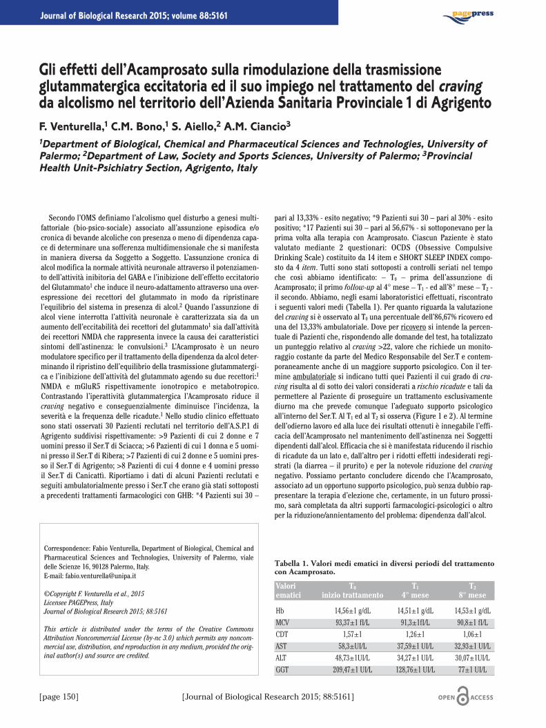
[page 150] [Journal of Biological Research 2015; 88:5161]
Journal of Biological Research 2015; volume 88:5161
Secondo l’OMS definiamo l’alcolismo quel disturbo a genesi multi-fattoriale (bio-psico-sociale) associato all’assunzione episodica e/ocronica di bevande alcoliche con presenza o meno di dipendenza capa-ce di determinare una sofferenza multidimensionale che si manifestain maniera diversa da Soggetto a Soggetto. L’assunzione cronica dialcol modifica la normale attività neuronale attraverso il potenziamen-to dell’attività inibitoria del GABA e l’inibizione dell’effetto eccitatoriodel Glutammato1 che induce il neuro-adattamento attraverso una over-espressione dei recettori del glutammato in modo da ripristinarel’equilibrio del sistema in presenza di alcol.2 Quando l’assunzione dialcol viene interrotta l’attività neuronale è caratterizzata sia da unaumento dell’eccitabilità dei recettori del glutammato1 sia dall’attivitàdei recettori NMDA che rappresenta invece la causa dei caratteristicisintomi dell’astinenza: le convulsioni.3 L’Acamprosato è un neuromodulatore specifico per il trattamento della dipendenza da alcol deter-minando il ripristino dell’equilibrio della trasmissione glutammatergi-ca e l’inibizione dell’attività del glutammato agendo su due recettori:1
NMDA e mGluR5 rispettivamente ionotropico e metabotropico.Contrastando l’iperattività glutammatergica l’Acamprosato riduce ilcraving negativo e conseguenzialmente diminuisce l’incidenza, laseverità e la frequenza delle ricadute.1 Nello studio clinico effettuatosono stati osservati 30 Pazienti reclutati nel territorio dell’A.S.P.1 diAgrigento suddivisi rispettivamente: >9 Pazienti di cui 2 donne e 7uomini presso il Ser.T di Sciacca; >6 Pazienti di cui 1 donna e 5 uomi-ni presso il Ser.T di Ribera; >7 Pazienti di cui 2 donne e 5 uomini pres-so il Ser.T di Agrigento; >8 Pazienti di cui 4 donne e 4 uomini pressoil Ser.T di Canicattì. Riportiamo i dati di alcuni Pazienti reclutati eseguiti ambulatorialmente presso i Ser.T che erano già stati sottopostia precedenti trattamenti farmacologici con GHB: *4 Pazienti sui 30 –
pari al 13,33% - esito negativo; *9 Pazienti sui 30 – pari al 30% - esitopositivo; *17 Pazienti sui 30 – pari al 56,67% - si sottoponevano per laprima volta alla terapia con Acamprosato. Ciascun Paziente è statovalutato mediante 2 questionari: OCDS (Obsessive CompulsiveDrinking Scale) costituito da 14 item e SHORT SLEEP INDEX compo-sto da 4 item. Tutti sono stati sottoposti a controlli seriati nel tempoche così abbiamo identificato: – T0 – prima dell’assunzione diAcamprosato; il primo follow-up al 4° mese – T1 - ed all’8° mese – T2 -il secondo. Abbiamo, negli esami laboratoristici effettuati, riscontratoi seguenti valori medi (Tabella 1). Per quanto riguarda la valutazionedel craving si è osservato al T0 una percentuale dell’86,67% ricovero eduna del 13,33% ambulatoriale. Dove per ricovero si intende la percen-tuale di Pazienti che, rispondendo alle domande del test, ha totalizzatoun punteggio relativo al craving >22, valore che richiede un monito-raggio costante da parte del Medico Responsabile del Ser.T e contem-poraneamente anche di un maggiore supporto psicologico. Con il ter-mine ambulatoriale si indicano tutti quei Pazienti il cui grado di cra-ving risulta al di sotto dei valori considerati a rischio ricadute e tali dapermettere al Paziente di proseguire un trattamento esclusivamentediurno ma che prevede comunque l’adeguato supporto psicologicoall’interno del Ser.T. Al T1 ed al T2 si osserva (Figure 1 e 2). Al terminedell’odierno lavoro ed alla luce dei risultati ottenuti è innegabile l’effi-cacia dell’Acamprosato nel mantenimento dell’astinenza nei Soggettidipendenti dall’alcol. Efficacia che si è manifestata riducendo il rischiodi ricadute da un lato e, dall’altro per i ridotti effetti indesiderati regi-strati (la diarrea – il prurito) e per la notevole riduzione del cravingnegativo. Possiamo pertanto concludere dicendo che l’Acamprosato,associato ad un opportuno supporto psicologico, può senza dubbio rap-presentare la terapia d’elezione che, certamente, in un futuro prossi-mo, sarà completata da altri supporti farmacologici-psicologici o altroper la riduzione/annientamento del problema: dipendenza dall’alcol.
Correspondence: Fabio Venturella, Department of Biological, Chemical andPharmaceutical Sciences and Technologies, University of Palermo, vialedelle Scienze 16, 90128 Palermo, Italy.E-mail: [email protected]
©Copyright F. Venturella et al., 2015Licensee PAGEPress, ItalyJournal of Biological Research 2015; 88:5161
This article is distributed under the terms of the Creative CommonsAttribution Noncommercial License (by-nc 3.0) which permits any noncom-mercial use, distribution, and reproduction in any medium, provided the orig-inal author(s) and source are credited.
Gli effetti dell’Acamprosato sulla rimodulazione della trasmissioneglutammatergica eccitatoria ed il suo impiego nel trattamento del cravingda alcolismo nel territorio dell’Azienda Sanitaria Provinciale 1 di AgrigentoF. Venturella,1 C.M. Bono,1 S. Aiello,2 A.M. Ciancio31Department of Biological, Chemical and Pharmaceutical Sciences and Technologies, University ofPalermo; 2Department of Law, Society and Sports Sciences, University of Palermo; 3ProvincialHealth Unit-Psichiatry Section, Agrigento, Italy
Tabella 1. Valori medi ematici in diversi periodi del trattamentocon Acamprosato.
Valori T0 T1 T2
ematici inizio trattamento 4° mese 8° mese
Hb 14,56±1 g/dL 14,51±1 g/dL 14,53±1 g/dLMCV 93,37±1 fI/L 91,3±1fI/L 90,8±1 fI/LCDT 1,57±1 1,26±1 1,06±1AST 58,3±UI/L 37,59±1 UI/L 32,93±1 UI/LALT 48,73±1UI/L 34,27±1 UI/L 30,07±1UI/LGGT 209,47±1 UI/L 128,76±1 UI/L 77±1 UI/L

[Journal of Biological Research 2015; 88:5161] [page 151]
Article
Bibliografia
1. De Witte P. Neuroprotective and abstinence-promoting effects ofacamprosate. CNS Drugs 2005;19:517-37.
2. Littleton JM. Acamprosate in alcohol Dependence: Implications ofa Unique Mechanism of Action. J Addict Med 2007;1:115-25.
3. Mason BJ, Heyser CJ. Acamprosate: a protorypic neuromodulatorin the treatment of alcohol dependence. CSN & NeurologicalDisorders-Drug Targets 2010;9:23-32.
Figura 1. Valutazione craving secondo l’Obsessive CompulsiveDrinking Scale al quarto mese (T1).
Figura 2. Valutazione craving secondo l’Obsessive CompulsiveDrinking Scale all’ottavo mese (T2).
The analysis carried out by Doores and colleagues1 and Venturiniand colleagues2 on the total bacterial load tested in wild and cultivatedfresh mushrooms highlight values ranging between 3.7 and 9.3 logufc/g. Due to the absence of pathogens, the microbiological quality ofmushrooms analysed by the above-mentioned authors has been con-sidered good. On the basis of data reported in literature we have alsoextended the observations with frozen and dried mushrooms in orderto evaluate the mesophilous bacterial and fungal load. In particular thepresence/absence of Escherichia coli (Migula,1895) Castellani &Chalmers, 1919, Salmonella spp. and Listeria monocytogenes (Murrayet al., 1926) Pirie 1940 was analysed. The aim of this paper is also toevaluate the quality and safety of mushrooms daily consumption byconsumers. Thirty samples of mushrooms (10 cultivated fresh mush-rooms identified with letter A 1-10; 10 dried mushrooms identifiedwith letter B 1-10 and, 10 frozen mushrooms identified with letter C 1-10) were taken from large-scale distribution markets, mini-marketsand small markets owned by migrants. The frozen mushrooms werecontained in packages of Agaricus bisporus (J.E. Lange) Imbach 1946and packages of Boletus edulis Bull. Besides mixed packages of A. bis-porus, Pholiota nameko (T. Itô) S. Ito & S. Imai, Boletus luteus L.,Pleurotus ostreatus (Jacq.) P. Kumm. and, Lentinula edoses (Berk.)Pegler were analysed. Other analyzed frozen mushrooms were a mix-ture of A. bisporus, Agrocybe aegerita (V. Brig.) Singer, P. ostreatus and,P. cornucopiae (Paulet) Rolland and a mixed package of A. bisporusand P. ostreatus. The fresh mushrooms (A. bisporus and Pleurotusostreatus) were purchaed from the grocery store. Dried mushrooms (L.edodes) were purchased from shops owned by Chinese migrants. Themicrobiological analysis were carried out in the laboratory of the
Center of Mycological Control belonging to the Sanitary Agency of theprovince of Palermo (southern Italy). The total bacterial load wasanalysed in 25 g of mushrooms (1:10 diluition) according to the ruleISO 4833:2004.3 Moulds and yeasts were analysed according to thereport ISTISAN 96/35.4 Salmonella sp. was checked through the crite-ria of analysis ruled by UNI EN ISO 6579:2008.5 The methodology ofISO 16649-2:20016 was used to test the Escherichia coli positive beta-glucorinidase. Listeria monocitogenes was tested accordino to the ruleISO 11290-1:2005.6 The total count in Petri dishes was made using theformula reported in ISO 7218:2007.7 The cultura media were providedby the concern Lickson srl (Vicari, province of Palermo). The nomen-clature follows the List of Prokaryotic Names with Standing inNomenclature (LPSN). L. monocitogenes and Salmonella spp. were notfound in the analysed mushrooms. On the contrary a sample of freshmushrooms from a supermarket of the town of Palermo was pollutedby E. coli. The count of E. coli positive beta-glucorinidase correspondto 1.7�104 cfu/g. The value of total bacterial count in all the mushroomsanalysed varies from a minimum of 3.8�102 cfu/g found in dried mush-rooms (L. edodes) to a maximum 2.6�108 ufc/g in a fresh sample ofmixed mushrooms (A. bisporus and P. ostreatus) in the supermarket.As regards moulds and yeasts the value varies from zero in driedmushrooms (L. edodes, B. edulis) to 4.4�104 ufc/g in fresh mushrooms(A. bisporus). The results obtained showed that an analyzed sample ofmushroom had a high pollutant load of E. coli. As known this bacteri-um is responsible of intestinal infections that can result in seriousextra-intestinal infections. Besides E. coli is involved in drug resist-ance and thus have a significant impact on human health. Since thepackaging of fresh mushrooms polluted by E. coli was purchased froma supermarket belonging to the mass distribution highlights the needand importance of sanitary controls for the protection of the con-sumers. The recent warning from the EFSA (European Food SafetyAuthority) that Italy is the second country in the EU as largest foodborne diseases (especially salmonellosis) reinforces the need torespect to meet the parameters set out in Regulation 2073/2005 butalso the checking of the proper handling of mushrooms during cultiva-tion and packaging, including compliance with good hygienic practicesby insiders.
References
1. Doores S, Kramer M, Beefman R. Evaluation and bacterial popula-tions associated with fresh mushrooms (Agaricus bisporus). DevCrop Sci 1986;10:283-94.
2. Venturini ME, Reyes JE, Rivera CS, et al. Microbiological qualityand safety of fresh cultivated and wild mushrooms commercializedin Spain. Food Microbiol 2011;28:1492-8.
Correspondence: Giuseppe Venturella, Department of Biological, Chemicaland Pharmaceutical Sciences and Technologies, University of Palermo, vialedelle Scienze 16, 90128 Palermo, Italy.E-mail: [email protected]
Acknowledgements: the authors wish to thank Dr. Gaetano Licata (Licksonsrl) for his kindly providing cultural media.
©Copyright D. Magli et al., 2015Licensee PAGEPress, ItalyJournal of Biological Research 2015; 88:5161
This article is distributed under the terms of the Creative CommonsAttribution Noncommercial License (by-nc 3.0) which permits any noncom-mercial use, distribution, and reproduction in any medium, provided the orig-inal author(s) and source are credited.
Evaluation of bacterial and fungal load in fresh, frozenand dried food mushroomsD. Magli,1 F. Venturella,2 M.L. Gargano,1 C. Sciarratta,3 A. Zummo,3 G. Venturella11Department of Agricultural and Forest Sciences, University of Palermo; 2Department ofBiological, Chemical and Pharmaceutical Sciences and Technologies, University of Palermo;3Provincial Health Unit 6, Palermo, Italy
[page 152] [Journal of Biological Research 2015; 88:5161]
Journal of Biological Research 2015; volume 88:5161

3. ISO. UNI EN ISO 4833:2004: Metodo orizzontale per la conta dimicroorganismi. Tecnica della conta delle colonia a 30 gradi. Rome:ISO; 2004.
4. De Medici D, Fenicia L, Orefice L, Stacchini A. Metodi di analisi peril controllo degli alimenti. Rome: Istituto Superiore di Sanità; 1996.
5. ISO. UNI EN ISO 6579:2008. Microbiologia di alimenti e mangimiper animali. Metodo orizzontale per la ricerca di Salmonella spp.Rome: ISO; 2008.
6. Carratù B, Aureli P. Criteri chimici e microbiologici per la valu-tazione di conformità degli integratori alimentari a base di piante.Rome: Istituto Superiore di Sanità; 2007.
7. ISO. International Standard ISO 7218. Microbiology of food andanimal feeding stuffs - General requirements and guidance formicrobiological examinations. Geneva: International Organizationfor Standardization; 2007.
[Journal of Biological Research 2015; 88:5161] [page 153]
Article
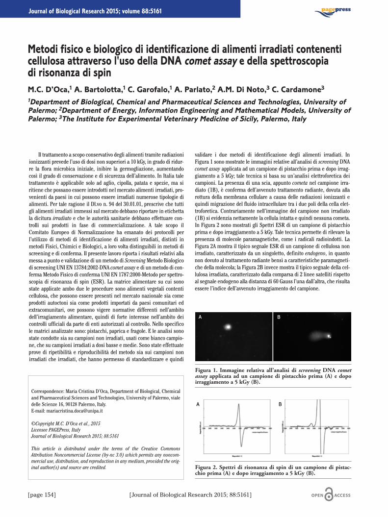
Il trattamento a scopo conservativo degli alimenti tramite radiazioniionizzanti prevede l’uso di dosi non superiori a 10 kGy, in grado di ridur-re la flora microbica iniziale, inibire la germogliazione, aumentandocosì il grado di conservazione e di sicurezza dell’alimento. In Italia taletrattamento è applicabile solo ad aglio, cipolla, patata e spezie, ma siritiene che possano essere introdotti nel mercato alimenti irradiati, pro-venienti da paesi in cui possono essere irradiati numerose tipologie dialimenti. Per tale ragione il Dl.vo n. 94 del 30.01.01, prescrive che tuttigli alimenti irradiati immessi sul mercato debbano riportare in etichettala dicitura irradiato e che le autorità sanitarie debbano effettuare con-trolli sui prodotti in fase di commercializzazione. A tale scopo ilComitato Europeo di Normalizzazione ha emanato dei protocolli perl’utilizzo di metodi di identificazione di alimenti irradiati, distinti inmetodi Fisici, Chimici e Biologici, a loro volta distinguibili in metodi discreening e di conferma. Il presente lavoro riporta i risultati relativi allamessa a punto e validazione di un metodo di ScreeningMetodo Biologicodi screening UNI EN 13784:2002-DNA comet assay e di un metodo di con-ferma Metodo Fisico di conferma UNI EN 1787:2000-Metodo per spettro-scopia di risonanza di spin (ESR). La matrice alimentare su cui sonostate applicate ambo due le procedure sono alimenti vegetali contenticellulosa, che possono essere presenti nel mercato nazionale sia comeprodotti autoctoni sia come prodotti importati da paesi comunitari edextracomunitari, ove possono vigere normative differenti nell’ambitodell’irragiamento alimentare, quindi di forte interesse nell’ambito deicontrolli ufficiali da parte di enti autorizzati al controllo. Nello specificole matrici analizzate sono: pistacchi, paprica e fragole. E le analisi sonostate condotte sia su campioni non irradiati, usati come bianco campio-ne, che su campioni irradiati a dosi basse e medie. Sono state effettuateprove di ripetibilità e riproducibilità del metodo sia sui campioni nonirradiati che irradiati, che hanno permesso di standardizzare e quindi
validare i due metodi di identificazione degli alimenti irradiati. InFigura 1 sono mostrate le immagini relative all’analisi di screening DNAcomet assay applicata ad un campione di pistacchio prima e dopo irrag-giamento a 5 kGy; tale tecnica si basa su un’analisi elettroforetica deicampioni. La presenza di una scia, appunto cometa nel campione irra-diato (1B), è conferma dell’avvenuto trattamento radiante, dovuta allarottura della membrana cellulare a causa delle radiazioni ionizzanti equindi migrazione del fluido intracellulare tra i due poli della cella elet-troforetica. Contrariamente nell’immagine del campione non irradiato(1B) si evidenzia nettamente la cellula intatta e quindi nessuna cometa.In Figura 2 sono mostrati gli Spettri ESR di un campione di pistacchioprima e dopo irraggiamento a 5 kGy. Tale tecnica permette di rilevare lapresenza di molecole paramagnetiche, come i radicali radioindotti. LaFigura 2A mostra il tipico segnale ESR di un campione di cellulosa nonirradiato, caratterizzato da un singoletto, definito endogeno, in quantonon dovuto al trattamento radiante bensì a caratteristiche paramagneti-che della molecola; la Figura 2B invece mostra il tipico segnale della cel-lulosa irradiata, caratterizzato dalla comparsa di 2 linee satelliti rispettoal segnale endogeno alla distanza di 60 Gauss l’una dall’altra, che risultaessere l’indice dell’avvenuto irraggiamento del campione.
Correspondence: Maria Cristina D’Oca, Department of Biological, Chemicaland Pharmaceutical Sciences and Technologies, University of Palermo, vialedelle Scienze 16, 90128 Palermo, Italy.E-mail: [email protected]
©Copyright M.C. D’Oca et al., 2015Licensee PAGEPress, ItalyJournal of Biological Research 2015; 88:5161
This article is distributed under the terms of the Creative CommonsAttribution Noncommercial License (by-nc 3.0) which permits any noncom-mercial use, distribution, and reproduction in any medium, provided the orig-inal author(s) and source are credited.
Metodi fisico e biologico di identificazione di alimenti irradiati contenenticellulosa attraverso l'uso della DNA comet assay e della spettroscopiadi risonanza di spinM.C. D’Oca,1 A. Bartolotta,1 C. Garofalo,1 A. Parlato,2 A.M. Di Noto,3 C. Cardamone31Department of Biological, Chemical and Pharmaceutical Sciences and Technologies, University ofPalermo; 2Department of Energy, Information Engineering and Mathematical Models, University ofPalermo; 3The Institute for Experimental Veterinary Medicine of Sicily, Palermo, Italy
[page 154] [Journal of Biological Research 2015; 88:5161]
Journal of Biological Research 2015; volume 88:5161
Figura 1. Immagine relativa all’analisi di screening DNA cometassay applicata ad un campione di pistacchio prima (A) e dopoirraggiamento a 5 kGy (B).
Figura 2. Spettri di risonanza di spin di un campione di pistac-chio prima (A) e dopo irraggiamento a 5 kGy (B).