bond strength of composite resin to enamel and dentin
TRANSCRIPT

INTRODUCTION
Laser tooth cutting with laser device requires more time compared with rotary cutting; however, it has some merits, which include low vibration and pain for the tooth and low uncomfortable sound for the patient during tooth preparation1-3). Therefore, its applications have gradually increased in clinics. The wavelength of erbium, chromium: yttrium scandium gallium garnet (Er,Cr:YSGG) laser is 2,780 nm, similar to that of to erbium: yttrium aluminum garnet (Er:YAG) laser (2,940 nm). Tooth cutting ability of Er,Cr:YSGG laser is superior to that of Er:YAG laser4). Er,Cr:YSGG laser possesses great tooth cutting ability and high intensity and frequency of irradiation. Its water/air ratio can be manipulated according to cutting conditions which is influenced by a hardness of tooth substance. Furthermore, use of new irradiation tips with Er,Cr:YSGG laser realized high speed tooth cutting equivalent to that of an air turbine5). The mechanism for tooth cutting using Er,Cr:YSGG laser is mainly based on the theory of hydrokinetics3), in which accelerated water spray crushes the tooth surface based on the high level of absorbed laser energy of water molecules4,5). Thermal accommodation has been reported to not easily occur on an irradiated tooth surface, with a low thermal increase in pulp6,7), when a cavity is prepared using Er,Cr:YSGG laser. However, tooth surface irradiated by laser is achieved using the thermal effect with sufficient water cooling8). Dentin surfaces prepared by Er:YAG laser is denatured by heat, which in turn shows a decrease in adhesion efficacy to composite resin9). The same finding has been reported in several other studies using Er,Cr:YSGG laser10-14). In clinics, it is recommended that
the heat-denatured layer be removed using excavation or low-speed cutting with round steel bur9). However, an excavator or rotary cutting device has proven to be inefficient in removing the heat-denatured layer after preparation with a Er,Cr:YSGG laser. In our previous report, we described the removal of the heat-denatured layer on the dentin surface by applying both phosphoric acid and sodium hypochlorite8). Therefore, an experimental adhesive systems including preconditioning both phosphoric acid and sodium hypochlorite would be expected to improve the bond strength of composite resin to enamel and dentin prepared with Er,Cr:YSGG laser.
The aim of the present study is to examine the effect of various adhesive systems on the bond strength of composite resin to enamel and dentin prepared with Er,Cr:YSGG laser. The null hypothesis is that adhesive systems would not affect the bond strength of composite resin to enamel and dentin prepared with Er,Cr:YSGG laser.
MATERIALS AND METHODS
The Human Research Committee of the Nippon Dental University School of Life Dentistry at Niigata reviewed and approved the present study (Approval number: ECNG-H-9)
Experimental materialThe experimental materials used in this study are presented in Table 1. A two-step self-etching system (Clearfil SE Bond, Kuraray Noritake Dental, Osaka, Japan) and a one-step self-etching system (Clearfil Tri-S Bond, Kuraray Noritake Dental) were used as adhesive
Bond strength of composite resin to enamel and dentin prepared with Er,Cr:YSGG laserMayo TAKADA1, Koichi SHINKAI2, Chikage KATO2 and Masaya SUZUKI2
1 Advanced Operative Dentistry, The Nippon Dental University Graduate School of Life Dentistry at Niigata, 1-8 Hamaura-cho, Chuo-ku Niigata, 951-8580, Japan
2 Department of Operative Dentistry, The Nippon Dental University School of Life Dentistry at Niigata, 1-8 Hamaura-cho, Chuo-ku Niigata, 951-8580, Japan
Corresponding author, Koichi SHINKAI; E-mail: [email protected]
We aimed to examine the effect of various adhesive systems on the bond strength of composite resin to enamel or dentin prepared with erbium, chromium: yttrium scandium gallium garnet (Er,Cr:YSGG) laser. Each laser-cut enamel or dentin surface was treated with a bonding agent (SBB, Group 1); self-etching primer (SBP) and SBB (Group 2 and control); phosphoric-acid (KET), SBP and SBB (Group 3); KET, sodium-hypochlorite (ADG), SBP and SBB (Group 4); all-in-one adhesive (TSB, Group 5); or KET, ADG and TSB (Group 6). The control group was only polished with wet silicon carbide papers. The enamel shear bond strength of Group 5 was significantly lower than that of other groups (p<0.01). The control group showed higher bond strength compared to Groups 1–6 (p<0.05). Preconditioning using phosphoric acid or phosphoric acid followed by sodium hypochlorite increased the bond strength of composite resin to enamel and dentin prepared using an Er,Cr:YSGG laser.
Keywords: Er,Cr:YSGG laser, Enamel bond strength, Dentin bond strength, Composite resin
Received Feb 6, 2015: Accepted Jun 23, 2015doi:10.4012/dmj.2015-053 JOI JST.JSTAGE/dmj/2015-053
Dental Materials Journal 2015; 34(6): 863–871

Table 1 Compositions of the different materials used in this study
Material Abbreviation Composition Lot no. Manufacturer
Clearfil SE BondPrimer
SBP
2-Hydroxyethyl methacrylate (HEMA), 10-methacryloyloxydecyl dihydrogen phosphate (MDB), hydrophilic aliphatic dimethacrylate, dl-camphorquinone, water, accelerators, dyes
00812A
Kuraray Noritake Dental
Clearfil SE Bond/Bond
SBB
Bisphenol A diglycidylmethacrylate, 2-hydroxyethylmethacrylate (HEMA), 10-methacryloyloxydecyl dihydrogen phosphate(MDB), hydrophobic aliphatic methacrylate, colloidal silica, dl-camphorquinone, initiators, accelerators
00812A
Clearfil Tri-S bond
TSB
Bisphenol A diglycidylmethacrylate, 2-hydroxyethyl methacrylate (HEMA), ethanol, 10-methacryloyloxydecyl dihydrogen phosphate (MDB), dl-camphorquinone, colloidal silica, water, initiators, accelerators
00116A
K-etchant gel KET Water, phosphoric acid 40 wt%, colloidal silica, dyes 00437A
AD gel ADGSodium hypochlorite 10 wt%, waterAlumina nano-filler
000803
Clearfil Majesty LV(Shade: A3)
CLVSilanated barium glass filler, silanated colloidal silica, triethyleneglycol dimethacrylate, hydrophobic aromatic, dimethacrylate, dl-camphorquinone, accelerators, pigments
00202A
Clearfil Majesty(Shade: A3)
CM
Bisphenol A diglycidylmethacrylate,hydrophobic aromatic, dimethacrylate,hydrophobic aliphatic dimethacrylate, silanated barium glass filler, prepolymerized organic filler, dl-camphorquinone, initiators, accelerators, pigments
00011E
system. A 40% phosphoric acid solution (K-etchant Gel, Kuraray Noritake Dental) and 10% sodium hypochlorite gel (AD Gel, Kuraray Noritake Dental) were used as pretreatment agents. A flowable composite resin (Clearfil Majesty LV, Kuraray Noritake Dental) and a universal composite resin (Clearfil Majesty, Kuraray Noritake Dental) were used as filling material.
Specimen preparationSeventy extracted human molars were stored in 0.1% thymol solution at 4°C immediately after extraction. Periodontal ligaments and dental calculus attached to their roots were removed with a hand scaler prior to use. The occlusal surfaces of the molars were ground with 120- and 600-grit silicon carbide papers to obtain flat enamel or dentin surfaces. The superficial enamel or dentin flat surfaces were prepared using an Er,Cr:YSGG laser (Waterlase MD, Biolase Technology, San Clemente, CA, USA) for the experimental groups. The control group was subjected to polishing only with wet silicon carbide papers. Waterlase MD was used according to the following setting: MX5 sapphire tip with turbo handpiece, focus mode at a focal distance of 3–5 mm, laser beam in focus of 500 μm in diameter, and a pulse
duration of 140 μs with a repetition rate of 20 Hz. The MX5 sapphire tip was perpendicularly positioned to the enamel or dentin surface and then irradiation with focus mode by free hand moving was performed in vertical directions for 5 s using output power of 3.0 W (75% of water spray and 85% of air spray) for the enamel surface and output power of 2.0 W (75% of water spray and 60% of air spray) for the dentin surface. The calculated energy density [output power (W)×irradiation time (s)/(a diameter of laser beam (mm)×0.5)2×3.14] for the enamel surface was 76.43 J/cm2 and that for the dentin surface was 50.96 J/cm2.
To ensure standardization of the exposed tooth surface, 3 mm (diameter)×3 mm (length) and 6 mm (diameter)×3 mm (length) plastic tubes were attached to the enamel surface and dentin surface, respectively. The experimental groups and bonding procedures are shown in Table 2. After each tooth surface was treated using one of the bonding procedures, the flowable composite resin of approximately 1-mm thickness was placed on the tooth surface to control intervention of air-babbles and generation of contraction gap between them, restricted by the tube, and photo-polymerized for 20 s using a halogen lamp (Candelux, J Morita, Osaka, Japan) with
864 Dent Mater J 2015; 34(6): 863–871
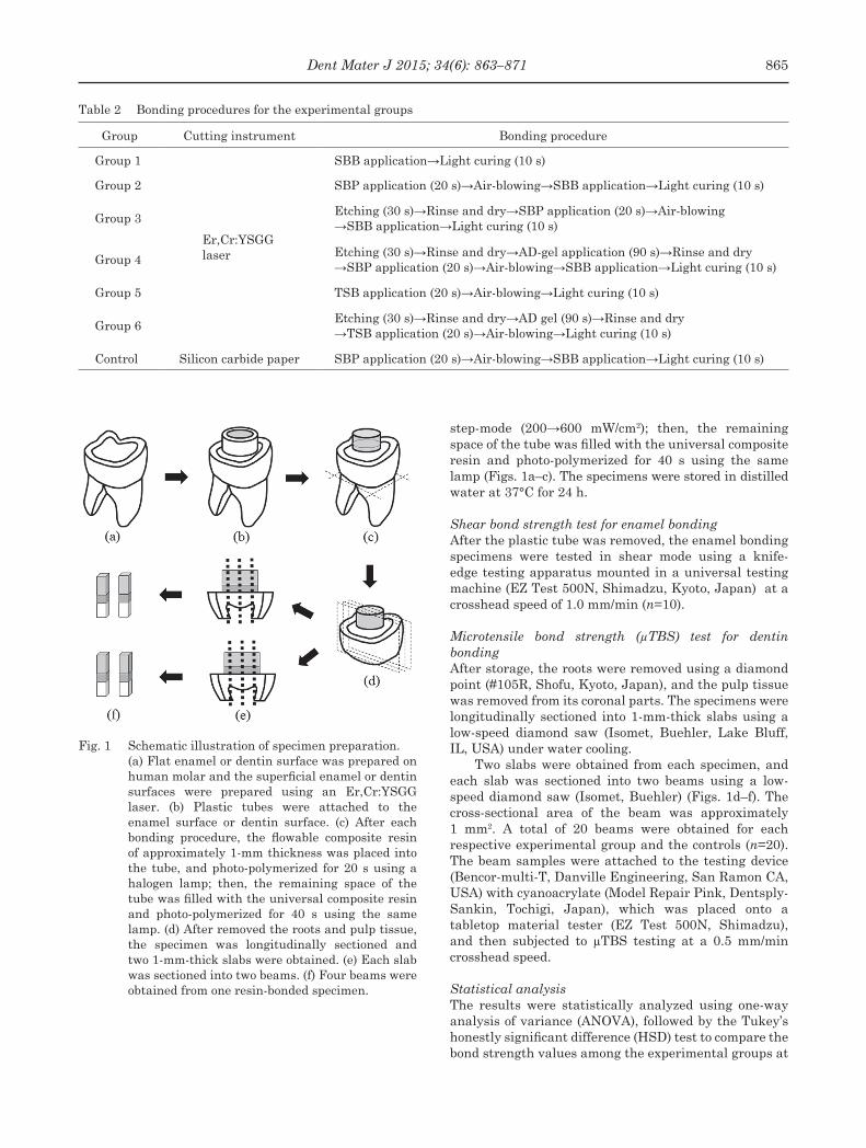
Table 2 Bonding procedures for the experimental groups
Group Cutting instrument Bonding procedure
Group 1
Er,Cr:YSGGlaser
SBB application→Light curing (10 s)
Group 2 SBP application (20 s)→Air-blowing→SBB application→Light curing (10 s)
Group 3Etching (30 s)→Rinse and dry→SBP application (20 s)→Air-blowing→SBB application→Light curing (10 s)
Group 4Etching (30 s)→Rinse and dry→AD-gel application (90 s)→Rinse and dry→SBP application (20 s)→Air-blowing→SBB application→Light curing (10 s)
Group 5 TSB application (20 s)→Air-blowing→Light curing (10 s)
Group 6Etching (30 s)→Rinse and dry→AD gel (90 s)→Rinse and dry→TSB application (20 s)→Air-blowing→Light curing (10 s)
Control Silicon carbide paper SBP application (20 s)→Air-blowing→SBB application→Light curing (10 s)
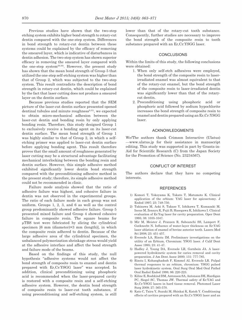
Fig. 1 Schematic illustration of specimen preparation. (a) Flat enamel or dentin surface was prepared on
human molar and the superficial enamel or dentin surfaces were prepared using an Er,Cr:YSGG laser. (b) Plastic tubes were attached to the enamel surface or dentin surface. (c) After each bonding procedure, the flowable composite resin of approximately 1-mm thickness was placed into the tube, and photo-polymerized for 20 s using a halogen lamp; then, the remaining space of the tube was filled with the universal composite resin and photo-polymerized for 40 s using the same lamp. (d) After removed the roots and pulp tissue, the specimen was longitudinally sectioned and two 1-mm-thick slabs were obtained. (e) Each slab was sectioned into two beams. (f) Four beams were obtained from one resin-bonded specimen.
step-mode (200→600 mW/cm2); then, the remaining space of the tube was filled with the universal composite resin and photo-polymerized for 40 s using the same lamp (Figs. 1a–c). The specimens were stored in distilled water at 37°C for 24 h.
Shear bond strength test for enamel bondingAfter the plastic tube was removed, the enamel bonding specimens were tested in shear mode using a knife-edge testing apparatus mounted in a universal testing machine (EZ Test 500N, Shimadzu, Kyoto, Japan) at a crosshead speed of 1.0 mm/min (n=10).
Microtensile bond strength (μTBS) test for dentin bondingAfter storage, the roots were removed using a diamond point (#105R, Shofu, Kyoto, Japan), and the pulp tissue was removed from its coronal parts. The specimens were longitudinally sectioned into 1-mm-thick slabs using a low-speed diamond saw (Isomet, Buehler, Lake Bluff, IL, USA) under water cooling.
Two slabs were obtained from each specimen, and each slab was sectioned into two beams using a low-speed diamond saw (Isomet, Buehler) (Figs. 1d–f). The cross-sectional area of the beam was approximately 1 mm2. A total of 20 beams were obtained for each respective experimental group and the controls (n=20). The beam samples were attached to the testing device (Bencor-multi-T, Danville Engineering, San Ramon CA, USA) with cyanoacrylate (Model Repair Pink, Dentsply-Sankin, Tochigi, Japan), which was placed onto a tabletop material tester (EZ Test 500N, Shimadzu), and then subjected to μTBS testing at a 0.5 mm/min crosshead speed.
Statistical analysisThe results were statistically analyzed using one-way analysis of variance (ANOVA), followed by the Tukey’s honestly significant difference (HSD) test to compare the bond strength values among the experimental groups at
865Dent Mater J 2015; 34(6): 863–871

Fig. 2 Shear bond strength of composite resin to laser-cut enamel.
Values with the same alphabets indicate no significant differences (p>0.05).
Fig. 3 Microtensile bond strength of composite resin to laser-cut dentin.
Values with the same alphabets indicate no significant differences (p>0.05).
Table 3 Criteria for judging each failure mode
Failure mode Definition
Adhesive failure Failure occurred entirely within the adhesive area
Cohesive failure in resin composite Failure occurred exclusively within the resin composite area
Cohesive failure in enamel or dentin Failure occurred exclusively within the enamel or dentin area
Mixed failureFailure continued from the adhesive into either the resin composite or enamel and dentin area
a 95% confidence level.
Failure mode analysis The fracture surfaces of the specimens were examined using a stereomicroscope (Leica EZ4D, Microsystems CMS, Wetzlar, Germany) at 35× magnification to determine the failure mode based on the classification of failure modes shown in Table 3.
Scanning electron microscopy (SEM) Several representative samples were selected from each group for further examination of the fracture surfaces using an SEM (S-800, Hitachi, Chiyoda, Tokyo, Japan). The specimens were sputter-coated with palladium and platinum and viewed using an acceleration voltage of 15 kV.
RESULTS
Shear bond strength of composite resin to enamelThe shear bond strength of each excremental group to the enamel prepared with Er,Cr:YSGG laser are shown in Fig. 2. Statistical analysis using one-way ANOVA detected significant differences among the experimental groups and control for enamel bonding. The Tukey’s HSD test revealed that the bond strength of Groups 3
and 4 was significantly higher than that of the other groups (p<0.05) and that of Group 5 was significantly lower than that of other groups (p<0.01).
μTBS of composite resin to dentinThe μTBS of each excremental group to the dentin prepared with Er,Cr:YSGG laser are shown in Fig. 3. Statistical analysis using one-way ANOVA detected significant differences among the experimental groups and control for dentin bonding. The Tukey’s HSD test revealed that the bond strength of Groups 1 and 2 was significantly lower than that of Groups 3, 4, and 6 (p<0.05) and that of Group 5 was significantly lower than that of Groups 3 and 4 (p<0.05). The control group showed higher bond strength compared to the experimental groups (Groups 1–6; p<0.05). No significant differences between Groups 5 and 6 and among Groups 1, 2, and 5 and Groups 3, 4 and 6 were observed.
Failure mode analysis of enamel bondingThe results of failure mode analysis of enamel bonding groups after shear bond strength testing are summarized in Table 4. In the control group, 70% of adhesive failure and 30% of mixed failure were accounted, and cohesive failure of enamel was not detected. On the other hand, cohesive failure of enamel was predominantly observed
866 Dent Mater J 2015; 34(6): 863–871

Fig. 4 Representative SEM pictures of Group 1, in which failure mode of the specimen was evaluated as mixed failure.
The enamel was fractured into small pieces on the surface of the resin rod. Magnification: A, ×20; B, ×100; C, ×1,000.
Fig. 5 Representative SEM pictures of Group 3, in which failure mode of the specimen was evaluated as cohesive failure in enamel.
Most of the fractured surface was covered with enamel. Magnification: A, ×20; B, ×100; C, ×1,000.
Table 4 Failure mode analysis of the experimental groups for enamel bonding
Failure modeGroups
1 2 3 4 5 6 Control
Adhesive failure (%) 0 0 0 0 40 0 70
Cohesive failure in enamel (%) 100 90 90 100 60 100 0
Mixed failure (%) 0 10 10 0 0 0 30
in the experimental groups, and only a few specimens showed adhesive failure or mixed failure. Representative SEM images of debonded surfaces are shown in Figs. 4 and 5.
Figure 4 shows representative SEM images of Group 1, which was determined to be a mixed failure. Small fragments of enamel were observed on the surface of the resin rod. Figure 5 shows representative SEM images of Group 3, which was determined to be a
cohesive failure in enamel. Most of the fractured surface was covered with enamel.
Failure mode analysis of dentin bondingThe results of failure mode analysis of dentin bonding groups after μTBS testing are summarized in Table 5. In total, the occurrence ratio of adhesive failure was the highest (83/140×100=59.3%) and that of mixed failure was 22.9% (32/140×100) and cohesive failure
867Dent Mater J 2015; 34(6): 863–871

Fig. 6 Representative SEM pictures of Group 3, in which failure mode of the specimen was evaluated as adhesive failure.
Both the surface of composite resin and dentin rod were mostly covered with an adhesive resin layer. Magnification: A and C, ×80; B and D, ×1,000.
Fig. 7 Representative SEM pictures of Group 1, in which failure mode of the specimens was evaluated as mixed failure.
A thin detached dentin layer was observed on the surface of composite resin rod and a thin detached composite resin layer was detected on the surface of dentin rod. Magnification: A and C, ×80; B and D, ×1,000.
Table 5 Failure mode analysis of the experimental groups for dentin bonding
Failure modeGroups
1 2 3 4 5 6 Control Total
Adhesive failure (%) 75 80 65 35 30 60 70 59.3
Cohesive failure in resin composite (%) 10 5 30 40 5 15 15 17.1
Cohesive failure in dentin (%) 0 0 0 0 0 0 5 0.7
Mixed failure (%) 15 15 5 25 65 25 10 22.8
in composite resin was 17.1% (24/140×100); cohesive failure in dentin was only observed in the controls and was the lowest (1/140×100=0.7%). Groups 1, 2, 3, and 6 as well as the control group predominantly showed adhesive failure. Groups 4 and 5 showed cohesive failure and mixed failure in composite resin, respectively.
Representative SEM images of adhesive failure and mixed failure are shown in Figs. 6 and 7. Representative images of Group 3, which was evaluated as adhesive failure, are shown in Fig. 6. Based on failure mode analysis results by stereomicroscopy, this specimen would be evaluated as adhesive failure. However, these
SEM images exhibited that both the surface of composite resin and dentin rod were almost covered with adhesive resin layer. Accordingly, failure in this specimen occurred within the adhesive layer.
Figure 7 presents representative SEM images of Group 1, which was classified as a mixed failure. A thin detached dentin layer was observed on the surface of the composite resin rod, and a thin detached composite resin layer was observed on the surface of the dentin rod.
868 Dent Mater J 2015; 34(6): 863–871

DISCUSSION
Enamel bondingSeveral studies have examined the bond strength of composite resin to enamel prepared with Er:YAG laser15-18). Owing to production of enamel microtags by laser cutting, steps of etching and rinsing could be omitted for the enamel in the etch and rinse adhesive system15,19). However, other studies have reported that etching with phosphoric acid was effective in increasing the bond strength of the composite resin to enamel prepared with Er:YAG laser16,17). On the other hand, enamel tags produced by laser cutting could be useful for enamel bonding because they facilitate mechanical interlocking.
Recently, a one- or two-step self-etching adhesive system has been widely used in clinics. Kameyama et al.18) compared the bond strengths of four types of self-etching adhesive systems to enamel prepared with Er:YAG laser. No significant difference between the bond strength of the one-step type and that of two-step type was observed as well as between the bond strength to laser-irradiated enamel and that to non-irradiated enamel on all adhesive system. Thus, opinions on the effect of an adhesive system on the bond strength of composite resin to laser-irradiated enamel extensively vary and no consensus has been achieved.
The present study revealed how experimental adhesive systems affected the bond strength of composite resin to laser-irradiated enamel. The results of this study have demonstrated that Groups 3 and 4 have significantly higher mean bond strength compared with the other groups including the controls, and the mean bond strength of Group 5 was markedly lower than that of the other groups. That is, the pretreatment using phosphoric acid etching or combination of phosphoric acid etching and sodium hypochlorite would contribute to increasing the bond strength of the self-etching systems to laser-irradiated enamel. Esteves-Oliveira et al.20) reported that the bond strength of the two-step self-etching system to rotary-cut tooth substance was the highest, followed by the Er:YAG laser irradiated tooth substance, and that of the Er,Cr:YSGG laser-irradiated tooth substance was markedly lower than that of the rotary-cut tooth substance. However, in this study, Er,Cr:YSGG laser cutting of enamel did not affect the bond strength of the two-step self-etching system to enamel based on the observation that Group 2 exhibited a slightly higher bond strength compared with that of the controls.
Furthermore, based on the finding that the enamel bond strength of experimental group used only bonding agent (Group 1) was similar to that of the controls, microtags produced by laser cutting could have contributed to the increase in enamel bond strength of the composite resin. In addition, the results of this study showed that the bond strength of the composite resin to laser-irradiated enamel was almost equivalent to that of the rotary-cut enamel when self-etch adhesives were employed. However, the laser-irradiated enamel surface
may have been weakened by thermal action because cohesive failure in the enamel was mostly observed after shear bond testing. Based on this result, adhesive degradation over time should be monitored.
Dentin bonding Several studies have shown that Er:YAG laser irradiation causes a decrease in dentin bond strength of composite resins21,22). Tachibana et al.23) compared the bond strengths of the two-step self-etching adhesive system to sound or caries-affected dentin prepared with various cutting apparatuses containing Er:YAG laser, which showed that the bond strength of the adhesive system to both laser-irradiated dentins were markedly lower than that of dentin prepared with other cutting apparatuses. The properties of the dentin surface irradiated by Er:YAG laser, which showed that heat induces denaturation of the layer as well as cracks on the laser-irradiated dentin, have been extensively studied24). These structural defects yielded by laser irradiation were considered as factors9,24-28) that could decrease the bond strength of composite resin to laser-irradiated dentin. In our previous study, we employed azan staining and light microscopy of dentin sections prepared by Er,Cr:YSGG laser and observed that the thickness of the heat-denatured layer was 13–16 μm. We also observed that this layer was eliminated when both phosphoric acid and sodium hypochlorite were applied on the laser-cut dentin; however, the application of only phosphoric acid could not elinimate the heat-denatured layer8).
The results of the present study demonstrated that Groups 3, 4, and 6, in which only phosphoric acid or both phosphoric acid and sodium hypochlorite were applied as a pretreatment, showed markedly higher dentin bond strength compared with Groups 1 and 2, in which pretreatments were not applied. Accordingly, pretreatment using phosphoric acid and sodium hypochlorite was effective in improving the bond strength of SE Bond to laser-cut dentin. In addition, Group 3 that was treated only with phosphoric acid exhibited similar dentin bond strength to that of Group 4, which was subjected to both phosphoric acid and sodium hypochlorite. These findings suggest that the presence of a heat-denatured layer does not necessarily disturb dentin bonding.
Our previous SEM observation of the laser-cut dentin surface subjected to both phosphoric acid and sodium hypochlorite showed that the dentin tubules were widely open and the intertubular matrices protruded8). Therefore, the application of sodium hypochlorite after acid etching possibly dissolved not only the collagen of decalcified dentin in the heat-denatured layer but also that of the sound dentin exactly underneath the heat-denatured layer. Therefore, excessive pretreatment of the laser-cut dentin could induce the weakening of the hybrid layer at the adhesive interface. Based on handling time and adhesive longevity, pretreatment using phosphoric acid etching should be recommended for laser-cut dentin.
869Dent Mater J 2015; 34(6): 863–871
Previous studies have shown that the two-step etching system exhibits higher bond strength to rotary-cut dentin compared with the one-step system. Differences in bond strength to rotary-cut dentin between these systems could be explained by the efficacy of removing the smeared layer, which is indicative of disturbances in dentin adhesion. The two-step system has shown superior efficacy in removing the smeared layer compared with the one-step system29,30). However, the present study has shown that the mean bond strength of Group 5 that utilized the one-step self-etching system was higher than that of Group 2, which was subjected to the two-step system. This result contradicts the description of bond strength in rotary-cut dentin, which could be explained by the fact that laser cutting does not produce a smeared layer on the dentin surface.
Because previous studies reported that the SEM picture of the laser-cut dentin surface presented opened dentinal tubules and minute roughness8,31), we expected to obtain micro-mechanical adhesion between the laser-cut dentin and bonding resin by only applying bonding resin. Therefore, this study designed Group 1 to exclusively receive a bonding agent on its laser-cut dentin surface. The mean bond strength of Group 1 was highly similar to that of Group 2, in which a self-etching primer was applied to laser-cut dentin surface before applying bonding agent. This result therefore proves that the small amount of roughness generated by laser cutting may be a structural advantage facilitating mechanical interlocking between the bonding resin and dentin surface. However, this simple adhesive method exhibited significantly lower dentin bond strength compared with the preconditioning adhesive method in the present study; therefore, its simple adhesive method could not be recommended in clinic.
Failure mode analysis showed that the ratio of adhesive failure was highest, and cohesive failure in dentin was not observed in the experimental groups. The ratio of each failure mode in each group was not uniform. Groups 1, 2, 3, and 6 as well as the control group predominantly showed adhesive failure. Group 5 presented mixed failure and Group 4 showed cohesive failure in composite resin. The square beams for μTBS test were fabricated by slicing the cylindrical specimen [6 mm (diameter)×3 mm (length)], in which the composite resin adhered to dentin. Because of the large adhesive area of the cylindrical specimen, the unbalanced polymerization shrinkage stress would yield at the adhesive interface and affect the bond strength and failure mode of the beams.
Based on the findings of this study, the null hypothesis “adhesive systems would not affect the bond strength of composite resin to enamel and dentin prepared with Er,Cr:YSGG laser” was accepted. In addition, clinical preconditioning using phosphoric acid is recommended when the laser-prepared cavity is restored with a composite resin and a self-etching adhesive system. However, the dentin bond strength of composite resin to laser-cut tooth substance, if using preconditioning and self-etching system, is still
lower than that of the rotary-cut tooth substance. Consequently, further studies are necessary to improve the bond strength of the composite resin to tooth substance prepared with an Er,Cr:YSGG laser.
CONCLUSIONS
Within the limits of this study, the following conclusions were obtained:
1. When only self-etch adhesives were employed, the bond strength of the composite resin to laser-irradiated enamel was almost equivalent to that of the rotary-cut enamel, but the bond strength of the composite resin to laser-irradiated dentin was significantly lower than that of the rotary-cut dentin.
2. Preconditioning using phosphoric acid or phosphoric acid followed by sodium hypochlorite increases the bond strength of composite resin to enamel and dentin prepared using an Er,Cr:YSGG laser.
ACKNOWLEDGMENTS
We/The authors thank Crimson Interactive (Ulatus) —www.ulatus.jp for their assistance in manuscript editing. This study was supported in part by Grants-in-Aid for Scientific Research (C) from the Japan Society for the Promotion of Science (No. 23234567).
CONFLICT OF INTEREST
The authors declare that they have no competing interests.
REFERENCES
1) Komori T, Yokoyama K, Takato T, Matumoto K. Clinical application of the erbium: YAG laser for apicoectomy. J Endod 1997; 23: 748-750.
2) Takizawa M, Aoki S, Takase Y, Ishikawa T, Kumazaki M, Inoue M, Zennyu K, Fujii B, Hasegawa K, Ishikawa I. Clinical evaluation of Er:Yag laser for cavity preparation. Oper Dent 1995; 38: 1035-1047.
3) Mir M, Meister J, Franzen R, Sabounchi SS, Lampert F, Gutknecht N. Influence of water-layer thickness on Er:YAG laser ablation of enamel of bovine anterior teeth. Lasers Med Sci 2008; 23: 451-457.
4) Eversole LA, Rizoiu IM. Preliminary investigations on the utility of an Erbium, Chromium: YSGG laser. J Calif Dent Assoc 1995; 23: 41-47.
5) Hadley J, Young DA, Eversole LR, Gornbein JA. A laser-powered hydrokinetic system for caries removal and cavity preparation. J Am Dent Assoc 2000; 131: 777-785.
6) Rizoiu I, Kohanghadosh F, Kimmel AI, Eversole LR. Pulpal thermal responses to an rebium, chromium: YSGG pulsed laser hydrokinetic system. Oral Surg Oral Med Oral Pathol Oral Radiol Endod 1998; 86: 220-223.
7) Kilinc E, Roshkind DM, Antonson SA, Antonson DE, Hardigan PC, Siegel SC, Thomas JW. Thermal safety of Er:YAG and Er,Cr:YSGG lasers in hard tissue removal. Photomed Laser Surg 2009; 27: 565-570.
8) Kato C, Taira Y, Suzuki M, Shinkai K, Katoh Y. Conditioning effects of cavities prepared with an Er,Cr:YSGG laser and an
870 Dent Mater J 2015; 34(6): 863–871

air-turbine. Odontology 2012; 100: 164-171.9) Fujitani M. Adhesive properties of resin bonding system
to Er:YAG or CO2 Lased Dentin —Does the resin bonding system exhibit its full adhesive performance to lased dentin? J Jpn Soc Laser Dent 2006; 17: 74-80.
10) Hossain M, Nakamura Y, Yamada Y, Kimura Y, Matsumoto N, Matsumoto K. Effects of Er,Cr:YSGG laser irradiation in human enamel and dentin: ablation and morphological studies. J Clin Laser Med Surg 1999; 17: 155-159.
11) Harashima T, Kinoshita J, Kimura Y, Brugnera A, Zanin F, Peccora JD, Matsumoto K. Morphological comparative study on ablation of dental hard tissues at cavity preparation by Er:YAG and Er,Cr:YSGG lasers. Photomed Laser Surg 2005; 23: 52-55.
12) Iwata A, Hayasihara H, Yasuo K, Onda K, Zennyuu K, Fukui M, Tanaka Y, Yoshikawa K, Yamamoto K. Study on dental hard tissues irradiated by an Er, Cr: YSGG laser: Surface characteristics and cutting efficiency. Oper Dent 2010; 53: 147-158.
13) Gholami GA, Fekrazad R, Esmaiel-Nejad A, Kalhori KA. An evaluation of the occluding effects of Er;Cr:YSGG, Nd:YAG, CO2 and diode lasers on dentinal tubules: a scanning electron microscope in vitro study. Photomed Laser Surg 2011; 29: 115-121.
14) Moretto SG, Azambuja N Jr. Arana-Chavez VE, Reus AF, Giannini M, Eduardo Cde P, De Freitas PM. Effects of ultramorphological changes on adhesion to lased dentin-Scanning electron microscopy and transmission electron microscopy analysis. Microsc Res Tech 2011; 74: 720-726.
15) Martines-Insua A. Dominguez LDS, Rivera FG, Santanapenin UA. Difference in bonding to acid-etched or Er:YAG-laser-treated enamel and dentin surface. J Prosthet Dent 2000; 84: 280-288.
16) De Munck J, Van Meerbeek B, Yudhira R, Lambrechts P, Vanherle G. Micro-tensile bond strength of two adhesive to Erbium: YAG-lased vs. bur-cut enamel and dentin. Eur J Oral Sci 2002; 110: 322-329.
17) Sasaki LH, Lobo PDC, Moriyama Y, Watanabe I, Villaverde AB, Tanaka CS, Moriyama EH, Brugnera Jr. A. Tensile bond strength and SEM analysis of enamel etched with Er:YAG laser and phosphoric acid: a comparative study in vitro. Braz Dent J 2008; 19: 57-61.
18) Kameyama A, Kato J, Aizwa K, Suemori T, Nakazawa Y, Ogata T, Hirai Y. Tensile bond strength of one-step self-etch adhesives to Er:YAG laser-irradiated and non irradiated enamel. Dent Mater 2008; 27: 386-391.
19) Attrill DC, Farrar SR, King TA, Dickinson MR, Davies RM, Blinkhorn AS. Er:YAG (=2.94 μm) laser etching of dental enamel as an alternative to acid etching. Laser Med Sci 2000; 15: 154-161.
20) Esteves-Oliveira M, Zezell DM, Apel C, Turbino ML, Aranha AC, Eduardo Cde P, Gutknecht N. Bond strength of self-etching primer to bur cut, Er,Cr:YSGG, and Er:YAG lased dental surfaces. Photomed Laser Surg 2007; 25: 373-380.
21) Sakakibara Y, Ishimaru K, Takamizu M. A Study on bond strength to dentin irradiated by Erbium:YAG Laser. Oper Dent 1998; 41: 207-219.
22) Kameyama A, Amagai T, Takizawa M, Hirai Y. Resin bonding to Er:YAG laser-irradiated dentin: A review. Shikwa Gakuho 2003; 103: 115-127.
23) Tachibana A, Marques MM, Soler JM, Matos AB. Erbium, chromium: yttrium scandium gallium garnet laser for caries removal: influence on bonding of a self-etching adhesive system. Lasers Med Sci 2008; 23: 435-441.
24) Fujitani M, Harima H, Shintani H. Does Er:YAG or CO2 laser ablation of dentin affect the adhesive properties of resin bonding systems? Excerpta Medica International Congress Series 2003; 1248: 193-199.
25) Shintani H, Fujitani M, Harima H, Kurosaki N, Hiri Y, Toda M, Okazaki M, Tanaka T. Studies on laser applications to dental hard tissues. Dentistry in Japan 2003; 39: 193(Abstract).
26) Sintani H, Fujitani M, Harima T, Kurosaki N, Hirai Y, Toda T, Okazaki M, Takata T. Studies on laser applications to dental hard tissues. JJDS 2003; 22: 62-69.
27) Iwata N, Iwamoto K, Fujito Y, Yoshikawa K, Inoue M, Inoue M. Study on dental hard tissues irradiated by an Er:YAG laser: Part 2 A morphological study of altered layer induction by Er:YAG laser irradiation. Oper Dent 2001; 44: 810-816.
28) Iwata N. Study on dental hard tissues irradiated by an Er:YAG laser. Oper Dent 2002; 45: 147-158.
29) Burrow MF, Kitasako Y, Thomas CD, Tagami J. Comparison of enamel and dentin microshear bond strengths of a two-step self-etching priming system with five all-in-one systems. Oper Dent 2008; 33: 456-460.
30) Chopra V, Sharma H, Prasad SD. A comparative evaluation of the bonding efficacy of two-step vs all-in-one bonding agents —An in-vitro study. J Conserv Dent 2009; 12: 101-104.
31) Lin S, Pan D, Lin Q, Yin S, Chen D, Yu L, Lin Z. Evaluation of phase, microstructure and composition of human dentine after Er,Cr:YSGG laser irradiation. J Nanosci Nanotechnol 2011; 11: 2421-2426.
871Dent Mater J 2015; 34(6): 863–871