bone degenerative disorders
DESCRIPTION
TRANSCRIPT

BONE & DEGENERATIVE DISORDERS
DEPARTMENT OF PATHOLOGYDEPARTMENT OF PATHOLOGY
www.freelivedoctor.com
OSTEOPOROSIS
Decreased Decreased bbone mass ( one mass ( oosteopenia.)steopenia.) r resulting in esulting in thin,fragile bones that are susceptible to fracture.thin,fragile bones that are susceptible to fracture.
Most common bone disorder in Most common bone disorder in U.S.A.(about 15 U.S.A.(about 15 million individuals have primary type)million individuals have primary type)
Most commonly occurs in postmenopausal Most commonly occurs in postmenopausal Caucasian women and the elderly.Caucasian women and the elderly.
www.freelivedoctor.com

PrimaryPrimary
PostmenopausalPostmenopausal
SenileSenile
SecondarySecondary
Endocrine Endocrine disordersdisorders
Hyperparathyroi-Hyperparathyroi-dismdism
Hypo-Hypo-hyperthyroidismhyperthyroidism
HypogonadismHypogonadism
Pituitary tumorsPituitary tumors
Diabetes, type 1Diabetes, type 1
Addison diseaseAddison disease
www.freelivedoctor.com
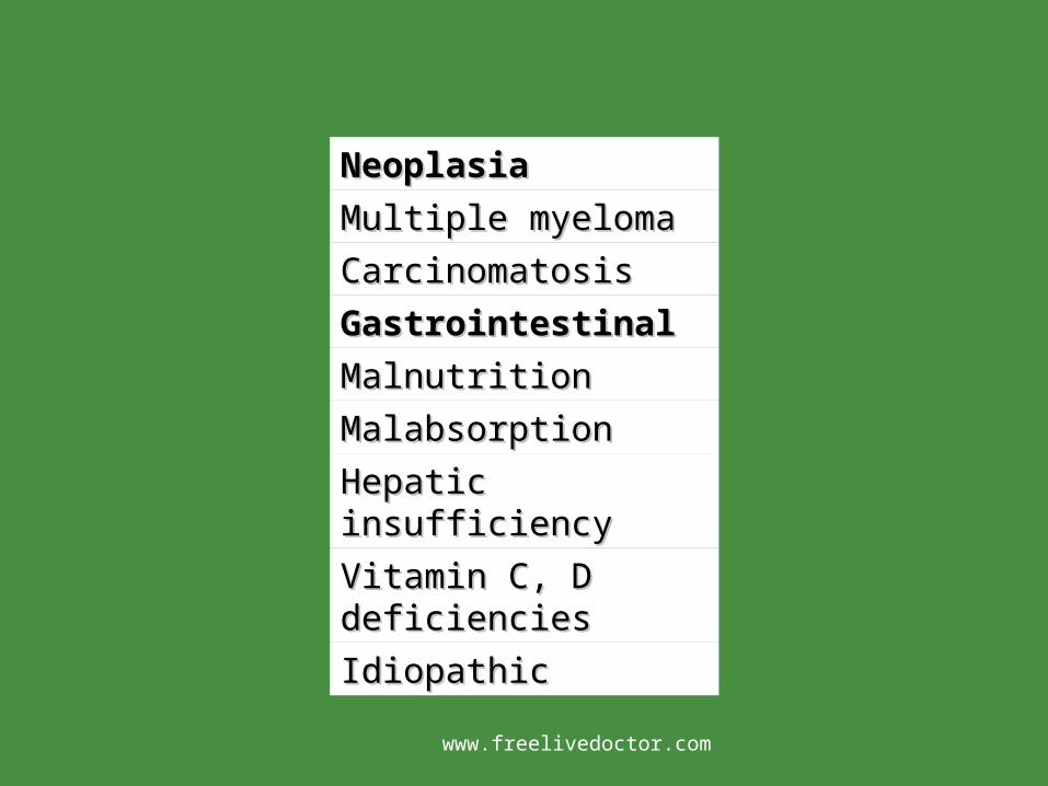
NeoplasiaNeoplasia
Multiple myelomaMultiple myeloma
CarcinomatosisCarcinomatosis
GastrointestinalGastrointestinal
MalnutritionMalnutrition
MalabsorptionMalabsorption
Hepatic insufficiencyHepatic insufficiency
Vitamin C, D Vitamin C, D deficienciesdeficiencies
IdiopathicIdiopathic
www.freelivedoctor.com

Rheumatologic Rheumatologic diseasedisease
DrugsDrugs
AnticoagulantsAnticoagulants
ChemotherapyChemotherapy
CorticosteroidsCorticosteroids
AnticonvulsantsAnticonvulsants
AlcoholAlcohol
MiscellaneousMiscellaneous
Osteogenesis Osteogenesis imperfectaimperfecta
ImmobilizationImmobilization
Pulmonary diseasePulmonary disease
HomocystinuriaHomocystinuria
AnemiaAnemiawww.freelivedoctor.com

www.freelivedoctor.com
OSTEOPOROSIS(cont.)
Patients may experience bone pain and Patients may experience bone pain and fractures.fractures.
Weight bearing bones are predisposed to Weight bearing bones are predisposed to fracturesfractures
Vertebrae ( compression fractures)Vertebrae ( compression fractures) Femoral neck ( Femoral neck ( hhip fracture)ip fracture) Distal radius ( Colles fracture)Distal radius ( Colles fracture) Pulmonary embolizationPulmonary embolization
www.freelivedoctor.com

www.freelivedoctor.com
OSTEOPOROSIS(cont.) Loss of heightLoss of height,, kyphos kyphoscoliosis, lordosiscoliosis, lordosis X-rays: generalized radiolucency of boneX-rays: generalized radiolucency of bone(( osteopenia osteopenia)) Dual Energy X Ray Absorptiometry ( DEXA.)Dual Energy X Ray Absorptiometry ( DEXA.) NORMAL SERUM CALCIUM,PHOSPHORUS,and NORMAL SERUM CALCIUM,PHOSPHORUS,and
ALKALINE PHOSPHATASE.ALKALINE PHOSPHATASE. Micro: Micro: tthinned cortical and trabecular bone hinned cortical and trabecular bone Treatment : Treatment : eestrogen replacement therapy , strogen replacement therapy , wweight bearing eight bearing
exercise,Calcium and Vitamin Dexercise,Calcium and Vitamin D,, Biphosphonate ( Biphosphonate (AlendroAlendro nate) nate) ,, Calcitonin. Calcitonin.
www.freelivedoctor.com

www.freelivedoctor.com

www.freelivedoctor.com

www.freelivedoctor.com

www.freelivedoctor.com

OSTEOMALACIA and RICKETS Both diseases are characterized by Both diseases are characterized by
decreased mineralization of newly formed decreased mineralization of newly formed bone, usually caused by deficiency or bone, usually caused by deficiency or abnormal metabolism of vitamin D. abnormal metabolism of vitamin D.
www.freelivedoctor.com

www.freelivedoctor.com
OSTEOMALACIA and RICKETS. Etiology: Etiology: ddietary deficiency of Vitamin Dietary deficiency of Vitamin D Intestinal malabsorptionIntestinal malabsorption(( bilis,pancreatic bilis,pancreatic
insuff.,celiac sprue,regional enteritis)insuff.,celiac sprue,regional enteritis) Lack of sunlightLack of sunlight Renal and liver diseaseRenal and liver disease Chronic use of antacids(Al OH binds to P)Chronic use of antacids(Al OH binds to P) Drugs(incr.rate of degradation of sterols:phe Drugs(incr.rate of degradation of sterols:phe
nytoin,phenobarbital,rifampin) nytoin,phenobarbital,rifampin)
www.freelivedoctor.com
RICKETS ( Children)
Prior to closure of the epiphysesPrior to closure of the epiphyses Both remodeled bone and bone formed at Both remodeled bone and bone formed at
the epypheseal growth plate are the epypheseal growth plate are undermineralizedundermineralized
Enchondral bone fromation is affected Enchondral bone fromation is affected leading to skeletal deformities.leading to skeletal deformities.
www.freelivedoctor.com
RICKETS (children) Craniotabes and frontal bossing: skull deformities.Craniotabes and frontal bossing: skull deformities. Rachitic rosary: deformity of the chest wall as a result of Rachitic rosary: deformity of the chest wall as a result of
an overgrowth of cartilage at the costochondral junctionan overgrowth of cartilage at the costochondral junction Pectus carinatum ( pigeon breast deformiy); outward Pectus carinatum ( pigeon breast deformiy); outward
protrusion of the sternum.protrusion of the sternum. Lumbar lordosis: Lumbar lordosis: sspinal curvaturepinal curvature Bowing of the legs: Bowing of the legs: ccurvature of femur, tibia due to weight urvature of femur, tibia due to weight
bearingbearing Fractures may also occur.Fractures may also occur.
www.freelivedoctor.com
OSTEOMALACIA. ( Adults)
Impaired mineralization of the osteoid Impaired mineralization of the osteoid matrix results in thin, fragile bones that are matrix results in thin, fragile bones that are susceptible to fracture.susceptible to fracture.
www.freelivedoctor.com

OSTEOMALACIA ( Adults)
Clinical Clinical ppresentation:resentation:
----Bone painBone pain
----Fractures of the vertebrae, hips and wrist.Fractures of the vertebrae, hips and wrist.
----X-rays X-rays :: diffuse radiolucency of bone diffuse radiolucency of bone(osteo(osteo
penia )penia )
--Lab: l--Lab: low serum calcium and phosphorusow serum calcium and phosphorus and/or and/or
raisedraised alkaline phosphatase alkaline phosphatase
www.freelivedoctor.com

BONE INFECTIONS.OSTEOMYELITIS,TB, Syphilis. PPYOGENICYOGENIC O OSTEOMYELITISSTEOMYELITIS.. Routes of infection: Routes of infection: Hematogenous spread : Most Hematogenous spread : Most common common
seeding of bone after bacteremia, seeding of bone after bacteremia, commonly affects the metaphysis.commonly affects the metaphysis.
www.freelivedoctor.com

www.freelivedoctor.com
www.freelivedoctor.com
OSTEOMYELITIS (cont.)
Routes of Infection:Routes of Infection: Hematogenous: from skin pustule,infected gums/teeth,IV Hematogenous: from skin pustule,infected gums/teeth,IV
puncture, UTIs, urologic procedurespuncture, UTIs, urologic proceduresends of long bonesends of long bones Direct inoculationDirect inoculation Spread from an adjacent site of infectionSpread from an adjacent site of infection Microbiology:Syaphylococcus aureus, (Microbiology:Syaphylococcus aureus, (mmost common), ost common),
Escherichia coli, Streptococci,Gonococci, Haemophilus Escherichia coli, Streptococci,Gonococci, Haemophilus influenzae, Salmonella (common in sickle cell disease) influenzae, Salmonella (common in sickle cell disease) Pseudomonas ( common in intravenous drug abusers and Pseudomonas ( common in intravenous drug abusers and diabeticsdiabetics))
www.freelivedoctor.com

OSTEOMYELITIS (cont.)
Clinical features:Clinical features: f feverever, localized pain, , localized pain, erythema and swelling, leukocytosiserythema and swelling, leukocytosis
X-ray : X-ray : mmay be normal for up to 2 weeksay be normal for up to 2 weeks,,
but it mbut it may initially show periosteal elevationay initially show periosteal elevation
Later: lLater: lytic focus with surrounding sclerosis.ytic focus with surrounding sclerosis.
www.freelivedoctor.com
OSTEOMYELITIS (cont.)
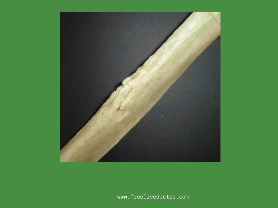
Pathology: Pathology: ssuppurative uppurative iinflammationnflammation Vascular insufficiencyVascular insufficiency Ischemic necrosis of boneIschemic necrosis of bone Sequestrum: necrotic bone.Sequestrum: necrotic bone. Involucrum: Involucrum: nnew bone formation that ew bone formation that
surrounds the sequestrumsurrounds the sequestrum
www.freelivedoctor.com
OSTEOMYELITIS(cont.)
Diagnosis: Diagnosis: Blood culture.Blood culture. Bone biopsy and cultureBone biopsy and culture Treatment: Antibiotics:+ Treatment: Antibiotics:+ ssurgical drainage.urgical drainage.
www.freelivedoctor.com

www.freelivedoctor.com

www.freelivedoctor.com
OSTEOMYELITIS(cont.)
Complications:Complications: FractureFracture Intraosseous ( Brodie) abscess.Intraosseous ( Brodie) abscess. AmyloidosisAmyloidosis Sinus tract formationSinus tract formation Squamous cell Ca. Squamous cell Ca. oof the skin at the site of f the skin at the site of
persistent draining sinus tractpersistent draining sinus tract Osteogenic sarcoma ( rare. ) Osteogenic sarcoma ( rare. )
www.freelivedoctor.com
TB OSTEOMYELITIS.
Affected individuals : adolescents or young Affected individuals : adolescents or young adults, inmigrants.adults, inmigrants.
U.S.AU.S.A:: VVictims tend to be older, excepting ictims tend to be older, excepting IImmunosupressed. mmunosupressed.
Occurs in 1% of cases of TB.Occurs in 1% of cases of TB.
www.freelivedoctor.com

www.freelivedoctor.com

www.freelivedoctor.com

www.freelivedoctor.com
www.freelivedoctor.com

www.freelivedoctor.com

www.freelivedoctor.com

www.freelivedoctor.com

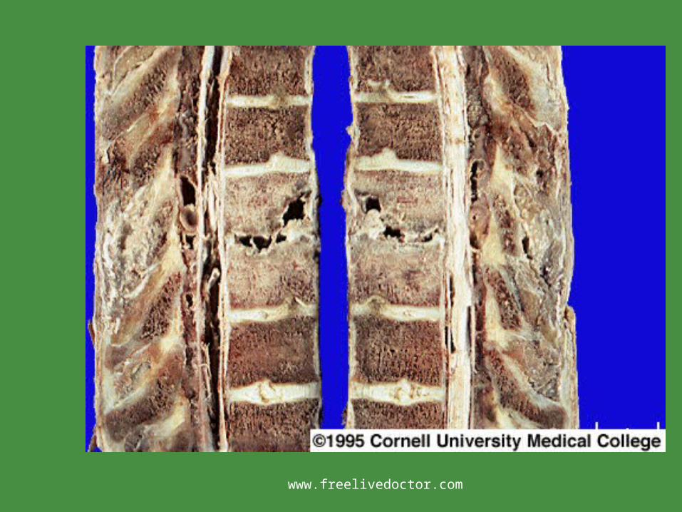
TB OSTEOMYELITIS(cont.)
Pain or tenderness, fever, night sweats, Pain or tenderness, fever, night sweats, weight loss.weight loss.
Caseating granulomas with extensive Caseating granulomas with extensive destruction of the bones.destruction of the bones.
Common site : ThoraCommon site : Thoraccic and lumbar ic and lumbar vertebrae ( Pottvertebrae ( Pott´s´s Disease) Disease)
www.freelivedoctor.com
TB OSTEOMYELITIS(cont.)
Complications.Complications. Vertebral compression fractureVertebral compression fracture Psoas abscessesPsoas abscesses AmyloidosisAmyloidosis
www.freelivedoctor.com
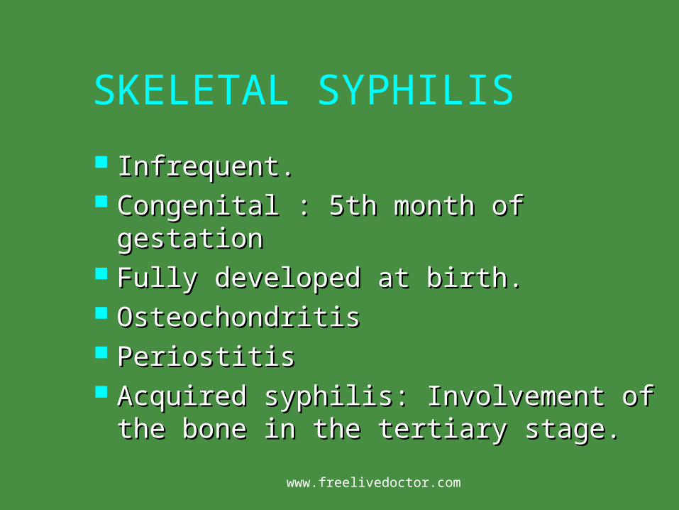
SKELETAL SYPHILIS
Infrequent.Infrequent. Congenital : 5th month of gestation Congenital : 5th month of gestation Fully developed at birth.Fully developed at birth. OsteochondritisOsteochondritis PeriostitisPeriostitis Acquired syphilis: Involvement of the bone Acquired syphilis: Involvement of the bone
in the tertiary stage.in the tertiary stage.
www.freelivedoctor.com

SKELETAL SYPHILIS(cont.)
2-5 years after the infection.2-5 years after the infection. Bones involved: Nose, palate, skull and Bones involved: Nose, palate, skull and
extremities.( Saber shin)extremities.( Saber shin) Histology: Edematous granulation tissue , Histology: Edematous granulation tissue ,
plasma cells and necrotic boneplasma cells and necrotic bone GummataGummata Silver stain for spirochetes.Silver stain for spirochetes.
www.freelivedoctor.com

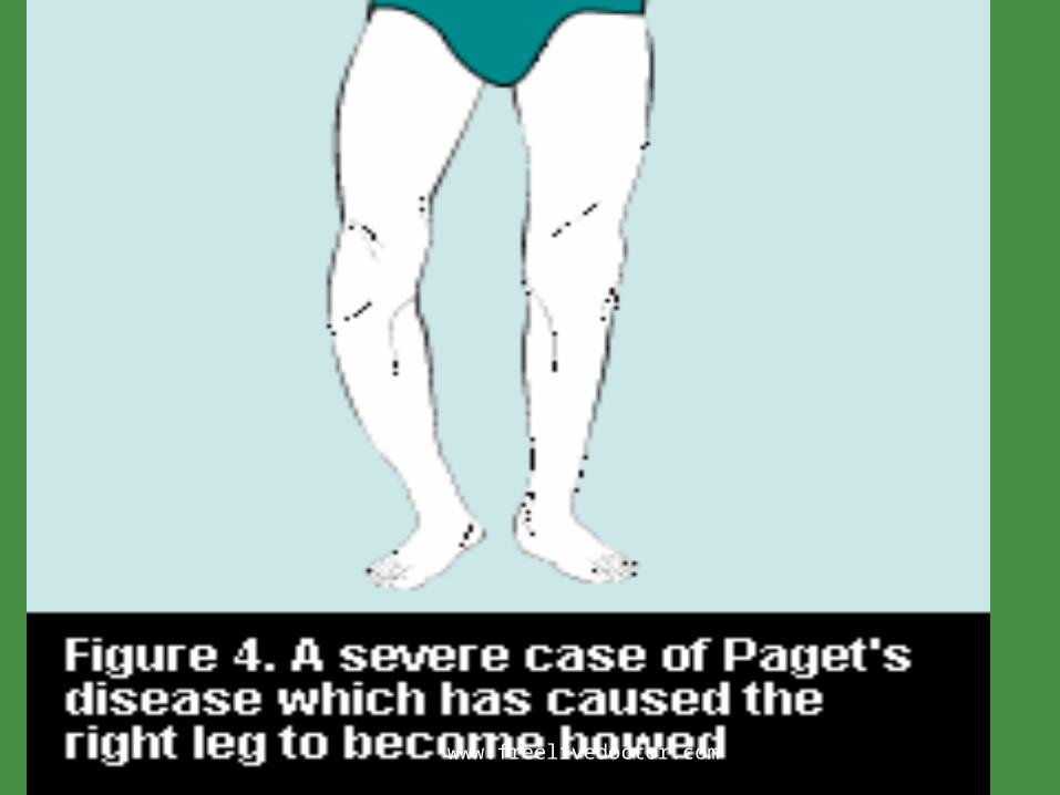
Paget´s disease(Osteitis deform.)
Begins after 40´s and is commonly seen in Begins after 40´s and is commonly seen in caucasians(3%), with some hereditary caucasians(3%), with some hereditary predisposition(autosomal dominant trait)predisposition(autosomal dominant trait)
Paramyxovirus may have a role as cause of the Paramyxovirus may have a role as cause of the disease showing 2 forms of presentation disease showing 2 forms of presentation a. Localized in tibia, femur, iliac, humerus, a. Localized in tibia, femur, iliac, humerus, vertebrae & skull(monostotic) in 15% of cases. vertebrae & skull(monostotic) in 15% of cases. b. Localized b. Localized in several bones(polyostotic) in 85% of cases.in several bones(polyostotic) in 85% of cases.
www.freelivedoctor.com

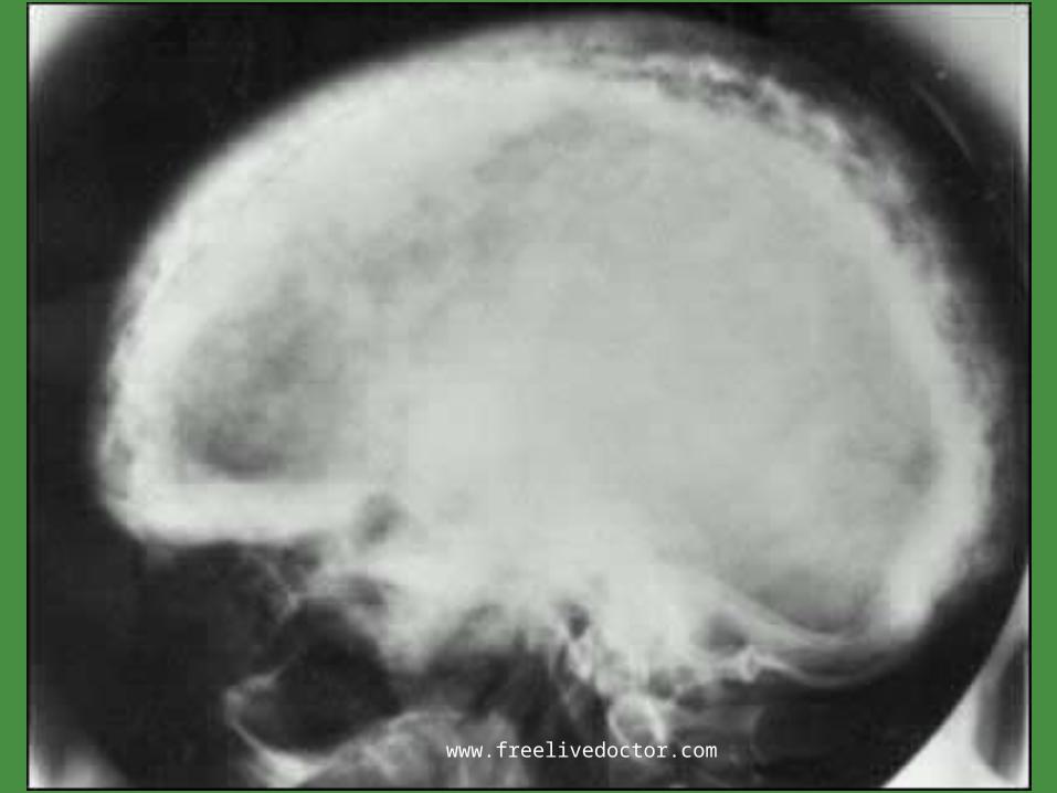
Paget´s disease(Osteitis deform.)
Pathology. There are Pathology. There are 3 stages: --Osteolytic 3 stages: --Osteolytic --Osteolytic- --Osteolytic-osteoblastic --osteoblastic --Osteosclerotic(“cold” stage)Osteosclerotic(“cold” stage)
Micro: --Micro: --Thickened trabecular & cortical bone Thickened trabecular & cortical bone --Abnormal arrangement of lamellar bone --Abnormal arrangement of lamellar bone (mosaic pattern) (mosaic pattern) --Woven(reactive) bone --Woven(reactive) bone --Giant multinucleated --Giant multinucleated osteoclasts(w/viral incl.) --Fibrotic osteoclasts(w/viral incl.) --Fibrotic marrow marrow
www.freelivedoctor.com

www.freelivedoctor.com

Paget´s disease(Osteitis deform.)
Complications. Complications. --Myelophthisic anemia --Myelophthisic anemia --Optic & auditory nerve compression --Optic & auditory nerve compression --Fx. --Fx. --Cardiac failure(high output due to large --Cardiac failure(high output due to large A/V shunts from blood vessels in fibrotic marrow A/V shunts from blood vessels in fibrotic marrow -- --Malignancies(about 1% of cases)Malignancies(about 1% of cases)osteo osteo genic sarcoma, fibrosarcoma(jaw,femur, genic sarcoma, fibrosarcoma(jaw,femur, pelvis)pelvis)
www.freelivedoctor.com
www.freelivedoctor.com

www.freelivedoctor.com
www.freelivedoctor.com

www.freelivedoctor.com

www.freelivedoctor.com

www.freelivedoctor.com

BONE & DEGENERATIVE DISORDERS
There are 206 bones diverse in size & shape There are 206 bones diverse in size & shape that have a role in mineral homeostasis, that have a role in mineral homeostasis, contains hematopoietic elements, determine contains hematopoietic elements, determine the body size/shape and provide support for the body size/shape and provide support for movement and protection. Long bones havemovement and protection. Long bones have
- Epiphysis - Epiphysial plate- Epiphysis - Epiphysial plate
- Metaphysis - Diaphysis(shaft)- Metaphysis - Diaphysis(shaft)
www.freelivedoctor.com

BONE & DEGENERATIVE DISORDERS
Also, a bone shows 2 gross anatomical forms in Also, a bone shows 2 gross anatomical forms in proportion to a particular function:proportion to a particular function:
1. Compact bone:dense outer shell(cortex) with 1. Compact bone:dense outer shell(cortex) with basic units(osteons) arranged in vertical columns basic units(osteons) arranged in vertical columns and lamellar structure that contain osteocytes & and lamellar structure that contain osteocytes & capillaries forming a haversyan system.capillaries forming a haversyan system.
2. Cancellous bone(spongious) arranged in 2. Cancellous bone(spongious) arranged in trabecules(osteocytes+osteoblasts+capillaries) trabecules(osteocytes+osteoblasts+capillaries) along with marrowalong with marrow
www.freelivedoctor.com

BONE & DEGENERATIVE DISORDERS
3. Woven bone(non-lamellar), a primitive 3. Woven bone(non-lamellar), a primitive form laid down in fetal development, with form laid down in fetal development, with irregular trabecula in a primitive matrix. In irregular trabecula in a primitive matrix. In adult life this type is seen in bone adult life this type is seen in bone regeneration(later replaced by lamellar regeneration(later replaced by lamellar bone) and tumorsbone) and tumors
www.freelivedoctor.com

www.freelivedoctor.com

www.freelivedoctor.com

BONE & DEGENERATIVE DISORDERS
Bone cells constitute about 2% of bone weight:Bone cells constitute about 2% of bone weight: Osteoprogenitor(STEM) cells in vacinity of surfaces Osteoprogenitor(STEM) cells in vacinity of surfaces Osteoblasts in surface of bones(synthesis& Osteoblasts in surface of bones(synthesis&
arrange of proteins) w/receptors for hormo arrange of proteins) w/receptors for hormo nes, GFs nes, GFs
Osteocytes(control of Ca++, P)Osteocytes(control of Ca++, P) Multinucleated osteoclasts(from granulocyte-mo Multinucleated osteoclasts(from granulocyte-mo
nocyte precursor in marrow) with nocyte precursor in marrow) with resorption pits (Howship´s resorption pits (Howship´s lacunae)lacunae)
www.freelivedoctor.com

BONE & DEGENERATIVE DISORDERS
Bone proteins include type I collagen(90% Bone proteins include type I collagen(90% of organic component) and non-collagenous of organic component) and non-collagenous proteins derived from osteoblastsproteins derived from osteoblastsmatrixmatrix
Bone is a blend of organic matrix(35%)and Bone is a blend of organic matrix(35%)and inorganic(65%)inorganic(65%)
Inorganic components are:Ca++hydroxyapa Inorganic components are:Ca++hydroxyapa tite(stores 99% of body Ca+ tite(stores 99% of body Ca++, 80% of P and 65% of Na & Mg)+, 80% of P and 65% of Na & Mg)
www.freelivedoctor.com

BONE & DEGENERATIVE DISORDERS
BONE DEPOSITION. BONE DEPOSITION. - From rows of osteoblasts laying - From rows of osteoblasts laying down lamellar collagen matrixdown lamellar collagen matrixincorporated incorporated into the bone and housed in small lacuna as into the bone and housed in small lacuna as osteocyteosteocytenon-calcified osteoid in the centre non-calcified osteoid in the centre of the osteon/surface of trabecula. Normally of the osteon/surface of trabecula. Normally calcification follows very quickly calcification follows very quickly -During osteoblastic activity ALKALINE -During osteoblastic activity ALKALINE PHOSPATASE is liberated into blood flow PHOSPATASE is liberated into blood flow
www.freelivedoctor.com

BONE & DEGENERATIVE DISORDERS
Bone & calcium homeostasis: Bone & calcium homeostasis: - - parathyroidsparathyroids
blood Ca++ PTH blood Ca++ PTH blood Ca++blood Ca++ bone (osteocyte rel/Ca++ bone (osteocyte rel/Ca++
www.freelivedoctor.com

BONE & DEGENERATIVE DISORDERS
MINERAL HOMEOSTASIS. MINERAL HOMEOSTASIS. 1. PTH in parathyroid glands Ca++ 1. PTH in parathyroid glands Ca++ PO++++ PO++++ 2. 1.25(OH2)D3 in proximal conv.& 2. 1.25(OH2)D3 in proximal conv.& straight tubules Ca+++ straight tubules Ca+++ PO++++ PO++++ 3. Calcitonin in “C” cells Ca++ 3. Calcitonin in “C” cells Ca++ PO++++ PO++++
www.freelivedoctor.com