bone marrow red yellow bone fat reticulin haematopoiesis – red, white, platelets lymphoid
TRANSCRIPT

Bone marrow
Red
Yellow
Bone
Fat
Reticulin
Haematopoiesis – red, white, platelets
lymphoid

Normal red cells
Central pale area
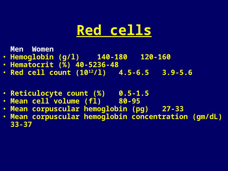
Red cellsMen Women
• Hemoglobin (g/l) 140-180 120-160• Hematocrit (%) 40-52 36-48• Red cell count (1012/l) 4.5-6.5 3.9-5.6
• Reticulocyte count (%) 0.5-1.5• Mean cell volume (fl) 80-95• Mean corpuscular hemoglobin (pg) 27-33• Mean corpuscular hemoglobin concentration (gm/dL) 33-37

Red cellspathological conditions:
I. decrease in the circulating red cell mass
(poss. with structural abnormalities)
very common - anaemia
II. increase in the circulating red cell mass
less common
polycythemia =erythrocytosis=polyglobuly

Polycythemia=increased concentration of red cells
• RELATIVE - decreased plasma volumedehydration, stress• ABSOLUTEprimary – neoplastic= polycythemia vera
= myeloproliferative neoplasm
secondary - increased erythropoietin stimulationAppropriate
reactive – low levels of oxygen in the PB (heart disease, high altitude)
Inappropropriate
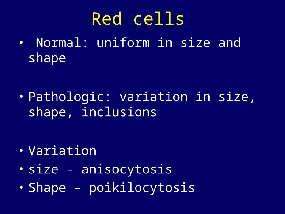
Red cells• Normal: uniform in size and shape
• Pathologic: variation in size, shape, inclusions
• Variation
• size - anisocytosis
• Shape – poikilocytosis
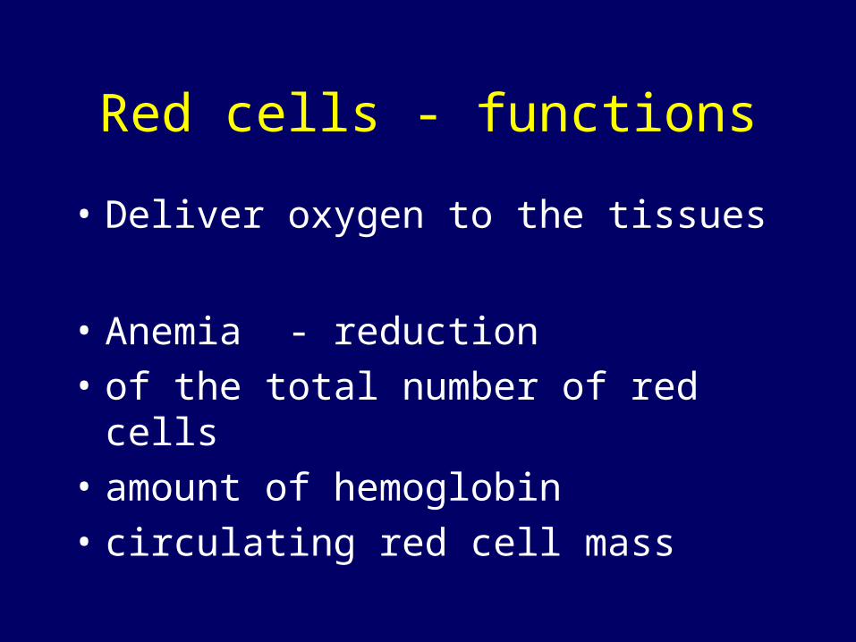
Red cells - functions
• Deliver oxygen to the tissues
• Anemia - reduction
• of the total number of red cells
• amount of hemoglobin
• circulating red cell mass

Consequences of anemia - symptoms
• ???????????
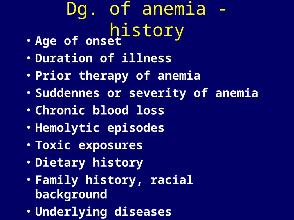
Dg. of anemia - history• Age of onset
• Duration of illness
• Prior therapy of anemia
• Suddennes or severity of anemia
• Chronic blood loss
• Hemolytic episodes
• Toxic exposures
• Dietary history
• Family history, racial background
• Underlying diseases

Anemia – consequences, symptoms
• Fatigue, syncope, dyspnea
• Impairment of organ function due to hypoxia
• Pallor, postural hypotension )decreased blood volume)
• Heart murmurs, heart failure . Increased cardiac output

Anemia
• Not a diagnosis per se
• Look for an underlying problem
• History, physical examination

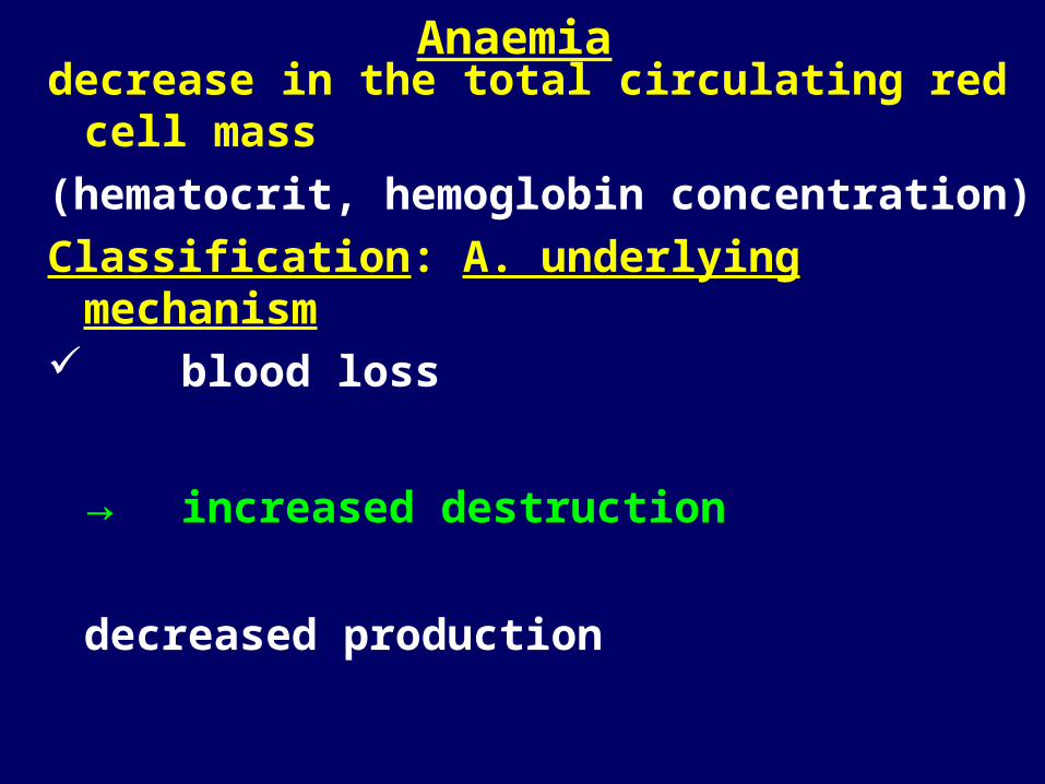
Anaemiadecrease in the total circulating red cell mass
(hematocrit, hemoglobin concentration)
Classification: A. underlying mechanism
blood loss
increased destruction
decreased production
B. morphology of erythrocytes
size (micro-, macro-, normocytic)
shape (spherocytosis, stomato-,...)
color (degree of hemoglobinization:normo- hypo-, hyperchromic)
M
A
Y
C
O
M
B
IN
E
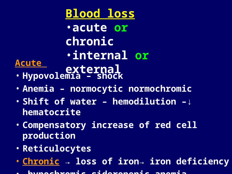
Acute
• Hypovolemia – shock
• Anemia – normocytic normochromic
• Shift of water – hemodilution –↓ hematocrite
• Compensatory increase of red cell production
• Reticulocytes
• Chronic → loss of iron→ iron deficiency
• hypochromic sideropenic anemia
Blood loss•acute or chronic•internal or external

Iron deficiency anemiamechanism: blood loss, decreased production
body iron = functional + storage
F - 2g, M - 6ginadequate intake for metabolic demands
Lack in diet or low absorption
most common nutritional disorder in the world
2. Increased requirement (children, pregn., lact)
!!!3. Chronic blood loss!!! - GIT, GYNmost important cause of iron deficiency
in the Western world
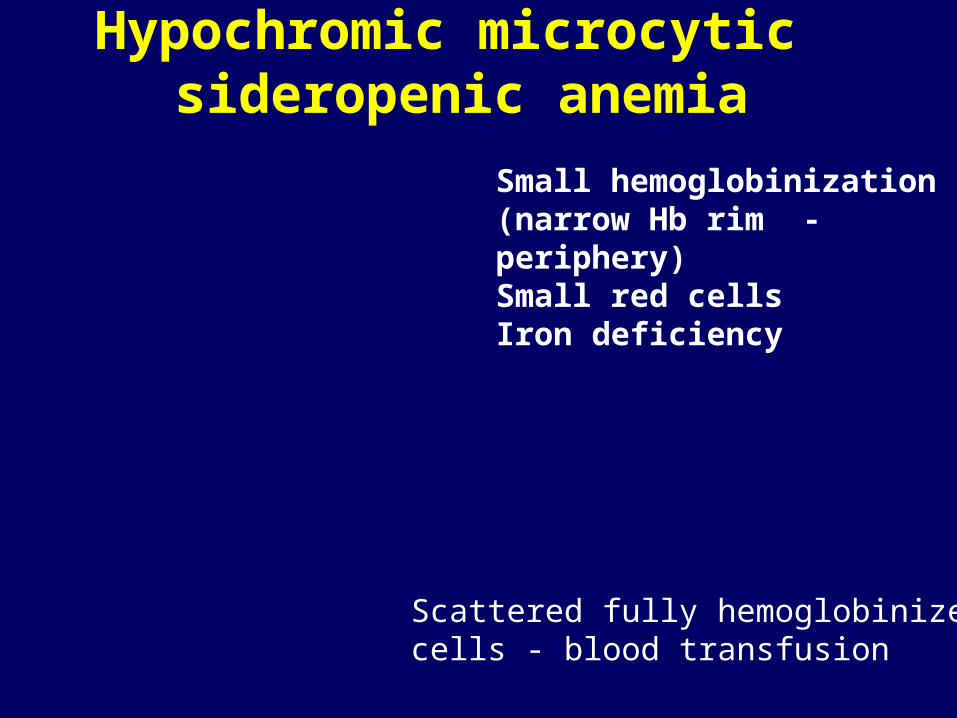
Hypochromic microcytic sideropenic anemia
Scattered fully hemoglobinized cells - blood transfusion
Small hemoglobinization(narrow Hb rim -periphery)Small red cellsIron deficiency

PB: ery pale + small
BM: erythroid hyperplasia, loss of iron
alopecia, koilonychia, atrophy of tongue, gastric mucosa
Plummer-Vinson (Kelly-Patterson) syndrome: siderop.an., atrophic glossitis, esophageal webs
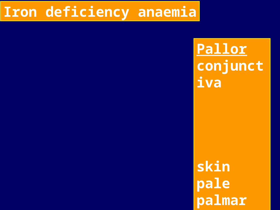
Iron deficiency anaemia
Pallorconjunctiva
skinpale palmar creases
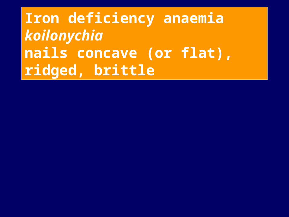
Iron deficiency anaemiakoilonychianails concave (or flat), ridged, brittle

Iron deficiency anaemiaangular cheilosisfissuring and ulceration; pallor

Iron deficiency anaemiaflattening and loss of papillaebald, fissured tongue

Causes of hypochromic anemia1. Disorders of iron metabolism
2. Disorders of heme synthesis
3. Disorders of globin synthesis (thalassemia)
Ad 1. Iron deficiency
• Blood loss
• Poor intake - growth, pregnancy, lactation
• Malabsorption
• Chronic infections or inflammatory states
• neoplasia
Anaemiadecrease in the total circulating red cell mass
(hematocrit, hemoglobin concentration)
Classification: A. underlying mechanism blood loss
→ increased destruction
decreased production
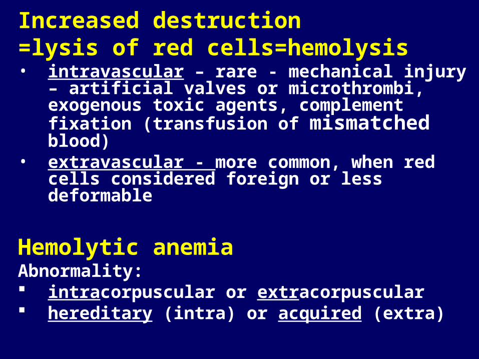
Increased destruction=lysis of red cells=hemolysis• intravascular – rare - mechanical injury – artificial
valves or microthrombi, exogenous toxic agents, complement fixation (transfusion of mismatched blood)
• extravascular - more common, when red cells considered foreign or less deformable
Hemolytic anemiaAbnormality: intracorpuscular or extracorpuscular hereditary (intra) or acquired (extra)

Hemolytic anemia• premature destruction of red cells
• accumulation of the products of the hemoglobin catabolism
• BM – increased erythropoiesis, extreme: extramedullary hematopoiesis
• PB: reticulocytosis
• high bilirubin –gallstones; jaundice, blr in urine
• chronic duration: hemosiderosis
Main clinical symptomsanemia, splenomegaly, jaundice; gallstones
Haemolytic anaemiasplenomegaly and jaundice

Haemolytic anaemiajaundice normal

Increased destruction of ery=hemolysisI. Intrinsic (intracorpuscular) causes
A. hereditary • membrane – cytoskeleton, lipid synthesis• enzymes – deficiencies - G6PD, glutathione
synthetase, pyruvate kinase• hemoglobin - deficient synthesis of globin,
structurally abnormal HbB. acquired
• membrane defect: paroxysmal nocturnal hemoglobinuria
• II. Extrinsic (extracorpuscular) causes• antibodies, trauma, infection, chemical injury
sequestration
Examples of hemolytic anemia
• Membrane defects –
• Proteins underlying the red cell membrane
• Shape, stability, flexibility

Hereditary spherocytosis (peripheral smear)
anisocytosis and several dark-appearing spherocytes with no central pallor. Howell-Jolly bodies (small dark nuclear remnants)

Hereditary spherocytosisAD (AR, sporadic);most common her. hemol. A.
Membrane defect – cytoskeleton – protein spectrin (and ankyrin) deficiencyRound erythrocyte= spherocyte, less deformableVulnerable to spleen sequestration and destruction
Main clinical symptomsanemia, splenomegaly, jaundice; gallstonesChronic hemolytic anemia (mild to normal)Acute anemic episodes:aplastic crisis (parvovirus)hemolytic crisis

Table 13-2. Adult Reference Ranges for Red Blood Cells*
A red cell squeezing from the red pulp cordsinto the sinus lumen. Note the degree of deformability required for red cells to passthrough the wall of the sinus.
Splenic sinus

Mutations weakening interactions involving α-spectrin, β-spectrin, ankyrin, band 4.2, or band 3 cause the normal biconcave red cell to lose membrane fragments and become sphericalspherocytic cells: less deformable than normal, become trapped in the splenic cords, phagocytosed by macrophages.
Red cell membrane cytoskeleton Alterations leading to spherocytosis and hemolysis

Pathophysiology of hereditary spherocytosis

Haemolytic anaemia: reticulocytes
precip. RNA

Hereditary elliptocytosis
Usually mild, rarely severe

Hemolytic anemia
• Intracorpuscular
• Enzyme deficiencies

G6PD deficiency
enzymes protecting the red cell against the oxidative stress
G6PD deficiency → loss of protection → oxidant injury
infections, drugs, beans (favism)
→ hemolysis; otherwise normal
morphologic changes of chronic HA rarely present
hundreds of genetic forms of G6PD
common pathologic alleles: G6PDA-, G6PD Mediterranean
X-linked→ males homozygous, women heterozygous
Mediterranean, Middle East, Africa
Protection against malaria

G6PD deficiency
• Clinical and laboratory findings
• Episode of acute hemolytic anemia in anotherwise healthy person; neonatal jaundice
• following oxid. injury – drug (antimal. – primaquine,; sulfoamides, nitrofurantoin, nalidixic acid; TNT, , infections, food
• Variable severity
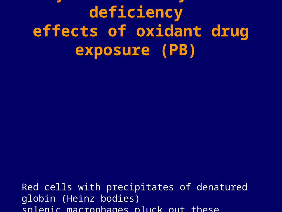
Enzyme deficiency: G6PD deficiency effects of oxidant drug exposure (PB)
Red cells with precipitates of denatured globin (Heinz bodies)splenic macrophages pluck out these inclusions → "bite cells"

Increased destruction of ery=hemolysisI. Intrinsic causes
A. hereditary membrane – cytoskeleton, lipid synthesisenzymes – deficiencies - G6PD, glutathione
synthetase, pyruvate kinase
→hemoglobin – abnormal
quantity (deficient synthesis of globin)
quality (structurally abnormal Hb)
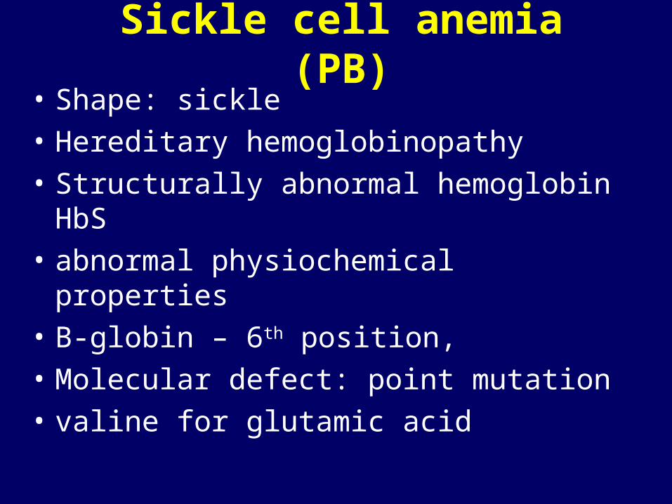
• Shape: sickle
• Hereditary hemoglobinopathy
• Structurally abnormal hemoglobin HbS
• abnormal physiochemical properties
• B-globin – 6th position,
• Molecular defect: point mutation
• valine for glutamic acid
Sickle cell anemia (PB)
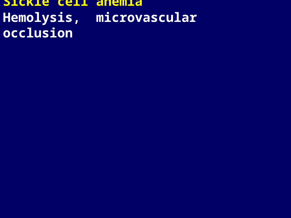
Sickle cell anemia Hemolysis, microvascular occlusion

Sickle cell anemia
• Oxyg. HbS: liquid
• Deoxyg.:viscous gel →fibers
HbS - aggregation and polymerization
• Sickle shape; Initially: reversible (with oxygenation)
• Repeated: irreversible sickling
• Membrane damage
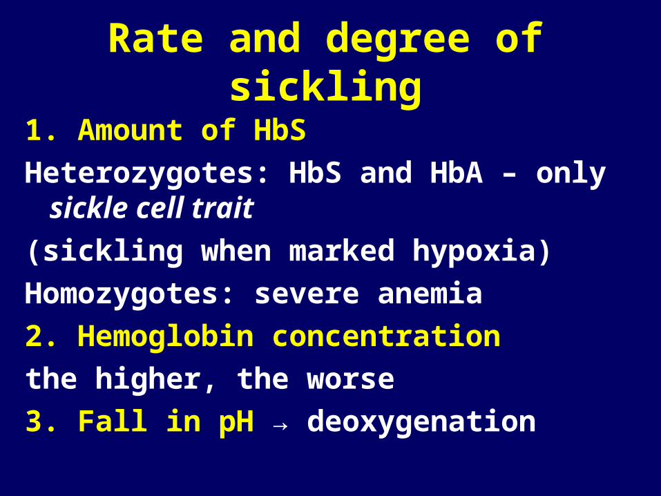
Rate and degree of sickling
1. Amount of HbS
Heterozygotes: HbS and HbA – only sickle cell trait
(sickling when marked hypoxia)
Homozygotes: severe anemia
2. Hemoglobin concentration
the higher, the worse
3. Fall in pH → deoxygenation

Sickle cell anemia - clinical manifestation1. chronic hemolytic anemia (ery survival 20 days)
chronic hyperbilirubinemia, Hbemia, jaundice, gallstonesaplastic crisis
2. occlusion of small vessels → thrombosis, ischemia, necrosis
painful crises
3. splenomegaly4. increased susceptibility to infections
5. activation of the bone marrow, extramedullary haematopoiesis
Diagnosis
• Clinical, laboratory – blood smear
• HbS - electrophoresis
• Clinical course variable
• Therapy symptomatic
Thalassemia
• Deficient synthesis of globin chains• Globin chain absent or amount reduced• β - major, minor (more common), intermedia• β +,0
• Homo/heterozygous
• α +,0

Scleral jaundiceHaemolytic autoimmune anaemia

Pathogenesis of β-thalassemia major
aggregates of unpaired α-globin chains not visible Blood transfusions correct the anemia reducethe stimulus for marrow expansion,but add to systemic iron overload

Thalassemia major: gallbladder - bilirubin gallstones

Immunohemolytic anemia
• Antibodies
• Coombs test

Raynaud phenomenonautoimmnune haemolytic anaemia
Erythroblastosis fetalis

Decreased production of red cellsDeficiency of vital substrates disorders of proliferation and differentiation stem cells erythroblastsImpaired: DNA synthesis – B12, folic acid – megaloblastic hemoglobin synthesis - heme
(lack of iron)
- globin Others: anemia of chron. dis., AA, PRCA

Megaloblastic anemia
• impaired DNA synthesis
• characteristic morphologic changes
blood (macrocytes), bone marrow (megaloblasts)
• Deficiency of vit. B12
• Folic acid

Megaloblastic anaemia

Vitamin B12 absorption

Deficiency of vit. B12
1. Decreased intake – diet, vegetarianism
2. Impaired absorption
Intrinsic factor deficiency – pernicious anemia,
gastrectomy
Malabsorption
Intestinal dis., resection of ileum
Parasitic uptake, bacterial overgrowth
3. Increased requirement
pregnancy, hyperthyroidism, disseminated cancer
Vit. B12 deficiency• BM and blood, CNS, (pernicious: GIT)
• GIT: beefy tongue – atrophic glossitis
• CNS – spinal cord - myelin degenaration laterodorsal tracts – balance, motoric, sensitive
• Pernicious: stomach: chronic gastritis, intestinal metaplasia, higher risk of carcinoma
Pernicious anemia
• Older people
• Autoimmune Ab
• Poss. with autoimmune thyroiditis, adrenalitis

Megaloblastic anaemia
Acute leukaemia
Diffferential diagnosis

Megaloblastic anaemiahypersegmented neutrophils (macropolycyte)

Megaloblastic anaemia - perniciouslemon-yellow appearancepallor (anaemia) + jaundice (ineffective erythropoiesis)

Pernicious anaemia (38 ys.)premature greying, blue eyes, vitiligo
Beefy tongueatrophic glossitis(Hunter)vit. B12 deficiency

Pernicious anaemiaDorsolateral spinal cord demyelination
Folate deficiency• Decreased intake• Increased requirements• Impaired use
• Relative deficiency
• Megaloblastic anemia; no neurological symptoms• Cheilosis, glossitis, dermatitis

Anemia of chronic disease
*Infections
*immunologic
*neoplasms
Mechanism: defect in reutilization of iron (transfer, cytokines)
! abundant storage iron
Anemia: normo, normo or hypo, micro
Aplastic anemia• Failure or suppression of myeloid stem cell• PANCYTOPENIA• primary OR secondary - drugs , chemicals
infectionsirradiationinherited – Fanconi
Or cause unknown…BM: hypocellular, PB: pancytopenia,
symptomsspleen normal
Special subgroup: pure red cell anemia
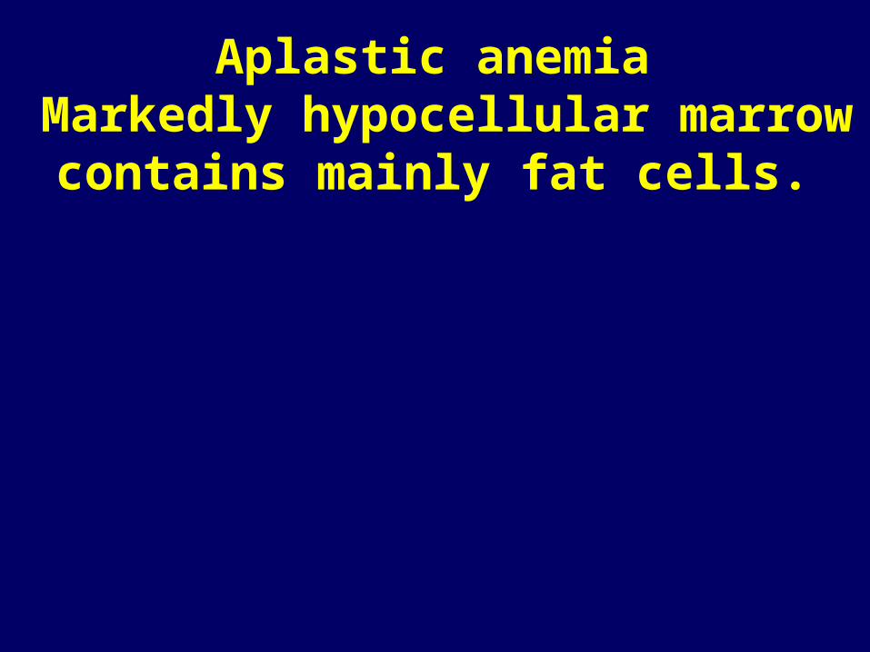
Aplastic anemia Markedly hypocellular marrow
contains mainly fat cells.