bone marrow–derived immune cells regulate vascular disease...
TRANSCRIPT

Research article
The Journal of Clinical Investigation http://www.jci.org Volume 114 Number 3 August 2004 419
Bone marrow–derived immune cells regulate vascular disease through a p27Kip1-
dependent mechanismManfred Boehm, Michelle Olive, Andrea L. True, Martin F. Crook, Hong San,
Xuan Qu, and Elizabeth G. Nabel
Cardiovascular Branch, National Heart, Lung, and Blood Institute, NIH, Bethesda, Maryland, USA.
The cyclin-dependent kinase inhibitors are key regulators of cell cycle progression. Although implicated in carcinogenesis, they inhibit the proliferation of a variety of normal cell types, and their role in diverse human diseases is not fully understood. Here, we report that p27Kip1 plays a major role in cardiovascular dis-ease through its effects on the proliferation of bone marrow–derived (BM-derived) immune cells that migrate into vascular lesions. Lesion formation after mechanical arterial injury was markedly increased in mice with homozygous deletion of p27Kip1, characterized by prominent vascular infiltration by immune and inflamma-tory cells. Vascular occlusion was substantially increased when BM-derived cells from p27–/– mice repopulated vascular lesions induced by mechanical injury in p27+/+ recipients, in contrast to p27+/+ BM donors. To deter-mine the contribution of immune cells to vascular injury, transplantation was performed with BM derived from RAG–/– and RAG+/+ mice. RAG+/+ BM markedly exacerbated vascular proliferative lesions compared with what was found in RAG–/– donors. Taken together, these findings suggest that vascular repair and regeneration is regulated by the proliferation of BM-derived hematopoietic and nonhematopoietic cells through a p27Kip1-dependent mechanism and that immune cells largely mediate these effects.
IntroductionVascular regeneration and repair are essential to the survival of blood vessels. Arterial healing requires the coordinated tempo-ral and spatial expression of proteins that regulate vascular cell proliferation. Inflammation and immunity are also essential components of the pathogenesis of cardiovascular diseases (ref. 1; reviewed in refs. 2–4), but the role of immune progenitors in vascu-lar proliferation and inflammation has not been established.
p27Kip1, a member of the Cip/Kip family of cyclin-dependent kinase inhibitors, binds and alters the activities of cyclin D–, cyclin E–, and cyclin A–dependent kinases in quiescent cells, lead-ing to failure of G1/S transition and cell cycle arrest (5, 6). An increase in the levels of p27Kip1 causes proliferating cells to exit the cell cycle, and a decrease in p27Kip1 is necessary for quiescent cells to resume division. Environmental stresses also regulate p27Kip1 levels; for example, hypoxia causes a hypoxia-inducible factor–1α–dependent increase in p27Kip1 (7). p27–/– mice develop hyperplasia in multiple organs, including endocrine tissues, thy-mus, and spleen (8–10); however, the vascular phenotype of these mice is not known. Defining the role of p27Kip1 in vascular diseas-es is critical not only to understanding disease pathogenesis, but importantly, to the design of vascular therapeutics. For example, p27Kip1 mediates the antiproliferative and antimigratory activity of sirolimus-coated stents (11–13).
Bone marrow–derived (BM-derived) cells are increasingly recog-nized as key components of vascular regeneration (14–16). After vascular injury, progenitor cells from arterial and BM compart-
ments are mobilized by cytokine activation (15). It is hypothesized that BM-derived cells circulate, home to sites of vascular damage, proliferate, and form arterial lesions in conjunction with cellular components from the local artery. However, the molecular mecha-nisms that regulate the contributions of BM-derived cells to vas-cular lesion formation are not well understood. We hypothesized that p27Kip1 directly regulates the proliferation of BM-derived cells that migrate into damaged blood vessels and reconstitute vascu-lar lesions. Surprisingly, we found that BM-derived hematopoietic and nonhematopoietic cells give rise to most of the cell prolifera-tion in blood vessels during repair, and that indeed, repopulation occurs through a p27Kip1-dependent mechanism. This mechanism directly links vascular inflammation and proliferation, and sug-gests that immunity and inflammation are important targets in treatment of cardiovascular diseases.
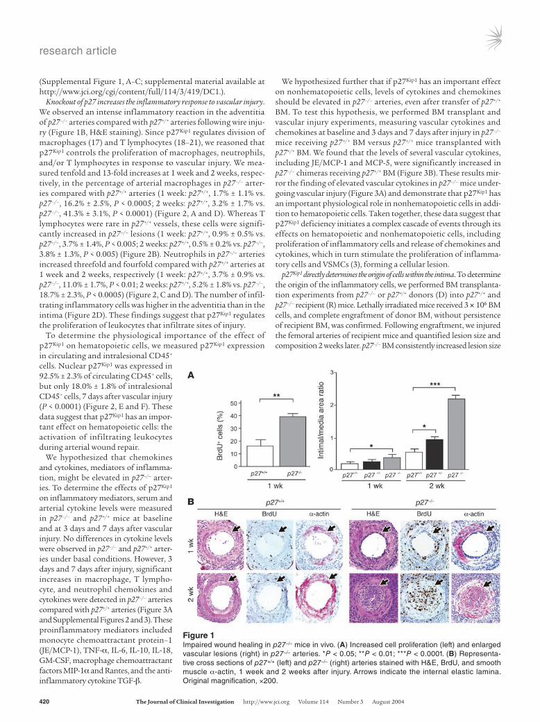
Resultsp27–/– mice develop extensive arterial lesions. To determine whether p27Kip1 regulates arterial wound repair, we subjected p27–/–, p27+/–, and p27+/+ mice to a wire injury in the femoral artery and examined cell proliferation and lesion formation 1 week and 2 weeks later. Cell proliferation was significantly increased in the intima of p27–/– arteries compared with p27+/+ arteries (p27–/–, 37.7% ± 1.4% vs. p27+/+, 18.0% ± 4.4%, P < 0.01) (Figure 1A, left). Arterial lesion size was also markedly increased in p27–/– mice compared with p27+/+ mice (intima/media ratios at 1 week: p27–/–, 0.4 ± 0.1 vs. p27+/+, 0.2 ± 0.1, P < 0.05; at 2 weeks: p27–/–, 2.2 ± 0.1 vs. p27+/+, 0.5 ± 0.1, P < 0.0001) (Figure 1A, right, and Figure 1B). p27Kip1 heterozygous mice pre-sented an intermediate phenotype (Figure 1A). Many of the intimal cells were putative VSMCs, determined by α-actin staining (Figure 1B), suggesting increased proliferation of this cell type. These find-ings are consistent with in vitro experiments in which p27Kip1-delet-ed VSMCs proliferate at accelerated rates compared with p27+/+ cells
Nonstandard abbreviations used: bone marrow (BM); monocyte chemoattractant protein–1 (JE/MCP-1).
Conflict of interest: The authors have declared that no conflict of interest exists.
Citation for this article: J. Clin. Invest. 114:419–426 (2004). doi:10.1172/JCI200420176.
research article
420 The Journal of Clinical Investigation http://www.jci.org Volume 114 Number 3 August 2004
(Supplemental Figure 1, A–C; supplemental material available at http://www.jci.org/cgi/content/full/114/3/419/DC1.).
Knockout of p27 increases the inflammatory response to vascular injury. We observed an intense inflammatory reaction in the adventitia of p27–/– arteries compared with p27+/+ arteries following wire inju-ry (Figure 1B, H&E staining). Since p27Kip1 regulates division of macrophages (17) and T lymphocytes (18–21), we reasoned that p27Kip1 controls the proliferation of macrophages, neutrophils, and/or T lymphocytes in response to vascular injury. We mea-sured tenfold and 13-fold increases at 1 week and 2 weeks, respec-tively, in the percentage of arterial macrophages in p27–/– arter-ies compared with p27+/+ arteries (1 week: p27+/+, 1.7% ± 1.1% vs. p27–/–, 16.2% ± 2.5%, P < 0.0005; 2 weeks: p27+/+, 3.2% ± 1.7% vs. p27–/–, 41.3% ± 3.1%, P < 0.0001) (Figure 2, A and D). Whereas T lymphocytes were rare in p27+/+ vessels, these cells were signifi-cantly increased in p27–/– lesions (1 week: p27+/+, 0.9% ± 0.5% vs. p27–/–, 3.7% ± 1.4%, P < 0.005; 2 weeks: p27+/+, 0.5% ± 0.2% vs. p27–/–, 3.8% ± 1.3%, P < 0.005) (Figure 2B). Neutrophils in p27–/– arteries increased threefold and fourfold compared with p27+/+ arteries at 1 week and 2 weeks, respectively (1 week: p27+/+, 3.7% ± 0.9% vs. p27–/–, 11.0% ± 1.7%, P < 0.01; 2 weeks: p27+/+, 5.2% ± 1.8% vs. p27–/–, 18.7% ± 2.3%, P < 0.0005) (Figure 2, C and D). The number of infil-trating inflammatory cells was higher in the adventitia than in the intima (Figure 2D). These findings suggest that p27Kip1 regulates the proliferation of leukocytes that infiltrate sites of injury.
To determine the physiological importance of the effect of p27Kip1 on hematopoietic cells, we measured p27Kip1 expression in circulating and intralesional CD45+ cells. Nuclear p27Kip1 was expressed in 92.5% ± 2.3% of circulating CD45+ cells, but only 18.0% ± 1.8% of intralesional CD45+ cells, 7 days after vascular injury (P < 0.0001) (Figure 2, E and F). These data suggest that p27Kip1 has an impor-tant effect on hematopoietic cells: the activation of infiltrating leukocytes during arterial wound repair.
We hypothesized that chemokines and cytokines, mediators of inflamma-tion, might be elevated in p27–/– arter-ies. To determine the effects of p27Kip1 on inflammatory mediators, serum and arterial cytokine levels were measured in p27–/– and p27+/+ mice at baseline and at 3 days and 7 days after vascular injury. No differences in cytokine levels were observed in p27–/– and p27+/+ arter-ies under basal conditions. However, 3 days and 7 days after injury, significant increases in macrophage, T lympho-cyte, and neutrophil chemokines and cytokines were detected in p27–/– arteries compared with p27+/+ arteries (Figure 3A and Supplemental Figures 2 and 3). These proinflammatory mediators included monocyte chemoattractant protein–1 (JE/MCP-1), TNF-α, IL-6, IL-10, IL-18, GM-CSF, macrophage chemoattractant factors MIP-1α and Rantes, and the anti-inflammatory cytokine TGF-β.
We hypothesized further that if p27Kip1 has an important effect on nonhematopoietic cells, levels of cytokines and chemokines should be elevated in p27–/– arteries, even after transfer of p27+/+ BM. To test this hypothesis, we performed BM transplant and vascular injury experiments, measuring vascular cytokines and chemokines at baseline and 3 days and 7 days after injury in p27–/– mice receiving p27+/+ BM versus p27+/+ mice transplanted with p27+/+ BM. We found that the levels of several vascular cytokines, including JE/MCP-1 and MCP-5, were significantly increased in p27–/– chimeras receiving p27+/+ BM (Figure 3B). These results mir-ror the finding of elevated vascular cytokines in p27–/– mice under-going vascular injury (Figure 3A) and demonstrate that p27Kip1 has an important physiological role in nonhematopoietic cells in addi-tion to hematopoietic cells. Taken together, these data suggest that p27Kip1 deficiency initiates a complex cascade of events through its effects on hematopoietic and nonhematopoietic cells, including proliferation of inflammatory cells and release of chemokines and cytokines, which in turn stimulate the proliferation of inflamma-tory cells and VSMCs (3), forming a cellular lesion.
p27Kip1 directly determines the origin of cells within the intima. To determine the origin of the inflammatory cells, we performed BM transplanta-tion experiments from p27–/– or p27+/+ donors (D) into p27+/+ and p27–/– recipient (R) mice. Lethally irradiated mice received 3 × 10⁶ BM cells, and complete engraftment of donor BM, without persistence of recipient BM, was confirmed. Following engraftment, we injured the femoral arteries of recipient mice and quantified lesion size and composition 2 weeks later. p27–/– BM consistently increased lesion size
Figure 1Impaired wound healing in p27–/– mice in vivo. (A) Increased cell proliferation (left) and enlarged vascular lesions (right) in p27–/– arteries. *P < 0.05; **P < 0.01; ***P < 0.0001. (B) Representa-tive cross sections of p27+/+ (left) and p27–/– (right) arteries stained with H&E, BrdU, and smooth muscle α-actin, 1 week and 2 weeks after injury. Arrows indicate the internal elastic lamina. Original magnification, ×200.

research article
The Journal of Clinical Investigation http://www.jci.org Volume 114 Number 3 August 2004 421
when transplanted into either p27+/+ or p27–/– recipients (intima/media ratios: Dp27–/– → Rp27+/+, 1.1 ± 0.1 vs. Dp27+/+ → Rp27+/+, 0.3 ± 0.1, P < 0.0005; Dp27+/+ → Rp27–/–, 1.8 ± 0.1 vs. Dp27–/– → Rp27–/–, 2.7 ± 0.2, P < 0.0001) (Figure 4, A and D). Importantly, we found that p27–/– BM also significantly increased the percentage of arterial macrophages in
p27+/+ or p27–/– recipient mice compared with p27+/+ donor BM (Dp27–/– → Rp27+/+, 14.2% ± 1.4% vs. Dp27+/+ → Rp27+/+, 3.8% ± 1.9%, P < 0.005; Dp27–/– → Rp27–/–, 43.6% ± 1.8% vs. Dp27+/+ → Rp27–/–, 29.6% ± 1.6%, P < 0.0001) (Figure 4, B and E). Thus, p27Kip1 directly regulates vascu-lar lesion size through its effects on BM-derived inflammatory cells.
Figure 2p27–/– mice develop arterial inflammation after vascular injury. (A–C) Accumulation of macrophages (*P < 0.0005; **P < 0.0001) (A), T lympho-cytes (†P < 0.005; ††P < 0.001) (B), and neutrophils (#P < 0.01; ##P < 0.0005) (C) in p27+/+ (white bars) and p27–/– (gray bars) arteries after injury. (D) Representative photomicrographs of cross sections of p27+/+ (left) and p27–/– (right) arteries immunostained for macrophages (arrow, red cytoplasmic staining) and neutrophils (arrow, brown cytoplasmic staining). Original magnification, ×200. (E) Immunofluorescence of circulating mononuclear (upper left) and intralesional cells (upper right) demonstrating coexpression of CD45 (membranes, green) and p27Kip1 (nuclei, red). Nuclear DAPI expression (blue) is also shown (lower panels). Original magnification, ×1000. (F) Quantitative analysis of coexpression of endogenous nuclear p27Kip1 in circulating CD45+ mononuclear cells (white bar) and intralesional cells (gray bar). Results are expressed as a percentage of CD45+, p27Kip1+ cells compared with CD45+ cells alone.

research article
422 The Journal of Clinical Investigation http://www.jci.org Volume 114 Number 3 August 2004
To determine whether p27Kip1 regulates the proliferation of BM-derived cells, we quantified hematopoietic BM cells from p27–/– and p27+/+ mice at baseline and at 3 days and 7 days fol-lowing arterial injury. We found a significant increase in CFCs in p27–/– mice compared with p27+/+ mice under baseline conditions and at 3 days and 7 days after injury (baseline: p27–/–, 73.6 ± 7.1 vs. p27+/+, 23.2 ± 3.6 colonies, P < 0.0005; 3 days: p27–/–, 100.2 ± 6.0 vs. p27+/+, 26.4 ± 1.8 colonies, P < 0.0001; 7 days: p27–/–, 61.0 ± 5.0 vs. p27+/+, 26.4 ± 5.5 colonies, P < 0.001) (Figure 4C). These data provide evidence that p27Kip1 directly mediates proliferation of BM-derived hematopoietic cells under basal conditions and dur-ing healing of a blood vessel.
We confirmed the BM origin of cells within the vascular lesions by performing BM transplants from male donors into female recipients and analyzing the presence of Y chromosomes within arteries. We found many Y chromosome–positive cells within the adventitia of p27–/– and p27+/+ recipient mice, suggesting the presence of BM-derived hematopoietic cells (T cells, monocytes, and neutrophils). In the intima, p27–/– donor BM yielded a sig-nificant increase in Y chromosome–positive cells compared with
p27+/+ donor BM (Figure 5A). Some of these intimal cells were Y chromosome+, CD45+, α-actin–, while other cells were Y chromo-some+, CD45–, α-actin+ (Figure 5, B and C). These findings confirm earlier reports by Sata et al. (15) that a significant portion of the neointima is formed by BM-derived cells. Our observations sug-gest that BM-derived cells repopulate vascular lesions through a p27Kip1-dependent mechanism.
p27Kip1 directly regulates BM-derived immune cells infiltrating vascu-lar lesions. The absence of p27Kip1 permits clonal expansion of T lymphocytes in vitro and in vivo (21). To determine whether this mechanism determines the abundance of BM-derived immune cells within the arteries of p27–/– mice, we performed vascular experiments in RAG–/– mice (22). First, we performed vascular inju-ry in RAG+/+ and RAG–/– arteries. We found that RAG–/– mice had significantly smaller lesions than did RAG+/+ mice (intima/media ratios: 1 week: RAG–/–, 0.10 ± 0.05 vs. RAG+/+, 0.15 ± 0.07; 2 weeks: RAG–/–, 0.21 ± 0.08 vs. RAG+/+, 0.55 ± 0.14; P < 0.05) (Figure 6A). RAG–/– arteries contained reduced numbers of inflammatory cells (data not shown). These findings suggest that reduced inflamma-tion in the native artery contributes to improved vascular heal-
Figure 3Cytokine levels are increased in p27–/– arteries following vas-cular injury. (A) Artery samples were collected from p27+/+ (white bars) and p27–/– (gray bars) mice at baseline (Co) and at 3 days and 7 days after injury. (B) Vascular tissues were extracted from p27+/+ (white bars) and p27–/– (gray bars) mice receiving p27+/+ BM at baseline and at 3 days and 7 days after injury. **P < 0.005; ***P < 0.0005.

research article
The Journal of Clinical Investigation http://www.jci.org Volume 114 Number 3 August 2004 423
ing. To ascertain whether an absence of T cells and B cells in the BM also protects against lesion development, we transplanted RAG+/+ or RAG–/– BM into p27+/+ or p27–/– recipient mice, confirmed engraftment, and then performed arterial injury. RAG–/– BM trans-planted into WT mice significantly reduced lesion formation com-pared with RAG+/+ BM (intima/media ratios: DRAG–/– → Rp27+/+, 0.16 ± 0.05 vs. DRAG+/+ → Rp27+/+, 0.33 ± 0.07, P < 0.05) (Figure 6B). RAG–/– donor BM also significantly reduced arterial lesion size in p27–/– recipients compared with RAG+/+ BM. Surprisingly, the magnitude of the RAG–/– BM effect was greater in p27–/– recipi-ents than in p27+/+ recipients, suggesting that native cells in p27–/– arteries also contribute to the size of vascular lesions (intima/media ratios: DRAG–/– → Rp27–/–, 0.4 ± 0.1 vs. DRAG+/+ → Rp27–/–, 1.8 ± 0.1, P < 0.0001) (Figure 6B).
To further delineate the relative contribution of T and B cells, we performed a thymectomy on recipient p27+/+ mice and transplanted p27–/– or p27+/+ BM. After successful engraftment, we performed vascular injury and examined the neointima 2 weeks later. After thymectomy in recipient mice, the percentage of circulating T cells
significantly decreased, from 38.2% ± 2.2% before thymectomy to 18.1% ± 2.2% at the time of sacrifice (P < 0.0005). As anticipated, the thymectomy did not produce a reduction in B cells. We found that the partial T cell depletion significantly reduced neointima forma-tion in thymectomized p27+/+ mice receiving p27–/– BM compared with nonthymectomized p27+/+ mice receiving p27–/– BM (intima/media ratios: Dp27–/– → Rp27+/+ thymectomized, 0.44 ± 0.06 vs. Dp27–/– → Rp27+/+ nonthymectomized, 1.07 ± 0.10, P < 0.0005) (Figure 6C). These findings further support the conclusion that mature T cells contribute to the inflammatory response during arterial wound repair.
DiscussionVascular diseases are characterized by inflammation and prolif-eration, but a mechanistic role for the immune system in these lesions has not been shown. Here we provide direct genetic evi-dence for p27Kip1 as a determinant of vascular inflammation and proliferation. In response to injury, p27Kip1 is required to promote the healing process and protect against the excessive proliferation
Figure 4p27Kip1 determines vascular proliferation and BM progenitor pool size. (A) p27–/– BM accelerates arterial lesion formation when transplanted into p27+/+ or p27–/– recipient mice. Following engraftment, arteries were injured and intima/media ratios were measured 2 weeks later. **P < 0.0005; ***P < 0.0001. (B) p27–/– BM significantly increased the percentage of arterial macrophages in p27+/+ and p27–/– recipient arteries compared with p27+/+ BM. *P < 0.005. (C) CFCs are significantly elevated in p27–/– BM (gray bars) compared with p27+/+ BM (white bars) at the indicated time points after vascular injury. Each data point was generated by three limiting dilutions. #P < 0.001. (D) Representative H&E-stained cross sections of recipient p27+/+ (left two panels) and p27–/– (right two panels) arteries following transplantation with donor p27+/+ and p27–/– BM, as indicated, followed by vascular injury. Original magnification, ×200. (E) Representative cross sections of recipient p27+/+ (left two panels) and p27–/– (right two panels) arteries immunostained for macrophages following transplantation with donor p27+/+ and p27–/– BM, as indicated, fol-lowed by vascular injury. Original magnification, ×200.

research article
424 The Journal of Clinical Investigation http://www.jci.org Volume 114 Number 3 August 2004
of inflammatory cells and VSMCs that occurs in pathological remodeling. We now report that p27Kip1 regulates these process-es through its effects on hematopoietic and nonhematopoietic cells, inhibiting both the proliferation of BM-derived cells that migrate into vascular lesions and the proliferation of local VSMCs. Cytokines and chemokines from these BM-derived inflammatory cells also drive the proliferation of vascular lesions. Furthermore, RAG+/+ donor BM exacerbates vascular proliferative lesions com-pared with RAG–/– donor BM when transplanted into p27+/+ or p27–/– recipients, and thymectomy experiments further confirm the contribution of mature T cells and the immune system to vas-cular injury and remodeling.
The role of BM cells in vascular repair and regeneration has not been well-defined. Accumulating evidence suggests that somatic stem
cells in the BM differentiate into various lineages, including vascular endothelial cells (23, 24) and smooth muscle cells (25, 26). In animal models of graft vasculopathy and hyperlipidemia-induced atheroscle-rosis, BM cells may give rise to a substantial percentage of VSMCs that contribute to arterial remodeling (15, 16, 27). The contribution of these cells to human vascular disease has not been proven, although circulating smooth muscle progenitor cells have been identified in human peripheral blood (28). We demonstrate here that p27Kip1, through its effects on the number of BM progenitor cells, directly reg-ulates the contribution of BM-derived cells that repopulate the vascu-lar neointima following injury. We find that p27–/– cells competitively infiltrate the vascular lesion in substantially higher percentages than do p27+/+ donor cells. Some of the proliferative effects on local VSMCs could be attributed to mature T cells, monocytes, and neutrophils.
Figure 5p27–/– BM–derived cells reconstitute the intima and adventitia of vascular lesions during repair. (A) The percentage of Y chromosome–positive nuclei in the intima was determined 2 weeks after injury using FISH techniques. *P < 0.0001. (B and C) Representative cross sections of recipient p27+/+ female arteries following transplantation of male p27+/+ (B) or p27–/– (C) donor BM. Left two panels: Y chromosome+ cells (yellow), α-actin+ cells (red), and nuclei (DAPI stain, blue). Arrows indicate the margins of the intima as determined by the internal and external elastic lamina. Right two panels: triple immunofluorescence detects Y chromosome+ cells (yellow), CD45+ cells (green), α-actin+ cells (red), and nuclei (DAPI stain, blue). Arrows indicate Y chromosome+ nuclei. Original magnification, ×100 (left two panels of B and C); right two panels: confocal microscopy.
Figure 6Knockout of RAG confers protection against vascular proliferation. (A) RAG–/– arteries are protected against abnormal lesion formation 1 week and 2 weeks after vascular injury compared with RAG+/+ arteries. *P < 0.05. (B) RAG–/– BM directly reduces arterial lesion size in p27+/+ or p27–/– recipient mice. **P < 0.0001. (C) Thymectomy (T cell depletion) in p27+/+ mice reduces neointima development following transplantation with p27–/– BM. ***P < 0.0005.

research article
The Journal of Clinical Investigation http://www.jci.org Volume 114 Number 3 August 2004 425
Inflammation is central to the pathogenesis of vascular diseases such as atherosclerosis. Directed migration of leukocytes through the endothelium initiates an inflammatory process in which monocytes, macrophages, and T lymphocytes in the vascular lesion interact with each other, endothelial cells, and VSMCs in complex ways through cell-cell contact and the production of chemokines, cytokines, and growth factors (reviewed in refs. 3, 29, 30). The adventitia is also an important source of vascular progenitor cells that can differentiate into VSMCs and contribute to vascular lesions (31). In this study, we also found a substantial percentage of Y chromosome+ cells in the adventitia. Many of these adventitial cells were CD45+, suggesting a hematopoietic origin, and were like-ly immune and inflammatory cells. These inflammatory cells may stimulate the proliferation of local VSMCs through the release of cytokines and chemokines. The observation that p27Kip1 regulates the proliferation of these BM-derived hematopoietic cells is sup-ported by recent observations of atherosclerotic lesion formation in apoE–/– mice receiving p27–/– BM (32).
Here we provide direct genetic evidence of immune system involvement in lesion progression and composition that is medi-ated by p27Kip1 regulation of the BM. We find that lymphocytes are obligatory for lesion progression, since RAG+/+ BM exacerbated lesions, whereas RAG–/– BM conferred protection against vascular proliferation. Furthermore, vascular proliferation was increased fourfold when RAG+/+ BM was transplanted into p27–/– recipients. The role of T cells in vascular repair was defined further in the thymectomy experiments; partial depletion of T cells resulted in a much smaller vascular lesion, confirming the physiological impor-tance of T cells in vascular inflammation and injury. Lymphocytes also play an important role in early atherogenesis, as demonstrated by genetic crosses of LDL receptor null and RAG null mice (33).
In summary, our findings indicate that p27Kip1 plays a major role in vascular repair and regeneration through its effects on the proliferation of BM-derived hematopoietic and nonhematopoietic cells. This mechanism directly links vascular proliferation and inflammation and suggests that therapeutics directed at immune and inflammatory cells are important in treating cancer and car-diovascular, autoimmune, and other human diseases.
MethodsGeneration of homozygous mice. We obtained heterozygous 129/BL6 p27+/– mice from Andrew Koff (Memorial Sloan-Kettering Cancer Center, New York, New York, USA). We backbred the mice for 12 generations against a C57BL/6 background and studied male and female mice at 10 weeks of age. p27+/+ littermates were used as controls. RAG–/– mice (strain B6.129S7-Rag1tm1Mom, on a C57BL/6 background) were obtained from The Jackson Laboratory (Bar Harbor, Maine, USA). RAG+/+ littermates were used as controls. Genotyping was performed by PCR amplification of mouse tail DNA using allele-specific probes. Each experimental group contained a minimum of five mice. All experiments were conducted according to the guidelines of the Animal Care and Use Committee of the National Heart, Lung, and Blood Institute.
Wire injury in mice. p27–/–, p27+/–, and p27+/+ mice were investigated using an established model of vascular wire injury (34). This procedure was per-formed by one surgeon (H. San) who was blinded to genotype. This injury led to complete endothelial denudation and medial VSMC apoptosis. The cellularity of the media was decreased and became repopulated by prolif-erating VSMCs. Each group consisted of at least five mice and ten arter-ies. The percentage of BrdU-positive cells, macrophages, T lymphocytes, and neutrophils in the intima, media, and adventitia were counted in 40
sections per artery and were analyzed by computer-assisted morphometry. BrdU (25 mg/kg) was injected 1 hour before sacrifice. Intima and media cross-section areas were measured by two independent observers blinded to genotype, using a computerized measuring system (34).
Thymectomy in adult mice. The mice were anesthetized and placed in a ventrodorsal position on a water heated pad. The skin was shaved from the neck to the thorax and the mice were intubated and connected to a ventilator with 100% oxygen. A longitudinal midline incision was made through the skin and superficial fascia from the level of the angle of the mandible to the fourth rib. The pretracheal muscles were separated down to the sternum and the trachea was exposed. The thymus was completely removed by performing sharp or blunt dissection and separating it from the pleural linings. The pretracheal muscles and the sternum were closed with Vicryl suture (Ethicon Inc., Somerville, New Jersey, USA), and the skin incision was closed with stainless steel staples.
BM transplantation. BM was obtained from 8- to 12-week-old male p27–/–, p27+/+, RAG–/–, and RAG+/+ mice after euthanizing with CO2. Marrow cell suspensions were flushed from femurs and tibias, filtered, and stored on ice until use. Recipient female mice were lethally irradiated with 9 Gy of whole-body irradiation. Three million unfractionated cells were injected intravenously into the tail vein of each recipient mouse. Twelve weeks later, successful engraftment was confirmed by quantitative PCR to determine the presence or absence of p27Kip1 or Sry to distinguish female and male BM cells and by FACS to determine the absence of T and B lymphocytes in RAG–/– BM. Five mice (ten femoral arteries) were studied per group. Four sections were analyzed per artery.
Tissue disaggregation and cell isolation. Circulating blood mononuclear cells were obtained by Ficoll gradient purification using lymphocyte separation medium (ICN Biomedicals Inc., Aurora, Ohio, USA). Vascular infiltrating leukocytes were obtained as described (35) with minor modifications. Sub-sequently, cells were spun down onto glass slides.
Immunohistochemistry and in situ hybridization. Immunohistochemistry was performed using an ABC immunoperoxidase (BrdU or MAC-2) or alkaline phosphatase (α-actin) protocol. An anti–α-actin antibody against vascular smooth muscle α-actin (1:1,000; Roche Diagnostics Corp., Indianapolis, Indiana, USA), a MAC-2 antibody against macrophages (1:16,000; Cedar-lane Laboratories Ltd., Hornby, Ontario, Canada), antibody CL8993B against neutrophils (1:10, Cedarlane Laboratories Ltd.), an antibody against CD3 used to identify T lymphocytes (Santa Cruz Biotechnology Inc., San Cruz, California, USA), and an alkaline phosphatase–conjugated mouse monoclonal antibody against BrdU (1 U/ml, Roche Diagnostics Corp.) were used as primary antibodies.
Immunofluorescence cytochemistry was performed using the Cytofix/Cytoperm system (BD Biosciences — Pharmingen, San Diego, California, USA). Double labeling was performed using a rabbit anti-p27Kip1 anti-body (C19) (1:50, Santa Cruz Biotechnology Inc.), an FITC-conjugated rat anti-mouse CD45 antibody (1:50, BD Biosciences — Pharmingen), and a TRITC-conjugated anti-rabbit secondary antibody (1:200; Sigma-Aldrich, St. Louis, Missouri, USA), followed by mounting in DAPI-containing media (Vector Laboratories Inc., Burlingame, California, USA).
In situ hybridization immunohistochemistry was performed as described (36) with modifications. Staining with α-actin was performed using a Cy3-conjugated antibody (Sigma-Aldrich). CD45 staining of sec-tions was followed by use of an FITC-labeled tyramide signal amplifica-tion system (PerkinElmer Inc., Boston, Massachusetts, USA), and Y chro-mosome was detected with a Cy5-labeled pY35316 RNA probe using the tyramide signal amplification system. DAPI counterstaining identified cell nuclei. Fluorescence emission images were obtained with a confocal microscope system and collected with a C-Apochromat (1.2 NA) water lens (Carl Zeiss Inc., Thornwood, New York, USA). For conventional fluo-

research article
426 The Journal of Clinical Investigation http://www.jci.org Volume 114 Number 3 August 2004
rescence microscopy, samples were viewed using a fluorescence micro-scope (Eclipse E800; Nikon Inc., Melville, New York, USA). A minimum of 100 cells was scored for each slide.
Serum and arterial cytokine concentrations. We measured the concentration of serum cytokines using a mouse SearchLight proteome array (Pierce Bio-technology Inc., Rockford, Illinois, USA).
CFC assay. CFC frequency was measured by standard protocol (37).Statistical analysis. Experimental data were analyzed by ANOVA followed
by Dunn correction or unpaired two-tailed t test. Results are expressed as mean ± SEM.
AcknowledgmentsWe thank A. Koff for p27–/– mice, and R. Weigert of NIH’s Nation-al Heart, Lung, and Blood Institute (NHLBI), C. Combs (NHLBI
Light Microscopy Facility), J.P. McCoy (NHLBI Flow Cytometry Core Facility), C. Dunbar (NHLBI), and E. Mezey of the National Institute of Neurological Disorders and Stroke (NINDS) for tech-nical assistance. These studies were supported by the NHLBI Divi-sion of Intramural Research.
Received for publication October 1, 2003, and accepted in revised form June 22, 2004.
Address correspondence to: Elizabeth G. Nabel, Cardiovascu-lar Branch, National Heart, Lung, and Blood Institute, National Institutes of Health, Building 50, Room 4523, 50 Center Drive, Bethesda, Maryland 20892, USA. Phone: (301) 496-1518; Fax: (301) 402-7560; E-mail: [email protected].
1. Ridker, P.M., Rifai, N., Rose, L., Burning, J.E., and Cook, N.R. 2002. Comparison of C-reactive pro-tein and low-density lipoprotein cholesterol levels in the prediction of first cardiovascular events. N. Engl. J. Med. 347:1557–1565.
2. Binder, C.J., et al. 2002. Innate and acquired immu-nity in atherogenesis. Nat. Med. 8:1218–1226.
3. Libby, P. 2002. Inflammation in atherosclerosis. Nature. 420:868–874.
4. Hansson, G.K., Libby, P., Schonbeck, U., and Yan, Z.Q. 2002. Innate and adaptive immunity in the pathogenesis of atherosclerosis. Circ. Res. 91:281–291.
5. Polyak, K., et al. 1994. Cloning of p27Kip1, a cyclin-dependent kinase inhibitor and a potential media-tor of extracellular antimitogenic signals. Cell. 78:59–66.
6. Sherr, C.J., and Roberts, J.M. 1999. CDK inhibitors: positive and negative regulators of G1-phase pro-gression. Genes Dev. 13:1501–1512.
7. Goda, N., et al. 2003. Hypoxia-inducible factor 1 is essential for cell cycle arrest during hypoxia. Mol. Cell. Biol. 23:359–369.
8. Nakayama, K., et al. 1996. Mice lacking p27kip1 display increased body size, multiple organ hyperplasia, retinal dysplasia, and pituitary tumors. Cell. 85:707–720.
9. Kiyokawa, H., et al. 1996. Enhanced growth of mice lacking the cyclin-dependent kinase inhibitor func-tion of p27kip1. Cell. 85:721–732.
10. Fero, M.L., et al. 1996. A syndrome of multi-organ hyperplasia with features of gigantism, tumorigenesis, and female sterility in p27kip1-deficient mice. Cell. 85:733–744.
11. Marks, A.R. 2003. Sirolimus for the prevention of in-stent restenosis in a coronary artery. N. Engl. J. Med. 349:1307–1309.
12. Marx, S.O., Jayaraman, T., Go, L.O., and Marks, A.R. 1995. Rapamycin-FKBP inhibits cell cycle reg-ulators of proliferation in vascular smooth muscle cells. Circ. Res. 76:412–417.
13. Poon, M., et al. 1996. Rapamycin inhibits vascu-lar smooth muscle cell migration. J. Clin. Invest.
98:2277–2283. 14. Dzau, V.J., Braun-Dullaeus, R.C., and Sedding,
D.G. 2002. Vascular proliferation and atheroscle-rosis: new perspectives and therapeutic strategies. Nat. Med. 8:1249–1256.
15. Sata, M., et al. 2002. Hematopoietic stem cells differentiate into vascular cells that participate in the pathogenesis of atherosclerosis. Nat. Med. 8:403–409.
16. Shimizu, K., et al. 2001. Host bone-marrow cells are a source of donor intimal smooth-muscle-like cells in murine aortic transplant arteriopathy. Nat. Med. 7:738–741.
17. Matsuoka, M., Nishimoto, I., and Asano, S. 1999. Interferon-gamma impairs physiologic downregulation of cyclin-dependent kinase inhibitor, p27Kip1, during G1 phase progression in macrophages. Exp. Hematol. 27:203–209.
18. Tsukiyama, T., et al. 2001. Down-regulation of p27Kip1 expression is required for development and function of T cells. J. Immunol. 166:304–312.
19. Mohapatra, S., Agrawal, D., and Pledger, W.J. 2001. p27Kip1 regulates T cell proliferation. J. Biol. Chem. 276:21976–21983.
20. Boussiotis, V.A., et al. 2000. p27Kip1 functions as an energy factor inhibiting interleukin 2 transcription and clonal expansion of alloreactive human and mouse helper T lymphocytes. Nat. Med. 6:290–297.
21. Zhang, S., Lawless, V.A., and Kaplan, M.H. 2000. Cytokine-stimulated T lymphocyte proliferation is regulated by p27Kip1. J. Immunol. 165:6270–6277.
22. Mombaerts, P., et al. 1992. RAG-1-deficient mice have no mature B and T lymphocytes. Cell. 68:869–877.
23. Asahara, T., et al. 1999. Isolation of putative pro-genitor endothelial cells for angiogenesis. Science. 275:964–967.
24. Yasmashita, J., et al. 2000. Flk1-positive cells derived from embryonic stem cells serve as vascular progenitors. Nature. 408:92–96.
25. Jiang, Y., et al. 2002. Pluripotency of mesenchymal stem cells derived from adult bone marrow. Nature. 418:41–49.
26. Hillebrands, J.L., et al. 2001. Origin of neointimal
endothelium and α-actin–positive smooth muscle cells in transplant arteriosclerosis. J. Clin. Invest. 107:1411–1422.
27. Saiura, A., Sata, M., Hirata, Y., Nagai, R., and Makuuchi, M. 2001. Circulating smooth muscle progenitor cells contribute to atherosclerosis. Nat. Med. 7:382–383.
28. Caplice, N.M., et al. 2003. Smooth muscle cells in human coronary atherosclerosis can originate from cells administered at marrow transplanta-tion. Proc. Natl. Acad. Sci. U. S. A. 100:4754–4759.
29. Glass, C.K., and Witztum, J.L. 2001. Atherosclero-sis: the road ahead. Cell. 104:503–516.
30. Libby, P., Ridker, P.M., and Maseri, A. 2002. Inflammation and atherosclerosis. Circulation. 105:1135–1143.
31. Hu, Y., et al. 2004. Abundant progenitor cells in the adventitia contribute to atherosclerosis of vein grafts in ApoE-deficient mice. J. Clin. Invest. 113:1258–1265. doi:10.1172/JCI200419628.
32. Diez-Juan, A., et al. 2004. Selective inactivation of p27Kip1 in hematopoietic progenitor cells increases neointimal macrophage proliferation and acceler-ates atherosclerosis. Blood. 103:158–161.
33. Song, L., Leung, C., and Schindler, C. 2001. Lymphocytes are important in early atheroscle-rosis. J. Clin. Invest. 108:251–259. doi:10.1172/JCI200111380.
34. Duckers, H.J., et al. 2001. Heme oxygenase-1 pro-tects against vascular constriction and prolifera-tion. Nat. Med. 7:693–698.
35. Hayn, C., et al. 2001. Differential cell cycle progres-sion patterns of infiltrating leukocytes and resident cells after balloon injury of the rat carotid artery. Arterioscler. Thromb. Vasc. Biol. 21:1948–1954.
36. Mezey, E., Chandross, K.J., Harta, G., Maki, R.A., and McKercher, S.R. 2000. Turning blood into brain: cells bearing neuronal antigens generated in vivo from bone marrow. Science. 290:1779–1782.
37. Cheng, T., Rodrigues, N., Dombkowski, D., Stier, S., and Scadden, D.T. 2000. Stem cell repopulation efficiency but not pool size is governed by p27(kip1). Nat. Med. 6:1235–1240.