boomershine wellness centers inflammatory arthritis · boomershine wellness centers personalized...
TRANSCRIPT
Boomershine Wellness CentersPersonalized Rheumatology Care
Inflammatory ArthritisChad S. Boomershine, MD, PhDMedical Director, Boomershine Wellness Centers
Medical Director, Elite Healthcare AllianceAssistant Professor of Medicine, Vanderbilt University
1195 Old Hickory Blvd, Suite 102Brentwood, TN 37027
Phone 615‐435‐3235 Fax 615‐435‐3275
Chad S. Boomershine, MD, PhDBoard Certified Rheumatologist
PhD in Molecular Immunology
Assistant Professor ‐ Vanderbilt University
Medical Director ‐ Boomershine Wellness Centers
Medical Director ‐ Elite Healthcare Alliance
Medical Director – Platinum Medical Group
DISCLOSURES
Speaker’s Bureau:
Takeda, Pfizer
Objectives‐As a result of participating in this activity:
1. The participant will be able to discriminate degenerative from inflammatory arthritis.
2. The participant will be able to differentiate between the various causes of inflammatory arthritis.
3. The participant will be able to discuss new treatment guidelines for gout.
4. The participant will be able to discuss various treatments for rheumatoid arthritis.

The Arthritis Epidemic
Exercise boom of 1970sBaby boomers in 70s
The Obesity epidemic
“Perfect storm of arthritis”
100 million in US suffer joint diseaseJoint complaints 10‐15% of PCP office visits
Arthritis Knowledge is Essential
The Gout epidemic
Rheumatologist shortage
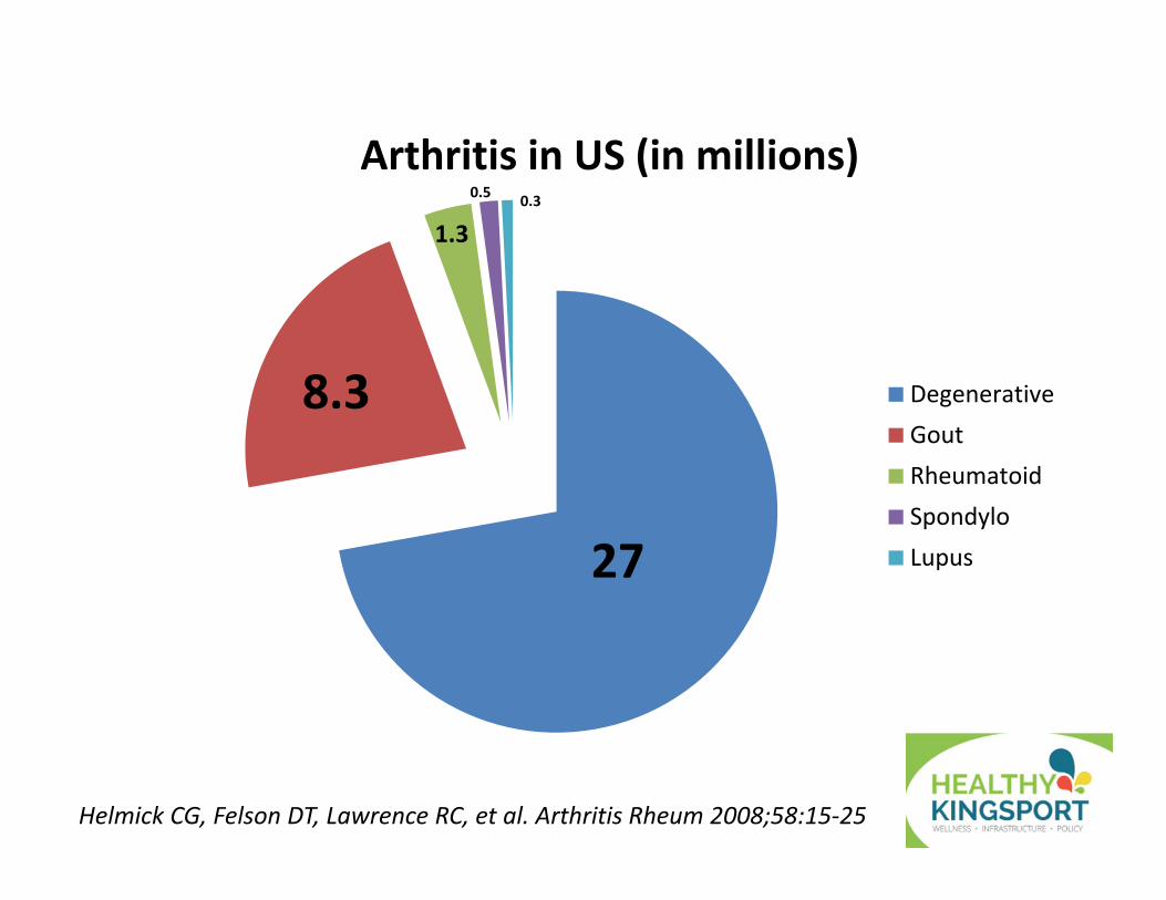
Arthritis in US (in millions)
DegenerativeGoutRheumatoidSpondyloLupus
8.3
1.3
0.5 0.3
Helmick CG, Felson DT, Lawrence RC, et al. Arthritis Rheum 2008;58:15‐25
27
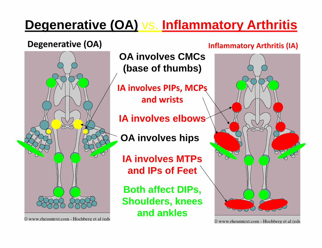
Degenerative (OA) vs. Inflammatory Arthritis
OA involves CMCs (base of thumbs)
IA involves PIPs, MCPs and wrists
OA involves hips
IA involves elbows
Degenerative (OA) Inflammatory Arthritis (IA)
IA involves MTPs and IPs of Feet
Both affect DIPs, Shoulders, knees
and ankles
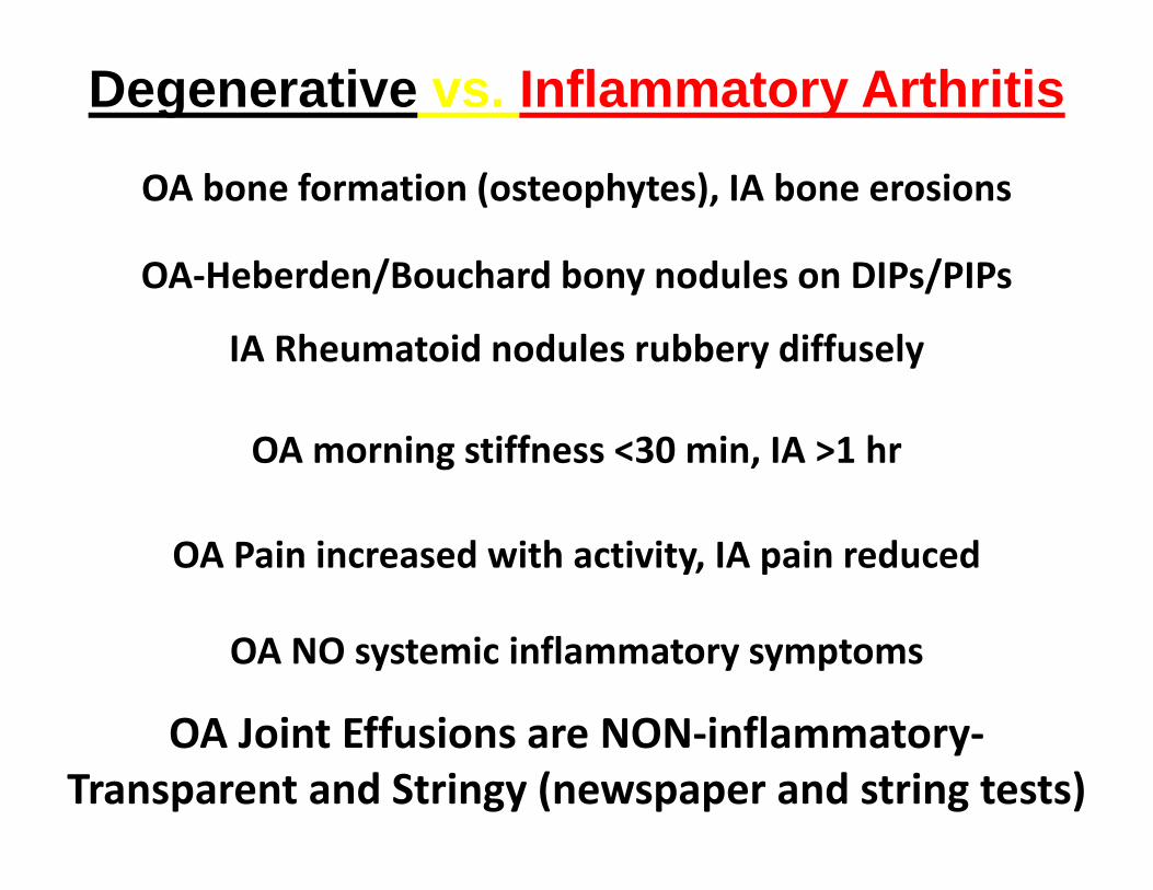
OA bone formation (osteophytes), IA bone erosions
OA‐Heberden/Bouchard bony nodules on DIPs/PIPs
IA Rheumatoid nodules rubbery diffusely
OA Pain increased with activity, IA pain reduced
OA morning stiffness <30 min, IA >1 hr
OA Joint Effusions are NON‐inflammatory‐Transparent and Stringy (newspaper and string tests)
OA NO systemic inflammatory symptoms
Degenerative vs. Inflammatory Arthritis
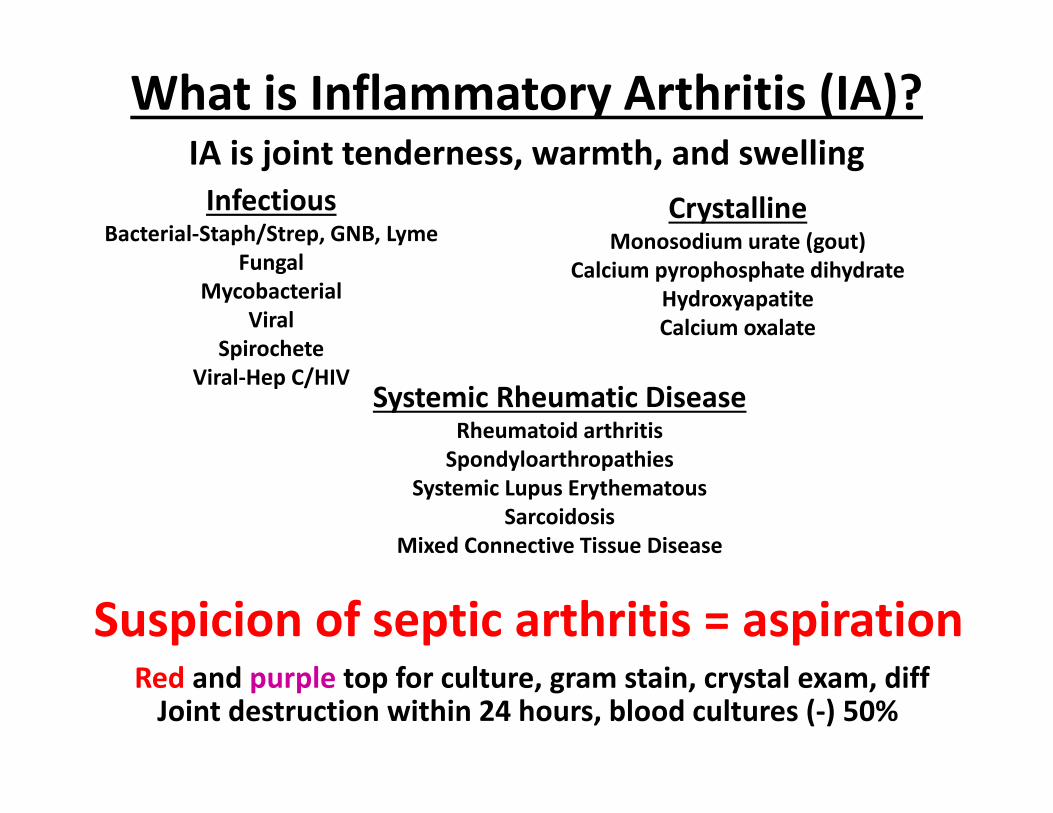
What is Inflammatory Arthritis (IA)?
InfectiousBacterial‐Staph/Strep, GNB, Lyme
Fungal Mycobacterial
ViralSpirochete
Viral‐Hep C/HIV
CrystallineMonosodium urate (gout)
Calcium pyrophosphate dihydrateHydroxyapatiteCalcium oxalate
Systemic Rheumatic DiseaseRheumatoid arthritisSpondyloarthropathies
Systemic Lupus ErythematousSarcoidosis
Mixed Connective Tissue Disease
Joint destruction within 24 hours, blood cultures (‐) 50%
Suspicion of septic arthritis = aspiration
IA is joint tenderness, warmth, and swelling
Red and purple top for culture, gram stain, crystal exam, diff
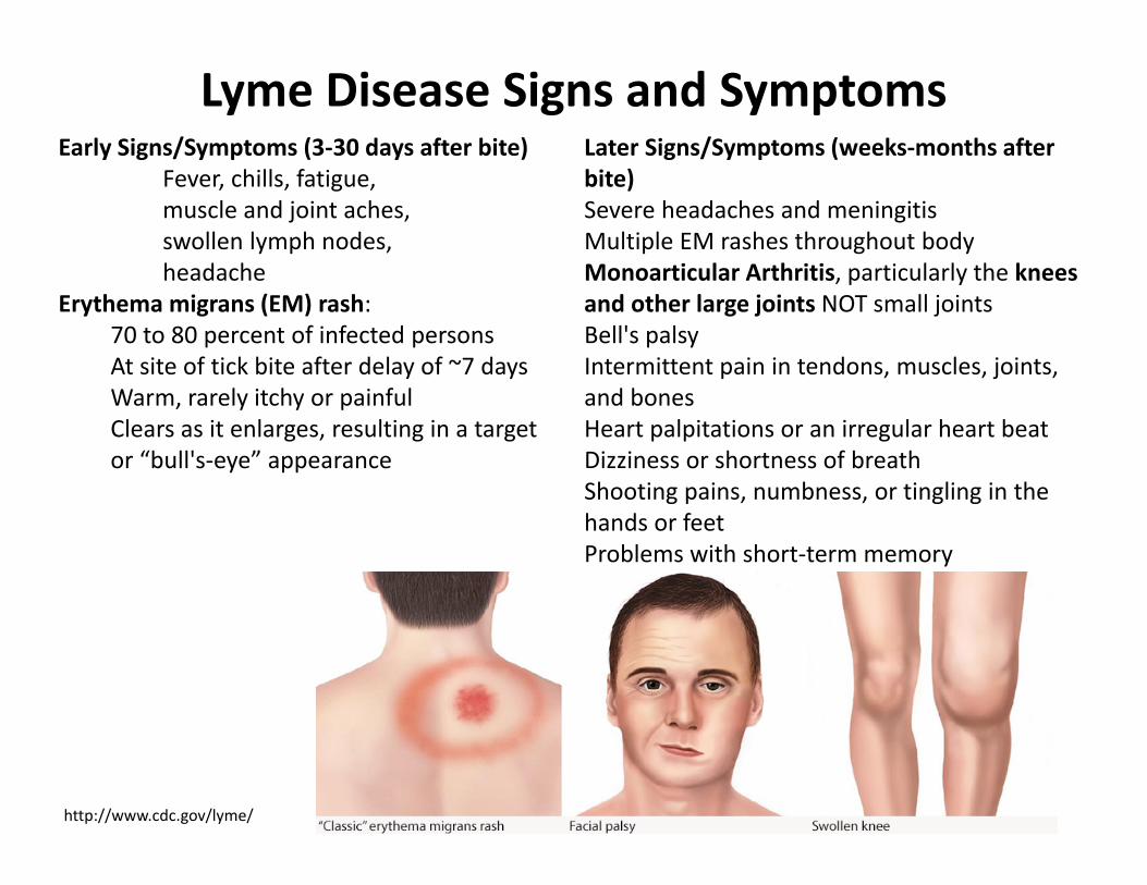
Early Signs/Symptoms (3‐30 days after bite)Fever, chills, fatigue, muscle and joint aches, swollen lymph nodes,headache
Erythema migrans (EM) rash:70 to 80 percent of infected personsAt site of tick bite after delay of ~7 daysWarm, rarely itchy or painfulClears as it enlarges, resulting in a target or “bull's‐eye” appearance
Later Signs/Symptoms (weeks‐months after bite)Severe headaches and meningitisMultiple EM rashes throughout bodyMonoarticular Arthritis, particularly the knees and other large joints NOT small jointsBell's palsy Intermittent pain in tendons, muscles, joints, and bonesHeart palpitations or an irregular heart beat Dizziness or shortness of breathShooting pains, numbness, or tingling in the hands or feetProblems with short‐term memory
Lyme Disease Signs and Symptoms
http://www.cdc.gov/lyme/

Who SHOULD be tested for Lyme Disease? Need High Pretest Probability of Positivity
A recent history of having resided in or traveled to an area endemic for Lyme disease.ANDA risk factor for exposure to ticks.ANDSymptoms consistent with early disseminated disease or late Lyme disease.
Patients with an erythema migrans (EM) rash should be TREATED not tested. Don’t test patients with non‐specific symptoms only (eg, fatigue, myalgias/arthralgias).
Who should NOT be tested for Lyme Disease? Everyone Else
Endemic Lyme Areas
http://www.cdc.gov/lyme/
http://www.cdc.gov/mmwr/preview/mmwrhtml/00038469.htm
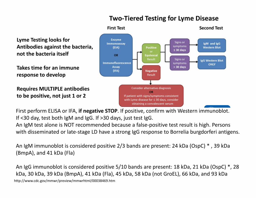
First perform ELISA or IFA, if negative STOP. If positive, confirm with Western immunoblot. If <30 day, test both IgM and IgG. If >30 days, just test IgG.An IgM test alone is NOT recommended because a false‐positive test result is high. Persons with disseminated or late‐stage LD have a strong IgG response to Borrelia burgdorferi antigens.
An IgM immunoblot is considered positive 2/3 bands are present: 24 kDa (OspC) * , 39 kDa(BmpA), and 41 kDa (Fla)
An IgG immunoblot is considered positive 5/10 bands are present: 18 kDa, 21 kDa (OspC) *, 28 kDa, 30 kDa, 39 kDa (BmpA), 41 kDa (Fla), 45 kDa, 58 kDa (not GroEL), 66 kDa, and 93 kDa
Lyme Testing looks for Antibodies against the bacteria, not the bacteria itself
Takes time for an immune response to develop
Requires MULTIPLE antibodies to be positive, not just 1 or 2
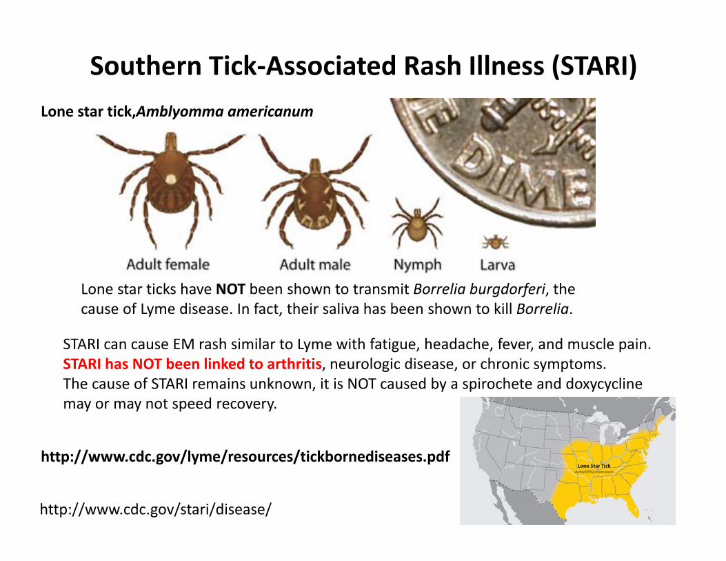
Lone star ticks have NOT been shown to transmit Borrelia burgdorferi, the cause of Lyme disease. In fact, their saliva has been shown to kill Borrelia.
Lone star tick,Amblyomma americanum
http://www.cdc.gov/stari/disease/
STARI can cause EM rash similar to Lyme with fatigue, headache, fever, and muscle pain. STARI has NOT been linked to arthritis, neurologic disease, or chronic symptoms.The cause of STARI remains unknown, it is NOT caused by a spirochete and doxycycline may or may not speed recovery.
Southern Tick‐Associated Rash Illness (STARI)
http://www.cdc.gov/lyme/resources/tickbornediseases.pdf
http://www.cdc.gov/lyme/resources/tickbornediseases.pdf
Am J Med Sci. 2013 Nov;346(5):427‐9
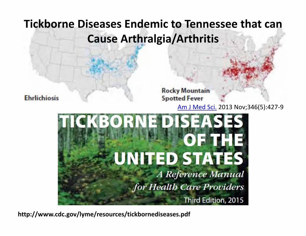
Tickborne Diseases Endemic to Tennessee that can Cause Arthralgia/Arthritis
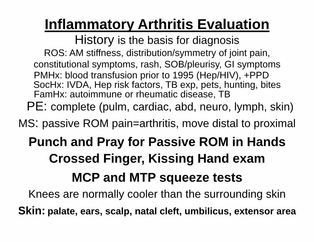
Inflammatory Arthritis Evaluation
PE: complete (pulm, cardiac, abd, neuro, lymph, skin)MS: passive ROM pain=arthritis, move distal to proximal
Knees are normally cooler than the surrounding skinSkin: palate, ears, scalp, natal cleft, umbilicus, extensor area
History is the basis for diagnosisROS: AM stiffness, distribution/symmetry of joint pain,
constitutional symptoms, rash, SOB/pleurisy, GI symptoms
SocHx: IVDA, Hep risk factors, TB exp, pets, hunting, bitesFamHx: autoimmune or rheumatic disease, TB
PMHx: blood transfusion prior to 1995 (Hep/HIV), +PPD
Crossed Finger, Kissing Hand examPunch and Pray for Passive ROM in Hands
MCP and MTP squeeze tests
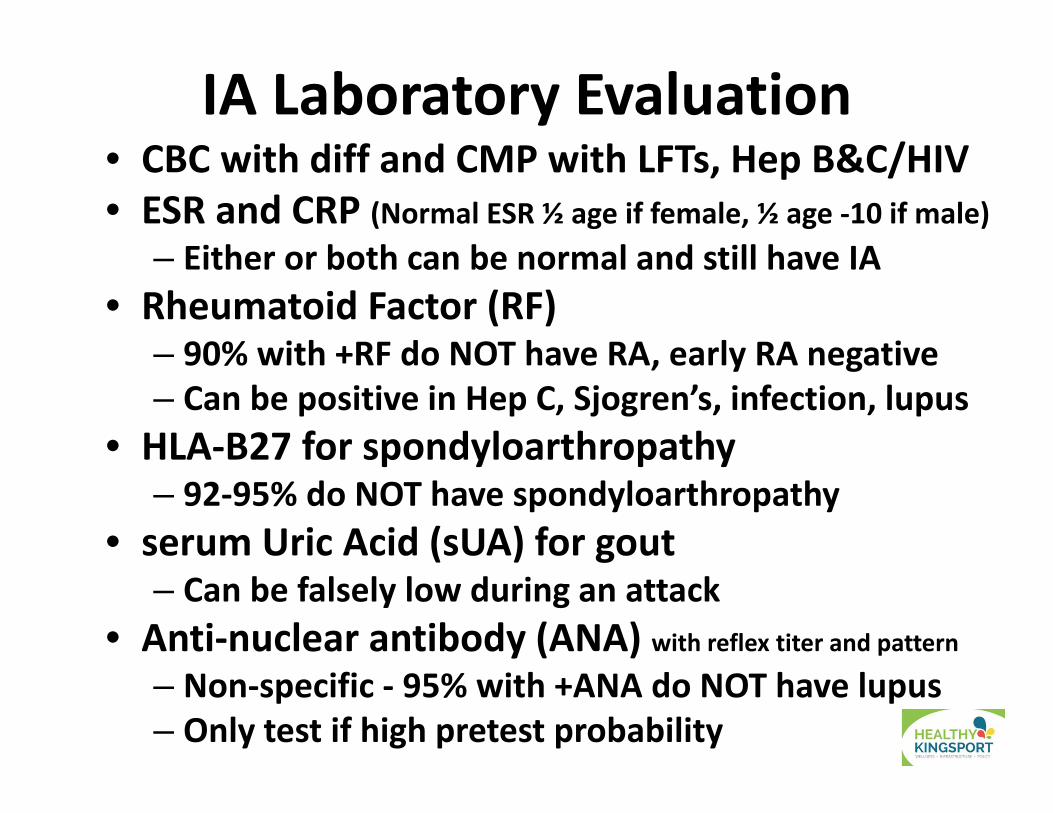
IA Laboratory Evaluation• CBC with diff and CMP with LFTs, Hep B&C/HIV• ESR and CRP (Normal ESR ½ age if female, ½ age ‐10 if male)
– Either or both can be normal and still have IA• Rheumatoid Factor (RF)
– 90% with +RF do NOT have RA, early RA negative– Can be positive in Hep C, Sjogren’s, infection, lupus
• HLA‐B27 for spondyloarthropathy– 92‐95% do NOT have spondyloarthropathy
• serum Uric Acid (sUA) for gout– Can be falsely low during an attack
• Anti‐nuclear antibody (ANA) with reflex titer and pattern– Non‐specific ‐ 95% with +ANA do NOT have lupus– Only test if high pretest probability
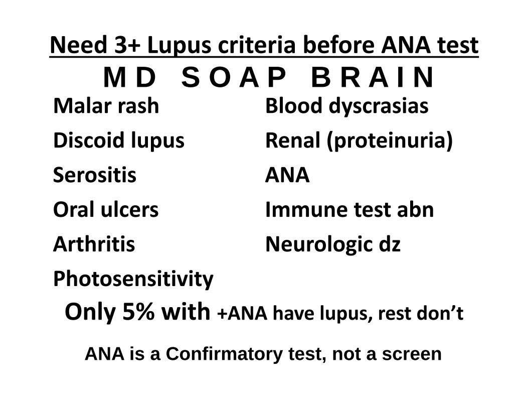
Need 3+ Lupus criteria before ANA test
Malar rashDiscoid lupusSerositisOral ulcersArthritisPhotosensitivity
Blood dyscrasiasRenal (proteinuria)ANAImmune test abnNeurologic dz
M D S O A P B R A I N
ANA is a Confirmatory test, not a screen
Only 5% with +ANA have lupus, rest don’t
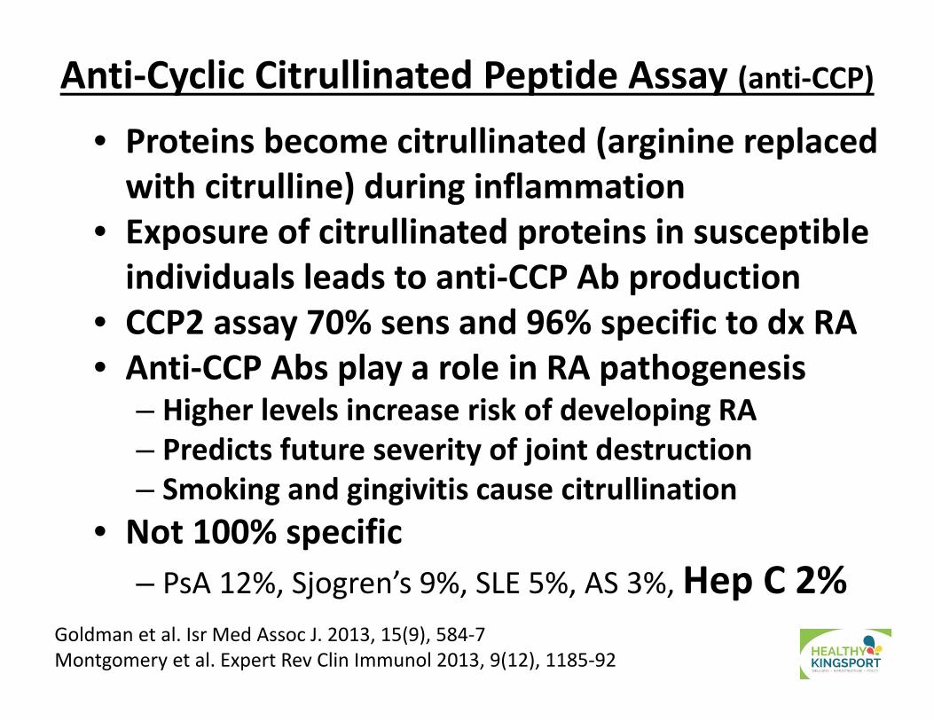
Anti‐Cyclic Citrullinated Peptide Assay (anti‐CCP)• Proteins become citrullinated (arginine replaced with citrulline) during inflammation
• Exposure of citrullinated proteins in susceptible individuals leads to anti‐CCP Ab production
• CCP2 assay 70% sens and 96% specific to dx RA• Anti‐CCP Abs play a role in RA pathogenesis
– Higher levels increase risk of developing RA– Predicts future severity of joint destruction– Smoking and gingivitis cause citrullination
• Not 100% specific – PsA 12%, Sjogren’s 9%, SLE 5%, AS 3%, Hep C 2%
Goldman et al. Isr Med Assoc J. 2013, 15(9), 584‐7Montgomery et al. Expert Rev Clin Immunol 2013, 9(12), 1185‐92
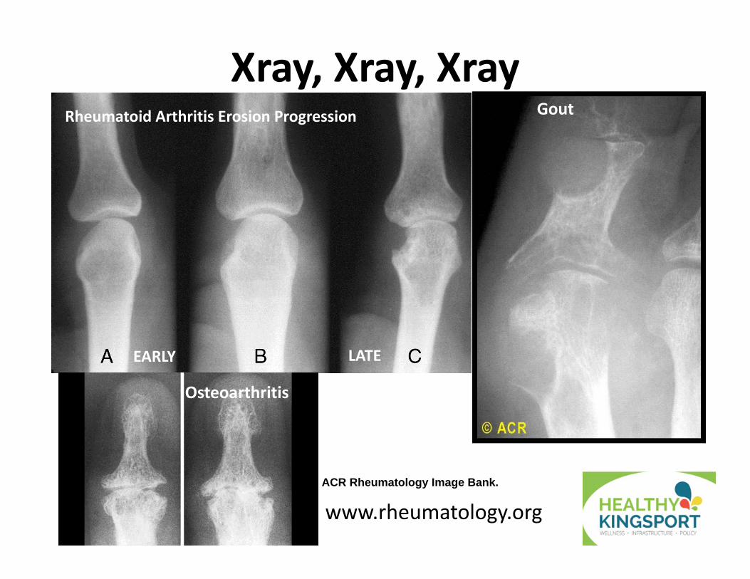
Xray, Xray, Xray
www.rheumatology.org
Rheumatoid Arthritis Erosion Progression Gout
Osteoarthritis
ACR Rheumatology Image Bank.
EARLY LATE
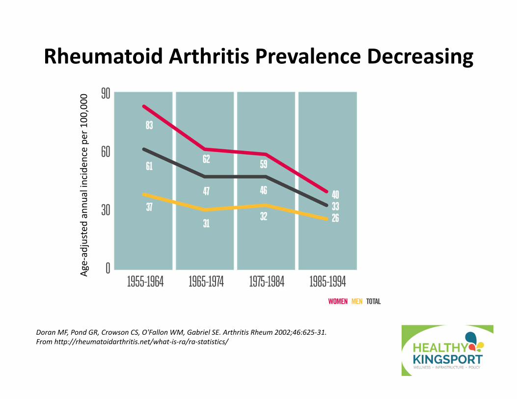
Doran MF, Pond GR, Crowson CS, O'Fallon WM, Gabriel SE. Arthritis Rheum 2002;46:625‐31.From http://rheumatoidarthritis.net/what‐is‐ra/ra‐statistics/
Rheumatoid Arthritis Prevalence DecreasingAg
e‐adjusted
ann
ual inciden
ce per 100,000
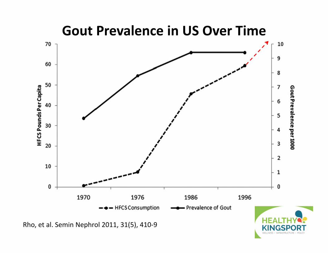
Rho, et al. Semin Nephrol 2011, 31(5), 410‐9
Gout Prevalence in US Over Time
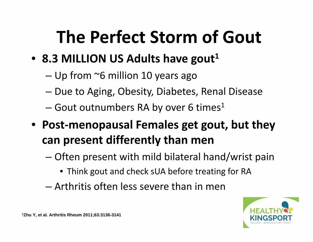
The Perfect Storm of Gout• 8.3 MILLION US Adults have gout1
– Up from ~6 million 10 years ago– Due to Aging, Obesity, Diabetes, Renal Disease– Gout outnumbers RA by over 6 times1
• Post‐menopausal Females get gout, but they can present differently than men– Often present with mild bilateral hand/wrist pain
• Think gout and check sUA before treating for RA
– Arthritis often less severe than in men
1Zhu Y, et al. Arthritis Rheum 2011;63:3136-3141
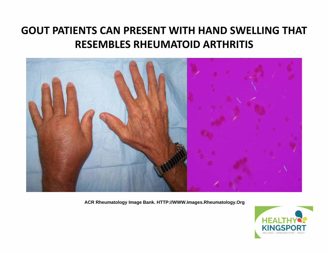
GOUT PATIENTS CAN PRESENT WITH HAND SWELLING THAT RESEMBLES RHEUMATOID ARTHRITIS
ACR Rheumatology Image Bank. HTTP://WWW.Images.Rheumatology.Org
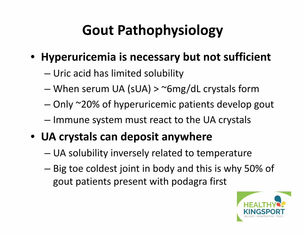
Gout Pathophysiology
• Hyperuricemia is necessary but not sufficient– Uric acid has limited solubility – When serum UA (sUA) > ~6mg/dL crystals form– Only ~20% of hyperuricemic patients develop gout– Immune system must react to the UA crystals
• UA crystals can deposit anywhere – UA solubility inversely related to temperature – Big toe coldest joint in body and this is why 50% of gout patients present with podagra first
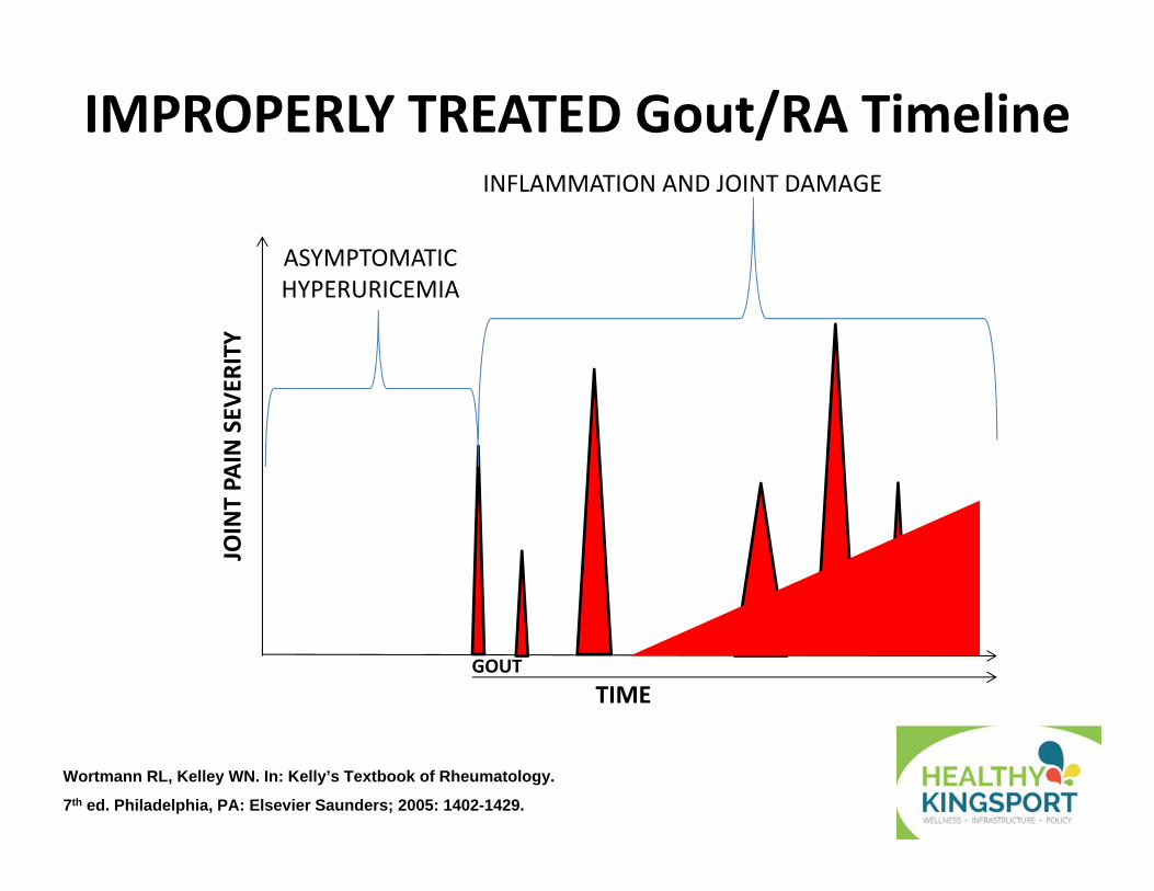
IMPROPERLY TREATED Gout/RA Timeline
Wortmann RL, Kelley WN. In: Kelly’s Textbook of Rheumatology.
7th ed. Philadelphia, PA: Elsevier Saunders; 2005: 1402-1429.
TIME
JOINT PA
IN SEV
ERITY
INFLAMMATION AND JOINT DAMAGE
ASYMPTOMATIC HYPERURICEMIA
GOUT
ACR Rheumatology Image Bank. HTTP://WWW.Images.Rheumatology.Org
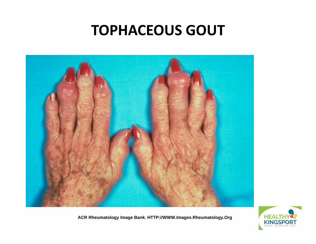
TOPHACEOUS GOUT
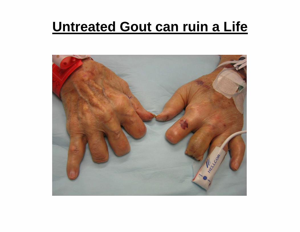
Untreated Gout can ruin a Life
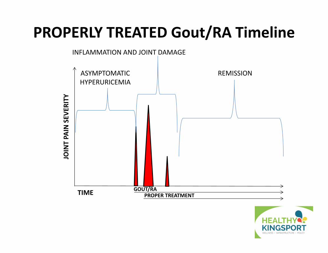
PROPERLY TREATED Gout/RA Timeline
TIME
JOINT PA
IN SEV
ERITY
INFLAMMATION AND JOINT DAMAGE
ASYMPTOMATIC HYPERURICEMIA
GOUT/RA PROPER TREATMENT
REMISSION
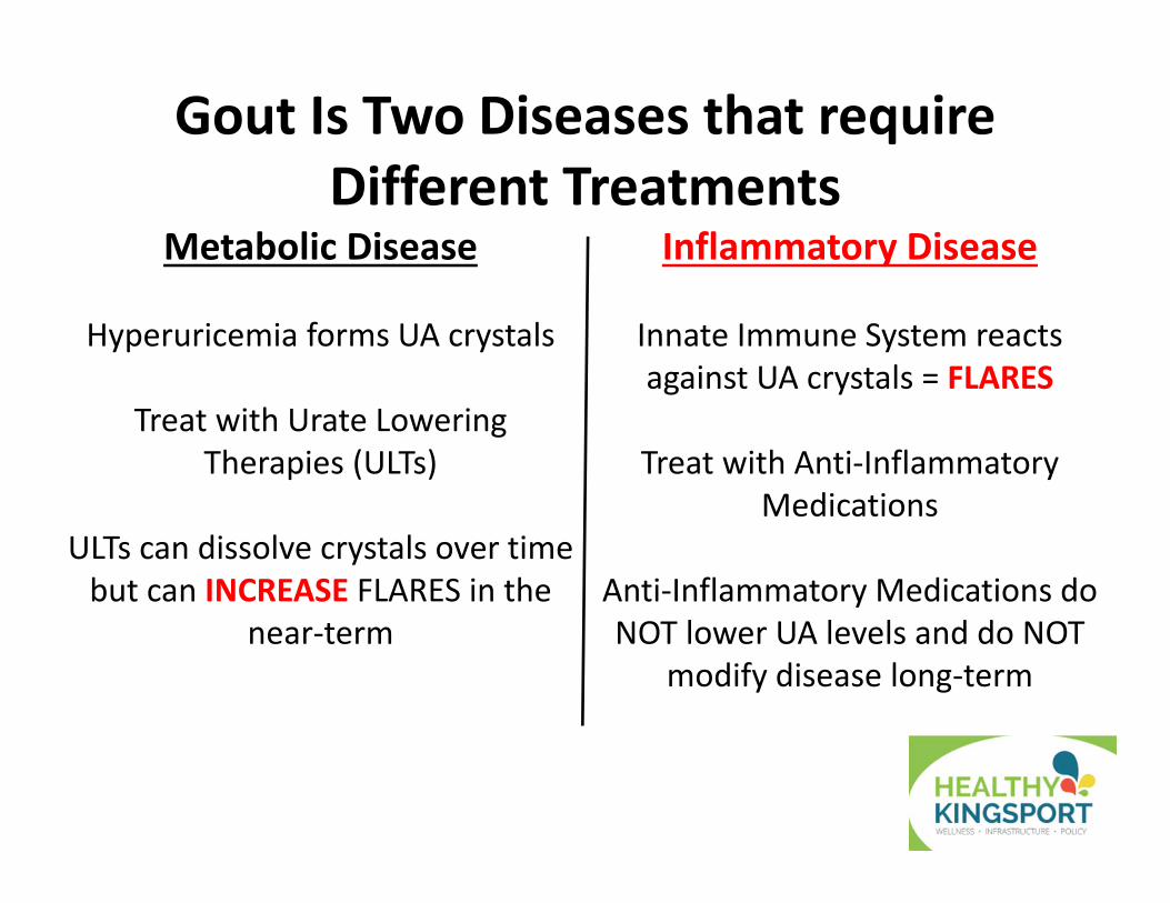
Gout Is Two Diseases that require Different Treatments
Inflammatory Disease
Innate Immune System reacts against UA crystals = FLARES
Treat with Anti‐Inflammatory Medications
Anti‐Inflammatory Medications do NOT lower UA levels and do NOT
modify disease long‐term
Metabolic Disease
Hyperuricemia forms UA crystals
Treat with Urate Lowering Therapies (ULTs)
ULTs can dissolve crystals over time but can INCREASE FLARES in the
near‐term

YOU MUST ASK ABOUT GOUT ATTACKS • Many patients do not know what gout is
– Runs in families‐may not know it’s a disease• Ask patient if they’ve ever had episode where joint got hot, red and painful that lasted a week
• Ask if they’ve ever received treatment or had a joint aspiration
• Ask how many episodes they’ve had and if anything seemed to precipitate the attacks– Seafood Buffet, NASCAR/Football/Hunting (beer)
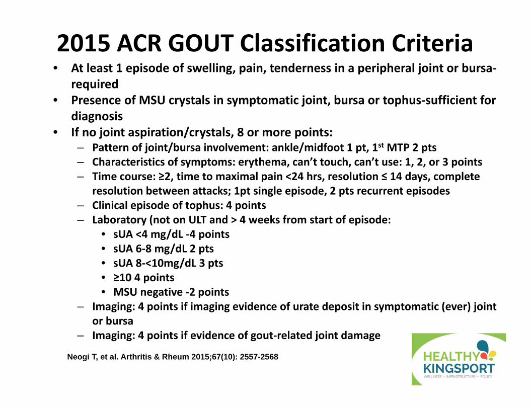
2015 ACR GOUT Classification Criteria• At least 1 episode of swelling, pain, tenderness in a peripheral joint or bursa‐
required• Presence of MSU crystals in symptomatic joint, bursa or tophus‐sufficient for
diagnosis• If no joint aspiration/crystals, 8 or more points:
– Pattern of joint/bursa involvement: ankle/midfoot 1 pt, 1st MTP 2 pts– Characteristics of symptoms: erythema, can’t touch, can’t use: 1, 2, or 3 points– Time course: ≥2, time to maximal pain <24 hrs, resolution ≤ 14 days, complete
resolution between attacks; 1pt single episode, 2 pts recurrent episodes– Clinical episode of tophus: 4 points– Laboratory (not on ULT and > 4 weeks from start of episode:
• sUA <4 mg/dL ‐4 points• sUA 6‐8 mg/dL 2 pts• sUA 8‐<10mg/dL 3 pts• ≥10 4 points• MSU negative ‐2 points
– Imaging: 4 points if imaging evidence of urate deposit in symptomatic (ever) joint or bursa
– Imaging: 4 points if evidence of gout‐related joint damage
Neogi T, et al. Arthritis & Rheum 2015;67(10): 2557-2568
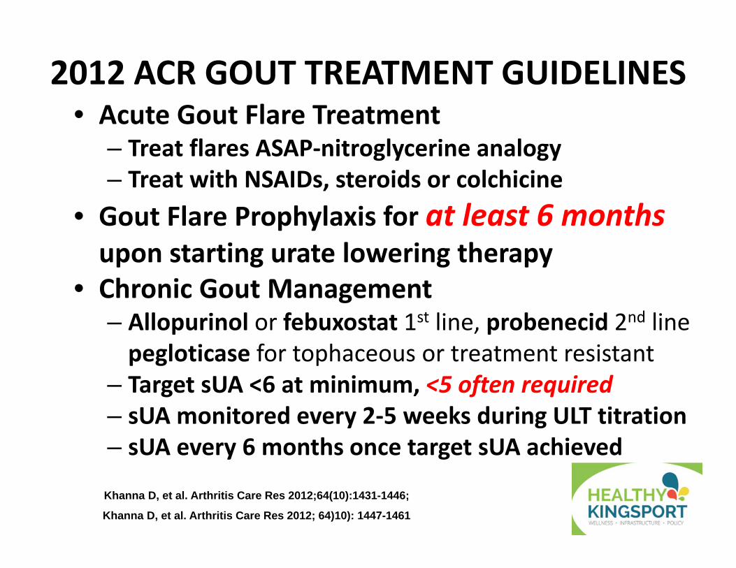
2012 ACR GOUT TREATMENT GUIDELINES• Acute Gout Flare Treatment
– Treat flares ASAP‐nitroglycerine analogy– Treat with NSAIDs, steroids or colchicine
• Gout Flare Prophylaxis for at least 6 months upon starting urate lowering therapy
• Chronic Gout Management– Allopurinol or febuxostat 1st line, probenecid 2nd linepegloticase for tophaceous or treatment resistant
– Target sUA <6 at minimum, <5 often required– sUA monitored every 2‐5 weeks during ULT titration – sUA every 6 months once target sUA achieved
Khanna D, et al. Arthritis Care Res 2012;64(10):1431-1446;
Khanna D, et al. Arthritis Care Res 2012; 64)10): 1447-1461
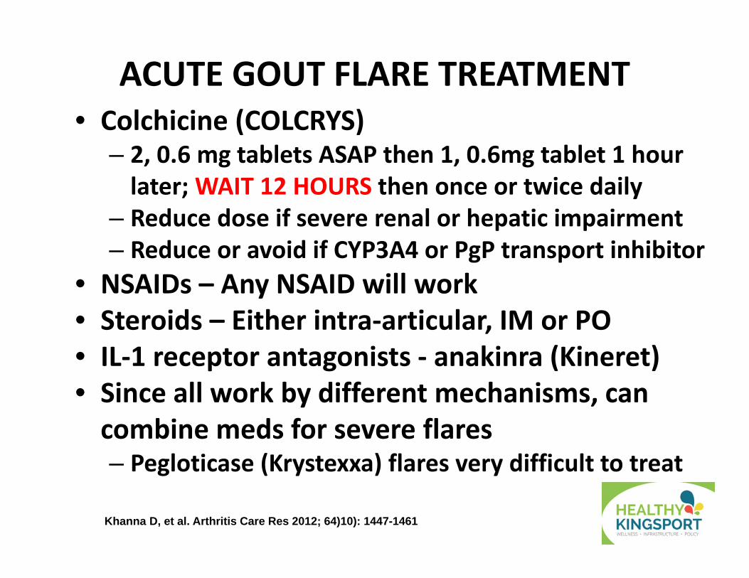
ACUTE GOUT FLARE TREATMENT• Colchicine (COLCRYS)
– 2, 0.6 mg tablets ASAP then 1, 0.6mg tablet 1 hour later; WAIT 12 HOURS then once or twice daily
– Reduce dose if severe renal or hepatic impairment– Reduce or avoid if CYP3A4 or PgP transport inhibitor
• NSAIDs – Any NSAID will work• Steroids – Either intra‐articular, IM or PO• IL‐1 receptor antagonists ‐ anakinra (Kineret)• Since all work by different mechanisms, can combine meds for severe flares– Pegloticase (Krystexxa) flares very difficult to treat
Khanna D, et al. Arthritis Care Res 2012; 64)10): 1447-1461
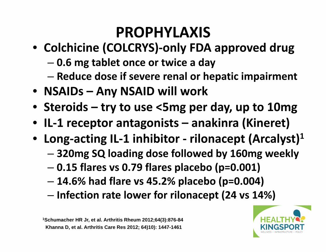
PROPHYLAXIS• Colchicine (COLCRYS)‐only FDA approved drug
– 0.6 mg tablet once or twice a day – Reduce dose if severe renal or hepatic impairment
• NSAIDs – Any NSAID will work• Steroids – try to use <5mg per day, up to 10mg• IL‐1 receptor antagonists – anakinra (Kineret)• Long‐acting IL‐1 inhibitor ‐ rilonacept (Arcalyst)1
– 320mg SQ loading dose followed by 160mg weekly– 0.15 flares vs 0.79 flares placebo (p=0.001)– 14.6% had flare vs 45.2% placebo (p=0.004)– Infection rate lower for rilonacept (24 vs 14%)
1Schumacher HR Jr, et al. Arthritis Rheum 2012;64(3):876-84Khanna D, et al. Arthritis Care Res 2012; 64)10): 1447-1461

CHRONIC GOUT MANAGEMENT• As long as there are UA crystals there will be inflammation– Joint damage can continue between flares– Must continue prophylaxis until all crystals gone
• Must keep sUA levels low dissolve all crystals– <6mg/dL at a minimum, <5mg/dL for many – The lower the sUA the faster crystals will dissolve– “Trust but verify” monitor sUA levels to ensure patients are compliant with therapy
– It takes ~2 years of low sUA to stop flares– If tophi present, 2 years after tophi resolve
Khanna D, et al. Arthritis Care Res 2012; 64)10): 1431-1446
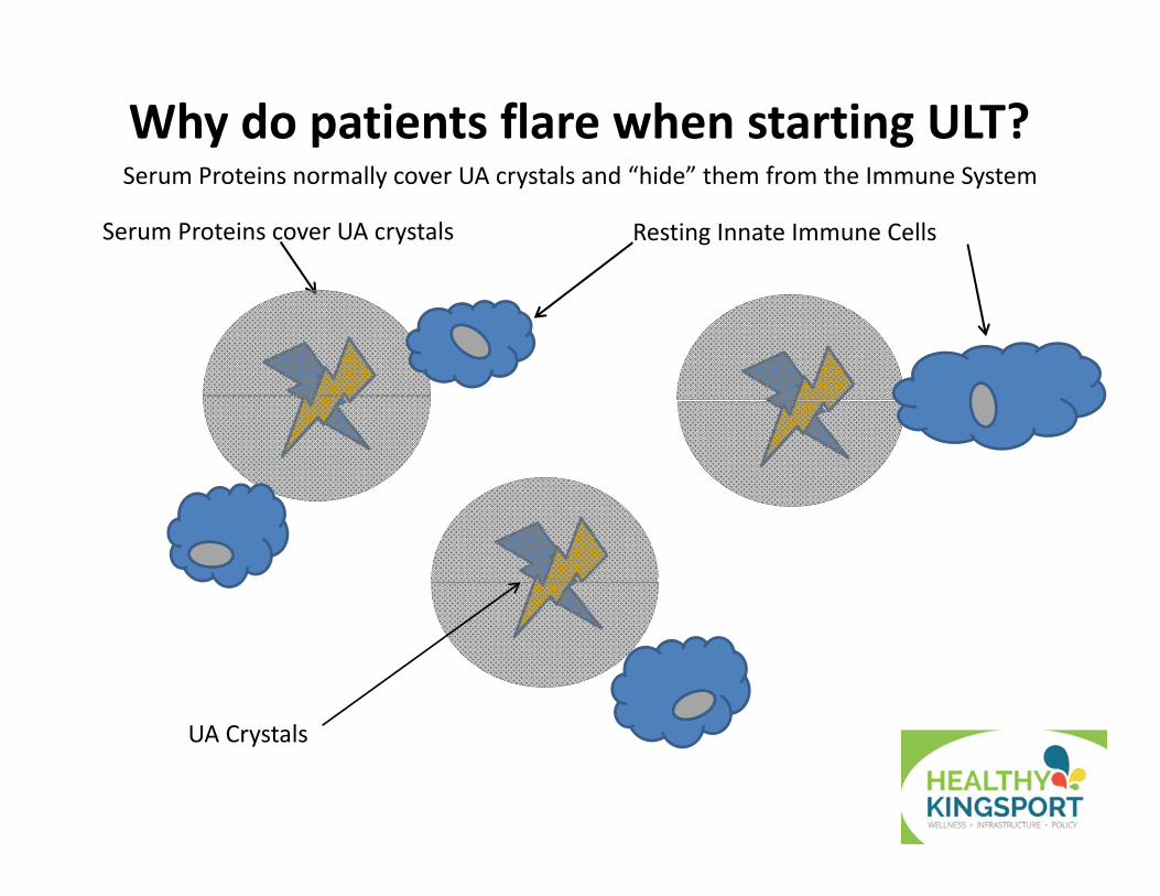
Why do patients flare when starting ULT?
Serum Proteins cover UA crystals Resting Innate Immune Cells
UA Crystals
Serum Proteins normally cover UA crystals and “hide” them from the Immune System
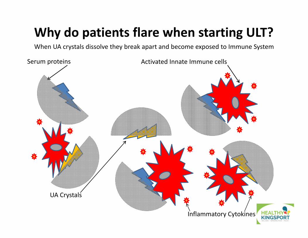
Serum proteins Activated Innate Immune cells
UA Crystals
Inflammatory Cytokines
Why do patients flare when starting ULT?When UA crystals dissolve they break apart and become exposed to Immune System
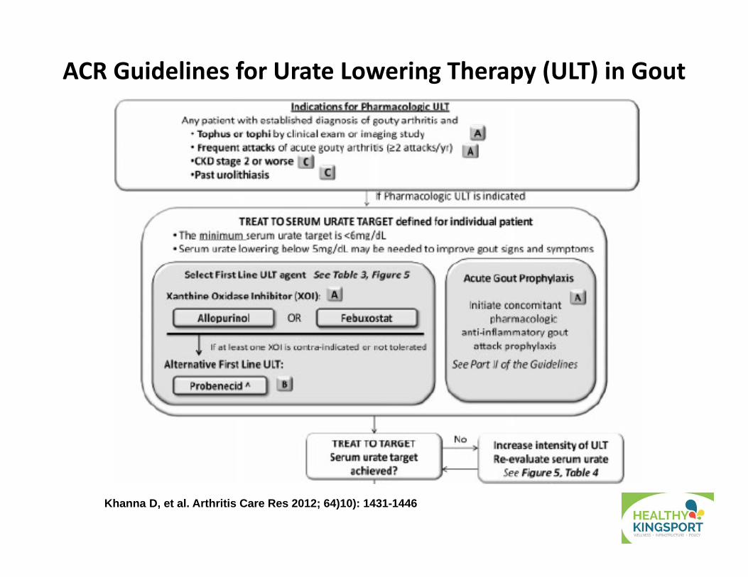
ACR Guidelines for Urate Lowering Therapy (ULT) in Gout
Khanna D, et al. Arthritis Care Res 2012; 64)10): 1431-1446
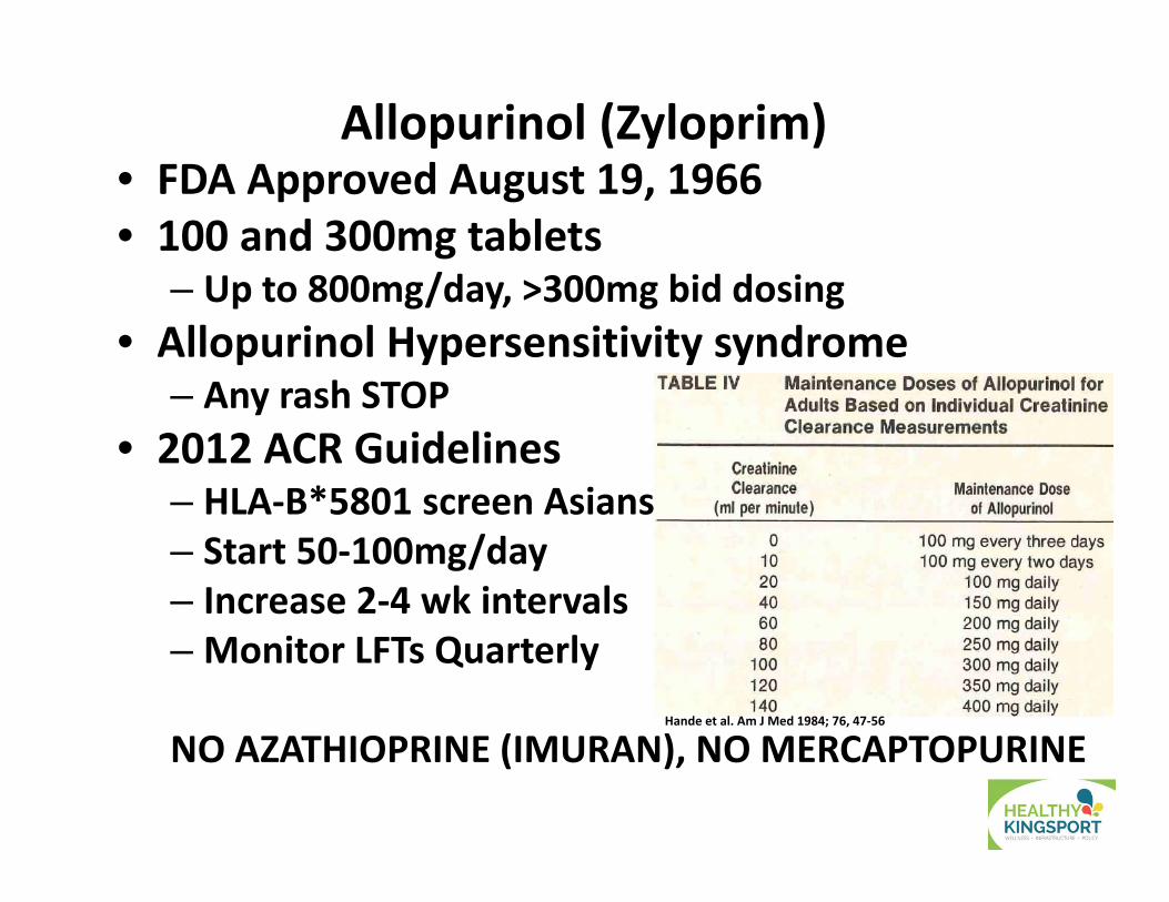
Allopurinol (Zyloprim)• FDA Approved August 19, 1966 • 100 and 300mg tablets
– Up to 800mg/day, >300mg bid dosing• Allopurinol Hypersensitivity syndrome
– Any rash STOP• 2012 ACR Guidelines
– HLA‐B*5801 screen Asians– Start 50‐100mg/day – Increase 2‐4 wk intervals– Monitor LFTs Quarterly
NO AZATHIOPRINE (IMURAN), NO MERCAPTOPURINEHande et al. Am J Med 1984; 76, 47‐56
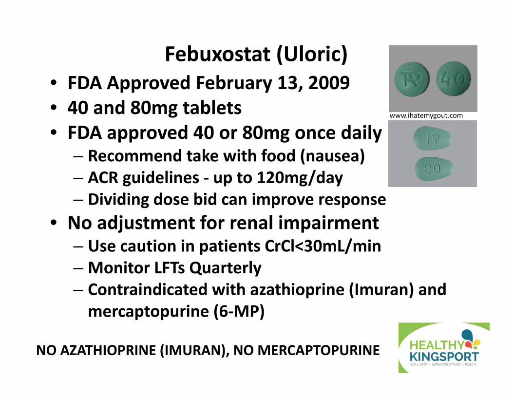
Febuxostat (Uloric)• FDA Approved February 13, 2009 • 40 and 80mg tablets• FDA approved 40 or 80mg once daily
– Recommend take with food (nausea)– ACR guidelines ‐ up to 120mg/day– Dividing dose bid can improve response
• No adjustment for renal impairment– Use caution in patients CrCl<30mL/min– Monitor LFTs Quarterly– Contraindicated with azathioprine (Imuran) and mercaptopurine (6‐MP)
www.ihatemygout.com
NO AZATHIOPRINE (IMURAN), NO MERCAPTOPURINE
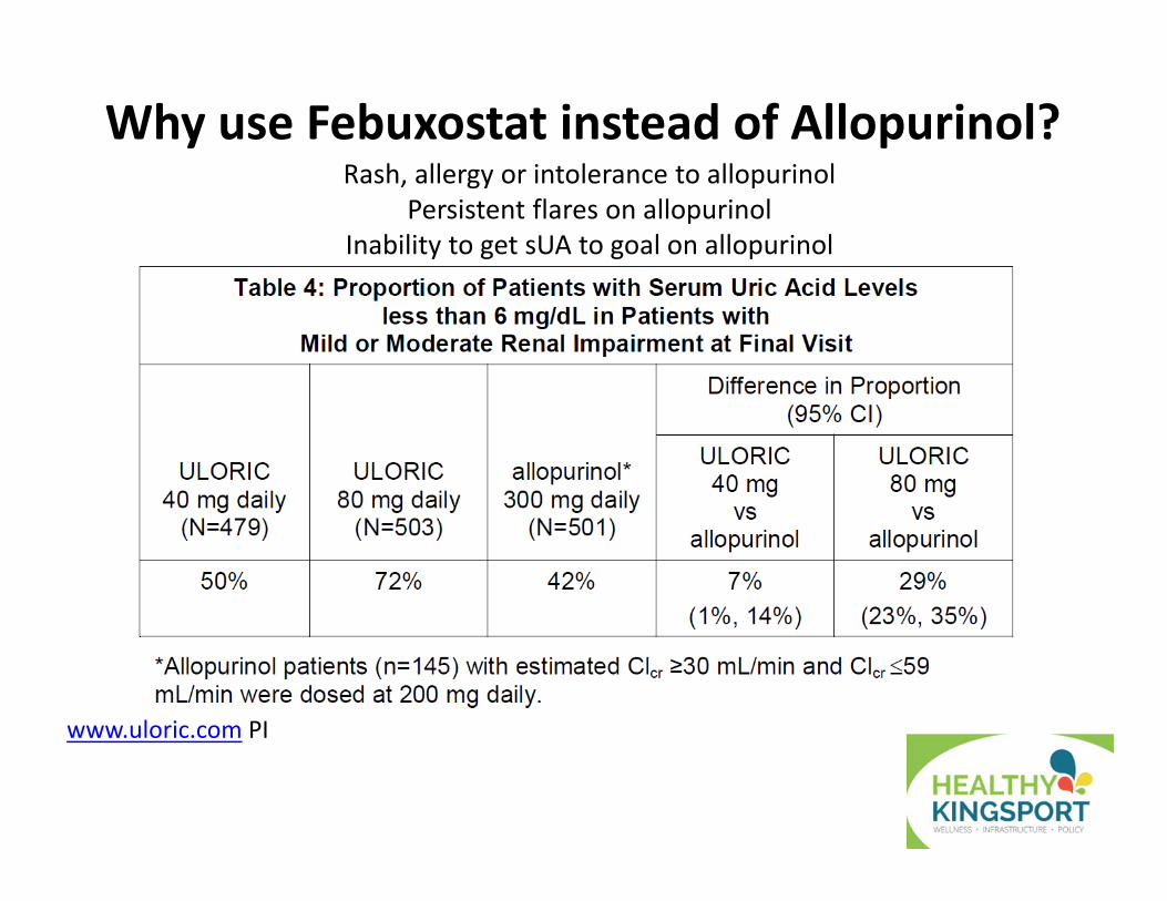
Why use Febuxostat instead of Allopurinol?
www.uloric.com PI
Rash, allergy or intolerance to allopurinolPersistent flares on allopurinol
Inability to get sUA to goal on allopurinol

Pegloticase (Krystexxa)• Pegylated uricase enzyme
– Patients that fail allopurinol and febuxostat– Drops sUA to ~0 mg/dL within 24 hrs
• Infused once every 2 weeks– Check sUA prior to infusion, don’t infuse if >6mg/dL– Anaphylaxis, CHF exacerbation– Pretreat with steroid, tylenol, and antihistamine
• FLARE PROPHYLAXIS essential• No renal restrictions• Cost prohibitive• Test for G6PD prior to dosing‐hemolytic anemia
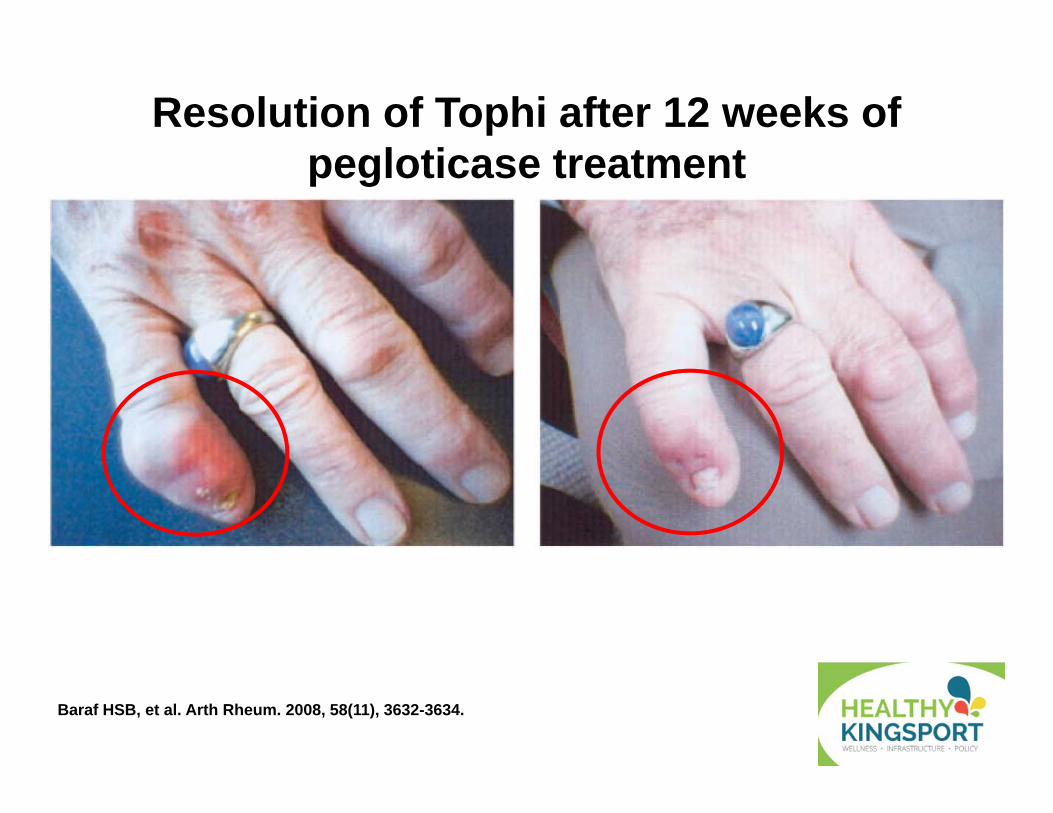
Resolution of Tophi after 12 weeks of pegloticase treatment
Baraf HSB, et al. Arth Rheum. 2008, 58(11), 3632-3634.
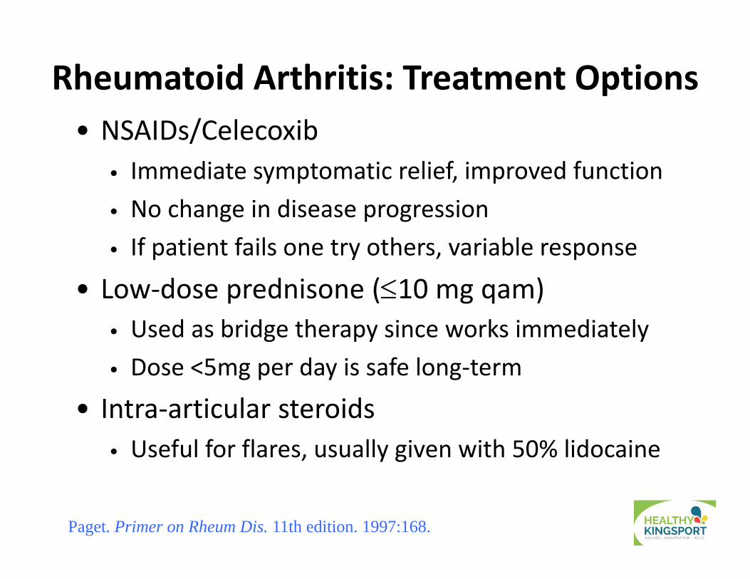
Rheumatoid Arthritis: Treatment Options • NSAIDs/Celecoxib
• Immediate symptomatic relief, improved function• No change in disease progression• If patient fails one try others, variable response
• Low‐dose prednisone (10 mg qam)• Used as bridge therapy since works immediately• Dose <5mg per day is safe long‐term
• Intra‐articular steroids • Useful for flares, usually given with 50% lidocaine
Paget. Primer on Rheum Dis. 11th edition. 1997:168.
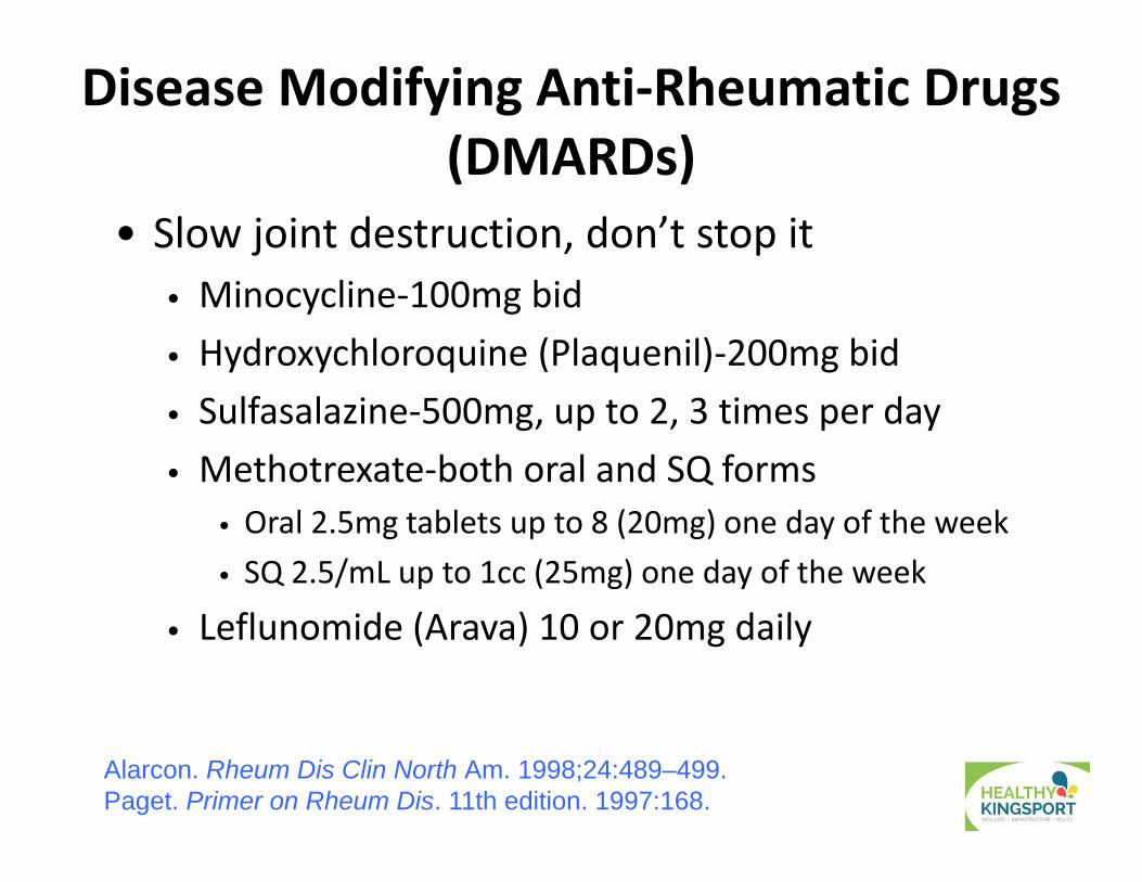
Disease Modifying Anti‐Rheumatic Drugs (DMARDs)
• Slow joint destruction, don’t stop it• Minocycline‐100mg bid• Hydroxychloroquine (Plaquenil)‐200mg bid• Sulfasalazine‐500mg, up to 2, 3 times per day• Methotrexate‐both oral and SQ forms
• Oral 2.5mg tablets up to 8 (20mg) one day of the week• SQ 2.5/mL up to 1cc (25mg) one day of the week
• Leflunomide (Arava) 10 or 20mg daily
Alarcon. Rheum Dis Clin North Am. 1998;24:489–499.Paget. Primer on Rheum Dis. 11th edition. 1997:168.
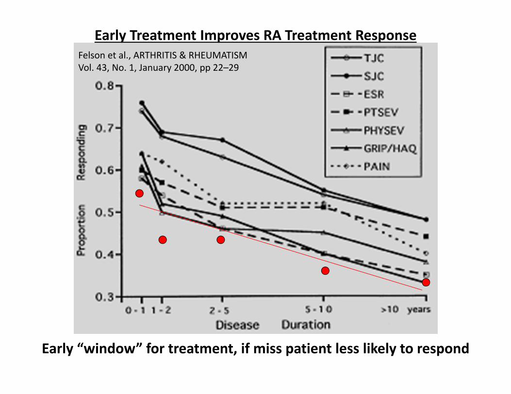
Felson et al., ARTHRITIS & RHEUMATISMVol. 43, No. 1, January 2000, pp 22–29
Early Treatment Improves RA Treatment Response
Early “window” for treatment, if miss patient less likely to respond
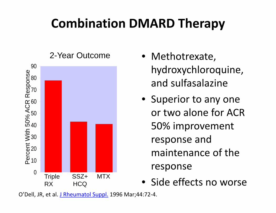
Combination DMARD Therapy
• Methotrexate, hydroxychloroquine, and sulfasalazine
• Superior to any one or two alone for ACR 50% improvement response and maintenance of the response
• Side effects no worse0
10
20
30
40
50
60
70
80
902-Year Outcome
Per
cent
With
50%
AC
R R
espo
nse
TripleRX
SSZ+HCQ
MTX
O’Dell, JR, et al. J Rheumatol Suppl. 1996 Mar;44:72‐4.
Monitoring Treatment With DMARDs• DMARDs require frequent monitoring• Blood, liver, lung, and kidney are frequent sites of adverse effects
• Interval of laboratory testing varies with the drug• 4‐ to 12‐week intervals are commonly needed
• Most patients need to be seen 2 to 6 times a year for physical exam• Monitor for lung disease, lymphoma, rash
Biologic Agents for RA Treatment• Biologics only treatments that STOP damage• Infliximab (Remicade) 3‐10mg/kg IV q4‐8 wks
• FDA approved 8/24/1998• chimeric monoclonal antibody that binds to TNFα, interes with endogenous TNFα activity
• Due to murine portion, can cause formation of neutralizing antibodies‐ given with MTX to prevent
• Typically used in Medicare patients since infusion covered under part B
• Largely replaced by SQ biologics
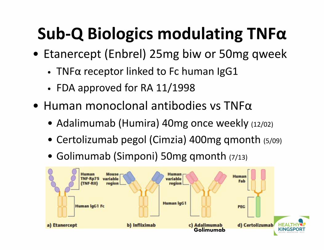
Sub‐Q Biologics modulating TNFα• Etanercept (Enbrel) 25mg biw or 50mg qweek
• TNFα receptor linked to Fc human IgG1 • FDA approved for RA 11/1998
• Human monoclonal antibodies vs TNFα• Adalimumab (Humira) 40mg once weekly (12/02)• Certolizumab pegol (Cimzia) 400mg qmonth (5/09)
• Golimumab (Simponi) 50mg qmonth (7/13)
Golimumab
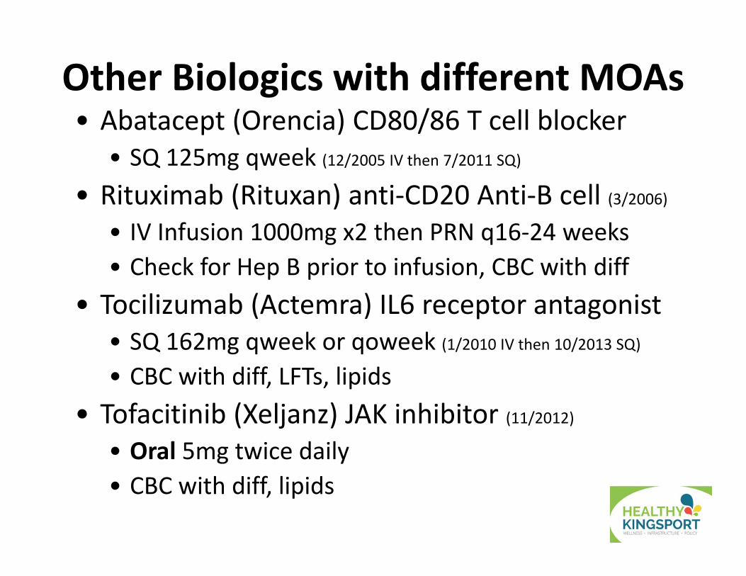
Other Biologics with different MOAs• Abatacept (Orencia) CD80/86 T cell blocker
• SQ 125mg qweek (12/2005 IV then 7/2011 SQ)
• Rituximab (Rituxan) anti‐CD20 Anti‐B cell (3/2006)• IV Infusion 1000mg x2 then PRN q16‐24 weeks • Check for Hep B prior to infusion, CBC with diff
• Tocilizumab (Actemra) IL6 receptor antagonist• SQ 162mg qweek or qoweek (1/2010 IV then 10/2013 SQ)
• CBC with diff, LFTs, lipids• Tofacitinib (Xeljanz) JAK inhibitor (11/2012)
• Oral 5mg twice daily• CBC with diff, lipids
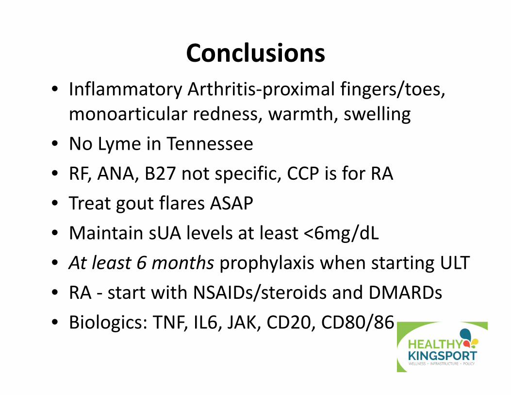
Conclusions• Inflammatory Arthritis‐proximal fingers/toes, monoarticular redness, warmth, swelling
• No Lyme in Tennessee• RF, ANA, B27 not specific, CCP is for RA• Treat gout flares ASAP• Maintain sUA levels at least <6mg/dL• At least 6 months prophylaxis when starting ULT• RA ‐ start with NSAIDs/steroids and DMARDs• Biologics: TNF, IL6, JAK, CD20, CD80/86
Boomershine Wellness CentersPersonalized Rheumatology Care
Inflammatory ArthritisChad S. Boomershine, MD, PhDMedical Director, Boomershine Wellness Centers
Medical Director, Elite Healthcare AllianceAssistant Professor of Medicine, Vanderbilt University
1195 Old Hickory Blvd, Suite 102Brentwood, TN 37027
Phone 615‐435‐3235 Fax 615‐435‐3275