both a monoclonal antibody and antisera …...these antisera have high titer in biological assays...
TRANSCRIPT
B O T H A M O N O C L O N A L A N T I B O D Y AND A N T I S E R A
SPECIFIC F O R D E T E R M I N A N T S U N I Q U E T O I N D I V I D U A L
C L O N E D H E L P E R T C E L L LINES CAN S U B S T I T U T E F O R
A N T I G E N A N D A N T I G E N - P R E S E N T I N G CELLS IN T H E
A C T I V A T I O N OF T CELLS*
By JONATHAN KAYE,* STEVEN PORCELLI, JOHN TITE, BARRY JONES, AND CHARLES A. JANEWAY, JR. a
From the Department of Pathology, Yale University School of Medicine, New Haven, Connecticut 06510
The proliferation of helper T cells and the T-dependent induction of B cell activation are events that are initiated by T cell recognition of antigen in association with Ia glycoproteins. However, the molecular basis of helper T cell antigen recognition remains unknown. Like several other laboratories, we have approached this problem by raising antibodies that are individually specific for functionally characterized cloned T cell lines. Clone-specific antibodies raised against alloreactive Ia-recognizing T cell clones (1), antigen-Ia-specific helper T cell hybrids (2), a human cytotoxic HLA-A recognizing T cell clone (3), and a murine H-2 recognizing cytotoxic T cell clone (4) have already been described. These clone-specific antibodies are either stimulatory themselves, as is the case of antisera raised to alloreactive T cell clones, or inhibit antigen-driven T cell activation, as are antibodies raised against helper T cell hybrids. However, stimulatory antibodies specific for major histocompatibility complex (MHC) 1 restricted, antigen-specific cloned helper T cell lines have not been described. In addition, the question of whether a monoclonal antibody of this specificity can activate a T cell has never been addressed.
In this paper, we describe antisera that both activate the T cells themselves and induce T cell-dependent B cell activation. In addition, we have produced a monoclonal antibody specific for a single cloned helper cell line that can also activate the T cells of this line. The anti-clone antibodies we have produced appear to completely replace the initial antigen-Ia recognition event by the T
* Supported by grants AI-14579 and CA-29606 from the National Institutes of Health (NIH), and by the Howard Hughes Medical Institute.
*J. Kaye was initially supported by NIH Training Grant T-32 AI-07019. 0 Investigator, Howard Hughes Medical Institute. Abbreviations used in this paper." BSA, bovine serum albumin; Con A, concanavalin A; FCS, fetal
calf serum; IL-1, interleukin 1; IL-2, interleukin 2; LPS, lipopolysaccharide; MHC, major histocom- patibility complex; OVA, ovalbumin; PBS, phosphate-buffered saline; RaMBr, rabbit anti-mouse brain serum; SDS-PAGE, sodium dodecyl sulphate polyacrylamide gel electrophoresis.
836 J. ExP. MED. © The Rockefeller University Press • 0022-1007/83/09/0836/21 $1.00 Volume 158 September 1983 836-856
KAYE ET AL. 837
cell, while apparently not affecting the growth factor requirements of the ensuing pathway of activation. These findings thus strongly support the supposition that clone-specific antibodies react with antigen and /o r Ia recognition sites.
The experiments presented here utilize antisera raised against two cloned helper T cell lines that have identical functional properties and genotype, but differ in nominal antigen specificity and in recognition of non-self Ia molecules. These antisera have high titer in biological assays and are absolutely specific for the immunizing clone. The ability of these sera, and the monoclonal antibody, to activate the appropriate T cell is dependent on a second signal from an accessory cell. The requirement for accessory cells, however, can be eliminated by the addition of interleukin-1 (IL-1) or interleukin-2 (IL-2). Thus, this system allows the analysis of the activation of a homogeneous T cell population in the complete absence of other cell types. Although similar to mitogen stimulation, the specificity of the interaction of T cells with anti-clone antibodies should make this system more analogous to an antigen-driven reaction. The effect of clone specific anti-T cell sera on T-dependent B cell activation, the major function of T helper cells, has also not previously been studied. The clone-specific antibodies we describe allow T cells to interact with B cells to induce B cell proliferation and immunoglobulin secretion in the absence of antigen recognition or MHC restriction. Thus, these antibodies mimic many facets of antigen-specific T cell interactions with Ia-bearing cells, and should allow a functional and ultimately molecular dissection of helper T cell "behavior."
Materials and Methods
Animals.
Animals were either purchased from the Jackson Laboratory, Bar Harbor, ME, or raised in our colony at Yale University. All animals were between 2 and 6 mo of age at the time they were used.
Production o fT Cell Clones D8 and D IO. To produce cloned helper T cell lines, the procedure of Sredni et al. (5) as modified
by our laboratory (6) was followed. In this case, two AKR/J mice were primed, each with 50 ug 5×-recrystallized hens egg albumin (OVA) (United States Biochemical Co., Cleve- land, OH) in complete Freund's adjuvant containing M. tuberculosis strain H37Ra (Difco Laboratories Inc., Detroit, MI). Mice were immunized in the hind footpads and base of the tail. 6 d later, the draining popliteal and inguinal lymph nodes were removed, the T cells purified by passage over Ig-anti-Ig (7) columns and cultured in Click's EHAA medium containing 200 #g/ml OVA and 5% fetal calf serum (FCS)selected for low mitogenicity. 20 × 10°lymph node T cells were cultured with 5 × 10⁸ mitomycin C-inactivated syngeneic spleen cells in 25-cm 2 flasks for 3 d. The cells were then pelleted and resus- pended in Click's medium containing 20% FCS, 200 #g/ml OVA, and 0.33% agar (Difco Bacto Agar). This mixture was layered into a 60-mm petri dish containing 2 ml of 0.5% agar in Click's medium with 20% FCS and 200/~g/mi OVA. After several days, numerous clusters or colonies of cells appeared. These were plucked with a pasteur pipette and transferred to a 96-well microtiter plate containing Click's medium, 200 /~g/ml OVA, 10% of a growth supporting FCS, 20% rat Con A supernatant as a source of growth factors and 2 x 10⁵ mitomycin C-treated syngeneic spleen cells/well. Most colonies gave rise to vigorous growth. 20 lines were expanded to 1-ml cultures in multiwell plates and tested for proliferative responses to OVA in the context of syngeneic mitomycin C
838 ST1MULATORY ANTIBODIES SPECIFIC FOR HELPER T CELL CLONES
inactivated spleen cells. 19 /20 gave significant responses to OVA. Two of these, D 10 and D8, were characterized extensively. D 10 was also recloned twice in succession by limiting dilution, first in U-bottom microtiter plates, where growth was obtained at low efficiency at 1 cell/well, and subsequently in V-bottom microtiter plates, where growth was obtained with close to 100% efficiency at 0.5 cells/well. Clone D10.G4. I was obtained from one of these wells. Cells were expanded in 25 and 75 cm 2 tissue culture flasks fed twice per week with medium and antigen, and weekly with 5-10 x 105 mitomycin C-inactivated spleen cells per milliliter of culture. Vigorous growth has been maintained for 18 mo under these conditions with these cloned lines. Recently, we have found that the United States Biochemical Co. lot of OVA we have used contains conalbumin as a contaminant. D 10 and its subclones have since been demonstrated to exhibit antigen specificity for this protein, and not ovalbumin, and are currently maintained using commercially purified conalbumin (Sigma Chemical Co., St. Louis, MO) as antigen. D8 has been grown with various batches of ovalbumin, including one commercially prepared lot that we have found to be conalbumin free (Sigma Chemical Co.).
Antibodies. Anti-DlO and Anti-D8. Anti-T cell antisera were produced by weekly immunizations
of BALB.K mice with 3-10 x 106 cloned T cells intraperitoneally in phosphate-buffered saline (PBS). Cloned T cells were harvested 7-10 d after the last addition of feeder cells and washed several times in PBS before injection. Mice were bled 1 wk after the last immunization and the sera collected and tested. After four injections with D10, or its subclones, the immune sera had detectable stimulatory activity in a T cell proliferation assay. These mice were subsequently boosted and bled on alternate weeks. Anti-D10 is a pooled serum from these animals, derived from multiple bleeds. Six out of six BALB.K mice have produced anti-D10 activity by this protocol. Two other BALB.K mice also produced anti-D10 activity following a single boost 3 wk after priming. Two out of four (AKR x B6)F1 mice have produced anti-Dl0 activity after six immunizations. The cloned T cells (D10.G4.1) used to immunize these F1 mice were grown exclusively with AKR/J feeder cells.
Two BALB.K mice were also immunized with D8 cells by the above procedure. One of these mice produced serum with detectable activity after the seventh immunization and was subsequently boosted. Anti-D8 is a pool of serum from this animal, derived from multiple bleeds.
Monoclonal Anti-DlO Antibody (3D3). A BALB.K mouse was immunized with 5 x 10⁶ D10.G4.1 intraperitoneally in PBS and boosted 3 wk later with 4 × 10⁶ D10.G.4.1 intravenously. 3 d later the immune spleen cells were fused to P3X63-Ag8.653 cells as described previously (8). The resulting hybridoma culture supernatants were screened for the induction of D 10.G4.1 proliferation in the absence of accessory cells and the presence of 1-5 % of an IL- 1 rich P388D 1 supernatent. Out of 120 hybridomas screened, 1, 3D3, was positive in this assay. 3D3 was subsequently cloned on agar and by limiting dilution. ~ 9 0 % of the 3D3 clones tested (39 total) were positive in the above assay. Two agar clones, 3D3.1 and 3D3.2, were used in experiments presented here. 3D3 is an IgGl- secreting hybridoma.
Monoclonal Anti-Fc Receptor. This antibody, 2.4G2, was the generous gift of Dr. I. Mellman (Yale University, New Haven, CT) and has been described previously (9).
Rabbit Anti-Mouse Brain (RaMBr). RaMBr serum was produced as described previously (10).
Assays. All assays, except the T-dependent induction of B cell immunoglobulin secretion, were
performed in Click's medium containing 5% FCS. The induction of polyclonal immuno- globulin secretion was performed in RPMI-1640 containing 10% FCS and 60 #M 2- mercaptoethanol. All proliferation assays were harvested with a PHD Cell Harvesting System (Cambridge Technology, Inc., Cambridge, MA). Other methods were as follows:
T Cell Proliferation. 2 × 10 ̀4 cloned T cells were cultured in 0.2 ml containing feeder
KAYE ET AL. 839
cells inactivated with mitomycin C (Sigma Chemical Co.) and various additions. Feeder cells were either whole spleen cells (2 × 10⁵ cells/culture) or splenic adherent cells (2 × 10⁴ cells/culture). Splenic adherent cells were prepared by a modification of the method ofSteinman et al. (11). Briefly, whole spleen cells (1 × 10⁷ cells/ml in Dulbecco's Modified Eagle Medium containing 10% FCS) were allowed to adhere to plastic petric dishes (100 mm; Costar, Cambridge MA). After 2 h at 37°C, nonadherent cells were removed by gentle washing with warmed medium in the absence of FCS. Medium containing FCS was replaced and the incubation continued overnight at 37°C. At the end of this incubation period, adherent cells were harvested by vigorous pipetting with warmed medium. Cell yields range from 2-8% of the input cell number. In some experiments soluble factors were added in place of feeder cells. Cultures were harvested after 72 h following a 3-4 h (70 Ci/mmol) or 16 h (6.7 Ci/mmol) 3H-TdR (New England Nuclear, Boston, MA) pulse of 1 ~Ci/culture.
T-Dependent B Cell Proliferation and Immunoglobutin Secretion. Cloned T cells were inactivated with mitomycin C and added to B cells as previously described (12). B cells were prepared by treatmt qt of whole spleen cells with a rat monoclonal antibody that reacts with a nonpolymorphic determinant on the Thy-1 molecule followed by rabbit complement. Proliferation ~ as measured by a 4 h ~H-TdR pulse of 1 ~Ci/culture following a 48-h culture period. B cell immunoglobulin secretion was assayed in a plaque-forming cell assay as previously described (13), culturing 3 × 10⁴ cloned T cells and 4 × 10⁵ B cells in 0.2-ml cultures. Triplicate cultures were pooled and assayed by this method after 3, 4, or 5 d in culture.
IL-2 Assay. The presence of IL-2 was determined by the ability to support the growth of the IL-2-dependent cell line HT-2, by a modification of the method of Gillis (14). 1 × 10⁴ HT-2 cells were cultured in 0.1 ml containing various amounts of supernatants to be assayed. Cultures were pulsed for the final 4 h of a 24-h culture period with 1 ~Ci 3H- TdR/cuhure . HT-2 cells were kindly provided by Dr. P. Marrack (National Jewish Hospital and Research Center, Denver, CO).
Supernatant Factors. Con A-Induced Spleen Cell Supernatant. The production of conconavalin A (Con A)
supernatants from rat spleen cells has been described previously (6). Residual Con A was inactivated by the addition of a-methyl-D-mannoside (20 mg/ml).
AOFS Supernatants. AOFS 21.10.9 cells were the kind gift of Dr. P. Marrack. IL-2 containing supernatants were produced by incubating AOFS 21.10.9 cells at an initial concentration of 5 × 10fi/ml with 2 t~g/ml Con A for 24 h at 37°C in medium lacking FCS. Supernatants were harvested and a-methyl-x)-mannoside (20 mg/ml) was added before use.
IL-I-Rich P388D1 Supernatant. A partially purified IL-l-rich supernatant from lipo- polysaccharide (LPS)-induced P388D1 cells was the generous gift of Dr. S. Durum (Yale University New Haven, CT). The production and purification of this supernatant has been described previously (15). Briefly, cultures of P388D 1 cells were stimulated 4 d with LPS at a concentration of 20 #g/ml. Supernatants were partially purified by a 65% saturated ammonium sulfate precipitation followed by Sephadex G-75 chromatography. Biologic activity (thymocyte proliferation assay) was contained in the 15,000-20,000- dalton fractions. This source of IL-1 contains no detectable IL-2 (see Table V).
Absorption ofAnti-DlO Mitogenic Activity. 1 ml of 1:1,000 anti-D10 diluted in Click's medium containing 5% FCS was absorbed with 250 ~1 of protein A-coupled Sepharose (Pharmacia Fine Chemicals Co., Sweden) or bovine serum albumin (BSA)-coupled Seph- arose 4B (BSA coupled to CNBr-activated Sepharose at 1 mg protein/ml of Sepharose). After 1 h at 4°C with frequent mixing, the Sepharose was pelleted and the supernatant assayed for the ability to induce D10.G4.1 proliferation. The Sepharose beads used for absorption were extensively washed in PBS and eluted with 1 ml of 0.1 M sodium citrate pH 3.5 for 15 min at 4°C. This eluate was dialyzed against medium and also assayed for activity.
840 STIMULATORY ANTIBODIES SPECIFIC FOR HELPER T CELL CLONES
Results DIO and D8 are Helper T Cell Clones.
The cloned helper T cell lines used in these studies have not been described previously; their production, recloning, and analysis are as follows: D 10 and D8 are T cell colonies produced from the pooled lymph node T cells of two OVA- primed AKR/J mice by the method of Sredni et al. (5), as modified by our laboratory (6). The D10 clone D10.G4, and its subclone D10.G4.1, were pro- duced by the method of limiting dilution. Analysis by SDS-PAGE has recently revealed that the commercially obtained batch of 5× crystallized OVA (USBC) used for in vivo priming and in vitro culturing of these clones was, in fact, a mixture of chicken egg white proteins, including conalbumin and OVA. Al- though we originally interpreted our data as demonstrating that D10 showed antigen specificity for OVA (16), we have since, using purified egg white proteins, established that the true antigen is conalbumin. All subsequent references to OVA in this paper will refer to conalbumin-free OVA. Table I shows the antigen reactivity patterns of D8 and of D 10 and its subciones. D8 proliferates to OVA but not conalbumin in the presence of H-2 k feeder cells. Conversely, D10 and its subclones proliferate to conalbumin, but not OVA, in an H-2-restricted manner. We have not observed alloreactivity with D8 when tested against eight independent haplotypes. D 10, however, was initially found to be alloreactive to H_2b.p. and q Upon further cloning to yield D 10.G4 and D 10.G4.1, only reactivity to H-2 b has been maintained.
Functionally, in vitro, D 10 and D8 induce hapten-specific plaque-forming cells in the presence of low doses of haptenated antigen (reference 16, and K. Bottomly, personal communication). At high antigen doses, D10 and D8 induce polyclonal B cell proliferation and immunoglobulin secretion. As expected (12, 16), all of these helper cell functions are antigen specific and H-2 restricted.
Anti-clone Sera Are Mitogenic and Specific For the Immun&ing Clone. In an attempt to identify cell surface molecules involved in helper cell functions, we have raised antisera against D10 and, more recently, against D8. The choice of BALB.K mice as recipients for these immunizations eliminated the potential production of anti-H-2 antibodies that might have influenced screening assays, while favoring the production of antibodies directed against other alloantigens. Individual mice received 3-10 × 106 cloned T ceils intraperitoneally in PBS on a weekly basis. Mice were bled 1 wk after the last immunization, and the sera collected and assayed for the ability to block or stimulate the proliferative response of the appropriate T cell clone. The antisera produced after four immunizations with D10 or its subclones was found to be mitogenic for the inducing clone. These mice were subsequently boosted and bled on alternate weeks. We have observed no qualitative differences in the activity of sera between individual mice immu- nized with a single D10 subclone, or between mice immunized with different D10 subclones. Anti-D10 refers to a pool of such sera that was used for all the experiments presented here. Anti-D8 was produced similarly, with the exception that no activity was detected until after the seventh immunization. We have not observed inhibition of antigen-induced T cell proliferation with any of our sera.
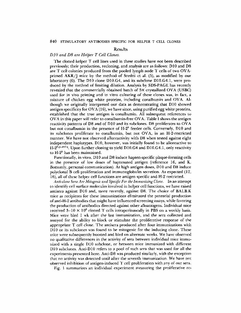
Fig. 1 summarizes an individual experiment measuring the proliferative re-
KAYE ET AL. 841
TABLE I
T Cell Clones DIO and D8 are H-2 Restricted and Antigen Specific: T Cell Proliferation
A. D10 and D8 proliferate to antigen in an H-2-restricted manner.
D81 D10 D10.G4 H-2I* Feeders* Antig ens Antigen Antigen A ,E
- - + - - + - - +
D10.G4.1 Antigen
- - +
k,k AKR 1,290 5,428 ~ 457 15,232 1,122 10,993 k,k B10.BR 1,250 7,826 333 9,163 k,k B10.A 1,611 12,320 k,k B10.A(2R) 738 13,094 b,b B10 215 233 7,873 7,419 b,b C57B1/6 32,300 26,809 8,272 5,835 b,k B10.A(5R) 174 111 10,294 10,808 18,078 15,160 d,d BALB/C 299 156 529 1,321 2,859 2,046 558 659 d,d B10.D2 1,911 639 322 1,285 (dxb) CB6FI 1,226 251 12,968 12,635 s,s B10.S 176 356 1,064 3,414 s,k B 10.S(9R) 2,621 1,846 p,p B10.F(13R) 1,045 1,843 7,185 9,310 3,304 2,906 q,q B10.G 167 318 9,152 8,711 4,709 3,925 r,r B10.RIII 76 118 544 2,072 2,389 2,740 f,f B10.M 436 1,048 765 1,831 2,011 2,030
B. D10 and D8 are antigen specific.
T Clone ! Antigen 0 Response**
D 10.G4.1 - - 236 D 10.G4.1 Conalbumin 14,237 D 10.G4.1 OVA 338
D8 - - 45 D8 Conalbumin 64 D8 OVA 10,925
* Haplotype of origin of the I-A and I-E subregions of various stimulator cells. Strains with I-E b''f'q express no I-E molecules on the cell surface.
5 2 x 10 mitomycin C treated whole spleen cells/0.2 ml culture. 200 #g/ml of OVA (DS) or conalbumin (D 10) added (+) or with no antigen (-).
I 2 x 10⁴ cloned T cells/0.2 ml culture. Mean cpm SH-TdR incorporation of triplicate cultures, pulsed for the final 3 h of a 72-h culture period; significant stimulation underlined.
** Mean cpm ~H-TdR incorporation in the presence of 2 x l0 s mitomycin C-treated whole spleen cells as feeders.
sponse of D10.G4.1 and D8 to anti-D10 and anti-D8. Anti-D10 is a potent mitogen for DI0.G4.1 with a titer greater than 1:1,000. However, this serum has no effect even at high doses on D8 cells, despite the fact that in this particular experiment D8 gave a more vigorous antigen response than D10.G4.1. Con- versely, anti-D8 stimulates D8 to proliferate while exhibiting no apparent effect on D 10.G4.1. The lower titer of anti-D8 may reflect a lower immunogenicity of these cells, consistent with the finding that a greater number of immunizations are required to induce the activity. Indeed, in recent experiments we have found that anti-D 10 can be induced following a single boost 3 wk after priming.
To further examine the specificity of anti-D 10, we have assayed normal AKR
I o
b x
r :
842 STIMULATORY ANTIBODIES SPECIFIC FOR HELPER T CELL CLONES
2 4
20
16
12
9
\ I
\ \
c.o
- - 6 - ..... ~ "8 L i i ~ :
1:200 I;400 1:800 1:l,600 1:5,200 1:6,400 NO ANTISERA
A N T I S E R A DILUTION
FIGURE 1. Clone-specific antisera induce T cell proliferation. D10.G4.1 (D) and D8 (O) proliferation measured in a 72-h assay containing 2 x 10⁴ cloned T cells and 2 x 10⁵ mitomycin C-treated BALB.K whole spleen cells per 0.2 ml culture in the presence of various concentra- tions of anti-D10 ( ) or anti-D8 ( ..... ). D10.G4.1 and D8 antigen responses are assayed similarly, with the addition of conalbumin (D 10.G4.1) or OVA (DS) at 200 ug /ml (histograms). Data are expressed as mean cpm 3H-TdR incorporation of triplicate cultures following a 16-h pulse,
a
~///~
~/ /~ ~///~
ANTIGEN
spleen cells, lymph node cells, and Con A blasts in a proliferative assay. In no case did anti-D10 induce a response from these cells. In addition, absorption with AKR spleen cells, Con A blasts, or the AKR thymoma BW5147 does not remove any of the mitogenic activity directed at D 10.G4.1 (data not shown). We have seen significant absorption of anti-D10 activity using D10 cells, and no absorption with D8 cells, but the technical difficulty in obtaining large numbers of cloned cells has made these experiments inconclusive. These studies will be repeated with D 10- and D8-derived T cell hybridomas currently being generated.
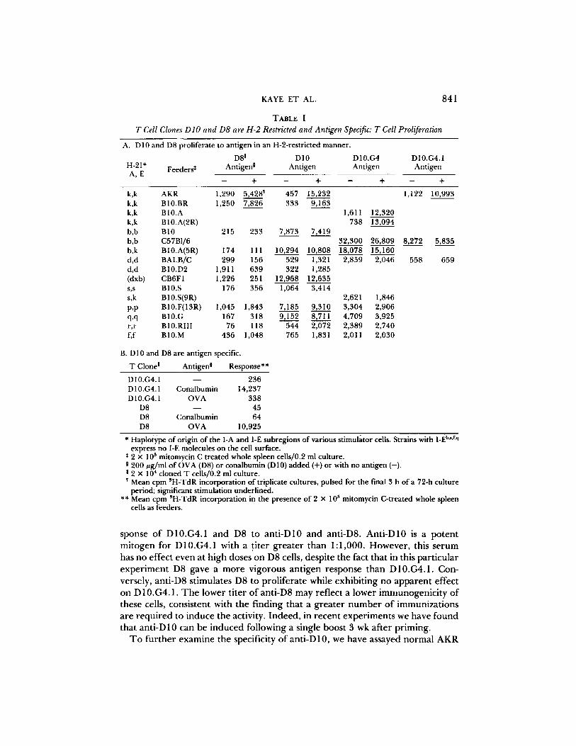
Anti-D lO Mitogenic Activity Is Due to Antibody. Although nonimmune BALB.K sera were not mitogenic for D10, we wanted
to verify that antibody was responsible for the observed activity. Anti-D10 was diluted to a break point in activity and absorbed with protein A-coupled Sepha- rose or, as a control, BSA-coupled Sepharose. Only the protein A-Sepharose was able to remove the mitogenic activity for D10.G4.1 (Fig. 2A). In addition, activity was recovered in the acid eluate of the protein A-Sepharose (Fig. 2B). Therefore, the ability of this serum to activate D10.G4.1 is the property of antibodies directed against the cloned T ceil.
Anti-clone Mitogenic Activity Requires Accessory Cells. Recent evidence suggests that the induction of T cell proliferation by antigen
or mitogen is a muhistep process (17, 18). Thus, we asked whether anti-D10 alone was sufficient to induce proliferation or whether a second signal from an accessory cell was also involved. Anti-D10 in the absence of accessory cells has almost no mitogenic activity for D10 or its subclones (Table IIA). The slight
K A Y E E T A L . 843
16
14
12
IO
b x 8
U 6
~ ~'~ t: 2pO0 1:8,000
A N T I S E R A DILUTION
52
:)8
24
8
4
I:?.pO0 I:1~000
A N T I S E R A DILUTION
20
b x 16
0 .
12
FIGURE 5). Anti-D10 mitogenic activity is specifically removed by protein A-Sepharose. Left: 1 ml of 1:1,000 anti-D 10 in medium was absorbed with 250 ~1 of protein A-Sepharose (O) or BSA-Sepharose (0). After 1 h on ice the mixture was centrifuged and the supernatant assayed for the ability to induce D10.G4.1 proliferation in a 72-h assay (see Fig. 1). Right: The protein A-Sepharose (O) or BSA-Sepharose (O) beads used for absorption were extensively washed and eluted with 1 ml of 0.1 M citrate pH 3.5. After dialysis against medium, the eluate was assayed as above. The data are expressed as the mean cpm 3H-TdR incorporation of triplicate cultures.
residual activity seen is not a consistent finding and may represent minor feeder cell contaminat ion o f the T cell clone, or direct accessory cell- independent activation. Anti-D8 also requires accessory cells to induce proliferat ion (for example, see Table IV). In o rde r to fu r the r characterize the cell involved in the anti-D10 response we have used spleen cells enr iched by adherence to plastic (splenic adheren t cells) as accessory cells. This preparat ion contains a variety of cell types. 10-fold fewer splenic adheren t cells than spleen cells support an equal or be t te r antigen response, consistent with the finding that such adheren t cells are enr iched for ant igen-presenting cells (19). When tested in the presence o f anti-D10, these same cells were also found to be enr iched for cells that support D10.G4.1 prol iferat ion (Table IIB).
Al though anti-D10 activity could not be absorbed out using spleen cells, and there fore was not likely to interact directly with the accessory cell population, it was possible that ant ibody aggregated on the T cell surface was interacting with Fc receptors to induce a second signal. Particularly relevant is the finding that aggregated immunoglobul in can induce the product ion of IL-1 f rom macro- phages (20). T h e addit ion of the monoclonal anti-Fc receptor ant ibody 2.4G2 (9), however, has no effect on the anti-D10 response whether spleen cells or splenic adheren t cells are used as accessory cells (Table II C). Rabbit anti-mouse brain (RaMBr) is known to be mitogenic for T cells, and RaMBr mitogenic activity requires a B cell as accessory cell, and an intact Fc (10). Note that the proliferative response o f D 10.G4.1 to RaMBr is almost totally el iminated in the
8 4 4 STIMULATORY ANTIBODIES SPECIFIC FOR HELPER T CELL CLONES
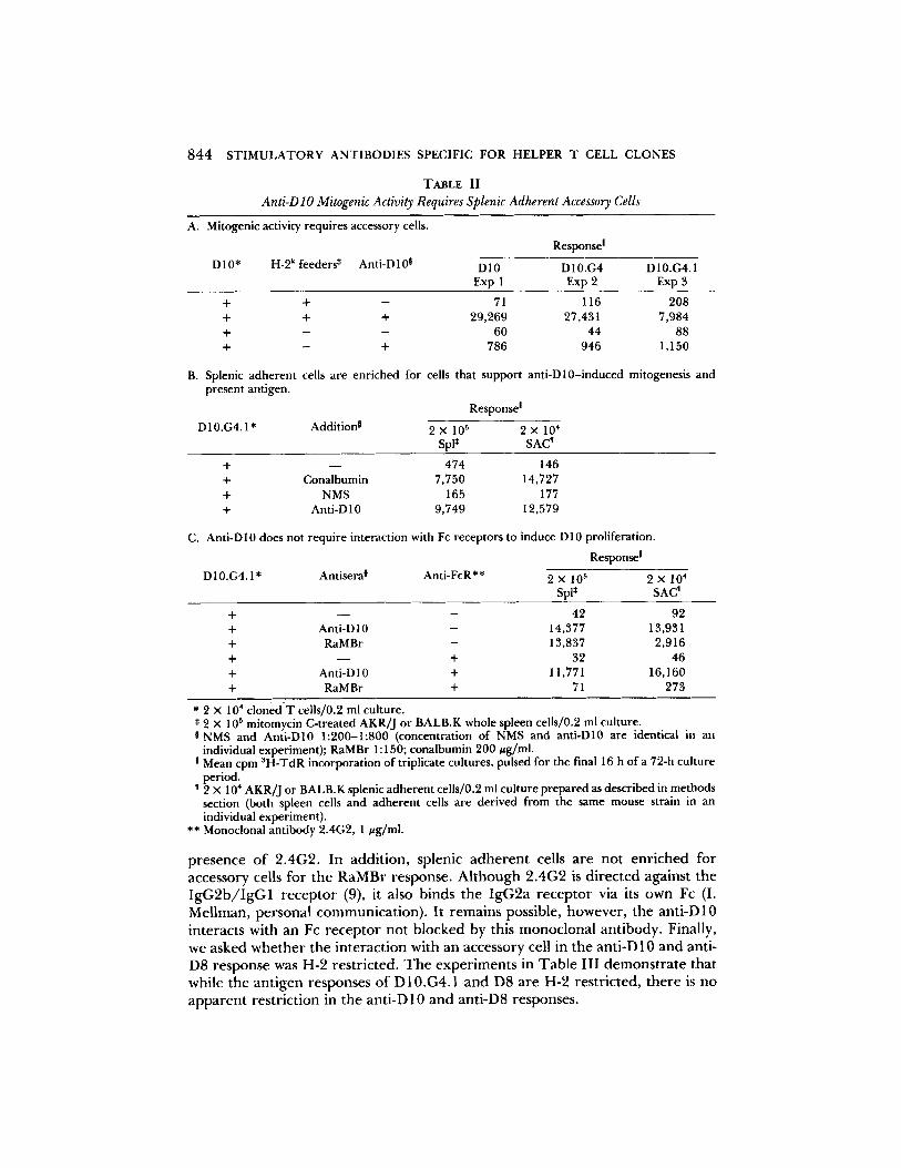
TABLE II
Anti-D lO Mitogenic Activity Requires Splenic Adherent Accessory Cells
A. Mitogenic activity requires accessory cells.
D 10* H-2 k feeders* Anti-D 10 °
Response !
D10 D10.G4 D10.G4.1 Exp 1 Exp 2 Exp 3
+ + - 71 116 208 + + + 29,269 27,431 7,984 + - - 60 44 88 + - + 786 946 1,150
B. Splenic adherent cells are enriched for cells that support anti-D10-induced mitogenesis and present antigen.
Response I
D10.G4.1* Addition j 2 x 10⁵ 2 x 10⁴ Spl* SAC ~
+ - - 474 146 + Conalbumin 7,750 14,727 + NMS 165 177 + Anti-D 10 9,749 12,579
C. Anti-Dl0 does not require interaction with Fc receptors to induce D10 proliferation.
Response I
D10.G4.1* Antisera 0 Anti-FcR** 2 x 10⁵ 2 × 10⁴ SpI* SAC'
+ - - - 42 92 + Anti-D10 - 14,377 13,931 + RaMBr - 13,837 2,916 + - - + 32 46 + Anti-Dl 0 + 11,771 16,160 + RaMBr + 71 273
* 2 x 104 cloned T cells/0.2 ml culture. * 2 x 10⁵ mitomycin C-treated AKR]J or BALB.K whole spleen cells/0.2 ml culture. I NMS and Anti-D10 1:200-1:800 (concentration of NMS and anti-D10 are identical in an
individual experiment); RaMBr 1 : 150; conalbumin 200 #g/ml. ! Mean cpm SH-TdR incorporation of triplicate cultures, pulsed for the final 16 h of a 72-h culture
period. I 2 x 10⁴ AKR/J or BALB.K splenic adherent cells/0.2 ml culture prepared as described in methods
section (both spleen cells and adherent cells are derived from the same mouse strain in an individual experiment).
** Monoclonal antibody 2.4G2, 1 #g/ml.
p r e s e n c e o f 2 . 4 G 2 . In a d d i t i o n , s p l e n i c a d h e r e n t cel ls a r e n o t e n r i c h e d f o r
a c c e s s o r y cel ls f o r t h e R a M B r r e s p o n s e . A l t h o u g h 2 . 4 G 2 is d i r e c t e d a g a i n s t t h e
I g G 2 b / I g G 1 r e c e p t o r (9), i t a l so b i n d s t h e I g G 2 a r e c e p t o r v ia its o w n Fc (I.
M e l l m a n , p e r s o n a l c o m m u n i c a t i o n ) . I t r e m a i n s pos s ib l e , h o w e v e r , t h e a n t i - D 1 0
i n t e r a c t s w i t h an Fc r e c e p t o r n o t b l o c k e d by this m o n o c l o n a l a n t i b o d y . F ina l ly ,
w e a s k e d w h e t h e r t h e i n t e r a c t i o n w i t h an a c c e s s o r y cel l in t h e a n t i - D 10 a n d an t i -
D 8 r e s p o n s e was H - 2 r e s t r i c t e d . T h e e x p e r i m e n t s in T a b l e I I I d e m o n s t r a t e t h a t
w h i l e t h e a n t i g e n r e s p o n s e s o f D 1 0 . G 4 . 1 a n d D 8 a r e H - 2 r e s t r i c t e d , t h e r e is n o
a p p a r e n t r e s t r i c t i o n in t h e a n t i - D 1 0 a n d a n t i - D 8 r e s p o n s e s .
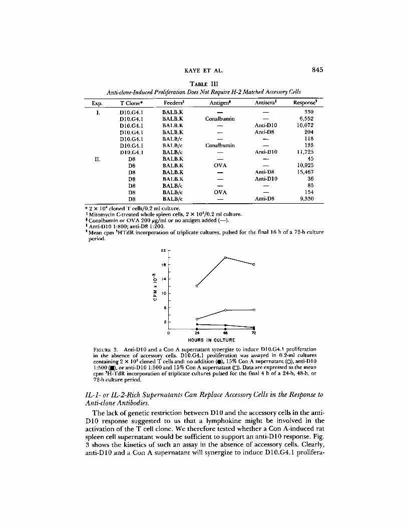
KAYE E T AL. 845
E x p .
I.
II.
Anti-clone-Induced Proliferation TABLE I I I
Does Not Require H-2 Matched Accessory Cells
T Clone* Feeders* Ant igen i Ant isera ! Response I
D 10.G4.1 BALB.K - - - - 330 D 10.G4.1 BALB.K Cona lbumin - - 6 ,552 D10.G4.1 BALB.K - - Ant i -D10 10,072 D 10.G4.1 BALB. K - - Anti-D8 204 D 10.G4.1 B A L B / c - - - - I 18 D 10.G4.1 B ALB/c Cona lbumin - - 133 D 10.G4.1 B A L B / c - - Ant i -D 10 11,725
D8 B ALB.K - - - - 45 D8 B ALB.K O V A - - 10,925 D8 BALB.K - - Anti-D8 15,467 D8 BALB.K - - Anti -D10 36 D8 B ALB/c - - - - 85 D8 B ALB/c O V A - - 154 D8 B A L B / c - - Anti -D8 9 ,330
* 2 X 104 c loned T cells/0.2 ml cul ture. * Mitomycin C-t reated whole spleen cells, 2 x 10⁵/0.2 ml cul ture.
C ona l bumi n or O V A 200 #g / ml o r no an t igen added ( - - ) . I Ant i -D10 1:800; ant i-D8 1:200.
Mean cpm 3 H T d R incorpora t ion o f triplicate cul tures, pulsed for the final 16 h o f a 72-h cul ture period.
2 2
10
? 0 14
K
~E I 0 It o
o
2
H O U R S I N C U L T U R E
FIGURE 3. Ant i -D10 and a Con A supe rna t an t synergize to induce D10.G4.1 prol iferat ion in the absence o f accessory cells. D10.G4.1 proliferat ion was assayed in 0.2-ml cul tures conta in ing 2 x 10⁴ c loned T cells and: no addi t ion (O), 15% Con A supe rna t an t (O), ant i -D10 1 : 5 0 0 ( i ) , o r ant i -D 10 1:500 and 15% Con A supe rna t an t (f-l). Data are expressed as the mean cpm 3H-TdR incorpora t ion o f triplicate cul tures pulsed for the final 4 h o f a 24-h, 48-h, or 72-h cu l ture period.
IL-1- or IL-2-Rich Supernatants Can Replace Accessory Cells in the Response to Anti-clone Antibodies.
The lack of genetic restriction between D 10 and the accessory cells in the anti- D10 response suggested to us that a lymphokine might be involved in the activation of the T cell clone. We therefore tested whether a Con A-induced rat spleen cell supernatant would be sufficient to support an anti-D 10 response. Fig. 3 shows the kinetics of such an assay in the absence of accessory cells. Clearly, anti-D10 and a Con A supernatant will synergize to induce D10.G4.1 prolifera-
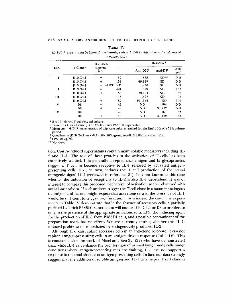
846 STIMULATORY ANTIBODIES SPECIFIC FOR HELPER T CELL CLONES
T A B L E IV IL-1-Rich Supernatant Supports Anti-clone-dependent T Cell Proliferation in the Absence of
Accessory Cells
I L- 1 -Rich Resp °nsea Exp. T Clone* superna- - -
tant , Anti.D10 ! Anti.D8! Anti- gen !
I D10.G4.1 - 37 878 ND** ND D10.G4.1 + 159 49,929 ND ND D10.G4.1 - +LPS ~ ND 1,256 ND ND
II D10.G4.1 - 491 329 ND 133 D10.G4.1 + 33 33,194 ND 32
III D10.G4.1 - 113 1,427 ND 92 D10.G4.1 + 87 161,144 120 190
IV D8 - 43 ND 104 ND D8 + 49 ND 31,772 ND
V D 8 - 43 ND 168 53 D8 + 48 ND 31,422 33
* 2 x 104 cloned T cells/0.2 ml culture. * Presence (+) or absence (-) of 1% IL-1 rich P388D1 supernatant. § Mean cpm SH-TdR incorporation of triplicate cultures, pulsed for the final 16 h of a 72-h culture
period. ! Conalbumin (D 10.G4.1) or OVA (D8), 200 ug/ml; anti-D 10 1:800; anti-D8 1:200. LPS, 50 ~zg/ml.
** Not done.
tion. Con A - i n d u c e d supe rna t an t s conta in m a n y soluble med ia to r s inc luding IL- 2 a nd IL-1. T h e ro le o f these pro te ins in the act ivat ion o f T cells has been extensively s tudied. It is genera l ly accep ted tha t an t igen and Ia g lycopro te ins t r igge r a T cell to b e c o m e recept ive to IL-1 re leased by act ivated ant igen- p resen t ing cells. IL-1, in turn , induces the T cell p r o d u c t i o n o f the actual mi togen ic signal IL-2 ( reviewed in r e fe rence 21). It is no t k n o w n at this t ime w h e t h e r the induc t ion o f recept ivi ty to IL-2 is also IL-1 dependen t . It was o f interest to c o m p a r e this p r o p o s e d mechan i sm o f act ivat ion to tha t obse rved with ant i -c lone antisera. I f such ant isera t r igge r the T cell c lone in a m a n n e r ana logous to an t igen and Ia, one migh t expec t tha t ant i -c lone sera in the p resence o f IL-1 would be sufficient to t r igge r prol i fera t ion. Th i s is indeed the case. T h e exper i - ments in Tab le IV d e m o n s t r a t e that in the absence o f accessory cells, a partially pur i f ied I L - l - r i ch P 3 8 8 D 1 supe rna t an t will induce D 10.G4.1 o r D8 to pro l i fe ra te only in the p resence o f the a p p r o p r i a t e ant i -c lone sera. LPS, the induc ing agen t fo r the p r o d u c t i o n o f IL- 1 f r o m P 3 8 8 D 1 cells, and a possible c o n t a m i n a n t o f the p r e pa ra t i on used, has no effect. We are cu r ren t ly test ing w h e t h e r this IL-1- i nduced pro l i fe ra t ion is m e d i a t e d by e n d o g e n o u s l y p r o d u c e d IL-2.
A l t h o u g h IL-1 can replace accessory cells in an ant i -c lone response , it can no t replace an t igen -p resen t ing cells in an an t igen-dr iven response (Table IV). This is consis tent with the work o f Mizel and Ben-Zvi (22) who have d e m o n s t r a t e d that , while IL-1 can e n h a n c e the pro l i fe ra t ion o f p r i m e d lymph n o d e cells u n d e r condi t ions whe re an t igen -p resen t ing cells are limiting, IL-1 can no t suppor t a response in the total absence o f an t igen -p resen t ing cells. In fact, o u r data s t rongly suggest tha t the addi t ion o f soluble an t igen and IL-1 to a he lper T cell c lone is
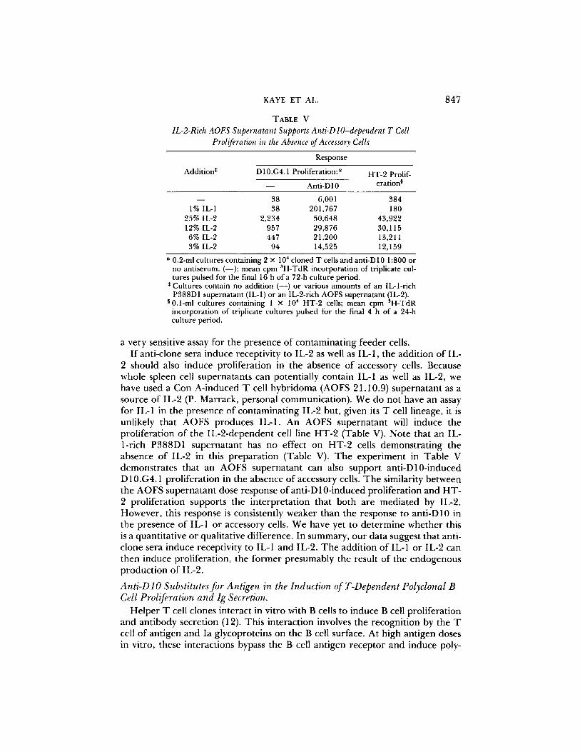
KAYE ET AL. 847
T A B L E V
IL-2-Rich AOFS Supernatant Supports Anti-D lO-dependent T Cell Proliferation in the Absence of Accessory Cells
Addition *
Response
D 10.G4.1 Proliferation:*
- - Anti-D 10 HT-2 Prolif-
eration 0
- - 38 6,001 384 1% IL-I 38 201,767 180
25% IL-2 2,234 50,648 43,922 12% IL-2 957 29,876 30,115 6% IL-2 447 21,200 13,211 3% IL-2 94 14,525 12,159
* 0.2-ml cultures containing 2 × 1 0 4 cloned T cells and anti-D10 1:800 or no antiserum. (--); mean cpm ~H-TdR incorporation of triplicate cul- tures pulsed for the final 16 h of a 72-h culture period.
* Cultures contain no addition (--) or various amounts of an IL-l-rich P388D1 supernatant (IL-1) or an IL-2-rich AOFS supernatant (IL-2).
0 0 . 1 _ m l cultures containing 1 × 10⁴ HT-2 cells; mean cpm 3H-TdR incorporation of triplicate cultures pulsed for the final 4 h of a 24-h culture period.
a very sensitive assay for the presence of contaminat ing feeder cells. I f anti-clone sera induce receptivity to IL-2 as well as IL-1, the addit ion of IL-
2 should also induce prol i ferat ion in the absence o f accessory cells. Because whole spleen cell supernatants can potentially contain IL-1 as well as IL-2, we have used a Con A-induced T cell hybr idoma (AOFS 21.10.9) superna tan t as a source o f IL-2 (P. Marrack, personal communicat ion) . We do not have an assay for IL-1 in the presence o f con tamina t ing IL-2 but, given its T cell lineage, it is unlikely that AOFS produces IL-1. An AOFS superna tant will induce the prol i ferat ion o f the IL-2-dependen t cell line H T - 2 (Table V). Note that an IL- l-r ich P388D1 superna tan t has no effect on H T - 2 cells demons t ra t ing the absence of IL-2 in this p repara t ion (Table V). T h e expe r imen t in Tab le V demons t ra tes that an AOFS superna tan t can also suppor t ant i -D10-induced D 10.G4.1 prol i ferat ion in the absence o f accessory cells. T h e similarity be tween the AOFS superna tan t dose response o f anti-D 10-induced prol i ferat ion and H T - 2 prol i ferat ion supports the in terpre ta t ion that bo th are media ted by IL-2. However , this response is consistently weaker than the response to anti-D10 in the presence of IL-1 or accessory cells. We have yet to de t e rmine whether this is a quant i ta t ive or qualitative difference. In summary , ou r data suggest that anti- clone sera induce receptivi ty to IL-1 and IL-2. T h e addit ion o f IL-1 or IL-2 can then induce prol iferat ion, the f o r m e r presumably the result o f the endogenous produc t ion o f IL-2.
Anti-D lO Substitutes for Antigen in the Induction ofT-Dependent Polyclonal B Cell Proliferation and Ig Secretion.
Hel pe r T cell clones interact in vitro with B cells to induce B cell prol i ferat ion and ant ibody secret ion (12). This interact ion involves the recogni t ion by the T cell o f ant igen and Ia glycoproteins on the B cell surface. At high ant igen doses in vitro, these interact ions bypass the B cell ant igen r ecep to r and induce poly-
848 S T I M U L A T O R Y A N T I B O D I E S SPECIFIC FOR HELPER T CELL CLONES
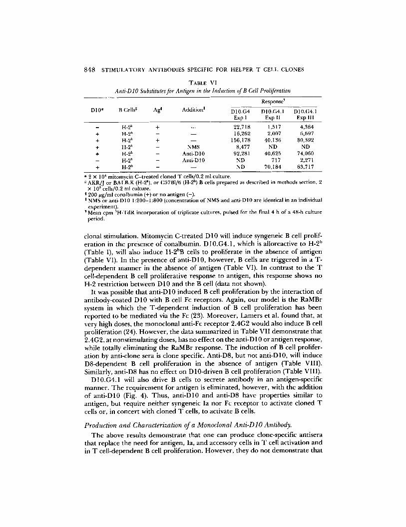
TABLE V I
Anti-D lO Substitutes for Antigen in the Induction of B Cell Proliferation
D10* B Cells* Ag a Addition !
Response I
D10.G4 D10.G4.1 D10.G4.1 Exp I Exp II Exp III
- H-2 k + - - 22,718 1,517 4,364 + H-2 k - - - 16,262 2,007 6,697 + H-2 k + - - 156,178 40,136 80,392 + H-2 k - NMS 8,477 ND ND + H-2 k - Anti-D10 92,281 40,625 74,060 - H-2 k - Anti-D 10 ND 717 2,271 + H-2 b - - - ND 70,184 63,717
* 2 x 104 mitomycin C- t rea ted cloned T cells/0.2 ml culture. * AKR/J or BALB.K (H-2k), or C57B1/6 (H-2 b) B cells prepared as described in methods section, 2
× l0 s cells/0.2 ml culture. 200 #g/ml conalbumin (+) or no antigen (-) .
! NMS or anti-D10 1:200-1:800 (concentration of NMS and anti-D10 are identical in an individual experiment). Mean cpm 3H-TdR incorporation of triplicate cultures, pulsed for the final 4 h of a 48-h culture period.
cional stimulation. Mitomycin C-treated D 10 will induce syngeneic B cell prolif- eration in the presence of conalbumin. D10.G4.1, which is alloreactive to H-2 b (Table I), will also induce H-2bB cells to proliferate in the absence of antigen (Table VI). In the presence of anti-D 10, however, B cells are triggered in a T- dependent manner in the absence of antigen (Table VI). In contrast to the T cell-dependent B cell proliferative response to antigen, this response shows no H-2 restriction between D 10 and the B cell (data not shown).
It was possible that anti-D 10 induced B cell proliferation by the interaction of antibody-coated D10 with B cell Fc receptors. Again, our model is the RaMBr system in which the T-dependent induction of B cell proliferation has been reported to be mediated via the Fc (23). Moreover, Lamers et al. found that, at very high doses, the monoclonal anti-Fc receptor 2.4G2 would also induce B cell proliferation (24). However, the data summarized in Table VII demonstrate that 2.4G2, at nonstimulating doses, has no effect on the anti-D 10 or antigen response, while totally eliminating the RaMBr response. The induction of B cell prolifer- ation by anti-clone sera is clone specific. Anti-D8, but not anti-D10, will induce D8-dependent B cell proliferation in the absence of antigen (Table VIII). Similarly, anti-D8 has no effect on D10-driven B cell proliferation (Table VIII).
D10.G4.1 will also drive B cells to secrete antibody in an antigen-specific manner. The requirement for antigen is eliminated, however, with the addition of anti-D10 (Fig. 4). Thus, anti-Dl0 and anti-D8 have properties similar to antigen, but require neither syngeneic Ia nor Fc receptor to activate cloned T cells or, in concert with cloned T cells, to activate B cells.
Production and Characterization of a Monoclonal Anti-DlO Antibody.
The above results demonstrate that one can produce clone-specific antisera that replace the need for antigen, Ia, and accessory cells in T cell activation and in T cell-dependent B cell proliferation. However, they do not demonstrate that

KAYE ET AL. 849
T A B L E V I I
Anti-D tO Does Not Require Interaction with Fc Receptors to Induce T-Dependent B Cell Proliferation
B cell proliferation in the presence of:
D 10.G4.1 * B Cells ¢ Addi t ion 0 Anti-FcR n
- - +
- + - - 1,8741 1,535 + + - - 3,187 2,930 + + Conaibumin 79,474 62,745 + + Ant i -Dl0 94,251 74,905 + + RaMBr 49,531 1,810
* 2 X 10 4 mitomycin-treated cloned T cells/0.2 ml culture. * 2 × 10 5 AKR/J B cells/0.2 ml culture.
Conalbumin 200 t~g/ml; anti-D10 1:800; RaMBr 1 : 150. ! Presence (+) or absence ( - ) o f monoclonal ant ibody 2.4G2, 1 #g/ml. 1 Mean cpm SH-TdR incorporat ion of triplicate cultures, pulsed for the
final 4 h o f a 48-h culture period.
TABLE V I I I
Induction of T-Dependent B Cell Proliferation by Anti-Clone Sera is Clone Spec~c
T Clone* B Ceils* Addi t ion O Response I
D10.G4.1 + - - 4,005 D 10.G4.1 + Conalbumin 65,403 D 10.G4.1 + Anti-D I 0 59,164 D 10.G4.1 + Anti-D8 3,944
D8 + - - 5,822 D8 + OVA 67,606 D8 + Anti-D8 56,530 D8 + Anti-D10 5,124 - - + Conalbumin 3,440 - - + O V A 3,500 - - + Anti-D 10 1,954 - - + Anti-D8 1,084
* 2 × 104 mitomycin C- t r ea t ed cloned T cells/0.2 ml culture. * 2 × 10⁵ AKR]J B ceils/0.2 ml culture. m Conalbumin and OVA, 200 #g/ml; anti-D10 1:800; anti-D8 1:200. ! Mean cpm SH-TdR incorporat ion o f triplicate cultures pulsed for the
final 4 h of a 48-h culture period.
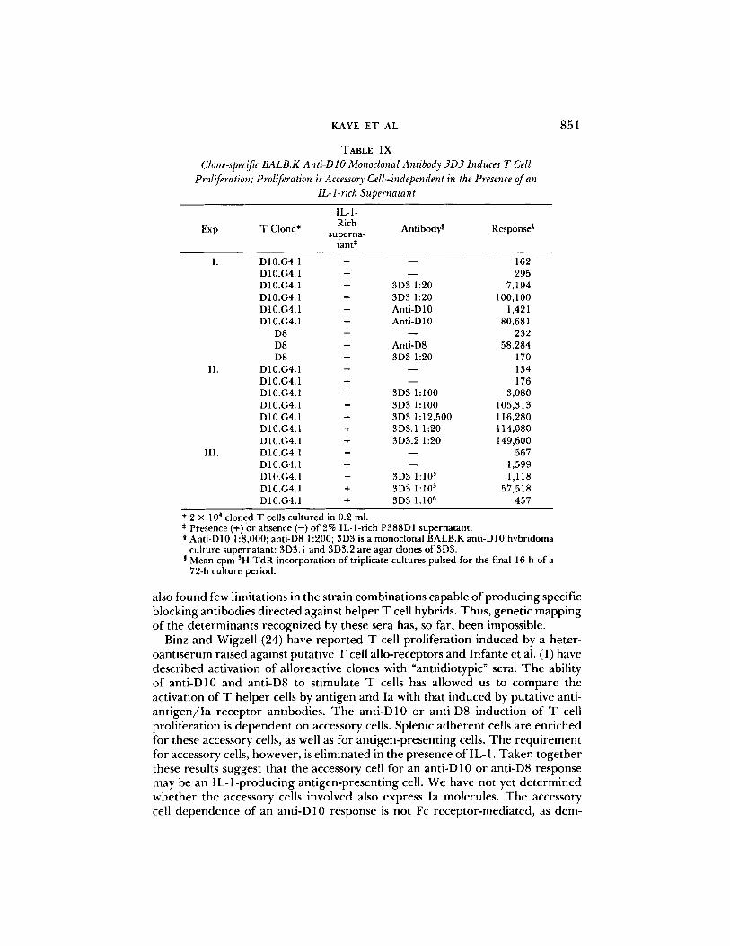
antibody of a single specificity can produce such effects. To approach this problem, BALB.K mice were primed with D10, boosted, and their spleen cells fused to P3X63-Ag8.653 cells to produce monoclonal antibodies. One such monoclonal anti-D10 antibody, and its subclones, is shown in Table IX. This monoclonal antibody induces D 10.G4.1 to proliferate, while having no effect on D8. Proliferation in the absence of accessory cells requires IL-1. The titer of culture supernatant of this hybridoma in a functional assay is remarkable; supernatant at 1:105 induces D 10.G4.1 proliferation (Table IX). Thus, a mono- clonal antibody can replace antigen and Ia-bearing antigen-presenting cells for
8 5 0 STIMULATORY ANTIBODIES SPECIFIC FOR HELPER T CELL CLONES
8 0
?, 0 70
n.. 6 o
~ 5o
~ 4O
z <
X 3O
?.o
io
0 0 3 4 5
DAYS IN CULTURE
FIGURE 4. Anti-D10 induces T-dependent polyclonal B cell antibody secretion. 3 × 10⁴ D10.G4.1 were cultured with 4 × 10⁵ BALB.K B cells in 0.2-ml cultures containing; no addition (O), 200 ug/ml conalbumin (I), or anti-D10 1:500 (&). Control cultures contained B cells and conalbumin (0) or B cells and anti-D10 (1"7) in the absence of cloned T cells. After a 3-, 4-, or 5-d culture period, triplicate cultures were pooled and assayed in a plaque-forming cell assay using protein A coupled sheep erythrocytes.
T cell activation in this system. We are using this monoclonal antibody to isolate the molecule, presumed to be the antigen-Ia receptor, from D 10.G4.1 cells.
Discussion
In an attempt to characterize the proteins involved in helper T cell antigen recognition we have raised antisera and a monoclonal antibody in BALB.K mice against two AKR-derived cloned helper T cell lines, D10 and D8. BALB.K anti- D10 serum, BALB.K anti-D8 serum, and monoclonal antibody 3D3 are potent inducers of T cell proliferation. However, these antibodies react only with the immunizing cell line, exhibiting no cross-reactivity between D10 and D8 cells. In addition, anti-D10 serum does not functionally react with normal or mitogen- stimulated AKR T cells. These results and the fact that D10 and D8 are functionally identical but differ in antigen specificity and alloreactivity, suggested to us that these antisera may react with a variable portion of the antigen/Ia receptor(s). Why such an immunization protocol should produce only antibodies directed against "idiotypic" and not framework determinants, is unknown. Three possibilities are apparent; a) T cell receptor constant regions may be less poly- morphic than immunoglobulins, with no polymorphism between AKR and BALB.K strains; (b) T cell receptor allotypes may be poorly immunogenic or (c) anti-T cell receptor allotypes are not detected in the assays used. We have also been able to produce a similar anti-D10 activity in two out of four (AKR x B6)F1 mice and are currently testing AKR anti-D 10 sera for activity. White et al. (2)
KAYE ET AL. 851
TABLE I X
Clone-specific BALB.K Anti-D lO Monoclonal Antibody 3D3 Induces T Cell Proliferation; Proliferation is Accessory Cell-independent in the Presence of an
IL- l-rich Supernatant
IL-1-
Exp T Clone* Rich Antibodyg Response ! superna-
tant *
I. DI0.G4.1 - - - 162 D 10.G4.1 + - - 295 D10.G4.1 - 3D3 1:20 7,194 D10.G4.1 + 3D3 1:20 100,100 D10.G4.1 - Anti-D10 1,421 D10.G4.1 + Anti-D10 80,681
D8 + - - 232 D8 + Anti-D8 58,284 D8 + 3D3 1:20 170
II. D10.G4.1 - - - 134 D10.G4.1 + - - 176 D10.G4.1 - 3D3 1:100 3,080 D10.G4.1 + 3D3 1:100 105,313 D10.G4.1 + 3D3 1:12,500 116,280 D 10.G4.1 + 3D3.1 1:20 114,080 D10.G4.1 + 3D3.2 1:20 149,600
III. D 10.G4.1 - - - 567 D 10.G4.1 + - - 1,599 D10.G4.1 - 3D3 1:105 1,118 D10.G4.1 + 3D3 1:105 57,518 D10.G4.1 + 3D3 1:106 457
* 2 × 104 cloned T cells cultured in 0.2 ml. * Presence (+)or absence ( - ) o f 2% IL-l-rich P388D1 supernatant. 0 Anti-D10 1:8,000; anti-D8 1:200; 3D3 is a monoclonal BALB.K anti-D10 hybridoma
culture supernatant; 3D3.1 and 3D3.2 are agar clones of 3D3. Mean cpm 3H-TdR incorporation of triplicate cultures pulsed for the final 16 h of a 72-h culture period.
a lso f o u n d few l imi t a t i ons in t he s t r a in c o m b i n a t i o n s c a p a b l e o f p r o d u c i n g specif ic b l o c k i n g a n t i b o d i e s d i r e c t e d aga ins t h e l p e r T cell hyb r id s . T h u s , g e n e t i c m a p p i n g o f t he d e t e r m i n a n t s r e c o g n i z e d by these se ra has, so far , b e e n imposs ib le .
Binz a n d Wigze l l (24) have r e p o r t e d T cell p r o l i f e r a t i o n i n d u c e d by a h e t e r - o a n t i s e r u m r a i s ed aga ins t p u t a t i v e T cell a l l o - r e c e p t o r s a n d I n f a n t e et al. (1) have d e s c r i b e d ac t i va t i on o f a l l o r e a c t i v e c lones wi th "an t i id io typ ic" sera . T h e ab i l i ty o f a n t i - D 1 0 a n d an t i -D8 to s t i m u l a t e T cells has a l l ow e d us to c o m p a r e t he ac t i va t i on o f T h e l p e r cells by a n t i g e n a n d Ia wi th t ha t i n d u c e d by p u t a t i v e ant i - a n t i g e n / I a r e c e p t o r a n t i b o d i e s . T h e a n t i - D 1 0 o r an t i -D8 i n d u c t i o n o f T cell p r o l i f e r a t i o n is d e p e n d e n t on accesso ry cells. Sp len ic a d h e r e n t cells a r e e n r i c h e d fo r these acces so ry cells, as well as fo r a n t i g e n - p r e s e n t i n g cells. T h e r e q u i r e m e n t fo r accesso ry cells, h o w e v e r , is e l i m i n a t e d in the p r e s e n c e o f IL-1 . T a k e n t o g e t h e r these resu l t s sugges t t ha t t he accesso ry cell fo r an a n t i - D 1 0 o r an t i -D8 r e s p o n s e m a y be an I L - l - p r o d u c i n g a n t i g e n - p r e s e n t i n g cell . W e have n o t ye t d e t e r m i n e d w h e t h e r t he acces so ry cells i n v o l v e d also e xp re s s Ia molecu les . T h e accessory cell d e p e n d e n c e o f an a n t i - D 1 0 r e s p o n s e is n o t Fc r e c e p t o r - m e d i a t e d , as d e m -
852 S T I M U L A T O R Y ANTIBODIES SPECIFIC FOR HELPER T CELL CLONES
onstrated by blocking studies with monoclonal antibody 2.4G2. Anti-D10 may therefore activate D10 to induce, via a soluble mediator or directly, IL-1 production from the accessory cell population. The lack of genetic restriction between the T cell and accessory cell in antibody-mediated responses would suggest that if direct cell contact is required, it is not mediated through a specific Ia recognition event. However, we have not ruled out the possibility that recognition of nonpolymorphic portions of Ia molecules is involved in this process. Mitogen activation of T cells also appears to involve induction of IL-1 from an accessory cell population in the absence of specific Ia recognition (26). Our data also suggest that anti-D 10 will induce receptivity to IL-2, independently of IL-1. It is unlikely, however, that when anti-D10 induces proliferation in the presence of accessory cells that this is the result of accessory T cell-released IL- 2, since adherent cells are enriched for accessory cells. In addition, anti-Thy-1 and complement-treated spleen cells are not depleted of accessory cells for an anti-D 10-response although deficient in IL-2 production induced by Con A (data not shown). By analogy, the induction of receptivity to IL-2 by antigen and Ia may also be the result of direct activation, independent of IL-1. Meuer et al. (3) have described a monoclonal antibody specific for an individual cloned human cytolytic T cell line which, although it inhibits antigen-induced proliferation, also induces increased receptivity to IL-2.
Infante et al. (1) have described similar clone-specific antisera raised against alloreactive T cell clones. These antisera induce IL-2 production and T cell proliferation (although variably) in the absence of accessory cells, and therefore apparently independent of IL-1. We are currently testing the requirements for the induction of IL-2 secretion by the antibodies described here. The antisera we have prepared also induce low level proliferation, in some experiments, in the absence of added accessory cells. However, minor feeder cell contamination of the cloned T cell population can create the appearance of an accessory cell- independent response. In experiments in which soluble antigen and IL-1 did not induce T cell proliferation, the anti-D10 or anti-D8 response in the absence of IL-1 was negligible (Table IV). These findings also clarify an issue raised by Durum and Gershon (15). These authors proposed that Ia recognition was required in antigen-driven T cell proliferation only for the induction of IL-1 secretion. Our more discriminating system, using cloned antigen-Ia specific T cells and IL-1, demonstrates that IL-1 and antigen are not sufficient for T cell activation. Thus, Ia is required for antigen recognition by helper T cells.
We have also demonstrated that a monoclonal antibody can induce D 10.G4.1 proliferation. This finding may have profound implications concerning the mechanism of activation of helper T cells by antigen in association with Ia molecules. Anti-immunoglobulin sera activate B cells, but only at concentrations well above the saturation level of binding (27). In addition, cross-linking of B cell surface immunogiobulin seems to be involved in this triggering process. Unless there are multiple determinants recognized by 3D3 on a single target molecule, monoclonal antibody 3D3 could not extensively cross-link the T cell surface. Also, based on the titer of 3D3 culture supernatants, very low concen- trations of antibody are sufficient to activate the T cell. If indeed this antibody binds the ant igen/Ia receptor, the interaction of these receptors and antigen-Ia
KAYE ET AL. 853
complexes may therefore be more analagous to the interaction of a hormone with its receptor than to the interaction of a multi-determinant antigen with B cell surface immunoglobulin. Anti-hormone receptor antibodies can, in some systems, mimic the hormone in the activation of a target cell and, in fact, cause pathology by this mechanism in autoimmune disease (28).
RaMBr sera are also mitogenic for D10 and D8. These sera recognize a nonpolymorphic determinant on the Thy-1 molecule (29). Although BALB.K and AKR strains are allelic at the Thy-1 locus, anti-D10 and anti-D8 mitogenic activities are not the result ofanti-Thy-1 antibodies for the following reasons: (a) D 10 and D8 are both Thy-1.1 +1.2- but anti-D 10 and anti-D8 are clone specific; (b) AKR Con A blasts or the Thy-l.1 + thymoma BW5147 do not absorb out anti-D10 mitogenic activity while removing anti-Thy-1 mitogenic activity from RaMBr and anti-Thy-1 cytotoxic activity from anti-D10; (c) (AKR x B6)F1 anti- D10 is also mitogenic for D10 but not D8; and (d) under conditions where RaMBr immunoprecipitates Thy-1 from surface-iodinated D10.G4.1, BW5147 absorbed anti-D 10, while still biologically active, does not (data not shown). It is possible, though, that Thy-1 acts as a carrier protein, increasing the immuno- genicity of the AKR T cells.
The finding that anti-clone sera substitute for antigen and Ia in the helper T cell-dependent activation of B cells is novel. Blocking studies with 2.4G2 suggest that B cell activation in this system is not mediated through an Fc receptor. Other possible mechanisms of B cell activation, particularly the release of a soluble factor, are currently under investigation. We plan to extend these studies to include antigen-specific responses, and an examination of the activation state of B cells capable of responding to T helper cells activated by these antisera. The fact that anti-D 10 and anti-D8 exhibit clone specificity in the induction of T- dependent B cell activation suggests that these antisera are reacting with the same determinant to induce both T cell and T-dependent B cell activation. Preliminary studies with the monoclonal anti-D 10 antibody have confirmed this (data not shown).
In preliminary biochemical studies using biosynthetically labeled cloned T cells we have found that these antisera precipitate a number of proteins. One protein is precipitated by anti-D10 from D10, but not D8 cells. Two-dimensional SDS- PAGE analysis (first dimension nonreducing, second dimension reducing condi- tions) reveals that this protein has an apparent molecular weight of 80,000 and is comprised of 40,000-dalton disulfide bonded subunits. Similar findings have been reported by two other laboratories using clone-specific monoclonal anti- bodies (3, 30). Two-dimensional gel analysis (first dimension nonequilibrium pH gradient electrophoresis, second dimension SDS-PAGE) of anti-D 10 immunopre- cipitates reveals that this protein is a heterodimer consisting of an acidic and a basic subunit. Immunoprecipitation studies using the clone-specific monoclonal anti-D10 antibody 3D3 have yielded similar results. A protein with similar characteristics has previously been isolated from a T cell tumor and was also demonstrated to be a constituent of normal T cells (31).
These studies demonstrate that cloned T cell lines can be activated by clone- specific antibodies, including a monoclonal antibody, in the complete absence of other cell types, provided exogenous IL-1 (or IL-2) is added. Thus, this system

854 STIMULATORY ANTIBODIES SPECIFIC FOR HELPER T CELL CLONES
is ideally suited for the biochemical analysis of the complex process of T cell activation, as only a single, cloned cell type is present. These antibodies should also serve as a means for isolating and characterizing the receptor involved in antigen-Ia recognition.
S u m m a r y
Two antisera and a monoclonal antibody raised in BALB.K mice against cloned, major histocompatibility complex (MHC)-restricted, antigen-specific helper T cell lines are described. These antibodies are specific for individual cloned T cell lines and are potent inducers of T cell proliferation. The induction of T cell proliferation by these antibodies requires the presence of an adherent accessory cell. There is no H-2 restriction between this accessory cell and the cloned T cell, nor is this antibody-induced proliferation blocked by a monoclonal anti-Fc receptor antibody. The requirement for an accessory cell, however, is eliminated in the presence of an IL-1- or IL-2-rich supernatant. Thus this system allows the analysis of helper T cell activation with only a single cell type present. Anti-T cell sera also induce T cell-dependent B cell proliferation and immun0- globulin secretion. The induction of T cell-dependent B cell activation by these sera does not require H-2-matched T cells and B cells. The specificity of these antibodies and their ability to stimulate cloned helper T cells in the absence of antigen and antigen-presenting cells strongly suggest that these antibodies are directed against antigen a n d / o r Ia recognition sites on the T ceil.
The authors wish to thank Pat Conrad and Barbara Broughton for technical support, and Maureen Wescott and Diane Mierz for help in generation of the monoclonal antibody. We also thank Ira Mellman and Pippa Marrack for gifts of cells and antibodies, and Scott Durum for generously supplying IL-l-rich supernatants.
Received for publication 21 April 1983 and in revised form 13June 1983.
R e f e r e n c e s 1. Infante, A.J., P. D. Infante, S. Gillis, and C. G. Fathman. 1982. Definition o f T cell
idiotypes using anti-idiotypic antisera produced by immunization with T cell clones. J. Exp. Med. 155:1100.
2. White,J., K. M. Haskins, P. Marrack, andJ. Kappler. 1983. Use of I region-restricted, antigen-specific T cell hybridomas to produce idiotypically specific anti-receptor antibodies. J. hnmunol. 130:1033.
3. Meuer, S. C., K. A. Fitzgerald, R. E. Hussey, J. C. Hodgdon, S. F. Schlossman, and E. L. Reinherz. 1983. Clonotypic structures involved in antigen-specific human T cell function.J. Exp. Med. 157:705.
4. Lancki, D. W., M. I. Lorber, M. R. Loken, and F. W. Fitch. 1983. A clone-specific monoclonal antibody that inhibits cytolysis of a cytolytic T cell clone. J. Exp. Med. 157:921.
5. Sredni, B., H. Y. Tse, and R. H. Schwartz. 1980. Direct cloning and extended cultures of antigen-specific, MHC restricted, proliferating T lymphocytes. Nature (Lond.). 283:581.
6. Janeway, C. A.,Jr., E. A. Lerner, P.J. Conrad, and B. Jones. 1982. The precision of self and non-self major histocompatibility complex encoded antigen recognition by cloned T cells. Behri~g h~st. Mitt. 70:200.
KAYE ET AL. 855
7. Wigzell, H. 1976. Specific affinity fractionation of lymphocytes using glass or plastic bead columns. In In Vitro Methods on Cell-Mediated and Tumor Immunity. B. R. Bloom andJ. R. David, editors. Academic Press, Inc., New York. p. 245-255.
8. Marion, T. N., and D. E. Briles. 1981. Analysis of autoimmune anti-DNA antibody responses using somatic cell hybridization. In Monoclonal Antibodies and T Cell Hybridomas. G.J. Hammerling, U. Hammerling, and J. F. Kearney, eds. Elsevier/ North-Holland, New York. p. 251-258.
9. Mellman, I. S., and J. C. Unkeless. 1980. Purification of a functional mouse Fc receptor through the use of a monoclonal antibody. J. Exp. Med. 152:1048.
10. Jones, B., and C. A. Janeway, Jr. 1981. Functional activities of antibodies against brain-associated T cell antigens. I. Induction of T cell proliferation. Eur. J. Immunol. 11:584.
11. Steinman, R. M., G. Kaplan, M. D. Witmer, and Z. A. Cohn. 1979. Identification of a novel cell type in peripheral lymphoid organs of mice.J. Exp. Med. 149:1.
12. Jones, B., and C. A. Janeway, Jr. 1981. Cooperative interaction of B lymphocytes with antigen-specific helper T lymphocytes is MHC restricted. Nature (Lond.). 292:547.
13. Gronowicz, E., A. Coutinho, and F. Melchers. 1976. A plaque assay for all cells secreting Ig of a given type or class. Eur. J. hnmunol. 6:588.
14. Gillis, S., M. Ferm, W. Ou, and K. Smith. 1978. T cell growth factor: parameters of production and a quantitative microassay for activity. J. Immunol. 120:2027.
15. Durum, S. K., and R. K. Gershon. 1982. Interleukin 1 can replace the requirement for I-A-positive cells in the proliferation of antigen-primed T cells. Proc. Natl. Acad. Sci. USA. 79:4747.
16. Bottomly, K., B. Jones, J. Kaye, and F. Jones III. Subpopulations of B cells distin- guished by cell surface expression of Ia antigens:correlation of Ia and idiotype during activation by cloned Ia-restricted T cells. J. Exp. Med. 97:265.
17. Larsson, E., and A Coutinho. 1980. Mechanism of T cell activation. I. A screening of"step one" ligands. Eur. J. hnmunol. 10:93.
18. Germain, R. N. 1981. Accessory cell stimulation o f T cell proliferation requires active antigen processing, la-restricted antigen presentation, and a separate non-specific 2nd signal.J, hnmunol. I27:1964.
19. Sunshine, G. H., D. R. Katz, and M. Feldmann. 1980. Dendritic cells induce T cell proliferation to synthetic antigens under Ir gene control. J. Exp. Med. 152:1817.
20. Blyden, G., and R. E. Handschumacher. 1977. Purification and properties of human lymphocyte activating factor (LAF). J. Immunol. 118:1631.
21. Moller, G. (editor). 1982. Interleukins and lymphocyte activation. Immunol. Rev. Vol. 63.
22. Mizel, S. B., and A. Ben-Zvi. 1980. Studies on the role of lymphocyte-activating factor (Interleukin 1) in antigen induced lymph node lymphocyte proliferation. Cell. Immunol. 54:382.
23. Jones, B. 1982. Functional activities of antibodies against brain-associated T cell antigens. II. Stimulation o f T cell-induced B cell proliferation. Eur. J. Immunol. 12:30.
24. Lamers, M. C., S. E. Heckford, and H. B. Dickler. 1982. Monoclonal anti-Fc IgG receptor antibodies trigger B lymphocyte function. Nature (Lond.). 298:178.
25. Binz, H., and H. Wigzell. 1981. T cell receptors with allo-major histocompatibility complex specificity from rat and mouse. J. Exp. Med. 154:126 I.
26. Rock, K. L. 1982. The role of Ia molecules on the activation of T lymphocytes. J. Immunol. 129:1360.
27. Sieckmann, D. G., R. Asofsky, D. E. Mosier, I. M. Zitron, and W. E. Paul. 1978. Activation of mouse lympbocytes by anti-immunoglobulin. I. Parameters of the
856 STIMULATORY ANTIBODIES SPECIFIC FOR HELPER T CELL CLONES
proliferative response. J. Exp. Med. 147:814. 28. Kahn, C. R., M. Kasuga, G. L. King, and C. Grunfeld. 1982. Autoantibodies to
insulin receptors in man: immunological determinants and mechanism of action. In Receptors, Antibodies and Disease. Ciba Foundation Symposium, 90. D. Evered and J. Whelan, editors. Pitman Books Ltd., London. p. 91.
29. Jones, B. 1983. Evidence that the Thy-1 molecule is the target for T-cell mitogenic antibody against brain-associated antigens. Eur. J. Immunol. In press.
30. Haskins, K., R. Kubo, J. White, M. Pigeon, J. Kappler, and P. Marrack. The MHC- restricted antigen receptor on T cells. I. Isolation with a monocional antibody. J. Exp. Med. 96:1149.
31. Allison, J. P., B. W. McIntyre, and D. Bloch. 1982. Tumor-specific antigen of murine T-lymphoma defined with monoclonal antibody. J. lmmunol. 129:2293.