bovine corneal opacity and permeability (bcop) assay · (opacity) •quantitativechange in the...
TRANSCRIPT

Bovine Corneal Opacity and Permeability (BCOP) Assay
ICCVAM Workshop on Best Practices for Assessing the Potential for Chemically Induced Eye Injuries
Presented by Hans Raabe
20 January 2011

2
Outline
• Key features of the BCOP• Application of the assay• Control and benchmark materials• Details of assay execution• Calculations
20 January 2011 ICCVAM Best Practices Workshop

3
Key Features of the BCOP:Test System and Exposure
• Viable corneas maintained in organ culture• Control over the exposure concentration• Control over the exposure time at the
specified concentration• Exposure over the whole corneal surface• Control over the post-exposure
(expression) period
20 January 2011 ICCVAM Best Practices Workshop

4
Key Features of the BCOP: Endpoints for Assessing Tissue Injury
• Quantitative change in light passage (opacity)
• Quantitative change in the barrier properties of the epithelium to small molecules (fluorescein penetration)
• Option for histology to assess the degree and depth of injury
• Additional endpoints:– Corneal hydration– Endothelial cell layer integrity
20 January 2011 ICCVAM Best Practices Workshop

A Continuum of Sensitivity
20 January 2011 ICCVAM Best Practices Workshop 5

6
Requirements for all Protocols
• Concurrent tested controls:– Negative controls– Positive controls– Acceptance criteria
• Whenever possible:– Concurrently tested benchmark or reference
materials• For submissions:
– Full Good Laboratory Practices compliant studies
20 January 2011 ICCVAM Best Practices Workshop

7
Negative Control: BCOP
• Accounts for non-specific changes in the test system and assay execution– Examples:
negative control, DI water, saline, or medium
• Corrects opacity and permeability values• Allows assessment of the quality of tissue
maintenance during the assay and of slide preparation by the histology laboratory
20 January 2011 ICCVAM Best Practices Workshop

8
Positive Control: BCOP1
• Ensures the integrity of the test system and proper execution of the assay
• Generally the same positive control is used with each assay trial• Ethanol or NaOH are used for liquid test articles• Imidazole is used for solid test articles
• The positive control must be included each time the assay is performed.
20 January 2011 ICCVAM Best Practices Workshop
1A proposal for OECD to reconsider the most appropriate positive control(s) for use in the BCOP will be discussed at WNT 2011.

Defined Acceptance Criteria• Determine the acceptability of a trial (run) of the assay• Acceptable range of responses is determined from historical results
20 January 2011 ICCVAM Best Practices Workshop 9
10
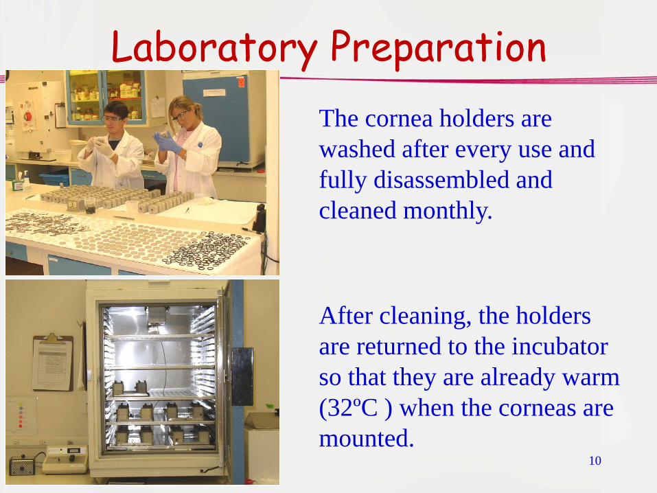
Laboratory PreparationThe cornea holders are washed after every use and fully disassembled and cleaned monthly.
After cleaning, the holders are returned to the incubator so that they are already warm (32ºC ) when the corneas are mounted.

11
Laboratory Preparation
Pipette calibration is checked.
The medium is pre-warmed in a 37º C water bath.
The laboratory area is prepared for harvesting and mounting of the corneas

12
Organ Culture MediumEagle’s Minimal Essential Medium
– Standard formulation includes bicarbonate
– 1% Fetal bovine serum added
– pH indicator, phenol red removed from incubation medium
• Reduces background absorbance (opacity)
• Eliminate color change
– Phenol red used in the rinsing medium to help identify residual test article (pH change)
20 January 2011 ICCVAM Best Practices Workshop

13
Cornea Holder • Several designs but all:
– Hold the cornea between the two halves
– Provide compartments for medium on both sides of the cornea
– Are constructed of relatively inert plastic
– Have access holes for changing the medium and removing bubbles
• Each holder is numbered so individual corneas are tracked by the holder number
20 January 2011 ICCVAM Best Practices Workshop

14
Source of the Bovine eyes
• Eyes are a by-product of food production
• Eyes are easily removed in this species
• Remove eyes as soon as possible in the process to reduce mechanical damage
• It is essential to work closely with the abattoir management to maintain quality control
20 January 2011 ICCVAM Best Practices Workshop

15
Holding and Transport of the Eyes• It may take several hours to obtain
the required number of eyes• The eyes must be kept wet and cool
– Hanks’ Balanced Salts Solution– Ice placed around the collection vessel
to cool it (Summer heat is deadly!)• Eyes arrive in the laboratory within 4-
5 hours of the first eyes being taken• Eyes are processed immediately
20 January 2011 ICCVAM Best Practices Workshop

16
Inspecting the Eyes• Assure that the eyes are
cool on arrival• Individual eyes are
inspected for corneal opacities and scratches– Keep the corneal surface wet– Some labs use dilute
fluorescein to mark scratches
• Approximately 30% of the eyes will be discarded (more in Summer)

17
Excising the Corneas
• The first cut is made with a scalpel
• The cornea is then cut from the globe with a 2-3 mm ring of sclera around the outside
18
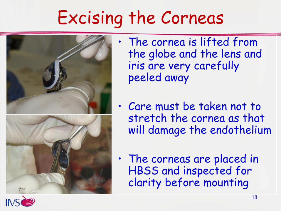
Excising the Corneas• The cornea is lifted from
the globe and the lens and iris are very carefully peeled away
• Care must be taken not to stretch the cornea as that will damage the endothelium
• The corneas are placed in HBSS and inspected for clarity before mounting

19
Mounting the Cornea in the Holder
• Corneas are handled only by the edge of the sclera to protect the epithelium and endothelium
• The cornea is placed on the posterior half of the holder with the sclera outside the O ringAssure that the cornea is not
dragged over the O ring20 January 2011 ICCVAM Best Practices Workshop

20
Mounting the Cornea in the HolderFitting and securing the anterior half over the cornea is a critical step to prevent damage to the endothelium
– Bring the anterior half over the posterior half so they are aligned
– Lower the anterior half onto the cornea without sideways motion
– Hold both halves steady while tightening the screws

21
Mounting the Cornea in the HolderAfter the screws are tight, phenol red-free medium is added to the holder
– The posterior chamber is filled first and plugged
– Care is taken to prevent foaming and to remove any air bubbles from the chamber
– The anterior chamber is filled and the corneas inspected

22
Post-Mounting Incubation• The holders are placed
on trays and returned to the incubator.
• The mounted corneas are incubated for at least one hour at 32±1oC to allow the corneas to resume normal metabolic activity
20 January 2011 ICCVAM Best Practices Workshop

23
Measuring the Passage of Light Through the Corneas
• Several types of instruments used in different laboratories
• Opacitometer (SpectroDesigns OP-KIT)– White light, dual light source
zeroing– Center weighted reading in
the cornea– Calibrated with three barrier
filters (linear)The readings should be linear within 2-3 units over the range of 0 to 225 opacity units
20 January 2011 ICCVAM Best Practices Workshop

24
Measuring the Passage of Light Through the Corneas
• Other instruments used:– BASF has recently developed a system fully
compliant with the OECD 437 specifications
20 January 2011 ICCVAM Best Practices Workshop
BASF Opacitometer ModelsBASF-OP2.0 and P3.0
25
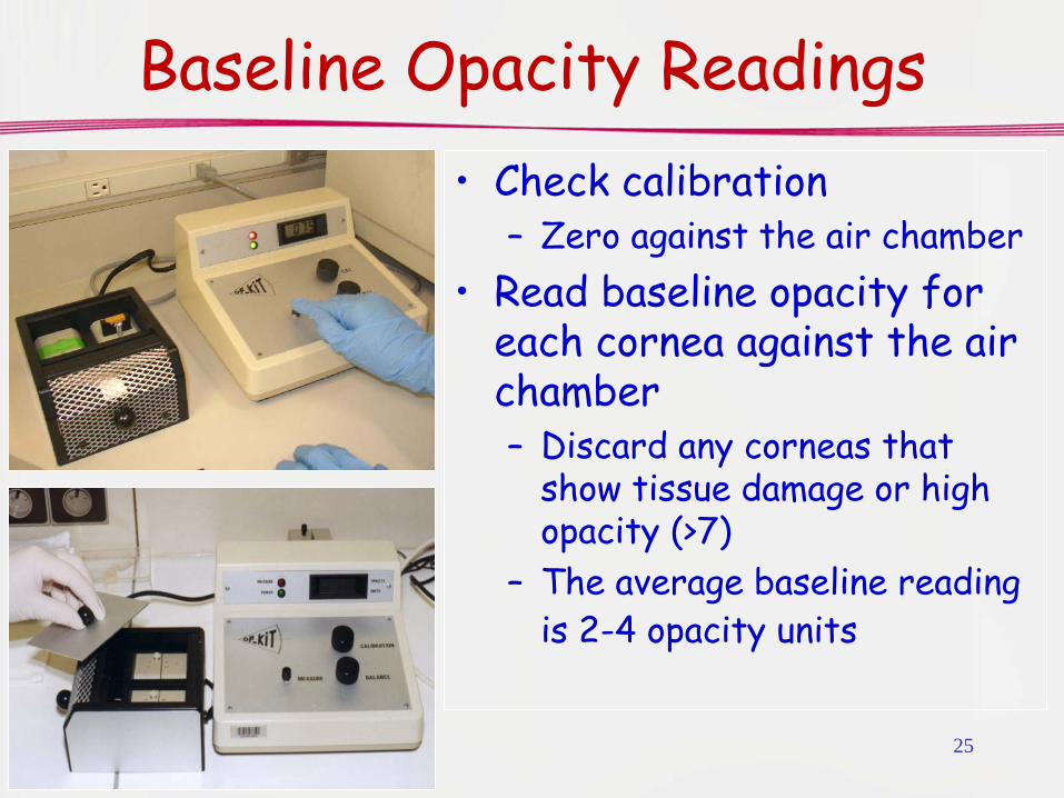
Baseline Opacity Readings• Check calibration
– Zero against the air chamber• Read baseline opacity for
each cornea against the air chamber– Discard any corneas that
show tissue damage or high opacity (>7)
– The average baseline reading is 2-4 opacity units

26
Parameters of the Assay• Test article concentration
– Non-aqueous liquids generally tested neat– Liquid formulations generally tested neat– Some materials diluted to address specific
product development questions or to better resolve relative toxicity• Concentrate vs end use concentration• Certain surfactant materials (with increased
exposure time)– Solids generally tested at 20% suspension in
water or saline• Intermediate solvents are not used to enhance
solubility20 January 2011 ICCVAM Best Practices Workshop

27
Parameters of the Assay• Exposure time
– Liquids exposed for 10 minutes– Longer exposures for diluted surfactants
or for increased sensitivity in the mild range
– Multiple exposure time protocols• 3 minute exposure suggested for organic
solvents (over prediction at 10 minutes)– Solids exposed as a 20% suspension for 4
hours20 January 2011 ICCVAM Best Practices Workshop
28
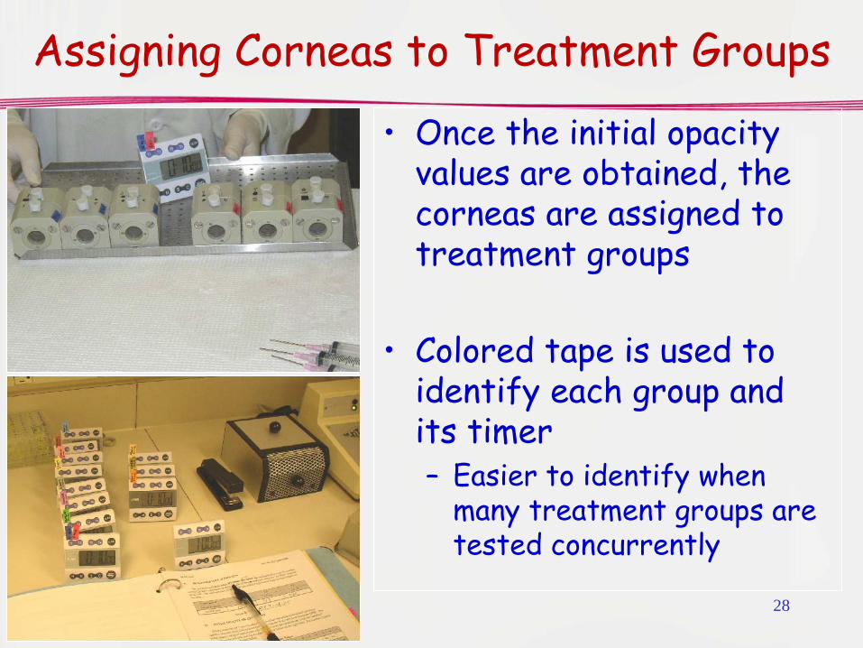
Assigning Corneas to Treatment Groups
• Once the initial opacity values are obtained, the corneas are assigned to treatment groups
• Colored tape is used to identify each group and its timer– Easier to identify when
many treatment groups are tested concurrently

29
Test and Control Article Dosing
Remove the medium from the anterior chamber
– It is critical to remove as much medium as possible (aspirate 2x)
– Some labs use a vacuum pump but should only be used with extreme care
– Needles are cut to remove the point and bevel

30
Test and Control Article Dosing• 750 µL of non-viscous
liquids are measured with a standard micropipette
• Cornea holders are tipped forward to allow addition of the test material without it touching the cornea
• Non-hazardous test materials are handled on the bench while potentially hazardous materials are handled in the safety cabinet

31
Test and Control Article Dosing
• When all the corneas are dosed, the holders are tipped upright to allow the liquid to flow over the corneas
• The timer is started• Exposures of 3 minutes
or less are performed on the bench
20 January 2011 ICCVAM Best Practices Workshop
• Otherwise, corneas are returned to the incubator

32
Rinsing the Corneas• The rinse solutions and
syringes are prepared.• The holders are removed
from the incubator.• At the designated time, the
test article is rapidly removed from the anterior chamber and first rinsed with 2-3 mL of medium containing phenol red.
• The posterior chamber is not rinsed.

33
Rinsing the Corneas• The holder is “swirled” for
~5 seconds so the rinse medium lifts the test material off the cornea.
• The rinse medium is removed and fresh rinse medium added.
• At least three rinse cycles are performed. More may be required to remove all of the test substance
• Phenol red color should show a neutral pH.

34
Rinsing the Corneas• After one rinse with phenol
red-free medium, the anterior chamber is refilled with phenol red-free medium.
• Assure that all bubbles are removed.
• The holders are then returned to the incubator (32º C) for the post-exposure incubation.
• An opacity reading may be taken

35
Handling Viscous Test Substances
• Viscous substances may be worked to improve spreading consistency
• The softened substance is pressure loaded into a positive displacement pipette for dosing.

36
Open Chamber DosingThe anterior chamber window can be removed to allow direct dosing of the viscous test material onto the cornea
37
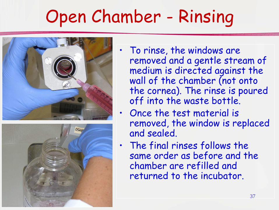
Open Chamber - Rinsing
• To rinse, the windows are removed and a gentle stream of medium is directed against the wall of the chamber (not onto the cornea). The rinse is poured off into the waste bottle.
• Once the test material is removed, the window is replaced and sealed.
• The final rinses follows the same order as before and the chamber are refilled and returned to the incubator.
38
Parameters of the Assay• Post-exposure (expression) incubation (after
rinsing and before the final opacity reading is taken)– Commonly 2 hours for liquids– Longer post-exposure expression periods used to
allow certain classes of materials to better express their toxicity (e.g., peroxides, bleaches, alkylators)• Classes showed delayed maximal toxicity in vivo• May be used for both solid and liquid test substances• Negative control corneas are essential to evaluate
nonspecific changes during the long incubation.20 January 2011 ICCVAM Best Practices Workshop

39
Final Opacity Measurement• At the end of the post-
exposure incubation, the final opacity is read against the air chamber.
• The medium is removed from both chambers and fresh medium added to the posterior chamber.– It is critical to avoid causing
bubbles or allowing any bubbles to remain in the chamber.
– Aspirate the anterior chamber 2x to remove all the medium.

40
Addition of Fluorescein• The posterior chambers are
plugged.• One mL of fluorescein solution in
DPBS is added to each anterior chamber.
• 0.5% Na Fl solution for solids.• 0.4% Na Fl solution for all
others
• Rotate the holders to the up position and return to the incubator for 90 minutes.

41
Measuring Fluorescein Permeability
• The fluorescein in the posterior chamber medium may be measured with a spectrophotometer or plate reader
– The path length should be 1 cm.– The medium must be well mixed
before the sample is taken from the holder
– The full 5 ml volume of the chamber should be collected.
– Tubes and plates should be prelabeled.

42
Collecting the Fluorescein Samples
The full volume of medium from the posterior chamber is collected and transferred to the prelabeled tube.

43
Measuring Fluorescein• The samples are mixed.• For measurement using a 96-well plate
reader, 360 µL are added to a well to replicate the 1 cm path length
• Include a solvent blank (medium) and a 1:1000 dilution of the stock fluorescein to verify its concentration.
• The absorbance is read at 490 nm (OD490).
• Depending upon the equipemnt, the absorbance may not be linear above an OD of 1.500
• Samples with OD values greater than that are diluted and re-read.

44
Fixing the Corneas• Histological assessment of
tissue lesions has become a major part of the BCOP assay
• In some cases, the protocol may specify to fix and save the corneas for possible histology
• Cassettes are prelabeled with our accession number and the cornea number
• A “sponge” is placed in the bottom to protect the corneal endothelium
20 January 2011 ICCVAM Best Practices Workshop

45
Fixing the Corneas• The holder is opened and the cornea
carefully lifted from the O ring.• The cornea is placed with its
endothelial side onto the sponge and immediately submerged in 10% neutral buffered formalin.
• Corneas are fixed for at least 24 hours

46
Calculations• Opacity Score:
– Subtract starting opacity value from the final opacity value for each cornea to obtain the net opacity.
– Subtract the average of the negative control net opacities from the net opacity of each treated cornea to obtain the corrected net opacity.
– Take the average for each treatment group.• Permeability Score:
– Subtract the plate blank from each OD490 value to obtain the net OD490 value (if using a plate reader)
– Multiply the OD490 value by the dilution factor if a dilution was made.
– Subtract the average of the net negative control OD490 values from the net OD490 value for each treated cornea to obtain the corrected net OD490 value.
– Take the average for each treatment group.20 January 2011 ICCVAM Best Practices Workshop

47
Using Opacity and Permeability Values
• The corrected opacity and permeability (OD490) values are used to calculate the In Vitro Score.
• In vitro Score=Opacity +(15xOD490)– Developed by Merck for large set of
pharmaceutical intermediates• Anionic and non-ionic surfactants and
surfactant formulations are often evaluated by permeability scores alone
• Other users evaluate opacity and permeability separately (Casterton)
20 January 2011 ICCVAM Best Practices Workshop

48
Prediction Model Developed by Merck
In Vitro Score Predicted Irritation Potential
<=25 Mild
25.1 – 55 Moderate
> 55.1 Severe
Sina, J.F., Galer, D.M., Sussman, R.G., Gautheron, P.D., Sargent, E.V., Leong, B., Shah, P.V., Curren, R.D., and Miller, K. (1995) A collaborative evaluation of seven alternatives to the Draize eye irritation test using pharmaceutical intermediates. Fundamental and Applied Toxicology 26:20-31.
This model should be used only with standard exposures and in conjunction with the responses of the benchmark materials. It is not appropriate for all classes of materials.
20 January 2011 ICCVAM Best Practices Workshop
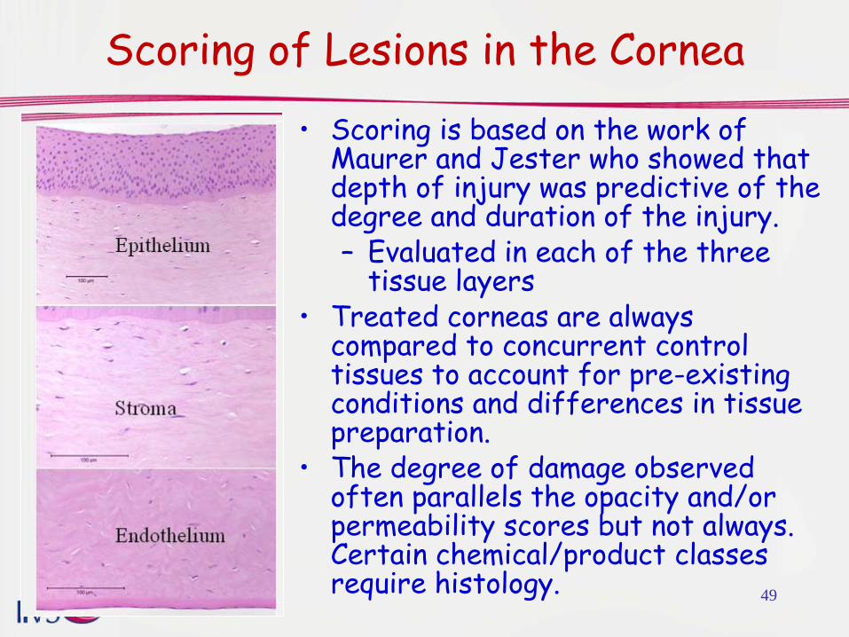
Scoring of Lesions in the Cornea• Scoring is based on the work of
Maurer and Jester who showed that depth of injury was predictive of the degree and duration of the injury.– Evaluated in each of the three
tissue layers• Treated corneas are always
compared to concurrent control tissues to account for pre-existing conditions and differences in tissue preparation.
• The degree of damage observed often parallels the opacity and/or permeability scores but not always. Certain chemical/product classes require histology. 49