brain abscess - journal of neurology, neurosurgery
TRANSCRIPT

Journal of Neurology, Neurosurgery, and Psychiatry, 1973, 36, 757-768
Brain abscessReview of 89 cases over a period of 30 years
A. J. BELLER, A. SAHAR, AND I. PRAISS
From the Department of Neurosurgery, Hadassah Hebrew University Hospital, Jerusalem, Israel
SUMMARY Eighty-nine cases of brain abscess, diagnosed over a period of 30 years, are reviewed.The incidence of this disease did not decline throughout the period. Abscesses of ear and nose
origin constituted the largest group (38%). Postoperative abscesses seem to have increased in inci-dence, presumably due to routine postoperative antibiotic treatment. Antibiotics were possiblyresponsible for the suppression of signs of infection in 4500 of the patients, who presented as
suffering from a space-occupying lesion. The most accurate diagnostic tool was angiography, whichlocalized the lesion in 9000 and suggested its nature in 61%. Brain scan may prove as satisfactory.Staphylococcus was cultured in about two-thirds of the cases. Mortality seemed to decrease con-
comitantly with the advent of more potent antibiotics. The treatment of choice in terms of bothmortality and morbidity seemed to be enucleation after previous sterilization. The hazards of radicalsurgery should be taken into consideration.
The multiplicity of factors involved in themanagement of brain abscess complicates itsevaluation. Surgical therapy in the pre-anti-biotic era carried a mortality of 61% (Websterand Gurdjian, 1950). The use of antimicrobialsand antibiotics improved the prognosis (Jooma,Pennybacker, and Tutton, 1951) but the highhopes expressed with the advent of these agentswere not realized. The improvement in morbidityand mortality since the use of antibiotics did notcontinue (Gregory, Messner, and Zinneman,1967) and the incidence of brain abscess did notdecrease (Garfield, 1969). This disturbinglymeagre progress necessitated the reassessment ofour own methods of treatment and the reevalua-tion of the variables involved. To this end therecords of patients with brain abscess admittedto our department during the last 30 years werereviewed. This period covered the three stages ofantimicrobial therapy and coincided with thedevelopment of the neurosurgical service in thehospital.A total of 89 cases was studied. In all cases the
presence of pus within the brain substance wasverified either at operation or at necropsy. Caseswith localized bacterial encephalitis and epidural
757
or subdural collections of pus were not includedin this study.
FINDINGS AND COMMENTS
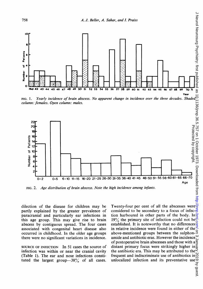
INCIDENCE The yearly incidence varied betweenone and 10 patients per annum, with no apparentchange among the three decades (Fig. 1). Onehas, however, to take into consideration the factthat the total number of beds in the hospital didnot change in this period. On the other hand,during the first eight years of the study period,ours was the only neurosurgical service in Israel.Although the population trebled during theperiod under study, there are now six neuro-surgical services in our country. Taking intoaccount all these factors, it may be assumed thatthe true incidence of brain abscess increasedduring this period of 30 years.A preponderance of males over females
(60:29) was found among our patients.The age distribution is presented in Fig. 2.
The highest incidence was among infants-15%.The 38 cases in children under the age of 15 con-stituted 4300 of all cases. The youngest patientwas 2 months old, the oldest 70 years. The pre-
Protected by copyright.
on 3 January 2019 by guest.http://jnnp.bm
j.com/
J Neurol N
eurosurg Psychiatry: first published as 10.1136/jnnp.36.5.757 on 1 O
ctober 1973. Dow
nloaded from
A. J. Beller, A. Sahar, and L Praiss
43 44 45 46 47 51 52 53 54 S5 56 57 58 59 60 61
Year
FIG. 1. Yearly incidence of brain abscess. No apparent change in incidence over the three decades. Shadedcolumn: females. Open column: males.
0 16c.T 14
0.1
E 6Z 4
2"
0-2 0-5 6-10 11-15 16-20 21-25 26-30 31-35 36-40 41-45 46-50 51
FIG. 2. Age distribution of brain abscess. Note the high incidence among infants.
dilection of the disease for children may bepartly explained by the greater prevalence ofparacranial and particularly ear infections inthis age group. This may give rise to brainabscess by contiguous spread. The four casesassociated with congenital heart disease alsooccurred in childhood. In the older age groupsthere were no significant variations in incidence.
SOURCE OF INFECTION In 51 cases the source ofinfection was within or near the cranial cavity(Table 1). The ear and nose infections consti-tuted the largest group-38% of all cases.
1-55 56-6061-65 66-70Age
Twenty-four per cent of all the abscesses wereconsidered to be secondary to a focus of infec-tion harboured in other parts of the body. In19% the primary site of infection could not beestablished. It is noteworthy that no differencesin relative incidence were found in either of theabove-mentioned groups between the sulphon-amide and antibiotic eras. However the incidenceof postoperative brain abscesses and those with adistant primary focus were strikingly higher inthe antibiotic era. This may be attributed to thefrequent and indiscriminate use of antibiotics inunlocalized infection and its preventative use
758
Protected by copyright.
on 3 January 2019 by guest.http://jnnp.bm
j.com/
J Neurol N
eurosurg Psychiatry: first published as 10.1136/jnnp.36.5.757 on 1 O
ctober 1973. Dow
nloaded from

Brain abscess
TABLE 1SOURCE OF INFECTION
1940-50 1940-71
I a. Ear and mastoid 7 27b. Paranasal sinuses 1 13 7 51c. Postoperative 1 101d. Post-traumatic 4) 7)
11 a. Pulmonary 2 5b. Paradoxical 2 .5 4 21c. Distant metastatic 1 12
III Unknown 5 17
23 89
after surgery. Many of the antibiotic drugs areonly bacteriostatic, thus the infection may betemporarily subdued and flare up after a dor-mant period. The following is illustrative of suchcomplication.
A 22 year old female was admitted in 1951 with an 11
year history of episodes of tachycardia accompaniedby the 'feeling' of an unpleasant smell and sometimesfollowed by loss of consciousness. The sole neuro-logical finding was a right lower quadrant homony-mous anopia. Radiographs of the skull showedcalcification in the left temporal region. At crani-otomy a dermoid cyst was removed from the floor ofthe left middle fossa. The patient was given routineantibiotic treatment postoperatively and recoverywas complete. Six months later the patient was re-admitted because of right-sided Jacksonian seizures.Several days later a fever of 40° C developed, to-gether with signs of increased intracranial pressure.On lumbar puncture the cerebrospinal fluid (CSF)was purulent and grew Staphylococcus aureus. Atrepeated craniotomy a thick-walled abscess was
enucleated from the site of the previous lesion. Thepatient made an uneventful recovery.
Four cases of the so-called 'paradoxical' brainabscess were included in this series. All thesepatients suffered from congenital heart diseasewhich resulted in an arteriovenous shunt. It isassumed (Wechsler and Kaplan, 1940) thatinfected emboli during transient bacteraemia by-passed the pulmonary capillaries and reached thegreater circulation and thus the brain via thecongenital heart defect. Before the antimicrobialera this type of brain abscess was diagnosed onlyat necropsy (Beller, 1951). Since the late '40s the
number of successfully treated 'paradoxical'abscesses has grown steadily. It should, how-ever, be kept in mind that, unless the cardiacdefect is repaired, the route for repeated infectionremains open. This is illustrated in the followingcase.
In 1948 a 6 year old boy with tetralogy of Fallot hada chronic abscess removed from the left parietal lobe.The child recovered completely. Repair of the cardiacdisease was not considered feasible. In 1958 the boywas admitted again with signs of a right frontal lobespace-occupying lesion. An abscess was diagnosedand evacuated by repeated aspirations. He againmade a complete recovery. He was admitted for thethird time in 1966 because of headaches and right-sided Jacksonian seizures. Only angiography re-vealed a left temporal avascular space-occupyinglesion. Pus was evacuated by repeated aspirations.The patient's recovery was again complete.
SITE OF SUPPURATION The abscesses weredivided equally between the right and left halves
TABLE 2SITE OF BRAIN ABSCESSES
Right Left Total
Frontal 11 10 21Parietal 6 9 15Temporal 14 16 30Occipital 6 4 10Cerebellar 8 5 13
45 44 89
TABLE 3SITE OF 34 OTOGENIC AND RHINOGENIC ABSCESSES
Frontal Temporal Parietal Cerebellar
Otitisipsilateral 1 11 5unilateralcontralateral 2bilateral 1 1 2 4
2 14 2 9
Frontal sinusitis 4Maxillary sinusitis 2Pansinusitis 1
5 2
759
Protected by copyright.
on 3 January 2019 by guest.http://jnnp.bm
j.com/
J Neurol N
eurosurg Psychiatry: first published as 10.1136/jnnp.36.5.757 on 1 O
ctober 1973. Dow
nloaded from

A. J. Beller, A. Sahar, and I. Praiss
of the brain (Table 2). The lobes affected mostfrequently were the temporal and frontal. Thedata presented in Table 3 further stress the roleof otogenic and rhinogenic infections as sourcesof brain abscess. The site of the abscess was ipsi-lateral or adjacent to the infected ear or sinus in32 out of 34 cases. This also suggests a contiguoustype of spread. In the remaining two cases it ispossible that otitis had previously been bilateraland had 'healed' by antibiotic treatment. Theintracranial complication became manifest inspite of disappearance of signs of infection at thesource.
BACTERIOLOGY Table 4 lists the types of bac-teria found in the abscesses in this series. A total
TABLE 4TYPES OF ORGANISM CULTURED
Staph. aureusStaph. albusStrep. haemolyticStrep. anhaemolyticProteusPseudomonasMeningococcusHaemophilusBacteroidesPneumococcus
Staph. and proteusStaph. and strep.Staph. and Esch. coliProteus and pyocyaneusStrep. and Esch. coliStrep. and proteusPseudomonas and klebsiella
Sterile
24
4
4 4S monobacterial
24J1 p17
21
2
1
17
74
No culture 15
of 74 cultures were obtained. Of these 45 grew asingle type of organism and in 12 more than onetype was found. Staphylococcus was by far thecommonest organism cultured (see Table 9); itwas found in 63% of the positive cultures. Thesecond largest group were streptococci, found inalmost 20% of the positive cultures. Both thesetypes of gram-positive cocci were evenly distri-buted throughout the period under study.
The 17 cases in which culture of the pus yieldedno growth were also evenly distributed during theperiod. The 'sterilization' of the pus is probablycaused by preoperative antimicrobial or anti-biotic treatment, again, not eradicating infectionbut rendering it sterile for in vitro growth.
CLINICAL FEATURES Of the 89 patients, 82 werediagnosed during life and treated. The mostprominent symptoms and signs were headache,focal neurological signs, either irritative or ofdeficit, and an altered state of consciousness.Forty ofthe patients presented with no symptomsor signs of an infective process (Table 5) andtherefore were diagnosed at first as sufferingfrom a space-occupying lesion. It should be
TABLE 5SYMPTOMATOLOGY OF BRAIN ABSCESSES
Neurological Neurological andsyndrome only signs of infection
1941-46 5 71948-53 8 181954-59 8 141960-65 13 61966-71 6 4
40 49
TABLE 6EEG FINDINGS
Focal disturbance 14Regional disturbance 17Hemispheric disturbance 4Signs of increased pressure only 2Diffuse disturbance 5
Normal 3*
* Two cases-cerebellar abscess.
noted that during the last decade of the studythere was a rise in the relative incidence of the'non-infective' cases. Such modification of theclinical features may be attributed to the use ofantibiotics before admission.
Sixteen patients were admitted with suddenonset of meningitis due to rupture of the abscess
76()
Protected by copyright.
on 3 January 2019 by guest.http://jnnp.bm
j.com/
J Neurol N
eurosurg Psychiatry: first published as 10.1136/jnnp.36.5.757 on 1 O
ctober 1973. Dow
nloaded from

Brain abscess
FIG. 3. Serial brain sections showing extensive nion-capsulated abscess. The patient had received steroids.
into the cerebrospinal fluid system. Theirclinical course was in no way different from thatof the other patients.When dealing with the clinical picture of brain
abscess, mention should be made of treatmentwith steroids. Four of our patients had hadsteroid treatment before their admission to ourdepartment. Only one of these survived. Steroidsprobably increase the hazards of local and generalinfection and mask its clinical manifestations.Moreover, they seem to prevent the local limita-tion of the inflammatory process by capsuleformation. The following case helps to illustratethis.
A 14 year old girl was treated by dilatations forcaustic constriction of the oesophagus. To preventexcessive scarring she was also given steroids and'preventive' antibiotics. One week before admission
to our department she complained of headaches anda neurologist found papilloedema with minimalpyramidal signs on the right. There were no systemicsigns of infection. On the day of transfer to neuro-surgery the girl suddenly lapsed into coma andCheyne-Stokes respiration, followed by hypotension.She died shortly afterwards. At necropsy an abscesswhich had destroyed a large part of the left hemi-sphere was found (Fig. 3).
The other two fatal cases had also extensivesupratentorial noncapsulated abscesses. Theydied with signs of acute brain-stem compression.
METHODS OF LOCALIZATION An electroenceph-alographic (EEG) examination was performedin 45 of the treated patients (Table 6). It showedthe focal disturbance considered typical for brainabscess (Kiloh and Osselton, 1961) in only 14
761
Protected by copyright.
on 3 January 2019 by guest.http://jnnp.bm
j.com/
J Neurol N
eurosurg Psychiatry: first published as 10.1136/jnnp.36.5.757 on 1 O
ctober 1973. Dow
nloaded from

A. J. Beller, A. Sahar, and L. Praiss
FIG. 4. Right carotid angiogram demonstrating alarge temporal avascular space-occupying lesion. Thisfinding but not the symptoms or signs suggested thepresence ofpus.
cases. In 17 additional cases this examinationwas useful in localizing the process and choice ofthe next diagnostic procedure. These efficacyrates of the EEG are in agreement with Garfield's(1969) data.
In 15 cases, most of them in the earlier years,the pus was located by direct puncture. In two ofthese, pus was aspirated during an attempt atventriculography. In an additional three casesthe site of a previous operation was punctured orre-explored (Table 7).
TABLE 7METHODS OF LOCALIZATION OF BRAIN ABSCESSES
Angiography Ventriculo- Pneumo- Directgraphy encephalo- puncture
Site Nature graphy
Studies (no.) 31 19 21 16 15tEquivocal or
negative 3* 4 6 3Accuracy (%) 90 61 81 63 83
* One cerebellar abscess.t In three cases the site of previous operation was punctured or re-explored. In two additional cases pus was aspirated at attemptingventriculography.
Sixteen patients were studied by pneumo-encephalography; none of them had signs ofincreased intracranial pressure or infection. Thisprocedure was the least effective for diagnosisand also frequently resulted in rapid aggravationof the patient's condition. Less frequent deteri-oration was observed during or after ventriculo-graphy, which was accurate in 81.%. It is possiblethat complications of ventriculography were lessappreciated, since in our practice it immediatelyprecedes definitive surgery.Angiography seemed to be the most accurate
and also the safest procedure. It helped localizethe process in 90%0 of the cases in which it wasperformed. Moreover, by demonstrating anavascular process it suggested its correct naturein 61%. The accuracy of this procedure, mainlyfrom this aspect, increased with the gain ofexperience and with multiphase angiography.
S
-A." ::
o..:~~~~~~~~~~~~~~~~~~~~~~~~~~~~~........ tH Bk . - r~~~~~~M
. ......- I F*:_ g-e _ _ _ _ _
wiB.:-Sti .a;
.;*. _r .* .|w. _. E-:.s * _s .. h w*.,0U.......... .. ... . w B;
*.:' :it: :R. :! 9tf..... &.- r B.* l; ws41t ol |es.,__2s_:.x_ .W *
a- _._- .... =E. ... :,r. 'ws._ s,.-7h_= w o2iii . & ::= :t
FIG. 5. 99Tc brain scan. Note the area of uptakewhich is triangular on anteroposterior view and ring-shaped on lateral view. The burr-hole was placed abovethe centre of the ring.
762
Protected by copyright.
on 3 January 2019 by guest.http://jnnp.bm
j.com/
J Neurol N
eurosurg Psychiatry: first published as 10.1136/jnnp.36.5.757 on 1 O
ctober 1973. Dow
nloaded from
Brain abscess
The importance of preoperative suspicion of thepresence of intracerebral pus need not be stressed(Fig. 4).
Brain scan with 99Tc was performed in fourcases and was positive in all. In one case (Fig. 5),due to the ring shape of the lesion, this study wasconsidered sufficient, as it proved to be. Thisexamination seems to be the least hazardous andpromises a high degree of accuracy with furtherexperience. It may also help identify multiplelesions (Tefft, Matson, and Neuhauser, 1966).
MORTALITY Eighty-two of the 89 patients hadsurgical treatment. The seven patients who diedbefore surgery had been ill with other diseaseswhich were complicated by brain abscess. Whenthe complication was recognized the patient wasmoribund and the diagnosis was made at nec-ropsy. Thus the overall mortality of treated brainabscess was 40%0. It seemed to decrease in thelast five year period.The basic factors influencing mortality are
reviewed below.
Age No significant correlation was found be-tween the age of the patient and mortality. Thiswas true even in infants (Table 8). This agreeswith findings of others (Dawes, 1961; Garfield,1969).
TABLE 8AGE DISTRIBUTION OF MORTALITY (TREATED CASES ONLY)
Age (yr) Cases Deaths Mortality(no.) (no.) (%)
0-2 12 (13)* 4 333-5 8 (9) 3 386-10 9 (9) 3 3311-15 6 (7) 2 3316-20 5 (5) 2 4021-40 20 (23) 9 4541-55 13 (14) 7 5456-70 9 (9) 3 33
All treated cases 82 33 40
* Figures in parentheses represent total numbers of patients.
Bacteriology From Table 9 it appears thatstaphylococci caused the largest number ofdeaths. They also constituted the largest group
TABLE 9MORTALITY ACCORDING TO ORGANISM
Organism Cases Deaths Mortality(no.) (no.) (%)
Staphylococcus 25 10 40Staph. and other bacteria 7 3 43Total no. of staph. 32 13 41
Streptococcus 7 1 14Strep. and other bacteria 3 3 100
Other bacteria 15 6 40
Sterile culture 17 7 41
Total* 74 30 41
* In additional 15 cases cultures were not obtained.
and the mortality rate did not differ from theaverage. There seemed to be a low death rate inabscesses produced by streptococci in mono-bacterial growth. This is contrary to otherobservations (Gregory et al., 1967; Garfield,1969). The virulence of other organisms is diffi-cult to assess due to the small numbers. Thequestion as to whether polybacterial growthcarries a higher mortality is difficult to answer. Itseems that the prognosis in such abscesses de-pends on the most virulent of the severalorganisms.
Sterile cultures did not carry any better prog-nosis. This suggests only that in vitro sterilitybears little or no relation to in vivo virulence(Sperl, MacCarty, and Wellman, 1959). 'Nogrowth' could be the result of preoperative anti-biotics (Ballantine and Shealy, 1959) or anaero-bic or slow growing organisms (Liske andWeikers, 1964; Salibi, 1964).
Arranging the cases by periods (Table 10)there seemed to be a drop in mortality in theyears 1951-55 and again since 1966. Theseperiods coincide with the more extensive use ofpenicillin and later with that of broad spectrumantibiotics. In the latter period more potentdrugs such as the synthetic penicillins andgaramycin became available.
Rupture ofabscess This is considered one of themost feared sequelae of brain abscess. It was themost frequent cause of death in several reports(Lewin, 1955; Ballantine and Shealy, 1959). In
763
Protected by copyright.
on 3 January 2019 by guest.http://jnnp.bm
j.com/
J Neurol N
eurosurg Psychiatry: first published as 10.1136/jnnp.36.5.757 on 1 O
ctober 1973. Dow
nloaded from

A. J. Beller, A. Sahar, and L Praiss
TABLE 10MORTALITY FROM BRAIN ABSCESS
Period All cases Treated cases
(no.) (%) (no.) (%)
1941-45 7 58 6 541946-50 5 45 5 451951-55 6 33 5 281956-60 13 65 8 401961-65 7 39 7 391966-71 2 20 2 20
Total 40 45 33 40
our series, however, it occurred only eighttimes-10% of all treated cases. Rupture of theabscess was fatal in five cases or 15% of all post-operative deaths. In four of these the rupturewas into the 4th ventricle. The rupture of asupratentorial abscess, therefore, should not beconsidered as an ominous sign.
Multiplicity Eight of the patients had eithermultilocular or multiple abscesses. Five of thesedied-a mortality of 63%. Two of the survivorshad multiloculated interconnecting abscesseswhich were enucleated. One patient had threeparadoxical abscesses on three separate occa-sions.
CAUSES OF DEATH Table 11 lists the causes ofdeath in the surgically treated cases. Uncon-trolled infection seemed to be the major cause ofdeath accounting for 16 out of the 33 fatalities.In the last decade however, this occurred in onlythree cases-no doubt due to the use of morepotent antibiotic agents.
TABLE 11CAUSES OF POSTOPERATIVE DEATH
1941-45 1941-71
Meningitis/ventriculitis 4 14Rupture into 4th ventricle 1 4Multilocular/multiple abscess 5Acute herniation 1 4Septicaemia 2Congestive heart failure 2Other 2
Total 6 33
Acute temporal or brain-stem herniation wasencountered in four postoperative cases and inall the seven who died before coming to surgery.Urea and mannitol administration or emergencysurgery were of limited help (Pennybacker, 1961).Four additional cases with rupture of cerebellarabscess into the 4th ventricle, died probably fromdirect and acute compression of the brain-stem.Death due to failure to recognize and localize
all lesions in multiple abscesses led to death infive cases (Tutton, 1953).
METHODS OF SURGICAL TREATMENT During mostof the reviewed period treatment was supervisedby one person (A.J.B.). The choice of thesurgical treatment was based on the currentneurosurgical thought, the preoperative diagno-sis, and the patient's condition. In the earlyperiod drainage was mainly performed (Table
TABLE 12ASSESSMENT OF SURGICAL PROCEDURE
Procedure Cases Survivals Deaths Mortality(no.) (%)
Drainage 6 4 2 33Aspiration 33 16 17 52Primary resection 26 17 9 35Aspirations and resection 17 12 5 30
Total treated 82 49 33 40untreated 7 7 lOO
Total no. of cases 89 49 40 45
12). As its dangers became apparent (King andTurney, 1954) treatment by repeated aspirationswas preferred. After aspiration the abscess cavitywas irrigated with an antibiotic solution and theniophendylate (Pantopaque) together with airwere introduced. Changes in the size of the cavityon radiography indicated the need for furtheraspirations (Fig. 6). Resection was performedeither as the primary procedure or after repeatedaspirations. Primary resection was attemptedwhen the abscess was located in a 'silent' area,when there was reason to believe that a chronicwall was present. or when the preoperativediagnosis was of a tumour (Fig. 7). Aspiration
764
Protected by copyright.
on 3 January 2019 by guest.http://jnnp.bm
j.com/
J Neurol N
eurosurg Psychiatry: first published as 10.1136/jnnp.36.5.757 on 1 O
ctober 1973. Dow
nloaded from

Brain abscess
FIG. 6. A-P and lateralrecumbent double contrastradiograms. Above: immedi-ately after evacuation ofpus;below: four days later. Noteshrinkage of cavity andabsorption of most antibioticsolution (layer between air andiophendylate) that had beeninstalled.
was chosen in the gravely ill, in acute abscesses,or in anticipation of a later resection.These considerations were reflected in the
results of surgery. Aspiration carried the highestmortality-52%. Resection either primary orsecondary was associated with the death of one-third of the patients.
LATE SEQUELAE Of the 49 survivors, 45 patientswere followed for more than one year, bypersonal examination. The late sequelae ofsurgery are dealt with below.
Recurrence This complication occurred fourtimes-that is, in 8% of the survivors. All thesepatients had been treated by aspirations and apneumoencephalogram had been performed atthe completion of treatment and read as normal(Pennybacker, 1961). This rate of recurrence is
similar to that reported by Jooma et al. (1951).Two of these patients survived by later aspirationfollowed by resection; in both recrudescenceoccurred within months of the primary proced-ure. The two other cases were fatal and occurredseveral years after the first operation. The fol-lowing is illustrative of such danger.
A 7 year old boy was admitted to hospital in 1952with left-sided Jacksonian seizures. Three days laterhe developed a fever of 390 C, severe headache, andsigns of meningitis. The CSF contained 1,000 leuco-cytes per cmm. A right frontal abscess was diagnosedby ventriculography. The child was treated byrepeated aspirations and made a complete recovery.Eleven years later, in 1963, when on military service,he suddenly became ill. The diagnosis was menin-gitis; however, he lapsed into coma and died within24 hours of the beginning of his disease. At necropsya right frontal lobe ruptured abscess was found.
765
Protected by copyright.
on 3 January 2019 by guest.http://jnnp.bm
j.com/
J Neurol N
eurosurg Psychiatry: first published as 10.1136/jnnp.36.5.757 on 1 O
ctober 1973. Dow
nloaded from

A. J. Beller, A. Sahar, and L Praiss., 4 . . ... | s , s . . . . . ......... e ~~~~~~~~.AL 1i9 ZO0 21 2.2 213 2-4
It may be assumed that the infective organismslay dormant in the retained capsule. This is onemore case in favour of resection of the capsulewhenever feasible (LeBeau, 1946; Tutton, 1953;Lewin, 1955), although some surgeons doubt theefficacy of this procedure in the prevention ofrecurrences (Bonnal, Descuns, and Duplay,1960).
Neurological deficit Twenty-two out of the 45patients followed had some degree of permanentneurological deficit (Table 13). In five of thesepatients the deficit could be attributed to previ-ous tumour or trauma or surgery performed forit. Of the remaining 17 patients, four were notaware of their deficit, which consisted of hemi-anopia, hypoaesthesia, or minimal paresis. This
FIG. 7. Right frontal en-capsulated abscess afterenucleation. Above: outersurface; below: abscess cut inhalf.
left 13 patients or 2900 of the followed survivorswith varying degrees of incapacitation. Three ofthese were totally disabled, whereas the remain-ing 10 were able to resume their previous dutiesin spite of the deficit, which was mostly hemi-paresis and sometimes some dysphasia. Only onechild had minimal mental retardation.Permanent neurological deficit was commonest
after resection either primary or secondary,occurring in one-third of the patients followed.Disability was least in patients treated by aspira-tion. Two out of 15 patients followed up hadmoderate hemiparesis. These disability rates areaverage between the data of Jooma et al., (1951)and Kerr, King, and Meagher, (1958). Suchresults of radical vs. the more conservative modesof treatment put in question the justification of
766
:%..: .:..: .:5:.
Protected by copyright.
on 3 January 2019 by guest.http://jnnp.bm
j.com/
J Neurol N
eurosurg Psychiatry: first published as 10.1136/jnnp.36.5.757 on 1 O
ctober 1973. Dow
nloaded from

Brain abscess
TABLE 13SEQUELAE OF BRAIN ABSCESS
Surgical Patients Deficit Seizuresprocedure followed
(no.) Unnoticed Moderate Severe
Drainage 4 2 2 1Aspiration 15 1 2 3Primary resection 15 3 2 (3)* 1Second resection 11 3 1 (2)* 1
Total followed 45t 4 10 3 6
* Figures in parentheses-patients in whom deficit was attributed topre-existing lesion or surgery.t Four cases of cerebellar abscess.
radical resection (LeBeau, 1946; Ballantine andShealy, 1959).
Parietal or dominant hemisphere lesions weremore prone to permanent and more severedeficit. Survivors of cerebellar surgery with anymethod remained symptomless.
Epilepsy Six of the patients followed updeveloped epileptic seizures. These constitute15% of the survivors with supratentorial abscess.This is an unexplicably low incidence as com-pared with numerous other series (Jooma et al.,1951; Lewin, 1955; Kerr et al., 1958).No relationship existed between the severity of
neurological deficit and the occurrence of epi-lepsy. One patient with no permanent deficit hadseizures. Another, with severe hemiparesis, alsohad incapacitating Jacksonian attacks.
Aspiration was apparently associated with thehighest incidence of seizures.
CONCLUSIONS
There is reason to believe that the incidence ofbrain abscess is increasing.
Routine use of antibiotics after brain surgeryor in paracranial infection does not always pre-vent abscess formation though it may delay itssymptomatology.The frequent use of antibiotics, even without
definite diagnosis, may mask the clinical pictureof brain abscess, thus making the preoperativediagnosis more difficult.
Paracranial suppuration is still the commonestsource of intracranial infection.The least disturbing and most accurate
diagnostic procedure both in terms of localiza-tion and in suggesting the nature of the lesion isangiography. Brain scan may prove to be evenmore accurate; moreover, it may demonstratemultiple lesions.
Preoperative use of steroids is extremelyhazardous.
Sensitivity of a given organism to the anti-biotic drug is a more important factor in progno-sis than its particular type. Antibiotics should beadministered in massive doses, when indicatedand by sensitivity.
Enucleation of the abscess in the 'cold' stageafter 'sterilization' by aspirations seems to bethe procedure of choice. Additional damage, dueto the radical surgery, should be taken into con-sideration.
REFERENCES
Ballantine, H. T., Jr., and Shealy, C. N. (1959). The role ofradical surgery in the treatment of abscess of the brain.Surgery, Gynecology, and Obstetrics, 109, 370-374.
Beller, A. J. (1951). The syndrome of brain abscess withcongenital cardiac disease. Report on a case with com-plete recovery. Journal of Neurosurgery, 8, 239-243.
Bonnal, J., Descuns, P., and Duplay, J. (1960). Les AbcesEncephaliques Li l',tre des Antibiotiques, pp. 154. Masson:Paris.
Dawes, J. D. K. (1961). Discussion on intracranial complica-tions of otogenic origin. Proceedings of the Royal Societyof Medicine, 54, 315-320.
Garfield, J. (1969). Management of supratentorial intra-cranial abscess: a review of 200 cases. British MedicalJournal, 2, 7-1 1.
Gregory, D. H., Messner, R., and Zinneman, H. H. (1967).Metastatic brain abscesses. A retrospective appraisal of 29patients. Archives of Internal Medicine, 119, 25-31.
Jooma, 0. V., Pennybacker, J. B., and Tutton, G. K. (1951).Brain abscess: aspiration, drainage, or excision ? Journal ofNeurology, Neurosurgery, and Psychiatry, 14, 308-313.
Kerr, F. W. L., King, R. B., and Meagher, J. N. (1958).Brain abscess-a study of forty-seven consecutive cases.Journal of the American Medical Association, 168, 868-872.
Kiloh, L. G., and Osselton, J. W. (1961). Clinical Electro-encephalography, pp. 135. Butterworths: London.
King, J. E. J., and Turney, F. (1954). Brain abscess: evolutionof the methods of treatment. Annals of Surgery, 139, 587-612.
LeBeau, J. (1946). Radical surgery and penicillin in brainabscess. A method of treatment in one stage with specialreference to the cure of three thoracogenic cases. Journal ofNeurosurgery, 3, 359-374.
Lewin, W. (1955). Recent developments in the managementof brain abscess. British Medical Journal, 1, 631-634.
Liske, E., and Weikers, N. J. (1964). Changing aspects ofbrain abscesses. Review of cases in Wisconsin 1940 through1962. Neurology (Minneap.), 14, 294-300.
Pennybacker, J. (1961). Discussion on intracranial complica-tions of otogenic origin. Proceedings of the Royal Society ofMedicine, 54, 309-315.
Salibi, B. S. (1964). Bacteroides infection of the brain.
767
Protected by copyright.
on 3 January 2019 by guest.http://jnnp.bm
j.com/
J Neurol N
eurosurg Psychiatry: first published as 10.1136/jnnp.36.5.757 on 1 O
ctober 1973. Dow
nloaded from
A. J. Beller, A. Sahar, and L Praiss
Successful management of case with three intracranialabscesses. Archives of Neurology, 10, 629-634.
Sperl, M. P., Jr., MacCarty, C. S., and Wellman, W. E.(1959). Observations on current therapy of abscess of thebrain. Archives of Neurology and Psychiatry, 81, 439-441.
Tefft, M., Matson, D. D., and Neuhauser, E. B. D. (1966).Brain abscess in children. Radiologic methods for earlyrecognition. American Journal of Roentgenology, 98, 675-688.
Tuitton, G. K. (1953). Cerebral abscess-the present position.
Annals of the Royal College of Surgeons of England, 13,281-311.
Webster, J. E., and Gurdjian, E. S. (1950). The surgicalmanagement of intracranial suppuration. Methods indiagnosis and management, and a report of 23 cases ofcivilian and 33 cases of military abscess. InternationalAbstracts of Surgery, 90, 209-235.
Wechsler, I. S., and Kaplan, A. (1940). Cerebral abscess(paradoxic) accompanying congenital heart disease. Reportof two cases. Archives of Internal Medicine, 66, 1282-1289.
768
Protected by copyright.
on 3 January 2019 by guest.http://jnnp.bm
j.com/
J Neurol N
eurosurg Psychiatry: first published as 10.1136/jnnp.36.5.757 on 1 O
ctober 1973. Dow
nloaded from